Embed Size (px)
Citation preview

GARREAU ET AL. VOL. 7 ’ NO. 4 ’ 2977–2987 ’ 2013
www.acsnano.org
2977
March 11, 2013
C 2013 American Chemical Society
Color Control in CoaxialTwo-Luminophore NanowiresAlexandre Garreau,†,* Florian Massuyeau,† Stephane Cordier,‡ Yann Molard,‡ Eric Gautron,†
Patricia Bertoncini,† Eric Faulques,† Jany Wery,† Bernard Humbert,† Alain Bulou,§ and Jean-Luc Duvail†,*
†Institut des Matériaux Jean Rouxel, Université de Nantes, CNRS, France, ‡Institut des Sciences Chimiques de Rennes, Université de Rennes, CNRS, France, and§Institut des Molécules et Matériaux du Mans/-PEC, Université du Maine, CNRS, France
Luminescent organic nanowires NWs areof great importance as building blocksin future miniaturized optoelectronic
and photonic devices because these sys-tems rely upon the ability to tune the opto-electronic characteristics of the constituentmaterials. In particular, the potential of or-ganic NWs as nanosources for applicationssuch as tagging,1,2 sensing,3�6 lasing,7,8 dis-play and lighting9,10 are extensively explored.In each case, the control of emitted light(stability, intensity, color) is a foremost point.For these reasons, many organic nanowiresand nanotubes have been fabricated duringthe last 10 years, either from conjugatedpolymers,9,11 or from oligomers or smallmolecules.12 Some original emissive behav-iors induced by the nanostructuration havealso been shown.13,14 In most cases the or-ganic one-dimensional nanosources are com-posed of a single luminophore, thus resultingin a single color emission. Then, the color offluorescence is directly related to the choiceof the luminophore among a broad rangeof dyes or nanoparticles. However, these
luminophores have also to meet many othercriterions depending on the targeted appli-cation, such as the emission quantum yield,the emission stability under illumination orin different media, the solubility, the bio-compatibility, etc. For this purpose, the studyof multiluminophore nanostructures is anemerging and promising domain.15,16 Themixing of luminophores into nanowires canresult in color tuning as well as white emis-sion.17�21 Moreover, by controlling the po-sition of two luminescent materials at thenanometer scale, it is possible to engineermulticolor nanosources22 and multisegmentednanowires.1,2 This strategy is not restrictedto pure organic materials but hybrid systemscombiningbothorganicand inorganicchromo-phores (quantum dots, phosphorescent met-al complexes) are also of great interest.20,22�25
However, the mixing of two or more emis-sivematerials can lead to complex and non-desired behaviors caused by charge andenergy transfers between the two lumino-phores. Reabsorption phenomenon (trivialenergy transfer), charge trapping,26 resonance
* Address correspondence [email protected],[email protected].
Received for review July 13, 2012and accepted March 11, 2013.
Published online10.1021/nn400763r
ABSTRACT We report a general and simple approach to take control of the
color of light-emitting two-luminophore hybrid nanowires (NWs). Our strategy is
based on the spatial control at the nanoscale (coaxial geometry) and the spectral
selection of the two kinds of luminophores in order to restrict complex charge and
energy transfers. Thus, it is possible to control the color of the photoluminescence
(PL) as an interpolation of the CIE (Commission Internationale de l'Eclairage)
coordinates of each luminophore. For this purpose, we selected a green-emitting semiconducting polymer and a red-emitting hexanuclear metal cluster
compound, (n-Bu4N)2Mo6Br8F6, dispersed in a poly(methyl-methacrylate) (PMMA) matrix. The great potential and the versatility of this strategy have been
demonstrated for two configurations. First, a yellow PL with a continuous change along the nanowire has been evidenced when the proportion of the PPV
shell versus the nanocomposite core, that is, the green/red volumic ratio, progressively shifts from 1:2 to 1:5. Second, an extremely abrupt change in the PL
color with red-green-yellow segments has been achieved. A simple model corroborates the effectiveness of this strategy. PL excitation and time-resolved
experiments also confirm that no significant charge and energy transfers are involved. The two-luminophore hybrid nanowires may find widespread
nanophotonic applications in multicolor emitting sources, lasers and chemical and biological sensors.
KEYWORDS: hybrid nanomaterial . coaxial nanowire . light-emitting polymer . cluster compounds . luminescence . energy transfer .nanophotonics
ARTIC
LE
GARREAU ET AL. VOL. 7 ’ NO. 4 ’ 2977–2987 ’ 2013
www.acsnano.org
2978
energy transfer (RET) process, i.e., dipole�dipole cou-pling (Förster energy transfer)27�29 and electron ex-change (Dexter energy transfer)30 are the main pheno-mena encountered and they complicate the predictionand the control of the properties.31 Also, these mech-anisms can result in the decrease of the emitted light ina part or in the whole spectral range, thus contributingto the decrease of the overall intensity.32,33
The tuning of optical emission by RET mechanismsadditionally depends on the pumping wavelength, theabsorption, the quantum yield of emitters and the RETefficiency. The RET efficiency depends on the donor-to-acceptor separation distance.34 Other studies haveshown that other parameters are also involved in RET,such as the particle shape and size, the relative spatialdistribution of acceptor and donor,29 the overlappingbetween the donor absorption spectrum and theacceptor emission spectrum and the relative orienta-tion of the emitters.21 As a consequence, the design ofa new generation of multiluminophore nanoparticleswhich prevent or restrict complex energy or chargetransfer could be suitable to tailor the color of emission.In this study, we show how a coaxial geometry com-
bining two types of luminophores at the nanometerscale can result in a very fine-tuning of the photolumi-nescence (PL) features by minimizing the charge andenergy transfermechanisms. Besides the local arrange-ment of the luminophores, this has been achieved byselecting luminophores which exhibit distinct color ofPL emission as well as no significant overlapping oftheir absorption and emission spectral range. Here, aπ-conjugated polymer poly(para-phenylene-vinylene)PPV has been used as a fluorescent green material andmetal-clusters compound (n-Bu4N)2Mo6Br8F6 as aphosphorescent red one. (n-Bu4N)2Mo6Br8F6 is basedon [Mo6Br8F6]
2� cluster units paired with two (n-Bu4Nþ)
counter cations (see Figure S1 in Supporting Information).(n-Bu4N)2Mo6Br8F6 is highly soluble and constitutes auseful one-nanometer diameter building block for theelaboration of hybrid nanocomposites35�37 and function-alization of metal organic framework (MOF).38 Here, thecluster units were dispersed in an insulating poly(methyl-methacrylate) (PMMA) matrix to separate the two lumi-nophores and to avoid charge transfers. The coaxialnanowires with a core of (n-Bu4N)2[Mo6Br8F6]@PMMAand a shell of PPV have been successfully synthesizedby a template strategy, as demonstrated by electronmicroscopy. On the basis of a systematic study of micro-PL coupled to micro-Raman investigations along thecoaxial NWs, we demonstrate that the resulting PLspectra can be simply decomposed by the sum ofthe spectrum of each luminophore weightedwith theirrespective proportion. To probe whether transfer me-chanisms are involved between the core and the shell,the time-resolved PL emission has been studied for PPVnanotubes, (n-Bu4N)2[Mo6Br8F6]@PMMA nanowires andfor the coaxial nanowires. This strongly suggests that
transfer processes are not significantly involved. Thus,for a given excitation wavelength, it is possible to anti-cipate and to control the color of the emitted light onthe chromaticity diagram as an interpolation of the CIE(Commission Internationale de l'Eclairage) coordinatesof each luminophore. Moreover, the original designproposed here also permits to get a very sharp colorchange within few nanometers, as demonstrated fornanowireswithgreen, red andyellowemitting segments.
RESULTS AND DISCUSSION
Design of Coaxial Two-Luminophore Nanowires. For twoor more luminophores based systems, the control ofthe color is usually achieved by simply mixing organicdyes, quantum dots or other nanoparticles with dif-ferent ranges of emission. An empirical approach isadopted that consists in changing the proportion ofeach luminophore. Generally, this strategy promotescomplex phenomena occurring when two (or more)kinds of emitters are into contact, that are Dexter andFörster energy transfers as well as charge transfers. Torestrain these phenomena, spatial separation of thetwo kinds of emitters can be achieved by preparing bi-or multilayered thin films. But it does not circumventthe self-absorption phenomenon due to overlappingof the absorption and emission spectra. The originaldesign proposed here enables us to address thesepoints and finally to anticipate the emitting color alongnanowires with a control at the nanoscale. This controlhas been demonstrated here with individual nano-wires exhibiting either a uniform yellow color, a pro-gressive change from yellow to orange, or a sharp shiftof the color from yellow to green and red segments(Figure 1).
First, we selected the PPV conjugated polymer as agreen emitter and (n-Bu4N)2[Mo6Br8F6] cluster com-pound as a red emitter because the absorption bandof the cluster unit (red dashed line, peak at ∼3.8 eV)has no significant overlap with the emission band ofthe PPV (green line, peak at ∼2.2 eV) as shown onFigure 1a. Thus, the absorption by the cluster units ofthe photons emitted by PPV cannot significantly occur.Figure 1b shows epi-fluorescence micrographs of ran-dom PPV nanotubes and (n-Bu4N)2[Mo6Br8F6]@PMMAcomposite nanowires mats synthesized as referencesystems. Uniform bright green and red characteristic ofPPV and (n-Bu4N)2[Mo6Br8F6] emissions are observed.
Aside from the necessary spectral separation, thespatial separation is also required. Indeed, Dexter energytransfer occurswhen thewave functions of the two typesof luminophores overlap. This implies that the exciteddonor and the ground-state acceptor are close enough(typically∼0.6�2 nm). For charge transfer phenomenon,a similar distance between the donor and the acceptor isneeded. Concerning Förster energy transfer, the processis efficient for typical distances 5�10 nm between the
ARTIC
LE

GARREAU ET AL. VOL. 7 ’ NO. 4 ’ 2977–2987 ’ 2013
www.acsnano.org
2979
donor and the acceptor.29 In our system, the spatialseparationwas achieved by dispersing the cluster unitsin an insulating PMMAmatrix to form a hybrid compos-ite (n-Bu4N)2[Mo6Br8F6]@PMMA and by using a sec-ondary template strategy. First, a thin layer of PPV wasdeposited by impregnation of porous alumina mem-branes. Then, a solution containing both the PMMAand the cluster units was used to fill PPV nanotubes.This results in a coaxial arrangement where the twoluminophores are spatially well-separated at the nano-scale with a composite (n-Bu4N)2[Mo6Br8F6]@PMMAcore and a PPV shell as illustrated in Figure 1b (see theMethods part for further details).
Coaxial Nanowires Characterization. The coaxial NWswere characterized using a scanning electron micro-scope (SEM), an atomic force microscope (AFM) inintermittent contact mode and a transmission electronmicroscope (TEM) equipped with an energy dispersive
X-ray spectrometer (EDX) after removing the aluminatemplate. A mat of flexible NWs (Figure 2a) is obtained,confirming that the procedure successfully generatedNWs at high yield (∼ 109 per template), as well as dis-persed individual NWs (insets of Figure 2a,b). Theirlengths were found in the range 10�20 μm while themembrane thickness is about 60 μm. This differencecan be explained by the alumina removal step and thesonication step required for their dispersion in solution,which result in shortening of the NWs.
The contrast provided by the heavy atom of mo-lybdenum (Z = 42) from the cluster units, present in thecore, compared to mainly carbon in the shell allows toclearly distinguish the coaxial morphology of NWs byTEM analysis (Figure 2c). Statistical analysis of micro-graphs yielded average values for NWs outer diameter,∼ 200�250 nm, and the shell thickness of about 20 nm,in agreement with TEM studies of the shell of PPVnanotubes published previously.39 The disparity ofNWs diameters reflected the inhomogeneous internalpore diameters within the template employed. Thecoaxial geometry is unambiguously demonstrated bythe EDX study. Figure 2d shows two EDX spectraperformed with 10 nm diameter spot size measuredfor the NWs core (blue circles) and the NWs shell (redcircles) in Figure 2c. It is clear from the peaks corre-sponding to molybdenum and bromine that the[Mo6Br8F6]
2‑ cluster units are only located within thecore of theNW,while the carbon dominates for the PPVshell. The presence of phosphorus and copper resultsfrom the removal of the template by phosphoric acidand from the TEM grid, respectively. The EDX peak at1.5 keV cannot be clearly attributed to bromine oraluminum and arises probably from a mixture of thetwo contributions. The lack of contrast within the corepart strongly suggests that the cluster units are homo-genously dispersed into the PMMA matrix. This is inagreement with selective area electron diffraction(SAED) study, not shown here. The absence of diffrac-tion features in SAED pattern indicates that there is nocrystallization of the cluster compounds. The (n-Bu4N)2-[Mo6Br8F6]@PMMA composite can thus be considered ashomogeneous, a foremost point for the targeted controlof the color all along the nanowire. Indeed, more con-centrated areas of cluster units would result into a locallymore intense redemissionand thus into inhomogeneouscolor along the NW due to a local change in the balancebetween the red and the green emission.
The successive impregnation of the template by thePPV and the composite simply produces the coaxialgeometry of nanowires. To demonstrate the versatilityto change the emission color permitted by this coaxialarrangement, two designs have been processed, asshown in Figure 1: (1) a continuous PPV shell, it resultsin a uniform bright yellow-orange emission along theNWs; (2) a discontinuous PPV shell results in multi-segmented NWs with red, green or yellow segments.
Figure 1. (a) Normalized absorption (dashed lines), excita-tion (dotted line, λem = 710 and 500 nm for (n-Bu4N)2-[Mo6Br8F6] cluster compound and PPV, respectively) andphotoluminescence (solid lines, λexc = 400 nm) spectra of the(n-Bu4N)2[Mo6Br8F6] cluster compound (red lines) andof thePPV polymer (green lines). These spectra are measured forspin-coated thin films. The cluster and polymer structuresare shown as insets. (b) Fluorescence microscopy images(λexc = 330�380 nm; λem > 420 nm) of dispersed PPVnanotubes (green), (n-Bu4N)2[Mo6Br8F6]@PMMA nanowires(red) and coaxial nanowires for the two coaxial arrange-ments: (1) continuous PPV shell (yellow NWs) and (2)discontinuous PPV shell (red-green-yellow segments): sche-matic illustration of (n-Bu4N)2[Mo6Br8F6]@PMMAcompositeNWs, PPV NTs and coaxial NWs are given in insets. Amagnified view of a single nanowire is shown on the rightside of each image.
ARTIC
LE

GARREAU ET AL. VOL. 7 ’ NO. 4 ’ 2977–2987 ’ 2013
www.acsnano.org
2980
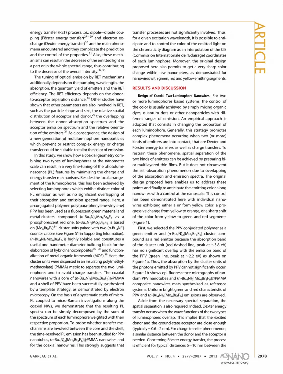
The relation between the proportions of each materialand the color along the NWs has been established bymicro-Raman and microphotoluminescence spectros-copy studies, as reported below.
Spectroscopic Studies along the Nanowires. To evaluatethe proportions of each material along the NWs andthe corresponding emitted color in the case of a con-tinuous PPV shell, the anodic alumina oxide (AAO)templated NWs were revealed by making a cross sectionas illustrated in Figure 3. This cross section was thenscanned by a focused laser andmicro-Raman (Figure 3a)and micro-PL (Figure 3c) spectra were recorded atdifferent positions from top to bottom of the mem-brane. The spot size of the focused laser beam on thesample was estimated to be about 1�2 μm. For theRaman study, a spectrum was recorded every 6 μmfor the 60-μm long NWs. The Raman peaks below300 cm�1 were attributed to the hexanuclear molyb-denum cluster,40�42 whereas peaks at 814, 882, 930,and 1453 cm�1 were attributed to PMMA.43�45 TheRaman characteristic peaks at 1174, 1550, 1588, and1631 cm�1 correspond to the stretching and thebending modes of carbon bonds in the PPV molecularbackbone.46 The presence of the peaks related to thePPV, PMMA and clusters corroborates the successfulsynthesis of coaxial NWs, as shown by TEM. Mostimportantly, the variation of the corresponding Raman
peak intensity along the scanning direction is a directsignature of the quantity of each material. This effecthas been exploited to evaluate the variation of theproportion of eachmaterial along the 60-μm longNWs.Indeed, the choice of the membrane and the wettingconditions can directly affect the quantity of matterdeposited inside the nanopores. We have shown re-cently that PPV NWs can be prepared in polycarbonatemembranes while PPV NTs are generally formed intoAAO membranes.47 Here, the evolution of the propor-tion of PPV and clusters compounds has been deter-mined from the change in the Raman intensity for 10spectra (Figure 3b). After a strong decrease of theRaman signal for both PPV and clusters from theimpregnation side, the intensity (normalized to thevalue at the first, i.e., top position) stabilizes around0.5�0.6 for the cluster units while the PPV one keepsfalling down to 0.2whenprobing down to the oppositeside of themembrane cross-section. Such a decrease ofthe intensity can directly be attributed to less matterdeposited. Indeed, it is expected for a solvent-assistedwetting synthesis that the quantity of polymer insolution decreases when it further penetrates fromthe top impregnation side to the bottom of the pores.This results in a thinner nanotube wall, also corrobo-rated by TEM investigation. Considering aNWdiameterof 200 nm, the thickness of the PPV shell can vary from
Figure 2. (a) SEM image of amat of flexible coaxial NWs. Inset: magnified SEM image of an isolatedNW. (b) AFM height imageof a network of wires deposited onto a glass substrate (Z-scale: 1 μm). Inset: magnified AFM height image of a NW withtopography line profile measured across the wire at the indicated location. (c) TEM micrograph of an isolated NW whichreveals the coaxial geometry, i.e., a PPV shell (indicated by the blues lines) and a (n-Bu4N)2[Mo6Br8F6]@PMMA composite coreas confirmed by EDX measurements (d) at the locations indicated on panel c (the circles represent roughly the probe area).Red and blue spectra correspond to the shell and the core of the fiber, respectively.
ARTIC
LE
GARREAU ET AL. VOL. 7 ’ NO. 4 ’ 2977–2987 ’ 2013
www.acsnano.org
2981
about 20 nm (top) to 10 nm (bottom). Then, the cor-responding volumic ratios of green (PPV) versus red((n-Bu4N)2[Mo6Br8F6]@PMMA) emitters have been es-timated to vary approximately from 1:2 (shell thicknessof 20 nm) to 1:5 (shell thickness of 10 nm) along thenanowires. We emphasize that the respective propor-tions of the two emitters are very similar in our coaxialapproach. Thus, it is possible to tune the color ofemission either in a slight way as shown here, or verysharply (see Figure 1) only by changing the thickness ofthe shell. Additionally, we propose two methods toincrease the tuning range. First, a larger change in theshell thickness along the coaxial nanowiremay result ina broader change of the covered color range: thesharper the variation of shell thickness, the strongerthe shift of color. Second, an increase in the clusterconcentration within PMMA may result in a largerweight of the red spectrum in the overall spectrum,i.e., a global red shift of the color range. A similar effectis expected for other couples of luminophores, whilerespecting the spectral restrictions reported above.This dramatically contrasts with the common strategyused for tuning the color that consists in the directmixing of the two (or more) emitters with typicaldonor�acceptor molar ratios of 100:1.18 Such a dis-crepancy between the two kinds of luminophores can
be a drawback for in-solution processes, because thelimit of solubility of the two emitters may constrain thedonor�acceptor ratio, i.e., the resulting color. Basedon this variation in theproportionof the two luminophoresand considering the quantum yield equals to about 40%for the PPV when processed into nanotubes47 and about20% for the (n-Bu4N)2[Mo6Br8F6],
48,49 a progressive shift ofthe color can indeed be anticipated from yellow-green toyellow-orange from top to bottom of the NWs.
The emissive features of the as-prepared NWs havebeen investigated by steady-state PL with a 2 μmdiameter microprobe at three positions along themembrane pores, as show in Figure 3c. The widespectra are composed of the characteristic PPV PLbands centered at 2.40, 2.24, 2.07, and 1.88 eV and ofthe cluster broad band with a maximum at 1.77 eV. It isclear from Figure 3c that the PL contribution of PPV isstrongly reduced from top to the bottom of the crosssection with respect to the nanocomposite signal. Thechange in the PL intensity for the 2.24 and the 1.77 eVbands is reported on the right axis of Figure 3b. The PLdecrease appears to be well-correlated to the Ramanintensity decrease. A more detailed analysis with asimple model to fit the PL spectra is given later.
Color Control and Spectral Modeling. The effect of themonochromatic pumping wavelength on the emission
Figure 3. (a) Micro-Raman spectra of array of coaxial NWs embedded in Al2O3 matrix for five laser positions (λexc = 785 nm)along the pores as illustrated in the scheme. The assignment of characteristic Raman peaks is also reported. (b) Micro-Ramanintensity (dashed lines) of PMMA (814 cm�1, blue), (n-Bu4N)2[Mo6Br8F6] (206 cm
�1, red) and PPV (1550 cm�1, green) versus thecross-sectional position in the membrane reported from Figure 3a. Right axis: PL intensity (solid lines) versus the position forthe 2.25 eV (green) and the 1.77 eV (red) bands related to PPV and clusters compounds, respectively. (c) Micro-PL spectra(λexc = 457.9 nm) of coaxial NWs for the three positions top-middle-bottom along the pores as illustrated in scheme. Thecorrespondence between the micro-PL spectra and the perception color is represented in a chromaticity diagram (d).CIE coordinates for pure PPV nanotubes and (n-Bu4N)2[Mo6Br8F6]@PMMA NWs are also reported.
ARTIC
LE
GARREAU ET AL. VOL. 7 ’ NO. 4 ’ 2977–2987 ’ 2013
www.acsnano.org
2982
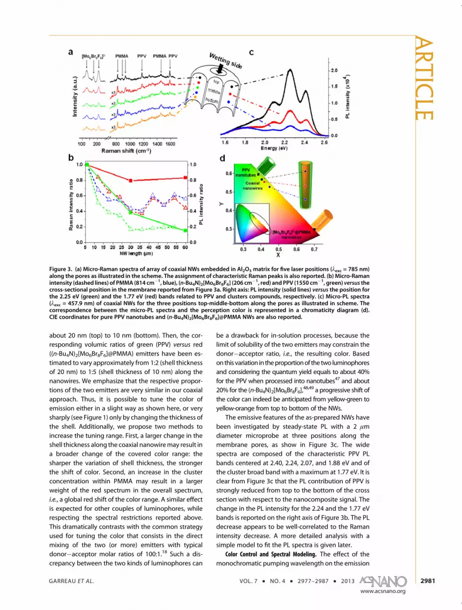
color was first determined. Considering that the PPVand the clusters units have different absorption spectra(Figure 1), the monochromatic pumping can favorresonant pumping of one species. Here, the PL spectrareported on Figure 4 were measured under a microp-robe at the same position along the nanowires and forpumping at 3.71 eV (334 nm), 2.71 eV (458 nm), and2.54 eV (488 nm). It is clear that the contribution of thePPV spectrum progressively increases with respect tothe (n-Bu4N)2[Mo6Br8F6]@PMMA one when the excita-tion line shifts from 3.71 to 2.71 and to 2.54 eV. Sucha variation can be anticipated from the absorption,emission and excitation spectra on Figure 1 (see alsophotoluminescence excitation maps in SupportingInformation), due to the preferential resonance pump-ing with the clusters for an excitation line in the UVrange andwith the PPV for excitation in the visible bluerange. It can also be noted a broadening of the overallspectrum for the excitation at 3.71 eV, which is due tothe superimposed broadened emissions of the twochromophores at this excitation energy. In addition,the sharp drop in the PL intensity at 2.50 eV for anexcitation at 2.54 eV is due to the edge filter used toeliminate the Rayleigh contribution. For these reasons,the points reported in the chromaticity diagramare notaligned (inset of Figure 4). In the following, the colorwas investigated for a fixed pumping wavelength.
We now address the color control. For this purpose,the coaxialNWswith a continuousPPV shell are considered
(see Figure 1b, case 1). The color emission correspondingto each PL spectrum (pumping wavelength: 2.71 eV) hasbeen determined for three positions along the NWs andreported in a chromaticity diagram (Figure 3d: top, x =0.41, y=0.57;middle, x=0.42, y=0.55; bottom, x=0.45,y = 0.53). The coordinates of (n-Bu4N)2[Mo6Br8F6]@PMMA NWs (x = 0.70, y = 0.30) and PPV nanotubes (x =0.38, y = 0.60) are also reported. It can be noted that theperception of the emission color for the coaxial NWs iscloser to the PPV one, due mainly to the sensitivity ofthe human eye. Moreover, the contribution of the PPVemission is favored by the higher quantum yield of PPVand the pumping wavelength (2.71 eV), as discussedabove. As anticipated from the Raman study, thereduced quantity of PPV from top (impregnation side)to bottom of the NWs results in a progressive shift ofthe color from yellow-green to yellow-orange. It is em-phasized that the coaxial design is a versatile strategywhich makes possible to have either a slight color shiftfor a progressive change of the shell thickness, or a verysharp color change for a discontinuous shell.
Next, we show that complex behaviors caused bycharge and energy transfers between the two lumino-phores are not significantly involved in the hybridcoaxial NWs. As discussed above, the spectral featuresof the selected two luminophores and the originaldesign with the coaxial geometry and the insulatingmatrix for the cluster units are expected to preventfrom reabsorption phenomenon, charge trapping, re-sonance energy transfer process and electron ex-change. If they occur, such phenomena would resultin the decrease of the donor (green) emission intensity,
Figure 4. Effect of the energy of excitation (2.54, 2.71, and3.71 eV) on the PL spectrum of the coaxial nanowires andtheir respective color on a chromaticity diagram (inset). Theinvestigated area along the NWs (in AAO template) is thesame for the three spectra.
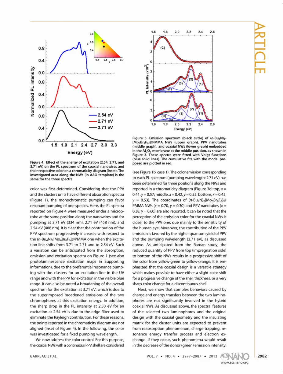
Figure 5. Emission spectrum (black circle) of (n-Bu4N)2-[Mo6Br8F6]@PMMA NWs (upper graph), PPV nanotubes(middle graph), and coaxial NWs (lower graph) embeddedin the Al2O3 membrane at the middle position, as shown inFigure 3. These spectra were fitted with Voigt functions(blue solid lines). The cumulative fits with the model pro-posed are plotted in red.
ARTIC
LE
GARREAU ET AL. VOL. 7 ’ NO. 4 ’ 2977–2987 ’ 2013
www.acsnano.org
2983
and the increase in the acceptor (red) emission inten-sitywhen compared to the casewithout transfer. This isobviously not the case as a dominant bright yellowemission is obtained for a volumic green (PPV):red(PMMA-cluster) ratio equal to 1:2 to 1:5 in the NWswith a continuous shell. This qualitative observation isconfirmed by the following quantitative analysis. Con-sidering that for the coaxial NWs, any coupling be-tween the two luminophoreswould involve changes inthe spectral shape with respect to the single lumino-phore case, a simplemodel has been tentatively imple-mented. It is proposed that the spectrum for the coaxialnanowires results from the sum of the PPV and clusterspectra weighted with only two parameters PPPV andPCluster related to the proportion of each luminophore:
[Coaxial spectrum] ¼ PPPV�[PPV spectrum]
þ PCluster�[Cluster spectrum]
In this model, for the three positions along the crosssection (top, middle and bottom, see Figure 3), thespectrum of the PPV nanotubes was fitted with fourVoigt functions (noted (1), (2), (3) and (4)) correspond-ing to the four bands usually identified for PPV,50 whilethe spectrum for (n-Bu4N)2[Mo6Br8F6]@PMMA compos-ite NWs was fitted with a Voigt function noted (C) (acomplete description of the fitting method is reportedin Supporting Information). The corresponding curvesare reported in Figure 5 for the spectrameasured in themiddle of the membrane cross sections (see Support-ing Information for the fits of the spectra at the top andbottompositions and for details of the fittingparameters).Then, the spectra for the coaxial two-luminophorenanowires were tentatively fitted by the sum of thePPV and cluster contributions with fixed values for theenergy of the emission bands and the width parameters.Only themagnitudes with respect to PPV and compositespectra were allowed to vary freely, corresponding tothe proportion of each material as described in themodel. Finally, the band widths were allowed toslightly vary. A very good fit was obtained for thetop, the middle and the bottom spectra, as shown inFigure 5 and Supporting Information. To further corro-borate the validity of our analysis, the fitting compo-nents areas (related to the width and the magnitude)for the four PPV bands and the cluster one in two-luminophore NWs were compared to the correspond-ing values for the one-luminophore NWs or NTs (FigureS4 and Table S3 in Supporting Information). Remark-ably, for each position (top, middle, and bottom), theareas for one-luminophore or two-luminophore sys-tems are very similar. Moreover, their intensity exhibitsa variation with the position along the NW close to theone observed for the Raman intensity. This analysisshows that for our system, the very simple model withonly two free parameters PPPV and PCluster related to theproportion of each luminophore is well verified.
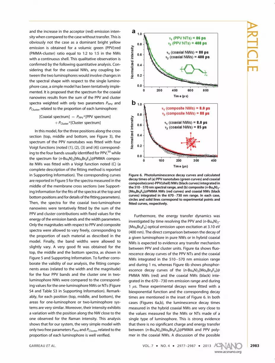
Furthermore, the energy transfer dynamics wasinvestigated by time resolving the PPV and (n-Bu4N)2-[Mo6Br8F6] optical emission upon excitation at 3.10 eV(400 nm). The direct comparison between the decay ofa given luminophore in pure NWs or in hybrid coaxialNWs is expected to evidence any transfer mechanismbetween PPV and cluster units. Figure 6a shows fluo-rescence decay curves of the PPV NTs and the coaxialNWs integrated in the 510�570 nm emission rangeand during 1 ns, whereas Figure 6b shows phosphor-escence decay curves of the (n-Bu4N)2[Mo6Br8F6]@PMMA NWs (red) and the coaxial NWs (black) inte-grated in the 670�730 nm emission range and during1 μs. These experimental decays were fitted with abiexponential function and the corresponding decaytimes are mentioned in the inset of Figure 6. In bothcases (Figures 6a,b), the luminescence decay timesmeasured in the hybrid coaxial NWs are very close tothe values measured for the NWs or NTs made of asingle type of luminophore. This is strong evidencethat there is no significant charge and energy transferbetween (n-Bu4N)2[Mo6Br8F6]@PMMA and PPV poly-mer in the coaxial NWs. A discussion of the possible
Figure 6. Photoluminescence decay curves and calculateddecay times of (a) PPV nanotubes (green curves) and coaxialcomposite(core)-PPV(shell) NWs (black curves) integrated inthe 510�570 nm spectral range, and (b) composite (n-Bu4N)2-[Mo6Br8F6]@PMMA NWs (red curves) and coaxial NWs (blackcurves) integrated in the 670�730 nm range. In each case,circles and solid lines correspond to experimental points andfitted curves, respectively.
ARTIC
LE

GARREAU ET AL. VOL. 7 ’ NO. 4 ’ 2977–2987 ’ 2013
www.acsnano.org
2984
effects of the coaxial morphology on energy transfersin our system is nowproposed. For the PPV, the excitondiffusion length is typically 5�12 nm.51,52 Thus, for aPPV shell thickness of about 20 nm, almost all theexcitons created in PPV could diffuse toward the inter-face and thus eventually interact with clusters unitsembedded in PMMA. A fluorescence resonance energytransfer (FRET) mechanism between the two lumino-phores should then strongly affect the contribution ofPPV to the overall PL spectrum and PL decay. From thestationary and time-resolved PL studies and analysis, itis reasonable to consider that no significant energytransfer takes place in our system. Additionally, theproportion of clusters located in the dipole�dipole likecoupling area close to PPV can be estimated. Indeed,the FRET efficiency is inversely proportional to the sixthpowerof thedistancebetween thedonor andacceptor.53
Previous studies in composites with quantum dotssuggested nonradiative resonant dipole�dipole inter-action to explain the observed efficient excitationtransfer from the conjugated polymer to the QD.54,55
They found Förster radii of 5�7 nmwhich correspondsto the distance between donor and acceptor where50% of the excitations are being transferred. The uppervalue of 7 nm for the Förster radius is considered herefor the estimation. On the basis of the morphologicalstudy, average values for the outer NW diameter andthe PPV shell thickness are 220 and 20 nm, respectively.The estimated proportion of clusters under influenceof FRET is about 15% of the total clusters amount. Thus,only a minor proportion of the clusters units couldinteract with PPV excitons. Dexter energy transfer canalso be ruled out in the emission pathways of thesehybrid nanowires. The Dexter mechanism is a short-range process involving electron exchange occurringwithin about 1 nm between donor and acceptor. Themain consequence of Dexter transfer is PL quenching.Thus, one should speculate that if this transfer exists inour hybrid nanowires it should affect only the core�shell interface, and thus involve a negligible fraction ofclusters at the interface. Similarly, only a very smallproportion of clusters would be affected by chargetransfers from the PPV shell to the clusters through theinsulating PMMA matrix by hopping mechanism. Ac-cordingly, Figure 6 shows that there is no significant PLquenching in the hybrid coaxial nanowires with re-spect to pure PPV nanotubes or cluster-PMMA NWs.
Beyond the original strategy proposed in this workto minimize the interactions between the luminophoresof each color, the coaxial structure can be extended to
any couple of luminophores, including the donor�acceptor systems. Then, the proportion of donorslocated in the shell and acceptors in the core whichexperiences charge and energy transfers could betuned by the control of the outer diameter and shellthickness in relation to the characteristic physicallengths involved. For a fixed shell thickness and whenRETmechanisms are involved, the relative contributionof donor�acceptor coupling would increase when thediameter is decreased. Thus, the coaxial design couldbe further exploited to explore the chromaticity dia-gram, including the white panel by incorporating blueemitters. It is also a promising way to help under-standing the complex coupling mechanisms impli-cated within nanosources made of at least two typesof luminophores. This is a central point in this veryactive field of research, in relation with some of thechallenges of nanophotonics, for example by forecast-ing the emission color of integrated devices.
CONCLUSION
Two-luminophore nanowires based on a coaxialarchitecture have been designed to take control ofthe color of hybrid nanosources. The two lumino-phores, (n-Bu4N)2[Mo6Br8F6] cluster compound andsemiconducting PPV conjugated polymer, were se-lected to restrain the absorption and emission spectraloverlappingwhile a spatial separation was achieved bythe coaxial geometry. A wetting template process wasused to fabricate the coaxial nanowires with a PPV shelland (n-Bu4N)2[Mo6Br8F6]@PMMA core. A systematicstudy of the micro-PL coupled with micro-Ramaninvestigations along the coaxial nanowires shows thatthe resulting PL spectra can be simply decomposedby the sum of the spectrum of each luminophoreweighted by the relative proportion of each material.A time-resolved PL study confirms that the couplingbetween the two luminophores can be neglected. Thepresent investigation reveals that it is possible toforecast the color of the emitting coaxial NW on thechromaticity diagram as an interpolation of the CIEcoordinates of each luminophore. Additionally, thiscoaxial morphology offers a promising way to get asharp change of the color at the nanoscale, as shownhere for red, green and yellow emitting segments. Thisoriginal design is a relevant way to produce hybridnanosources by in-solution processing with a greatversatility in the color control, and thus to address animportant challenge of nanophotonics for tagging,sensing, lasing and lighting development.
METHODS
Cluster Synthesis and Composite Preparation. (n-Bu4N)2[Mo6Br8F6]waspreparedaccording toprocedure reported.38 For the fabrication
of the (n-Bu4N)2[Mo6Br8F6]@PMMA composite, acetone (opticalspectroscopy grade, Carlo Erba) andPMMA (Mw: 120 000g 3mol�1,Sigma-Aldrich) were usedwithout further purification. To get anappropriate viscosity for the PMMA and clusters in acetone, a
ARTIC
LE

GARREAU ET AL. VOL. 7 ’ NO. 4 ’ 2977–2987 ’ 2013
www.acsnano.org
2985
solution containing 4 wt % of (n-Bu4N)2[Mo6Br8F6] and 10 wt %of PMMA was prepared.
Fabrication of the Coaxial NWs. The coaxial NWswere fabricatedby a wetting template method in anodic alumina oxide (AAO)nanoporous template. Commercial AAOmembranes purchasedfrom Whatman (anodisc 13) have been used. They are 60 μmthick with a real pore diameter showing a polydispersity between200 nm and around 250 nm, as revealed by SEM analysis.
For the synthesis of coaxial NWs, two steps of wetting arerequired. The first step consists in the wetting of AAO mem-brane with the PPV precursor in methanol solution. The sulfo-nium polyelectrolyte used as a precursor polymer of PPV wassynthesized in our laboratory via the standard procedure.56 Toobtain PPV nanotubes, the concentration of PPV precursor inmethanol was chosen at 0.5 mg/mL and 200 μL of this solutionwas drop-casted on AAO membrane, as described elsewhere.39,47
The precursor embedded in AAO membrane was then thermallyconverted under a dynamic secondary vacuum (∼10�6 Torr) for4 h to obtain PPV. The coaxial nanowires are then obtained by asecondary-template strategy. The AAO membrane containingPPV nanotubes was wetted with the solution containing TBAþ
[Mo6Br8F6]2� and PMMA in acetone. Thewetted templates were
left overnight under ambient condition to allow solvent eva-poration. For some characterization, the AAO templates con-taining coaxial NWs was completely etched in H3PO4 (25 wt %)overnight and washed several time with DI water. The coaxialNWs were homogeneously dispersed in DI water solution byultrasonication during ∼10 s with a power of 140 W (FisherScientific FB 15052). Ultrasonication results in the shortening ofthe nanowire length.
Structural and Optical Characterizations. A field-effect scanningelectron microscope (JEOL, JSM-7600F operating at 5 kV), anAFM (Nanowizard II, JPK instruments) working in intermittent-contact mode in air with Si tips (PPP-NCHR, Nanosensors) and atransmission electron microscope (Hitachi H9000 NAR operat-ing at 300 kV) were used to investigate themorphology and thecomposition of the nanowires. For SEM, AFM and fluorescencemicroscopy experiments, a drop of solution (∼10 μL) containingthe nanowires was deposited onto silicon or glass substratesafter the dissolution of the template. For TEM experiments(imaging, electron diffraction and EDX), a drop of the solutionwas deposited on TEM copper grids covered with a thin holeycarbon film. The microscope is equipped with a Noran KevexSi�Li detector for EDX spectroscopy. Epi-fluorescence micro-graphs were acquired using a calibrated microscope (Eclipse Ti,Nikon) equippedwith a�60objective andaCCDcamera, 130WHglamp and fluorescence filter cube (EX 330-380, Dm: 400, BA 420).
Spectroscopic Characterizations. All characterizations were per-formed at room temperature. Absorption and photolumines-cence of the [Mo6Br8F6]
2� clusters were measured on spin-coated thin films (1500 rpm, 30 s) deposited on glass from asolution containing 4 wt % in acetone and from a PPV precursor(PPV precursor: 0.5 mg 3mL�1 in methanol, 1500 rpm, 30 s,thermal conversion at 300 �C for 3 h). UV�vis absorption on thinfilm sample was performed with a Perkin-Elmer double beamspectrophotometer equipped with an integrating sphere; wecarried out PLE measurements using a Jobin-Yvon Fluorolog 3equipped with a CCD camera and PL experiments on film wereachieved under 400 nm excitation at 0.5 mW by a Spectra-Physics Hurricane X laser. For micro-Raman and microphotolu-minescence studies, the template containing the nanowireswas broken to reveal a cross section and consequently thewhole length of the nanowires. Micro-Raman spectra wererecorded using a Renishaw inVia Raman microscope equippedwith a 785 nm line of a HPNIR diode laser. Wemeasured steady-state micro-PL spectra with a Jobin-Yvon T64000 spectrometerunder 457.9 nm laser excitation obtained by an argon ion laser.In each case, the spot size of the focused laser beam on thesample was estimated to be about 1�2 μm. The output laserpower incident on the sample and the acquisition time werefixed at 5 mW and 90 s, and at 10 μW and 10 s for each Ramanand PL spectrum, respectively. Investigation of the effect of thepumping wavelength was performed with Jobin-Yvon T64000and Horiba Jobin-Yvon iHR 320 spectrometers under 457.9,488, and 334 nm laser excitation from an argon ion laser.
The transient PL experiments have been achieved under 400 nmexcitation using Spectra-Physics Hurricane X laser system (82 fs,1 kHz) onto mat of NWs. The collected emission was temporallydetected with a streak camera (Hamamatsu C7700) coupledwith an imaging spectrograph. The laser pump power imping-ing on sample was kept at 0.5 mW to minimize sample photo-bleaching.
Conflict of Interest: The authors declare no competingfinancial interest.
Acknowledgment. A. Garreau benefits from a Region Paysde la Loire grant through the Nanofonc network. This work hasalso been supported by a financial support of the C'Nano Nord-Ouest network. The authors thank J. Y. Mevellec for his helpin micro-Raman spectroscopy studies and N. Stephant andS. Grolleau for SEM characterization. We gratefully acknowledgeA. Moissette from LASIR Lille I, France, for the PL spectroscopymeasurements under UV excitation.
Supporting Information Available: The structure of the[Mo6Br
i8F
a6]2� anionic unit, the description of the method used
for the fit of PL spectra, and the photoluminescence excitationmaps for thin film of PPV and (n-Bu4N)2[Mo6Br8F6]@PMMA arereported. Thismaterial is available free of charge via the Internetat http://pubs.acs.org.
REFERENCES AND NOTES1. Li, X.; Wang, T.; Zhang, J.; Zhu, D.; Zhang, X.; Ning, Y.; Zhang,
H.; Yang, B. Controlled Fabrication of Fluorescent BarcodeNanorods. ACS Nano 2010, 4, 4350–4360.
2. Park, D. H.; Hong, Y. K.; Cho, E. H.; Kim, M. S.; Kim, D.-C.;Bang, J.; Kim, J.; Joo, J. Light-Emitting Color BarcodeNanowires Using Polymers: Nanoscale Optical Character-istics. ACS Nano 2010, 4, 5155–5162.
3. Gu, F.; Zhang, L.; Yin, X.; Tong, L. Polymer Single-NanowireOptical Sensors. Nano Lett. 2008, 8, 2757–2761.
4. Zhao, Y. S.; Wu, J.; Huang, J. Vertical Organic NanowireArrays: Controlled Synthesis and Chemical Sensors. J. Am.Chem. Soc. 2009, 131, 3158–3159.
5. Zang, L.; Che, Y.; Moore, J. S. One-Dimensional Self-Assembly of Planar pi-Conjugated Molecules: AdaptableBuilding Blocks for Organic Nanodevices. Acc. Chem. Res.2008, 41, 1596–1608.
6. Che, Y.; Yang, X.; Loser, S.; Zang, L. Expedient VaporProbing of Organic Amines Using Fluorescent NanofibersFabricated from an n-Type Organic Semiconductor. NanoLett. 2008, 8, 2219–2223.
7. O'Carroll, D.; Lieberwirth, I.; Redmond, G. MicrocavityEffects and Optically Pumped Lasing in Single ConjugatedPolymer Nanowires. Nat. Nanotechnol. 2007, 2, 180–184.
8. Zhao, Y. S.; Peng, A.; Fu, H.; Ma, Y.; Yao, J. NanowireWaveguides andUltraviolet Lasers Based on Small OrganicMolecules. Adv. Mater. 2008, 20, 1661–1665.
9. Kuo, C.-C.; Lin, C.-H.; Chen, W.-C. Morphology and Photo-physical Properties of Light-Emitting Electrospun Nano-fibers Prepared from Poly(fluorene) Derivative/PMMABlends. Macromolecules 2007, 40, 6959–6966.
10. Yang, H.; Lightner, C. R.; Dong, L. Light-Emitting CoaxialNanofibers. ACS Nano 2012, 6, 622–628.
11. Lu, X.; Zhang, W.; Wang, C.; Wen, T.-C.; Wei, Y. One-Dimensional Conducting Polymer Nanocomposites: Syn-thesis, Properties and Applications. Prog. Polym. Sci. 2011,36, 671–712.
12. Kim, F. S.; Ren, G.; Jenekhe, S. A. One-Dimensional Nano-structures of π-Conjugated Molecular Systems: Assembly,Properties, and Applications from Photovoltaics, Sensors,and Nanophotonics to Nanoelectronics. Chem. Mater.2011, 23, 682–732.
13. Long, Y.-Z.; Li, M.-M.; Gu, C.; Wan, M.; Duvail, J.-L.; Liu, Z.;Fan, Z. Recent Advances in Synthesis, Physical Propertiesand Applications of Conducting Polymer Nanotubes andNanofibers. Prog. Polym. Sci. 2011, 36, 1415–1442.
14. Zhao, Y. S.; Fu, H.; Peng, A.; Ma, Y.; Liao, Q.; Yao, J.Construction and Optoelectronic Properties of Organic
ARTIC
LE

GARREAU ET AL. VOL. 7 ’ NO. 4 ’ 2977–2987 ’ 2013
www.acsnano.org
2986
One-Dimensional Nanostructures. Acc. Chem. Res. 2009,43, 409–418.
15. Dennis, A. M.; Bao, G. Quantum Dot-Fluorescent ProteinPairs as Novel Fluorescence Resonance Energy TransferProbes. Nano Lett. 2008, 8, 1439–1445.
16. Stoferle, T.; Scherf, U.; Mahrt, R. F. Energy Transfer in HybridOrganic/Inorganic Nanocomposites. Nano Lett. 2009, 9,453–456.
17. Camposeo, A.; Di Benedetto, F.; Cingolani, R.; Pisignano, D.Full Color Control and White Emission from ConjugatedPolymer Nanofibers. Appl. Phys. Lett. 2009, 94, 043109.
18. Zhao, Y. S.; Fu, H. B.; Hu, F. Q.; Peng, A. D.; Yang, W. S.;Yao, J. N. Tunable Emission from Binary Organic One-Dimensional Nanomaterials: An Alternative Approach toWhite-Light Emission. Adv. Mater. 2008, 20, 79–83.
19. Fardy, M.; Yang, P. Materials Science: Lilliputian LightSticks. Nature 2008, 451, 408–409.
20. Vohra, V.; Calzaferri, G.; Destri, S.; Pasini, M.; Porzio, W.;Botta, C. Toward White Light Emission through EfficientTwo-Step Energy Transfer in Hybrid Nanofibers. ACS Nano2010, 4, 1409–1416.
21. Giansante, C.; Raffy, G.; Schafer, C.; Rahma, H.; Kao, M.-T.;Olive, A. G. L.; Del Guerzo, A. White-Light-Emitting Self-Assembled NanoFibers and Their Evidence by Microspec-troscopy of Individual Objects. J. Am. Chem. Soc. 2011, 133,316–325.
22. Basak, S.; Chandrasekar, R. Multiluminescent Hybrid Or-ganic/Inorganic Nanotubular Structures: One-Pot Fabrica-tion of Tricolor (Blue-Red-Purple) Luminescent Paralle-pipedic Organic Superstructure Grafted with EuropiumComplexes. Adv. Funct. Mater. 2011, 21, 667–673.
23. Sui, X. M.; Shao, C. L.; Liu, Y. C. White-Light Emission ofPolyvinyl Alcohol/ZnO Hybrid Nanofibers Prepared byElectrospinning. Appl. Phys. Lett. 2005, 87, 113115.
24. Moynihan, S.; Iacopino, D.; O'Carroll, D.; Doyle, H.; Tanner,D. A.; Redmond, G. Emission Colour Tuning in Semicon-ducting Polymer Nanotubes by Energy Transfer to Organo-Lanthanide Dopants. Adv. Mater. 2007, 19, 2474–2479.
25. Zhang, H.; Song, H.; Dong, B.; Han, L.; Pan, G.; Bai, X.; Fan, L.;Lu, S.; Zhao, H.; Wang, F. Electrospinning Preparation andLuminescence Properties of Europium Complex/PolymerComposite Fibers. J. Phys. Chem. C 2008, 112, 9155–9162.
26. Bredas, J.-L.; Beljonne, D.; Coropceanu, V.; Cornil, J. Charge-Transfer and Energy-Transfer Processes in pi-ConjugatedOligomers and Polymers: A Molecular Picture. Chem. Rev.2004, 104, 4971–5003.
27. McGehee, M. D.; Bergstedt, T.; Zhang, C.; Saab, A. P.;O'Regan, M. B.; Bazan, G. C.; Srdanov, V. I.; Heeger, A. J.Narrow Bandwidth Luminescence from Blends with En-ergy Transfer from Semiconducting Conjugated Polymersto Europium Complexes. Adv. Mater. 1999, 11, 1349–1354.
28. Forster, T. Transfer Mechanisms of Electronic Excitation.Discuss. Faraday Soc. 1959, 27, 7–17.
29. Halivni, S.; Sitt, A.; Hadar, I.; Banin, U. Effect of NanoparticleDimensionality on Fluorescence Resonance Energy Trans-fer in Nanoparticle-Dye Conjugated Systems. ACS Nano2012, 6, 2758–2765.
30. Dexter, D. L. A Theory of Sensitized Luminescence inSolids. J. Chem. Phys. 1953, 21, 836–850.
31. Scholes, G. D.; Rumbles, G. Excitons in Nanoscale Systems.Nat. Mater. 2006, 5, 683–696.
32. Kamtekar, K. T.; Monkman, A. P.; Bryce, M. R. RecentAdvances in White Organic Light-Emitting Materials andDevices (WOLEDs). Adv. Mater. 2010, 22, 572–582.
33. Gather, M. C.; Köhnen, A.; Meerholz, K. White OrganicLight-Emitting Diodes. Adv. Mater. 2011, 23, 233–248.
34. Simbrunner, C.; Hernandez-Sosa, G.; Quochi, F.; Schwabegger,G. N.; Botta, C.; Oehzelt, M.; Salzmann, I.; Djuric, T.; Neuhold, A.;Resel, R.; et al. Color Tuning of Nanofibers by PeriodicOrganic-Organic Hetero-Epitaxy. ACS Nano 2012, 6, 4629–4638.
35. Molard, Y.; Dorson, F.; Cîrcu, V.; Roisnel, T.; Artzner, F.;Cordier, S. Clustomesogens: Liquid Crystal Materials Con-taining Transition-Metal Clusters. Angew. Chem., Int. Ed.2010, 49, 3351–3355.
36. Mocanu, A. S.; Amela-Cortes, M.; Molard, Y.; Circu, V.;Cordier, S. Liquid Crystal Properties Resulting from Syner-getic Effects between Non-Mesogenic Organic Moleculesand a One Nanometre Sized Octahedral Transition MetalCluster. Chem. Commun. 2011, 47, 2056–2058.
37. Molard, Y.; Ledneva, A.; Amela-Cortes, M.; Cîrcu, V.;Naumov, N. G.; Mériadec, C.; Artzner, F.; Cordier, S. IonicallySelf-Assembled Clustomesogen with Switchable Magnetic/Luminescence Properties Containing [Re6Se8(CN)6]n- (n = 3,4) Anionic Clusters. Chem. Mater. 2011, 23, 5122–5130.
38. Dybtsev, D.; Serre, C.; Schmitz, B.; Panella, B.; Hirscher, M.;Latroche, M.; Llewellyn, P. L.; Cordier, S.; Molard, Y.; Haouas,M.; et al. Influence of [Mo6Br8F6]
2� Cluster Unit Inclusionwithin the Mesoporous Solid MIL-101 on Hydrogen Stor-age Performance. Langmuir 2010, 26, 11283–11290.
39. Lorcy, J. M.; Massuyeau, F.; Moreau, P.; Chauvet, O.; Faul-ques, E.; Wery, J.; Duvail, J. L. Coaxial Nickel/Poly(p-pheny-lene vinylene) Nanowires as Luminescent Building BlocksManipulatedMagnetically.Nanotechnology 2009, 20, 405601.
40. Bublitz, D.; Preetz, W. Schwingungsspektren und Normal-koordinatenanalysen der 92Mo-, 100Mo-, 35Cl- und 37Cl-Isotopomeren der Clusteranionen [(Mo6X8
i )Y6a]2�; Xi = Cl,
Br; Ya = F, Cl, Br, I. Z. Anorg. Allg. Chem. 1996, 622, 1107–1117.
41. Ramirez-Tagle, R.; Arratia-Pérez, R. Electronic Structure andMolecular Properties of the [Mo6X8L6]
2� ; X = Cl, Br, I; L =F, Cl, Br, I Clusters. Chem. Phys. Lett. 2008, 460, 438–441.
42. Schoonover, J. R.; Zietlow, T. C.; Clark, D. L.; Heppert, J. A.;Chisholm, M. H.; Gray, H. B.; Sattelberger, A. P.; Woodruff,W. H. Resonance Raman Spectra of [M6X8Y6]
2� ClusterComplexes (M = Mo, W; X, Y = Cl, Br, I). Inorg. Chem.1996, 35, 6606–6613.
43. Willis, H. A.; Zichy, V. J. I.; Hendra, P. J. The Laser-Raman andInfra-Red Spectra of Poly(methyl methacrylate). Polymer1969, 10, 737–746.
44. Xu, X.; Ming, H.; Zhang, Q.; Zhang, Y. Properties of RamanSpectra and Laser-Induced Birefringence in PolymethylMethacrylate Optical Fibres. J. Opt. A: Pure Appl. Opt. 2002,4, 237.
45. Thomas, K. J.; Sheeba, M.; Nampoori, V. P. N.; Vallabhan,C. P. G.; Radhakrishnan, P. Raman Spectra of PolymethylMethacrylate Optical Fibres Excited by a 532 nm DiodePumped Solid State Laser. J. Opt. A: Pure Appl. Opt. 2008,10, 055303.
46. Mulazzi, E.; Perego, R.; Aarab, H.; Mihut, L.; Lefrant, S.;Faulques, E.; Wery, J. Photoconductivity and Optical Prop-erties in Composites of Poly(paraphenylene vinylene) andSingle-Walled Carbon Nanotubes. Phys. Rev. B 2004, 70,155206.
47. Massuyeau, F.; Duvail, J. L.; Athalin, H.; Lorcy, J. M.; Lefrant,S.;Wery, J.; Faulques, E. Elaboration of Conjugated PolymerNanowires and Nanotubes for Tunable Photolumines-cence Properties. Nanotechnology 2009, 20, 155701.
48. Maverick, A. W.; Najdzionek, J. S.; MacKenzie, D.; Nocera,D. G.; Gray, H. B. Spectroscopic, Electrochemical, andPhotochemical Properties of Molybdenum(II) and Tungsten-(II) Halide Clusters. J. Am. Chem. Soc. 1983, 105, 1878–1882.
49. Szczepura, L.; Edwards, J.; Cede~no, D. Luminescent Proper-ties of Hexanuclear Molybdenum(II) Chloride ClustersContaining Thiolate Ligands. J. Cluster Sci. 2009, 20, 105–112.
50. Mulazzi, E.; Ripamonti, A.; Wery, J.; Dulieu, B.; Lefrant, S.Theoretical and Experimental Investigation of Absorptionand Raman Spectra of Poly(paraphenylene vinylene). Phys.Rev. B 1999, 60, 16519–16525.
51. Stubinger, T.; Brutting, W. Exciton Diffusion and OpticalInterference in Organic Donor-Acceptor PhotovoltaicCells. J. Appl. Phys. 2001, 90, 3632–3641.
52. Markov, D. E.; Amsterdam, E.; Blom, P. W. M.; Sieval, A. B.;Hummelen, J. C. Accurate Measurement of the ExcitonDiffusion Length in a Conjugated Polymer Using a Hetero-structure with a Side-Chain Cross-Linked Fullerene Layer.J. Phys. Chem. A 2005, 109, 5266–5274.
53. Lakowicz, J. R. Principles of Fluorescence Spectroscopy, 3rded.; Springer: New York, 2006.
ARTIC
LE
GARREAU ET AL. VOL. 7 ’ NO. 4 ’ 2977–2987 ’ 2013
www.acsnano.org
2987
54. Anni, M.; Manna, L.; Cingolani, R.; Valerini, D.; Creti, A.;Lomascolo, M. Forster Energy Transfer from Blue-EmittingPolymers to Colloidal CdSe/ZnS Core Shell Quantum Dots.Appl. Phys. Lett. 2004, 85, 4169–4171.
55. Kaufmann, S.; Stoferle, T.; Moll, N.; Mahrt, R. F.; Scherf, U.;Tsami, A.; Talapin, D. V.; Murray, C. B. Resonant EnergyTransfer Within a Colloidal Nanocrystal Polymer HostSystem. Appl. Phys. Lett. 2007, 90, 071108.
56. Stenger-Smith, J. D.; Lenz, R. W.; Wegner, G. Spectroscopicand Cyclic Voltammetric Studies of Poly(p-phenylenevinylene) Prepared from Two Different Sulphonium SaltPrecursor Polymers. Polymer 1989, 30, 1048–1053.
ARTIC
LE


