Embed Size (px)
Citation preview

The EMBO Journal vol.14 no.16 pp.3925-3936, 1995
Dominant negative mutants of the Cdc2 kinaseuncouple cell division from iterative plantdevelopment
Adriana Hemerlyl2,Janice de Almeida Engler1,Catherine Bergounioux3,Marc Van Montagu1'4, Gilbert Engler5,Dirk Inze5 and Paulo Ferreira1'2'Laboratorium voor Genetica, Universiteit Gent, K.L. Ledeganckstraat,B-9000 Gent, Belgium, 3Laboratoire de Physiologie Wgetale,Universite d'Orsay, F-91405 Orsay, France and 5Laboratoire associe del'Institut National de la Recherche Agronomique (France), UniversiteitGent, B-9000 Gent, Belgium2Present address: Laborat6rio de Bioenergetica, Departamento deBioquimica Medica, Universidade Federal de Rio de Janeiro, CEP21941-590, Rio de Janeiro, RJ, Brasil
4Corresponding author
Communicated by M.Van Montagu
Because plant cells do not move and are surroundedby a rigid cell wall, cell division rates and patterns arebelieved to be directly responsible for generating newstructures throughout development. To study the rela-tionship between cell division and morphogenesis,transgenic tobacco and Arabidopsis plants were con-structed expressing dominant mutations in a key regu-lator of the Arabidopsis cell cycle, the Cdc2a kinase.Plants constitutively overproducing the wild-typeCdc2a or the mutant form predicted to acceleratethe cell cycle did not exhibit a significantly altereddevelopment. In contrast, a mutation expected to arrestthe cell cycle abolished cell division when expressed inArabidopsis, whereas some tobacco plants constitutivelyproducing this mutant protein were recovered. Theseplants had a reduced histone Hi kinase activity andcontained considerably fewer cells. These cells were,however, much larger and underwent normal differen-tiation. Morphogenesis, histogenesis and developmentaltiming were unaffected. The results indicate that, inplants, the developmental controls defining shape canact independently from cell division rates.Keywords: ArabidopsislCdc2 kinase/cell division/morpho-genesis/tobacco
IntroductionAlthough plant development has been characterizedextensively, the relationship between cell division anddevelopment is unclear. Since plant cells are surroundedby a rigid cell wall, a precise spatial and temporalregulation of cell division is expected to play an importantrole in morphogenesis. Therefore, plant morpho-genesis studies often emphasize frequencies and patternsof cell division as being directly responsible for generatingshape.
Alternatively, some plant developmental processes show
flexibility in morphogenetic controls. Most plant morpho-genesis takes place after embryogenesis, by iterativedevelopment at the meristems. The regulation of morpho-genic processes as they start in the embryo greatly differsfrom their subsequent regulation in the developing plant.As in animals, plant embryogenesis shows a sequentialdevelopment in which patterns of cell division are, ingeneral, reasonably fixed (reviewed by Jiirgens, 1992;West and Harada, 1993). In contrast, apical meristemsproduce repeated units that can be modulated dependingon environmental conditions. The morphogenic plasticityof plants reflects a degree of flexibility in the intrinsicprogramme that controls patterns and frequencies of celldivision during iterative development.
In a cycling plant cell, however, cell division must beunder stringent control. The molecular and biochemicalmechanisms of cell cycle control in plants are beingelucidated and appear to be largely similar to all othereukaryotes (reviewed by Jacobs, 1992). Perturbations inthe cell clock would probably lead to abnormal divisions,and the analysis of its effects on plant development couldhelp to reveal the extent of correlation between celldivision and morphogenesis in plants. The major regulatorsof the eukaryotic cell cycle are cyclin-dependent kinases(Cdks), whose activity is controlled mainly by post-translational events (reviewed by Murray, 1993; Solomon,1993). Dominant mutations that affect the normal controlsof cell cycle progression have been described for the yeastCdks (cdc2/cdc28) (Booher and Beach, 1986; Mendenhallet al., 1988; Gould and Nurse, 1989). They can eithercause cells to enter mitosis prematurely (Wee phenotype)or arrest cell division (Cdc2- phenotype). We, therefore,employed a novel strategy for plants: the construction ofdominant mutations in a plant Cdk and its subsequentoverexpression in plants in an attempt to deregulate thenormal function of the wild-type gene.
Arabidopsis has an apparently small Cdk family. Sofar, two members have been cloned, cdc2a and cdc2b(Ferreira et al., 1991; Imajuku et al., 1992). For ourstudies, cdc2a was chosen because it is the only functionalhomologue and it is expressed in all plant meristems,suggesting a possible role during plant development(Martinez et al., 1992; Hemerly et al., 1993). We showthat a mutation that inhibits kinase activity is lethal whenoverexpressed in Arabidopsis, but tobacco plants with lowlevels of the Cdc2a protein could be rescued. Analyses ofthese mutant plants showed that, despite a reducedfrequency of cell division, their development paralleledthat of control plants. The plants consist of fewer cells,but undergo normal morphogenesis and histogenesis, withnormal developmental timing. This observation demon-strates that cell division and plant development may bepartially uncoupled.
© Oxford University Press 3925

A.Hemerly et al.
A
B
AtCdc2a
HsCdk2
SpCdc2
A F
G E G T Y G V V Y
G E G TY G VV Y
IK 20
K 20
G E G T Y G V V Y K' 20N
AtCdc2a D F G L A R A F G 155
HsCdk2 D F G L A R A F G 153
SpCdc2 D F G L A R S [G 160
PCR mutagenesis
AF
T 35S Icdc2a13Nos cdc2a.A1 4F1 5N
-Ezz :EzJ- cdc2a.N 147
qI I Icdc2a+a/
Arobidopsis tobacco
Fig. 1. Diagram of the construction of cdc2a mutants. (A) Alignedamino acid sequence of regions of the Arabidopsis (At) Cdc2a, human(Hs) Cdk2 and S.pombe (Sp) Cdc2, indicating the mutated residues inCdc2a. Boxes surround residues conserved in the three kinases.(B) Schematic diagram of the 35S-cdc2a mutant construction used togenerate transgenic plants (see Materials and methods). Two cdc2amutant cDNAs were created by PCR mutagenesis: cdc2a.A14FJ5 andcdc2a.N147. Both mutants and the wild-type cDNA (cdc2a+) werecloned under control of CaMV 35S and introduced into Arabidopsisand tobacco plants.
ResultsConstruction of point mutations in theArabidopsis cdc2a cDNABased on dominant mutations described in yeasts, twomutant Arabidopsis cdc2a cDNAs were generated (FigureIA). In one of the mutations, the D147 residue wasreplaced with an N147 residue. Both in Saccharomycescerevisiae and in humans, this mutation has been shownto inactivate the kinase causing an arrest of the cell cycle(Mendenhall et al., 1988; van den Heuvel and Harlow,1993). With the resolution of the crystal structure of thehuman Cdk2, it was shown that the D residue (D145 inthe human Cdk2) is essential for Mg-ATP phosphatebinding and possibly drives the phosphate reorientationfor kinase activation (De Bondt et al., 1993; reviewed byPines, 1993).
In the second mutant form of cdc2a, T14 and Y15 werereplaced with A14 and F15. In Schizosaccharomycespombe, phosphorylation of Y15 inhibits the kinase activity,and the mutation of Y15 to F15 causes cells to entermitosis prematurely (Gould and Nurse, 1989). In highereukaryotes, the adjacent residue, T14, is also phosphoryl-ated and seems to act cooperatively in the inhibition ofthe kinase activity (Krek and Nigg, 199 la; Norbury et al.,1991). Because a detailed analysis of the ArabidopsisCdc2a phosphorylation sites was unavailable, we choseto mutate both residues.The biological activity of the mutant Cdc2a proteins
was assayed in yeast. The mutated cDNAs were clonedin an expression vector under the control of the thiamine-repressible promoter, nmtl (Maundrell, 1990), and intro-
F~~~~~~?.t> - _ 'Is :sBib:sM
ALII l.s
Fig. 2. Expression of cdc2a wild-type and mutant forms in Spombe.S.pombe cdc2-33 strain expressing the various cdc2a forms under thecontrol of the thiamine-repressible promoter were constructed asdescribed in Materials and methods. Photomicrographs were takenafter 16 h induction in thiamine-free medium. (A) Control cellstransformed with the vector pREP3. (B) Cells expressing cdc2a+.Cells are slightly smaller in size than the control. (C) Cells expressingcdc2a.A14FJ5. A population with abnormally varied cell sizes isobserved. (D) Cells expressing cdc2a.N147A223. Elongated cells areindicative of cycle arrest.
duced into fission yeast. As shown in Figure 2B, cellsexpressing cdc2a+ were smaller than the control cellstransformed with the vector alone (Figure 2A). This weakWee phenotype is probably the result of an inefficientregulation of the overproduced Cdc2 homologue. Theoverexpression of cdc2a.A14FJ5 had more drastic effects,generating a mixed population of cells with abnormalphenotypes, similar to the effects obtained with the mutatedform of the fission yeast Cdc2 (Gould and Nurse, 1989)(Figure 2C). Cells overexpressing cdc2a.N147 exhibitedan elongated shape, typical of cell cycle arrest. Theseresults show that the mutant Cdc2a proteins are biologic-ally functional, causing the expected phenotypes in yeast.
In the course of this work, further sequence analysesrevealed that an extra nucleotide was mutated incdc2a.N147 cDNA during the polymerase chain reaction(PCR) amplification. It resulted in a substitution of theresidue T223 with A223. To confirm that the Cdc2-phenotype was due only to the N147 mutation, cdc2acDNAs containing each of the mutations separately wereconstructed and expressed in yeast. Cells producingCdc2a.A223 and Cdc2a+ had the same weak Wee pheno-type, whereas cells transformed with the N147 singlemutation exhibited the Cdc- phenotype (data not shown).This result demonstrates that the N147 mutation on cdc2ais sufficient to inactivate the kinase. (Note that hereafterthe double mutation will be referred to as cdc2a.N147A223.)
The wild-type (cdc2a+) and mutant forms of cdc2awere cloned under control of the constitutive cauliflowermosaic virus (CaMV) 35S promoter, which drives expres-sion in all plant meristems (Odell et al., 1985), andwere introduced into Arabidopsis and tobacco plants(Figure lB).
Analyses of 35S-cdc2a transgenic plantsTen independent Arabidopsis lines transformed with 35S-cdc2a+ or 35S-cdc2aAJ4FJ5 constructs were analysedin the R2 generation. High levels of transgene mRNAand protein were found in all lines of Cdc2a+- andCdc2a.A14F15-overproducing plants. Two representativelines of each mutant are shown on Figure 3A. Nine
3926
Uncoupling of cell division and plant development
-4
w '' w~~~~~~~~~~~~~~~~~~~~~....
A
4L 4)
0 N N
c) i- * N
..-
C
N0
-P - ~
qp m cdc2a mRNA
CI
051
4
0
Ara;dosi
ErCL
0~0
5a
_ 40
c0
C3
__2CLnoLC 1
Ia-tobacco
_. cdc2o protein
_ Histone Hi__ilw
tobacco
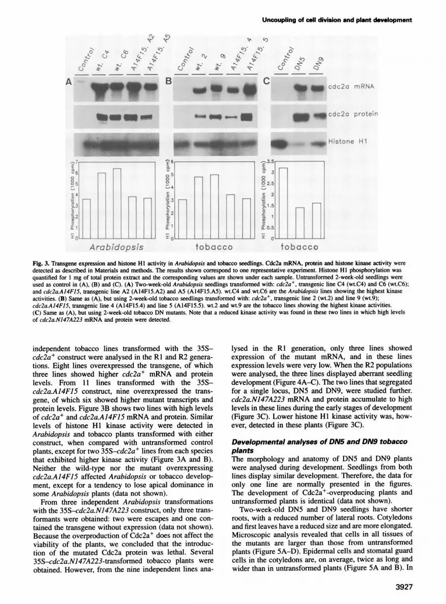
Fig. 3. Transgene expression and histone HI activity in Arabidopsis and tobacco seedlings. Cdc2a mRNA, protein and histone kinase activity weredetected as described in Materials and methods. The results shown correspond to one representative experiment. Histone HI phosphorylation wasquantified for 1 mg of total protein extract and the corresponding values are shown under each sample. Untransformed 2-week-old seedlings wereused as control in (A), (B) and (C). (A) Two-week-old Arabidopsis seedlings transformed with: cdc2a+, transgenic line C4 (wt.C4) and C6 (wt.C6);and cdc2a.A14F15, transgenic line A2 (A14FI5.A2) and A5 (A14FI5.A5). wt.C4 and wt.C6 are the Arabidopsis lines showing the highest kinaseactivities. (B) Same as (A), but using 2-week-old tobacco seedlings transformed with: cdc2a+, transgenic line 2 (wt.2) and line 9 (wt.9);cdc2a.A14F15, transgenic line 4 (A14F15.4) and line 5 (A14F15.5). wt.2 and wt.9 are the tobacco lines showing the highest kinase activities.(C) Same as (A), but using 2-week-old tobacco DN mutants. Note that a reduced kinase activity was found in these two lines in which high levelsof cdc2a.N147A223 mRNA and protein were detected.
independent tobacco lines transformed with the 35S-cdc2a+ construct were analysed in the RI and R2 genera-tions. Eight lines overexpressed the transgene, of whichthree lines showed higher cdc2a+ mRNA and proteinlevels. From 11 lines transformed with the 35S-cdc2a.AJ4FJ5 construct, nine overexpressed the trans-gene, of which six showed higher mutant transcripts andprotein levels. Figure 3B shows two lines with high levelsof cdc2a+ and cdc2a.AJ4FJ5 mRNA and protein. Similarlevels of histone HI kinase activity were detected inArabidopsis and tobacco plants transformed with eitherconstruct, when compared with untransformed controlplants, except for two 35S-cdc2a+ lines from each speciesthat exhibited higher kinase activity (Figure 3A and B).Neither the wild-type nor the mutant overexpressingcdc2a.A14F15 affected Arabidopsis or tobacco develop-ment, except for a tendency to lose apical dominance insome Arabidopsis plants (data not shown).From three independent Arabidopsis transformations
with the 35S-cdc2a.N147A223 construct, only three trans-formants were obtained: two were escapes and one con-tained the transgene without expression (data not shown).Because the overproduction of Cdc2a+ does not affect theviability of the plants, we concluded that the introduc-tion of the mutated Cdc2a protein was lethal. Several35S-cdc2a.N147A223-transformed tobacco plants wereobtained. However, from the nine independent lines ana-
lysed in the RI generation, only three lines showedexpression of the mutant mRNA, and in these linesexpression levels were very low. When the R2 populationswere analysed, the three lines displayed aberrant seedlingdevelopment (Figure 4A-C). The two lines that segregatedfor a single locus, DN5 and DN9, were studied further.cdc2a.N147A223 mRNA and protein accumulate to highlevels in these lines during the early stages of development(Figure 3C). Lower histone HI kinase activity was, how-ever, detected in these plants (Figure 3C).
Developmental analyses of DN5 and DN9 tobaccoplantsThe morphology and anatomy of DN5 and DN9 plantswere analysed during development. Seedlings from bothlines display similar development. Therefore, the data foronly one line are normally presented in the figures.The development of Cdc2a+-overproducing plants anduntransformed plants is identical (data not shown).
Two-week-old DN5 and DN9 seedlings have shorterroots, with a reduced number of lateral roots. Cotyledonsand first leaves have a reduced size and are more elongated.Microscopic analysis revealed that cells in all tissues ofthe mutants are larger than those from untransformedplants (Figure 5A-D). Epidermal cells and stomatal guardcells in the cotyledons are, on average, twice as long andwider than in untransformed plants (Figure 5A and B). In
3927

A.Hemerly et al.
Fig. 4. Development of tobacco DN5 and DN9 plants. (A) DN5 line R2 generation, 3 weeks after germination. Seedlings were germinated in vitro,in medium without selection. Arrows indicate untransformed seedlings. There is a variation in phenotype severity between the various mutantseedlings. (B) Close-up of one DN5 seedling, 2 weeks after germination, showing early development drastically affected compared with (C), anuntransformed seedling of the same age. (D) Four-week-old DN5 and (E) control plants, growing in soil. Mutant leaves, while significantly smaller,gradually acquire shapes similar to those of the untransformed plants. The same number of leaves is found in DN mutants and control plants.(F) Two-month-old DN5 (right side) and untransformed (left side) plants, growing in soil. Mutant plants are shorter than the controls, and matureleaves, although slightly smaller, exhibit normal shape. (G) Side view of mature flowers of untransformed plants (left side), and representativeflowers of DN5 and DN9 plants (centre). In older mutant plants, the floral development is more severely affected (right side). (H) Same as (G), butshowing an upper view. (I) Mature seed pods of untransformed (left side) and DN5 (right side) plants. The mutants produce only a few viable seeds.
roots, epidermal cells are also longer, but cell width didnot increase (Figure 5C and D). Cross-sections of a firstleaf (Figure 6F), cotyledon and root (data not shown)showed that the anatomical structure of these organs isnot affected in mutant plants. Tissue identity and thenumber of cell layers remain unchanged. The shoot apical
meristem in the mutant has a reduced size, but the cellsare no larger than those of the control. It consists ofthe three layers (LI, LII and LIII) typically found inuntransformed plants (Figure 6A and B). Interestingly,cells in the root apical meristem are disorganized in thepromeristem region (Figure 6D). The normally tiered set
3928

Uncoupling of cell division and plant development
E0E., , , yS:...4 :£
Fig. 5. Cell size in tobacco DN5 and DN9 mutants. In (A)-(D), photomicrographs were taken of chlorolactophenol-cleared whole-mount seedlings.In (E) and (F), the material was vibro-sliced, cleared and analysed microscopically (see Materials and methods). Note that all cells in the mutant arelarger than those in the control. (A) Cotyledon of a 2-week-old untransformed seedling, and (B) a 2-week-old DN5 seedling, showing the epidermis.(C) Root of a 2-week-old untransformed seedling, and (D) 2-week-old DN5 seedling, showing the epidermis. (E) Cross-section through a fullyexpanded leaf of an untransformed flowering plant, and (F) a DN9 flowering plant. Bars = 100 rm.
of initials, typical of the closed-type of meristem found intobacco (Figure 6C) (Clowes, 1981) cannot be completelyrecognized. Distinct collumela and lateral root cap cellsare found, composed of larger cells.The width and length of leaves of mutant and control
plants growing in soil were measured. As shown in Figure7C, the ratio between width and length of cotyledons andfirst leaves was lower in the mutants. This reflects theelongated shape of cotyledons and first leaves of themutants. Nevertheless, as the mutant plants grow, newleaves progressively exhibit a normal shape (Figures 4Dand 7A-C), although they never reach the sizes found incontrol plants. While they are structurally normal, all cellsare larger compared with the control (Figure 5E and F).
Stems in the mutant plants grow less than those inuntransformed plants; consequently the internodes aresmaller and the final size of the plant is slightly shorter(Figure 4F). Flowers are also smaller and their size varieswithin the same plant (Figure 4G and H). Nevertheless,they have all organs, albeit with a reduced size, and arefertile. Interestingly, in contrast to all other plant organs,all cells in the flowers have normal sizes (data not shown).The subsequent seed pods are small and contain less seeds(Figure 41). Mature embryos have smaller cotyledons, butthe cells have a normal size (data not shown).
Developmental timing is not affected in DN5 and DN9
mutants. New leaves are initiated at the same rate as inthe untransformed plants. The increase in leaf size as afunction of time was measured, and showed that leafgrowth in mutants parallels that of the control plants(Figure 7D). The passage from juvenile to adult phase isalso unaltered, with mutants flowering concomitantly withthe untransformed plants.
cdc2a.N147A223 mRNA and protein levels fluctuatedduring the life cycle bf DN5 and DN9 plants (data notshown). Such variations were not observed in Cdc2a+-overproducing plants. This suggested the existence of acomplex control mechanism of cdc2a.N147A223 expres-sion. However, there is always a direct correlation betweenlevels of the mutant protein and the severity of thephenotype.
Cdc2a.N147A223 is out-competing the wild-typeproteinThe negative dominance of the overproduced Cdc2a.N147A223 over the endogenous wild-type protein wasexpected to be achieved by the titration of regulatorymolecules. As already demonstrated, much lower histoneHI kinase activity was found in the mutants, supportinga possible competition between the wild-type andmutant allele.To further test this hypothesis, transgenic tobacco
3929

A
Al
I..~~~~~~lj 6x t
4 4.
4~~~fOI §.
* .45I1 .D
Fig. 6. Anatomy of tobacco DN5 mutants. Photomicrographs of thin sections were prepared as described in Materials and methods. (A) Medianlongitudinal section through a shoot apical meristem of an untransformed tobacco seedling, and (B) DN5 seedling, 5 days after germination. Themeristem organization of the mutant resembles the typical LI, LII and LIII layers within the control plants, but with a reduced number of cells.(C) Median longitudinal section through a root apical meristem from untransformed tobacco seedling, and (D) DN5 seedling, 5 days aftergermination. The control plant exhibits the typical tiers of initials (PM). The corresponding meristematic region in the mutant has a disordered cellorganization. The collumela and lateral root cap cells are larger than in the control (RC). (E) Cross-section through the first leaf from anuntransformed tobacco seedling, and (F) a DN5 seedling, 2 weeks after germination. The same number of cell layers is found in both the mutant andcontrol leaves. Cell differentiation is also unaffected in the mutants: the distinct cell types are present. Cells in the mutants are larger than in thecontrol. Bars = 50 ,um. PM, promeristem; RC, root cap.
plants overproducing Cdc2a+, homozygous line 2, were
reciprocally crossed with DN5 and DN9 homozygousplants. Plants from all three lines were, in addition,reciprocally crossed with untransformed plants. Theheterozygous Wt.2 seedlings did not exhibit an alteredphenotype (Figure 8A). All heterozygote mutant linesshowed the mutant phenotype. Most of the population hadthe less severe phenotype (Figure 8B) with the earlydevelopment of only a few seedlings severely affected(Figure 8C). In contrast, the progenies of the cross betweenCdc2a+ and mutant plants showed a completely normaldevelopment (Figure 8D). Microscopic analyses confirmedthat cell sizes in these plants were no larger than in thecontrols (data not shown). High levels of cdc2a mRNAand protein were found in these plants (data not shown).The overproduced Cdc2a+, therefore, might be out-
competing the mutant protein, and thereby nullifyingits dominant negative effect. These results support thehypothesis that a titration mechanism dictates the domin-ance of the cdc2a- mutant over the wild-type.
Protoplast analyses of the tobacco cdc2a mutantsTo study the effect of cdc2a mutations on the cell divisionof individual plant cells, protoplasts of plants producingCdc2a+ (lines 2 and 9), Cdc2a.A14F15 (lines 4 and 5),
Cdc2a.N147A223 (lines DN5 and DN9) and untrans-formed plants were isolated and induced to divide. Trans-genic tobacco plants were used, because Arabidopsisplants expressing the cdc2a.N147A223 mutation could notbe obtained.
Freshly isolated leaf mesophyll protoplasts of DN5 andDN9 plants are larger than those from the other lines, inagreement with the larger mesophyll cells found in DNmutant leaves (Figure 9A). While after 3 days of cultivation-90% of Cdc2a+, Cdc2a.A14F15 and untransformed cellshave divided or are undergoing division (Figure 9B 1), nodivision of DN5 and DN9 cells was detected (Figures 9B2and 3). After 15 days of culture, when cells from Cdc2a+,Cdc2a.A14F15 and untransformed plants had alreadyundergone several rounds of division and formed smallmicrocalli (Figure 9C1 and 2), DN5 and DN9 cells hadnot divided. Very large cells were found (Figure 9C3 and5), but no cytokinesis or nuclear division occurred (Figure9C4 and 6).Cdc2a protein levels and histone HI kinase activities
were monitored during the first round of cell division. Noincrease in kinase activity was found in DN5 and DN9cultured protoplasts (Figure 9D). This observation is inagreement with the absence of cell divisions in thesemutant protoplasts. A gradual increase in kinase activity
3930
A.Hemerly et aL
BLi Lll LillI;.-
-:f...F
i
x,".4
I
i.,,4eAI
ViI;
r N..... .
I.\
E
-4,
I
F
r
I 0.
0
.IIm4 ,k.iI, "a
. ...:.-
2

Uncoupling of cell division and plant development
Cl.,C11.
0
50.tca)o .1-Cc-
-u O.,3
O.
D 46
14
2
E ', 0,
_r_aa71
4
4r2
2
0~~~~~~00
0
0
50
010
')0
10
:0
- SR1I DN9 .' DN5 . .
* .
2 3 4 : rv,eeks
100C_. SRI' DN9
80 DN5-j~~~~~
EC
4C:2
2 0
.
v3eesweeks
11*SR1MDN9~~DN5
Fig. 7. Leaf development in tobacco DN mutants. (A) Display of the cotyledons (left side) until the fourth leaf (right side) from 3.5-week-old DN5(upper panel) and untransformed (lower panel) seedlings, growing in vitro. Mutant cotyledons have a more elongated shape. Leaf shape in themutants gradually parallels that in the control. (B) Mature leaf of untransformed (left side) and DN5 (right side) plant. Their shape and size arenearly the same. (C) Measurements of the ratio between width and length (W/L) in representative untransformed (SRI) and mutant (DN5 and DN9)leaves. These values were used to evaluate differences in shape between mutant and control leaves. In the mutant cotyledons (COT), the W/L ratio ismuch lower, reflecting their elongated shape. As plants grow, the W/L ratios found in newly formed leaves from the mutants (L2-L8) graduallyparallel the control, reflecting their similar shape. (D) Measurement of the increase in length (left side) and width (right side) of three representativefifth leaves of control (SR1) and mutant (DN5 and DN9) plants during development. Measurements were taken every 4 days. They show that thekinetics of leaf growth in the mutants is similar to that of the control. Both reach maximum leaf expansion after similar periods, although leaves inthe mutants are in general slightly smaller.
was observed in the cell cultures of the other transgenicand untransformed plants analysed. The highest levels ofkinase activity were detected after 3 days, when mostcells were dividing (Figure 9D).
Nuclear DNA content in DN plantsWe showed that overproduction of Cdc2a.N 147A223resulted in plants consisting of fewer cells. These cellsunderwent normal differentiation but were much larger.We were interested to know whether there was an increasein their nuclear DNA content.
Microscopic analyses of a number of different cell typesfrom DN plants showed that no nuclear division occurredwithout cytokinesis. There was always a single nucleuspresent per cell. To investigate whether endoreduplicationmight occur in the large mutant cells, their nuclear DNAcontent was monitored by flow cytometry (data not shown).The percentage of 2C (G1) and 4C (G2) cells in thedifferent organs of 2-week-old seedlings was similar inthe DN5 mutant and the control. In mature leaves of 2-month-old plants, the same percentages of 2C and 4Ccells were also found in DN5 and control plants. Nopolyploid cells were detected at these stages of develop-ment. The results show that extra DNA replication doesnot occur in the DN plants during development.
DiscussionMorphogenesis and histogenesis occur throughout plantlife due to continuous iterative growth at the meristems.This implies that, in contrast with animals, cell division,elongation and differentiation occur throughout develop-ment and must be perfectly coordinated. However, theextent to which these processes are linked with eachother, and coupled to plant development, remains largelyunknown.To approach this question, we disrupted the function of
a key regulator of the Arabidopsis cell cycle, the Cdc2akinase, by introducing dominant mutations in its catalyticsubunit and ectopically expressing them in plants. Weshowed that dominant mutations can be used successfullyas an alternative approach to study gene function in plants.
Cdc2a overproduction does not affect plantdevelopmentAs a control in our experiments, we have overexpressedthe wild-type cdc2a in Arabidopsis and tobacco. Plantswith high levels of Cdc2a+ developed normally, exceptfor a tendency to lose apical dominance. However, we
cannot distinguish between a direct effect of Cdc2afunction or a more general stress response to proteinoverproduction. Results obtained when tobacco protoplasts
3931

A.Hemerly et a.
Fig. 8. Dominance of the Cdc2a.N147A223 mutant protein over thewild-type Cdc2a in tobacco DN mutants. (A) A 2-week-old seedlingfrom a cross between the homozygote Wt.2 and SRI plants. Theheterozygote Wt.2 seedlings exhibited the same phenotype as theuntransformed plants. (B) and (C) Phenotypes of 2-week-old seedlingsfrom a cross between homozygote DN5 and SR1 plants. Most of themutant heterozygote plants exhibited the less severe mutant phenotypeshown in (B). (D) A representative 2-week-old seedling from a crossbetween homozygote Wt.2 and DN5 plants. All progenies from thesecrosses lost the mutant phenotype.
overproducing Cdc2a+ were induced to divide showedthat the kinetics of kinase activation and cell division areunaffected. It had already been demonstrated that cdc2overexpression has no effects on cell division in S.pombe(Durkacz et al., 1986) or in HeLa cells overexpressingthe chicken cdc2+ (Krek and Nigg, 1991b). This is notsurprising, considering that the kinase activation involvesa cascade of events and requires regulatory proteins that
might be limiting. For the same reasons, ectopic expressionof cdc2a could not be expected to promote cell division.In addition, studies of cdc2a expression in plants suggestedthat it can be present in particular tissues without triggeringcell division (Martinez et al., 1992; Hemerly et al., 1993).In practice, only a 2-fold increase in kinase activity couldbe obtained in some plant lines overproducing Cdc2a+.
Does reversible tyrosine phosphorylation play arole in plant cell cycle control?In a number of eukaryotes, reversible Y15 phosphorylationof Cdc2 was determined to be an important regulator ofcell cycle progression (reviewed by Atherton-Fessler et al.,1993). Based on this, we constructed a Cdc2a mutantwhere the Y15 residue, together with T14, were substitutedin order to disrupt this control. No effects on developmentwere observed in Arabidopsis and tobacco plants produc-ing high levels of the mutant Cdc2a.A14F15, except fora tendency to lose apical dominance in Arabidopsisplants. The same phenotype was exhibited by the Cdc2a+-overproducing plants. Cultivated tobacco mesophyllprotoplasts producing Cdc2a.A14F15 did not show anyperturbation in cell cycle progression.Our data did not allow us to define clearly the role of
tyrosine phosphorylation in Cdc2a regulation. First, wecan suppose that the tyrosine phosphorylation controlexists in plants, and takes part in the regulation of Cdc2aactivation. Nevertheless, perturbed cell divisions occurringin Cdc2a.A14F15-overproducing plants might have a smallchance of affecting development (see below). Our attemptsto show tyrosine phosphorylation in Cdc2a using anti-phosphotyrosine antibodies did not give a clear result(data not shown). Hence, a second possibility is that plantCdc2s are not phosphorylated at Y15. Finally, there isstill the possibility that Y15 is phosphorylated in plantCdc2s, but its regulation is not crucial for entry intomitosis, as described for S.cerevisiae (Amon et al., 1992;Sorger and Murray, 1992).
Mutation of residue D147T233 of Cdc2ainactivates the kinase and is lethal to plantswhen present at high levelsWe showed that the double mutation N147A223 has aninhibitory effect on the Arabidopsis Cdc2a. The work onS.pombe suggests that, as in yeast and humans, themutation of D147 would be sufficient to inactivate theArabidopsis kinase. No transgenic Arabidopsis plantsproducing detectable levels of Cdc2a.N147A223 proteinwere obtained when this mutation was expressed in plantsunder the control of the CaMV 35S promoter. Threetobacco transformants expressing moderate levels of thetransgene were obtained. Lower levels of kinase activityand a reduced number of cells were found in these plants.When crossed with Cdc2a+-overproducing plants, theirprogenies developed completely normally (typical cellsizes and numbers). Additionally, no increase in eitherkinase activity or cell division was observed in protoplastsof the tobacco DN plants induced to divide. Taken together,these data strongly indicate that the Cdc2a.N147A223protein is an inactive kinase that probably binds toregulatory factors, titrating them out of the wild-typekinase. The ability to rescue some tobacco plants mightresult from a less efficient interaction of the mutant
3932

Uncoupling of cell division and plant developmentXw::
si :1t. /aEg:.., @ipJpA S.
*j:: {M', :Wt >' ..... .t:i;-' A}'t .g:.a. "S'
_, s ^e
r . i .,,,* / ^,.
- A: 5.
X *. .tZ, 8*@:;i.
j B wM
,BusWiwr j ,.
e
*
DN5ti t2 t3
D SR1ti t2 t3
S - P. c d c 2 a p r o t e n
Histone Hi
Fig. 9. Effect of Cdc2a mutations on cell division of tobacco protoplasts. Leaf mesophyll protoplasts were prepared from transgenic tobacco linesWt.2 and Wt.9 (overexpressing cdc2a+), A14F15.A4 and A14F15.A5 (overexpressing cdc2a.A14F15), DN5 and DN9 (expressing cdc2a.N147A223)and untransformed SRI plants as described in Materials and methods. Protoplasts were cultivated in hormone medium to induce cell division. Theresults obtained with the Cdc2a+ and Cdc2a.A14F15 lines were similar to those of control SRI plants and are not presented. (A) Freshly isolatedprotoplasts from untransformed (1) and DN5 (2) plants. Protoplasts from DN5 plants are larger than from control. Bars = 50 gm. (B) Cells 3 daysafter cultivation. Most of the untransformed cells (1) have divided or are undergoing division, whereas DN5 cells did not divide (2). Some DN5 cellsexhibited an elongated shape (3). Bars = 50 jm. (C) Cells 15 days after cultivation. Control cells had divided several times and formed smallmicrocalli (1), whereas DN5 cells exhibited elongated and budded shapes, but without cytokinesis (3 and 5). The same cells as shown in (1), (3) and(5) were visualized for DAPI staining (2, 4 and 6). Nuclei are indicated with an arrow. Note that nuclear division did not occur in DN5 cells, sincethey have only one nucleus. Bars = 100 gm. (D) Detection of Cdc2a protein and histone HI kinase activities in DN5 and control cells after 1 (tl),2 (t2) and 3 (t3) days of culture. Higher Cdc2a protein levels were found in DN5 cells (right, upper panel) compared with control (left, upper panel).In this experiment, two Cdc2a forms with distinct mobilities were detected that could represent different phosphorylated Cdc2a. A gradual increasein kinase activities was observed in control cells (left, lower panel). In contrast, kinase activities did not rise in DN5 cells (right, lower panel).
Arabidopsis protein with regulatory molecules in a hetero-logous system. Therefore, the overproduced mutant proteinis dominant in plants and causes an arrest of the cell cycle.The complete blockage of cell division in cultured DN
protoplasts, contrasting with a reduction of cell divisions inthe meristems of the same plants, remains to be elucidated.Besides the distinct physiological state of both cell types, itis possible that the larger mesophyll cells have lost theircompetence to divide, or that the titration of regulatorymolecules worked more efficiently in protoplasts due to thepresence of lower levels of endogenous Cdc2a.
The points in the cell cycle where Cdc2a function isessential could not be determined precisely. The datasuggest that, in the tobacco DN mutants, progressionthrough the cell cycle is not blocked, but is probablydelayed where Cdc2a activity is required. Analyses ofnuclear DNA content from DN seedlings did not show an
increase in the population of cells at a particular phase ofthe cell cycle, compared with the control. In addition,only two members of the Cdk family were identified inArabidopsis. So far, yeast complementation studies haveshown that only Cdc2a is a functional Cdc2 homologue.
3933
A-~4
.,
B
C i
.s.'AN
.'A.
@: ;
...
fi Z
V.''L,L.. "".xbl._, ' \ > '@
te
^ :X
j* *:
9 %
_t * .,
, .'z! E_{; .Rna. {.:.., .._.. ) ::
*e m*
*:.^ 4.
3X. S
gis$",->..2. j
W,t
S aF- :t
A.Hemerly et al.
Hence, as in yeast, it is possible that Cdc2a operates bothin G1/S and G2/M transitions.
Cell division can be uncoupled from plantdevelopmentAs a plant cannot grow without cell divisions, it is obviousthat the constitutive overexpression of the cdc2a.N147A223 mutation in plants is lethal. However, in plantswith moderate levels of Cdc2a.N147A223, a reducedfrequency of cell divisions was found. Mutant plantsunderwent completely normal histogenesis. Celldifferentiation was unaffected, and all tissues were formed,though with reduced cell divisions. The plant structurewas maintained, with all organs conserving their normalnumber of cell layers. The lower frequency of cell divisionsdid not have a significant effect on plant growth. Theproblem of fewer cells was compensated for by a sig-nificant increase in cell growth. All cells in the plant werelarger, except in flowers and embryos. Interestingly, thisgrowth was polarized. The morphology of mutant plantswas similar to that of the untransformed control. Theyexhibited smaller organs but with normal shape. Thesedata support the lack of a causal relationship betweencell division and plant morphogenesis/histogenesis. Themechanisms controlling plant structure and shape probablyact independently of the frequency of cell divisions.
Studies in the animal field have also suggested a lackof correlation between cell division and morphogenesis.Recently, Busturia and Lawrence (1994) have shown thatin the animal kingdom a rigid programming of cellnumbers is not an absolute requirement for the formationof complex structures during development. By studyingthe growth of Drosophila embryos with a reduced numberof cells in the abdominal primordia, they ruled out theregulation of cell number in the fly abdomen.Our results obtained by reducing the activity of an
essential kinase of the plant cell cycle support the observa-tions made in a study of y-irradiated wheat seedlingsgrowing without DNA synthesis and cell division (Foardand Haber, 1961; Haber, 1962). These studies showed thatthe initial development of irradiated seedlings parallelsthat of non-irradiated wheat.
In our study, the entire plant life cycle was analysed.The lower frequency of cell divisions in the DN mutantsdid not affect the timing of morphogenic processes. Theprecise coordination of organ initiation was maintained.The transition from juvenile to mature phase was notdelayed. This suggests that most developmental controlsin plants act independently of the rate of cell divisions.
Embryo development and iterative developmentat meristemsOur data allowed the uncoupling of cell division fromiterative plant growth. However, the same concept cannotbe extended to embryo development. The embryo exhibitsa sequential development where most of the initial celldivisions are formative and in fixed orientations. It canbe expected that a lack of, or a delayed, cell divisionwould have drastic consequences on subsequent develop-ment. The analysis of Arabidopsis pattern mutants showedthat perturbed cell divisions are involved in the abnormalembryo development (Berleth and Jiirgens, 1993; Mayeret al., 1993). Preliminary data on Arabidopsis plants
expressing cdc2a.NJ47A223 specifically during embryo-genesis suggest that this negative mutation affects embryodevelopment.
In tobacco, the organ size and shape of developing DNseedlings is most affected. The disturbed shape mightmerely result from higher Cdc2a.N147A223 protein levelsduring this phase of development, reducing the numberof cell divisions to below that needed by the plant toundergo normal morphogenesis. However, the observedphenotype could also be a consequence of abnormalembryo development. The 35S promoter drives expressionin embryos (Benfey et al., 1990), but a detailed analysisof the expression during early phases of embryo formationhas not been made. Northern analyses of total seed podsshowed high levels of cdc2a.NJ47A223 mRNA (data notshown). Embryo cotyledons are smaller and contain lesscells. In spite of the embryo pattern being unaltered, thecotyledon and the shoot apical meristem might becomeperturbed later in embryogenesis.
In meristems, most cell divisions are proliferative, asthey simply increase cell number. The contribution of ameristematic cell to the formation of the plant bodydepends on its position in the meristem. Clonal analysessuggest that a cell's final fate can vary according to itsposition (Stewart, 1978; Poethig, 1987). Chimera studiesalso showed that plant ontogenesis can vary greatly,without having an effect on form (Poethig, 1987). Hence,it appears that the large ontogenetic flexibility allowsperturbations of cell divisions at meristems to be repairedsubsequently by neighbouring sister cells in order to keepthe final shape. Still, a particular number of cell divisions,in a certain position, would be necessary to generate theright form. The data obtained with the DN mutants showthat a plant can reach its final form with a smaller numberof cells. Possibly, in order to compensate for this, amechanism that promotes cell growth is activated. There-fore, we can infer that cell divisions are a source of growthrather than generators of form.An intriguing observation was made in the roots of
tobacco DN plants. The promeristem is formed by dis-organized proliferating cells which retain the ability togenerate roots with normal structure and shape. It suggeststhat pattern and shape controls governing root developmentcan affect organ fate independent of cell division patternsat the meristem. Analyses of the shoot meristemless mutantfrom Arabidopsis have also suggested that an organizedshoot apical meristem might not be essential for leafformation (Barton and Poethig, 1993). The shoot apicalmeristem in the tobacco DN mutants, while organized,appears to contain fewer cells than in control plants.Nevertheless, the initiation of new leaves is not delayed,and there is no loss of apical dominance. This indicatesthat a shoot apical meristem consisting of less cells canbe perfectly functional.
Organismal theory for the origin of plantmulticellularityOur analyses of the relationship between cell division andplant development led us to an extensive discussionconcerning the origin of multicellularity in plants. Incontrast to the cellular theory in animals, the organismaltheory for plants (reviewed in Kaplan and Hagemann,1991; Hagemann, 1992; Kaplan, 1992) states that cells
3934

Uncoupling of cell division and plant development
are purely parts of the organism and that rather than beingthe cause of growth, they merely represent markers of it.This implies that the shape of an organism develops inde-pendently of cell division. It has already been shown thatorgan morphogenesis would always involve growth, withor without actual cell division (Green, 1976). Together withthe results obtained with y-irradiated wheat seedlings, ourdata, obtained from analyses of the DN mutants, furthersupport the hypothesis that the mechanisms controllingplant morphogenesis operate independently of cell division.The independence of the morphological complexity of
organisms from a multicellular structure is found naturallyin marine algae. In different species it has been shownthat morphogenesis does not require cell division, but isinstead associated with organelle proliferation, essentiallyof the nuclei (reviewed by Kaplan and Hagemann, 1991).It was assumed that morphogenesis is correlated withnuclear replication: an increase in organ and protoplasmicvolume coincides with nuclear divisions. However, in ourstudies, we show that cell growth in the DN mutants isnot followed by nuclear replication or endoreduplication.Although this is not a natural situation, it seems that thecapacity for morphogenesis in higher plants is, at least inpart, independent of DNA replication and nuclear division.
In conclusion, our data emphasize a concept introduced>100 years ago, which stresses the importance of studyingplant development at both the structural and organismallevel. The uncoupling of cell division from iterative plantdevelopment illustrates the flexibility of some develop-mental programmes, and suggests a redundancy of themechanisms involved in triggering plant morphogenesis.Further analyses of this flexible machinery will be of greatimportance in our understanding of plant development.
Materials and methodsMutagenesis, plasmid construction and plant transformationArabidopsis cdc2a cDNA was mutagenized by PCR (using a DNAamplification instrument; Techne, Cambridge, UK) essentially asdescribed by Landt et al. (1990). The following oligonucleotides wereused as primers: A (5') ATTATTCAGGAATCCATGGATCAGTA, con-taining a NcoI restriction site; B (3') CAGTCCAAAATTAGCAAGCTT,where the codon for D147 (GAT) was changed to N147 (AAT); C (5')ATTCAGGAATCCATGGATCAGTACGAGAAAGTTGAGAAGATT-GGTGAAGAAGCTTTCGGT, where the codons for T14Y15 (ACTTAC)were changed to A14F15 (GCITTC) and containing a NcoI restrictionsite; and D (3') AATGAAGGAGATCTCTGGTl-TTATGCC, containingaBglll restriction site. To obtain the cdc2a.N147A223 cDNA, the first PCRwas performed with oligonucleotides A and B, and the amplified productwas used as a primer together with oligonucleotide D in a second PCR.The cdc2a.A14F15 cDNA was produced by only one PCR using theoligonucleotides C and D as primers. NcoI and BglII restriction sites wereintroduced in the wild-type cdc2a cDNA by performing a PCR with theoligonucleotides A and D as primers. The PCR products corresponding tothe mutated cDNAs were blunt-ended, cloned into pUC 18 and sequenced.The CaMV 35S promoter was transferred from the vector pDE9 (Plant
Genetic Systems N.V.) totheplasmidpHPH I (Plant Genetic Systems N.V.)containing the 3' NOS, which originated from the vector pH35S. Thecdc2a.N147A223, cdc2a.A14FJ5 and cdc2a+ cDNAs were removed frompUC18 by NcoI and BglII digestion and cloned into the NcoI and BamHIrestriction sites of the vector pH35S. The cassettes 35S-cdc2a-3' NOSwere cloned in the binary vector pGSV4 (Herouart et al., 1994) andtransferred to Agrobacterium tumefaciens. The constructs were introducedinto Arabidopsis thaliana ecotype C24 and Nicotiana tabacum cv Petithavana (SRI) plants by the root transformation method (Valvekens et al.,1988) and the leaf disc protocol (Horsch et al., 1985), respectively.
cdc2a expression in S.pombeThe cdc2a cDNA and its mutant forms were transcriptionally fused tothe thiamine-repressible promoter (nmtl), in the Spombe expression
vector, pREP3 (Maundrell, 1993) containing LEU2 as a selectablemarker. The cDNAs were restricted with PstI or Sail, according to theirorientation in pUC18, blunt-ended, released after restriction with BglII,and cloned into the MscI and BamHI sites of pREP3. Plasmid constructswere transformed into the Spombe strain cdc2-33 leul-32h-, by thelithium acetate method (Okasaki et al., 1990). The transformed colonies(Leu+) were inoculated in minimal liquid medium containing 5 jMthiamine and grown overnight at 25°C. Cells were washed in minimalmedium without thiamine and grown in this medium, at 25°C, for 16 h,to induce the promoter. Samples were analysed by light microscopy.
Kinase assaysHistone H1 kinase assays were performed with Cdk complexes purifiedfrom total plant protein extracts by pl3sucl-Sepharose affinity binding,according to Azzi et al. (1992). Briefly, pl3sucl was purified from anoverproducing Escherichia coli strain by chromatography in SephacrylS2000, and conjugated to CNBr-activated Sepharose 4B (Pharmacia),according to the manufacturer's instructions. Total plant protein extracts(300 ,ug) were incubated with 50 jl 50% (v/v) pl3sucl-Sepharose beadsfor 2 h at 4'C. The washed beads were combined with 30 jl kinase buffercontaining 1 mg/ml histone HI (IIIS, Sigma), 150 mM ATP and 1 jiCi of[y-32P]ATP (Amersham). After 20 min incubation at 30°C, samples wereanalysed by SDS-PAGE and autoradiographed. [32P]Phosphate incorpora-tion in histone H1 was quantified by excising the corresponding proteinsfrom dried gels and Cerenkov counting in a scintillation counter.
RNA, protein and nuclear DNA analysesRNA extraction, Northern analyses and hybridization reactions wereperformed as described by Hemerly et al. (1993). Radiolabelled antisenseRNA (Riboprobe Gemini H Core System; Promega) synthesized fromthe full-length cdc2a cDNA was used as probe.
Protein extracts were obtained by the procedure described for the kinaseassays. Polyclonal antibodies were raised against the entire Cdc2a protein(P.Ferreira, unpublished results). Total protein (15 jg) was subjected toelectrophoresis on 12% SDS-PAGE gels (Laemmli, 1970) and electroblot-ted to nitrocellulose membrane (Hybond C super). Filters were blockedovernight with 2% milk in phosphate-buffered saline (PBS) buffer, washedthree times with PBS, incubated for 2 h with the cdc2a antiserum at 1:5000dilution, in PBSTA (PBS containing 0.5% Tween-20 and 0.1% albumin),washed for 1 h with PBST; incubated for 1-2 h with peroxidase-conjugatedsecondary antibody at 1:10 000 dilution in PBSTA, and washed for 1 hwith PBST. Protein detection was by the chemiluminescent procedure(ECL, Western blotting detection system, Amersham).
Nuclei from tobacco seedlings and leaf cells were isolated and sortedby flow cytometry as described (Bergounioux and Brown, 1990).
Protoplast analysesProtoplasts were prepared from leaves of 2-month-old tobacco SRIplants growing in soil as described by Caboche (1980), with somemodifications. Leaf cells were enzymatically digested with 0.5% cellulaseand 0.2% macerozyme in K3M medium [K3 basal medium (Nagy andMaliga, 1976) containing 0.4 M mannitol], overnight. Protoplasts werecultivated, at a density of 105/ml, in K3M medium containing 20 g/lsucrose, 1 mgA a-naphthaleneacetic acid (NAA) and 0.2 mg/l 6-benzylaminopurine. Samples of 5x 10⁵ protoplasts were collected everyday for protein analyses and histone H1 kinase assays. After 5 days ofculture, cells were subcultured in the same medium, except that theconcentration of NAA was decreased to 0.1 mg/l, at a density of 103cells/ml. The cultures were analysed microscopically. Cell nuclei werestained with 50 ng/ml 4',6-diamidino-2-phenylindole (DAPI), in K3Mmedium, and immediately visualized. The mitotic index was measuredafter DAPI staining.
Microscopic analysesWhole-mount Arabidopsis and tobacco seedlings and embryos were ana-lysed with a Zeiss Jenalumar microscope after clearing with chlorolacto-phenol (CLP) (Beeckman and Engler, 1994). Fresh slices of varioustobacco organs were prepared using a vibro-slicer (Campden Ltd., London,UK), cleared in CLP and analysed microscopically. Thin sections of plantorgans were made according to a standard embedding protocol (Hemerlyet al., 1993). Sections were stained in 0.05% toluidine blue.
AcknowledgementsThe authors are grateful to Dr Paul Nurse for providing the Spombestrain and to Dr K.Maundrell for the plasmid pREP3 and thank Drs
3935

A.Hemerly et aL
Peter Doerner, Paul Nurse, Anton Gerats and Jonathan Clarke for criticalreading, Chris Genetello for technical assistance, Martine De Cock forpreparation of the manuscript, and Karel Spruyt and Vera Vermaerckefor photographs and drawings. This work was supported by grants fromthe Belgian Programme on Interuniversity Poles of Attraction (PrimeMinister's Office, Science Policy Programming, No. 38), and 'VlaamsActieprogramma Biotechnologie' (ETC 002). A.S.H. and P.F. areindebted to the Coordena,ao de Aperfei,oamento de Pessoal de NivelSuperior (CAPES 1387/89-12) and Conselho Nacional de Desenvolvi-mento Cientifico e Tecnologico (CNPq 204081/82-2) for predoctoralfellowships, respectively. G.E. and D.I. are research engineer and researchdirector of the Institut National de la Recherche Agronomique (France),respectively.
ReferencesAmon,A., Surana,U., Muroff,I. and Nasmyth,K. (1992) Regulation of
p34CDc28 tyrosine phosphorylation is not required for entry intomitosis in S.cerevisiae. Nature, 355, 368-371.
Atherton-Fessler,S., Hannig,G. and Piwnica-Worms,H. (1993) Reversibletyrosine phosphorylation and cell cycle control. Semin. Cell Biol., 4,433-442.
Azzi,L., Meijer,L., Reed,S.L., Pidikiti,R. and Tung,H.Y.L. (1992)Interaction between the cell-cycle control proteins p34cdc2 andpgcKShs2. Evidence for two cooperative binding domains in pgCKShs2.Eur. J. Biochem., 203, 353-360.
Barton,M.K. and Poethig,R.S. (1993) Formation of the shoot apicalmeristem in Arabidopsis thaliana: an analysis of development in thewild type and in the shoot meristemless mutant. Development, 119,823-831.
Beeckman,T. and Engler,G. (1994) An easy technique for the clearingof histochemically stained plant tissue. Plant Mol. Biol. Rep., 11,54-59.
Benfey,P.N., Ren,L. and Chua,N.-H. (1990) The CaMV 35S enhancercontains at least two domains which can confer different developmentaland tissue-specific expression patterns. EMBO J., 9, 1677-1684.
Bergounioux,C. and Brown,S.C. (1990) Plant cell cycle analysis withisolated nuclei. Methods Cell Biol., 33, 563-573.
Berleth,T. and Jurgens,G. (1993) The role of the monopteros genein organising the basal body region of the Arabidopsis embryo.Development, 118, 575-587.
Booher,R. and Beach,D. (1986) Site-specific mutagenesis of cdc2+, acell cycle control gene of the fission yeast Schizosaccharomycespombe. Mol. Cell. Biol., 6, 3523-3530.
Busturia,A. and Lawrence,P.A. (1994) Regulation of cell number inDrosophila. Nature, 370, 561-563.
Caboche,M. (1980) Nutritional requirements of protoplast-derived,haploid tobacco cells grown at low cell densities in liquid medium.Planta, 149, 7-18.
Clowes,F.A.L. (1981) The differences between open and closedmeristems. Ann. Bot., 48, 761-767.
De Bondt,H.L., Rosenblatt,J., Jancarik,J., Jones,H.D., Morgan,D.O. andKim,S.-H. (1993) Crystal structure of cyclin-dependent kinase 2.Nature, 363, 595-602.
Durkacz,B., Carr,A. and Nurse,P. (1986) Transcription of the cdc2 cellcycle control gene of the fission yeast Schizosaccharomyces pombe.EMBO J., 5, 369-373.
Ferreira,P.C.G., Hemerly,A.S., Villarroel,R., Van Montagu,M. and Inze,D.(1991) The Arabidopsis functional homolog of the p34Cdc2 proteinkinase. Plant Cell, 3, 531-540.
Foard,D.E. and Haber,A.H. (1961) Anatomic studies of gamma-irradiatedwheat growing without cell division. Am. J. Bot., 48, 438-446.
Gould,K.L. and Nurse,P. (1989) Tyrosine phosphorylation of the fissionyeast cdc2+ protein kinase regulates entry into mitosis. Nature, 342,39-45.
Green,P.B. (1976) Growth and cell pattern formation on an axis: critiqueof concepts, terminology, and modes of study. Bot. Gaz., 137, 187-202.
Haber,A.H. (1962) Nonessentiality of concurrent cell divisons for degreeof polarization of leaf growth. I. Studies with radiation-induced mitoticinhibition. Am. J. Bot., 49, 583-589.
Hagemann,W. (1992) The relationship of anatomy to morphology inplants: a new theoretical perspective. Int. J. Plant Sci., 153, S38-S48.
Hemerly,A.S., Ferreira,P.C.G., de Almeida Engler,J., Van Montagu,M.,Engler,G. and Inze,D. (1993) cdc2a expression in Arabidopsis thalianais linked with competence for cell division. Plant Cell, 5, 1711-1723.
Hrouart,D., Van Montagu,M. and Inz6,D. (1994) Developmental and
environmental regulation of the Nicotiana plumbaginifolia cytosolicCu/Zn-superoxide dismutase promoter in transgenic tobacco. PlantPhysiol., 104, 873-880.
Horsch,R.B., Fry,J.E., Hoffman,N.L., Eichholtz,D., Rogers,S.G. andFraley,R.T. (1985) A simple and general method for transferring genesinto plants. Science, 227, 1229-1231.
Imajuku,Y., Hirayama,T., Endoh,H. and Oka,A. (1992) Exon-intronorganization of the Arabidopsis thaliana protein kinase genes CDC2aand CDC2b. FEBS Lett., 304, 73-77.
Jacobs,T. (1992) Control of the cell cycle. Devel. Biol., 153, 1-15.Jurgens,G. (1992) Pattern formation in the flowering plant embryo. Curr.
Opin. Genet. Dev., 2, 567-570.Kaplan,D.R. (1992) The relationship of cells to organisms in plants:
problems and implications of an organismal perspective. Int. J. PlantSci., 153, S28-S37.
Kaplan,D.R. and Hagemann,W. (1991) The relationship of cell andorganism in vascular plants. Bioscience, 41, 693-703.
Krek,W. and Nigg,E.A. (1991a) Differential phosphorylation of vertebratep34cdc2 kinase at the G1/S and G2/M transitions of the cell cycle:identification of major phosphorylation sites. EMBO J., 10, 305-316.
Krek,W. and Nigg,E.A. (1991b) Mutations of p34cdc2 phosphorylationsites induce premature mitotic events in HeLa cells: evidence for adouble block to p34cdc2 kinase activation in vertebrates. EMBO J.,10, 3331-3341.
Laemmli,U.K. (1970) Cleavage of structural proteins during the assemblyof the head of bacteriophage T4. Nature, 227, 680-685.
Landt,O., Grunert,H.-P. and Hahn,U. (1990) A general method for rapidsite-directed mutagenesis using the polymerase chain reaction. Gene,96, 125-128.
Martinez,M.C., J0rgensen,J.-E., Lawton,M.A., Lamb,C.J. and Doerner,P.W. (1992) Spatial pattern of cdc2 expression in relation to meristemactivity and cell proliferation during plant development. Proc. NatlAcad. Sci. USA, 89, 7360-7364.
Maundrell,K. (1990) nmtl of fission yeast. J. Biol. Chem., 265,10857-10864.
Maundrell,K. (1993) Thiamine-repressible expression vectors pREP andpRIP for fission yeast. Gene, 123, 127-130.
Mayer,U., Buttner,G. and Jurgens,G. (1993) Apical-basal patternformation in the Arabidopsis embryo: studies on the role of the gnomgene. Development, 117, 149-162.
Mendenhall,M.D., Richardson,H.E. and Reed,S.I. (1988) Dominantnegative protein kinase mutations that confer a GI arrest phenotype.Proc. Natl Acad. Sci. USA, 85, 4426-4430.
Murray,A.W. (1993) Turning on mitosis. Curr Biol., 3, 291-293.Nagy,J.I. and Maliga,P. (1976) Callus induction and plant regeneration
from mesophyll protoplasts ofNicotiana sylvestris. Z. Pflanzenphysiol.,78, 453-455.
Norbury,C., Blow,J. and Nurse,P. (1991) Regulatory phosphorylation ofthe p34cdc2 protein kinase in vertebrates. EMBO J., 10, 3321-3329.
Odell,J.T., Nagy,F. and Chua,N.-H. (1985) Identification of DNAsequences required for activity of the cauliflower mosaic virus 35Spromoter. Nature, 313, 810-812.
Okazaki,K., Okazaki,N., Kume,K., Jinno,S., Tanaka,K. and Okayama,H.(1990) High-frequency transformation method and library transducingvectors for cloning mammalian cDNAs by trans-complementation ofSchizosaccharomyces pombe. Nucleic Acids Res., 18, 6485-6489.
Pines,J. (1993) Clear as crystal? Curr: Biol., 3, 544-547.Poethig,R.S. (1987) Clonal analysis of cell lineage patterns in plant
development. Am. J. Bot., 74, 581-594.Solomon,M.J. (1993) Activation of the various cyclin/cdc2 protein
kinases. Curr. Opin. Cell Biol., 5, 180-186.Sorger,P.K. and Murray,A.W. (1992) S-phase feedback control in budding
yeast independent of tyrosine phosphorylation of p34cdc28. Nature,355, 365-368.
Stewart,R.N. (1978) Ontogeny of the primary body in chimeral formsof higher plants. In Subtelny,S. and Sussex,I.M. (eds), The ClonalBasis of Development (The 36th Symposium of the Society forDevelopmental Biology). Academic Press, New York, pp. 132-160.
Valvekens,D., Van Montagu,M. and Van Lijsebettens,M. (1988)Agrobacterium tumefaciens-mediated transformation of Arabidopsisroot explants by using kanamycin selection. Proc. Natl Acad. Sci.USA, 85, 5536-5540.
van den Heuvel,S. and Harlow,E. (1993) Distinct roles for cyclin-dependent kinases in cell cycle control. Science, 262, 2050-2054.
West,M.A.L. and Harada,J.J. (1993) Embryo'genesis in higher plants: anoverview. Plant Cell, 5, 1361-1369.
Received on November 30, 1994; revised on May 18, 1995
3936


