Embed Size (px)
Citation preview
E2 interaction and dimerization in the crystal structure of TRAF6
Qian Yin1,2, Su-Chang Lin1, Betty Lamothe3, Miao Lu1, Yu-Chih Lo1, Gregory Hura4, LixinZheng5, Rebecca L. Rich6, Alejandro D. Campos3, David G. Myszka6, Michael J. Lenardo5,Bryant G. Darnay3, and Hao Wu1,2,*1 Weill Medical College of Cornell University, New York, NY 100212 Tri-institutional Training Program in Chemical Biology, New York, NY 100213 Department of Experimental Therapeutics, University of Texas MD Anderson Cancer Center,Houston, Texas 770304 Advanced Light Source, Lawrence Berkeley National Laboratory, Berkeley, CA 947205 Laboratory of Immunology, National Institute of Allergy and Infectious Diseases, National Institutesof Health, Bethesda, MD 208926 Center for Biomolecular Interaction Analysis, School of Medicine, University of Utah, Salt LakeCity, Utah 84132
AbstractTRAF6 mediates Lys63 (K63)-linked polyubiquitination for NF-κB activation via its N-terminalRING and zinc finger domains. Here we report the crystal structures of TRAF6 and its complex withthe ubiquitin conjugating enzyme (E2) Ubc13. The RING and zinc fingers of TRAF6 assume a rigid,strikingly elongated structure. Interaction of TRAF6 with Ubc13 involves direct contacts of the RINGand the preceding residues while the first zinc finger plays a structural role. Surprisingly, this regionof TRAF6 is dimeric both in the crystal and in solution, different from the trimeric C-terminal TRAFdomain. Structure-based mutagenesis reveals that TRAF6 dimerization is critical for polyubiquitinsynthesis and auto-ubiquitination. Fluorescence energy transfer analysis shows that TRAF6dimerization induces higher order oligomerization of full-length TRAF6. The mismatch of dimericand trimeric symmetry may provide a mode of infinite oligomerization that facilitates ligand-dependent signal transduction of many immune receptors.
Tumor necrosis factor (TNF) receptor associated factors (TRAFs) play important roles inintracellular signal transduction of many receptor families such as the TNF receptorsuperfamily, the IL-1 receptors (IL-1R), the Toll-like receptors (TLR), T-cell receptors (TCR)and B-cell receptors (BCR) 1,2. Upon receptor activation, TRAFs are directly or indirectlyrecruited to the intracellular domains of these receptors. They subsequently engage othersignaling proteins to activate the inhibitor of κB (IκB) kinase (IKK) and MAP kinases, leadingultimately to activation of transcription factors such as NF-κB and AP-1 to induce immuneand inflammatory responses and confer protection from apoptosis.
Most TRAFs contain an N-terminal domain with RING (really interesting gene) and a variablenumber of zinc fingers and a C-terminal TRAF domain that comprises a coiled coil domainand a conserved TRAF-C domain (Fig. 1a). Previous biochemical and structural studies have
*Correspondence to Hao Wu, Ph.D., Department of Biochemistry, Weill Medical College of Cornell University, 1300 York Avenue,New York, NY 10021, Phone: 212-746-6451, Fax: 212-746-4843, [email protected] codes. Protein Data Bank: Coordinates for TRAF6 and the TRAF6/Ubc13 complex in two crystal forms have been depositedwith accession code XXXX, XXXX and XXXX, respectively.
NIH Public AccessAuthor ManuscriptNat Struct Mol Biol. Author manuscript; available in PMC 2010 March 9.
Published in final edited form as:Nat Struct Mol Biol. 2009 June ; 16(6): 658–666. doi:10.1038/nsmb.1605.
NIH
-PA Author Manuscript
NIH
-PA Author Manuscript
NIH
-PA Author Manuscript
revealed that the TRAF domain forms a mushroom-shaped trimeric structure with the TRAF-C domain as the head for interaction with receptors and adaptor proteins and the coiled coildomain as the stalk for trimerization 3–5. Remarkably, TRAF6 is uniquely pleiotropic inparticipating in the signal transduction of many receptor systems while TRAF2, TRAF3 andTRAF5 appear to signal only within the TNF receptor superfamily 5.
The downstream signaling mechanism of TRAFs was first revealed from biochemical andcellular studies of TRAF6 to show the involvement of K63-linked polyubiquitination 6–8.Ubiquitination is one of the most prevalent post-translational modifications 9. It isaccomplished in three steps, 1) ATP-dependent attachment of ubiquitin (Ub) via a thioesterbond to a Ub activating enzyme (E1), 2) transfer of Ub from E1 to the active site Cys of a Ubconjugating enzyme (E2), and 3) transfer of Ub from the E2 active site to Lys residues ofsubstrates (including other molecules of Ub) with the aid of a Ub ligase (E3) 10–12. There aretwo types of well characterized E3s. The HECT domain-containing E3s harbor an essentialcatalytic Cys residue and promote substrate polyubiquitination via an E3 intermediate with athioester linked Ub. The RING domain-containing E3s do not appear to exhibit catalyticactivity but provide a bridge between E2s and substrates. TRAF6 is a RING-type E3 thatfacilitates K63-linked polyubiquitination. Unlike K48-linked polyubiquitin (poly-Ub) chainsthat are hallmarks for proteasomal degradation, the K63 linkage is non-degradative and hasbeen discovered to function as a signaling moiety in DNA damage repair processes and innateimmunity pathways 10,13.
Upon activation by the relevant signaling pathways after ligand stimulation, TRAF6 promotesK63-linked polyubiquitination of itself and downstream signaling proteins, a process thatrequires the heterodimeric E2 of Ubc13 and the ubiquitin E2 variant (Uev) known as Uev1A10. Crystal structure of the complex between Ubc13 covalently bound to donor Ub and a Uevknown as Mms2 has elegantly revealed that Uev possesses an acceptor Ub binding site andorientates the acceptor Ub for K63-linkage with the donor Ub 14. The K63-linked poly-Ubchains function as anchors to recruit the TAK1 kinase complex and IKK to activate both theMAP kinase pathway and the NF-κB pathway 15. TAK1 directly phosphorylates MAP kinaseswhile IKK-mediated phosphorylation of IκB leads to its degradation to free NF-κB fortranscription.
Despite extensive studies, how TRAF6 as well as other E3s promote polyubiquitination ispoorly understood. Here we report biochemical, structural and cell biological studies on theN-terminal region of human TRAF6, which reveal both specificity and mechanism of TRAF6-mediated polyubiquitination. We show that the RING domain of TRAF6 does not functionalone; instead, residues preceding the RING directly interact with Ubc13 and the first zincfinger plays a structural role. Surprisingly, we show that the N-terminal region of TRAF6 isdimeric, in contrast to the trimeric symmetry of its C-terminal region. Investigation on thefunctional consequence of this specific TRAF6 dimerization reveals unforeseen aspects of itsE3 activity and mode of signal transduction.
RESULTSInteraction with Ubc13 requires RING and zinc finger 1 (RZ1)
TRAF6-mediated K63-linked polyubiquitination requires the heterodimeric E2 complex ofUbc13 and Uev1A 10. While Ubc13 mediates direct interaction with an E3, Uev1A providesthe linkage specificity 14,16–18. To understand how TRAF6 interacts with Ubc13, weconstructed a number of deletion constructs of human TRAF6 containing the RING alone(residues 50-120), RZ1 (residues 50-159), RZ12 (residues 50-187) and RZ123 (residues 50-211).Surprisingly, gel filtration chromatography analysis of complex formation showed that theRING domain alone was not sufficient for Ubc13 interaction (Fig. 1b). This is in contrast to
Yin et al. Page 2
Nat Struct Mol Biol. Author manuscript; available in PMC 2010 March 9.
NIH
-PA Author Manuscript
NIH
-PA Author Manuscript
NIH
-PA Author Manuscript
many other RING domains which are necessary and sufficient for E2 interaction 19. Instead,TRAF6 RZ1 (residues 50-159) is the shortest construct that is necessary and sufficient forformation of a complex with Ubc13 (Fig. 1b). The first 49 residues of TRAF6 do not possessany recognizable domains and are dispensable for poly-Ub synthesis (see below). They causedsevere aggregation when included in any TRAF6 constructs.
We used surface plasmon resonance (SPR) to quantitatively measure the interaction betweenTRAF6 and Ubc13. An average dissociation constant of approximately 1.6 μM was obtainedfor the binding of Ubc13 to two coupling densities of TRAF6 RZ123 (Fig. 1c, 1d,Supplementary Fig. 1). This modest affinity is compatible with the necessity of an E2 to shuttlebetween its E1 and E3 20,21. As shown from the TRAF6/Ubc13 complex structure (below),substantial contributions to the TRAF6/Ubc13 interaction are afforded by additionalinteractions from residues preceding the RING and by a structural role of the first zinc fingerdomain. The RING domain per se of TRAF6 exhibits weak affinity to Ubc13 as shown fromNMR studies of the TRAF6 RING domain comprising residues 67-124 (KD ≈ 2 mM) 22.
Elongated structure of RING and zinc fingers 1–3 (RZ123) of TRAF6The structure of human TRAF6 RZ123 was determined at 2.6 Å resolution by single wavelengthanomalous diffraction and refined at 2.2 Å resolution (Table 1, Fig. 1e, Supplementary Fig.2). The monomer structure is elongated and resembles the shape of a golf club with the RINGdomain as the head of approximately 35 Å in length and the three zinc fingers as the shaft ofapproximately 100 Å in length (Fig. 1e). Surprisingly, instead of beads on a string, the structureis rigid as exemplified in the small RMSD of 0.5 Å between the two independent, dimericallyrelated TRAF6 molecules in the crystallographic asymmetric unit. The zinc fingers align in alinear fashion along the long axis of the molecule with rotations of approximately 110° betweenthe successive fingers (Fig. 1e). The fixed relationship between the successive fingers is atleast partly due to the fixed sequence spacing between them. There are always three residuesbetween the last Cys residue of the previous finger to the first β-strand of the next finger (Fig.1f, Supplementary Fig. 3). Together with the last Cys in the previous finger, these three residuesform a classical type I β-turn, which interacts with both zinc fingers to join them together. Thefirst and second zinc finger region (Z12) of TRAF6 is superimposable to the second and thirdzinc finger region (Z23) with an RMSD of 0.9 Å (Fig. 1g). The fourth zinc finger (Z4) may bemodeled based on its relationship with the previous zinc finger. This sequence spacing isconserved in different species of TRAF6 and in TRAF2, TRAF3 and TRAF5 (SupplementaryFig. 3), suggesting that it represents a conserved feature of the zinc finger arrangement inTRAFs.
Structure of the TRAF6 RZ1/Ubc13 complexThe structures of the human TRAF6 RZ1/Ubc13 complex were solved independently in 2crystal forms at 2.6 Å and 2.1 Å resolutions, respectively, by molecular replacement using theRZ1 model from the TRAF6 RZ123 structure and the previously determined Ubc13 structure16 (Table 1, Fig. 2a). Since the structures are similar with pair wise RMSDs of 0.8 Å, thedescription below is based on the higher resolution structure. The TRAF6/Ubc13 complexburies approximately 1,000 Å2 surface areas, most of which are hydrophobic (Fig. 1f, 2a, 2b).The architecture of the interaction is similar to the RING/E2 interaction in the c-Cbl/UbcH7complex 23 and to the U-box/E2 interaction in the CHIP/Ubc13 complex 24,25. RING and U-box domains share similar folds but the latter do not coordinate any metal ions 26.
Despite the general resemblance, a structural comparison showed that there is marked structuralplasticity at these E3/E2 interfaces. This is true even when comparing the TRAF6/Ubc13interaction with the CHIP/Ubc13 interaction in which Ubc13 is the E2 in both complexes.When TRAF6 is aligned with CHIP, Ubc13 molecules in the two complexes exhibit rotational
Yin et al. Page 3
Nat Struct Mol Biol. Author manuscript; available in PMC 2010 March 9.
NIH
-PA Author Manuscript
NIH
-PA Author Manuscript
NIH
-PA Author Manuscript
differences of approximately 10°. Some residues such as R6 of Ubc13 at the interface withTRAF6 show Cα position differences of up to 2 Å with the Ubc13 in complex with CHIP.Larger differences are also seen elsewhere in the Ubc13 structure. In addition, the interfacialresidues exhibit considerable side chain conformational variability (Supplementary Fig. 4,Supplementary Fig. 5).
In the TRAF6/Ubc13 complex, seven residues within the RING domain of TRAF6, Glu69,Pro71, Ile72, Leu74, Met75, Ala101 and Pro106, form the major contact site with Ubc13 (Fig.1f, 2b). Among these interactions, Ile72 and Leu74 are completely buried at the interface andcontribute the most surface areas. Residues Ile72 and Leu74 of TRAF6 correspond to Ile236and Phe238 of CHIP, respectively. Glu69 of TRAF6, which is Cys233 in CHIP, forms saltbridge with Arg14 of Ubc13. In the CHIP/Ubc13 complex, the same Arg14 residue is hydrogenbonded with Asp230 of CHIP (Supplementary Fig. 5).
Surprisingly, in addition to the RING of TRAF6, residues preceding it (residues 54-66)contribute further to the Ubc13 interaction, which may be the reason for an enhanced affinityin comparison with the RING domain per se. At least two residues in this region, Gln54 andAsp57, form direct interactions with Ubc13, with Asp57 in salt bridges with Arg6 and Lys10of Ubc13 (Fig. 2b). These interactions are completely absent in the CHIP/Ubc13 complex andArg6 of Ubc13 points to opposite directions in the two complexes. Involvement of Gln54 andAsp57 preceding the RING domain in Ubc13 interaction revealed an indirect structural role ofthe first zinc finger in Ubc13 interaction. Residues 59-61 in the sequence preceding the RINGdomain form a β-strand (β1) that interacts with the β-hairpin (β5 and β6) in the first zinc finger(Fig. 1e, 1f). This interaction is important for maintaining the proper conformation of the regionfor residues such as Gln54 and Asp57 to interact with Ubc13. The TRAF6 RING domainprotein containing the preceding sequence but without the first zinc finger (RING, residues50-120) did not form a complex with Ubc13 on gel filtration chromatography (Fig. 1b).Participation of residues preceding the RING in interaction with other E2s has also beenobserved in the c-Cbl complex with UbcH7 23, in which the linker helix of c-Cbl contacts thesimilar N-terminal region of the E2.
Structure based mutations confirmed the TRAF6/Ubc13 interactionWe designed structure-based mutations on TRAF6 residues Ile72 and Leu74, which bury themost surface areas, and on Asp57, a residue preceding the RING and contributing polar contactswith Ubc13. Mutations D57K, I72D, L74E, and L74K were generated with the goal ofdisrupting the TRAF6/Ubc13 interaction. Gel filtration analysis using the TRAF6 RZ123protein showed that all mutants no longer interacted with Ubc13 (Fig. 2c). These results werefurther confirmed by yeast two hybrid assays using full length TRAF6 (SupplementaryMethods and Supplementary Fig. 6), demonstrating the importance of these residues in Ubc13interaction.
Other TRAFs exhibit undetectable interactions with Ubc13While all interfacial residues with Ubc13 are conserved among TRAF6 sequences fromdifferent species, most are not conserved in TRAF2, TRAF3 and TRAF5 (Fig. 1f). This issurprising because at least TRAF2 and TRAF5 are known to mediate NF-κB activation throughK63-linked polyubiquitination2,27–29. Indeed, using yeast two hybrid assays, we showed thatfull length TRAF2, TRAF3 and TRAF5 did not interact with Ubc13 while TRAF6 interactedwell with Ubc13 (Fig. 2d).
To understand the molecular basis for the lack of interactions, we generated mutations onTRAF6 that switch to the corresponding sequences in these TRAFs, I72A (TRAF2), I72K(TRAF3), I72F (TRAF5), L74H (TRAF3 and 5) and L74R (TRAF2) (Fig. 1f). Using gel
Yin et al. Page 4
Nat Struct Mol Biol. Author manuscript; available in PMC 2010 March 9.
NIH
-PA Author Manuscript
NIH
-PA Author Manuscript
NIH
-PA Author Manuscript
filtration and yeast two hybrid analyses, all these mutants were shown to be defective in theirinteractions with Ubc13 in comparison with the wild type (Fig. 2e, Supplementary Fig. 6),confirming that these substitutions could underlie the failure of these TRAFs to interact withUbc13. Therefore, despite being in the same family of signaling proteins, TRAF2, TRAF3 andTRAF5 do not use the dimeric E2 Ubc13/Uev1A. Evolutionarily, TRAF6 is the oldest TRAFfamily member. It is likely that an initial ability to interact with Ubc13 becomes lost. One waythat this loss is compensated may be through association with a Ubc13-interacting E3. In thisregard, it is known that TRAF2 is constitutively associated with RING-containing proteinscIAP1 and cIAP2 30. Sequence analysis predicts that cIAPs can interact with Ubc13(Supplementary Fig. 3) and in vitro they promote polyubiquitination in the presence of Ubc13(data not shown). These observations are consistent with recent reports showing the role ofcIAPs in formation of the TNF receptor signaling complex 31 and the critical roles of cIAPsin TNF-mediated NF-κB activation32.
Poly-Ub synthesis, auto-ubiquitination and NF-κB activationBecause many E3s promote poly-Ub synthesis 11, we tested the ability of TRAF6 to stimulatepoly-Ub chain assembly by the E2 complex Ubc13/Uev1A in vitro. TRAF6 RZ123 (residues50-211), as well as RZ1 (residues 50-159) and RZ1234 (residues 50-279), strongly promotedpoly-Ub chain synthesis in the presence of E1, E2, Ub and ATP (Fig. 2f and data not shown).The interaction between TRAF6 and Ubc13 is required as TRAF6 mutants generated to disruptthe interaction or to mimic other TRAFs were all defective in promoting poly-Ub chainsynthesis (Fig. 2f).
TRAF6 undergoes auto-ubiquitination upon receptor stimulation by ligands such as IL-1 andthis auto-ubiquitination is required for NF-κB activation 8. To determine whether the observedTRAF6/Ubc13 interaction is also required for TRAF6 auto-ubiquitination and NF-κBactivation upon stimulation, we used wild type and mutant TRAF6 to rescue TRAF6-deficientmouse embryonic fibroblasts (MEFs). Retroviral infection of wild type TRAF6 rescuedTRAF6 auto-ubiquitination upon IL-1 treatment (Fig. 3a). In contrast to the wild type, TRAF6with disruptive mutations for Ubc13 interaction failed to rescue TRAF6 auto-ubiquitination inresponse to IL-1 (Fig. 3a). Furthermore, we determined whether IKK was activated in thesecells by pulling down IKK using antibody against the IKK regulatory subunit NEMO. In vitrokinase assay in phosphorylating purified GST-IκBα showed that IKK was active only in MEFsinfected with wild type TRAF6, and not Ubc13-binding defective TRAF6 mutants (Fig. 3b).These data support a critical role of the observed Ubc13 interaction in TRAF6 function in cells.
TRAF6 dimerization in the crystal and in solutionThe N-terminal region of TRAF6 forms a strikingly elongated non-crystallographic dimer inthe crystal (Fig. 4a). We did not anticipate this because the C-terminal coiled coil and TRAF-C domains form trimeric structures 3,4,6,7. The dimeric N-terminal domain of TRAF6 causesa symmetry mismatch in the full-length TRAF6 structure. The dimerization interface is formedvia the RING and part of the linker helix α2, and buries a total of 1,270 Å2 surface areas, mostof which are hydrophobic (Fig. 1f, 4a, 4b). Along the 2-fold axis, the solvent exposedhydrophobic side chains of Phe118 of both monomers stack against each other and likely formthe core of the interface. At the adjacent region, Lys67, Gln82, Arg88 and Phe122 form anotherpatch of the interface.
We generated potentially disruptive TRAF6 mutations F118A, F122A, R88A and the doublemutation R88A/F122A, and the more conservative substitutions F118Y and F118W on TRAF6RZ123. In comparison with wild type RZ123, the gel filtration elution positions of F118A, R88Aand the R88A/F122A double mutants of TRAF6 RZ123 shifted towards lower molecular weight(Fig. 4c). This suggests that the F118A, R88A and the R88A/F122A double mutations disrupted
Yin et al. Page 5
Nat Struct Mol Biol. Author manuscript; available in PMC 2010 March 9.
NIH
-PA Author Manuscript
NIH
-PA Author Manuscript
NIH
-PA Author Manuscript
TRAF6 dimerization. The elution position of F118Y shifted slightly towards lower molecularweight, suggesting that it is partially defective in dimerization.
To determine if the TRAF6 dimer in solution is the elongated dimer we observed in the crystal(Fig. 4a), we performed small angle X-ray scattering (SAXS) on the wild type RZ123 proteinof TRAF6 and its F118A mutant. The scattering profile from wild type TRAF6 was used toderive its global shape parameters such as the overall radius of gyration, maximum dimension,and the radius of gyration of the cross section. These model-independent, experimentallyderived parameters showed excellent agreement with those calculated from the dimeric, butnot the monomeric, TRAF6 crystal structure (Fig. 4d). Low resolution shape reconstructionfrom the SAXS data of the wild type TRAF6 showed an elongated molecular envelope thatsuperimposes well with the dimeric TRAF6 structure (Fig. 4e). The only region of TRAF6 thatprotrudes outside the molecular envelope corresponds to the α1-β4 loop, a region with high Bfactors in the crystal.
We analyzed the scattering profiles measured from wild type and the F118A mutant TRAF6by comparing them against those calculated from the crystal structures (Fig. 4f). The χ2 value,as defined in the program CRYSOL 33, was used as a metric of agreement. For wild typeTRAF6, the scattering profile matched that calculated from the dimeric crystal structure witha good χ2 agreement of 1.41 while that from a monomer gave a poor χ2 agreement of 11.2. TheSAXS curve of the F118A mutant fit to a mixture of 44% monomer and 56% dimer with agood χ2 agreement of 1.30. As compared with that from the gel filtration profile, this apparenthigher dimeric proportion may be due to the much higher protein concentration used in theSAXS measurement. These data demonstrated that TRAF6 exists as the elongated dimer insolution.
TRAF6 dimerization is critical for its biological functionsGiven the dimeric structure of TRAF6, we were wondering if this dimerization is importantfor TRAF6 function. Interestingly, residues at the dimerization interface are much moreconserved than residues for Ubc13 interaction (Fig. 1f). They are conserved not only amongthe different species of TRAF6 but also among TRAF2, TRAF3 and TRAF5. Remarkably,TRAF6 RZ123 mutants with defective dimerization were impaired in their ability to promotepoly-Ub chain synthesis in vitro (Fig. 4g). The three TRAF6 mutants that were most defectivein dimerization, F118A, R88A and R88A/F122A, were severely impaired in poly-Ub synthesiswith R88A/F122A being the most impaired. F118Y was partially impaired in its ability topromote poly-Ub formation, consistent with its partial defectiveness in dimerization.Therefore, a high correlation was present in the dimerization tendencies of wild type and mutantTRAF6 and their E3 activities. The TRAF6 dimerization interface is away from the Ubc13interaction interface and we confirmed the ability of the dimerization mutants in interactingwith Ubc13 (Supplementary Fig. 7).
TRAF6 is both an E3 and a substrate that undergoes functionally important auto-ubiquitination8. Full-length TRAF6 defective in dimerization also failed to undergo auto-ubiquitination (Fig.5a). The major auto-ubiquitination site in TRAF6 has been mapped to Lys124 8, which resideson the linker helix between the RING domain and the first zinc finger domain (Fig. 1f).Although Lys124 resides on the same linker helix involved in dimerization, its side chainprotrudes away from the interface (Fig. 4a). In fact, the K124R mutant with defective TRAF6auto-ubiquitination 8 is still dimeric and interacts with Ubc13 (data not shown). Consistentwith the defective auto-ubiquitination, dimerization mutants of TRAF6 also exhibited impairedabilities to restore IKK activation in TRAF6−/− MEFs (Fig. 5b). The R88A/F122A mutant wasthe most defective, with R88A and F118A showing residual and delayed IKK activation,consistent with the severity of impairment in poly-Ub synthesis in vitro (Fig. 4g).
Yin et al. Page 6
Nat Struct Mol Biol. Author manuscript; available in PMC 2010 March 9.
NIH
-PA Author Manuscript
NIH
-PA Author Manuscript
NIH
-PA Author Manuscript
TRAF6 dimerization induces higher order oligomerizationBecause the C-terminal region of TRAF6 is trimeric, we wondered what kind of oligomer isfull-length TRAF6 and what consequence disrupting dimerization would lead to. To determinethis, we fused TRAF6 to the N-terminus of Cerulean or Venus and performed fluorescenceenergy transfer (FRET) experiments. When BJAB lymphoma cells were co-transfected withwild type TRAF6-Cerulean and TRAF6-Venus fusion constructs, intense FRET signals wereobserved (Fig. 6a). In contrast, when the F118A-Venus fusion construct was co-transfectedwith either wild type TRAF6-Cerulean or F118A-Cerulean, much less FRET signals wereobserved (Fig. 6a). The larger FRET signals correlated with a spontaneous aggregation seenunder confocal microscopy of wild type TRAF6 in BJAB cells upon expression of the fusionconstructs (data not shown). In contrast, the F118A mutant fusion constructs only gave asmeared expression pattern, suggesting that TRAF6 dimerization is also critical for higherorder oligomerization of TRAF6.
To determine whether spontaneous aggregation is also observed upon receptor stimulationunder endogenous conditions, we used lipopolysaccharides (LPS) to stimulate Toll-likereceptors on BJAB cells. Endogenous TRAF6 was detected using anti-TRAF6 antibodyfollowed by visualization with Alexa Fluor 568-coupled secondary antibody. Comparison ofTRAF6 distribution before and after LPS treatment showed a clear coalescence of TRAF6 indiscernable clusters to the cell surface upon activation (Fig. 6b, 6c). The fact that thecoalescence is visible microscopically suggests that a large number of individual TRAF6molecules participate in this signaling induced aggregation, similar to the spontaneousaggregation observed under TRAF6 over-expression.
DISCUSSIONRING domains and their variants may comprise the largest family of ubiquitin ligases 19. Theestablished concept is that the RING interacts with E2 to bring the substrate to the proximityof E2 for ubiquitination. Our studies show that the RING of TRAF6 does not function alone,but function together with the neighboring sequences in TRAF6 including residues precedingthe RING and a structural role of the first zinc finger. Despite the availability of several E2/E3 complex structures 26,27, these details in the interaction of TRAF6 with Ubc13 could nothave been predicted. Surprisingly, this interaction is specific for TRAF6, but not other membersof the TRAF family, perhaps highlighting the unique critical biological roles of TRAF6 inmultiple receptor signaling pathways.
Despite the trimeric symmetry of the C-terminal domain of TRAF6, the N-terminal domain ofTRAF6 is dimerized via its RING domain and the linker helix to enable the formation of adimeric TRAF6/Ubc13/Uev1A complex (Fig. 6d). While RING dimerization has beenobserved before 34,35, its functional consequence has not been fully investigated. In the caseof TRAF6, dimerization is important for its ability to promote poly-Ub synthesis, auto-ubiquitination and NF-κB activation and correlates with its higher order oligomerization.However, how TRAF6 dimerization facilitates these activities remains to be elucidated. Forsynthesizing long poly-Ub chains, it is crucial to preferentially re-use poly-Ub intermediatesin the following cycle of ubiquitination. Therefore, a simplest explanation might be that TRAF6dimerization results in increased local concentration of the Ubc13/Uev1A complex to traplocally generated poly-Ub intermediates. Heterologous dimerization has been shown toactivate the E2 cdc34 E2 directly in the absence of E3 36. In addition, when Ubc13 is loadedwith donor Ub, the E2/E3 complex dimers may further tetramerize through the low affinityinteraction between Uev1A and the Ub 14 (Fig. 6e). The tendency to form oligomers for Ub-loaded E2 UbcH5 has been shown and is required for processive BRCA1-directedubiquitination 37.
Yin et al. Page 7
Nat Struct Mol Biol. Author manuscript; available in PMC 2010 March 9.
NIH
-PA Author Manuscript
NIH
-PA Author Manuscript
NIH
-PA Author Manuscript
For TRAF6 auto-ubiquitination, it is possible that dimerization of the TRAF6 N-terminalregion is required because it promotes higher order aggregation. We have shown that the N-terminal region per se is not sufficient for TRAF6 auto-ubiquitination in cells; instead, thetrimeric coiled coil region is also needed (data not shown). On the molecular level, the observedhigher order aggregation of TRAF6 is consistent with an infinite expandable model of full-length TRAF6 in its activated state as a consequence of the symmetry mismatch between itsN- and C-terminal regions. In this model, the dimeric and trimeric symmetry axes are roughlyparallel to each other and both perpendicular to the membrane surface to enable multipleengagements of receptor trimers (Fig. 6f). The size of the aggregate may depend on TRAF6concentration, receptor engagement and interaction with other signaling proteins in the cell.Both dimerization and trimerization would cooperate in this model to form the lattice ofinteractions and the expanded lattice would in turn strengthen dimerization and trimerization.It is likely that the lattice of interactions, as well as the inherent flexibility at the N- and C-terminal region junctions, provides the necessary geometry required for transfer of poly-Ubchains, in trans, from one E2 to the TRAF6 auto-ubiquitination site at K124 of another TRAF6/E2 complex.
In the context of this model, the difference in FRET signals between wild type TRAF6 and theF118A mutant may have been resulted from stabilization of TRAF6 trimerization in theexpanded lattice of interactions. The distance between the C-terminus of TRAF6 in theprotomers of the trimer is approximately 26 Å in the crystal structure of the C-terminal regionof TRAF6 3,5, while the estimated distance between dimerically related C-termini is more than200 Å. Alternatively, because the linker region between the end of zinc finger 4 and thebeginning of the coiled coil is predicted to lack secondary structures and therefore may beflexible, it is also possible that different C-terminal trimers may be brought into close proximityas well in this higher order aggregation, leading to enhanced FRET signals.
Therefore, our studies have not only unveiled interesting aspects of TRAF6 E2 interaction anddimerization, but also revealed an unexpected platform of oligomerization in mediating TRAF6function. The massive increase in local concentrations of the associated signaling proteins inthis aggregation platform likely acts as a factory to promote poly-Ub synthesis, auto-ubiquitination and recruitment of downstream proteins such as the TAK1 complex and the IKKcomplex. In keeping with this concept, recent studies suggest that the critical role ofoligomerization in ubiquitination in general may be more prevalent than previously anticipated38–40. Interestingly, the proposed model of TRAF6 aggregation bears unexpected similarity toassembly of the death receptor signaling complex upon activation of Fas, a death receptor inthe TNF receptor superfamily 41–43. The aggregations in both systems could provide an elegantscaffold to facilitate proximity-induced caspase activation, ubiquitination or kinase activation.On a conceptual level, these observations signify a convergence of the signaling mechanismsof caspase mediated death pathways and the TRAF mediated survival pathways in immunereceptor signal transduction.
METHODSProtein expression, purification and mutagenesis
We constructed human TRAF6 RING (residues 50-120), RZ1 (residues 50-159), RZ12(residues 50-187), RZ123 (residues 50-211) and RZ1234 (residues 50-279) with C-terminalpolyhistidine tags and human Ubc13 and Uev1A with N-terminal polyhistidine tags. Weexpressed these proteins in BL21-CodonPlus®(DE3) cells and purified them by Ni-affinitychromatography (Qiagen) and gel filtration chromatography (Superdex 200, GE Healthcare).We removed the N-terminal polyhistidine tags by thrombin (GE Healthcare) cleavage. Toobtain the Ubc13/Uev1A complex, we mixed equimolar purified tagless Ubc13 and Uev1Aand passed the mixture through the gel filtration column. For complex formation with Ubc13,
Yin et al. Page 8
Nat Struct Mol Biol. Author manuscript; available in PMC 2010 March 9.
NIH
-PA Author Manuscript
NIH
-PA Author Manuscript
NIH
-PA Author Manuscript
we incubated a given TRAF6 construct (concentrations from 0.16 to 0.6 mM) with excessUbc13 (molar ratios from 1:1.2 to 1:2) for gel filtration chromatography. Injection volumesranged from 450μl to 735 μl. We performed mutagenesis using the QuikChange® Site-DirectedMutagenesis Kit (Stratagene).
Crystallization and structure determinationWe crystallized TRAF6 RZ123 and the RZ1/Ubc13 complex using hanging drop vapor diffusionat 20 °C. The crystallization condition for RZ123 is 1.19–1.33 M (NH4)2SO4 and 0.1 M CHES(pH 9.4–9.5). Crystals formed in space group C2 with two molecules per crystallographicasymmetric unit. The crystallization condition for the RZ1/Ubc13 complex in both the P1 andthe C2 space groups is 8 % (w/v) PEG 4000. The P1 and C2 crystals contain one and twocomplexes per crystallographic asymmetric unit, respectively (Table 1). We collected alldiffraction data at beam line X4A of Brookhaven National Lab and processed them usingHKL2000 45.
We determined the RZ123 structure by single wavelength anomalous diffraction (SAD) 46 fromthe intrinsic zinc atoms using the program SOLVE and RESOLVE 47. We performed iterativemodel building in WinCoot 48 and refinement in CNS 1.2 49. We used molecular replacementto solve the RZ1/Ubc13 structure using the CCP4 suite 50. We generated all structuralpresentations using Pymol (DeLano Scientific) and Setor 51. The final atomic models ofRZ123 in the C2 space group and RZ1/Ubc13 in the P1 and C2 space groups contain 86.8%,83.5% and 79.2% residues, respectively, in the most favored regions and 100%, 100% and99.5% residues, respectively, in the allowed regions of the Ramachandran Plots.
Ubiquitination assaysWe incubated 100 nM mouse E1, 200 nM E2, 2μM wild type or mutant RZ123, RZ1 orRZ1234, and 20 μM Ub at 37 °C for 45 min in reaction buffer containing 25 mM Tris-HCl atpH 7.5, 2.5 mM MgCl2, 0.1 mM DTT, 2 mM ATP, 5 mM creatine phosphate, 0.6 units ml−1
creatine kinase and 0.6 units ml−1 inorganic pyrophosphatase. We quenched the reactions usingSDS loading buffer and resolved them on 15 % (v/v) SDS-PAGE. We transferred the proteinbands to PVDF membranes using a Tran-Blot® SD Semi-Dry Transfer Cell (BioRad). Weprobed the membranes with anti-Ub primary antibody (Santa Cruz Biotechnology) in 1:1000dilution, washed them and then incubated them with HRP-linked anti-mouse secondary IgG(Cell Signaling) in 1:4000 dilution. We used the SuperSignal West Pico Trial Kit (Pierce) tovisualize the reactive bands as instructed by the manufacturer.
Surface plasmon resonance (SPR)We assayed the interaction between TRAF6 RZ123 and Ubc13 at 25 °C using a Biacore 2000optical biosensor equipped with a CM4 sensor chip and equilibrated with 20 mM Tris-HCl atpH 7.5, 100 mM NaCl, 5 mM DTT, 0.005 % (v/v) Tween-20, and 0.1 mg ml−1 BSA. Weimmobilized TRAF6 RZ123 using amine coupling chemistry to sensor chips at 580 and 900RU respectively in running buffer containing 20 mM Tris-HCl at pH 7.5, 100 mM NaCl, and5 mM DTT. We test binding of Ubc13 to the surface-tethered TRAF6 in two-fold dilutionseries of 0.12, 0.23, 0.47, 0.94, 1.88, 3.75, 7.5, and 15.0 μM. We double referenced the bindingresponses 52 and fit them to a simple binding isotherm to determine the affinities using theprogram Scrubber 2 (BioLogic Software Ltd., Campbell, Australia).
Small angle X-ray scattering (SAXS)We collected SAXS data at the SIBYLS beam line (12.3.1) at the Advanced Light Source inLawrence Berkeley National Laboratory. The incident wavelength used in the experiment was1.54 Å with a q range of 0.011 – 0.21 Å−1 (q = 4π sin (θ/2)/λ where θ is the scattering angle
Yin et al. Page 9
Nat Struct Mol Biol. Author manuscript; available in PMC 2010 March 9.
NIH
-PA Author Manuscript
NIH
-PA Author Manuscript
NIH
-PA Author Manuscript

and λ is the wavelength). The detector was a MAR 165 CCD area detector. We used a 1 mmthick cuvette with sample volumes of 15 μL. We collected data sets at concentrations ofapproximately 10 mg ml−1 for both the wild type and the F118A mutant and processed themsimilarly as described previously 53. Briefly, we used a Guinier plot from scattering curvesextrapolated to zero concentration to determine the radius of gyration RG (Guinier) using thefreeware program PRIMUS 54. We subjected the scattering curves to an indirect Fouriertransform using the GNOM program 55 to yield the pair distribution function P(r), from whichwe derived the maximum dimension and the radius of gyration RG (P(r)) 56. We also generatedthe low resolution ab initio model from the values of GNOM output using the dummy atomapproach as implemented in GASBOR 57. Ten runs of GASBOR with and without 2 foldsymmetry yielded similar results. Results from each independent run agreed well with oneanother as judged by the program DAMAVER 58. We calculated the scattering profiles fromPDB files using the program CRYSOL 33 with an option which best fits the data by adjustingproperties of the hydration shell. We used the algorithm for elongated proteins to extract theradius of gyration of cross-section (Rxc) 59.
Supplementary MaterialRefer to Web version on PubMed Central for supplementary material.
AcknowledgmentsWe thank Drs. Tongpil Min and Jee Y. Chung for their earlier work on the project, Dr. Xuejun Jiang and Dr. XinjiangWang of the Sloan-Kettering Institute for purified E1, Dr. Zhijian (James) Chen of University of Texas SouthwesternMedical School for the expression constructs of Ubc13 and Uev1A, Randy Abramowitz and John Schwanof of X4Aof NSLS for data collection, and Jin Wu for maintaining our X-ray and computer equipment. This work was supportedby National Institute of Health (RO1 AI045937 to HW and RO1 AR053540 to BGD), Department of Defense (DOEContract DE-AC02-05CH11231 for GH), the Intramural Research Program of the National Institute of Allergy andInfectious Diseases (to LZ and MJL) and institutional start-up funds to BGD. SCL and YCL were postdoctoral fellowsof the Cancer Research Institute and ML was a postdoctoral fellow of the American Heart Association.
References1. Wu H. Assembly of post-receptor signaling complexes for the tumor necrosis factor receptor
superfamily. Adv Protein Chem 2004;68:225–79. [PubMed: 15500863]2. Pineda G, Ea CK, Chen ZJ. Ubiquitination and TRAF signaling. Adv Exp Med Biol 2007;597:80–92.
[PubMed: 17633019]3. Park YC, Burkitt V, Villa AR, Tong L, Wu H. Structural basis for self-association and receptor
recognition of human TRAF2. Nature 1999;398:533–8. [PubMed: 10206649]4. McWhirter SM, et al. Crystallographic analysis of CD40 recognition and signaling by human TRAF2.
Proc Natl Acad Sci U S A 1999;96:8408–13. [PubMed: 10411888]5. Ye H, et al. Distinct molecular mechanism for initiating TRAF6 signalling. Nature 2002;418:443–7.
[PubMed: 12140561]6. Deng L, et al. Activation of the IkappaB kinase complex by TRAF6 requires a dimeric ubiquitin-
conjugating enzyme complex and a unique polyubiquitin chain. Cell 2000;103:351–61. [PubMed:11057907]
7. Wang C, et al. TAK1 is a ubiquitin-dependent kinase of MKK and IKK. Nature 2001;412:346–51.[PubMed: 11460167]
8. Lamothe B, et al. Site-specific Lys-63-linked tumor necrosis factor receptor-associated factor 6 auto-ubiquitination is a critical determinant of I kappa B kinase activation. J Biol Chem 2007;282:4102–12. [PubMed: 17135271]
9. Hershko A, Ciechanover A. The ubiquitin system. Annu Rev Biochem 1998;67:425–79. [PubMed:9759494]
10. Pickart CM, Eddins MJ. Ubiquitin: structures, functions, mechanisms. Biochim Biophys Acta2004;1695:55–72. [PubMed: 15571809]
Yin et al. Page 10
Nat Struct Mol Biol. Author manuscript; available in PMC 2010 March 9.
NIH
-PA Author Manuscript
NIH
-PA Author Manuscript
NIH
-PA Author Manuscript
11. Hochstrasser M. Lingering mysteries of ubiquitin-chain assembly. Cell 2006;124:27–34. [PubMed:16413479]
12. Dye BT, Schulman BA. Structural mechanisms underlying posttranslational modification byubiquitin-like proteins. Annu Rev Biophys Biomol Struct 2007;36:131–50. [PubMed: 17477837]
13. Pickart CM, Fushman D. Polyubiquitin chains: polymeric protein signals. Curr Opin Chem Biol2004;8:610–6. [PubMed: 15556404]
14. Eddins MJ, Carlile CM, Gomez KM, Pickart CM, Wolberger C. Mms2-Ubc13 covalently bound toubiquitin reveals the structural basis of linkage-specific polyubiquitin chain formation. Nat StructMol Biol 2006;13:915–20. [PubMed: 16980971]
15. Chen ZJ. Ubiquitin signalling in the NF-kappaB pathway. Nat Cell Biol 2005;7:758–65. [PubMed:16056267]
16. VanDemark AP, Hofmann RM, Tsui C, Pickart CM, Wolberger C. Molecular insights intopolyubiquitin chain assembly: crystal structure of the Mms2/Ubc13 heterodimer. Cell 2001;105:711–20. [PubMed: 11440714]
17. Moraes TF, et al. Crystal structure of the human ubiquitin conjugating enzyme complex, hMms2-hUbc13. Nat Struct Biol 2001;8:669–73. [PubMed: 11473255]
18. McKenna S, et al. Noncovalent interaction between ubiquitin and the human DNA repair proteinMms2 is required for Ubc13-mediated polyubiquitination. J Biol Chem 2001;276:40120–6.[PubMed: 11504715]
19. Ardley HC, Robinson PA. E3 ubiquitin ligases. Essays Biochem 2005;41:15–30. [PubMed:16250895]
20. Eletr ZM, Huang DT, Duda DM, Schulman BA, Kuhlman B. E2 conjugating enzymes must disengagefrom their E1 enzymes before E3-dependent ubiquitin and ubiquitin-like transfer. Nat Struct MolBiol 2005;12:933–4. [PubMed: 16142244]
21. Huang DT, et al. Structural basis for recruitment of Ubc12 by an E2 binding domain in NEDD8’sE1. Mol Cell 2005;17:341–50. [PubMed: 15694336]
22. Mercier P, et al. Structure, interactions, and dynamics of the RING domain from human TRAF6.Protein Sci 2007;16:602–14. [PubMed: 17327397]
23. Zheng N, Wang P, Jeffrey PD, Pavletich NP. Structure of a c-Cbl-UbcH7 complex: RING domainfunction in ubiquitin-protein ligases. Cell 2000;102:533–9. [PubMed: 10966114]
24. Zhang M, et al. Chaperoned ubiquitylation--crystal structures of the CHIP U box E3 ubiquitin ligaseand a CHIP-Ubc13-Uev1a complex. Mol Cell 2005;20:525–38. [PubMed: 16307917]
25. Xu Z, et al. Structure and interactions of the helical and U-box domains of CHIP, the C terminus ofHSP70 interacting protein. Biochemistry 2006;45:4749–59. [PubMed: 16605243]
26. Ohi MD, Vander Kooi CW, Rosenberg JA, Chazin WJ, Gould KL. Structural insights into the U-box,a domain associated with multi-ubiquitination. Nat Struct Biol 2003;10:250–5. [PubMed: 12627222]
27. Rothe M, Wong SC, Henzel WJ, Goeddel DV. A novel family of putative signal transducers associatedwith the cytoplasmic domain of the 75 kDa tumor necrosis factor receptor. Cell 1994;78:681–92.[PubMed: 8069916]
28. Nakano H, et al. TRAF5, an activator of NF-kappaB and putative signal transducer for thelymphotoxin-beta receptor. J Biol Chem 1996;271:14661–4. [PubMed: 8663299]
29. Ishida TK, et al. TRAF5, a novel tumor necrosis factor receptor-associated factor family protein,mediates CD40 signaling. Proc Natl Acad Sci U S A 1996;93:9437–9442. [PubMed: 8790348]
30. Rothe M, Pan MG, Henzel WJ, Ayres TM, Goeddel DV. The TNFR2-TRAF signaling complexcontains two novel proteins related to baculoviral inhibitor of apoptosis proteins. Cell 1995;83:1243–52. [PubMed: 8548810]
31. Santoro MM, Samuel T, Mitchell T, Reed JC, Stainier DY. Birc2 (cIap1) regulates endothelial cellintegrity and blood vessel homeostasis. Nat Genet 2007;39:1397–402. [PubMed: 17934460]
32. Mahoney DJ, et al. Both cIAP1 and cIAP2 regulate TNFalpha-mediated NF-kappaB activation. ProcNatl Acad Sci U S A 2008;105:11778–83. [PubMed: 18697935]
33. Svergun D, Baraberato C, Koch MH. CRYSOL - a Program to evaluate X-ray Solution Scattering ofBiological Macromolecules from Atomic Coordinates. J Appl Cryst 1995;28:768–773.
Yin et al. Page 11
Nat Struct Mol Biol. Author manuscript; available in PMC 2010 March 9.
NIH
-PA Author Manuscript
NIH
-PA Author Manuscript
NIH
-PA Author Manuscript
34. Kostic M, Matt T, Martinez-Yamout MA, Dyson HJ, Wright PE. Solution structure of the Hdm2C2H2C4 RING, a domain critical for ubiquitination of p53. J Mol Biol 2006;363:433–50. [PubMed:16965791]
35. Knipscheer P, Sixma TK. Protein-protein interactions regulate Ubl conjugation. Curr Opin StructBiol 2007;17:665–73. [PubMed: 17933515]
36. Gazdoiu S, et al. Proximity-induced activation of human Cdc34 through heterologous dimerization.Proc Natl Acad Sci U S A 2005;102:15053–8. [PubMed: 16210246]
37. Brzovic PS, Lissounov A, Christensen DE, Hoyt DW, Klevit RE. A UbcH5/ubiquitin noncovalentcomplex is required for processive BRCA1-directed ubiquitination. Mol Cell 2006;21:873–80.[PubMed: 16543155]
38. Hao B, Oehlmann S, Sowa ME, Harper JW, Pavletich NP. Structure of a Fbw7-Skp1-cyclin Ecomplex: multisite-phosphorylated substrate recognition by SCF ubiquitin ligases. Mol Cell2007;26:131–43. [PubMed: 17434132]
39. Tang X, et al. Suprafacial orientation of the SCFCdc4 dimer accommodates multiple geometries forsubstrate ubiquitination. Cell 2007;129:1165–76. [PubMed: 17574027]
40. Peschard P, et al. Structural basis for ubiquitin-mediated dimerization and activation of the ubiquitinprotein ligase Cbl-b. Mol Cell 2007;27:474–85. [PubMed: 17679095]
41. Yang JK, et al. Crystal structure of MC159 reveals molecular mechanism of DISC assembly and FLIPinhibition. Mol Cell 2005;20:939–49. [PubMed: 16364918]
42. Carrington PE, et al. The structure of FADD and its mode of interaction with procaspase-8. Mol Cell2006;22:599–610. [PubMed: 16762833]
43. Siegel RM, et al. SPOTS: signaling protein oligomeric transduction structures are early mediators ofdeath receptor-induced apoptosis at the plasma membrane. J Cell Biol 2004;167:735–44. [PubMed:15557123]
44. Svergun DI. Restoring low resolution structure of biological macromolecules from solution scatteringusing simulated annealing. Biophys J 1999;76:2879–86. [PubMed: 10354416]
45. Otwinowski Z, Minor W. Processing of X-ray diffraction data collected in oscillation mode. MethodsEnzymol 1997;276:307–326.
46. Hendrickson WA. Analysis of protein structures from diffraction measurements at multiplewavelengths. Trans Am Crystallogr Assoc 1985;21:11.
47. Terwilliger T. SOLVE and RESOLVE: automated structure solution, density modification and modelbuilding. J Synchrotron Radiat 2004;11:49–52. [PubMed: 14646132]
48. Emsley P, Cowtan K. Coot: model-building tools for molecular graphics. Acta Crystallogr D BiolCrystallogr 2004;60:2126–32. [PubMed: 15572765]
49. Brunger AT, et al. Crystallography & NMR system: A new software suite for macromolecularstructure determination. Acta Crystallogr 1998;D54:905–21.
50. Collaborative Computational Project, N. The CCP4 Suite: Programs for Protein Crystallography.Acta Cryst 1994;D50:760–763.
51. Evans SV. SETOR: hardware-lighted three-dimensional solid model representations ofmacromolecules. J Mol Graph 1993;11:134–8. [PubMed: 8347566]
52. Myszka DG. Improving biosensor analysis. J Mol Recognit 1999;12:279–84. [PubMed: 10556875]53. Iyer RR, et al. The MutSalpha -PCNA interaction in human DNA mismatch repair. J Biol Chem.
200854. Konarev PV, Volkov VV, Sokolova AV, MHJK, Svergun DI. PRIMUS: a Windows PC-based system
for small-angle scattering data analysis. J Appl Cryst 2003;36:1277–1282.55. Svergun DI. Determination of the regularization parameter in indirect-transform methods using
perceptual criteria. J Appl Cryst 1992;25:495–503.56. Nagar B, et al. Organization of the SH3-SH2 unit in active and inactive forms of the c-Abl tyrosine
kinase. Mol Cell 2006;21:787–98. [PubMed: 16543148]57. Svergun DI, Petoukhov MV, Koch MH. Determination of domain structure of proteins from X-ray
solution scattering. Biophys J 2001;80:2946–53. [PubMed: 11371467]58. Kozin MB, Svergun DI. Automated matching of high- and low-resolution structural models. J Appl
Cryst 2001;34:33–41.
Yin et al. Page 12
Nat Struct Mol Biol. Author manuscript; available in PMC 2010 March 9.
NIH
-PA Author Manuscript
NIH
-PA Author Manuscript
NIH
-PA Author Manuscript
59. Glater, O.; Kratky, O. Small Angle X-ray Scattering. Academic Press; London: 1982.
Yin et al. Page 13
Nat Struct Mol Biol. Author manuscript; available in PMC 2010 March 9.
NIH
-PA Author Manuscript
NIH
-PA Author Manuscript
NIH
-PA Author Manuscript
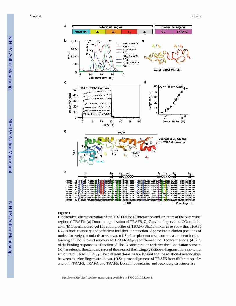
Figure 1.Biochemical characterization of the TRAF6/Ubc13 interaction and structure of the N-terminalregion of TRAF6. (a) Domain organization of TRAF6. Z1-Z4: zinc fingers 1–4. CC: coiledcoil. (b) Superimposed gel filtration profiles of TRAF6/Ubc13 mixtures to show that TRAF6RZ1 is both necessary and sufficient for Ubc13 interaction. Approximate elution positions ofmolecular weight standards are shown. (c) Surface plasmon resonance measurement for thebinding of Ubc13 to surface coupled TRAF6 RZ123 at different Ubc13 concentrations. (d) Plotof the binding response as a function of Ubc13 concentration to derive the dissociation constant(Kd). ± refers to the standard error of the mean of the fitting. (e) Ribbon diagram of the monomerstructure of TRAF6 RZ123. The different domains are labeled and the rotational relationshipsbetween the zinc fingers are shown. (f) Sequence alignment of TRAF6 from different speciesand with TRAF2, TRAF3, and TRAF5. Domain boundaries and secondary structures are
Yin et al. Page 14
Nat Struct Mol Biol. Author manuscript; available in PMC 2010 March 9.
NIH
-PA Author Manuscript
NIH
-PA Author Manuscript
NIH
-PA Author Manuscript
labeled. Zinc-coordinating residues are shaded in gray. Residues at the Ubc13 interface arehighlighted in red for those that bury more than 60 Å2 surface areas and in blue for those withsurface area burials of 20–60 Å2. Residues at the dimerization interface are highlighted inmagenta for those that bury more than 100 Å2 surface areas and in green for those with surfacearea burials of 40–100 Å2. The major TRAF6 auto-ubiquitination site at K124 is highlightedin yellow. “*“ in the TRAF2 sequence represents an insertion of “ VHEGIYEEG”. h: human; r:rat; c: cow; m: mouse; zf: zebra fish; f: fowl; d: drosophila. (g) Superposition of the zinc finger1 and 2 structure to the zinc finger 2 and 3 structure.
Yin et al. Page 15
Nat Struct Mol Biol. Author manuscript; available in PMC 2010 March 9.
NIH
-PA Author Manuscript
NIH
-PA Author Manuscript
NIH
-PA Author Manuscript
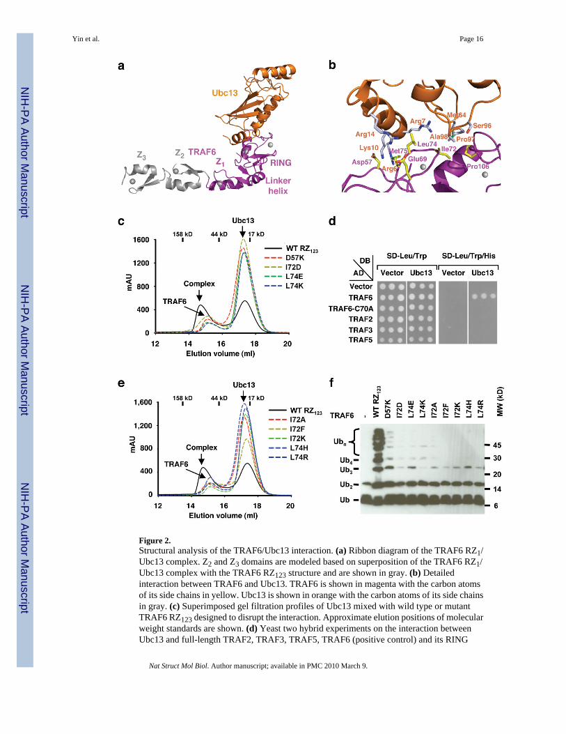
Figure 2.Structural analysis of the TRAF6/Ubc13 interaction. (a) Ribbon diagram of the TRAF6 RZ1/Ubc13 complex. Z2 and Z3 domains are modeled based on superposition of the TRAF6 RZ1/Ubc13 complex with the TRAF6 RZ123 structure and are shown in gray. (b) Detailedinteraction between TRAF6 and Ubc13. TRAF6 is shown in magenta with the carbon atomsof its side chains in yellow. Ubc13 is shown in orange with the carbon atoms of its side chainsin gray. (c) Superimposed gel filtration profiles of Ubc13 mixed with wild type or mutantTRAF6 RZ123 designed to disrupt the interaction. Approximate elution positions of molecularweight standards are shown. (d) Yeast two hybrid experiments on the interaction betweenUbc13 and full-length TRAF2, TRAF3, TRAF5, TRAF6 (positive control) and its RING
Yin et al. Page 16
Nat Struct Mol Biol. Author manuscript; available in PMC 2010 March 9.
NIH
-PA Author Manuscript
NIH
-PA Author Manuscript
NIH
-PA Author Manuscript
mutant C70A (negative control). (e) Superimposed gel filtration profiles of Ubc13 mixed withwild type or mutant TRAF6 RZ123 with interface residues switched to the correspondingsequences in other TRAFs: I72A (mutation to the corresponding TRAF2 sequence), I72K(TRAF3), I72F (TRAF5), L74H (TRAF3 and 5), and L74R (TRAF2). Approximate elutionpositions of molecular weight standards are shown. (f) Promotion of poly-Ub chain synthesisby wild type and mutant TRAF6 RZ123 in the presence of the E2 complex Ubc13/Uev1A andE1.
Yin et al. Page 17
Nat Struct Mol Biol. Author manuscript; available in PMC 2010 March 9.
NIH
-PA Author Manuscript
NIH
-PA Author Manuscript
NIH
-PA Author Manuscript
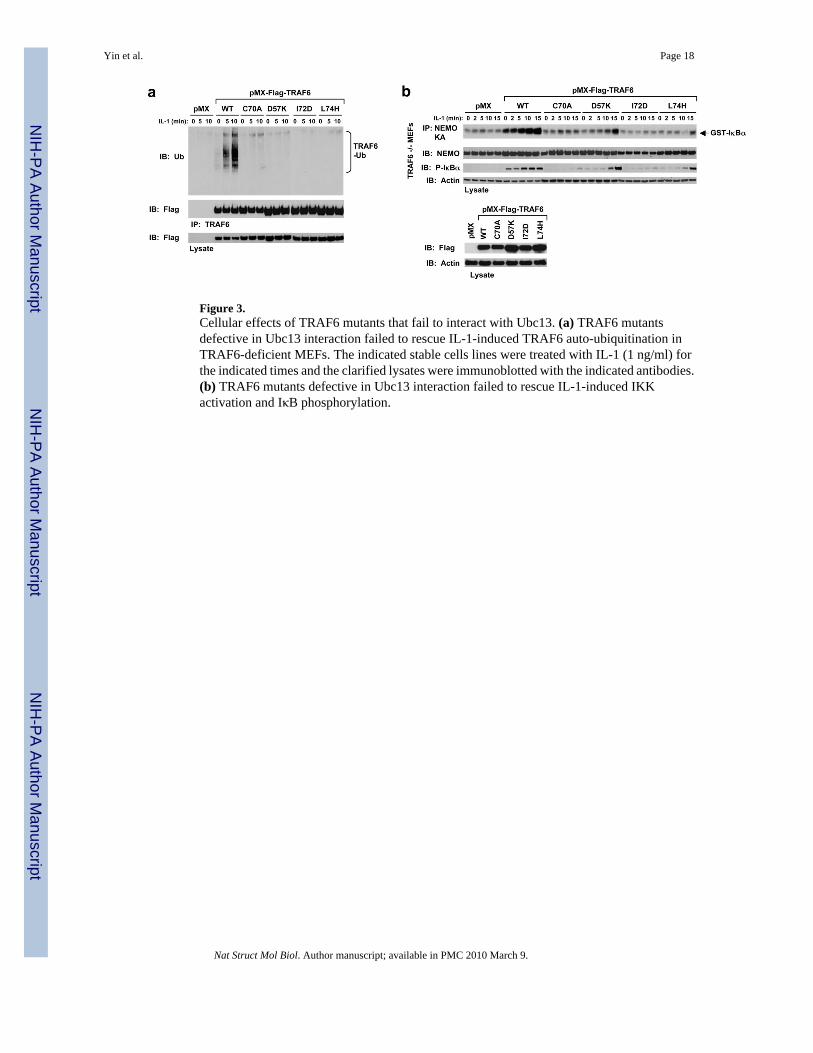
Figure 3.Cellular effects of TRAF6 mutants that fail to interact with Ubc13. (a) TRAF6 mutantsdefective in Ubc13 interaction failed to rescue IL-1-induced TRAF6 auto-ubiquitination inTRAF6-deficient MEFs. The indicated stable cells lines were treated with IL-1 (1 ng/ml) forthe indicated times and the clarified lysates were immunoblotted with the indicated antibodies.(b) TRAF6 mutants defective in Ubc13 interaction failed to rescue IL-1-induced IKKactivation and IκB phosphorylation.
Yin et al. Page 18
Nat Struct Mol Biol. Author manuscript; available in PMC 2010 March 9.
NIH
-PA Author Manuscript
NIH
-PA Author Manuscript
NIH
-PA Author Manuscript
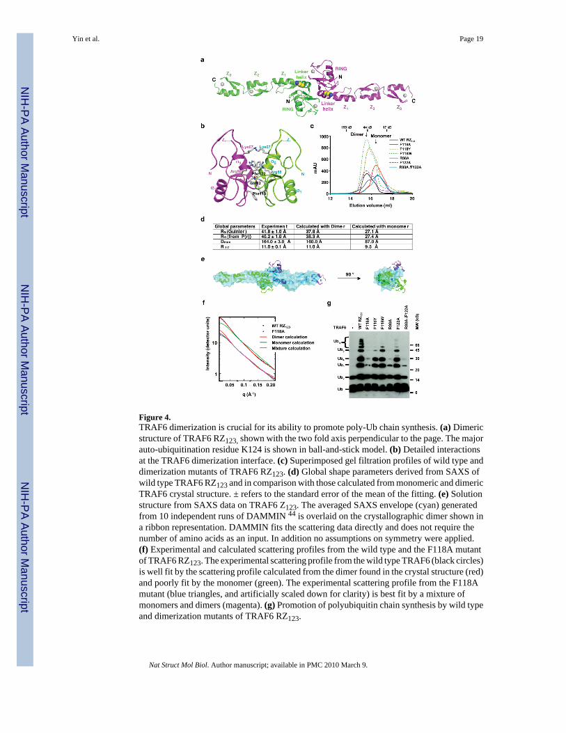
Figure 4.TRAF6 dimerization is crucial for its ability to promote poly-Ub chain synthesis. (a) Dimericstructure of TRAF6 RZ123, shown with the two fold axis perpendicular to the page. The majorauto-ubiquitination residue K124 is shown in ball-and-stick model. (b) Detailed interactionsat the TRAF6 dimerization interface. (c) Superimposed gel filtration profiles of wild type anddimerization mutants of TRAF6 RZ123. (d) Global shape parameters derived from SAXS ofwild type TRAF6 RZ123 and in comparison with those calculated from monomeric and dimericTRAF6 crystal structure. ± refers to the standard error of the mean of the fitting. (e) Solutionstructure from SAXS data on TRAF6 Z123. The averaged SAXS envelope (cyan) generatedfrom 10 independent runs of DAMMIN 44 is overlaid on the crystallographic dimer shown ina ribbon representation. DAMMIN fits the scattering data directly and does not require thenumber of amino acids as an input. In addition no assumptions on symmetry were applied.(f) Experimental and calculated scattering profiles from the wild type and the F118A mutantof TRAF6 RZ123. The experimental scattering profile from the wild type TRAF6 (black circles)is well fit by the scattering profile calculated from the dimer found in the crystal structure (red)and poorly fit by the monomer (green). The experimental scattering profile from the F118Amutant (blue triangles, and artificially scaled down for clarity) is best fit by a mixture ofmonomers and dimers (magenta). (g) Promotion of polyubiquitin chain synthesis by wild typeand dimerization mutants of TRAF6 RZ123.
Yin et al. Page 19
Nat Struct Mol Biol. Author manuscript; available in PMC 2010 March 9.
NIH
-PA Author Manuscript
NIH
-PA Author Manuscript
NIH
-PA Author Manuscript
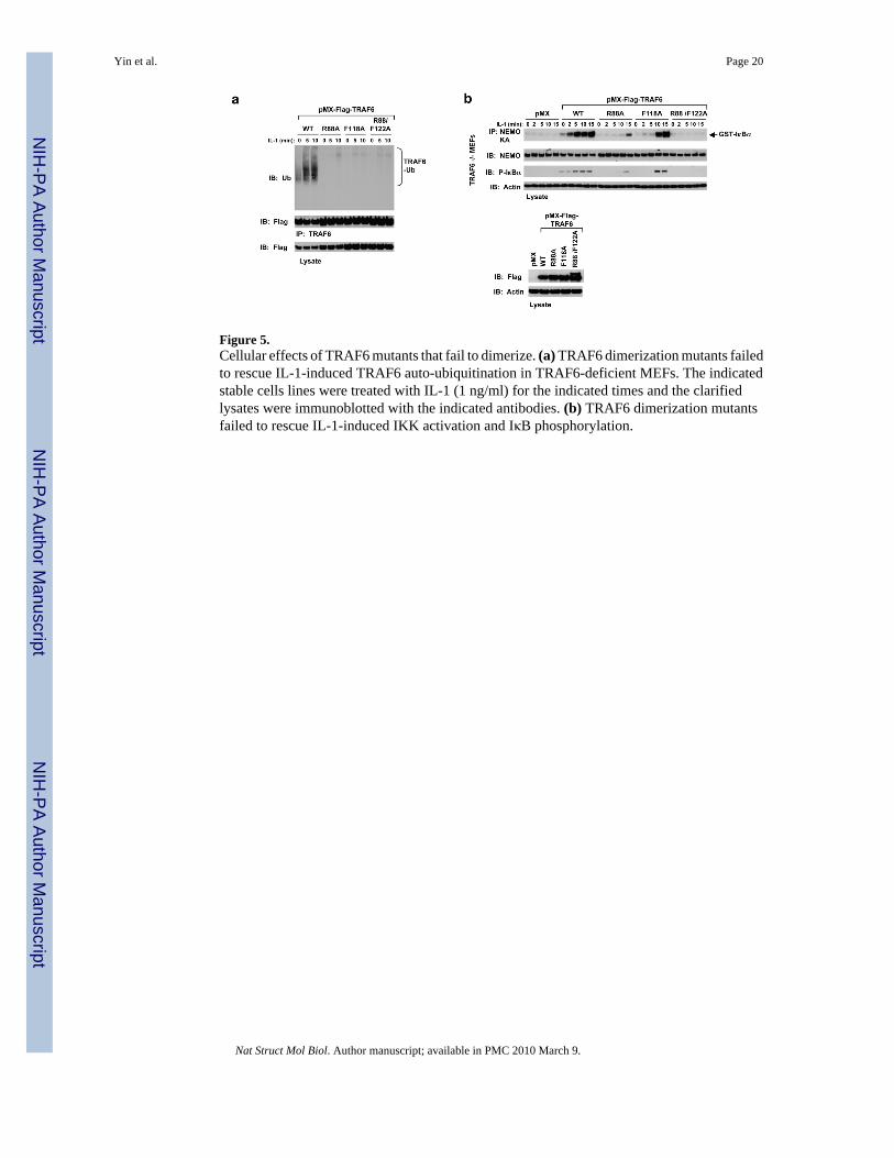
Figure 5.Cellular effects of TRAF6 mutants that fail to dimerize. (a) TRAF6 dimerization mutants failedto rescue IL-1-induced TRAF6 auto-ubiquitination in TRAF6-deficient MEFs. The indicatedstable cells lines were treated with IL-1 (1 ng/ml) for the indicated times and the clarifiedlysates were immunoblotted with the indicated antibodies. (b) TRAF6 dimerization mutantsfailed to rescue IL-1-induced IKK activation and IκB phosphorylation.
Yin et al. Page 20
Nat Struct Mol Biol. Author manuscript; available in PMC 2010 March 9.
NIH
-PA Author Manuscript
NIH
-PA Author Manuscript
NIH
-PA Author Manuscript
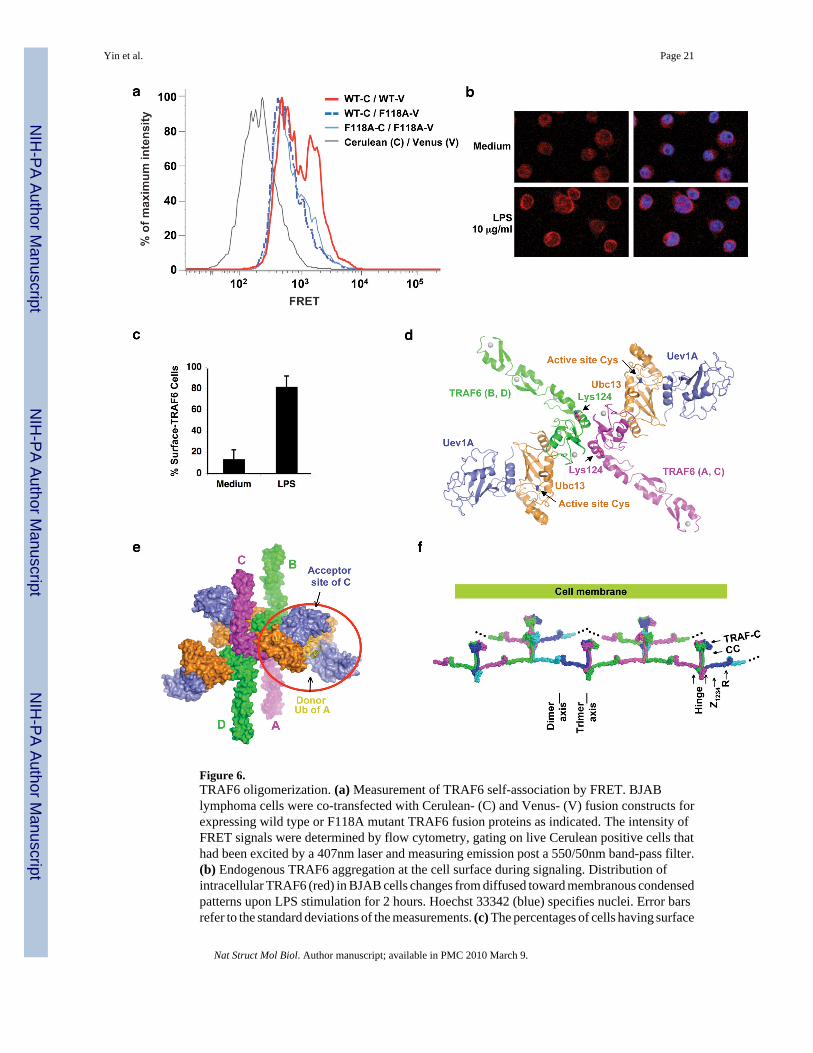
Figure 6.TRAF6 oligomerization. (a) Measurement of TRAF6 self-association by FRET. BJABlymphoma cells were co-transfected with Cerulean- (C) and Venus- (V) fusion constructs forexpressing wild type or F118A mutant TRAF6 fusion proteins as indicated. The intensity ofFRET signals were determined by flow cytometry, gating on live Cerulean positive cells thathad been excited by a 407nm laser and measuring emission post a 550/50nm band-pass filter.(b) Endogenous TRAF6 aggregation at the cell surface during signaling. Distribution ofintracellular TRAF6 (red) in BJAB cells changes from diffused toward membranous condensedpatterns upon LPS stimulation for 2 hours. Hoechst 33342 (blue) specifies nuclei. Error barsrefer to the standard deviations of the measurements. (c) The percentages of cells having surface
Yin et al. Page 21
Nat Struct Mol Biol. Author manuscript; available in PMC 2010 March 9.
NIH
-PA Author Manuscript
NIH
-PA Author Manuscript
NIH
-PA Author Manuscript
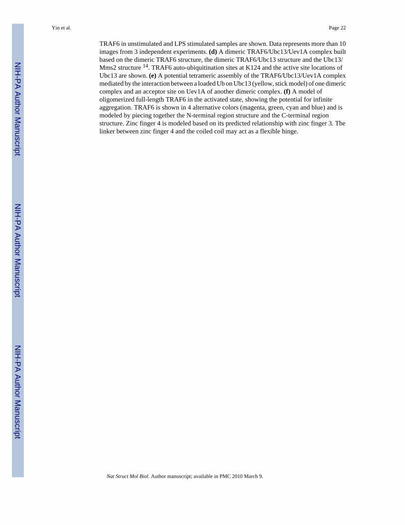
TRAF6 in unstimulated and LPS stimulated samples are shown. Data represents more than 10images from 3 independent experiments. (d) A dimeric TRAF6/Ubc13/Uev1A complex builtbased on the dimeric TRAF6 structure, the dimeric TRAF6/Ubc13 structure and the Ubc13/Mms2 structure 14. TRAF6 auto-ubiquitination sites at K124 and the active site locations ofUbc13 are shown. (e) A potential tetrameric assembly of the TRAF6/Ubc13/Uev1A complexmediated by the interaction between a loaded Ub on Ubc13 (yellow, stick model) of one dimericcomplex and an acceptor site on Uev1A of another dimeric complex. (f) A model ofoligomerized full-length TRAF6 in the activated state, showing the potential for infiniteaggregation. TRAF6 is shown in 4 alternative colors (magenta, green, cyan and blue) and ismodeled by piecing together the N-terminal region structure and the C-terminal regionstructure. Zinc finger 4 is modeled based on its predicted relationship with zinc finger 3. Thelinker between zinc finger 4 and the coiled coil may act as a flexible hinge.
Yin et al. Page 22
Nat Struct Mol Biol. Author manuscript; available in PMC 2010 March 9.
NIH
-PA Author Manuscript
NIH
-PA Author Manuscript
NIH
-PA Author Manuscript
NIH
-PA Author Manuscript
NIH
-PA Author Manuscript
NIH
-PA Author Manuscript
Yin et al. Page 23
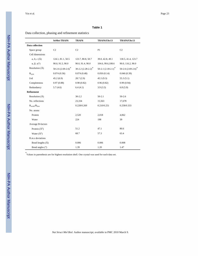
Table 1
Data collection, phasing and refinement statistics
SeMet-TRAF6 TRAF6 TRAF6/Ubc13 TRAF6/Ubc13
Data collection
Space group C2 C2 P1 C2
Cell dimensions
a, b, c (Å) 124.1, 81.1, 50.5 123.7, 80.8, 50.7 39.0, 42.8, 49.1 130.5, 41.4, 123.7
α, β, γ(°) 90.0, 91.5, 90.0 90.0, 91.4, 90.0 104.6, 99.6,108.6 90.0, 116.2, 90.0
Resolution (Å) 50-2.6 (2.69-2.6)* 30-2.2 (2.28-2.2)* 50-2.1 (2.18-2.1)* 50-2.6 (2.69-2.6)*
Rsym 0.074 (0.36) 0.074 (0.48) 0.050 (0.14) 0.046 (0.39)
I/σI 45.2 (6.9) 28.7 (2.9) 45.5 (9.3) 55.3 (5.1)
Completeness 0.97 (0.88) 0.98 (0.82) 0.96 (0.82) 0.99 (0.94)
Redundancy 5.7 (4.6) 6.4 (4.1) 3.9 (3.5) 6.8 (5.9)
Refinement
Resolution (Å) 30-2.2 50-2.1 50-2.6
No. reflections 23,334 15,563 17,678
Rwork/Rfree 0.228/0.269 0.210/0.251 0.258/0.333
No. atoms
Protein 2,520 2,018 4,062
Water 224 198 39
Average B-factors
Protein (Å2) 51.2 47.1 80.6
Water (Å2) 68.7 57.3 63.4
R.m.s deviations
Bond lengths (Å) 0.006 0.006 0.008
Bond angles (°) 1.39 1.20 1.47
*Values in parenthesis are for highest resolution shell. One crystal was used for each data set.
Nat Struct Mol Biol. Author manuscript; available in PMC 2010 March 9.


