Embed Size (px)
Citation preview

HAL Id: hal-03366016https://hal.archives-ouvertes.fr/hal-03366016
Submitted on 8 Oct 2021
HAL is a multi-disciplinary open accessarchive for the deposit and dissemination of sci-entific research documents, whether they are pub-lished or not. The documents may come fromteaching and research institutions in France orabroad, or from public or private research centers.
L’archive ouverte pluridisciplinaire HAL, estdestinée au dépôt et à la diffusion de documentsscientifiques de niveau recherche, publiés ou non,émanant des établissements d’enseignement et derecherche français ou étrangers, des laboratoirespublics ou privés.
Endothelial Cell Orientation and Polarity AreControlled by Shear Stress and VEGF Through Distinct
Signaling PathwaysAnne-Clémence Vion, Tijana Perovic, Charlie Petit, Irene Hollfinger, EireenBartels-Klein, Emmanuelle Frampton, Emma Gordon, Lena Claesson-Welsh,
Holger Gerhardt
To cite this version:Anne-Clémence Vion, Tijana Perovic, Charlie Petit, Irene Hollfinger, Eireen Bartels-Klein, etal.. Endothelial Cell Orientation and Polarity Are Controlled by Shear Stress and VEGFThrough Distinct Signaling Pathways. Frontiers in Physiology, Frontiers, 2021, 11, pp.623769.�10.3389/fphys.2020.623769�. �hal-03366016�

fphys-11-623769 February 24, 2021 Time: 17:6 # 1
ORIGINAL RESEARCHpublished: 02 March 2021
doi: 10.3389/fphys.2020.623769
Edited by:Anna Rita Cantelmo,
Université Lille Nord de France,France
Reviewed by:Stephan Huveneers,
Amsterdam University Medical Center(UMC), Netherlands
Elizabeth Anne Vincent Jones,KU Leuven, Belgium
*Correspondence:Anne-Clémence Vion
[email protected] Gerhardt
Specialty section:This article was submitted to
Vascular Physiology,a section of the journalFrontiers in Physiology
Received: 30 October 2020Accepted: 08 December 2020
Published: 02 March 2021
Citation:Vion A-C, Perovic T, Petit C,Hollfinger I, Bartels-Klein E,
Frampton E, Gordon E,Claesson-Welsh L and Gerhardt H(2021) Endothelial Cell Orientation
and Polarity Are Controlled by ShearStress and VEGF Through Distinct
Signaling Pathways.Front. Physiol. 11:623769.
doi: 10.3389/fphys.2020.623769
Endothelial Cell Orientation andPolarity Are Controlled by ShearStress and VEGF Through DistinctSignaling PathwaysAnne-Clémence Vion1,2* , Tijana Perovic1, Charlie Petit1, Irene Hollfinger1,Eireen Bartels-Klein1, Emmanuelle Frampton3, Emma Gordon3,4, Lena Claesson-Welsh4
and Holger Gerhardt1,5,6*
1 Integrative Vascular Biology Laboratory, Max Delbruck Center for Molecular Medicine, Berlin, Germany, 2 Universitéde Nantes, CNRS, INSERM, l’institut du thorax, Nantes, France, 3 Institute for Molecular Bioscience, The Universityof Queensland, Brisbane, QLD, Australia, 4 Beijer and Science for Life Laboratories, Rudbeck Laboratory, Departmentof Immunology, Genetics and Pathology, Uppsala University, Uppsala, Sweden, 5 DZHK (German Center for CardiovascularResearch), Berlin, Germany, 6 Berlin Institute of Health (BIH), Berlin, Germany
Vascular networks form, remodel and mature under the influence of multiple signals ofmechanical or chemical nature. How endothelial cells read and interpret these signals,and how they integrate information when they are exposed to both simultaneouslyis poorly understood. Here, we show using flow-induced shear stress and VEGF-A treatment on endothelial cells in vitro, that the response to the magnitude of amechanical stimulus is influenced by the concentration of a chemical stimulus, andvice versa. By combining different flow levels and different VEGF-A concentrations,front-rear polarity of endothelial cells against the flow direction was established ina flow and VEGF-A dose-response while their alignment with the flow displayed abiphasic response depending on the VEGF-A dose (perpendicular at physiologicaldose, aligned at no or pathological dose of VEGF-A). The effect of pharmaceuticalinhibitors demonstrated that while VEGFR2 is essential for both polarity and orientationestablishment in response to flow with and without VEGF-A, different downstreameffectors were engaged depending on the presence of VEGF-A. Thus, Src familyinhibition (c-Src, Yes, Fyn together) impaired alignment and polarity without VEGF-Awhile FAK inhibition modified polarity and alignment only when endothelial cells wereexposed to VEGF-A. Studying endothelial cells in the aortas of VEGFR2Y949F mutantmice and SRCiEC−KO mice confirmed the role of VEGFR2 and specified the role ofc-SRC in vivo. Endothelial cells of VEGFR2Y949F mutant mice lost their polarity andalignment while endothelial cells from SRCiEC−KO mice only showed reduced polarity.We propose here that VEGFR2 is a sensor able to integrate chemical and mechanicalinformation simultaneously and that the underlying pathways and mechanisms activatedwill depend on the co-stimulation. Flow alone shifts VEGFR2 signaling toward a Srcfamily pathway activation and a junctional effect (both in vitro and in vivo) while flow andVEGF-A together shift VEGFR2 signaling toward focal adhesion activation (in vitro) bothmodifying cell responses that govern orientation and polarity.
Keywords: endothelial cell, shear stress, VEGF, blood flow, signaling/signaling pathways
Frontiers in Physiology | www.frontiersin.org 1 March 2021 | Volume 11 | Article 623769

fphys-11-623769 February 24, 2021 Time: 17:6 # 2
Vion et al. Endothelial Responses to Flow and VEGF
INTRODUCTION
During embryonic development, all vertebrates initially establisha primitive network of vessels that subsequently remodels intoa hierarchical vascular structure. This involves the creationof a primary vascular plexus that expands by sproutingangiogenesis (Isogai et al., 2003; Potente et al., 2011) followedby vascular remodeling to adapt vessel organization, shapeand size; in its course, superfluous and inefficient segmentsare pruned away by active regression (Franco et al., 2015).The cellular and molecular regulation of this process isinfluenced by blood flow, hypoxia and metabolism. In thiscontext, cells need to respond appropriately to both mechanicaland chemical cues to ensure healthy tissue developmentand homeostasis.
Endothelial cells (ECs), which constitute the inner layer ofvessels, are in particular under constant mechanical strainsexerted by blood flow. Interestingly, ECs are able to sensesmall variations in the direction, magnitude, and regularity ofblood flow–induced shear stress (Wang et al., 2013; Givens andTzima, 2016) and respond to such changes by controlling theirnumber, shape and movement (Culver and Dickinson, 2010;Baeyens et al., 2016a). Adaptation of ECs to flow is criticalfor both the development and the maintenance of a well-functioning cardiovascular system, as modification of capillarypatterning allows for efficient oxygen and nutrient supply, whileinward and outward remodeling of main arteries maintainsappropriate blood pressure over the entire body (Baeyens et al.,2015). Physiological shear stress level, as found when thenetwork is mature will favor EC elongation and orientationparallel to the flow direction. Furthermore, the Golgi of EC willposition itself upstream of the nucleus, thus pointing againstthe flow direction (Franco et al., 2015) indicating their currentmigratory direction.
ECs are also well equipped to sense hypoxia. Hypoxicconditions drive the expression of vascular endothelial growthfactors (VEGFs) by the surrounding tissues, which initiatesendothelial sprouting through binding and activation of VEGFreceptors (VEGFRs). Signaling downstream of these receptors isessential for vascular morphogenesis, as they control processessuch as EC migration, proliferation and vessel permeability(Simons et al., 2016) and can influence arterial differentiation(Carmeliet and Tessier-Lavigne, 2005).
A long-standing question in developmental and cell biologyrelates to how cells integrate mechanical and chemical signalsto orchestrate the morphogenic behaviors that ensure adequatetissue patterning. When looking at the receptors and signalingcascades implicated in both flow and chemical responses in ECs,it is clear that they are largely redundant and involve the sameplayers (Jin et al., 2003; Koch and Claesson-Welsh, 2012). Thissuggests a cooperative or competitive action of chemical andmechanical stimuli during vascular bed formation, patterning,maturation and maintenance. In this context, VEGFR2 signalingis one of the most interesting examples. VEGFR2 is essentialfor VEGF-A-driven biological effects (Koch et al., 2011). Itbecomes activated and phosphorylated on tyrosine residues inresponse to VEGF-A: Y951, Y1059, Y1175, and Y1214 (human
sequence numbers) (Matsumoto et al., 2005). The Y951 phospho-site (Y949 in mouse VEGFR2) presents a specific binding sitefor the T cell-specific adaptor which is implicated in VEGF-A-induced permeability, by regulating VEGFR2-dependent SRCsignaling pathway at EC junctions (Sun et al., 2012). TheY1059 residues, located on the tyrosine kinase activationloop, are required for full kinase activity (Koch et al., 2011).The phosphorylated Y1175 (Y1173 in mouse VEGFR2) bindsphospholipase Cg, which is of importance for endothelialextracellular-signal-regulated kinase ERK1/2 pathway activation(Takahashi et al., 2001). A phenylalanine knock-in mouseVegfr2Y1173F/Y1173F is embryonically lethal due to an arrestin EC development (Sakurai et al., 2005). Interestingly thisphosphosite has also been described to be activated by flow,independently of VEGF, activating ERK1/2 and JNK pathwaysas well as eNOS (Chen et al., 1999; Jin et al., 2003). Ithas also been shown to activate NFκB both in vitro (Tzimaet al., 2005; Coon et al., 2015) and in vivo (Baeyens et al.,2015). Finally, VEGFR2 Y1214 signaling induces activationof ERK1/2 and Akt pathways required for c-Myc-dependentgene regulation, endothelial proliferation, and vessel stability(Testini et al., 2019).
MATERIALS AND METHODS
Mice and TreatmentsThe following mouse strains were used: VEGFRY949F mice(knock-in of phenylalanine (F) to replace the tyrosine (Y)at position 949 of VEGFR2 (Li et al., 2016) and c-Src-flox,Cdh5-CreERT2 mice designated as SRCiEC−KO mice (Cdh5-CreERT2 mice were provided by Ralf Adams (MPI, Muünster,Germany) (Kogata et al., 2006; Wang et al., 2010). c-Src-floxedmice were delivered from the Nice Mice, National ResourceCenter for Mutant Mice, Model Animal Research Center,China) (Schimmel et al., 2020). Mice were maintained at theUppsala University under standard husbandry conditions. Allanimal work was approved by the Uppsala University boardof animal experimentation (permit 5.2.18-8927-16). To induceCre-mediated deletion, tamoxifen (Sigma-Aldrich) was injectedi.p. (100 µg) at P1, P2 and P3. Aortas were then collectedat P6 onward. The investigators were blinded to genotypeduring experiments.
Metatarsal AssayMetatarsals were isolated from E16.5 mice using a protocoladapted from Song et al. (2015). After dissection, one metatarsalper well was placed in a µ-Plate 24 well ibiTreat plate with a1.5 polymer coverslip (Ibidi) and left in 170 µl of MEM-alpha(Gibco) with 10% FCS and 1% penicillin/streptomycin (Sigma).After 3 days, media were replaced with 300 µl MEM-alpha + 10%FCS + 1% pen/strep per well and media changed every 48 h.To induce Cre activity, cells were treated with 1 µM of 4-hydroxytamoxifen (Sigma) after 5 days. After 14 days, metatarsalswere fixed in 4% PFA in PBS for 20 min and antibodies wereadded in 3% Triton X-100, 1% Tween and 0.5% BSA in PBS.
Frontiers in Physiology | www.frontiersin.org 2 March 2021 | Volume 11 | Article 623769

fphys-11-623769 February 24, 2021 Time: 17:6 # 3
Vion et al. Endothelial Responses to Flow and VEGF
The following antibodies were used: GM130 (ref 560066, mouse,1:500, BD Biosciences), ERG (ref ab92513, rabbit, 1:500, Abcam).
Cell Culture and Microfluidic ChamberExperimentsHUVECs (passage 2–6; PromoCell) were routinely cultured inEBM-Bulletkit (Promocell). For flow experiments including staticcondition leading to WB lysate collection or immunofluorescencefrom Figures 2, 3, cells were cultured on 0.2% gelatin-coatedslides (Menzel Glazer) and unidirectional laminar shear stresswas applied using peristaltic pumps (Gilson) connected to a glassreservoir (ELLIPSE) and the chamber containing the slide. Thisdevice allows the circulation of 10 ml of medium on the slides,static slides has been exposed to 10 ml of medium without anymedium circulation. For flow experiments under high shear stress(Figure 4) and immunofluorescence staining, cells were culturedon 0.2% gelatin-coated 0.4 ibidi slides (IBIDI) and unidirectionallaminar shear stress was applied using the pumping system andcontrol unit form IBIDI, allowing the circulation of 10 ml ofmedium as for the peristaltic system.
Local shear stress was calculated using Poiseuille’s law andaveraged to 2 (Low SS) or 20 dyn/cm2 (High SS). For VEGF-A treatment, cells were exposed to shear stress for 24 h usingEBM media (Promocell) without VEGF-A supplement and co-stimulated with 0, 0.5, 10 or 200, ng/ml of human VEGF-A165(PeproTech ref 450-32) under the different flow conditions.For inhibition experiments, VEGFR2 inhibitors (SU1498, 1.5µM; ZM323881, 4 nM), Src family inhibitor (SU6656, 500 nM),FAK inhibitor (PND-1186, 3 nM) and p38 inhibitor (SB203580,1 µM) were added to the media 30 min prior flow start.Control condition were treated with DMSO diluted at 1/1,000 asall inhibitor used.
Western BlottingHUVECs were washed with cold PBS and scraped off inM-PER (Mammalian Protein Extraction Reagent; ThermoFisher Scientific) completed protease and phosphatase inhibitors(Roche). Lysates were centrifuged and protein supernatant wasquantified using the Lowry protein assay (Bio-Rad). Lysateswere mixed with reducing sample buffer for electrophoresisand subsequently transferred onto polyvinylidene fluoridemembranes. Equal loading was checked using Ponceau redsolution. Membranes were incubated with primary antibodies(see below). After incubation with secondary antibodies (1:3,000;GE Healthcare), immunodetection was performed using anenhanced chemiluminescence kit (SuperSignal West Dura;Pierce), and bands were revealed using the Las-4000 imagingsystem. After initial immunodetection, membranes were strippedof antibodies, probed with total form of the phosphorylatedform when suitable (PXL) and then reprobed with anti–GAPDHantibody. Values reported from Western blots were obtainedby band density analysis using FIJI (ImageJ) and expressedas the ratio of the protein of interest to GAPDH or as theratio of phosphorylated form to total form of the proteinof interest. The following antibodies were used: GAPDH (refMAB374, goat; 1:10,000; Millipore), VEGFR2 (ref 2479, rabbit,
1:1,000; cell signaling), p1175-VEGFR2 (ref 3770, rabbit, 1:1,000;cell signaling), p951-VEGFR2 (ref 2471, rabbit, 1:1,000; cellsignaling), VE-cadherin (ref ab33168, rabbit, 1:1,000; abcam),p685-VE-cadherin (ref ab119785, rabbit, 1:1,000; abcam), ZO-1(ref 61-7300, rabbit, 1:1,000; Thermo Fisher Scientific), FAK(ref 3285, rabbit, 1:1,000; cell signaling), Claudin5 (ref 34-1600, rabbit, 1:1,000; Invitrogen), Paxillin (ref 610051, rabbit,1:1,000; BD Biosciences), p118-Paxillin (ref 2541, rabbit, 1:1,000;cell signaling).
Immunofluorescence StainingHUVECs were fixed with 4% PFA in PBS for 10 min at roomtemperature (RT). Blocking/permeabilization was performedusing blocking buffer consisting of 5% BSA (Sigma-Aldrich),0.5% Triton X-100 (Sigma-Aldrich), 0.01% sodium deoxycholate(Sigma-Aldrich), and 0.02% sodium azide (Sigma-Aldrich) inPBS at pH 7.4 for 45 min at RT. Primary antibodies wereincubated at the desired concentration in 1:1 Blocking buffer/PBSat RT for 2 h and secondary antibodies were incubated at thedesired concentration in 1:1 blocking buffer/PBS for 1 h atRT. Aortas were fixed with 4% PFA in PBS overnight at 4◦C.Blocking/permeabilization was performed using blocking bufferconsisting of 1% FBS, 3% BSA (Sigma-Aldrich), 0.5% TritonX-100 (Sigma-Aldrich), 0.01% sodium deoxycholate (Sigma-Aldrich), and 0.02% sodium azide (Sigma-Aldrich) in PBS at pH7.4 for 1 h at RT. Primary antibodies were incubated at the desiredconcentration in 1:1 Blocking buffer/PBS overnight at 4◦C andsecondary antibodies were incubated at the desired concentrationin 1:1 blocking buffer/PBS for 2 h at RT. DAPI (Sigma-Aldrich,1/10,000, 5 min) was used for nuclear labeling. Cells and aortaswere mounted in Mowiol. The following antibodies were usedin vitro: VE-cadherin (ref sc-6458, goat; 1:100; Santa cruz), ZO-1 (ref 61-7300, rabbit, 1:500; Thermo Fisher Scientific), GM130(ref 610822, mouse, 1:1,000;BD Biosciences). The followingantibodies were used in vivo: GOLPH4 (ref ab28049, rabbit; 1:500;Abcam), VE–cadherin (ref 555289; rat; 1/100; BD Biosciences).
Microscope Image AcquisitionImages from fluorescently labeled HUVECs were acquiredusing a LSM 700 upright microscope equipped with a Plan-Apochromat 20×/0.8 NA Ph2 objective. Images were taken atroom temperature using Zen 2.3 software. Bright-field imageswere taken using a Leica DMIL LED microscope equipped witha 10×/0.22 NA Ph1 objective and a CCD camera (DFC3000 G).Images were acquired at room temperature while the cells werestill in their culture medium using LAS X software (Leica). Imagesof aortas were taken using a LSM 780 inverted microscope (Zeiss)equipped with a Plan-Apochromat 20×/0.8 NA Ph2 objectiveor with a Plan-Apochromat 63×/1.4 NA DIC objective. Themicroscope was equipped with a photon multiplier tube detector.Images were taken at room temperature using Zen 2.3 software(Zeiss). Images of metatarsals were acquired using a LSM 710inverted microscope equipped with Plan-Apochromat 10×/0.45NA and 20×/0.8 NA objectives. For all animal experiments, theinvestigators were unaware of the genotypes of the animals whileacquiring images.
Frontiers in Physiology | www.frontiersin.org 3 March 2021 | Volume 11 | Article 623769

fphys-11-623769 February 24, 2021 Time: 17:6 # 4
Vion et al. Endothelial Responses to Flow and VEGF
Cell Junction Activity AnalysisCell junction morphology analysis was done in HUVECs stainedfor VE-cadherin using the patch and classified Matlab codepreviously developed by Bentley et al. (2014) adapted for 2Dimages. Two status were defined: activated vs. stabilized, anddivided into 3 level: from low to high. Activated junctions weredefined as serrated or reticular and stabilized as straight thickjunctions. Each image taken was divided into small pieces ofimages allowing to visualized only a portion of the junctionand these small images were presented blindly and randomlyto the user who then classified the junction accordingly. Thistechnic ensured an unbiased analysis of the junctions regardingthe treatment and regarding the global shape of the cell.
Flow-Induced Orientation AnalysisTo analyze the orientation of cells, we calculated the angle formedbetween the vector of the flow direction (obtained by knowledgeof flow direction within the slide) and the “orientation vector”given by orientation of the main axe of each cell. Each valuefor each cell is then used to plot the hemi-roses presented inFigure 2A. To plot the bar graphs presented in Figures 2B,4B, 5B, 6B the cells were classified in 2 categories: aligned withthe flow direction, and not aligned with the flow direction. Forin vitro experiment, aligned with the flow was defined as anabsolute value of angle between 0 and 45◦, not aligned withthe flow as an absolute value of angle between 45 and 90◦; forin vivo experiments, aligned with the flow was defined as anabsolute value of angle between 0 and 15◦ and not aligned withthe flow as an absolute value of angle between 15 and 90◦. Anglecalculation and roses presentation was done automatically usinga homemade Matlab script validated on the first experiment by acomparison to hand calculation with FIJI.
Flow-Induced Polarity AnalysisTo analyze the polarization of cells, we calculated the angleformed between the vector of the flow direction (obtained byknowledge of flow direction within the slide) and the “golgivector” given by the line from the center of the nucleus to thecenter of the Golgi. Of note, static experiments are not exposedto flow and therefor calculation has been made using an arbitrarydirection given by the geometry of the ibidi slide. Each value foreach cell is then used to plot the roses presented in Figure 2C. Toplot the bar graphs presented in Figures 2D, 4D, 5D, 6D, the cellswere classified in 3 categories: against the flow direction, sidedto the flow direction and with the flow direction. For in vitroexperiment, with the flow was defined as an absolute value ofangle between 0 and 45◦, sided as an absolute value of anglebetween 45 and 135◦ and against the flow as an absolute valueof angle between 135 and 180◦; for in vivo experiment with theflow was defined as an absolute value of angle between 0 and30◦, sided as an absolute value of angle between 30 and 150◦and against the flow as an absolute value of angle between 150and 180◦. Angle calculation and roses presentation was doneautomatically using a homemade Matlab script validated on thefirst experiment by a comparison to hand calculation with FIJI.Metatarsal polarity analysis was performed using FIJI with values
defined as for in vivo experimental values (with: 0–30◦, sided:30–150◦, and against: 150–180◦).
Statistical AnalysisStatistical analysis was performed using GraphPad Prismsoftware. For in vitro and in vivo experiments, two-way ANOVA(data distribution was assumed to be normal) were used, followedby a Tukey test or a Fisher LSD test when conditions wereconsidered experimentally independent. Details of the statisticaltest used for each experiment can be found in the figure legends.The investigators were blinded to genotype during experimentsand quantification.
EXPERIMENTAL APPROACH
To study how physical forces and growth factors jointlyinfluence front-rear polarity and cell elongation toward the flowdirection (alignment/orientation), we subjected ECs to eitherstatic conditions, low or high shear stress (SS, 2 or 20 dyn/cm2
for 24 h) and to different VEGF-A concentrations ranging fromphysiological (0.5 and 10 ng/mL) to pathological (200 ng/mL)levels. Cell orientation refers to the long axis of ECs as theyadopt their prototypical elongated cell shape under the influenceof blood flow in vivo, or equivalent shear stress induced bymedium flow in vitro. Alignment in this context depicts whetherthis long-axis of the ECs is aligned with the direction of theflow. Similarly, front-rear polarity is also assessed in relation tothe flow direction. Unlike elongation or alignment, the polarityvector assigns a front and a rear to the cell, in a way that isnormally associated with dynamic movement of cells in a certaindirection. When they migrate, many cell types, including ECs,position their centrosome and Golgi ahead of the nucleus inthe direction of migration. Therefore, determining the center ofmass of the Golgi in relation to the center of mass of the cellnucleus delivers a vector that can be used as a proxy for front-rear polarity of migrating cells. Recent work illustrated that ECsunder flow in vivo and in vitro follow the same principle asthey orientate and migrate against the direction of flow duringvascular remodeling (Kupfer et al., 1982; Franco et al., 2015).We therefore determined the Golgi-nucleus vector to establishfront-rear polarity under all conditions of SS and growth factorstimulation. Throughout the analysis below, cell alignment andpolarity in relation to the direction of flow will serve as referencesystem for the phenotypical flow response of ECs (Figure 1).
As the polarized movement of ECs requires junctionalrearrangements and turnover, both during angiogenic sproutingand vascular remodeling under flow (Koch et al., 2011;Conway et al., 2013; Neto et al., 2018), we also assessedendothelial junctional patterns under the various conditions.Previous studies classified junctional features that correlatewith junctional dynamics and VE-cadherin turnover (Bentleyet al., 2014), providing a useful quantitative framework forthe assessment of flow and growth factor effects. We usedthis classification tool adapted for a 2D layer to evaluatejunctional activity in our different in vitro conditions. Wecomplemented these results with biochemical analysis of VE-cadherin phosphorylation on Tyrosine 685, which contributes
Frontiers in Physiology | www.frontiersin.org 4 March 2021 | Volume 11 | Article 623769

fphys-11-623769 February 24, 2021 Time: 17:6 # 5
Vion et al. Endothelial Responses to Flow and VEGF
FIGURE 1 | Experimental approach. In vitro, We subjected ECs to either static condition, low or high shear stress (SS, 2 or 20 dyn/cm2 for 24 h) and to differentVEGF-A concentrations ranging from physiological (0.5 and 10ng/mL) to pathological (200 ng/mL) levels. In vivo, we used the linear part of aorta which is exposed tohigh SS and free from VEGF-A influence. Phenotypical response: Cell orientation/alignment refers to the long axis of ECs as they adopt their prototypicalelongated cell shape under the influence of blood flow in vivo, or equivalent shear stress induced by medium’s flow in vitro. Determining the center of mass of thegolgi in relation to the center of mass of the cell nucleus delivers a vector used as a proxy for front-rear polarity of migrating cells. Functional response: biochemicalanalysis of VEGFR2 phosphorylation on Tyrosine 1175 and 951; VE-cadherin phosphorylation on Tyrosine 685; Paxillin phosphorylation on Tyrosine 118.classification of junction type through VE-cadherin staining. Pathways: inhibition through drug treatment (in vitro) or Knock-Out strategies (in vivo) and analysis of thephenotypical response.
to VE-cadherin internalization (Orsenigo et al., 2012). SinceVEGFR2 is a flow sensor (Tzima et al., 2005) when associated toPECAM and VE-cadherin, in addition to its VEGF-A receptoractivity, we then employed biochemical analysis to study itsactivation. We exposed ECs to selective inhibitor treatmentsto identify pathway activity patterns and their relevance forthe different aspects of the endothelial phenotypical responseto flow. Finally, we validated our results using an in vivoapproach. The aorta is exposed to high SS (similar level asused in our in vitro experiments) in its linear, thoracic partand free from VEGF-A influence. In this model, ECs formingthe linear part are highly elongated and aligned with the flowdirection and mostly polarized against the flow. In contrastEC display rounded shape and random alignment in the aorticarch exposed to turbulent flow generating low SS (Gimbroneand García-Cardeña, 2013). We took advantage of these aortic
characteristics to assess EC alignment in deficient mice modelsto validate our in vitro finding on phenotypical response toflow (Figure 1).
RESULTS
Combination of Flow Exposure andVEGF-A Treatment Modifies EndothelialCell Orientation and PolarityAs VEGF-A induces proliferation of ECs (Koch et al., 2011), wefirst controlled whether our experimental condition influencedcell density and therefore the analyses. In all conditions,static, low SS (2 dyn/cm2), or high SS (20 dyn/cm2),VEGF-A (0.5–200 ng/ml) did not affect the EC number
Frontiers in Physiology | www.frontiersin.org 5 March 2021 | Volume 11 | Article 623769

fphys-11-623769 February 24, 2021 Time: 17:6 # 6
Vion et al. Endothelial Responses to Flow and VEGF
FIGURE 2 | Dose-dependent effect of shear stress and VEGF-A concentration on cell orientation and polarity. (A) Representative picture (phase contrast) ofendothelial cells exposed to shear stress for 24 h (static: 0 dyn/cm2; Low SS: 3 dyn/cm2; High SS: 20 dyn/cm2) and associated orientation quantification shown ascircular plots (N = 3; between 1,500 and 3,000 cells analyzed). (B) Quantification of percentage of cells aligned with the flow direction (in between 45◦ around theflow axis, N = 3, between 1,500 and 3,000 cells analyzed), Data presented as Mean + SEM. (C) Representative picture (Immunofluorescence) of endothelial cellsexposed to shear stress for 24 h (Low SS: 3 dyn/cm2; High SS: 20 dyn/cm2) (scale bars: 30 µm). (D) Quantification of golgi position around the nucleus comparedto the flow direction (N = 3; with: in between 0 and 45◦ around the flow axis, side: 45–135◦, against: 135–180◦; between 1,500 and 3,000 cells analyzed). Two-wayANOVA; Tukey’s post hoc, *p < 0.05; **p < 0.01; ***p < 0.001.
(Supplementary Figure 1A). Both high and low SS exposurereduced the number of cells to about 75% of that in the staticconditions. Alignment analyses showed that the ECs main axis instatic condition (no flow, 0 dyn/cm2) was randomly distributed,with no effect of VEGF-A addition (Figures 2A,B). The onlyeffect observed was an increase in cell elongation after treatingECs with 200 ng/mL of VEGF-A (Supplementary Figure 1B).
Low SS (2 dyn/cm2, no VEGF-A) had no effect on cellalignment, which remained random. VEGF-A addition (0.5
ng/ml) to low SS promoted alignment perpendicular to the flowdirection while treatment with 200 ng/ml VEGF-A promotedEC alignment with the flow (Figures 2A,B). This showed thatthe effect of VEGF-A under low SS was biphasic, dependenton the dose used.
High SS itself (20 dyn/cm2, no VEGF-A) enhanced alignmentwith the flow. VEGF-A addition (0.5 ng/ml) to high SS madeECs align perpendicular to the flow direction. At high VEGF-A concentrations (200 ng/ml), alignment with the flow was
Frontiers in Physiology | www.frontiersin.org 6 March 2021 | Volume 11 | Article 623769

fphys-11-623769 February 24, 2021 Time: 17:6 # 7
Vion et al. Endothelial Responses to Flow and VEGF
FIGURE 3 | Effect of flow and VEGF-A combination on VEGFR2, junctional and focal adhesion activity. (A) Representative picture (Immunofluorescence) ofendothelial cells exposed to shear stress for 24 h (Low SS: 3 dyn/cm2; High SS: 20 dyn/cm2). Red arrows indicate gaps in the ECs monolayer (B) Quantification ofjunction status based on their morphology (N = 3; 100 patches analyzed blinded by images, 5–8 images per N) (scale bars: 20 µm, same magnification for allimages). (C) WB analysis of VE-cadherin phosphorylation, N = 5, (D) WB analysis of VEGFR2 phosphorylation on Tyr951. N = 4 (E) WB analysis of VEGFR2phosphorylation on Tyr1175. N = 4 (F) WB analysis of Paxilllin phosphorylation on Tyr118. N = 4 (G) WB representative images. Data presented as Mean + SEM.Two-way ANOVA; Fisher LSD test, *p < 0.05; **p < 0.01; ***p < 0.001 compared to static 0 VEGF; ##p < 0.01; ###p < 0.001 (compared to LSS 0 VEGF);!!p < 0.01 (compared to HSS 0 VEGF); $$p < 0.01; $$$p < 0.001 compared to static from the same VEGF concentration; ∧∧p < 0.05; ∧∧∧p < 0.01 compared toLow SS from the same VEGF concentration.
Frontiers in Physiology | www.frontiersin.org 7 March 2021 | Volume 11 | Article 623769

fphys-11-623769 February 24, 2021 Time: 17:6 # 8
Vion et al. Endothelial Responses to Flow and VEGF
FIGURE 4 | Pathways involved in flow and VEGF responses. (A) Representative picture of junction (VE-cadherin mentioned as VE-cadh) of endothelial cells exposedto shear stress for 24 h (High SS: 20 dyn/cm2) without or with inhibitors (scale bars: 40 µm, same magnification for all images). (B) Quantification of percentage ofcells aligned with the flow direction (in between 30◦ around the flow axis. DMSO, N = 5, inhibitors N = 3). (C) Representative picture polarity of endothelial cellsexposed to shear stress for 24 h (High SS: 20 dyn/cm2) without or with inhibitors (scale bars: 40 µm, same magnification for all images). (D) Quantification of golgiposition around the nucleus compared to the flow direction (DMSO, N = 5, inhibitors N = 3. with: in between 0 and 45◦ around the flow axis, side: 45–135◦, against:135–180◦). Arrow represents flow direction (High SS). Two-way ANOVA; *p < 0.05; **p < 0.01; ***p < 0.001. VEGFR2 inhibitors (SU1498, 1.5 mM; ZM323881,4 nM), Src family inhibitor (SU6656, 500 nM), FAK inhibitor (PND-1186, 3 nM) and p38 inhibitor (SB203580, 1 mM) were added to the media 30 min prior flow start.
Frontiers in Physiology | www.frontiersin.org 8 March 2021 | Volume 11 | Article 623769

fphys-11-623769 February 24, 2021 Time: 17:6 # 9
Vion et al. Endothelial Responses to Flow and VEGF
increased, as observed under low SS (Figures 2A,B) but evenmore pronounced.
When assessing polarity, ECs in static culture remainedrandomly polarized in the absence and presence of VEGF-A(Figures 2C,D). At low SS in the absence of VEGF-A, ECspolarized in the direction “with” the flow, meaning that theirGolgi was mostly placed behind the nucleus compared to the flowdirection. On the contrary, high SS triggered a partial polarizationagainst the flow. In both low and high SS, VEGF-A promotedpolarity against the flow, in a dose-response manner: the higherthe VEGF-A concentration, the better cells polarized against theflow (Figures 2C,D).
Taken together, these data show that ECs align and orientthemselves relative to flow by integrating chemical and flowconditions and that the magnitude of the stimuli direct theresponse. With the more pronounced combined stimulation, ECsaligned efficiently and polarized against the flow.
Combination of Flow Exposure andVEGF-A Treatment Affects VEGFR2Phosphorylation, Adherens JunctionStability and Focal Adhesion ActivityWe then looked at cell adherens junctions. Under staticconditions, VE-cadherin staining and quantification of stabilized(straight) and activated (serrated) junctions did not showany effect of VEGF-A treatment after 24 h (SupplementaryFigures 2A,B). ZO-1 appeared localized at the junctionfor all VEGF-A concentration (Supplementary Figure 2A).Some discontinuities in the adherens junctions were observedat 200 ng/mL (Supplementary Figure 2, red arrow). Thiswas not associated to a change in VE-cadherin and ZO-1protein levels upon SS and VEGF-A exposure (SupplementaryFigures 3B,C).
Upon low SS alone (no VEGF-A), ECs showed a high level ofactivated/serrated junctions (Figures 3A,B). VEGF-A treatmenthad no additional effect on junction activation which remainedhigh. In contrast, high SS induced stabilization/straighteningof the junctions and increasing the concentration of VEGF-Acorrelated with an increase in serrated VE-cadherin junctions(Figures 3A,B). Junctional ZO-1 was higher in ECs exposed tohigh SS than in ECs exposed to low SS (Figure 3A). VEGF-A treatment induced a loss of this junctional ZO-1 both underlow and high SS (Figure 2A). Additionally, both under low andhigh SS, high VEGF-A concentrations (10 and 200 ng/mL) led toloosening of the adherens junctions, which displayed increasednumber of gaps in the endothelial layer (indicated as red arrowson Figure 3A), to a higher extent at high SS.
To better understand the junctional modifications, weanalyzed VE-cadherin phosphorylation on Tyrosine 685, whichcontributes to VE-cadherin internalization (Orsenigo et al.,2012). Under static condition, VEGF-A treatment increasedVEcadhY685 level in a dose dependent manner (Figures 3C,G).ECs exposed to low SS have high VE-cadherin phosphorylationlevel independently of VEGF-A concentration (Figures 3C,G),matching the observation of high level of serrated junction in thisflow condition. VEcadhY685 levels remained low under high SS
independently of VEGF-A concentration and despite increasingserrated junctions.
Since VEGFR2 is a flow sensor (Tzima et al., 2005) whenassociated to PECAM and VE-cadherin in addition to itsVEGF-A receptor activity, we evaluated its phosphorylationstatus in our conditions. First, we confirmed that bothtyrosine 1175 and 951 were phosphorylated in a dose-responsemanner upon VEGF-A treatment under static conditions(Figures 3D,E,G). VEGFR2 expression was stable in staticcondition but significantly increased in response to flow inthe absence of VEGF-A (Supplementary Figure 3A). Of note,high VEGF-A treatment (200 ng/mL) slightly but significantlydecreased VEGFR2 expression, suggesting modification inprotein turn over. VEGFR2Y1175 phosphorylation level wasincreased by SS (Figures 3E,G) in the absence of VEGF-A, withhigh SS inducing more VEGFR2Y1175 phosphorylation than lowSS. In comparison, VEGFR2Y951 phosphorylation was increasedby SS but at an equivalent level between low and high SS(Figures 3D,G). When ECs were exposed to low SS, VEGF-A treatment strongly increased VEGFR2Y951 and VEGFR2Y1175
phosphorylation in a dose-response manner while this effect wasnot present (VEGFR2Y951) or mild (VEGFR2Y1175) when ECswere exposed to high SS (Figures 3D,G).
As VEGFR2 activation can also affect focal adhesion formation(Pietilä et al., 2019), we then checked the expression levelsof FAK and Paxillin and the phosphorylation level of Paxillinon tyrosine 118, which controls focal adhesion turn over,affinity to FAK and cell migration (López-Colomé et al., 2017).FAK and Paxillin displayed stable expression over all of thetested conditions (Figure 3F and Supplementary Figure 3F).PaxillinY118 phosphorylation was activated by both low and highSS in the absence of VEGF-A compared to static condition(Figures 3F,G). At low SS, increasing VEGF-A concentrationdid not significantly change PaxillinY118 phosphorylation level.In contrast, at high SS, VEGF-A treatment increased it in adose-response manner (Figures 3F,G).
Combined, these data show that low flow induced an“activated” junctional morphology and phosphorylation ofVEGFR2 and VE-cadherin; these effects were further augmentedby VEGF-A. In contrast, high flow combined with VEGF-Apreferentially induced focal adhesion signaling.
VEGFR2 Is Involved in Orientation andPolarity Responses Through DifferentSignaling CascadesIn order to understand better how activation of VEGFR2contributed to focal adhesion or adherens junction turn-over in our setting and what was the role of each onein orientation and polarity, respectively, we exposed ECsto pharmacological inhibitors targeting the known pathwaysdownstream of VEGFR2 activation. We decided to focus on thecondition that displayed the clearest dichotomic effects of flowand VEGF-A on polarity and alignment respectively, namely highSS with or without 10 ng/mL of VEGF-A.
VEGFR2 inhibition by SU1498 and ZM323881 (two differentkinases inhibitor with a high selectivity for VEGFR2 but targeting
Frontiers in Physiology | www.frontiersin.org 9 March 2021 | Volume 11 | Article 623769

fphys-11-623769 February 24, 2021 Time: 17:6 # 10
Vion et al. Endothelial Responses to Flow and VEGF
different downstream effectors) had a strong effect on cellelongation and alignment with the flow in absence of VEGF-A; ECs were less elongated (Supplementary Figure 4) andlost their alignment with the flow (Figures 4A,B). Surprisingly,SU1498 had no significant effect on cell alignment and elongationin presence of VEGF-A (Figures 4A,B and SupplementaryFigure 4) and ZM323881 tended to restore alignment with theflow in presence of VEGF-A. VEGFR2 inhibition also impairedECs polarization against the flow both without (SU1498) and withVEGF-A (ZM323881) (Figures 4C,D).
We then inhibited SRC family kinases members (SU6656),FAK (PND-1186) and p38 (SB203580) in order to target pathwaysresponsible for ECs cellular processes implicated in adhesion andmigration. SRC family kinases inhibition significantly reducedECs alignment with the flow and polarity against the flow inthe absence of VEGF-A but had no effect in presence of VEGF-A. In contrast, FAK inhibition significantly impaired alignmentwith the flow and polarity against the flow only when ECs wereexposed to VEGF-A (Figures 3A–D) but not when VEGF-Awas absent. This demonstrated that cell orientation and polaritywere under the control of different pathways in the presenceor absence of VEGF-A. Interestingly, p38 inhibition whichprevents stress activation and migration of ECs, blocked ECalignment and elongation both with and without VEGF-A, buthad no effect on cell polarity; ECs remained partially polarizedagainst the flow as in control condition (Figures 4A–D andSupplementary Figure 4).
These data support the notion that alignment in response toflow is dependent on VEGFR2 and that alignment and polarityare differently regulated by pathways downstream of VEGFR2.
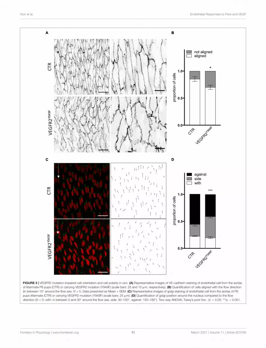
VEGFR2 and SRC Control EndothelialCell Orientation and Polarity in vivo inMatures ArteriesAs SRC family kinases inhibition impaired ECs response to flowin the absence of VEGF-A in vitro, we evaluated the relevanceof the VEGFR2—SRC pathway in vivo. First, As VEGFR2-949phosphorylation by VEGF stimulation recruits activated Srcto EC junctions to phosphorylate VE-cadherin, we analyzedthe aortas of VEGFR2Y949F mutant mice. We observed thatECs from the aorta of VEGFR2Y949F mutant mice lost theiralignment in response to flow (Figure 5A) and had a reducedcell length (Supplementary Figures 5A,B), similar to whatwe observed in vitro following treatment with the VEGFR2inhibitor ZM323881 (Supplementary Figure 4). Loss of VEGFR2phosphorylation on this specific site also impaired polarity of ECsagainst the flow; the proportion of ECs well polarized against theflow was decreased compared to control mice (Figure 5C). Tostudy whether this effect was associated specifically with SRC andnot YES or FYN in vivo, we assessed alignment and polarity ofECs in aortas of mice lacking endothelial SRC (SRCiEC−KO mice).
Cell shape and junction morphology was similarly affectedin SRCiEC−KO aortas as in VEGFR2Y949Faortas. Surprisingly,however, EC alignment with the flow was not impaired inSRCiEC−KO aortas (Figures 6A,B) nor was there any effect onEC length (Supplementary Figure 5B). Nevertheless, polarity
against the flow was reduced in SRCiEC−KO mice compared tocontrol (Figures 6C,D) similarly to VEGFR2Y949F mutant mice.Finally, to confirm the flow specificity of these observations,we used an ex vivo sprouting assay from mouse metatarsals(Song et al., 2015; Schimmel et al., 2020), which reproducedangiogenesis in a “no flow” but high growth factor condition.Polarity of ECs at the tip position was not altered in metatarsalexplants from SRCiEC−KO mice compared to control mice(Supplementary Figure 6). These results indicate that SRCactivity is not generally required for ECs to polarize, butselectively involved in flow induced cell polarity.
In conclusion, the in vitro and in vivo data agree on theimportance of VEGFR2, specifically through pY951 signaling,in endothelial cell alignment and polarity. Moreover, the Srcpathway regulates polarity but not alignment of ECs.
DISCUSSION
In the past decade, many efforts have been made to elucidate howmechanical forces and chemical signals contribute to vascularformation and patterning. Several mechano-sensory pathwayscontrolling cell shape (Levesque and Nerem, 1985; Wojciak-Stothard and Ridley, 2003; Noria et al., 2004), polarity (Francoet al., 2015; Kwon et al., 2016) and migration (Baeyens et al.,2016b; Rochon et al., 2016) during vascular patterning havebeen described and the basic cellular and molecular mechanismscontrolling angiogenesis have been well characterized (Potenteet al., 2011; Potente and Mäkinen, 2017). Nevertheless, how ECsintegrate signals coming from both mechanical and chemicalstimuli at the same time is not well understood, despite itsimportance in physiology and pathology. Here, we observedthat these signals can have synergistic or antagonistic effectsdepending on the feature observed.
We first confirm that VEGF-A treatment increases cellelongation, VEGFR2Y1175, VEGFR2Y951 and VEcadhY685
phosphorylation levels in a dose dependent manner under staticcondition, as expected from the literature (Simons et al., 2016;Cao et al., 2017). We also observe already described effectsof low SS and high SS on cell alignment (Chien, 2007; Wanget al., 2013), junctions aspect (Chiu and Chien, 2011) andchanges in VEGFR2Y1175 phosphorylation (Jin et al., 2003) inthe absence of VEGF-A. However, beyond these observations,we also identify new features associated with the combinationof flow and VEGF-A. Polarity against the flow is establishedin a dose-response to VEGF-A both under low and high SS,but only high SS could trigger polarity against the flow withoutVEGF-A. Furthermore, alignment displays a biphasic responsedepending on the VEGF-A level (aligned without VEGF orat pathologically high dose of VEGF-A, but perpendicular toflow at physiological levels of VEGF-A). This indicates thatorientation and polarity are controlled by different mechanisms.Interestingly, the effect of VEGF-A on polarity under high SS wasseen even from the lowest concentrations used (0.5 ng/mL) whilea higher concentration (10 ng/ml) was required to reach the samepercentage of polarity against the flow at low SS. This suggeststhat both parameters mutually control the cells’ sensitivity to
Frontiers in Physiology | www.frontiersin.org 10 March 2021 | Volume 11 | Article 623769
fphys-11-623769 February 24, 2021 Time: 17:6 # 11
Vion et al. Endothelial Responses to Flow and VEGF
FIGURE 5 | VEGFR2 mutation impaired cell orientation and cell polarity in vivo. (A) Representative images of VE-cadherin staining of endothelial cell from the aortasof littermate P6 pups (CTR) or carrying VEGFR2 mutation (Y949F) (scale bars: 25 and 10 µm, respectively). (B) Quantification of cells aligned with the flow direction(in between 15◦ around the flow axe, N = 5, Data presented as Mean + SEM. (C) Representative images of golgi staining of endothelial cell from the aortas of P6pups littermate (CTR) or carrying VEGFR2 mutation (Y949F) (scale bars: 25 µm). (D) Quantification of golgi position around the nucleus compared to the flowdirection (N = 5; with: in between 0 and 30◦ around the flow axe, side: 30–150◦, against: 150–180◦). Two-way ANOVA; Tukey’s post hoc, *p < 0.05; ***p < 0.001.
Frontiers in Physiology | www.frontiersin.org 11 March 2021 | Volume 11 | Article 623769

fphys-11-623769 February 24, 2021 Time: 17:6 # 12
Vion et al. Endothelial Responses to Flow and VEGF
FIGURE 6 | SRC deletion in ECs mutation impaired cell orientation and cell polarity in vivo. (A) Representative images of VE-cadherin staining of endothelial cell fromthe aortas of P6 pups littermate (CTR) or deleted for SRC in ECs (SRCiEC-KO) (scale bars: 25 and 10 µm, respectively). (B) Quantification of cells aligned with theflow direction (in between 15◦ around the flow axe, WT, N = 6; KO, N = 7, Data presented as Mean + SEM. (C) Representative images of golgi staining of endothelialcell from the aortas of P6 pups littermate (CTR) or deleted for SRC in ECs (SRCiEC-KO) (scale bars: 25 µm). (D) Quantification of golgi position around the nucleuscompared to the flow direction (WT, N = 6; KO, N = 7; with: in between 0 and 30◦ around the flow axe, side: 30–150◦, against: 150–180◦). Two-way ANOVA; Tukey’spost hoc, *p < 0.05; ***p < 0.001.
the other. Shear Stress modifies the sensitivity to VEGF-A, butVEGF-A in turn also affect the cell’s ability to respond to flow.
When inhibiting VEGFR2, the effects of both SS and VEGF-Aon alignment and polarity are lost suggesting that VEGFR2 couldbe the hub controlling these responses. The effect of SU1498 onpolarity was significant only in the absence of VEGF-A, whereasZM323881 significantly affected polarity only with VEGF-A.
Studies using these inhibitors highlight their selectivity forVEGFR2 but also show that SU1498 prevents ERK1/2 signalingcascade (Boguslawski et al., 2004) while ZM323881 inhibits ratherp38 and Rac1 pathways (Whittles et al., 2002; Garrett et al., 2007).Altogether this suggests that these inhibitors might differentlyrestrict pathways downstream of VEGFR2 that contribute topolarity establishment with or without VEGF-A.
Frontiers in Physiology | www.frontiersin.org 12 March 2021 | Volume 11 | Article 623769

fphys-11-623769 February 24, 2021 Time: 17:6 # 13
Vion et al. Endothelial Responses to Flow and VEGF
Inhibiting downstream effectors of VEGFR2, namely FAK andSRC family members, uncovered that the orientation and polarityof ECs are controlled differently in the presence or absence ofVEGF-A. Without VEGF-A, alignment with the flow and polarityagainst the flow are dependent on SRC family activity while inthe presence of VEGF-A, alignment and polarity against the floware dependent on FAK activity. Interestingly, p38 inhibition onlyimpaired alignment with the flow, not polarity which remainsmostly against the flow. In this context, p38 inhibition suggeststhat ECs can establish and modify their planar polarity withoutchanging their cell shape.
These conclusions are further supported by our in vivoanalysis, as VEGFR2 phosphorylation at Y951 contributes to ECsalignment with the flow and polarity against the flow in theabsence of VEGF-A. By genetically deleting SRC specifically inECs, we highlight that SRC is essential for polarity control butnot for alignment, although this was expected from our inhibitorexperiments in vitro. Interestingly, the inhibitor used in vitro hasa strong affinity toward Yes and Fyn, the two other SRC familymembers. While Yes and Fyn are structurally highly similar toSRC, evidence for their distinct roles in the endothelium arecurrently emerging (Eliceiri et al., 1999; Gordon et al., 2016;Schimmel et al., 2020). Combining our in vivo and in vitro results,we can propose that loss of SRC impairs endothelial polarityagainst the flow, which is not compensated by Fyn or Yes. Loss ofSRC had no effect on ECs alignment in vivo suggesting that thisfeature could be under the control of Yes or Fyn. Further workwill need to establish the exact contribution of Yes and Fyn to thealignment of ECs in response to flow. In the same line as for thep38 inhibition in vitro, we highlight that polarity and alignmentcan occur independently, but here showing that change in cellshape does not required planar polarity establishment.
By pointing their Golgi better against the flow at high SS,ECs display a cellular organization that is characteristic fortheir migration against the flow. In both SS conditions, VEGF-A will cause an activation that will enable ECs to adjust theirjunctions and basal adhesions more dynamically and polarizebetter against the flow even if exposed to a lower unidirectionalforce. Whether polarity always correlates with cell displacement(migration), however, remains unclear. Two hypotheses arisefrom the increased polarity against the flow; the first one is thatECs are indeed migrating better against the flow at high SS or withVEGF-A addition, the second would be that while ECs polarizeagainst the flow at high SS or with VEGF-A, they migrate verylittle due to high counteracting apical forces.
Both hypotheses raise the question of tension sensing andbalance between basal and lateral forces. Force transmissionoccurs through different structures in ECs (Campinho et al.,2020; Gordon et al., 2020); cell-cell junctions and cell adhesionsto the matrix have been both well described as mechano-sensitive elements. In our study, the structural changes ofVE-cadherin junctions do not correlate with alteration ofVEcadhY685 phosphorylation, suggesting that other junctionalplayers are involved. Interestingly ZO-1 has been shown tocontrol endothelial cell-cell tension and its loss, while looseningtension in-between cells favors focal adhesion formation(Tornavaca et al., 2015) and therefore reinforces basal adhesion.
In our setting, ZO-1 localization at junctions is specificallydecreased by VEGF-A when ECs are exposed to SS comparedto static condition and could be the missing player explainingthe visual modifications of adherens junctions. Additionally,increased Paxillin phosphorylation also correlates with the lossof junctional ZO-1. Together with our observation, this supportsour hypothesis that tension forces in between ECs could be highlydifferent in between our different in vitro conditions. VEGF-Aaddition under SS, would trigger loss of cell-cell tension (lateral)while increasing cell-matrix tension (basal). This effect of VEGF-A appears to be more pronounced at high SS compared to low SS.
Another situation where ECs adapt their shape and polarityis when they migrate to close a wound. In such a case, drivenby the first row of cells directly in contact with the free edge,ECs polarize collectively and migrate toward the wound. In thatsituation, the origin of the signal is different from a flow situation.In a wound assay, most cells are not exposed to the free edge andreceive an indirect cue for migration through force transmissionvia the lateral junctions. Recent work from Carvalho et al. (2019)shows that by decreasing VE-cadherin tension (therefore lateralforces) in between cells, ECs failed to collectively polarize towardthe wound. Under flow, every cell is independently exposed tothe same directional signal: SS at their apical side. Combiningthese facts and observations allows to hypothesize that the originof the mechanical signal and its way of transmission in-betweencells is crucial to determine if loosening of the lateral tensionwill lead to loss or reinforcement of polarity. In the case of awound closure, loosening junctions decreases collective polaritytoward the wound because ECs distant from the wound becomeblind to its location. In our settings, loosening junctions increasespolarity against the flow because ECs become more capable ofdetecting flow direction as signal from the lateral junctions doesnot interfere with the apical cue each cell perceive.
Interestingly, while testing inhibitor effects on polarity andalignment under high SS, we find that SRC family inhibition isefficient only without VEGF-A, i.e., a situation in which ZO-1is present at the junctions and phospho-Paxillin is low, lateraltension should be high and basal tension low. In contrast, FAKinhibition is efficient only with VEGF-A, a situation in which ZO-1 is delocalized from the junctions and phospho-Paxillin is high,suggesting low lateral tension and high basal tension. Therefore,it is tempting to speculate that rather than having differentpathways controlling polarity and alignment independently, thesame pathways could be in charge of polarity and alignment, buttheir relative contribution would vary depending on the presenceor absence of VEGF-A. SRC family members would participatewhen VEGF-A is absent and FAK pathways would take over onceVEGF-A is present to loosen the junction.
Finally, modification in flow sensitivity in ECs has beenascribed to the mechanosensitive complex formed by VEGFRs-PECAM-VE-cadherin (Baeyens et al., 2015), and in particular tothe ratio of VEGFR3 or VEGFR2 engaged in this complex, thusmodifying at which range of SS ECs align with the flow. Here weshow that the joint presence of flow and VEGF-A can also act asa lever to influence ECs alignment and polarity mostly throughchanges in the balance of the different VEGFR2 phospho-sitesthat become activated. Whether or not alignment and polarity go
Frontiers in Physiology | www.frontiersin.org 13 March 2021 | Volume 11 | Article 623769

fphys-11-623769 February 24, 2021 Time: 17:6 # 14
Vion et al. Endothelial Responses to Flow and VEGF
precisely through the same type of complex and if VEGFR3 couldalso play a role will need to be demonstrated.
DATA AVAILABILITY STATEMENT
The raw data supporting the conclusions of this article will bemade available by the authors, without undue reservation, to anyqualified research.
ETHICS STATEMENT
The animal study was reviewed and approved by theUppsala University board of animal experimentation(permit 5.2.18-8927-16).
AUTHOR CONTRIBUTIONS
A-CV designed the study, performed experiments, analyzedthe results, and wrote the manuscript. TP and CP performedthe experiments and analyzed the results. EB-K, IH, and EFperformed the experiments. EG performed the experiments andreviewed the manuscript. LC-W provided the mice, designedthe in vivo study, and reviewed the manuscript. HG designedthe study and wrote the manuscript. All authors contributed tothe article and approved the submitted version.
FUNDING
This work was supported by the German Centre forCardiovascular Research, the German Ministry of Educationand Research, the Fondation pour la Recherche Medical(ARF20170938625) (A-CV), the European Research Councilconsolidator grant 311719 Reshape (HG), the Knut andAlice Wallenberg foundation project grant and WallenbergScholar grant (KAW 20150030 and KAW 2015.0275 to LC-W), and the Fondation Leducq transatlantic network ofexcellence grant in neurovascular disease (17 CVD 03, HG andLC-W). EG was supported by the Wenner-Gren FoundationPostdoctoral Fellowship.
ACKNOWLEDGMENTS
We thank Dr. Anna Szymborska for helpful commentson the manuscript.
SUPPLEMENTARY MATERIAL
The Supplementary Material for this article can be foundonline at: https://www.frontiersin.org/articles/10.3389/fphys.2020.623769/full#supplementary-material
Supplementary Figure 1 | VEGF-A treatment does not affect cell number butincreases cell elongation. (A) Quantification of endothelial cells number under flowand VEGF-A. (B) Quantification of aspect ratio (length of main axis/length of shortaxis) of endothelial cells under flow and VEGF-A. N = 3, between 1,500 and 3,000cells analyzed. N = 3, around 1,500 cells analyzed. Data presented asMean + SEM. Two-way ANOVA; Tukey’s post hoc, ∗∗∗p < 0.001 compared tostatic 0 VEGF; ###p < 0.001 (compared to LSS 0 VEGF); ! !!p < 0.001(compared to HSS 0 VEGF).
Supplementary Figure 2 | VEGF treatment does not change junctions activationunder static condition. (A) Representative picture (Immunofluorescence) ofendothelial cells exposed to VEGF-A for 24 h. Red arrows indicate gaps in theECs monolayer (B) Quantification of junction status based on their morphology(N = 3; 100 patches analyzed blinded by images, 5–8 images per N).
Supplementary Figure 3 | Proteins expression upon flow and VEGF treatment.(A) VEGFR2 expression assessed by WB, N = 6. (B) ZO1 expression assessed byWB, N = 3. (C) VE-cadherin expression assessed by WB, N = 5. (D) FAKexpression assessed by WB, N = 5. ANOVA followed by Tukey post Hoc;∗p < 0.05; ∗∗p < 0.01. (E) Representative pictures of the quantified WB.
Supplementary Figure 4 | Inhibitors effect on cell elongation. Quantification ofaspect ratio (length of main axis/length of short axis) of endothelial cells under highSS with or without VEGF-A (10 ng/mL) and with or without inhibitors (DMSO,N = 5, inhibitors N = 3) One-way ANOVA; Tukey’s post hoc, ∗∗∗p < 0.001compared to DMSO 0 VEGF; ###p < 0.001 compared to DMSO 10 VEGF.
Supplementary Figure 5 | VEGFR2 mutation impairs cell length but not SRCdeletion in vivo. (A) Quantification of ECs length in the aortas of P6 pups littermate(CTR) or carrying VEGFR2 mutation (Y949F). N = 5 (B) Distribution of cell length.N = 5 (C) Quantification of ECs length in the aortas of P6 pups littermate (CTR) ordeleted for SRC in ECs (SRCiEC−KO). N = 3–5 (D) Distribution of cell length.Unpaired T-Test; ∗∗∗p < 0.001.
Supplementary Figure 6 | ECs polarity is not impaired during directionalsprouting upon loss of c-Src. Representative images and quantification of polarityof ECs sprouting out of metatarsal ex vivo. With means that ECs polarize in thedirection of the sprout (0–30◦); side, ECs present their golgi on the side of the cell(30–150◦); against, ECs polarize in the opposite direction of the sprout (150–180◦).N = 294 cells analyzed from 12 metatarsals from 2 independent experiments.
Supplementary Table 1 | p-Values for each comparison for Figures 2B,D.Two-way ANOVA; Tukey’s post hoc, ∗p < 0.05; ∗∗p < 0.01; ∗∗∗p < 0.001. Graylines: statistics presented on the graphs. Bold: significant difference.
Supplementary Table 2 | p-Values for each comparison for Figures 3C–F.Two-way ANOVA; Fisher LSD. ∗p < 0.05; ∗∗p < 0.01; ∗∗∗p < 0.001. Gray lines:statistics presented on the graphs. Bold: significant difference.
REFERENCESBaeyens, N., Bandyopadhyay, C., Coon, B. G., Yun, S., and
Schwartz, M. A. (2016a). Endothelial fluid shear stress sensingin vascular health and disease. J. Clin. Invest. 126, 821–828.doi: 10.1172/jci83083
Baeyens, N., Larrivée, B., Ola, R., Hayward-Piatkowskyi, B., Dubrac, A., Huang,B., et al. (2016b). Defective fluid shear stress mechanotransduction mediates
hereditary hemorrhagic telangiectasia. J. Cell Biol. 214, 807–816. doi: 10.1083/jcb.201603106
Baeyens, N., Nicoli, S., Coon, B. G., Ross, T. D., Van den Dries, K., Han, J., et al.(2015). Vascular remodeling is governed by a VEGFR3-dependent fluid shearstress set point. eLife 4:e04645.
Bentley, K., Franco, C. A., Philippides, A., Blanco, R., Dierkes, M., Gebala, V., et al.(2014). The role of differential VE-cadherin dynamics in cell rearrangementduring angiogenesis. Nat. Cell Biol. 16, 309–321. doi: 10.1038/ncb2926
Frontiers in Physiology | www.frontiersin.org 14 March 2021 | Volume 11 | Article 623769

fphys-11-623769 February 24, 2021 Time: 17:6 # 15
Vion et al. Endothelial Responses to Flow and VEGF
Boguslawski, G., McGlynn, P. W., Harvey, K. A., and Kovala, A. T.(2004). SU1498, an inhibitor of vascular endothelial growth factor receptor2, causes accumulation of phosphorylated ERK kinases and inhibitstheir activity in vivo and in vitro. J. Biol. Chem. 279, 5716–5724.doi: 10.1074/jbc.m308625200
Campinho, P., Vilfan, A., and Vermot, J. (2020). Blood flow forces in shaping thevascular system: a focus on endothelial cell behavior. Front. Physiol. 11:552.doi: 10.3389/fphys.2020.00552
Cao, J., Ehling, M., März, S., Seebach, J., Tarbashevich, K., Sixta, T., et al. (2017).Polarized actin and VE-cadherin dynamics regulate junctional remodelling andcell migration during sprouting angiogenesis. Nat. Commun. 8:2210.
Carmeliet, P., and Tessier-Lavigne, M. (2005). Common mechanisms of nerve andblood vessel wiring. Nature 436, 193–200. doi: 10.1038/nature03875
Carvalho, J. R., Chia, M., Dufton, N., Almagro, L. O., Conway, D. E., Randi,A. M., et al. (2019). Non-canonical Wnt signaling regulates junctionalmechanocoupling during angiogenic collective cell migration. eLife 8:e45853.
Chen, K. D., Li, Y. S., Kim, M., Li, S., Yuan, S., Chien, S., et al. (1999).Mechanotransduction in response to shear stress. Roles of receptor tyrosinekinases, integrins, and Shc. J. Biol. Chem. 274, 18393–18400. doi: 10.1074/jbc.274.26.18393
Chien, S. (2007). Mechanotransduction and endothelial cell homeostasis: thewisdom of the cell. Am. J. Physiol. Heart Circ. Physiol. 292, H1209–H1224.
Chiu, J.-J., and Chien, S. (2011). Effects of disturbed flow on vascular endothelium:pathophysiological basis and clinical perspectives. Physiol. Rev. 91, 327–387.doi: 10.1152/physrev.00047.2009
Conway, D. E., Breckenridge, M. T., Hinde, E., Gratton, E., Chen, C. S., Schwartz,M. A., et al. (2013). Fluid shear stress on endothelial cells modulates mechanicaltension across VE-cadherin and PECAM-1. Curr. Biol. 23, 1024–1030. doi:10.1016/j.cub.2013.04.049
Coon, B. G., Baeyens, N., Han, J., Budatha, M., Ross, T. D., Fang, J. S., et al.(2015). Intramembrane binding of VE-cadherin to VEGFR2 and VEGFR3assembles the endothelial mechanosensory complex. J. Cell Biol. 208, 975–986.doi: 10.1083/jcb.201408103
Culver, J. C., and Dickinson, M. E. (2010). The effects of hemodynamic force onembryonic development. Microcirculation 17, 164–178. doi: 10.1111/j.1549-8719.2010.00025.x
Eliceiri, B. P., Paul, R., Schwartzberg, P. L., Hood, J. D., Leng, J., Cheresh, D. A.,et al. (1999). Selective requirement for Src kinases during VEGF-inducedangiogenesis and vascular permeability. Mol. Cell 4, 915–924. doi: 10.1016/s1097-2765(00)80221-x
Franco, C. A., Jones, M. L., Bernabeu, M. O., Geudens, I., Mathivet, T., Rosa, A.,et al. (2015). Dynamic endothelial cell rearrangements drive developmentalvessel regression. PLoS Biol. 13:e1002125. doi: 10.1371/journal.pbio.1002125
Garrett, T. A., Van Buul, J. D., and Burridge, K. (2007). VEGF-induced Rac1activation in endothelial cells is regulated by the guanine nucleotide exchangefactor Vav2. Exp. Cell Res. 313, 3285–3297. doi: 10.1016/j.yexcr.2007.05.027
Gimbrone, M. A., and García-Cardeña, G. (2013). Vascular endothelium,hemodynamics, and the pathobiology of atherosclerosis. Cardiovasc. Pathol. 22,9–15. doi: 10.1016/j.carpath.2012.06.006
Givens, C., and Tzima, E. (2016). Endothelial mechanosignaling: does one sensorfit all? Antioxid. Redox Signal. 25, 373–388. doi: 10.1089/ars.2015.6493
Gordon, E., Schimmel, L., and Frye, M. (2020). The importance of mechanicalforces for in vitro endothelial cell biology. Front. Physiol. 11:684. doi: 10.3389/fphys.2020.00684
Gordon, E. J., Fukuhara, D., Weström, S., Padhan, N., Sjöström, E. O., vanMeeteren, L., et al. (2016). The endothelial adaptor molecule TSAd is requiredfor VEGF-induced angiogenic sprouting through junctional c-Src activation.Sci. Signal. 9:ra72. doi: 10.1126/scisignal.aad9256
Isogai, S., Lawson, N. D., Torrealday, S., Horiguchi, M., and Weinstein, B. M.(2003). Angiogenic network formation in the developing vertebrate trunk.Development 130, 5281–5290. doi: 10.1242/dev.00733
Jin, Z.-G., Ueba, H., Tanimoto, T., Lungu, A. O., Frame, M. D., Berk, B. C., et al.(2003). Ligand-independent activation of vascular endothelial growth factorreceptor 2 by fluid shear stress regulates activation of endothelial nitric oxidesynthase. Circ. Res. 93, 354–363. doi: 10.1161/01.res.0000089257.94002.96
Koch, S., and Claesson-Welsh, L. (2012). Signal transduction by vascularendothelial growth factor receptors. Cold Spring Harb. Perspect. Med.2:a006502. doi: 10.1101/cshperspect.a006502
Koch, S., Tugues, S., Li, X., Gualandi, L., and Claesson-Welsh, L. (2011). Signaltransduction by vascular endothelial growth factor receptors. Biochem. J. 437,169–183. doi: 10.1042/bj20110301
Kogata, N., Arai, Y., Pearson, J. T., Hashimoto, K., Hidaka, K., Koyama,T., et al. (2006). Cardiac ischemia activates vascular endothelial cadherinpromoter in both preexisting vascular cells and bone marrow cells involvedin neovascularization. Circ. Res. 98, 897–904. doi: 10.1161/01.res.0000218193.51136.ad
Kupfer, A., Louvard, D., and Singer, S. J. (1982). Polarization of the Golgi apparatusand the microtubule-organizing center in cultured fibroblasts at the edge ofan experimental wound. Proc. Natl. Acad. Sci. U.S.A. 79, 2603–2607. doi:10.1073/pnas.79.8.2603
Kwon, H.-B., Wang, S., Helker, C. S., Rasouli, S. J., Maischein, H. M., Offermanns,S., et al. (2016). In vivo modulation of endothelial polarization by Apelinreceptor signalling. Nat. Commun. 7:11805.
Levesque, M. J., and Nerem, R. M. (1985). The elongation and orientation ofcultured endothelial cells in response to shear stress. J. Biomech. Eng. 107,341–347. doi: 10.1115/1.3138567
Li, X., Padhan, N., Sjöström, E. O., Roche, F. P., Testini, C., Honkura, N., et al.(2016). VEGFR2 pY949 signalling regulates adherens junction integrity andmetastatic spread. Nat. Commun. 7:11017.
López-Colomé, A. M., Lee-Rivera, I., Benavides-Hidalgo, R., and López, E. (2017).Paxillin: a crossroad in pathological cell migration. J. Hematol. Oncol. 10:50.
Matsumoto, T., Bohman, S., Dixelius, J., Berge, T., Dimberg, A., Magnusson,P., et al. (2005). VEGF receptor-2 Y951 signaling and a role for the adaptermolecule TSAd in tumor angiogenesis. EMBO J. 24, 2342–2353. doi: 10.1038/sj.emboj.7600709
Neto, F., Klaus-Bergmann, A., Ong, Y. T., Alt, S., Vion, A. C., Szymborska, A., et al.(2018). YAP and TAZ regulate adherens junction dynamics and endothelial celldistribution during vascular development. eLife 7:e31037.
Noria, S., Xu, F., McCue, S., Jones, M., Gotlieb, A. I., Langille, B. L., et al. (2004).Assembly and reorientation of stress fibers drives morphological changes toendothelial cells exposed to shear stress. Am. J. Pathol. 164, 1211–1223. doi:10.1016/s0002-9440(10)63209-9
Orsenigo, F., Giampietro, C., Ferrari, A., Corada, M., Galaup, A., Sigismund, S.,et al. (2012). Phosphorylation of VE-cadherin is modulated by haemodynamicforces and contributes to the regulation of vascular permeability in vivo. Nat.Commun. 3:1208.
Pietilä, I., Van Mourik, D., Tamelander, A., Kriz, V., Claesson-Welsh, L., Tengholm,A., et al. (2019). Temporal dynamics of VEGFA-induced VEGFR2/FAK co-localization depend on SHB. Cells 8:1645. doi: 10.3390/cells8121645
Potente, M., Gerhardt, H., and Carmeliet, P. (2011). Basic and therapeutic aspectsof angiogenesis. Cell 146, 873–887. doi: 10.1016/j.cell.2011.08.039
Potente, M., and Mäkinen, T. (2017). Vascular heterogeneity and specialization indevelopment and disease. Nat. Rev. Mol. Cell Biol. 18, 477–494. doi: 10.1038/nrm.2017.36
Rochon, E. R., Menon, P. G., and Roman, B. L. (2016). Alk1 controls arterialendothelial cell migration in lumenized vessels. Development 143, 2593–2602.doi: 10.1242/dev.135392
Sakurai, Y., Ohgimoto, K., Kataoka, Y., Yoshida, N., and Shibuya, M. (2005).Essential role of Flk-1 (VEGF receptor 2) tyrosine residue 1173 invasculogenesis in mice. Proc. Natl. Acad. Sci. U.S.A. 102, 1076–1081. doi:10.1073/pnas.0404984102
Schimmel, L., Fukuhara, D., Richards, M., Jin, Y., Essebier, P., Frampton, E., et al.(2020). c-Src controls stability of sprouting blood vessels in the developingretina independently of cell-cell adhesion through focal adhesion assembly.Development 147:dev185405. doi: 10.1242/dev.185405
Simons, M., Gordon, E., and Claesson-Welsh, L. (2016). Mechanisms andregulation of endothelial VEGF receptor signalling. Nat. Rev. Mol. Cell Biol. 17,611–625. doi: 10.1038/nrm.2016.87
Song, W., Fhu, C. W., Ang, K. H., Liu, C. H., Johari, N. A., Lio, D., et al. (2015). Thefetal mouse metatarsal bone explant as a model of angiogenesis. Nat. Protoc. 10,1459–1473. doi: 10.1038/nprot.2015.097
Sun, Z., Li, X., Massena, S., Kutschera, S., Padhan, N., Gualandi, L., et al. (2012).VEGFR2 induces c-Src signaling and vascular permeability in vivo via theadaptor protein TSAd. J. Exp. Med. 209, 1363–1377. doi: 10.1084/jem.20111343
Takahashi, T., Yamaguchi, S., Chida, K., and Shibuya, M. A. (2001). singleautophosphorylation site on KDR/Flk-1 is essential for VEGF-A-dependent
Frontiers in Physiology | www.frontiersin.org 15 March 2021 | Volume 11 | Article 623769

fphys-11-623769 February 24, 2021 Time: 17:6 # 16
Vion et al. Endothelial Responses to Flow and VEGF
activation of PLC-gamma and DNA synthesis in vascular endothelial cells.EMBO J. 20, 2768–2778. doi: 10.1093/emboj/20.11.2768
Testini, C., Smith, R. O., Jin, Y., Martinsson, P., Sun, Y., Hedlund, M., et al. (2019).Myc-dependent endothelial proliferation is controlled by phosphotyrosine 1212in VEGF receptor-2. EMBO Rep. 20:e47845.
Tornavaca, O., Chia, M., Dufton, N., Almagro, L. O., Conway, D. E., Randi, A. M.,et al. (2015). ZO-1 controls endothelial adherens junctions, cell-cell tension,angiogenesis, and barrier formation. J. Cell Biol. 208, 821–838. doi: 10.1083/jcb.201404140
Tzima, E., Irani-Tehrani, M., Kiosses, W. B., Dejana, E., Schultz, D. A.,Engelhardt, B., et al. (2005). A mechanosensory complex that mediatesthe endothelial cell response to fluid shear stress. Nature 437, 426–431.doi: 10.1038/nature03952
Wang, C., Baker, B. M., Chen, C. S., and Schwartz, M. A. (2013). Endothelialcell sensing of flow direction. Arterioscler. Thromb. Vasc. Biol. 33, 2130–2136.doi: 10.1161/atvbaha.113.301826
Wang, Y., Nakayama, M., Pitulescu, M. E., Schmidt, T. S., Bochenek, M. L.,Sakakibara, A., et al. (2010). Ephrin-B2 controls VEGF-induced angiogenesisand lymphangiogenesis. Nature 465, 483–486. doi: 10.1038/nature09002
Whittles, C. E., Pocock, T. M., Wedge, S. R., Kendrew, J., Hennequin, L. F.,Harper, S. J., et al. (2002). ZM323881, a novel inhibitor of vascular endothelialgrowth factor-receptor-2 tyrosine kinase activity. Microcirculation 9, 513–522.doi: 10.1038/sj.mn.7800164
Wojciak-Stothard, B., and Ridley, A. J. (2003). Shear stress-induced endothelial cellpolarization is mediated by Rho and Rac but not Cdc42 or PI 3-kinases. J. CellBiol. 161, 429–439. doi: 10.1083/jcb.200210135
Conflict of Interest: The authors declare that the research was conducted in theabsence of any commercial or financial relationships that could be construed as apotential conflict of interest.
Copyright © 2021 Vion, Perovic, Petit, Hollfinger, Bartels-Klein, Frampton, Gordon,Claesson-Welsh and Gerhardt. This is an open-access article distributed under theterms of the Creative Commons Attribution License (CC BY). The use, distributionor reproduction in other forums is permitted, provided the original author(s) andthe copyright owner(s) are credited and that the original publication in this journalis cited, in accordance with accepted academic practice. No use, distribution orreproduction is permitted which does not comply with these terms.
Frontiers in Physiology | www.frontiersin.org 16 March 2021 | Volume 11 | Article 623769


