Embed Size (px)
Citation preview
517
J. Exp. Med.
The Rockefeller University Press • 0022-1007/97/08/517/11 $2.00Volume 186, Number 4, August 18, 1997 517–527http://www.jem.org
Endothelial-dependent Mechanisms Regulate LeukocyteTransmigration: A Process Involving the Proteasome andDisruption of the Vascular Endothelial–CadherinComplex at Endothelial Cell-to-Cell Junctions
By Jennifer R. Allport,
*
Han Ding,
*
Tucker Collins,
*
Mary E. Gerritsen
‡
and Francis W. Luscinskas
*
From
*
The Vascular Research Division, Departments of Pathology, Brigham and Women’s Hospital and Harvard Medical School, Boston, Massachusetts 02115; and
‡
Bayer Corporation, West Haven, Connecticut 06516
Summary
Although several adhesion molecules expressed on leukocytes (
b
1 and
b
2 integrins, platelet en-dothelial cell adhesion molecule 1 [PECAM-1], and CD47) and on endothelium (intercellularadhesion molecule 1, PECAM-1) have been implicated in leukocyte transendothelial migra-tion, less is known about the role of endothelial lateral junctions during this process. We haveshown previously (Read, M.A., A.S. Neish, F.W. Luscinskas, V.J. Palambella, T. Maniatis, andT. Collins. 1995.
Immunity.
2:493–506) that inhibitors of the proteasome reduce lymphocyte andneutrophil adhesion and transmigration across TNF-
a
–activated human umbilical vein endothelialcell (EC) monolayers in an in vitro flow model. The current study examined EC lateral junctionproteins, principally the vascular endothelial (VE)–cadherin complex and the effects of protea-some inhibitors (MG132 and lactacystin) on lateral junctions during leukocyte adhesion, to gain abetter understanding of the role of EC junctions in leukocyte transmigration. Both biochemicaland indirect immunofluorescence analyses of the adherens junction zone of EC monolayers re-vealed that neutrophil adhesion, not transmigration, induced disruption of the VE–cadherincomplex and loss of its lateral junction localization. In contrast, PECAM-1, which is located atlateral junctions and is implicated in neutrophil transmigration, was not altered. These findingsidentify new and interrelated endothelial-dependent mechanisms for leukocyte transmigrationthat involve alterations in lateral junction structure and a proteasome-dependent event(s).
L
ocalized leukocyte accumulation is the cellular hall-mark of inflammation. Although this has been recog-
nized for more than a century, it is only in the past decadethat the role of the endothelium has been appreciated. Thenotion that the vascular endothelium actively participatesin leukocyte recruitment initially gained support from invitro studies demonstrating that treatment of cultured en-dothelium with inflammatory cytokines TNF-
a
or IL-1,and certain Gram-negative bacterial endotoxin could “acti-vate” the endothelium to become adhesive for blood leu-kocytes and cell lines (1–3). Subsequently, the work ofmany investigators has identified and molecularly clonedseveral such endothelial cell (EC)
1
adhesion molecules and
their cognate ligands on leukocytes, which support leuko-cyte adhesion to endothelium.
Recent reports have shown that the proteasome pathwayis involved in activation of NF-
k
B, which is a transcriptionfactor necessary for activation of EC gene transcriptionof E-selectin (CD62E), intercellular adhesion molecule 1(ICAM-1) (CD54), and vascular cell adhesion molecule 1(VCAM-1) (CD106) (4). Small peptide aldehyde inhibitors(MG132, MG115) of the proteasome can dramatically re-duce TNF-
a
–induced expression of E-selectin, VCAM-1,and ICAM-1 in human umbilical vein ECs (4). Function-ally, neutrophil adhesion was reduced by 50%, and transmi-gration was reduced by
.
60%. Live-time video micros-copy showed that many adherent neutrophils had flattenedand extended pseudopods into the EC junctions, but were
1
Abbreviations used in this paper:
ALLM,
N
-acetyl-leucinyl-leucinyl-methional; DPBS, Dulbecco’s phosphate-buffered saline; EC, endothelialcell; HUVEC, human umbilical vein endothelial cell; HSA, human serumalbumin; ICAM, intercellular adhesion molecule; PECAM-1, platelet en-dothelial cell adhesion molecule 1; TBS, Tris-buffered saline; VCAM 1,vascular cell adhesion molecule 1.
This manuscript was presented in abstract form at the IX InternationalVascular Biology Meeting held in Seattle, WA on September 4–9, 1996,and at Experimental Biology ’97 held in New Orleans, LA on April 6–9,1997.
on Septem
ber 29, 2014jem
.rupress.orgD
ownloaded from
Published August 18, 1997
518
Endothelial Regulation of Neutrophil Transmigration
unable to transmigrate. This result raises the possibility thatthe proteasome regulates an essential endothelial-depen-dent component(s) during transendothelial passage.
In light of our previous observations that proteasome in-hibitors prevent firmly adherent neutrophils from penetrat-ing between endothelial cell–cell lateral junctions, we rea-soned that the function of endothelial cell-to-cell lateraljunctions may be critical during the process of leukocytetransmigration. The molecular structure and organization ofendothelial cell–cell lateral junctions has been reviewed (5–7).We focused our attention on the adherens type junctionswhich appear to serve as a focal point for the connectionsbetween the EC plasma membrane and its underlyingactin-cytoskeleton complex. The adherens type junctionscontain cadherins (for review see reference 7), a family ofsingle-span transmembrane glycoproteins which directly as-sociate with structural components of the cytoskeleton andmediate Ca
2
1
-dependent cell–cell adhesion in a homotypicfashion. Cadherin-5, also termed vascular endothelial (VE)–cadherin, is specific to vascular endothelium and localizesexclusively to lateral junctions of intact, confluent endothe-lium (8, 9). A recent study (8) has revealed that VE–cadherinassociates with the cytosolic proteins
a
- and
b
-catenins toform a complex and organize at nascent endothelial cell-to-cell contacts. Plakoglobin (also named
g
-catenin) associatedwith VE–cadherin and
a
- and
b
-catenins at cell-to-cell con-tacts, through an undefined mechanism(s), only as EC ap-proached confluence. p120 (p120
cas
), initially identified asone of several substrates of the tyrosine kinase pp60
src
(10,11), and a closely associated molecule termed p100 alsohave been reported to associate with
a
- and
b
-catenins andVE-cadherin in umbilical vein endothelium (12). That theVE–cadherin complex is dynamic and involved in regulat-ing cell-to-cell contact is suggested by wounding (8) orCa
2
1
depletion (5) experiments where VE-cadherin andplakoglobin rapidly and reversibly retract from the endo-thelial lateral junctions.
To better understand the molecular basis of inhibition ofneutrophil transmigration by proteasome inhibitors and thepotential role of the components of the EC adherens junc-tions, we have evaluated the effects of two structurally differ-ent proteasome inhibitors, MG132 and lactacystin (13), on theassociation of VE–cadherin with
a
-,
b
-, and
g
-catenin (plako-globin), and p120/p100 in 6-h TNF-
a
–activated EC usingboth indirect immunofluorescence microscopy and immuno-precipitation followed by immunoblotting. Second, we havecoincubated human neutrophils with control or protea-some inhibitor–treated TNF-
a
–activated endothelium, andevaluated the staining patterns and biochemical associationof members of the VE-cadherin complex, and as a control,platelet endothelial cell adhesion molecule 1 (PECAM-1;CD31), which has been shown previously to colocalize toEC lateral junctions (14) and has been implicated in leuko-cyte transmigration in in vivo and in vitro studies (15). Theresults of these experiments suggest that novel endothelial-dependent mechanisms regulate neutrophil transmigrationwhich involves structural alterations of the VE–cadherincomplex, as well as a proteasomal-dependent step(s).
Materials and Methods
Materials
Dulbecco’s phosphate-buffered saline (DPBS) with Ca
2
1
andMg
2
1
, DPBS, M199, DMEM, RPMI-1640 with 25 mM Hepes,and 1 M solution of Hepes were purchased from BioWhittakerBioproducts (Walkersville, MD). Human
r
TNF-
a
(produced in
Escherichia coli
) was the gift of Dr. Baker (Genentech Inc., SouthSan Francisco, CA), and a concentration of 25 ng/ml gave maxi-mal response and contained
,
10 pg/ml of endotoxin as reportedpreviously (16). The proteasome inhibitors, MG132 (carboben-zoxyl-leucinyl-leucinyl-leucinal-H) and lactacystin were the giftof Dr. J. Adams (ProScript, Inc., Cambridge, MA). Each inhibi-tor was dissolved in DMSO at 40 mM and stored at
2
80
8
C. Foruse in experiments, aliquots of inhibitors were thawed at 37
8
Cand diluted directly into appropriate culture media, or as other-wise noted in the text. Calpain inhibitor II (also abbreviated asALLM [
N
-acetyl-leucinyl-leucinyl-methional]) and aprotinin werepurchased from Calbiochem Corp. (La Jolla, CA). EDTA, Protein–GSepharose, PMSF, leupeptin, DMSO, BSA, and Hepes were pur-chased from Sigma Chemical Co. (St. Louis, MO). Human se-rum albumin (HSA) was obtained from Baxter Healthcare Corp.(Glendale, CA).
mAbs
The following murine mAbs have been reported previously:anti–E-selectin (H4/18 or 7A9, each an IgG
1
; reference 16), anti–ICAM-1 (Hu5/3, IgG
1
; reference 17), anti–VCAM-1 mAb E1/6(IgG
1
; reference 18), anti–PECAM-1 (obtained from Iowa Hy-bridoma Bank, Iowa City, IA, or from Immunotech, Inc., West-brook, ME), and anti–HLA-A,B (W6/32, IgG
2a
) mAb (19).These mAb were used as hybridoma culture supernatant fluid forsurface immunofluorescence assays and as pure IgG for immuno-fluorescence microscopy, immunoprecipitations, and Western blot-ting. Murine mAb directed to VE–cadherin (clone TEA1/31,IgG
1
) was purchased from Immunotech Inc. or from BIODE-SIGN Intl. (Kennebunk, ME). Murine mAb directed to plako-globin (clone PG5.1, IgG
2b
) was purchased from BIODESIGNIntl. Murine mAb to
a
- and
b
-catenins and p120 (clone 5, 14,and 98, respectively; all IgG
1
) were purchased from TransductionLabs. (Lexington, KY). A second mAb to
a
-catenin (Zymed, S.San Francisco, CA) was used for immunofluorescence studies.
Cell Culture
ECs were isolated from two to five umbilical cord veins,pooled, and established as detailed (19). Primary human umbilicalvein endothelial cell (HUVEC) cultures were serially passaged (1:3 split ratio) and maintained in M199 containing 10% FCS, ECgrowth factor, porcine intestinal heparin, and antibiotics.
Experimental Protocols.
For immunofluorescence staining as-says, ECs (passage 1–2) were plated at confluent density on 4-wellchamber glass slides (Lab-Tek; Nunc, Inc., Naperville, IL) until 2 dafter confluent. For flow assays, ECs were plated at confluence onhuman fibronectin (2
m
g/cm
2
)-coated 25-mm glass coverslips(20), and used 3 d later. For quantitative surface immunofluores-cence assays, ECs were plated on fibronectin-coated microtiterplates (C96; Costar, Cambridge, MA), and used 2–3 d after con-fluence. For immunoprecipitation assays, EC were plated on 0.1%gelatin-coated 100-mm diameter plastic petri dishes (Costar), andused 3 d after attaining confluence. EC monolayers were not ma-nipulated or fed for 48 h before use.
on Septem
ber 29, 2014jem
.rupress.orgD
ownloaded from
Published August 18, 1997
519
Allport et al.
Leukocyte Isolation
Human neutrophils were purified from whole blood of volun-teer donors as previously detailed (19). Isolated neutrophils (94%pure) were resuspended in cold DPBS containing 0.75 mM Ca
2
1
,0.75 mM Mg
2
1
, and 0.2% HSA (assay buffer).
Endothelial–Leukocyte Interactions in a Parallel-plateFlow Chamber
The parallel-plate flow chamber used in this study has been de-scribed in detail (20–22). A wall shear stress of 1.8 dynes/cm
2
wasachieved with a flow rate of 0.85 ml/min (20). EC monolayerswere assayed for leukocyte adhesion and transmigration underflow conditions as before (16, 20).
Immunofluorescence Assays
Quantitative surface immunofluorescence assays for adhesionmolecule expression on EC were performed in triplicate wells asreported previously (17) using appropriate primary mAb–detectedFITC-conjugated goat anti–mouse (F(ab
9
)
2
, 1:50 dilution; CaltagLabs., S. San Francisco, CA).
Indirect Immunofluorescence Staining.
Indirect immunofluores-cence staining of endothelial surface molecules was performed us-ing the protocol of Gerritsen et al. (23). In brief, confluent ECin 4-well chambers were activated as detailed above and thenwashed with assay buffer. Neutrophils (0.3
3
10
6
) or assay bufferwere added to wells and incubated at 37
8
C for 10 min. Wellswere washed twice with DPBS, and then fixed with methanol(
2
20
8
C) on ice for 5 min, followed by three washes. Immunoflu-orescence staining was performed as detailed (23). Fields of FITCfluorescence–stained EC were visualized on a fluorescence mi-croscope (Microfot FXA; Nikon, Inc., Melville, NY) equippedwith a
3
20 objective and the images were captured using acooled charged-coupled device video camera. Exposure timeswere matched in each instance (typically 1–3 s, final magnifica-tion of all images was 320, except where noted).
Immunoprecipitation of Endothelial Proteinsand Immunoblotting
Immunoprecipitation of Junctional Proteins.
VE–cadherin com-plex proteins were immunoprecipitated using a modification of
the method of Lampugnani et al. (8). In brief, EC were incubatedwith inhibitors, ALLM, or carrier (0.02% DMSO) and/or TNF-
a
,and then washed three times with assay buffer. EC were incu-bated at 37
8
C for 10 min under static conditions with either 10
7
human neutrophils (5 ml, ratio of neutrophils to EC was 2.5:1) orassay buffer alone. Nonadherent neutrophils were removed bywashing (2 times) the monolayers with DPBS alone. Plates wereplaced on ice and the Triton X-100 soluble fraction extracted for30 min in lysis buffer (10 mM Tris, 150 mM NaCl, 1 mM PMSF,40 U/ml aprotinin, 15
m
g/ml leupeptin, 0.36 mM 1,10-phenan-throline, 2 mM CaCl
2
(Tris-buffered saline [TBS] and proteaseinhibitors), 1% NP-40, and 1% Triton X-100), mixing every fewminutes. For total lysates, monolayers were lysed in lysis buffercontaining 0.5% SDS. The supernatant was collected, microcen-trifuged at 14,000
g
for 5 min, and stored at
2
80
8
C. The lysedmonolayers were washed 3 times with TBS and protease inhibitors,and the Triton X-100 insoluble fraction was extracted as detailed(8) and stored at
2
80
8
C.For immunoprecipitation, protein G–Sepharose was washed with
TBS, and a 60-
m
l suspension (1:1 suspension with TBS) was cou-pled to mAb by mixing for 1 h at ambient temperature with 8–10
m
gof one of several mAb in a final volume of 500
m
l. The aboveconcentrations of mAbs were optimal for detection in our system.The IgG protein G–Sepharose was microcentrifuged and washed(3 times) with TBS containing 1 mM PMSF, 40 U/ml aprotinin,1 mM CaCl
2
, and 2% (vol/vol) Triton X-100 (buffer 1). The celllysates (representing 0.5–1
3
10
6
EC) were precleared by incu-bating with protein G–Sepharose, pelleted, and the supernatantswere incubated with the IgG protein G–Sepharose for 1 h at RT.The immunoprecipitates were then washed (3 times) with TBScontaining 1 mM PMSF, 40 U/ml aprotinin, 0.05% (vol/vol)NP-40, and 2 mM CaCl
2
, and boiled for 5 min in 2
3
sampleloading buffer. The entire supernatant of samples was resolved bySDS-PAGE (24) on a 7.5% (wt/vol) polyacrylamide gel andtransferred to nitrocellulose membrane (Hybond; Amersham Corp.,Arlington Heights, IL).
Immunoblot Analysis.
Immunoblots were performed using amodification of the method of Donnelly et al. (25). The membranewas blocked for 1 h with 5% dried milk protein in PBS (blockingbuffer) and then incubated for 2 h with primary mAb (either 5
m
g/ml anti–VE–cadherin, 1
m
g/ml anti–
a
-catenin, anti–
b
-cate-
Table 1.
Effects of Proteasome Inhibitor Lactacystin on EC Expression of Adhesion Molecules.
Treatment E-selectin ICAM-1 VCAM-1 p96
None 13
6
22 29
6
3 2
6
3 500
6
101 h carrier; TNF-
a
4 h 272
6
29‡ 239 6 3‡ 253 6 8‡ 488 6 281 h Lac; TNF-a 4 h 72 6 7§ 72 6 3§ 72 6 8§ 432 6 421 h ALLM; TNF-a 4 h 232 6 45‡ 220 6 3 247 6 18 428 6 18TNF-a 4 h; carrier 2 h 235 6 73 334 6 20 352 6 9 444 6 42TNF-a 4 h; Lac 2 h 237 6 19 350 6 4 334 6 22 498 6 45TNF-a 4 h; ALLM 2 h 245 6 20 351 6 17 343 6 8 465 6 45
*Surface expression of adhesion molecules was determined using a two-step indirect surface immunofluorescence assay (19) and the mAb listed inMaterials and Methods. Aliquots of lactacystin (Lac, 10 mM) and calpain inhibitor II (ALLM, 40 mM) were thawed and immediately diluted intoculture media to final concentration of 20 mM. Carrier control was 0.02% DMSO. The results are the average of triplicate wells and are representa-tive of two separate experiments. Background nonbinding control mAb K16/16 was 6 6 4.‡Value is significantly higher (P ,0.05) than media control of unactivated EC monolayers§Value is significantly lower than carrier TNF-a–activated EC monolayers (P ,0.01).
on Septem
ber 29, 2014jem
.rupress.orgD
ownloaded from
Published August 18, 1997
520 Endothelial Regulation of Neutrophil Transmigration
nin or anti-p120, or 5 mg/ml anti-plakoglobin or anti–PECAM-1diluted in block). The membrane was washed six times at 5 minintervals with PBS containing 0.05% (vol/vol) Tween 20 and0.05% (wt/vol) BSA Tween 20, and then the primary mAbs weredetected with anti–mouse IgG conjugated to horseradish peroxi-dase (Sigma Chemical Co., 1/10,000 dilution, 1 h at room tem-perature). The immunoreactive bands were visualized using en-hanced chemiluminescence (Amersham Corp.).
StatisticsAdhesion data was collected by analyses of variance and Stu-
dent’s two sample t test was used to calculate statistical signifi-cance (Minitab Software, State College, PA).
Results
Lactacystin Inhibits Neutrophil Transendothelial Migration.We have found previously that pretreatment of endothelialmonolayers with the aldehyde peptide 20S proteasome in-hibitor MG132 before TNF-a activation of EC dramati-cally reduces the induction of E-selectin, ICAM-1, andVCAM-1 gene transcription (4). Functionally, this resultsin reduced neutrophil and lymphocyte adhesion and inhi-bition of transendothelial migration under flow conditions.Lactacystin (13) is a Streptomyces metabolite that potentlyinhibits the 20S proteasome, and is structurally distinct fromMG132 (4). Pretreatment of EC with lactacystin (20 mM)before TNF-a also significantly inhibited surface expressionof adhesion molecules (Table 1), reduced adhesion by 88%,and essentially ablated neutrophil transmigration (.95% in-hibition).
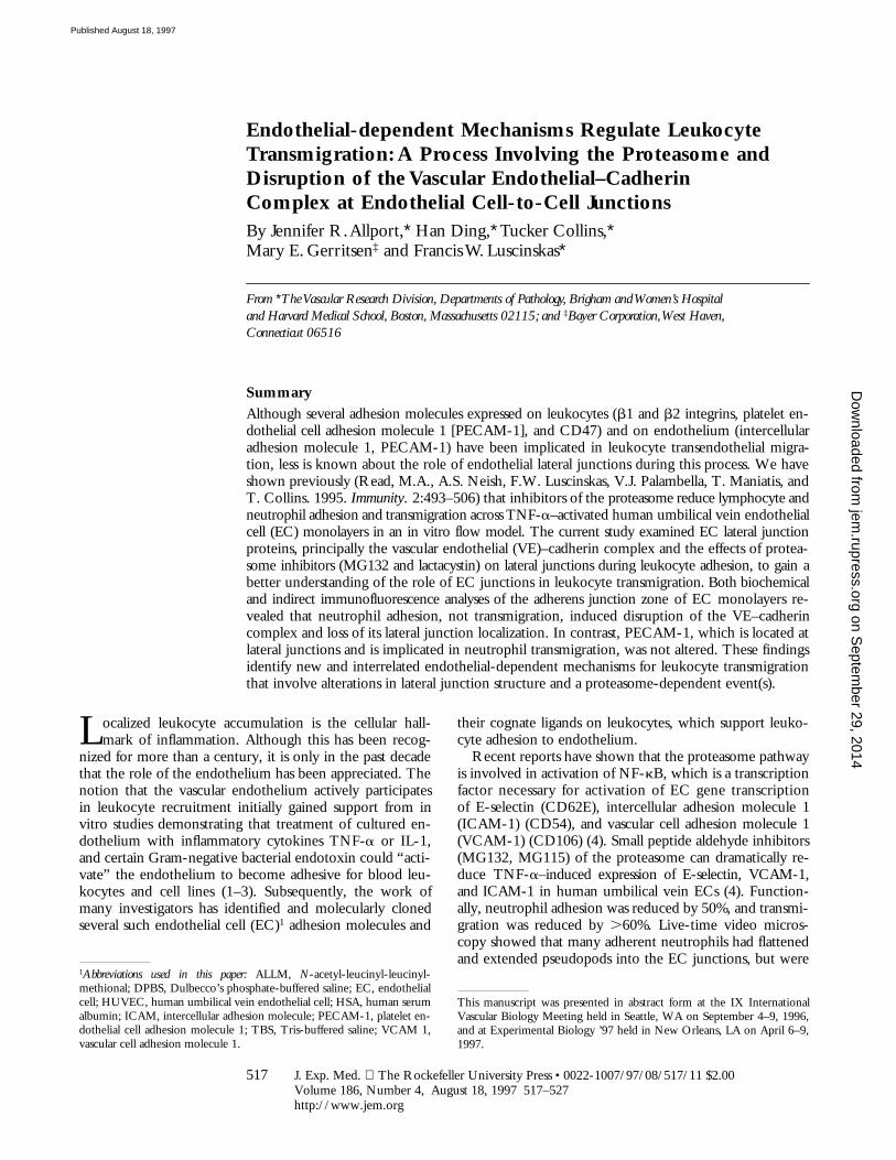
To distinguish between the effects of the proteasome in-hibitors on expression of endothelial leukocyte adhesionmolecules and the process of neutrophil transmigration,MG132, lactacystin, or carrier control were added to 4 hTNF-a–activated EC, and the monolayers further incu-bated. Under such conditions, both inhibitors have no ef-fect on neutrophil adhesion (Fig. 1) or expression of endo-
thelial adhesion molecules (Table 1 and data not shown),but reduce neutrophil transmigration by .50% (Fig. 1).The effect on transmigration was observed by 60 min, andby 120 min the level of blockade was .70% for MG132and .60% for lactacystin. A direct effect of the inhibitorson neutrophils is not likely because a 5-min pretreatmentof neutrophils with 5 mM MG132 before perfusion did notalter neutrophil adhesion or transmigration (vehicle, 51.5 65.9% transmigrated versus MG132 treated, 51.5 6 6.3%; n 53 experiments). We conclude that MG132 and lactacystin,two structurally distinct inhibitors of the proteasome, acton the endothelium to block transmigration, separately fromtheir effect on adhesion molecule expression.
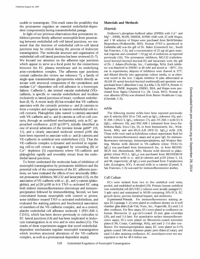
The Endothelial VE–Cadherin Complex Is Not Altered byTreatment with TNF-a, Proteasomal Inhibitors, or Their Com-bination. Inhibition of neutrophil penetration suggeststhat the function of lateral junctions is an important endo-thelial-dependent component(s) for transendothelial passageof the leukocyte. To examine the effects of TNF-a, pro-teasomal inhibitors or both on the VE–cadherin complex,confluent EC monolayers were incubated with or withoutinhibitors and then with or without TNF-a for a total of6 h. The effect of these treatments on the VE–cadherincomplex was determined in Triton X-100–soluble lysates byimmunoprecipitation with anti–VE–cadherin mAb and im-munoblot analysis using specific mAb, and by a second in-dependent analysis using immunofluorescence photomi-croscopy. VE–cadherin, a- and b-catenins, and plakoglobinwere clearly identified (Fig. 2, lane C), and their migrationin SDS-PAGE under reduced conditions was consistent withprevious reports (8, 12). Similarly, immunoprecipitation withanti–VE–cadherin mAb, and subsequent immunoblottinganalysis with an anti-p120 mAb, detected a single band mi-grating at 120 kD (data not shown), but the results were notconsistently found in every EC culture examined. How-ever, analysis of EC lysates with anti-p120 mAb followedby blotting with anti-p120 consistently revealed a band at120 kD and a second band at 100 kD, consistent with a re-cent analysis in endothelium derived from brain (12). Theendothelial VE–cadherin complex was not altered by 6 h ofincubation with TNF-a, MG132, ALLM, lactacystin, orcarrier control (Fig. 2, compare lane C with lanes T, MG,
Figure 1. Proteasome inhibitors MG132 and lactacystin prevent neu-trophil migration under flow at 1.8 dynes/cm2. Confluent EC monolay-ers were incubated with TNF-a for 4 h before addition of proteasome in-hibitors (MG, 20 mM MG132; LAC, 20 mM lactacystin) or DMSOcarrier (Cont.) for 1 (left) or 2 h (right), and neutrophil adhesion and trans-migration were assessed as detailed in Materials and Methods. *P ,0.05.
Figure 2. The endothelialVE–cadherin complex is not al-tered by incubation with protea-somal inhibitors with or withoutTNF-a treatment. ConfluentEC monolayers were treatedwith inhibitors before or afteraddition of TNF-a, washedtwice, and extracted in lysis
buffer 1 (Materials and Methods). The VE–cadherin complex was immu-noprecipitated using anti–VE–cadherin mAb, separated by SDS-PAGE,and subjected to immunoblotting using mAbs that recognize a- andb-catenins, plakoglobin, and p120. The results with anti-p120 mAb werevariable and p120 was not always detected, even though the other com-ponents shown were consistent and reproducible. The x-axis labels are:C, 0.02% DMSO; T, 25 ng/ml of TNF-a for 4 h; MG, MG132 (20mM) for 4 h; ALLM, ALLM (20 mM) treatment for 4 h.
on Septem
ber 29, 2014jem
.rupress.orgD
ownloaded from
Published August 18, 1997
521 Allport et al.
and ALLM; data with lactacystin not shown). Similarly,treatment with TNF-a for 4 h followed by 2 h of coincu-bation with inhibitors, ALLM, or carrier control did not al-ter the VE–cadherin complex (data not shown).
Analysis of Triton X-100 cell lysates also revealed thatthe majority of VE–cadherin and other members of thecadherin complex remained in the Triton X-100–solublefraction, not in the cytoskeleton-associated Triton X-100–insoluble lysate (data not shown), and this distribution wasnot altered by any of the above treatments.
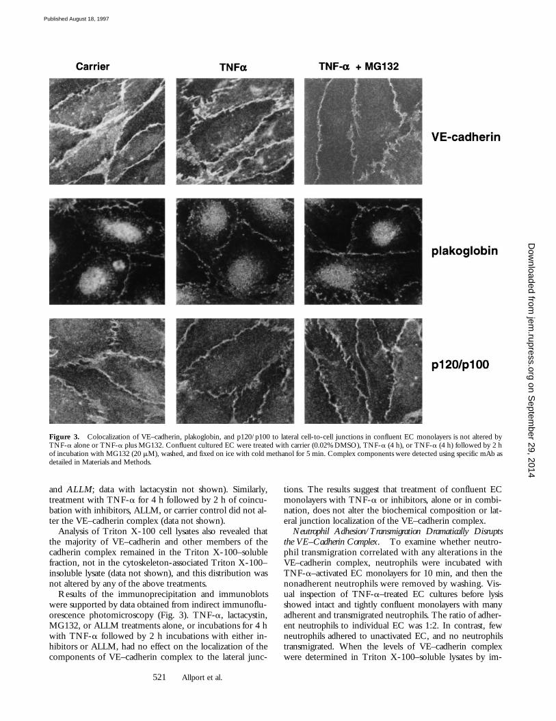
Results of the immunoprecipitation and immunoblotswere supported by data obtained from indirect immunoflu-orescence photomicroscopy (Fig. 3). TNF-a, lactacystin,MG132, or ALLM treatments alone, or incubations for 4 hwith TNF-a followed by 2 h incubations with either in-hibitors or ALLM, had no effect on the localization of thecomponents of VE–cadherin complex to the lateral junc-
tions. The results suggest that treatment of confluent ECmonolayers with TNF-a or inhibitors, alone or in combi-nation, does not alter the biochemical composition or lat-eral junction localization of the VE–cadherin complex.
Neutrophil Adhesion/Transmigration Dramatically Disruptsthe VE–Cadherin Complex. To examine whether neutro-phil transmigration correlated with any alterations in theVE–cadherin complex, neutrophils were incubated withTNF-a–activated EC monolayers for 10 min, and then thenonadherent neutrophils were removed by washing. Vis-ual inspection of TNF-a–treated EC cultures before lysisshowed intact and tightly confluent monolayers with manyadherent and transmigrated neutrophils. The ratio of adher-ent neutrophils to individual EC was 1:2. In contrast, fewneutrophils adhered to unactivated EC, and no neutrophilstransmigrated. When the levels of VE–cadherin complexwere determined in Triton X-100–soluble lysates by im-
Figure 3. Colocalization of VE–cadherin, plakoglobin, and p120/p100 to lateral cell-to-cell junctions in confluent EC monolayers is not altered byTNF-a alone or TNF-a plus MG132. Confluent cultured EC were treated with carrier (0.02% DMSO), TNF-a (4 h), or TNF-a (4 h) followed by 2 hof incubation with MG132 (20 mM), washed, and fixed on ice with cold methanol for 5 min. Complex components were detected using specific mAb asdetailed in Materials and Methods.
on Septem
ber 29, 2014jem
.rupress.orgD
ownloaded from
Published August 18, 1997

522 Endothelial Regulation of Neutrophil Transmigration
munoprecipitation with anti–VE–cadherin mAb and im-munoblotting with specific mAb to each component, dra-matic alterations were observed. Neutrophil adhesion and/ormigration across TNF-a–activated EC monolayers inducedloss of VE–cadherin, b-catenin, and plakoglobin, whereasthe level of a-catenin was not decreased (Fig. 4 A, com-pare lane 3 to 4). Over the course of our experiments, wenoted that the native immunoreactive species of b-catenin,plakoglobin, and p120/p100 were always below detectablelevels, whereas there was often retention of a small amountof VE–cadherin. Coincubation of control unactivated ECmonolayers with unactivated neutrophils consistently had nosignificant effect on the VE–cadherin complex (compare
lane 1 and 2). Since equal numbers of EC were used foreach sample, and this ECL detection system is very sensi-tive, we infer the losses are not due to unequal sampleloading.
Neutrophil Adhesion Induces Rapid Disruption and Degrada-tion of VE–Cadherin Complex. The disruption of the VE–Cadherin complex by addition of neutrophils was accom-panied by significant loss and proteolytic cleavage of eachcomponent, except a-catenin. This was addressed by usingtotal EC monolayer lysates (soluble in 1% Triton X-100,1% NP-40, and 0.5% SDS) and performing immunopre-cipitation with specific mAb directed against each compo-nent. Immunoprecipitated proteins were detected subse-
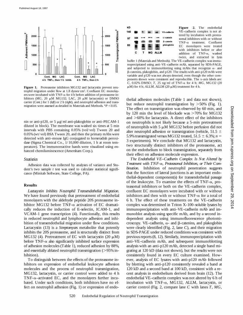
Figure 4. Effect of neutrophil adhesion on the endothelial VE–cad-herin complex. (A) Confluent EC monolayers were incubated with me-dia alone or media containing TNF-a for 6 h and washed. Neutrophils(107) in DPBS-HSA or DPBS-HSA alone were added for 10 min at 378Cunder static conditions. Nonadherent neutrophils were removed bywashing and the monolayers were extracted on ice for 30 min. Proteinswere immunoprecipitated with anti–VE–cadherin mAb as detailed inMaterials and Methods and detected by immunoblotting with specificmAb. (B) Confluent EC monolayers were treated with media alone ormedia containing TNF-a for 4 h, washed twice, and 107 neutrophilswere added. After incubation for 0, 3, or 10 min at 378C, nonadherentneutrophils were removed, and monolayers were extracted with lysisbuffer 1 containing 0.5% SDS. Proteins were immunoprecipitated usingspecific mAb directed against each individual component and subse-quently detected by immunoblotting with a specific mAb. The Mr formolecular mass standards are shown on the left margin and 0, 3, and 10refer to the time period of neutrophil–EC coincubation at 378C. (C) Me-dia alone (lane 1), resting neutrophils (107 cells, lane 2), neutrophil mem-branes representing 107 cells (lane 3), or conditioned media from coincu-bations of neutrophils (107 cells) with TNF-a–activated HUVEC (lane 4)were added to control (lane 1) or TNF-a–activated HUVEC monolayers(lanes 2–4) and incubated at 378C for 10 min. HUVEC were washedtwice and extracted with ice-cold lysis buffer 1 (without 0.5% SDS). Pro-teins were immunoprecipitated with anti–VE–cadherin mAb as detailedin Materials and Methods, and detected by immunoblotting with specificmAb. (D) Neutrophil membranes representing 3 3 106 cells were incu-bated with 4 h TNF-a–activated EC in wells of chamber slides for 10min at 378C. The monolayers were washed twice and fixed for 5 min at48C with ice-cold methanol. Junctional proteins were detected by indi-rect immunofluorescence.
on Septem
ber 29, 2014jem
.rupress.orgD
ownloaded from
Published August 18, 1997
523 Allport et al.
quently by immunoblotting with the same mAb. Additionof SDS dissociates the complex from the cytoskeletal com-ponents (8), and thus allows for determination of the totalcellular content (soluble and cytoskeletal-associated) of eachcomponent of the complex. Total lysates were preparedfrom EC treated with TNF-a and incubated with mediaalone or media with neutrophils for 0, 3, or 10 min (Fig. 4B, lanes 0, 3, and 10, respectively, for each mAb listed). Inparallel, adhesion and transmigration was assessed by phasecontrast microscopy. The levels of native b-catenin, VE–cadherin, plakoglobin, and p120/p100 were decreased dra-matically in TNF-a–activated EC coincubated with neu-trophils for 3 min, a time point when neutrophil adhesion,but no transmigration, had occurred. By 10 min, when manyneutrophils had adhered and transmigrated, the levels of eachcomponent, except a-catenin, was reduced dramatically orundetectable (Fig. 4 B, lanes labeled 10). The level and ap-parent Mr of a-catenin was stable at each time point. Theseresults are consistent with the immunoblots of the VE–cad-herin complex shown in Fig. 4 A. In contrast, PECAM-1,which has been demonstrated to localize to endothelialcell-to-cell lateral junctions and is involved in neutrophil
migration (15), remained at similar levels at both 0 and 10min.
Inspection of immunoblots of total lysates (Fig. 4 B) fromTNF-a–activated EC coincubated with neutrophils usinganti–VE–cadherin mAb revealed the native VE–cadherinspecies (140 kD, Fig. 4 B, large arrow, VE–cadherin, lane 0),and two immunoreactive bands with an apparent Mr ofz100 kD (small arrow) after 3 or 10 min. That a-catenin re-mains associated with VE–cadherin, as demonstrated in Fig.4 A, may be due to its continued association with these100-kD VE–cadherin immunoreactive degradation productsidentified in Fig. 4 B. Previous studies have reported thata-catenins can associate directly with VE–cadherin in somecells types (26), but not in others (27). Immunoblots of totallysates with mAb to b-catenin revealed an immunoreactivedegradation product at z75 kD, and essentially total loss ofimmunoreactive bands for p120/p100 and plakoglobin.These data rule out the possibility that components of theVE–cadherin complex shift their distribution from TritonX-100–soluble to –insoluble fraction (i.e., cytoskeletal as-sociated) after neutrophil adhesion. From these findings,we infer that neutrophil adhesion alone is sufficient to in-duce rapid endothelial-dependent disruption and partialdegradation of members of the VE–cadherin complex.
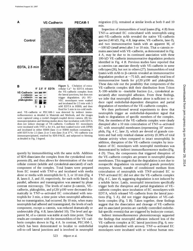
We then performed several experiments to show thatneutrophils trigger an endothelial-dependent signal thatleads to degradation of specific members of the complex.First, the members of the VE–cadherin complex were clearlydisrupted after a 10 min coincubation of TNF-a–activatedEC with neutrophil membranes (representing 107 neutro-phils, Fig. 4 C, lane 3), which are devoid of granule con-tents and had only residual elastase activity (0.38% of totalelastase activity when compared to intact neutrophils). Inaddition, disruption of the VE–cadherin complex by incu-bation of EC monolayers with neutrophil membranes wasdemonstrated by indirect immunofluorescence studies (Fig.4 D). Thus, the components that triggered disruption ofthe VE–cadherin complex are present in neutrophil plasmamembranes. This suggests that the degradation is not due tononspecific degradation via neutrophil granule proteolyticenzymes. Second, transfer of conditioned medium fromcoincubation of neutrophils with TNF-activated EC toTNF-activated EC did not alter the VE–cadherin complex(Fig. 4 C, lane 4), suggesting degradation is not induced bya soluble factor. Lastly, neutrophil interactions specificallytrigger both the disruption and partial degradation of VE–cadherin complex since incubation of EC monolayers withEDTA, which released VE–cadherin from the lateral junc-tions (Fig. 5 A), did not trigger degradation of VE–cad-herin complex (Fig. 5 B). Taken together, these findingssuggest that the dissociation and cleavage of VE–cadherinand its associated proteins are carefully controlled endothe-lial-specific event(s) that are triggered by leukocyte contact.
Indirect immunofluorescence photomicroscopy supportedthe findings that neutrophil adhesion induced loss of theVE–cadherin complex at EC lateral junctions (Fig. 6; neu-trophils are identified with arrows). TNF-a–activated ECmonolayers were incubated with or without human neu-
Figure 5. Chelation of extra-cellular Ca21 by EDTA releasesthe VE–cadherin complex fromthe lateral junctions, but does nottrigger degradation. (A) HU-VEC monolayers were washedand incubated for 2.5 min with 3mM EDTA in HBSS, and thenfixed for 5 min in ice-cold meth-
anol. VE–cadherin or PECAM-1 was detected by indirect immu-nofluorescence as detailed in Materials and Methods, and the imageswere captured using a cooled charged-coupled device camera. (B) Im-munoprecipitation and blotting studies show that incubation with EDTAdoes not induce cleavage of VE–cadherin. Confluent EC monolayerswere incubated with TNF-a for 4 h, and then washed twice with DPBSand incubated in either HBSS (lane 1) or HBSS medium containing 3mM EDTA for 2.5 (lane 2) or 5 min (lane 3) at 378C. VE–cadherin wasimmunoprecipitated, resolved by SDS-PAGE, and subjected to blottingas detailed in Fig. 3 legend.
on Septem
ber 29, 2014jem
.rupress.orgD
ownloaded from
Published August 18, 1997

524 Endothelial Regulation of Neutrophil Transmigration
trophils for 5 min, fixed, and then stained with mAb to theVE–cadherin complex. The loss is clearly demonstrated formonolayers stained to detect VE–cadherin, b-catenin, p120/p100, and plakoglobin, whereas the level and junctionalcolocalization of PECAM-1 remained constant. A signifi-cant overall reduction in immunoreactive staining of b-cate-nin, p120/p100, and plakoglobin was observed in the pres-ence of neutrophils, whereas VE–cadherin was lost specificallyfrom regions of neutrophil adhesion/transmigration (smallarrows). This is consistent with the immunoprecipitationdata where, in the majority of experiments, a small amountof native VE–cadherin is retained, whereas other compo-nents of the complex are not detectable.
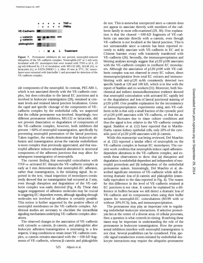
To investigate whether the degradation of the VE–cad-herin complex involved the cellular proteasome system, theeffects of proteasome inhibitors were evaluated. As shownin Fig. 7, disruption of the VE–cadherin complex was notprevented by 2 h of incubation with 20 mM lactacystin orMG132, even though such treatments significantly reducedtransmigration (Fig. 1). For comparison, the components ofthe VE–cadherin complex in TNF-a–activated EC re-mained constant in the absence of neutrophils. In addition,shorter incubations (5 or 30 min) with 50 mM MG132 did
not prevent neutrophil-dependent dissociation of the VE–cadherin complex (data not shown). These findings estab-lish that neutrophil adhesion triggers rapid EC-dependentdegradation of the VE–cadherin complex through an enzy-matic pathway that appears to be independent of the pro-teasome.
Discussion
The current work demonstrates that neutrophil adhe-sion to TNF-a–activated endothelial monolayers dramati-cally alters the molecular composition and organization ofthe endothelial cell VE–cadherin complex, which has beenimplicated in maintenance of endothelial cell-to-cell adhe-sion and cell-to-cytoskeletal integrity. Neutrophil adhesionto confluent TNF-a–activated endothelial monolayers in-duced rapid and near complete dissociation of b-catenin,p120/p100, and plakoglobin from VE–cadherin and loss oftheir lateral junction colocalization. Biochemical analysis ofTNF-a–activated EC monolayers using immunoprecipita-tion and immunoblotting revealed that these proteins aredegraded rapidly through a proteolytic mechanism that isdependent on the endothelium and does not involve gran-
Figure 6. Photographs showVE–cadherin, b-catenin, p120/p100, plakoglobin, and PECAM-1staining patterns. Indirect immu-nofluorescence microscopy showsthat TNF-a–activated EC mono-layers express abundant levels ofVE–cadherin, b-catenin, p120/p100, plakoglobin, and PECAM-1at lateral junctions before neu-trophil adhesion (left). After 10min of neutrophil adhesion, thelateral staining of VE–cadherin,b-catenin, p120/p100, and plako-globin is lost, whereas PECAM-1is still abundantly expressed atlateral junctions (right). Adher-ent neutrophils are identified(right, arrows), and intact VE–cad-herin junctional staining (top as-terisks). Original magnificationwas 320, except VE–cadherinwhich was 640.
on Septem
ber 29, 2014jem
.rupress.orgD
ownloaded from
Published August 18, 1997
525 Allport et al.
ule components of the neutrophil. In contrast, PECAM-1,which is not associated directly with the VE–cadherin com-plex, but does colocalize to the lateral EC junctions and isinvolved in leukocyte transmigration (28), remained at con-stant levels and retained lateral junction localization. Giventhe rapid and specific cleavage of the components of VE–cadherin complex by the endothelial cells, we suspectedthat the cellular proteasome was involved. Surprisingly, twodifferent proteasome inhibitors, MG132 or lactacystin, didnot prevent dissociation or degradation of components ofthe VE–cadherin complex, however, both inhibitors didprevent .60% of neutrophil transmigration, specifically bypreventing neutrophil penetration of the lateral junctions.Taken together, the results demonstrate that leukocyte ad-hesion and transmigration across the vascular endotheliumis more complex than previously appreciated, and that neu-trophil adhesion induces substantial alterations in structuralcomponents of the adherens junctions that may allow forsubsequent transmigration of neutrophils.
The current finding that neutrophil coincubation withTNF-a–activated EC disrupts the VE–cadherin complex asearly as 3 min demonstrates that neutrophil–EC adhesion,rather than transmigration, is the initiating signal. As re-ported in the text, visual inspection of monolayers consis-tently showed that no transmigration had occurred at 3 min,even though disruption and degradation of the VE–cad-herin complex was easily detected (Fig. 4 B). These datasuggest engagement of adhesion molecules may be crucialin triggering EC-dependent events, although signaling throughmolecules not involved in adhesion is certainly possible.This notion is further supported by the positive effects ofneutrophil membranes on the VE–cadherin complex. Ad-ditional studies are necessary to elucidate the intracellularsignaling mechanisms underlying VE–cadherin complex alter-ations.
The observed changes in the association of VE–cadherinwith cytosolic a-catenin, b-catenin, and plakoglobin afterleukocyte adhesion/transmigration is interesting in a fewrespects. Using conditions to retain intact VE–cadherin com-plex, a-catenin remains associated with the z100-kD frag-ments of VE–cadherin, whereas b-catenin and plakoglobin
do not. This is somewhat unexpected since a-catenin doesnot appear to associate directly with members of the cad-herin family in most cells examined (29, 30). One explana-tion is that the cleaved z100-kD fragments of VE–cad-herin can associate directly with a-catenin, even thoughVE–cadherin is not localized at the lateral junction. This isnot unreasonable since a-catenin has been reported re-cently to stably associate with VE–cadherin in EC and inChinese hamster ovary cells transiently transfected withVE–cadherin (26). Secondly, the immunoprecipitation andblotting analyses strongly suggest that p120/p100 associateswith the VE–cadherin complex in confluent EC monolay-ers. Although the association of p120/p100 with VE–cad-herin complex was not observed in every EC culture, directimmunoprecipitation from total EC extracts and immuno-blotting with anti-p120 mAb consistently detected twospecific bands at 120 and 100 kD, which is in line with thereport of Staddon and co-workers (31). Moreover, both bio-chemical and indirect immunofluorescence evidence showedthat neutrophil coincubation with activated EC rapidly ledto the degradation and loss of lateral junctional staining ofp120/p100. One possible explanation for the inconsistencyof immunoprecipitation experiments using anti–VE–cad-herin mAb is that only a small fraction of the cytosolic poolof p120/p100 associates with VE–cadherin, or that the as-sociation fluctuates due to tissue culture conditions andthus the signal is low relative to the VE–cadherin complexsignal. Staddon et al. (12) have reported that in Mardin-Darby canine kidney epithelial cells, only 20% of the cyto-solic pool of p120/p100 associates with b-catenin.
While this manuscript was being reviewed, Del Maschioet al. (32) reported a similar effect of neutrophils on theVE–cadherin complex in human EC monolayers. The cur-rent work confirms that neutrophils induce rapid adhesion-dependent alterations in the VE–cadherin complex, and ex-tends these observations to show that (a) disruption anddegradation is endothelial-dependent and independent of neu-trophil proteolysis and (b) independent of the endothelialproteasome system. Interestingly, Del Maschio et al. de-scribed significant retention of VE–cadherin while still de-tecting dramatic loss of b-catenin and plakoglobin (essen-tially equivalent to the data reported in Fig. 4). The reasonfor this difference in the level of VE–cadherin retained atEC junctions is not clear. It cannot be explained by a dif-ference in buffers because we still detect a dramatic loss ofVE–cadherin and its components when using their buffersystem for neutrophil-EC coincubations (M199 with orwithout 20% FCS), lysis, and immunoprecipitation.
The proteasome may play an important role in regulat-ing endothelial–leukocyte interactions. If selective proteol-ysis lies at the center of a diverse array of cellular processes,then a question is what controls its timing. Resolving theseissues may be important in understanding the role of theproteasome in leukocyte transmigration. How the protea-somal inhibitors interfere with neutrophil transmigration isnot clear. Several possibilities can be considered. First, spe-cific signal transduction events initiated by endothelial–leu-kocyte interactions may require the ubiquitin–proteasome
Figure 7. Proteasome inhibitors do not prevent neutrophil-induceddisruption of the VE–cadherin complex. Neutrophils (107 in 5 ml) wereincubated with EC monolayers that were treated with TNF-a (4 h, 25ng/ml) followed by 2 h of incubation with MG132 (M), ALLM (A), orlactacystin (L), or 0.02% DMSO (2). After 10 min at 378C, EC mono-layers were extracted with lysis buffer 1 and processed for detection of theVE–cadherin complex.
on Septem
ber 29, 2014jem
.rupress.orgD
ownloaded from
Published August 18, 1997
526 Endothelial Regulation of Neutrophil Transmigration
proteolytic pathway. The paradigm for this is well estab-lished for several processes (13, 33). Second, the protea-some may degrade key architectural components of thejunctional complex. The VE–cadherin complex is probablynot the target of these events. Endothelial–leukocyte inter-actions could alter the activity of the endothelial protea-some by altering the composition or by increasing the ac-tivity of specific catalytic components of the proteasomefacilitating transmigration. Third, the proteasome inhibitorsmay act indirectly by altering the activity of important pro-cesses other than the proteasome. The structure–functionrelationship between the inhibitors used in this study suggeststhat this possibility is unlikely. Two structurally distinctproteasome inhibitors, lactacystin and MG132, specificallyblocked leukocyte transmigration. A structurally homolo-gous peptide aldehyde (ALLM), which inhibits cathepsin Band calpain but is a much weaker inhibitor of the protea-some, did not block transmigration. Collectively, these find-ings distinguish the proteasome pathway rather than cathep-sin/calpain-mediated protein degradation or processing asan important step in leukocyte transmigration.
The Process of Leukocyte Transendothelial Migration. Thecurrent report, and several previous studies, further expandthe notion that EC–leukocyte adhesion triggers EC-depen-dent changes that correlate with leukocyte transmigration.Huang et al. (34) showed that intracellular Ca21 gradientsin EC were coupled to transendothelial migration. Neutro-phil adhesion to EC induced rapid and transient several foldincreases in cytosolic Ca21 concentrations ([Ca21]i), whichparalleled the time course of neutrophil transmigration acrosscytokine-activated EC grown on human amnion prepara-
tions. Pharmacological clamping of [Ca21]i with an intra-cellular Ca21 chelator, bis-(2-amino-5-methylphenoxyle-thane-N,N,N9,N9-tetraacetic acid tetraacetoxymethyl ester,inhibited .90% of migration, but had no effect on adhe-sion. Pfau et al., working with lymphocytes and EC (35),and Zeigelstein et al., using both neutrophils and mono-cytes interacting with endothelial monolayers under flowconditions in vitro (36), also reported leukocyte adhesion–dependent changes of [Ca21]i in EC and the leukocyte.Neither of these studies report on the spatial resolution ofthe Ca21 flux, so it remains to be determined whether thecationic flux relates to an adhesion event or to a migratoryevent. Thus, the role of increases of [Ca21]i in EC appearcompelling, whereas the relative role in leukocytes is lessclear. Interestingly, in a static assay system, pharmocologicclamping of [Ca21]i in neutrophils did not dramatically altertheir migration across resting or cytokine-activated EC,suggesting that changes in neutrophils [Ca21]i are not a pre-requisite (37). In addition, Yoshida et al. (31) have reportedthat binding of leukocytes to 4-h IL-1–activated EC induceE-selectin linkage at its cytoplasmic domain to the EC actincytoskeleton. This link, or similar mechanisms, may supplycues to the EC that influence downstream processes in-volved in stable adhesion or in transmigration.
The results presented here raise several issues concerningthe function of lateral junctions during leukocyte traffick-ing at sites of inflammation. A basic understanding of theseprocesses may lead to insight into therapies for preventionof tissue damage and abnormal wound healing that occur asa result of a pathological inflammatory response.
The authors wish to thank Ms. Kay Case and Mr. William Atkinson for providing cultured vascular endo-thelium, and Drs. Michael A. Gimbrone, Jr., James Madara, and Douglas Goetz for helpful discussions.
This work was supported by National Institutes of Health grants HL36028 (F.W. Luscinskas, T. Collins),HL47646 and HL53939 (F.W. Luscinskas, J.R. Allport) and a postdoctoral fellowship from The Crohn’s andColitis Foundation of America (J.R. Allport).
Address correspondence to Dr. Francis W. Luscinskas, Brigham and Women’s Hospital, 221 Longwood Ave.,Boston, MA 02115. Phone: 617-732-6004; FAX 617-732-5933; E-mail: [email protected]
Received for publication 6 November 1996 and in revised form 9 June 1997.
References1. Bevilacqua, M.P., J.S. Pober, M.E. Wheeler, R.S. Cotran,
and M.A. Gimbrone, Jr. 1985. Interleukin-1 acts on culturedhuman vascular endothelium to increase the adhesion ofpolymorphonuclear leukocytes, monocytes, and related leu-kocyte cell lines. J. Clin. Invest. 76:2003–2011.
2. Schleimer, R.P., and R.K. Rutledge. 1986. Cultured humanvascular endothelial cells acquire adhesiveness for leukocytesfollowing stimulation with interleukin-1, endotoxin and tu-mor-promoting phorbol-esters. J. Immunol. 136:649–656.
3. Gamble, J.R., J.M. Harlan, S.J. Klebanoff, A.F. Lopez, andM.A. Vadas. 1985. Stimulation of the adherence of neutro-phils to umbilical vein endothelium by recombinant tumornecrosis factor. Proc. Natl. Acad. Sci. USA. 82:8667–8670.
4. Read, M.A., A.S. Neish, F.W. Luscinskas, V.J. Palombella,T. Maniatis, and T. Collins. 1995. The proteasome pathwayis required for cytokine-induced endothelial–leukocyte adhe-sion molecule expression. Immunity. 2:493–506.
5. Dejana, E., M. Corada, and G. Lampugnani. 1995. Endothe-lial cell-to-cell junctions. FASEB J. 9:910–918.
6. Geiger, B. 1991. The cytoplasmic domain of adherens-typejunctions. Cell Motil.Cytoskel. 20:1–6.
7. Anderson, J.M., M.S. Balda, and A.S. Fanning. 1993. Thestructure and regulation of tight junctions. Curr. Opin. CellBiol. 5:772–778.
8. Lampugnani, M.G., M. Corada, L. Caveda, F. Breviario, O.Ayalon, B. Geiger, and E. Dejana. 1995. The molecular or-
on Septem
ber 29, 2014jem
.rupress.orgD
ownloaded from
Published August 18, 1997
527 Allport et al.
ganization of endothelial cell to cell junctions: differential as-sociation of plakoglobin, b-catenin, and a-catenin with vas-cular endothelial cadherin (VE-cadherin). J. Cell Biol. 129:203–218.
9. Lampugnani, M.G., M. Resnati, M. Raiteri, R. Pigott, A.Pisacane, G. Houen, L.P. Ruco, and E. Dejana. 1992. A novelendothelial-specific membrane protein is a marker of cell-cellcontacts. J. Cell Biol. 118:1511–1522.
10. Kanner, S.B., A.B. Reynolds, and J.T. Parsons. 1991. Ty-rosine phosphorylation of a 120-kilodalton pp60src substrateupon epidermal growth factor and platelet-derived growthfactor stimulation and in polymovirus middle–T-antigen–transformed cells. Mol. Cell. Biol. 11:713–720.
11. Reynolds, A.B., J. Daniel, P.D. McCrea, M.J. Wheelock, J.Wu, and Z. Zhang. 1994. Identification of a new catenin: thetyrosine kinase substrate p120cas associates with E-cadherincomplexes. Mol. Cell. Biol. 14:8333–8342.
12. Staddon, J.M., C. Smales, C. Schulze, F.S. Esch, and L.L.Rubin. 1995. p120, a p120-related protein (p100), and thecadherin/catenin complex. J. Cell Biol. 130:369–381.
13. Fenteany, G., R.F. Standaert, W.S. Lane, S. Choi, E.J. Corey,and S.L. Schreiber. 1995. Inhibition of proteasome activitiesand subunit amino-terminal threonine modification by lacta-cystin. Science (Wash. DC). 268:726–731.
14. Muller, W.A., C.M. Ratti, S.L. McDonnell, and Z.A. Cohn.1989. A human endothelial cell–restricted, externally dis-posed plasmalemmal protein enriched in intercellular junctions.J. Exp. Med. 170:399–414.
15. Muller, W.A., S.A. Weigl, X. Deng, and D.M. Phillips.1993. PECAM-1 is required for transendothelial migration ofleukocytes. J. Exp. Med. 178:449–460.
16. Luscinskas, F.W., H. Ding, P. Tan, D. Cumming, T.F. Ted-der, and M.E. Gerritsen. 1996. L-selectin and P-selectin butnot VLA-4 mediate monocyte initial attachment and rollingon TNF-a activated vascular endothelium under flow invitro. J. Immunol. 156:326–335.
17. Luscinskas, F.W., M.I. Cybulsky, J.-M. Kiely, C.S. Peckins,V.M. Davis, and M.A. Gimbrone, Jr. 1991. Cytokine-activatedhuman endothelial monolayers support enhanced neutrophiltransmigration via a mechanism involving both endothelial–leukocyte adhesion molecule–1 and intercellular adhesion mol-ecule-1. J. Immunol. 146:1617–1625.
18. Rice, G.E., J.M. Munro, and M.P. Bevilacqua. 1990. Induc-ible cell adhesion molecule 110 (INCAM-110) is an endo-thelial receptor for lymphocytes. A CD11/CD18 indepen-dent adhesion mechanism. J. Exp. Med. 171:1369–1374.
19. Luscinskas, F.W., A.F. Brock, M.A. Arnaout, and M.A. Gim-brone, Jr. 1989. Endothelial–leukocyte adhesion molecule–1(ELAM-1)–dependent and leukocyte (CD11/CD18)-depen-dent mechanisms contribute to polymorphonuclear leukocyteadhesion to cytokine-activated human vascular endothelium.J. Immunol. 142:2257–2263.
20. Luscinskas, F.W., G.S. Kansas, H. Ding, P. Pizcueta, B.E.Schleiffenbaum, T.F. Tedder, and M.A. Gimbrone, Jr. 1994.Monocyte rolling, arrest and spreading on IL-4–activated vas-cular endothelium under flow is mediated via sequential ac-tion of L-selectin, b1-integrins, and b2-integrins. J. Cell Biol.125:1417–1427.
21. Shen, J., F.W. Luscinskas, A. Connolly, C.F. Dewey, Jr., andM.A. Gimbrone, Jr. 1992. Fluid shear stress modulates cyto-solic free calcium in vascular endothelial cells. Am. J. Physiol.262:C384–C390.
22. Luscinskas, F.W., H. Ding, and A.H. Lichtman. 1995. P-selectin
and VCAM-1 mediate rolling and arrest of CD41 T-lymphocyteson TNF-a–activated vascular endothelium under flow. J.Exp. Med. 181:1179–1186.
23. Gerritsen, M.E., C. Shen, M.C. Mchugh, W.J. Atkinson, M.Kiely, D.S. Milstone, F.W. Luscinskas, and M.A.J. Gimbrone.1995. Activation-dependent isolation and culture of murine pul-monary microvascular endothelium. Microcirculation. 2:151–163.
24. Laemmli, U.K. 1970. Cleavage of structural proteins duringthe assembly of the head of bacteriophage T4. Nature (Lond.).227:680–685.
25. Donnelly, L.E., R.S. Boyd, and J. MacDermot. 1992. Gsalpha is a substrate for mono(ADP-ribosyl)transferase ofNG108-15 cells. ADP-ribosylation regulates Gs alpha activityand abundance. Biochem. J. 228:331–336.
26. Breviario, F., L. Caveda, M. Corada, I. Martin-Padura, P.Navarro, J. Golay, M. Introna, D. Gulino, M.G. Lampug-nani, and E. Dejana. 1995. Functional properties of humanvascular endothelial cadherin (7B4/cadherin-5), an endothe-lium-specific cadherin. Arterioscler. Thromb. Vasc. Biol. 15:1229–1239.
27. Aberle, H., H. Schwartz, and R. Kemler. 1996. Cadherin–catenin complex: protein interactions and their implicationsfor cadherin function. J. Cell. Biochem. 61:514–523.
28. Muller, W.A. 1995. The role of PECAM-1 (CD31) in leu-kocyte emigration: studies in vitro and in vivo. J. LeukocyteBiol. 57:523–528.
29. Gumbiner, B.M. 1996. Cell adhesion: the molecular basis oftissue architecture and morphogenesis. Cell. 84:345–357.
30. Kemler, R. 1993. From cadherins to catenins: cytoplasmicprotein interactions and regulation of cell adhesion. TIG(Trends Genet.). 9:317–321.
31. Yoshida, M., W.F. Westlin, N. Wang, D.E. Ingber, A.Rosenzweig, N. Resnick, and M.A. Gimbrone, Jr. 1996.Leukocyte adhesion to vascular endothelium induces E-selec-tin linkage to the actin cytoskeleton. J. Cell Biol. 133:445–455.
32. Del Maschio, A., A. Zanetti, M. Corada, Y. Rival, L. Ruco,M.G. Lampugnani, and E. Dejana. 1996. Polymorphonuclearleukocyte adhesion triggers the disorganization of endothelialcell-to-cell adherens junctions. J. Cell Biol. 135:497–510.
33. Kopan, R., E.H. Schroeter, H. Weintraub, and J.S. Nye.1996. Signal transduction by activated mNotch: importanceof proteolytic processing and its regulation by the extracellu-lar domain. Proc. Natl. Acad. Sci. USA. 93:1683–1688.
34. Huang, A.J., J.E. Manning, T.M. Bandak, M.C. Ratau, K.R.Hanser, and S.C. Silverstein. 1993. Endothelial cell cytosolicfree calcium regulates neutrophil migration across monolayersof endothelial cells. J. Cell Biol. 120:1371–1380.
35. Pfau, S., D. Leitenberg, H. Rinder, B.R. Smith, R. Pardi,and J.R. Bender. 1995. Lymphocyte adhesion–dependent cal-cium signaling in human endothelial cells. J. Cell Biol. 128:969–978.
36. Ziegelstein, R.C., S. Corda, R. Pili, A. Passaniti, D. Lefer,J.L. Zweier, A. Fraticelli, and M.C. Capogrossi. 1994. Initialcontact and subsequent adhesion of human neutrophils ormonocytes to human aortic endothelial cells releases an en-dothelial intracellular calcium store. Circulation. 90:1899–1907.
37. Kuijpers, T.W., M. Hoogerwerf, and D. Roos. 1992. Neutro-phil migration across monolayers of resting or cytokine-acti-vated endothelial cells. Role of intracellular calcium changesand fusion of specific granules with the plasma membrane. J.Immunol. 148:72–77.
on Septem
ber 29, 2014jem
.rupress.orgD
ownloaded from
Published August 18, 1997


