Embed Size (px)
Citation preview
ORIGINAL ARTICLE
Enzyme replacement therapy for mucopolysaccharidosis VI:long-term cardiac effects of galsulfase (Naglazyme®) therapy
E. Braunlin & H. Rosenfeld & C. Kampmann & J. Johnson &
M. Beck & R. Giugliani & N. Guffon & D. Ketteridge &
C. M. Sá Miranda & M. Scarpa & I. V. Schwartz &
E. Leão Teles & J. E. Wraith & P. Barrios &
E. Dias da Silva & G. Kurio & M. Richardson &
G. Gildengorin & J. J. Hopwood & M. Imperiale &
A. Schatz & C. Decker & P. Harmatz & MPSVI StudyGroup
Received: 17 October 2011 /Revised: 7 March 2012 /Accepted: 14 March 2012 /Published online: 5 June 2012
Abstract Characteristic cardiac valve abnormalities and leftventricular hypertrophy are present in untreated patients withmucopolysaccharidosis type VI (MPSVI). Cardiac ultrasoundwas performed to investigate these findings in subjects duringlong-term enzyme replacement therapy (ERT) with recombi-nant human arylsulfatase B (rhASB, rhN-acetylgalactosamine4-sulfatase, galsulfase, Naglazyme®). Studies were conductedin 54 subjects before ERTwas begun and at specific intervals
for up to 96 weeks of weekly infusions of rhASB at 1 mg/kgduring phase 1/2, phase 2, and phase 3 trials of rhASB. Atbaseline, mitral and aortic valve obstruction was present andwas significantly greater in those ≥12 years of age.Mild mitraland trace aortic regurgitation were present, the former beingsignificantly greater in those <12 years. Left ventricular hy-pertrophy, with averaged z-scores ranging from 1.6–1.9 SDgreater than normal, was present for ages both <12 and
Communicated by: Gregory M. Pastores
*The MPS VI Study Group co-investigators (see Acknowledgmentsection)
E. BraunlinPediatric Cardiology, University of Minnesota,Minneapolis, MN, USA
H. Rosenfeld :G. KurioCardiology,Children’s Hospital & Research Center Oakland,Oakland, CA, USA
C. KampmannDepartment of Congenital Heart Diseases /Pediatric Cardiology / GUCH, University Medicine,Center for Diseases in Childhood and Adolescence,Mainz, Germany
J. Johnson : P. Harmatz (*)Gastroenterology,Children’s Hospital & Research Center Oakland,Oakland, CA, USAe-mail: [email protected]
M. BeckCentre for Lysosomal Storage Diseases,University Children’s Hospital,Mainz, Germany
R. Giugliani : I. V. Schwartz : P. BarriosDepartment of Genetics, UFRGS,Porto Alegre, RS, Brazil
R. Giugliani : I. V. Schwartz : P. BarriosMedical Genetics Service, HCPA,Porto Alegre, RS, Brazil
R. GiuglianiINAGEMP – Instituto Nacional de Genética Médica Populacional,Porto Alegre, RS, Brazil
N. GuffonHôpital Femme Mère Enfant,Lyon, France
D. KetteridgeMetabolic Unit, SA Pathology atWomen’s and Children’s Hospital,Adelaide, Australia
J Inherit Metab Dis (2013) 36:385–394DOI 10.1007/s10545-012-9481-2
# The Author(s) 2012. This article is published with open access at Springerlink.com

≥12 years. After 96 weeks of ERT, ventricular septal hyper-trophy regressed in those <12 years. For those ≥12 years,septal hypertrophy was unchanged, and aortic regurgitationincreased statistically but not physiologically. Obstructivegradients across mitral and aortic valves remained unchanged.The results suggest that long-term ERT is effective in reducingintraventricular septal hypertrophy and preventing progres-sion of cardiac valve abnormalities when administered tothose <12 years of age.
Introduction
Mucopolysaccharidosis type VI (MPS VI, Maroteaux-Lamysyndrome) is a lysosomal storage disease caused by functionalabsence of the enzyme N-acetylgalactosamine 4-sulfatase(arylsulfatase B or ASB; E.C. 3.1.6.12). Absence of this en-zyme results in the accumulation of dermatan-sulfated glyco-saminoglycans (GAGs) within lysosomes of various tissuesincluding bones, cartilage, lungs, airways, and the cardiovas-cular system (Neufeld and Muenzer 2001). The progressiveaccumulation of these substances results in multi-organ systemdysfunction such as joint contractures, short stature, dysostosismultiplex, decreased pulmonary function, cardiac abnormali-ties and, ultimately, shortened life span (Neufeld and Muenzer2001). The severity of the clinical findings is variable but, in arecent survey of 121 MPS VI subjects, an accelerated clinicalcourse was associated with urinary excretion of GAGs inexcess of 200 μg/mg creatinine (Swiedler et al. 2005).
Evaluation of the heart in individuals with untreated MPSVI by cardiac ultrasound has been reported by several inves-tigators (Lael et al. 2010; Dangel 1998; Wippermann et al.1995; Azevedo et al. 2004; Scarpa et al. 2009; Fesslova etal. 2009). The characteristic cardiac abnormalities of MPS
VI include ventricular hypertrophy and a progressive thick-ening of mitral and aortic valves resulting in valvular regur-gitation, stenosis, or both. Until now the effects of enzymereplacement therapy (ERT) upon the heart in MPS VI havenot been reported.
Previous phase 1, 2, and 3 studies have shown thattreatment of individuals with MPS VI by ERT with recom-binant human N-acetylgalactosamine 4-sulfatase (rhASB;galsulfase; Naglazyme®) is safe, rapidly reduces urinaryGAG levels, and improves endurance as measured by 6-or 12-min walk and pulmonary function testing (Harmatz etal. 2004, 2005, 2006). Further analysis of pooled data fromthe clinical ERT trials and the survey study has demonstrat-ed safety and significant long-term increases in endurance,pulmonary function, and growth when galsulfase is admin-istered for 96 weeks or more (Harmatz et al. 2008, 2010;Decker et al. 2010). The purpose of this report is to analyzecardiac ultrasound data obtained during phase 1, 2, and 3clinical ERT trials to determine the effects of 96 weeks ofERT upon the characteristic cardiac findings in individualswith MPS VI.
Methods
Previous reports have detailed the study design and out-comes of the phase I/2, 2, and 3 galsulfase trials in subjectswith MPS VI (Harmatz et al. 2004, 2005, 2006, 2008,2010); details of the clinical trials are outlined in Table 1.Cardiac ultrasound was performed as part of the clinicalevaluation of subjects in the phase 1/2, 2, and 3 clinicaltrials at baseline, before ERT was begun, and at intervals of24–48 weeks and 72–96 weeks after initiation of ERT(Table 2). An Institutional Review Board (IRB) or Ethics
C. M. Sá MirandaInstituto de Biologia Molecular e Celular,Unidade de Biologia do Lisossoma e Peroxisoma,Porto, Portugal
M. ScarpaDepartment of Pediatrics, University of Padova,Padova, Italy
E. Leão TelesUnidade de Doenças Metabólicas, Departmento de Pediatria,Hospital S. João,Porto, Portugal
J. E. WraithGenetic Medicine, St. Mary’s Hospital,Manchester M13 9WL, UK
E. Dias da SilvaCardiologia Pediatrica, Departamento de Pediatria,Hospital de S. João,Porto, Portugal
M. RichardsonCardiology Department, Women’s and Children’s Hospital,Adelaide, Australia
G. GildengorinPediatric Clinical Research Center,Children’s Hospital & Research Center Oakland,Oakland, CA, USA
J. J. HopwoodLysosomal Diseases Research Unit,SA Pathology at Women’s and Children’s Hospital Adelaide,North Adelaide, Australia
M. Imperiale :A. Schatz :C. DeckerBioMarin Pharmaceutical, Inc.,Novato, CA, USA
386 J Inherit Metab Dis (2013) 36:385–394
Committee (EC) at each participating clinical site approvedeach study. All adult patients and parent/guardians gavewritten consent; patients younger than 18 years old gavewritten assent according to local IRB regulations.
Archived ultrasound data, collected from subjects whohad participated in phase 1/2, 2, or 3 clinical trials, werereviewed and tabulated for this study. The original echoeswere obtained and analyzed at the individual sites; originalecho tapes were unavailable for further review. Each ar-chived measurement was reviewed for reliability by a singleperson (E.B.). Discrepancies were resolved by discussionwith individual sites. The entire data set was then subjectedto statistical evaluation. Height, weight, and urinary GAGcontent at study entry were analyzed for the 54 subjects whoparticipated in phase 1/2, 2, or 3 galsulfase trials.
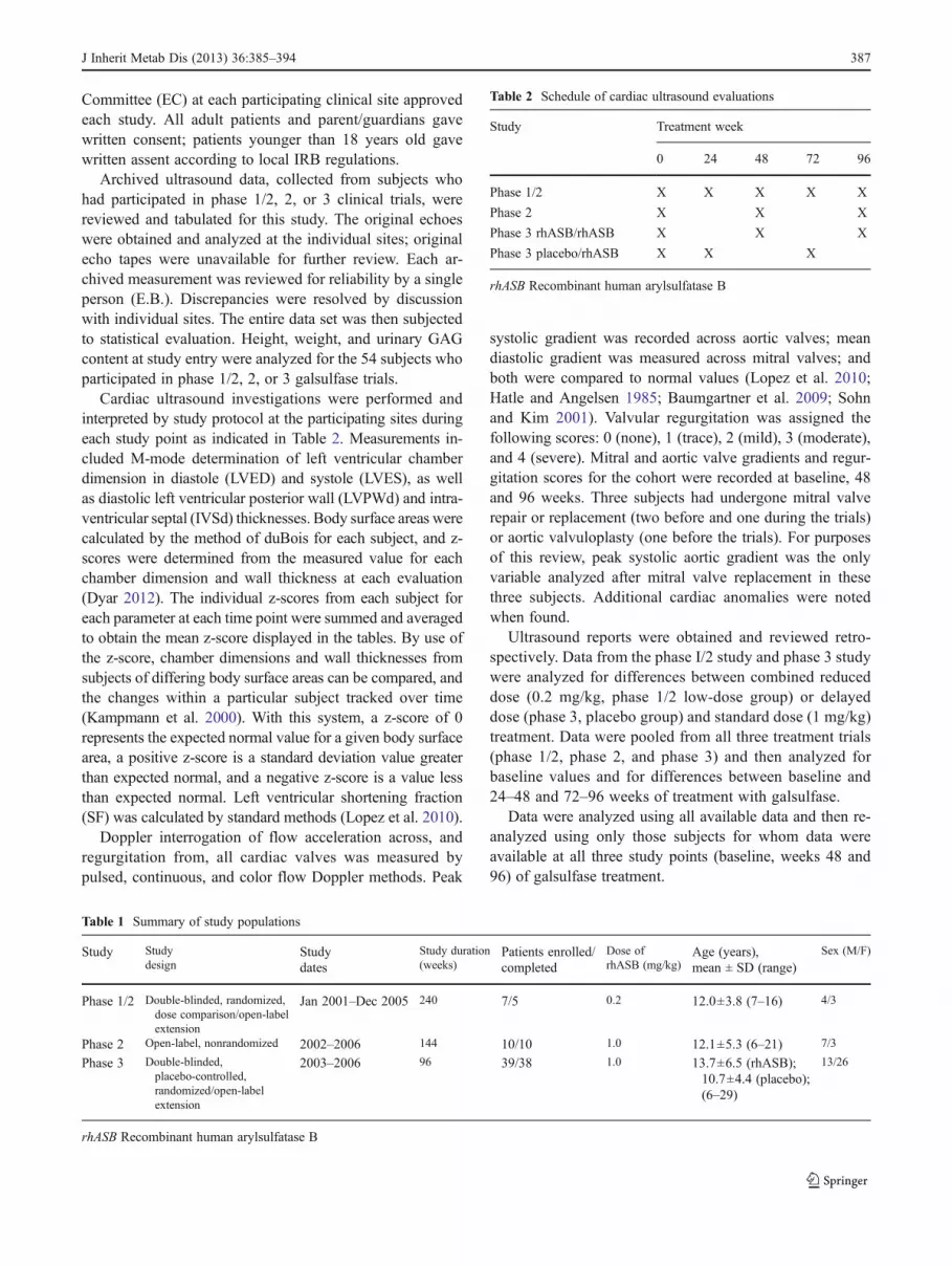
Cardiac ultrasound investigations were performed andinterpreted by study protocol at the participating sites duringeach study point as indicated in Table 2. Measurements in-cluded M-mode determination of left ventricular chamberdimension in diastole (LVED) and systole (LVES), as wellas diastolic left ventricular posterior wall (LVPWd) and intra-ventricular septal (IVSd) thicknesses. Body surface areas werecalculated by the method of duBois for each subject, and z-scores were determined from the measured value for eachchamber dimension and wall thickness at each evaluation(Dyar 2012). The individual z-scores from each subject foreach parameter at each time point were summed and averagedto obtain the mean z-score displayed in the tables. By use ofthe z-score, chamber dimensions and wall thicknesses fromsubjects of differing body surface areas can be compared, andthe changes within a particular subject tracked over time(Kampmann et al. 2000). With this system, a z-score of 0represents the expected normal value for a given body surfacearea, a positive z-score is a standard deviation value greaterthan expected normal, and a negative z-score is a value lessthan expected normal. Left ventricular shortening fraction(SF) was calculated by standard methods (Lopez et al. 2010).
Doppler interrogation of flow acceleration across, andregurgitation from, all cardiac valves was measured bypulsed, continuous, and color flow Doppler methods. Peak
systolic gradient was recorded across aortic valves; meandiastolic gradient was measured across mitral valves; andboth were compared to normal values (Lopez et al. 2010;Hatle and Angelsen 1985; Baumgartner et al. 2009; Sohnand Kim 2001). Valvular regurgitation was assigned thefollowing scores: 0 (none), 1 (trace), 2 (mild), 3 (moderate),and 4 (severe). Mitral and aortic valve gradients and regur-gitation scores for the cohort were recorded at baseline, 48and 96 weeks. Three subjects had undergone mitral valverepair or replacement (two before and one during the trials)or aortic valvuloplasty (one before the trials). For purposesof this review, peak systolic aortic gradient was the onlyvariable analyzed after mitral valve replacement in thesethree subjects. Additional cardiac anomalies were notedwhen found.
Ultrasound reports were obtained and reviewed retro-spectively. Data from the phase I/2 study and phase 3 studywere analyzed for differences between combined reduceddose (0.2 mg/kg, phase 1/2 low-dose group) or delayeddose (phase 3, placebo group) and standard dose (1 mg/kg)treatment. Data were pooled from all three treatment trials(phase 1/2, phase 2, and phase 3) and then analyzed forbaseline values and for differences between baseline and24–48 and 72–96 weeks of treatment with galsulfase.
Data were analyzed using all available data and then re-analyzed using only those subjects for whom data wereavailable at all three study points (baseline, weeks 48 and96) of galsulfase treatment.
Table 1 Summary of study populations
Study Studydesign
Studydates
Study duration(weeks)
Patients enrolled/completed
Dose ofrhASB (mg/kg)
Age (years),mean ± SD (range)
Sex (M/F)
Phase 1/2 Double-blinded, randomized,dose comparison/open-labelextension
Jan 2001–Dec 2005 240 7/5 0.2 12.0±3.8 (7–16) 4/3
Phase 2 Open-label, nonrandomized 2002–2006 144 10/10 1.0 12.1±5.3 (6–21) 7/3
Phase 3 Double-blinded,placebo-controlled,randomized/open-labelextension
2003–2006 96 39/38 1.0 13.7±6.5 (rhASB);10.7±4.4 (placebo);(6–29)
13/26
rhASB Recombinant human arylsulfatase B
Table 2 Schedule of cardiac ultrasound evaluations
Study Treatment week
0 24 48 72 96
Phase 1/2 X X X X X
Phase 2 X X X
Phase 3 rhASB/rhASB X X X
Phase 3 placebo/rhASB X X X
rhASB Recombinant human arylsulfatase B
J Inherit Metab Dis (2013) 36:385–394 387

Statistical analysis
Initial analyses were based on analysis of variance modelsor Student’s t-test to assess differences in demographicvariables at baseline between the various subgroups com-pared. Analysis of differences by gender and by age(<12 years versus ≥12 years) were made both at baselineand between weeks 24–48 and 72–96. Finally, the majorhypotheses were evaluated using general linear models andperformed for all subjects for whom data were available andagain for subjects for whom data were available at all threestudy points of galsulfase treatment (Pettersen et al. 2008;Diggle et al. 2012). The models investigated differencesover the three time points between and within each of thetwo treatment groups (reduced or delayed vs. standarddose). To estimate more efficient and unbiased regressionparameters of data collected as repeated measures over time,the generalized estimating equation approach of Zeger andLiang was used (Diggle et al. 2012). This allowed specifi-cation of a working correlation matrix that accounts for thewithin-subject correlations. A significance level of 0.05 wasused for all statistical tests. Data were analyzed using SASversion 9.2.
Results
Baseline studies
The mean age for the entire group was 11.8±5.4 (range 6–29) years; 37 (68 %) were females and 17 (32 %) males. Themean urinary GAG of 341.4±120.5 μg GAG/mg creatininewas elevated and concomitant mean age-adjusted stature(102.5±13 cm) reduced (Table 3), suggesting more severeMPS VI disease in this group of individuals as has beenpreviously reported by others (Swiedler et al. 2005). Height,weight, and body surface area differed by age (p00.008,0.0008, and 0.001, respectively, data not shown); however,there was no difference in height, weight, or urinary GAGeither by gender (p00.373, 0.373, and 0.235, respectively)or in GAG by age (p00.224).
Cardiac dimensions and function
Cardiac findings for the 54 subjects before the initiation ofERT are listed in Table 4. Males and females did not signif-icantly differ in age (12.2±6 vs. 11.7±5.2 years, p00.742)or in any measured cardiac parameter (data not shown), thusboth genders were grouped together for all subsequent anal-yses. The mean z-score of left ventricular end-diastolicdimension (LVED) was within 1 SD of normal at baselinefor all participants, regardless of age. By contrast, the meanz-score of LVES was 1.2 SD smaller than normal in those≥12 years of age, a finding that just reached statisticalsignificance (p00.039) when compared to those <12 years.Despite this finding, SF was within normal limits for allparticipants, regardless of age. The mean z-scores for leftventricular wall thicknesses (LVPWd, IVSd) were increasedto 1.4 –1.9 SD greater than normal at baseline for all partic-ipants regardless of age (p00.858 and 0.229, respectively) inkeeping with the infiltrative nature of the lysosomal storagediseases.
Cardiac valves
Before ERT, the mean mitral valve gradient of 7.28±5.68 mmHg was higher than normal (<5 mmHg) (Lopez etal. 2010; Hatle and Angelsen 1985; Baumgartner et al.2009; Sohn and Kim 2001), indicating obstruction to flow(mitral stenosis) prior to institution of galsulfase therapy.The mitral gradient was significantly greater for subjects≥12 vs. <12 years (9.69±6.46 vs. 5.15±3.98 mmHg, re-spectively, p00.022) (Table 4). Two of the three mitral valvereplacements were performed in those >12 years of age. Forthe entire cohort, the mitral valve was mildly regurgitant atbaseline (score 1.35±1.05). In contrast to mitral stenosis,mitral regurgitation was significantly less in older subjects(mitral regurgitation score 1.00±0.93 vs. 1.58±1.01 forsubjects ≥12 vs. <12 years, p00.044).
The aortic valve was less affected than the mitral beforeERT. The peak systolic gradient across the aortic valve forthe entire group of 13.72±10.78 mmHg was at the upperlimit of childhood normal values (11–13 mmHg) (Lopez etal. 2010; Hatle and Angelsen 1985; Baumgartner et al.
Table 3 Demographics
**p00.008 for height by agegroups
Subjects Baseline height (cm) Baseline GAG (μg glycosaminoglycan/mg creatinine)Mean ± SD Mean ± SD
All (n054) 102.54±12.99 341.43±120.53
<12 years (n033) 98.9±11.0** 357.5±119.5
≥12 years (n021) 108.3±13.98** 316.23±120.65
Male (n017) 104.9±13.4 312.5±101.4
Female (n037) 101.5±12.8 354.72±127.44
388 J Inherit Metab Dis (2013) 36:385–394

2009; Sohn and Kim 2001). As with the mitral valve, theaortic gradient was significantly greater in older participants(18.77±13.68 vs. 9.24±3.90 mmHg, p00.015). Aortic val-vuloplasty was performed in one subject >12 years of agebefore the initiation of therapy. Aortic regurgitation wasbarely perceptible at baseline (aortic regurgitation score of0.53±0.87 equivalent to trace regurgitation) and did notdiffer between older and younger subjects (p00.104 forage ≥12 vs. <12 years).
Other cardiac anomalies
Other cardiac anomalies were rare in this group of subjects,but included one subject with partial anomalous pulmonaryvenous return, sinus venosus atrial septal defect, and pul-monary stenosis, who had undergone pulmonary valvotomy.
Studies after galsulfase treatment
Cardiac dimensions and function
Data from the entire cohort are presented in Table 5, whiledata from subjects in whom all three time points were avail-able (before ERT, weeks 24–48, and weeks 72–96) are pre-sented in Table 6. After 96 weeks of enzyme, the mean z-scorefor LVED remained unchanged (p00.150, p00.146, respec-tively) and normal (Table 5, Table 6) for both groups. Themean z-score for left ventricular systolic dimension (LVES)increased significantly (p00.034) during therapy for the entirecohort but not for those in whom all three time points wereavailable (p00.053), but this change did not adversely affectcardiac function since the SF remained normal for both groupsthroughout the study (p00.208, p00.135, respectively). Forboth the entire cohort and for those in whom all three time
points were available, the mean z-score for left ventricularposterior wall thickness (LVPWd) remained increased and didnot change (p00.551, p00.510, respectively) after 96 weeksof enzyme, but the mean z-score for IVSd decreased signifi-cantly (p<0.0001and p<0.0001, respectively) during this pe-riod. One subject was lost to follow-up after baseline studies.Analysis of data with and without his inclusion did not alterresults. Prior to combining data from the two groups, we hadexamined all data as a function of time, comparing standardvs. reduced or delayed enzyme. We observed a significanttime versus group interaction (p00.0396) for IVSd (Table 5,footnote 1, Figure A).
Because there was no statistical difference in wall thick-ness between the entire cohort and those in whom all threemeasurement points were available, we analyzed the datafor those <12 years of age vs. those ≥12 years of age fromthe entire cohort. Treatment with galsulfase appeared tohave more effect when administered to those <12 years ofage (Table 7). For those subjects <12 years of age, themean z-score for IVSd reflected significantly less hyper-trophy (p<0.0001) while for those ≥12 years it did notchange (p00.318).
Cardiac valves
After 96 weeks of enzyme, neither mitral valve obstruction(MMV) nor mitral valve regurgitation (MRS) changed ineither the entire cohort or in those in whom all three mea-surement points were available (MMV: p00.552, p00.971,respectively, and MRS: p00.459, p00.402, respectively,Tables 5 and 6). Aortic valve obstruction remained un-changed for both the entire cohort and for those in whomall three time points were available (p00.150, p00.516,respectively) during the studies, but aortic regurgitation
Table 4 Baseline data from 54 MPS VI subjects enrolled in galsulfase trials
Measurement Normal value Total <12 years ≥12 years p valuen054 n033 n021
Age - 11.8±5.4 (n054) 8.3±1.6 (n033) 17.3±4.6 (n021)
LVED mean z-score 0 −0.16±1.25 (n054) 0.08±1.33 (n033) −0.54±1.02 (n021) 0.076
LVES mean z-score 0 −0.74±1.40 (n054) −0.43±1.31 (n033) −1.23±1.43 (n021) 0.039*
SF (%) ≥28 42.20±6.63 (n054) 41.49±6.28 (n033) 43.31±7.15 (n021) 0.331
LVPWd mean z-score 0 1.86±0.99 (n053) 1.88±1.08 (n033) 1.83±0.85 (n020) 0.858
IVSd mean z-score 0 1.57±0.95 (n054) 1.69±0.99 (n033) 1.37±0.88 (n021) 0.229
AoPSG (mmHg) <10 13.72±10.78 (n034) 9.24±3.90 (n018) 18.77±13.68 (n016) 0.015*
MMV (mmHg) <5 7.28±5.68 (n032) 5.15±3.98 (n017) 9.69±6.46 (n015) 0.022*
Aortic regurgitation 0 0.53±0.87 (n054) 0.68±0.97 (n033) 0.29±0.64 (n021) 0.104
Mitral regurgitation 0 1.35±1.01 (n051) 1.58±1.01 (n031) 1.00±0.93 (n020) 0.044*
LVED Left ventricular chamber dimension in diastole, LVES left ventricular chamber dimension in systole, SF shortening fraction, LVPWd diastolic leftventricular posterior wall thickness, IVSd diastolic intraventricular septal thickness, AoPSG aortic peak systolic gradient,MMV mean mitral valve gradient
*p-value comparing age <12 years to ≥12 years
J Inherit Metab Dis (2013) 36:385–394 389

increased significantly (p00.004, p0−0.008, respectively)while still remaining within the “trace to mild” category. Asdiscussed above, prior to combining data from the twogroups, we examined the pattern over time for each group(standard vs. reduced or delayed) to learn if it was similarfor the two groups. We observed a significant time versusgroup interaction (p00.019) for aortic regurgitation (Table 5,footnote 2, Figure B) although again we could not identifyany reasonable explanation as to why this would be the case.
When the entire cohort was analyzed by age <12 vs.≥12 years, neither mitral nor aortic valve stenosis changedafter 96 weeks of therapy. Mitral regurgitation did not in-crease in either age group (Table 7) after 96 weeks oftreatment, but aortic valve regurgitation score increased inthose ≥12 years of age (p00.015).
Discussion
The cardiovascular system is progressively and unambigu-ously affected in individuals with MPS VI. Left ventricularhypertrophy, as well as anatomic and functional abnormal-ities of the mitral and aortic valves, have previously beenwell described by others (Lael et al. 2010; Dangel 1998;Wippermann et al. 1995; Azevedo et al. 2004; Scarpa et al.2009; Fesslova et al. 2009). Our data obtained prior to theinitiation of ERT from a large number of severely affectedsubjects with MPS VI support these observations but, moreimportantly, describe the cardiac effects of long-term treatmentwith galsulfase ERT.
Prior to ERT, left ventricular hypertrophy and mitralvalve stenosis were the most prominent cardiac features
Table 5 ECHO data from MPS VI subjects during galsulfase trials
Echo parameter Baseline (n054) 24–48 weeks (n053) 72–96 weeks (n052) p-value (based on repeated measuresmodel comparing all three time points)
LVED mean z-score −0.16±1.25 (n054) 0.11±1.17 (n053) −0.10±1.15 (n051) 0.150
LVES mean z-score −0.74±1.40 (n054) −0.28±1.37* (n052) −0.53±1.30 (n051) 0.034
SF (%) 42.20±6.63 (n054) 40.35±6.65 (n053) 40.40±8.11 (n051) 0.208
LVPWd mean z-score 1.86±0.99 (n053) 1.62±1.94 (n053) 1.64±0.97 (n051) 0.551
IVSd mean z-score 1.57±0.95 (n054) 1.14±0.86* (n053) 0.97±0.89* (n051) <0.0001a
AoPSG (mmHg) 13.72±10.78 (n034) 11.78±6.89 (n037) 12.99±9.00 (n034) 0.150
MMV (mmHg) 7.28±5.68 (n032) 7.75±6.63 (n034) 7.13±5.64 (n030) 0.552
Aortic regurgitation 0.53±0.87 (n054) 0.73±0.97* (n053) 0.88±0.99* (n050) 0.004b
Mitral regurgitation 1.35±1.01 (n051) 1.42±0.89 (n051) 1.51±0.73 (n047) 0.459
LVED Left ventricular chamber dimension in diastole, LVES left ventricular chamber dimension in systole, SF shortening fraction, LVPWd diastolic leftventricular posterior wall thickness, IVSd diastolic intraventricular septal thickness, AoPSG aortic peak systolic gradient,MMVmean mitral valve gradient
*p<0.05 compared to baselineaWe observed a significant time versus group interaction (p00.0396) for IVSd. Data shown in Figure A as mean ± SD
Figure A: IVSd z-score
Weeks
0 48 96
IVS
d z
-sco
re
-0.5
0.0
0.5
1.0
1.5
2.0
2.5
3.0
Reduced or Delayed DoseStandard Dose
bWe observed a significant time versus group interaction (p00.019) for aortic regurgitation. Data shown in Figure B as mean ± SD
Figure B: Aortic Regurgitation Score
Weeks
0 48 96
Ao
rtic
Reg
urg
itat
ion
Sco
re
-1.0
-0.5
0.0
0.5
1.0
1.5
2.0
2.5
Reduced or Delayed DoseStandard Dose
390 J Inherit Metab Dis (2013) 36:385–394

found by this study. Left ventricular hypertrophy was se-vere, with mean z-scores approaching 2 SD greater thannormal, and was found in subjects of all ages. The meangradient across the mitral valve was elevated at baseline andincreased significantly when those <12 years of age werecompared to those ≥12. Mitral valve replacement, per-formed in three subjects before, or during, these studiesconfirmed the severity of this finding. By contrast, mitralvalve regurgitation, usually a more common finding in mostMPS VI pediatric reports (Lael et al. 2010; Dangel 1998;Wippermann et al. 1995; Azevedo et al. 2004; Scarpa et al.2009; Fesslova et al. 2009) was only mild in our subjects.The presence of mitral stenosis, rather than regurgitation,has been found in older individuals with MPS VI (Diggle etal. 2012; Marwick et al. 1992; Tan et al. 1992) and isconsistent with the older age of the subjects in this study.
Aortic valve obstruction at baseline was significantlygreater in those 12 years of age or older but, when compared
to mitral obstruction, was milder. Only one subject under-went aortic valvuloplasty prior to initiation of enzyme treat-ment. Only trace aortic regurgitation was present at baseline,a finding considered not physiologically significant.
Long-term enzyme replacement therapy with galsulfasewas associated with maintenance of normal left ventricularfunction in all subjects in this study and regression of leftventricular septal hypertrophy in those who initiated treat-ment before 12 years of age. This remained true when weevaluated the entire cohort as well as when we analyzedonly those in whom all three measurement points wereavailable. During the course of this study, cardiac valvestenosis neither worsened nor improved, regardless of age.Aortic valve regurgitation increased significantly—but notphysiologically—after 96 weeks of enzyme replacement insubjects ≥12 years of age. Although this finding had littlephysiologic consequence to these individuals, it may implythat cardiac valve pathology, once begun, may not be
Table 7 ECHO data from MPS VI subjects by age during galsulfase trials
Echo parameter Age <12 years p-value Age ≥12 years p-value
Baseline 96 weeks Baseline 96 weeks
LVED mean z-score 0.08±1.33 (n033) −0.30±1.15 (n025) 0.219 −0.54±1.02 (n021) −0.09±1.14 (n026) 0.584
LVES mean z-score −0.43±1.31 (n033) −0.59±1.19 0.830 1.37±0.88 (n021) 1.04±0.91 (n026) 0.265
SF (%) 41.49±6.28 (n033) 38.74±9.80 0.247 43.31±7.15 (n021) 42.00±5.83 (n026) 0.747
LVPWd mean z-score 1.88±1.08 (n033) 1.56±1.03 (n025) 0.368 1.83±0.85 (n020) 1.72±0.93 (n026) 0.980
IVSd mean z-score 1.69±0.99 (n033) 0.89±0.87 (n025) <0.0001 1.37±0.88 (n021) 1.04±0.91 (n026) 0.318
AoPSG (mmHg) 9.24±3.90 (n018) 10.06±4.48 (n018) 0.993 18.77±13.68 (n016) 16.29±12.33 (n016) 0.790
MMV (mmHg) 5.15 ±3.98 (n017) 6.57±6.80 (n014) 0.3745 9.69±6.46 (n015) 7.62±4.58 (n016) 0.999
Aortic regurgitation 0.68±0.97 (n033) 0.81±0.83 0.080 0.29±0.64 (n021) 0.94±1.13 (n026) 0.015
Mitral regurgitation 1.58±1.01 (n031) 1.56±0.68 (n024) 0.983 1.00±0.93 (n020) 1.46±0.78 (n023) 0.218
LVED Left ventricular chamber dimension in diastole, LVES left ventricular chamber dimension in systole, SF shortening fraction, LVPWd diastolic leftventricular posterior wall thickness, IVSd diastolic intraventricular septal thickness, AoPSG aortic peak systolic gradient,MMV mean mitral valve gradient
Table 6 ECHO data from MPS VI subjects during galsulfase trials in which subjects had data on all three time points for each particular variable
ECHO Baseline(mean ± SD)
48 weeks(mean ± SD)
96 weeks(mean ± SD)
ANOVA p-value comparingweeks 0, 48, 96
LVED mean z-score (n050) −0.13±1.20 0.14±1.10 −0.11±1.16 0.146
LVES mean z-score (n049) −0.76±1.39 −0.31±1.37 −0.55±1.31 0.053
SF % (n050) 42.58±6.71 50.73±6.54 39.72±9.95 0.135
LVPWd mean z-score (n049) 1.91±1.00 1.63±2.00 1.68±0.96 0.510
IVSd mean z-score (n050) 1.61±0.97 1.13±0.86 0.99±0.88 <0.0001
AoPSG (mmHg) (n027) 13.28±11.92 11.81±7.48 12.49±10.20 0.516
MMV (mmHg) (n028) 7.19±4.61 7.14±5.31 7.26±5.77 0.971
Aortic regurgitation (n048) 0.53+ 0.83 0.70±0.93 0.88±0.99 0.008
Mitral regurgitation (n043) 1.33±0.96 1.34±0.85 1.48±0.75 0.402
LVED Left ventricular chamber dimension in diastole, LVES left ventricular chamber dimension in systole, SF shortening fraction, LVPWd diastolic leftventricular posterior wall thickness, IVSd diastolic intraventricular septal thickness, AoPSG aortic peak systolic gradient,MMV mean mitral valve gradient
J Inherit Metab Dis (2013) 36:385–394 391

reversible. This progression of aortic regurgitation in the≥12 years of age group is most likely due to underlyingdisease. It is difficult to assess causal relationship with ERTtreatment given that the treatment group was followed for96 weeks, placebo for 24 weeks, and echocardiography wasnot assessed after 24 weeks of treatment or placebo.
Valve obstruction is identified by the measurement of in-creased Doppler flow velocities across cardiac valves. With anormal cardiac output, the maximum normal Doppler velocityacross the mitral valve is 1.3 m/s or 6.7 mmHg in children(Hatle and Angelsen 1985). The mean mitral gradient, a moreaccurate measure of obstruction, is obtained by averaging theinstantaneous mitral flow velocities throughout diastole, result-ing in a lower value. Meanmitral valve gradients <5 mmHg areconsistent withmildmitral obstruction in adults; no values havebeen established for children (Baumgartner et al. 2009). Themaximum normal Doppler velocities across the aortic valve inadults and children are 1.7–1.8m/s, respectively, correspondingto peak aortic gradients of 11–13 mmHg (Hatle and Angelsen1985). At baseline the mean mitral and aortic valve gradients inour subjects exceeded normal values and were greater in oldersubjects, consistent with the progressive nature of MPS VI.
The lack of response of the cardiac valves to ERT issimilar to that reported in a small series of MPS VI patientswho underwent hematopoietic stem cell transplantation(HSCT) and were studied an average of 5 (range 1.8–9)years after the procedure (Herskhovitz et al. 1999). Therelatively avascular nature of cardiac valve tissue (Dowand Harper 1932) may, in part, explain the lack of improve-ment in valve morphology and function with either HSCTorERT. Irreversible valve damage accruing over years mayalso explain the lack of response in older children and youngadults (such as the subjects of this study) to any type ofintervention. In support of early intervention, galsulfase,given from 8 weeks of life, has been shown to preventcardiac abnormalities altogether (McGill et al. 2010).
Although this study had a large number of subjects withMPS VI who were studied over a lengthy time period, thereare limitations to the study. The placebo group was followedfor only the first 24 weeks of the phase 3 study. If follow-uphad been extended for a longer time period, other differencesbetween the placebo and treated groups may have been iden-tified. The second limitation of this study is that ultrasoundswere performed and analyzed at local sites. The concept of acentral echocardiographic facility to provide reliable and re-producible data for multicenter pediatric cardiac studiesemerged during the course of these trials (Lipschultz et al.2001). Cardiac ultrasound in subjects with MPS can be diffi-cult due to abnormalities of the thorax, poor lung expansionfrom hepatomegaly and restrictive lung disease, and inabilityto extend the neck. Comparison of M-mode measurementsmade in the field versus the central location in two differentpediatric studies (Lipschultz et al. 2001; Dai et al. 1999)
suggests that repeated measurements from the field may bemore reliable than a single measurement. Thus although inter-institutional differences may have affected the absolute valuesobtained in this study, it is likely that trends (repeated valuesfrom the same sites) were less affected.
Summary
Left ventricular hypertrophy and significant mitral valveobstruction are reported in 54 individuals with MPS VIranging from 6 to 29 years of age undergoing treatmenttrials with galsulfase. Long-term enzyme replacement withgalsulfase was associated with stable left ventricular func-tion in all subjects and regression of intraventricular septalhypertrophy when enzyme therapy began before the age of12 years. Despite long-term enzyme therapy, cardiac valvestenosis remained unchanged in all subjects. Aortic regurgi-tation increased statistically, but not physiologically, only inthose who began enzyme at 12 or more years of age. Theevidence presented suggests that the underlying pathologi-cal changes due to GAG accumulation, especially in thevalves, may be poorly reversible and that early initiationof galsulfase therapy may be beneficial.
Acknowledgments We acknowledge the participation of studypatients and their families and the expert assistance of all study sitecoordinators and study site personnel. This study was an investigator-initiated study sponsored by BioMarin Pharmaceutical Inc., and sup-ported, in part, with funds provided by the National Center for ResearchResources, 5 M01 RR-01271 (Dr. Harmatz), 5 M01 RR-00400 (Dr.Whitley), M01 RR-00334 (Dr. Steiner), and UL1-RR-024134 (Dr.Kaplan). The content is solely the responsibility of the authors and doesnot necessarily represent the official views of the National Center forResearch Resources or the National Institutes of Health. The support ofthe European Consortium for Lysosomal Diseases (EUCLYD, 7th Frame-work program, European Union) is acknowledged (Dr. Beck).
BioMarin reviewed the manuscript to insure the accuracy of allstatements regarding enzyme replacement therapy with galsulfase.All authors participated in the galsulfase clinical trials, collection ofdata, development and writing of the manuscript and are fully respon-sible for its content.
*The MPS VI Study Group co-investigators are John Waterson,MD, PhD and Elio Gizzi, MD, Children’s Hospital & Research CenterOakland, Oakland, CA; Yasmina Amraoui, MD, Children’s Hospital,University of Mainz, Germany; Bonito Victor, MD, Unidade deDoenças Metabólicas, Departamento Pediatria, Hospital de Sao João,Porto, Portugal; Javier Arroyo, MD, Hospital San Pedro de Alcantara,Hospital de día de Pediatría, Caceres, Spain; D.N. Bennett-Jones, MD,Consultant General and Renal Physician, Whitehaven, UK; PhilippeBernard, MD, Centre Hospitalier d’Arras, Arras, France; Prof. Billettede Villemeur, Hôpital Trousseau, Paris, France; Raquel Boy, MD,Hospital Universitário Pedro Ernesto, Rio de Janeiro, Brazil; EduardoCoopman, MD, Hospital del Cobre De. Salvador, Calama, Chile; Prof.Rudolf Korinthenberg, Universitätsklinikum Freiburg, Zentrum fürKinderheilkunde und Jugendmedizin, Klinik II Neuropädiatrie undMuskelerkrankungen, Freiburg, Germany;Michel Kretz, MD, HôpitalCivil de Colmar, Le Parc Centre de la Mère et de l’Enfant, Colmar,France; Shuan-Pei Lin, MD, MacKay Memorial Hospital, Departmentof Genetics, Taipei, Taiwan; Ana Maria Martins, MD, UNIFESP,
392 J Inherit Metab Dis (2013) 36:385–394

Instituto de Oncologia Pediátrica, GRAACC/UNIFESP, Departamentode Pediatria, São Paulo, Brazil; Anne O’Meara, MD, Our Lady’sHospital for Sick Children, Dublin, Ireland; Gregory Pastores, MD,PhD, NYU Medical Center, Rusk Institute, New York, NY; LorenzoPavone, MD, Rita Barone, MD, Agata Fiumara, MD, and Prof.Giovanni Sorge, Department of Pediatrics, University of Catania,Catania, Italy; Silvio Pozzi, MD, Ospedale Vito Fazzi, UO Pediatria,Lecce, Italy; Uwe Preiss, MD, Universitätsklinik und Poliklinik fűrKinder, Halle, Germany; Emerson Santana Santos, MD, FundaçãoUniversidade de Ciências da Saúde de Alagoas Governador, Departa-mento de Pediatria, Maceió, Brazil; Isabel Cristina Neves de Souza,MD, and Luiz Carlos Santana da Silva, PhD, Universidade Federaldo Pará, Centro de Ciências Biológicas, Hospital Universitário João deBarros Barreto, Belém, Brazil; Eugênia Ribeiro Valadares, MD, PhD,Hospital das Clínicas, Faculdade de Medicina da Universidade Federalde Minas Gerais-UFMG, Avenida Professor Alfredo Balena, BeloHorizonte-Minas Gerais, Brazil; Laura Keppen, MD, Department ofPediatrics, University of South Dakota School of Medicine, SiouxFalls, SD; David Sillence, MD, Children’s Hospital, Westmead, Aus-tralia; Lionel Lubitz, MD, Royal Children’s Hospital, Melbourne,Australia;William Frischman, MD, The Townsville Hospital, Towns-ville, Australia; Julie Simon, RN, Children’s Hospital & ResearchCenter Oakland, Oakland, CA; Claudia Lee, MPH, Children’s Hospi-tal & Research Center Oakland, Oakland, CA; Stephanie Oates, RN,Metabolic Unit, SA Pathology at Women’s and Children’s HospitalAdelaide, North Adelaide, Australia; Lewis Waber, MD, PhD, Pediat-ric Genetics and Metabolism, University of Texas Southwest MedicalCenter, Dallas, TX;Ray Pais, MD, Pediatric Hematology/Oncology, EastTennessee Children’s Hospital, Knoxville, TN; Laila Arash, MD, Child-ren's Hospital, University of Mainz, Germany; Robert Steiner MD,Departments of Pediatrics and Molecular and Medical Genetics, OregonHealth & Science University, Portland, OR; Chester B Whitley, PhD,MD, University of Minnesota Medical School, Minneapolis, MN; PaigeKaplan, MD, Children’s Hospital of Philadelphia, Philadelphia, PA;Barbara Plecko, MD, Univ. Klinik für Kinder und Jugendheilkunde,Graz, Austria.
Conflict of interest Drs. Harmatz, Beck, and Giugliani have provid-ed consulting support to BioMarin Pharmaceutical Inc., Novato, CA.Drs. Harmatz, Beck, Scarpa, and Braunlin reported receiving a speak-er’s honorarium and travel support from BioMarin. Drs. Harmatz andScarpa have received research grants from BioMarin. BioMarin is asupporter of the Lysosomal Disease Network’s WORLD Symposiumorganized by Dr. Whitley. Drs. Decker and Imperiale are employees ofBioMarin Pharmaceutical Inc.; both are stockholders. Andrea Schatz isan employee of BioMarin.
Open Access This article is distributed under the terms of the CreativeCommons Attribution License which permits any use, distribution, andreproduction in any medium, provided the original author(s) and thesource are credited.
References
Azevedo ACMM, Schwartz IV, Kalakun L, Brustolin S, Burin MG,Beheregaray APC, Leistner S, Giugliani C, Rosa M, Barrios P,Marinho D, Esteves P, Valadares E, Boy R, Horovitz D, Mabe P,de Silva LCA, de Souza ICN, Ribeiro M, Martins AM, PalharesD, Kim CA, Giugliani R (2004) Clinical and biochemical study of28 patients with mucopolysaccharidosis type VI. Clin Genet66:208–213
Baumgartner H, Hung J, Bermejo J, Chambers JB, Evangelista A,Griffin BP, Iung B, Otto CM, Pellikka PA, Quinones M(2009) Echocardiographic assessment of valve stenosis:EAE/ASE recommendations for clinical practice. J Am SocEchocardiogr 22:1–23
Dai S, Ayres NA, Harrist RB, Bricker JT, Labarthe DR (1999) Validityof echocardiographic measurement in an epidemiological study:Project HeartBeat! Hypertension 34:236–241
Dangel JH (1998) Cardiovascular changes in children with mucopoly-saccharide storage disease and related disorders—clinical andechocardiographic findings in 64 patients. Eur J Pediatr157:534–538
Decker C, Yu ZF, Giugliani R, Schwartz IV, Guffon N, Teles EL,Miranda MC, Wraith JE, Beck M, Arash L, Scarpa M, KetteridgeD, Hopwood JJ, Plecko B, Steiner R, Whitley CB, Kaplan P,Swiedler SJ, Conrad S, Harmatz P (2010) Enzyme replacementtherapy for mucopolysaccharidosis VI: growth and pubertaldevelopment in patients treated with recombinant human N-acetylgalactosamine 4-sulfatase. J Pediatr Rehabil Med 3(2):89–100
Diggle PJ, Heagerty P, Liang KY, Zeger SL (2002) Analysis of longi-tudinal data, 2nd ed. Oxford University Press, New York
Dow DR, Harper WF (1932) The vascularity of the valves of thehuman heart. J Anat 66(Pt 4):610–617
Dyar D (2012) Parameter(z). Pediatric and fetal echo z-score calcula-tors.http://parameterz.blogspot.com/
Fesslova V, Corti P, Sersale G, Rovelli A, Russo P, Mannarino S,Butera G, Parini R (2009) The natural course and the impact oftherapies of cardiac involvement in the mucopolysaccharidoses.Cardiol Young 19:170–178
Hachida M, Nonoyama M, Bonkohara Y, Hanyama N, Koyanagi H(1996) Combined aortic and mitral valve replacement in an adultwith mucopolysaccharidosis (Maroteaux-Lamy syndrome). HeartVessel 11:215–217
Harmatz P, Whitley CB, Waber L, Pais R, Steiner R, Plecko B, KaplanP, Simon J, Butensky E, Hopwood JJ (2004) Enzyme replacementtherapy in mucopolysaccharidosis VI (Maroteaux-Lamy syn-drome). J Pediatr 144:574–580
Harmatz P, Ketteridge D, Giugliani R, Guffon N, Teles EL, Sa MirandaMC, Yu Z-F, Swiedler SJ, Hopwood JJ, and for the MPS VI StudyGroup (2005) Direct comparison of measures of endurance, mobil-ity, and joint function during enzyme-replacement therapy of muco-polysaccharidosis VI (Maroteaux-Lamy syndrome): results after 48weeks in a phase 2 open-label clinical study of recombinant humanN-acetylgalactosamine 4-sulfatase. Pediatrics 115:e681–e689
Harmatz P, Giugliani R, Schwartz, Guffon N, Teles EL, Sa MirandaMC, Wraith JE, Beck M, Arash L, Scarpa M, Yu Z-F, Wittes J,Berger KI, Newman MS, Iowe AM, Kakkis E, Swiedler SJ, forthe MPS VI Phase 3 Study Group (2006) Enzyme replacementtherapy for mucopolysaccharidosis VI: a phase 3, randomized,double-blind, placebo-controlled, multinational study of recombi-nant human N-acetylgalactosamine 4-sulfatase (recombinant humanarylsulfatase B or rhASB) and follow-on, open-label extensionstudy. J Pediatr 148:533–539
Harmatz P, Giugliani R, Schwartz IDV, Guffon N, Teles EL, SaMiranda MC, Wraith JE, Beck M, Aash L, Scarpa M, KetteridgeD, Hopwood JJ, Plecko B, Steiner R, Whitley CB, Kaplan P, YuZ-F, Swiedler SJ, Decker C, for the MPS VI Study Group (2008)Long-term follow-up of endurance and safety outcomes duringenzyme replacement therapy for mucopolysaccharidosis VI: finalresults of three clinical studies of recombinant human N-acetylgalactosamine 4-sulfatase. Mol Gen Metab 94:469–475
Harmatz P, Yu Z-F, Giugliani R, Schwartz IVD, Guffon N, Teles EL, SaMirandaMC,Wraith JE, BeckM, Arash L, ScarpaM, Ketteridge D,Hopwood JJ, Plecko B, Steiner R, Whitley CB, Kaplan P, SwiedlerSJ, Hardy K, Berger KI, Decker C (2010) Enzyme replacement
J Inherit Metab Dis (2013) 36:385–394 393

therapy for mucopolysaccharidosis VI: evaluation of long-termpulmonary function in patients treated with recombinant human N-acetylgalactosamine 4-sulfatase. J Inherit Metab Dis 33:51–60
Hatle L, Angelsen B (1985) In: Doppler ultrasound in cardiology.Physical principles and clinical applications, 2nd ed. Philadelphia:Lea-Febiger, p 93
Herskhovitz E, Young E, Rainer J, Hall CM, Lidchi V, Chong K, VellodiA (1999) Bone marrow transplantation for Maroteaux-Lamy syn-drome (MPS VI): long-term follow-up. J Inher Metab Dis 22:50–62
Kampmann C, Wiethoff CM, Wenzel A, Stolz G, Betancour M,Wippermann C-F, Huth R-G, Habermehl P, Knuf M, EmschermannT, StopfkuchenH (2000) Normal values ofMmode echocardiograph-ic measurements of more than 2000 healthy infants and children incentral Europe. Heart 83:667–672
Lael GN, de Paula AC, Leone C, Kim CA (2010) Echocardiographicstudy of paediatric patients with mucopolysaccharidosis. CardiolYoung 20:254–261
Lipschultz SE, Easley KA, Orav EJ, Kaplan S, Starc S, Bricker JT, LaiWW, Moodie DS, Sopko G, Schluchter MD, Colan SD (2001)Reliability of multicenter pediatric echocardiographic measure-ments of left ventricular structure and function: the prospectiveP2C2 HIV study. Circulation 104:310–316
Lopez L, Colan SD, Frommelt PC, Ensing GL, Kendall K, YounoszaiAK, Lai WW, Geva T (2010) Recommendations for quantificationmethods during the performance of a pediatric echocardiogram: areport from the Pediatric Measurements Writing Group of theAmerican Society of Echocardiography Pediatric and CongenitalHeart Disease Council. J Am Soc Echocardiog 23:465–495
Marwick TH, Bastian B, Hughes CF, Bailey BP (1992) Mitral stenosisin the Maroteaux-Lamy syndrome: a treatable cause of dyspnoea.Postgrad Med J 68:287–288
McGill JJ, Inwood AC, Coman DJ, Lipke ML, de Lore D, Swiedler SJ,Hopwood JJ (2010) Enzyme replacement therapy for mucopoly-saccharidosis Vi from 8 weeks of age—a sibling control study.Clin Genet 77:492–498
Neufeld EF, Muenzer J (2001) The mucopolysaccharidoses. In: ScriverCR, Beaudet AL, Valle D, Sly WS (eds) The metabolic basis ofinherited diseases, 8th ed. Mc Graw-Hill, New York, pp 3421–3452
Pettersen MD, Du W, Skeens ME, Humes RA (2008) Regressionequations for calculation of z scores of cardiac structures in alarge cohort of healthy infants, children, and adolescents: anechocardiographic study. J Am Soc Echocardiogr 21:922–934
Scarpa M, Barone R, Fiumara A, Astarita L, Parenti G, Rampazzo A,Sala S, Sorge G, Parini R (2009) Mucopolysaccharidosis VI: theItalian experience. Eur J Pediatr 168:1203–1206
Sohn S, Kim HS (2001) Doppler aortic flow velocity measurement inhealthy children. J Korean Med Sci 16:140–144
Swiedler SJ, Beck M, Balbouj M, Giugliani R, Schwartz I, Harmatz P,Wraith JE, Roberts J, Ketteridge D, Hopwood JJ, Guffon N, SaMiranda MC, Teles EL, Berger KI, Pisca-Nichols C (2005)Threshold effect of urinary glycosaminoglycans and the walk testas indicators of disease progression in a survey of subjects withmucopolysaccharidosis VI (Maroteaux-Lamy Syndrome). Am JMed Gen 134A:144–150
Tan CTT, Schaff HV, Fletcher AM Jr, Edwards WD, Karnes PS (1992)Valvular heart disease in four patients with Maroteaux-Lamysyndrome. Circulation 85:188–195
Wippermann C-F, Beck M, Schranz D, Huth R, Michel-Behnke I,Jungst B-K (1995) Mitral and aortic regurgitation in 84patients with mucopolysaccharidoses. Eur J Pediatr 154:98–101
394 J Inherit Metab Dis (2013) 36:385–394


![[Efficacy and safety of idursulfase therapy in patients with mucopolysaccharidosis type II with and without comparison to placebo: systematic review and meta-analysis]](https://img.pdfslide.net/doc/110x75/63336c90a290d455630a2394/efficacy-and-safety-of-idursulfase-therapy-in-patients-with-mucopolysaccharidosis.jpg)


