Embed Size (px)
Citation preview

Rapid Publication
Exaggerated and Persistent Cutaneous Delayed-type Hypersensitivity inTransgenic Mice Whose Epidermal Keratinocytes Constitutively ExpressB7-1 AntigenAdnan Nasir,* Barbara Ferbel,* William Salminen,* Richard K. Barth,"11 and Anthony A. Gaspari*011*Departments of Dermatology, tMicrobiology and Immunology, 'Strong Children's Research Center, and IICancer Center, University ofRochester School of Medicine and Dentistry, Rochester, New York 14642
Introduction
Since mouse keratinocytes are tolerogenic antigen presentingcells for T cell activation, the expression of second signalmolecules such as B7-1 was targeted to epidermal keratino-cytes (KC) in vivo in transgenic mice. The expression vectorused to create transgenic mice consisted of a keratin 14 pro-moter fused 5' to the full length open reading frame of thecDNAencoding mouse B7-1 (between 10 and 30 copies of thetransgene per genome). Expression of B7-1 cell surface pro-tein was assessed by in situ immunostaining of cryostat sec-tions of tail skin with CTLA-41Ig fusion protein, revealinghigh levels of cell surface expression of B7 by all epidermalKC of transgenic mice, and a lack of such expression innontransgenic animals. The skin of such transgenic mice (de-rived from three different founder mice) was grossly andhistologically normal, with normal numbers of Langerhanscells and dendritic epidermal T cells. Immunologic challengeof transgenic mice with epicutaneous haptens such as fluo-rescein isothiocyanate revealed enhanced and persistent de-layed-type hypersensitivity responses, with an altered kineticsof resolution when compared with nontransgenic controls.These data indicate that in normal, nontransgenic mice, toler-ogenic antigen presentation by KCplays an important physi-ologic role in damping T cell-mediated inflammation in theskin by competing with professional APCfor TCRoccupancyin antigen specific T-lymphocytes that migrate into the epi-dermis. This also implies that altered regulation of B7-1 geneexpression by epidermal cells may account for skin "hyper-responsiveness" encountered in some chronic dermatologicdisorders. (J. Clin. Invest. 1994.94:892-898.) Key words: B7-1 antigen * costimulation * keratinocytes * transgenic mice -
peripheral tolerance
Address all correspondence to Anthony A. Gaspari, Dermatology De-partment, Box 697, University of Rochester School of Medicine andDentistry, 601 Elmwood Ave., Rochester, NY 14642.
Received for publication 14 February 1994 and in revised form 11May 1994.
1. Abbreviations used in this paper: APC, antigen-presenting cell;DETC, dendritic epidermal T cells; EC, epidermal cells; KC, keratino-cytes; LC, langerhans cells.
Previous in vitro studies of antigen presentation by class IIMHC-bearing keratinocytes (KC)' indicated that these cells failto activate hapten specific T-helper cells (1). Instead, antigenpresentation by KC is tolerogenic, resulting in T cell clonalanergy (2, 3). Similarly, in an in vivo model of antigen presenta-tion, naive mice that are first exposed to hapten modified, classII MHC-bearing KC are hyporesponsive to subsequent sensiti-zation by epicutaneous application of hapten, suggesting that,in vivo, antigen presentation by KCis also tolerogenic, presum-ably inducing clonal anergy in potentially hapten reactive T-helper cells (4).
These observations may be explained by the current twosignal model for T-lymphocyte triggering. This model describesT cell receptor occupancy by antigen-presenting cell (APG)derived class H MHC-peptide complex as the first signal forT cell activation (5); the second costimulatory signal for T cellactivation has been demonstrated to be APC derived and isneither antigen specific nor MHCrestricted. A large family ofAPC-derived costimulatory molecules has been described (6-12). The B7 antigens (B7-1, B7-2, B7-3) are a group of mole-
cules that effectively deliver this second signal, prevent theinduction of clonal anergy and induce IL-2 production (10, 11,13-15). The T cell-derived costimulatory signal receptors areCD28 and CTLA-4 (10, 11, 16, 17, 17a,b). Based on this modelof antigen presentation, one may explain defective antigen pre-sentation by KC to be based on their inability to provide T-helper cells adequate costimulation because they do not expressfunctional second signal molecules. Compatible with this hy-pothesis is our observation that stable expression of B7-1 bycultured human KC reconstitutes defective accessory cell andalloantigen presenting functions (18), two in vitro functionsusually associated with professional APCsuch as dendritic cells,activated macrophages, or B-lymphocytes. Because KCare nat-urally occurring tolerogenic APC, we hypothesize that epider-mal KC may play an important role in maintaining peripheraltolerance by counterbalancing the potent T-helper cell immuno-stimulatory capacities of epidermal Langerhans cells (LG) (19).To test this hypothesis, we developed a line of transgenic micewhose epidermal KCexpress, in situ, high levels of cell surfaceB7-1 antigen. In this report, we describe these mice: they haveexaggerated and persistent cutaneous delayed type hypersensi-tivity reactions.
MethodsMice. (DBA/2J x C57BL/6)Fl mice were obtained from The JacksonLaboratories (Bar Harbor, ME) and housed in a specific pathogen-freeanimal barrier facility at the University of Rochester.
892 Nasir et al.
Abstract
J. Clin. Invest.© The American Society for Clinical Investigation, Inc.0021-9738/94/08/0892/07 $2.00Volume 94, August 1994, 892-898

pKmB7C
K14 mB7 13
- 'DI
EcoRI BamHI
Figure 1. Expression vector construct. K14-mB7-C construct was lin-earized by digestion of pKmB7Cowith EcoRI and HindIII. The linear-ized DNAwas further digested with Scal and separated from pGEM3Zvector (flanking K14-mB7-C,,, not shown) by centrifugation over a 10-40% sucrose gradient. The resultant DNAwas dialyzed and microin-jected into the pronuclei of fertilized mouse eggs, which were transferredto pseudo-pregnant females as described in Methods to create B7-1transgenic mice.
Reverse transcription-polymerase chain reaction. RNApurification,cDNA, and PCRamplification were carried out as described with slightmodifications (20). RNAwas isolated from cells in guanidinium isothio-cyanate lysing buffer with subsequent CsCl step gradient purificationas described. RNA was extracted from mouse C3HIHeJ spleen cells,and quantitated using spectrophotometry; cDNA was then prepared byreverse transcription with Moloney murine leukemia virus reverse tran-scriptase (GIBCO/ BRL, Gaithersburg, MD). The PCRwas performedusing Taq polymerase (Boehringer Mannheim, Indianapolis, IN) and a5' oligonucleotide primer of B7 (corresponding to nucleotides 229-260of published cDNA sequence) (21) paired with a 3' oligonucleotideprimer of B7 (corresponding to nucleotides 1236-1207 of the publishedcDNA sequence) (21); these oligonucleotides fully encompass the openreading frame (nucleotides 249-1166). Denaturation, annealing, andextension reactions were carried out at 940C for 15 s, 560C for 15 s,and 72°C for 30 s, respectively, for 30 cycles. Three isoforms of theB7 RNAmolecule were identified: 1.0, 0.9, and 0.6 kb (data not shown).The identity of the longest product was confirmed by restriction mappingand sequencing to be that of the published B7 cDNA containing theentire open reading frame. This was cloned directly into the pCRII TAcloning vector (Invitrogen, San Diego, CA).
Expression vector construct. To target the expression of B7 to epider-mal KC, we used a K14 promoter (gift of Dr. Elaine Fuchs, Universityof Chicago), which has previously been used to target transgenes toepidermal KC (22). The K14 cassette, which is incorporated intopGEM3Zvector (Promega, Madison, WI), was modified to incorporatesplice donor and acceptor sites. The K14 poly A region was excised bydigesting with BamHI and Hind JI. The third and fourth exons of theC,3 gene (23), which include a 3' untranslated region and polyadenylationsignals, were incorporated into the K14 poly A deficient cassette at theBamHI/ Hind HI sites. The 1.0 kb mouse B7 cDNA was then clonedinto the BamHI site of the hybrid vector to yield pKmB7C, (See Fig. 1).Using DNAmediated gene transfection, we have previously demonstratedthat this expression construct is sufficient to induce cell surface expressionof functional mouse B7-1 by cultured mouse and human KC (24).
Transgenic mice. pKmB7C, DNAwas digested with EcoRlI/HindiIto liberate K14-mB7-C,8 (3.6 kb) from pGEM3Z, and further digestedwith Scal, which digests a single site in pGEM3Z(2.7 kb) to create twosmaller fragments (1.8, 0.9 kb). The insert DNAwas separated from thetwo fragments by 10-40% sucrose density gradient centrifugation asdescribed (25), dialyzed extensively in H2O, quantitated, and resuspendedin 1 x injection buffer at a concentration of 10 micrograms/ml. The DNAwas then microinjected into (DBA/2J x C57BLJ6)F1 fertilized eggswhich were then implanted in pseudopregnant females using standardtechniques (26).
Southern blot. Genomic DNAwas extracted from tail skin, electro-phoresed on 2.5% agarose gel, transferred to nitrocellulose and probed
for the expression of the transgene with 32P-labeled linearized K14-mB7-C,, using standard methodology (27).
In vitro costimulation assay. Purified T-lymphocytes were preparedfrom spleen cells (nontransgenic Fl mice) as previously described, usinga nylon wool column followed by antibody and complement (28). 10 dpassaged mouse KC (derived from transgenic or nontransgenic mice),devoid of LC, were plated into microtiter plates and allowed to adherefor 1 wk. Immediately before the assay, the KC were irradiated with2,000 rad (1 rad = 0.01 Gy) to prevent cell proliferation. These KCwere assayed for their ability to costimulate Concanavalin A (SigmaChemical Co., St. Louis, MO) (10 jig/ml final concentration) inducedT cell proliferation in T cell medium (1). 105 purified T cells wereincubated with the adherent KCin the presence or absence of Concanav-alin A. The 96-well plates were incubated at 370C, 5%CO2 for 72 h,and pulsed with 1 jiCi of [3H]TdR per well (1 Ci = 37GBq) (DupontNEN, Boston, MA) during the last 18 h of culture, harvested, andcounted using scintillation counting. Counts are represented as meancpm of triplicate wells. SEMwere < 10%.
Antibodies andflow cytometry. Primary epidermal cell (EC) suspen-sions were made as previously described (29), except that mouse skinswere incubated with a 1:10 dilution of the enzyme dispase (CollaborativeResearch, Boston, MA) (for 30 min at 370C), which was substituted for0.5% trypsin. 106 cells were incubated in 10% normal goat serum for10 min at room temperature to saturate any potential Fc receptor binding.The cells were then washed in FACSwash buffer (0.1% bovine serumalbumin/0.01% sodium azide) and then incubated with uncoupled pri-mary antibodies for 20-60 min at 4°C. The primary antibodies were asfollows: CTLA-4/Ig (a gift from Dr. Peter Linsley, Bristol-MyersSquibb, Seattle, WA), control Ig, M5/114 (anti-class II MHC,hybridomacells from ATCC, Rockville, MD), 30-H12 (anti-Thy 1.2, hybridomacells from ATCC), 53-67.2 (anti-CD8, hybridoma from ATCC) andGK-1.5 (anti-CD4, hybridoma cells from ATCC). Staining was detectedusing second step reagents: fluorescein conjugated F(ab)2' fragments ofgoat anti-human IgG (1:320 dilution) (to detect staining with CTLA-4/Ig or control Ig) (Cappel, Organon Teknika, Durham, NC) or fluoresceinconjugated F(ab)2' fragments of goal anti-rat IgG (1:40 dilution) (Cap-pel). One color flow cytometric analysis was performed on an EPICSflow cytometer (Coulter Immunology, Hialeah, FL). Logarithmicallyamplified fluorescence data were collected on 104 viable cells as deter-mined by forward light scatter intensity. These data were analyzed usingthe software program Cytologos (Coulter Immunology, Hialeah, FL).
Immunnohistochemistry. Biopsy specimens were obtained from distaltail, embedded in OCTmedium (Miles Laboratories, Elkhart, IN), snapfrozen, and 5-pm-thick cryostat sections were cut onto glass slides. They
Mouse S
9014)
9.4kB-6.6kB-
4.4kB-
.2.3kB-2.0kB-
Figure 2. Identification ofa B7-1 transgenic foundermouse. Genomic DNAwas extracted from tailskin from 3 different ani-mals, digested withBamHI, electrophoresed,transferred to nitrocellu-lose, and probed for theexpression of the B7transgene using 3P la-beled, linearized K14-mB7-C,3 construct. Copycontrol number indicatesthat transgenic founder(number 26) has between10-30 copies of thetransgene per genome.Using the above describedmethods, two othertransgenic mouse lineswere also identified (datanot shown).
Epidermal Keratinocyte B7 Transgenic Mice 893

Figure 3. In situ expression of B7-1 in the skin of transgenic and nontransgenic mice. Cryostat sections of tail skin (Line 26) were stained for B7-1 expression by indirect immunofluorescence as described in Methods. (A) Transgenic mouse, with bright staining with CTLA-4/1g, indicating ahigh level of cell surface expression of B7-1 on Virtually all epidermal KC. Similar staining patterns were noted in two other transgenic mousefounders (data not shown). (B) Non-transgenic mouse, with no detectable B7 expression observed (x25).
were fixed for 5 min at 40C in acetone, and then washed. The specimenswere then stained with uncoupled 10 Ag/mL CTLA4/Ig or control Ig.Staining was detected using fluorescein coupled F(ab)2' fragments of goatanti-human Ig. The specimens were examined using a Jena Lumar epifluo-rescence microscope (Zeiss, Federal Republic of Germany); photomicro-graphs were also obtained using the same microscope.
Contact hypersensitivity assay. Contact sensitization to FITC was as-sayed as previously described (30). Ear swelling was measured using thick-ness gauge calipers (Swiss Precision Instruments, Carlstadt, NJ) at the timepoints indicated after challenge on the pinna of the ear. Treatment groupsincluded sensitized and naive mice (n = 4 in each group), with bothtransgenic and nontransgenic mice being studied. Data is reported as themean change in ear thickness, AT = [(Thickness after challenge) - (Base-line Thickness)], plus/minus the standard error of the mean.
For the in vivo blocking study of the ear swelling response intransgenic mice, groups (four animals in each group) of naive transgenicmice were challenged on the pinna of the ear with a single epicutaneousapplication of 20 j1 of 0.5% FITC. Before this challenge, one group ofanimals was treated with a single intradermal injection of CTLA-4/1g20 .tg in a 20 kdI volume (blockade group); another group of transgenicmice received 20 itl of phosphate buffered saline (control group). Athird group of mice (nontransgenics) served as a negative control for theear swelling response in naive mice, and received a single epicutaneousapplication of 0.5% FITC without any prior injections. Ear swellingwas then measured at the indicated time intervals.
Statistical analysis. The ear swelling data were analyzed for statisti-
cal significance using the one tailed, paired student's t test (softwareprogram Statview SE + graphics, Abacus Concepts, Berkeley, CA.) Pvalues < 0.05 were considered significant.
Results
Transgenic mice were identified using Southern blotting ofgenomic DNA(BamHI digest) extracted from tail skin. As de-picted in Fig. 2, genomic DNAfrom three different mice indi-cated that mouse number 26 was transgenic, and the correspond-ing littermates (numbers 29, 14) were nontransgenic. Compari-son of the intensity of the band resulting from the presence ofthe transgene to that of the copy number control indicated thatthere were between 10-30 tandem copies of the transgene pergenome in mouse number 26, the founder of the transgenicmouse line that we established. We also identified two othertransgenic founder mice by southern blotting (data not shown).To confirm that our founder mice indeed expressed B7-1 cellsurface protein, we studied frozen sections of mouse skin byimmunofluorescence microscopy for CTLA-4/Ig binding. Asindicated in Fig. 3 A, there was bright cell surface staining ofall layers of epidermal KC by CTLA-4/Ig in all transgenicmouse lines, indicating strong B7 expression. This pattern was
894 Nasir et al.
B7 EXPRESSIONBY TRANSGENICANDWILD TYPE ECA
* CONA+KC
* CONA + B7 KC
CONA
O MED.
[3H] TdR INCORPORATION
WILD TYPE
ControlCTLA-4/lg -
z0
4
EZ -
Ou E
0T
E
4
t-.o
B10
0
0 1 2
LOGFLUORESCENCEINTENSITY
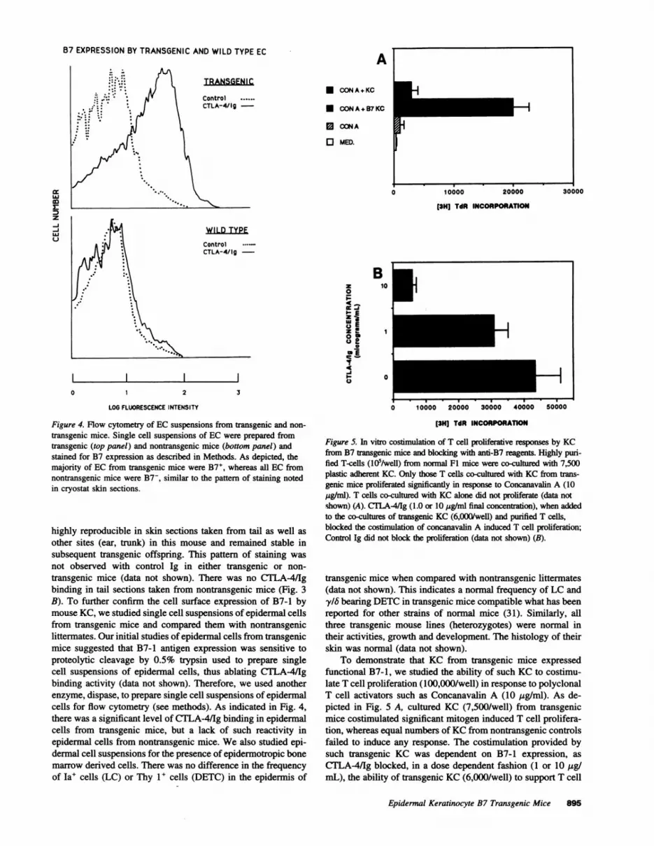
Figure 4. Flow cytometry of EC suspensions from transgenic and non-
transgenic mice. Single cell suspensions of EC were prepared fromtransgenic (top panel) and nontransgenic mice (bottom panel) andstained for B7 expression as described in Methods. As depicted, themajority of EC from transgenic mice were B7', whereas all EC fromnontransgenic mice were B7-, similar to the pattern of staining notedin cryostat skin sections.
highly reproducible in skin sections taken from tail as well as
other sites (ear, trunk) in this mouse and remained stable insubsequent transgenic offspring. This pattern of staining was
not observed with control Ig in either transgenic or non-
transgenic mice (data not shown). There was no CTLA-4/Igbinding in tail sections taken from nontransgenic mice (Fig. 3B). To further confirm the cell surface expression of B7-1 bymouse KC, we studied single cell suspensions of epidermal cellsfrom transgenic mice and compared them with nontransgeniclittermates. Our initial studies of epidermal cells from transgenicmice suggested that B7-1 antigen expression was sensitive toproteolytic cleavage by 0.5% trypsin used to prepare singlecell suspensions of epidermal cells, thus ablating CTLA-4/1gbinding activity (data not shown). Therefore, we used anotherenzyme, dispase, to prepare single cell suspensions of epidermalcells for flow cytometry (see methods). As indicated in Fig. 4,there was a significant level of CTLA-4/Ig binding in epidermalcells from transgenic mice, but a lack of such reactivity inepidermal cells from nontransgenic mice. Wealso studied epi-dermal cell suspensions for the presence of epidermotropic bonemarrow derived cells. There was no difference in the frequencyof Ia' cells (LG) or Thy 1+ cells (DETC) in the epidermis of
3
0 10000 20000 30000 40000 so500
[3H] TdR INCORPORATION
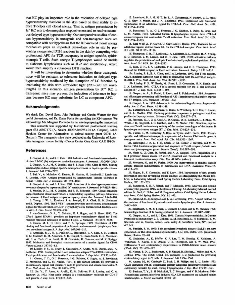
Figure 5. In vitro costimulation of T cell proliferative responses by KCfrom B7 transgenic mice and blocking with anti-B7 reagents. Highly puri-fied T-cells (105/well) from nonnal F1 mice were co-cultured with 7,500plastic adherent KC. Only those T cells co-cultured with KC from trans-genic mice proliferated significantly in response to Concanavalin A (10jig/ml). T cells co-cultured with KC alone did not proliferate (data notshown) (A). CTLA4/Ig (1.0 or 10 tig/ml final concentration), when addedto the co-cultures of transgenic KC (6,000/well) and purified T cells,blocked the costimulation of concanavalin A induced T cell proliferation;Control Ig did not block the proliferation (data not shown) (B).
transgenic mice when compared with nontransgenic littermates(data not shown). This indicates a normal frequency of LC andy/6 bearing DETCin transgenic mice compatible what has beenreported for other strains of normal mice (31). Similarly, allthree transgenic mouse lines (heterozygotes) were normal intheir activities, growth and development. The histology of theirskin was normal (data not shown).
To demonstrate that KC from transgenic mice expressedfunctional B7-1, we studied the ability of such KC to costimu-late T cell proliferation (100,000/well) in response to polyclonalT cell activators such as Concanavalin A (10 ug/ml). As de-picted in Fig. 5 A, cultured KC (7,500/well) from transgenicmice costimulated significant mitogen induced T cell prolifera-tion, whereas equal numbers of KCfrom nontransgenic controlsfailed to induce any response. The costimulation provided bysuch transgenic KC was dependent on B7-1 expression, as
CTLA-4/Ig blocked, in a dose dependent fashion (1 or 10 ug/mL), the ability of transgenic KC (6,000/well) to support T cell
Epidermal Keratinocyte B7 Transgenic Mice 895
z-J-jw

SENSITIZED MICE proliferation (Fig. 5 B). Control Ig did not block the transgenicA 25 -o TG KC costimulation response (data not shown).
-0-- NTG-s Using the sensitizer 0.5% FITC (30), we studied contact
C 2 hypersensitivity in transgenic mice (two different transgenicLn 20- mouse lines) and nontransgenic littermates. At 24 h, the peak
of the typical ear swelling response, there was no significantdifference when comparing transgenic to nontransgenic lit-
9- x /_termates (See Fig. 6, A and B). However, at later time pointsW
a/, !s|in the transgenic mice, but not the nontransgenic mice, there
LU z- °10 k/ > was a continued crescendo in the ear swelling that persisted for
a 4 IJ/ is 144 h. This difference between transgenic and nontransgenicZ hi/ mice at the 84-h time point was statistically significant (P0 51/)' \+ i < 0.05). This same pattern of exaggerated and persistent ear
swelling was noted in a second line of B7-1 transgenic mice0 ,., . ...,. (24 h ear swelling of sensitized mice: Transgenic AT = 16±3;
0 25 50 75 100 125 150 non-transgenic AT = 11+2; 144 h ear swelling of sensitized
TIME (HOURS) mice: Transgenic AT = 14±4; nontransgenic AT = 2±1).Interestingly, there was also a difference in the kinetics of
the ear swelling response of naive control animals, with thetransgenic mice again demonstrating a crescendo in ear swellingup to 144 h. The progressive in vivo ear swelling response in
25VE MICE transgenic mice was dependent on B7-1 expression, as a singleB 25 -fr-- TG-n subcutaneous injection of 20 ,g of CTLA-4/Ig before the epicu-
* NTG-n taneous application of hapten prevented this phenomenon in" o20 CTLA-4/Ig treated, but not sham treated mice (See Fig. 6 C).Z Westudied the histology of the sustained ear swelling re-
sponse in transgenic and nontransgenic naive controls in ourc o 15 contact hypersensitivity assay. Biopsy specimens taken from
the pinna of the ear of transgenic mice 21 d after the application10 of FITC were dramatically different than specimens taken from
SM x/ ' nontransgenic controls. In specimens from transgenic mice,< there was epithelial hyperplasia, vasodilation of dermal capillar-Z 5 ies as well as an inflammatory cell infiltrate comprised of mono-
nuclear cells, neutrophils and melanophages (Fig. 7 A). All
0 2 of these changes were absent in biopsy specimens taken from25 50 75 100 125 150 nontransgenic controls, in which the inflammation had resolved
TIME (HOURS) completely, resulting in histologically normal skin (Fig. 7 B).
DiscussionBLOCKING STUDY There is great interest in understanding the outcome of interactions
C 1-- NTg between T-helper cells and APC. Numerous studies have demon--*-o Tg+PBS strated that the characteristics of the APC determine the outcome
- 1Tg+CTLA-4/g of this interaction: T cell activation or tolerance induction (clonalS/l anergy) (5). Whereas professional APC express abundant class IIs./ MHCas well as costimulatory molecules, nonlymphoid cells such
as KCmay be induced to express class II MHCbut do not expresscritical costimulatory molecules (1-3). Numerous in vitro models
Z a g 4 of antigen presentation have confirmed the importance of multiplei1 APC-derived second signal molecules (B7-1, B7-2, B7-3 and possi-
Z 5- / < < * b Tbly other members of the immunoglobulin supergene family) (6-
Z / 15). Likewise, there are at least two T-cell receptors for this costimu-T/ V _X latory activity (CD28, CTLA-4 and possibly other heretofore un-
o0 1- > characterized molecules) (17ab). The redundancy of this system0 25 50 75 100 125 150
TIME (HOURS)
Figure 6. Time course of the ear swelling response to epicutaneoushapten application in sensitized and naive mice and blocking with anti- we treated a group of transgenic mice with a single subcutaneous injec-B7 reagents. Transgenic or nontransgenic animals were challenged on tion of CTLA-4/Ig (20 jig in a 20 yl volume); another group ofthe pinna of the ear 6 d after painting abdominal skin with 0.5% FITC transgenic mice received 20 yL of PBS before being challenged with(sensitized) (A) or without previous painting (naive) (B). In both the an epicutaneous application of 20 pAI of 0.5% FITC; a group of non-sensitized and naive groups, the transgenic mice developed a persistent transgenic mice served as a negative control, and received only a 0.5%ear swelling response. To block the progressive ear swelling response, FITC challenge (C).
896 Nasir et al.

- .w, -4P.
Figure 7. Histologic study of the per-sistent delayed type hypersensitivityreaction in the skin of transgenicmice. 21 d after the application of0.5% FITC to the pinna of the ear ofnaive control transgenic or non-transgenic mice, biopsy specimenswere taken for histologic examina-tion. (A) Ear skin from a transgenicmouse, with persistent inflammation.(B) Ear skin from a non-transgenicmouse, in which the inflammatory re-sponse has completely subsided (He-matoxylin and Eosin, x40).
indicates its importance in immune function, but also renders theinterpretation of in vivo studies difficult. For example, disruption ofthe B7-CD28 activation pathway in transgenic CD28-knockout miceresulted in a partial immunodeficiency state (32), suggesting thatCD28 is not required for all T cell responses in vivo and thatalternative costimulatory pathways exist and may be important.
Our transgenic mice are the first example of the aberrant ex-pression of this costimulatory ligand by naturally occurring tolero-genic APC (i.e., KC). Since stable expression of B7-1 antigen invitro by DNAmediated gene transfection renders KC as well asother B7- cell lines to become competent APC (18, 33, 34), wereasoned that targeting B7-1 to the epidermal KC of transgenicmice would induce such KC to become competent APC. Theabsence of tolerogenic APC may result in a loss of peripheraltolerance, and possibly skin disease. Wehave not observed anyspontaneous dermatologic abnormalities in these heterozygous
mice (observations of three different transgenic mouse lines up to9 mo of age, data not shown). However, our studies of cutaneousdelayed type hypersensitivity indicate that B7-1 transgenic micediffer in their responses to allergens, despite a normal skin architec-ture and normal numbers of LC and DETC. The nature of contacthypersensitivity differs qualitatively and quantitatively when com-pared to nontransgenic mice in that the amplitude and kinetics oflater phases of the contact hypersensitivity reaction are signifi-candy dissimilar. It is interesting that the most profound alterationsin contact hypersensitivity were observed in the resolution phaseof the inflammatory response. It is known that during contacthypersensitivity reactions, class II MHCantigen expression is in-duced on the cell surface of KC, presumably because of interferon-y released by activated T-lymphocytes that have migrated to theskin (35).
Based on our previous studies of Ia' KC, we hypothesized
Epidermal Keratinocyte B7 Transgenic Mice 897
..: *Korea' "',
Ill..
that KC play an important role in the resolution of delayed typehypersensitivity reactions in the skin based on their ability to in-duce T-helper cell clonal anergy. That is, antigen presentation byIa' KCacts to downregulate responsiveness and to resolve cutane-ous delayed type hypersensitivity. Our comparative studies of con-tact hypersensitivity in transgenic and non-transgenic mice arecompatible with our hypothesis that the KC-induced clonal anergymechanism plays an important physiologic role in situ by pre-venting exaggerated DTHreactions in the skin by competing withprofessional APCfor TCRoccupancy in antigen specific, epider-motropic T cells. Such anergic T-lymphocytes would be unableto elaborate lymphokines such as IL-2 and interferon-y, whichwould then amplify a cutaneous DTHreaction.
It will be interesting to determine whether these transgenicmice will be resistant to tolerance induction to delayed typehypersensitivity mediated by the disruption of LC function byirradiating the skin with ultraviolet light (290-320 nm wave-length). In this scenario, antigen presentation by B7' KC intransgenic mice may prevent the induction of tolerance to hap-tens because KC may substitute for LC as competent APC.
Acknowledgments
We thank Drs. David Scott, John Frelinger and Garvin Warner for theiruseful discussions, and Dr. Elaine Fuchs for providing the K14 cassette. Weacknowledge Ms. Margaret Piscitello for her role in preparing the manuscript.
This research was supported by Institutional postdoctoral traininggrant 532 AR07472 (A. Nasir), 1R29AR40933-01 (A. Gaspari), JohnsHopkins Center for Alternatives to animal testing grant 95041 (A.Gaspari). The transgenic mice were produced at the University of Roch-ester transgenic mouse facility (Cancer Center Core Grant CAl 1198-5).
References
1. Gaspari, A. A., and S. I. Katz. 1988. Induction and functional characterizationof class II MHC(Ia) antigens on murine keratinocytes. J. ImmnoL 140:2956-2963.
2. Gaspari, A. A., M. K. Jenkins, and S. I. Katz. 1988. Class II MHC-bearingkeratinocytes induce antigen-specific unresponsiveness in hapten-specific clones.J. ImmunoL. 141:2216-2220.
3. Bal, V., A. McIndoe, G. Denton, D. Hudson, G. Lombardi, J. Lamb, andR. Lechler. 1990. Antigen presentation by keratinocytes induces tolerance inhuman T-cells. Eur. J. ImmunoL 20:1893-1897.
4. Gaspari, A. A., and S. L. Katz. 1991. Induction of in vivo hyporesponsivenessto contact allergens by hapten-modified IaW keratinocytes. J. ImmunoL 147:4155-4161.
5. Mueller, D. L., M. K Jenkins, and R. H. Schwartz. 1989. Clonal expansionversus functional clonal inactivation: a costimulatory signalling pathway determinesthe outcome of T-cell antigen receptor occupancy. Anna. Rev. Immunol. 7:445-480.
6. Young, J. W., L. Koulova, S. A. Soergel, E. A. Clark, R. M. Steinman,and B. Dupont. 1992. The B7/BB-1 antigen provides one of several costimulatorysignals for the activation of CD4' T lymphocytes by human blood dendritic cellsin vitro. J. Clin. Invest. 90:229-237.
7. van-Seventer, G. A., Y. Shimizu, K. J. Hogan, and S. Shaw. 1990. TheLFA-1 ligand ICAM-1 provides an important costimulatory signal for T-cellreceptor-mediated activation of resting T-cells. J. Immunol. 144:4579-4586.
8. Dustin, M. I., D. Olive, and T. A. Springer. 1989. Correlation of CD2binding and functional properties of multimeric and monomeric lymphocyte func-tion associated antigen-3. J. Exp. Med. 169:503-517.
9. Armitage, R. J., W. C. Fanslow, L. Strockbine, T. A. Sato, K. N. Clifford,B. M. Macduff, D. M. Anderson, S. D. Gimpel, T. Davis-Smith, C. R. Maliszew-ski, E. A. Clark, C. A. Smith, K. H. Grabstein, D. Cosman, and M. K. Spriggs.1992. Molecular and biological characterization of a murine ligand for CD40.Nature (Lond.). 357:80-82.
10. Linsley, P. S., W. Brady, L. Grosmaire, A. Aruffo, N. K. Damle, and J. A.Ledbetter. 1991. Binding of the B cell activation antigen B7 to CD28 costimulatesT cell proliferation and Interleukin-2 accumulation. J. Exp. Med. 173:721-730.
11. Gimmi, C. D., G. J. Freeman, J. G. Gribben, K. Sugita, A. S. Freedman,C. Morimoto, and L. M. Nadler. 1991. B-cell surface antigen B7 provides acostimulatory signal that induces T cells to proliferate and secrete Interleukin-2.Proc. Nat!. Acad. Sci. USA. 88:6575-6579.
12. Liu, Y., Y. Jones, A. Aruffo, K. M. Sullivan, P. S. Linsley, and C. A.Janeway, Jr. 1992. Heat-stable antigen is a costimulatory molecule for CD4 Tcell growth. J. Exp. Med. 175:437-445.
13. Lenschow, D. J., G. H.-T. Su, L. A. Zuckerman, N. Nabavi, C. L. Jellis,G. S. Gray, J. Miller, and J. A. Bluestone. 1993. Expression and functionalsignificance of an additional ligand for CTLA-4. Proc. Natd. Acad. Sci. USA.90:11054-11058.
14. Boussiotis, V. A., G. J. Freeman, J. G. Gribben, J. Daley, G. Gray, andL. M. Nadler. 1993. Activated human B lymphocytes express three CTLA-4counterreceptors that costimulate T-cell activation. Proc. NatL. Acad. Sci. USA.90:11059-11063.
15. Razi-Wolf, Z., F. Galvin, G. Gray, and H. Reiser. 1993. Evidence for anadditional ligand, distinct from B7, for the CTLA-4 receptor. Proc. NatL Acad.Sci. USA. 90:11182-11186.
16. Thompson, C. B., T. Lindsten, J. A. Ledbetter, S. L. Kunkel, H. A. Young,S. G. Emerson, J. M. Leiden, and C. H. June. 1989. CD28 activation pathwayregulates the production of multiple T cell-derived lymphokines/cytokines. Proc.Nat!. Acad. Sci. USA. 86:1333-1337.
17. June, C. H., J. A. Ledbetter, P. S. Linsley, and C. B. Thompson. 1990.Role of the CD28 receptor in T-cell activation. Immunol. Today. 11:211-216.
17a. Linsley, P. S., E. A. Clark, and J. A. Ledbetter. 1990. The T-cell antigen,CD28, mediates adhesion with B cells by interacting with the activation antigen,B7/BB-l. Proc. NatL. Acad Sci. USA. 87:5031-5035.
17b. Linsley, P. S., W. Brady, M. Urnes, L. S. Grosmaire, N. K. Damle, andJ. A. Ledbetter. 1991. CTLA-4 is a second receptor for the B cell activationantigen B7. J. Exp. Med. 174:561-569.
18. Gaspari, A. A., B. Ferbel, F. Razvi, and R. Polakowska. 1993. Accessoryand alloantigen-presenting cell function of A431 keratinocytes that stably expressthe B7 antigen. Cell. Immunol. 149:291-302.
19. Gaspari, A. A. 1993. Advances in the understanding of contact hypersensi-tivity. AnL J. Cont. Derm 4:138-149.
20. Yamamura, M., K. Uyemura, R. Dians, K. Weinberg, T. H. Rea, B. Bloom,and R. L. Modlin. 1991. Defining protective responses to pathogens: cytokineprofiles in Leprosy lesions. Science (Wash. DC). 254:277-279.
21. Freeman, G. J., G. S. Gray, C. D. Gimmi, D. B. Lombard, L.-J. Zhou, M.White, J. D. Fingeroth, J. G. Gribben, and L. M. Nadler. 1991. Structure, expres-sion, and T-cell costimulatory activity of the murine homologue of the human Blymphocyte activation antigen B7. J. Exp. Med. 174:625-631.
22. Vassar, R., M. Rosenberg, S. Ross, A. Tyner, and E. Fuchs. 1989. Tissue-specific and differentiation-specific expression of a human K14 keratin gene intransgenic mice. Proc. NatL. Acad Sci. USA. 86:1563-1567.
23. Gascoigne, J. R. J., Y.-H. Chien, D. M. Becker, J. Kavaler, and M. M.Davis. 1984. Genomic organization and sequence of T-cell receptor 13-chain con-stant- and joining-region genes. Nature (Lond.). 310:387-391.
24. Nasir A., Z. Chen, B. Ferbel, and A. A. Gaspari. 1993. Preparation of aK14 promoter driven mouse B7 cDNA construct and functional analysis in atransient co-stimulation assay. Clin. Res. 41:606a. (Abstr.)
25. Moroson, H., and M. Furlan. 1970. An improvement in alkaline sucrosedensity gradient sedimentation of mammalian cell DNA. Radiation Research.44:713-726.
26. Hogan, B., F. Costantini, and E. Lacy. 1986. Introduction of new geneticinformation into the developing mouse embryo. In Manipulating the Mouse Em-bryo. A Laboratory Manual. Cold Spring Harbor Laboratory Press, Cold SpringHarbor, NY. 152-203.
27. Sambrook, J., E. F. Fritsch, and T. Maniatis. 1989. Analysis and cloningof eukaryotic genomic DNA. In Molecular Cloning: A Laboratory Manual, secondedition. N. Ford, C. Nolan, and M. Ferguson, editors. Cold Spring Harbor Labora-tory Press, Cold Spring Harbor, NY. 9.1-62.
28. Julius, M. H., E. Simpson, and L. A. Herzenberg. 1973. A rapid method forthe isolation of functional thymus-derived murine lymphocytes. Eur. J. Immunol.3:645-649.
29. Breathnach, S. M., S. I. Katz, L. Clement, L Green, and E. M. Shevach. 1978.Immunologic function of Ia bearing epidermal LC. J. Immunot 121:2005-2013.
30. Gaspari, A. A., and S. I. Katz. 1991. Contact Hypersensitivity. In CurrentProtocols in Immunology. J. E. Coligan, A. M. Kruisbeek, D. H. Margulies, E. M.Shevach, and W. Strober, editors. John Wiley & Sons/New York, NY. Section4.2:1-5.
31. Streilein, J. W. 1990. Skin associated lymphoid tissues (SALT): the nextgeneration. In The Skin Immune System (SIS). J. D. Bos, editor. CRCpress/BocaRaton, Florida. 25-48.
32. Shahinian, A., K. Pfeffer, K. P. Lee, T. M. Kundig, K. Kishihara, A.Wakeham, K. Kawai, P. S. Ohashi, C. B. Thompson, and T. W. Mak. 1993.Differential T cell costimulatory requirements in CD28-deficient mice. Science(Wash. DC). 261:609-612.
33. Norton, S. D., L. Zuckerman, K. B. Urdahl, R. Shefner, J. Miller, and M. K.Jenkins. 1992. The CD28 ligand, B7, enhances IL-2 production by providingcostimulatory signal to T cells. J. Immunol. 149:1556-1561.
34. Azuma, M., M. Cayabyab, D. Buck, J. H. Phillips, and L. L Lanier. 1992.CD28 Interaction with B7 costimulates primary allogeneic proliferative responses andcytotoxicity mediated by small, resting T lymphocytes. J. Emp. Med 175:353-360.
35. Basham, T. Y., B. H. Nickoloff, T. C. Merigan, and V. B. Morhenn. 1984.Recombinant gamma interferon induces HLA-DR expression on cultured humankeratinocytes. J. Invest. Dermnatol. 83:88-90.
898 Nasir et al.


