Embed Size (px)
Citation preview

Molecular Cell, Vol. 20, 65–75, October 7, 2005, Copyright ©2005 by Elsevier Inc. DOI 10.1016/j.molcel.2005.08.012
Exon-Junction Complex Components SpecifyDistinct Routes of Nonsense-Mediated mRNA Decaywith Differential Cofactor Requirements
Niels H. Gehring,1,2 Joachim B. Kunz,1,2
Gabriele Neu-Yilik,1,2 Stephen Breit,2
Marcelo H. Viegas,1 Matthias W. Hentze,1,3,*and Andreas E. Kulozik1,2,*1Molecular Medicine Partnership UnitUniversity of Heidelberg andEuropean Molecular Biology LaboratoryHeidelberg 69120Germany2Department for Pediatric Oncology, Hematology
and ImmunologyUniversity Hospital HeidelbergHeidelberg 69120Germany3Gene Expression UnitEuropean Molecular Biology LaboratoryHeidelberg 69117Germany
Summary
Messenger RNAs (mRNAs) bearing premature trans-lation termination codons (PTCs) are degraded bynonsense-mediated mRNA decay (NMD). For mamma-lian NMD, current models propose a linear pathwaythat involves the splicing-dependent deposition ofexon-junction complexes (EJCs) and the sequentialaction of the NMD factors UPF3, UPF2, and UPF1. Weshow here that different EJC proteins serve as entrypoints for the formation of distinguishable NMD-acti-vating mRNPs. Specifically, Y14, MAGOH, and eIF4A3can activate NMD in an UPF2-independent manner,whereas RNPS1-induced NMD requires UPF2. Weidentify the relevant regions of RNPS1, eIF4A3, Y14,and MAGOH, which are essential for NMD and provideinsights into the formation of complexes, that classifyalternative NMD pathways. These results are integ-rated into a nonlinear model for mammalian NMD in-volving alternative routes of entry that converge at acommon requirement of UPF1.
Introduction
NMD distinguishes between normal termination co-dons and PTCs in a process that requires active trans-lation (Maquat, 2004). Mammalian NMD also criticallydepends on splicing, which imprints the mRNA withEJCs that consist of multiple proteins that have beenshown to facilitate mRNA export (REF/Aly) and to en-able recognition of PTCs and activation of NMD (eIF4A3,BTZ, Y14, MAGOH, and RNPS1) (Kataoka et al., 2000,2001; Le Hir et al., 2000, 2001a, 2001b; Zhou et al.,2000; Lykke-Andersen et al., 2001; Chan et al., 2004;Degot et al., 2004; Ferraiuolo et al., 2004; Palacios etal., 2004; Shibuya et al., 2004). Generally, termination
*Correspondence: [email protected] (M.W.H.); [email protected] (A.E.K.)
codons more than 55 nucleotides (nt) upstream of the3#-most exon-exon junction elicit NMD (Maquat, 2004;Holbrook et al., 2004).
Current NMD models postulate a linear pathway inwhich the EJC sequentially recruits UPF3, UPF2, andUPF1 to trigger NMD (Kim et al., 2001; Lykke-Andersenet al., 2001). UPF1 is thought to link the EJC and theUPF proteins to the translation termination event, as itcan directly interact with the release factors (Czaplinskiet al., 1998). The essential role of UPF1 in NMD is un-derscored by the fact that RNAi-mediated depletion ofUPF1 stabilizes all known NMD substrates (Mendell etal., 2002, 2004; Gehring et al., 2003). UPF3 (of whichtwo isoforms exist in mammalian cells) directly associ-ates with the EJC, whereas UPF2 binds to UPF3 and toUPF1 (Lykke-Andersen et al., 2000, 2001; Mendell et al.,2000; Kim et al., 2001; Kadlec et al., 2004). UPF2 is thusthought to serve as an obligatory biochemical link inthe chain of UPF-protein interactions. Consistent withthis, depletion of UPF2 limits NMD of different sub-strates in mammalian cells (Mendell et al., 2002, 2004).
The DExH/D box helicase eIF4A3 (DDX48) appearsto play an important role in EJC deposition by bindingdirectly to the spliced mRNA. As such, it constitutes atleast part of the platform that anchors additional EJCproteins to the mRNA (Shibuya et al., 2004). eIF4A3 in-teracts with the Y14-MAGOH heterodimer (Chan et al.,2004; Palacios et al., 2004; Shibuya et al., 2004) andwith Barentsz (BTZ, also known as MLN51 or CASC3;Palacios et al., 2004). eIF4A3 and BTZ interact in theyeast two-hybrid assay and in GST pull-down experi-ments, indicating that their binding is direct (Palacios etal., 2004). Depletion of Y14 (RBM8A), BTZ, and eIF4A3 bymeans of RNAi limits NMD in mammalian cells (Gehringet al., 2003; Ferraiuolo et al., 2004; Palacios et al., 2004;Shibuya et al., 2004).
Another EJC component, RNPS1, was initially iden-tified as a general activator of splicing (Mayeda et al.,1999). It binds preferentially to spliced mRNAs (Mayedaet al., 1999; Le Hir et al., 2000) and activates NMD whentethered to an NMD reporter (Lykke-Andersen et al.,2001). RNPS1 coimmunoprecipitates with human NMDfactors, namely UPF1, UPF2, and UPF3a/b, and it isthought to act by supporting the association of the UPFproteins with the EJC in an as yet unidentified way(Lykke-Andersen et al., 2001).
This work uncovers that NMD-competent mRNPscan be generated by distinct routes that are charac-terized by different EJC protein entry points and dis-tinct cofactor requirements. NMD activated by tetheredY14, MAGOH, UPF3b, or eIF4A3 is insensitive to UPF2depletion. By contrast, RNPS1-induced NMD displaysstrong UPF2 requirement. Both routes involve UPF3band require UPF1 regardless of the NMD entry point.We identify cellular mRNAs that are controlled by NMDin a UPF1-dependent way and that display strong de-pendence either on RNPS1 and UPF2 or on BTZ. ThesemRNAs appear to represent physiological substrates ofthe two branches implicated and defined by the teth-ered function analyses.

Molecular Cell66
Results
The λN-peptide-boxB interaction enabled the analysisof NMD activation by UPF3b, and Y14 tethered to the3#UTR of NMD-reporter mRNAs (Gehring et al., 2003).A similar system using the MS2-coat protein identifiedUPF1, UPF2, UPF3a, UPF3b, and RNPS1 as activatorsof NMD when tethered to reporter mRNAs (Lykke-Andersen et al., 2000, 2001). To systematically analyzedifferent EJC and UPF proteins, we generated a set ofMS2- or λN-boxB-based tethering reporters (Figure S1available in the Supplemental Data with this article on-line). The majority of tested proteins decrease both theMS2- and λNV5-boxB-based reporter mRNAs. How-ever, RNPS1 and eIF4A3 function better bearing anMS2 tag, whereas MAGOH is more active when fusedto a λN tag (data not shown).
Different EJC Components Define Distinct Branchesof the NMD Pathway with DifferentialCofactor RequirementsWe combined tethering of the EJC proteins Y14, MAGOH,UPF3b, RNPS1, or eIF4A3 with the simultaneous siRNA-mediated depletion of either UPF1, UPF2, or BTZ toexplore whether all EJC components share the sameNMD cofactor requirements. Transfection of an siRNAagainst luciferase mRNA (Luc) served as negative con-trol. Depletion efficiencies were assessed by immu-noblotting of lysates from cells transfected with theUPF1, UPF2, or BTZ-specific siRNAs and compared tocells that were transfected with Luc-siRNA (Figures 1A–1C). This analysis shows that UPF1, UPF2, and BTZ areefficiently depleted by the respective siRNAs to levelsof less than 10% (UPF1, Figure 1A), w10% (UPF2, Fig-ure 1B), and w15% (BTZ, Figure 1C).
We next determined whether the depletion of UPF1,UPF2, eIF4A3, or BTZ inhibits NMD mediated by thedifferent tethered EJC proteins. Northern blot analysisdemonstrates that the abundance of NMD reportermRNAs with tethered EJC components is strongly de-creased when none of the NMD factors is ablated byRNAi (i.e., after transfection of the Luc-siRNA; Figure1D, lanes 1–6; Figure 1E, lanes 1 and 2). When UPF1is depleted, the NMD activity of all factors is severelyimpaired (w4- to 5-fold; Figure 1D, lanes 7–12; Figure1E, lanes 3 and 4). Thus, tethering of any of these EJCproteins recapitulates mammalian NMD that is charac-terized by its UPF1 dependence.
Assuming a widely accepted linear pathway of NMD(EJC/UPF3/UPF2/UPF1), one would expect thatUPF2 depletion should inactivate the NMD activity oftethered EJC components. In accordance with thisexpectation, NMD of reporter mRNAs with tetheredRNPS1 is exquisitely UPF2 dependent (Figure 1D; lanes6 and 18). By contrast, Y14, MAGOH, UPF3b, andeIF4A3 show normal and unexpected NMD activationafter the depletion of UPF2 under the same conditions(Figure 1D, lanes 13–16; Figure 1E, lanes 5 and 6).These results show that tethered RNPS1 displays theexpected requirement for UPF2, whereas tetheredUPF3b, eIF4A3, Y14, or MAGOH are all NMD active inUPF2-depleted cells.
Conversely, RNPS1-mediated NMD is resistant to de-
pNi1Yp1MdpdfddtAiebpptrU
CtTsippNtss
aftcSdaispubNtPRssoabGgsNt
letion of BTZ (Figure 1D, lanes 23 and 24), whereasMD induced by Y14, MAGOH, eIF4A3, and UPF3b is
mpaired 2.1- to 3.1-fold in BTZ-deficient cells (FigureD, lanes 19–22; Figure 1E lanes 7 and 8). Furthermore,14-MAGOH-induced NMD is sensitive to eIF4A3 de-letion, whereas RNPS1-induced NMD is not (FigureF). These data show that the EJC components Y14,AGOH, eIF4A3, and UPF3b can act with severely re-uced levels of UPF2, whereas Y14 and MAGOH de-end on BTZ and eIF4A3. By contrast, RNPS1 is UPF2ependent but BTZ and eIF4A3 independent. These
indings suggest that mammalian NMD surprisinglyoes not follow a linear, unbranched pathway but thatifferent EJC components define distinct branches ofhe NMD pathway with distinct cofactor requirements.lternatively, the surprising differences between UPF2-
ndependent NMD induced by tethered Y14, MAGOH,IF4A3, and UPF3b and UPF2-dependent NMD inducedy tethered RNPS1 may be caused by a potential non-hysiological assembly of NMD-competent mRNP com-lexes during the tethering assay. To distinguish betweenhese two alternatives, we next sought for cellular NMD-egulated transcripts with differential sensitivities toPF2, RNPS1, or BTZ depletion.
ellular mRNAs Regulated by UPF1, which Followhe RNPS1/UPF2 or BTZ Branch of NMDo identify cellular mRNAs that are regulated by repre-entatives of the two branches of the NMD pathwaymplicated by our tethered function analyses, we ex-lored the gene expression profiles of HeLa cells de-leted of UPF1, UPF2, RNPS1, or BTZ. Putative cellularMD substrates that were defined by their sensitivity
o UPF1 depletion were examined regarding their re-ponse to depletions of UPF2, RNPS1, and BTZ, re-pectively.Eight cellular mRNAs were selected for in-depth
nalysis (Table S1). Seven of these display structuraleatures (uORFs, 3#UTR introns) that would predisposehem to NMD (Mendell et al., 2004), and all eight in-rease in their abundance after UPF1 depletion (Table1 and Figure 2). The tethered function analyses pre-ict the existence of NMD substrates that are primarilyffected by depletion of RNPS1 and UPF2 while show-
ng minor responses to BTZ depletion; by contrast,ubstrates belonging to the other NMD branch shouldrimarily respond to BTZ depletion and be relativelynaffected by UPF2 and RNPS1 depletion. The un-ranched linear pathway, however, predicts that allMD substrates respond in similar ways to the deple-
ion of their common cofactors. As shown in Figure 2,TGS2, TGM2, and EPAS1 mRNAs are induced byNPS1 or UPF2 depletion nearly as strongly or moretrongly, respectively, as by depletion of UPF1; they re-pond very little to BTZ depletion (Figure 2). On thether hand, SC35 1.7 Kb and SC35 1.6 Kb (Sureau etl., 2001) are induced by depletion of BTZ and UPF1,ut not by depletion of RNPS1 and UPF2 (Figure 2). The8, TIMP1, and SLC3A2 transcripts represent a thirdroup of cellular NMD targets. All three transcriptshow a reproducible but less marked sensitivity toMD-protein depletion of both branches (Figure 2). Al-
hough there are no obvious distinguishing structural

Complexes Specifying Mammalian NMD67
Figure 1. Cofactor Requirements of NMD Induced by Different Tethered EJC Proteins
(A) Immunoblot analysis of protein lysates from HeLa cells transfected with siRNAs against luciferase (lanes 1–4) or UPF1 (lane 5) with a UPF1antibody. Dilutions corresponding to 50%, 20%, or 10% (lanes 2–4) of the initial protein amount (lane 1) from Luc-siRNA-transfected cellswere loaded to assess the efficiency of the UPF1 depletion. Reprobing with a tubulin-specific antibody was performed to control for loading.(B) Immunoblot analysis of protein lysates from HeLa cells transfected with siRNAs against luciferase (lanes 1–4) or UPF2 (lane 5) with aUPF2 antibody as described in (A).(C) Immunoblot analysis of protein lysates from HeLa cells transfected with siRNAs against luciferase (lanes 1–4) or BTZ (lane 5) with a BTZantibody as described in (A).(D) Northern blot analysis of RNA isolated from HeLa cells transfected with the indicated siRNAs (Luc, lanes 1–6; UPF1, lanes 7–12; UPF2,lanes 13–18; and BTZ, lanes 19–24). 48 hr later, the cells were cotransfected with plasmids for the transfection efficiency control (ctrl.), theNMD reporters, and the unfused λNV5 (lanes 1, 7, 13, and 19), MS2 (cp, lanes 5, 11, 17, and 23), λNV5-Y14 (lanes 2, 8, 14, and 20), λNV5-MAGOH (lanes 3, 9, 15, and 21), λNV5-UPF3b (4, 10, 16, and 22), or MS2-RNPS1 (lanes 6, 12, 18, and 24). Percentages ± SEM are meansfrom at least three independent experiments.(E) Northern blot analysis of RNA from HeLa cells transfected with the indicated siRNAs (Luc, lanes 1 and 6; UPF1, lanes 3 and 4; UPF2,lanes 5 and 6; and BTZ, lanes 7 and 8). 48 hr later, the cells were transfected with vectors for the 6MS2 reporter, the control (ctrl.), and MS2-eIF4A3 (lanes 2, 4, 6, and 8) or MS2 (cp, lanes 1, 3, 5, and 7).(F) Northern blot analysis of RNA from HeLa cells that were transfected with the indicated siRNAs (Luc, lanes 1–5; eIF4A3, lanes 6–10). 48 hrlater, the cells were cotransfected with vectors for the control (ctrl.), the NMD reporters, and unfused λNV5 (lanes 1 and 6), MS2 (cp, lanes 4and 9), λNV5-Y14 (lanes 2 and 7), λNV5-MAGOH (lanes 3 and 8), or MS2-RNPS1 (lanes 5 and 10).
features of these mRNAs, the observed differential re-sponses of endogenous NMD targets to NMD cofactordepletion clearly contradict the expectation of a linearpathway but support the existence of two branches ofthe NMD pathway.
Functional Complementation of UPF1and UPF2 DepletionsThe important conclusions reached above require vali-dation by controlling for potential off-target effects ofthe RNAi strategy (Jackson et al., 2003). We thus trans-fected cells treated with UPF1 siRNAs with a plasmid
that expresses a FLAG-tagged UPF1 that is resistantto the UPF1-siRNA. Immunoblotting shows that UPF1levels can be fully restored (Figure 3A). Consistent withthe data shown in Figure 1, UPF1 depletion increasesthe abundance of all reporter mRNAs with tetheredRNPS1, Y14, MAGOH, UPF3b, and eIF4A3 (Figures 3Cand 3D). When the UPF1 depletion is complementedwith the siRNA-resistant, FLAG-UPF1 expression plas-mid, these effects are completely reversed (Figures 3Cand 3D).
A similar approach was used to validate the data ob-tained with the UPF2 depletion. Again, immunoblot

Molecular Cell68
Figure 2. NMD of Cellular Targets Shows Dif-ferential Sensitivity to RNPS1-UPF2 or BTZDepletion
UPF1, UPF2, BTZ, and RNPS1 protein levelswere depleted by specific siRNAs to lessthan 10% (UPF1, RNPS1), w10% (UPF2),and w15% (BTZ; see Figures 1A–1C andFigure S5). The abundance of cellular NMD-regulated transcripts was determined byquantitative RT-PCR using RNA from HeLacells transfected with UPF1-, UPF2-, RNPS1-,BTZ-, or Luc-siRNA (negative control). Rela-tive expression levels were normalized toGAPD mRNA levels. Fold increases relate tolevels in Luc-siRNA-treated cells. The errorbars indicate SEM that are based on fourindependent experiments. Two differentsiRNAs targeting RNPS1 gave comparableresults.
analysis confirms that expression of the UPF2-siRNA-resistant V5-UPF2 plasmid replenishes UPF2 levels(Figure 3B). The complementation of the depleted UPF2reestablishes the NMD activity of tethered RNPS1 (Fig-ure 3E). Thus, the observed increased abundance ofthe reporter mRNAs is a specific result of the UPF1 andUPF2 depletions and not caused by unspecific or off-target effects of the UPF1- or UPF2-siRNAs.
The S Domain of RNPS1 Is Required to Trigger NMDand Recruits UPF3b and UPF2We next sought to identify the structural basis for thefunctionally defined alternative NMD pathways. RNPS1is unique amongst the EJC components analyzed herein that it triggers fully UPF2-dependent NMD. It con-sists of four major structural domains (Figure 4A): theN terminus (residues 1–68), an S domain (res. 69–121),an RRM domain (res. 163–243), and the C terminus (res.251–305). We generated RNPS1 deletion mutants (Fig-ure 4A) to identify the region(s) required for its NMDactivity and for its interaction with the UPF-proteins.These mutants were examined in the tethering assayand compared to full-length (wt) RNPS1. Surprisingly,the NMD activity of RNPS1 is lost only when the S do-main is removed (�69–121; Figure 4B, lane 3), and dele-tion of the entire RRM or the N- or C termini is tolerated(Figure 4B, lanes 4–6). The abundance of the deletionmutants in whole-cell extracts is comparable to wtRNPS1 (Figure 4C, top), whereas less �69–121 andmore �1–68 and �252–305 protein is detected in cyto-plasmic extracts (Figure 4C, bottom). Tethering ofRNPS1�69–121 induces an additional band detected inthe Northern blot (Figure 4B), which could represent anaberrantly processed reporter transcript. The genera-tion of this side product, however, cannot account forthe decreased NMD activity of the RNPS1�69–121 mu-tant, because aberrant splicing or 3# end processingwould be expected to decrease, not increase, theamount of reporter mRNA. In addition, we directly ana-lyzed the steady-state localization of GFP-tagged RNPS1
ddttfd
RffRfa�tbfftwtiTiao
YBWipMethE(wo
erivatives (Figure S2). Like full-length RNPS1, all testedeletion mutants exhibit a predominantly nuclear localiza-ion, although the pattern of nuclear speckles formed byhe RNPS1 mutants appears to differ from the pattern ofull-length RNPS1. In summary, we conclude that the Somain is critical for NMD induced by tethered RNPS1.As shown in Figures 1 and 3, the NMD activity of
NPS1 requires UPF2. This suggests that RNPS1 mayorm a complex with UPF2 and presumably additionalactors. Indeed, it has been reported previously thatNPS1 coimmunoprecipitates all human UPF proteins
rom cell extracts (Lykke-Andersen et al., 2001). We an-lyzed FLAG-tagged RNPS1 and the deletion mutants1–68, �69–121, and �163–243 for coimmunoprecipita-
ion of UPF2 and UPF3b. Western blotting confirms thatoth UPF2 and UPF3b are immunoprecipitated with
ull-length RNPS1 (Figure 4D, lane 2). Importantly, theunctionally active �1–68 and �163–243 can also bindhese two UPF proteins (Figure 4D, lanes 2, 4, and 5),hereas the functionally inactive �69–121 coprecipi-
ates strikingly less UPF3b than wt RNPS1, and UPF2s almost completely absent (Figure 4D, lanes 2 and 3).he functional dependence between UPF2 and RNPS1
n one of the alternative NMD pathways is thus structur-lly reflected by the interaction between the S domainf RNPS1 and UPF2.
14-MAGOH Heterodimers Interact with eIF4A3,TZ, and UPF3b to Activate NMDe next characterized structural requirements for the
nteractions necessary for NMD activation by the EJCroteins of the other NMD pathway (eIF4A3, Y14, andAGOH). Heterodimerization of Y14 and MAGOH is
ssential for their NMD activity. In addition, surfaceshat are thought to recruit essential NMD factors to theeterodimer are defined by the residues K16/F17, D66/68, R85/E87, K130/F134 (MAGOH), and L106/R108
Y14) (Fribourg et al., 2003). Based on this information,e investigated the effect of known and new mutationsf surface-exposed residues (Figure S3) together with

Complexes Specifying Mammalian NMD69
Figure 3. Functional Complementation of UPF1 or UPF2 Depletion
(A) Immunoblot analysis of UPF1 depletion and complementation was performed as described in Figure 1A. For complementation of theUPF1 depletion, cells were transfected with 0.1 �g FLAG-UPF1R (lane 3).(B) Analysis of UPF2 depletion and complementation was performed as described in Figure 1B. To complement the UPF2 depletion, cellswere transfected with 1.0 �g V5-UPF2R (lane 3).(C) Northern blot analysis of RNA from HeLa cells transfected with siRNAs against luciferase (lanes 1–6) or UPF1 (lanes 7–12). 48 hr later, thecells were transfected with plasmids expressing NMD reporters and the indicated MS2- or λNV5-tagged proteins as in Figure 1D. The UPF1depletion was complemented as in Figure 2A.(D) Northern blot analysis of RNA isolated from HeLa cells transfected with siRNAs against luciferase (lanes 1 and 2) or UPF1 (lanes 3–6). 48hr later, the cells were transfected with plasmids expressing the 6MS2 reporter and MS2-coat protein or MS2-eIF4A3 as described in Figure1E. The UPF1 depletion was complemented as in Figure 2A.(E) Northern blot analysis of RNA isolated from HeLa cells transfected with siRNAs against luciferase (lanes 1 and 2) or UPF2 (lanes 3–6). 48hr later, the cells were transfected with plasmids expressing the 4MS2 reporter and MS2-coat protein or MS2-tagged RNPS1 as in Figure 1D.For the complementation of the UPF2 depletion, cells were cotransfected with 1 �g of the V5-UPF2R plasmid as in Figure 2B.
two known mutations interfering with Y14-MAGOH het-erodimerization and a new Y14 mutation predicted tointerrupt Y14-MAGOH binding (Lau et al., 2003). Weconfirmed that most of the previously analyzed mutantsare impaired in their NMD function (Figures 5A and 5C;Fribourg et al., 2003), whereas two MAGOH mutantsare fully active (E72R/D73K and R85E/E87R; Figure 5A,lanes 6 and 7). The D66R/E68R MAGOH mutation leadsto a slight decrease of NMD activity (Figure 5A, lane 5),the K16E/F17A exerts an intermediate decline of NMDactivity (Figure 5A, lane 3), and all other mutations(K41D/N42A, K130E/F134A, and L136R) abolish NMDactivity of MAGOH completely (Figure 5A, lanes 4 and6–9). Similarly, the Y14 mutant E82R/E83R reducesNMD slightly (Figure 5C, lane 3), whereas all other mu-tations (L106E/R108E, L118R, and C149K/F150A) abol-
ish Y14-induced NMD (Figure 5C, lanes 4–6). The pro-tein expression levels of the MAGOH and Y14 mutantsare similar to the respective wt proteins, except for theMAGOH mutant L136R, which reproducibly shows a re-duced expression (Figures 5A and 5C, bottom panels).Hence, differential expression does not generally ac-count for the observed effects of the mutations.
Next, we analyzed these mutants for complex forma-tion with the other EJC proteins using FLAG-taggedMAGOH (Figure 5B) or FLAG-tagged Y14 and their re-spective mutants (Figure 5D). We included PYM/p29(WIBG) in this analysis, because this protein has beenshown to interact with Y14-MAGOH and thus repre-sents a putative NMD factor, although its function is notyet fully defined (Bono et al., 2004). FLAG-tagged wtMAGOH and FLAG-tagged Y14 coimmunoprecipitate

Molecular Cell70
Figure 4. The S Domain of RNPS1 Is Critical for Its NMD Function
(A) Schematic representation of the domain architecture of RNPS1. The S domain (residues 69–121) and the RRM (residues 163–243) areshown in light and dark gray, respectively.(B) Northern blot analysis of RNA isolated from HeLa cells transfected with vectors for the 4MS2 reporter (4MS2), the control (ctrl.) and MS2(cp, lane 1), MS2-tagged full-length RNPS1 (wt, lane 2), or the indicated deletion mutants (lanes 3–6). An additional band that is induced bythe expression of the �69–121 mutant is marked by an asterisk.(C) Immunoblot analysis of total (top) or cytoplasmic (bottom) lysates from the HeLa cells analyzed in (B) with an MS2 antibody. The blot wasreprobed with a GFP antibody (ctrl.).(D) Immunoprecipitations of RNaseA-treated lysates from HeLa cells that were transfected with FLAG-tagged full-length RNPS1 (wt, lane 2),the indicated deletion mutants (lane 3–5), or unfused FLAG (lane 1). 44 hr after transfection, cells were harvested and complexes wereimmunoprecipitated by using anti-FLAG (M2) agarose beads. Immunoblot analysis was performed with UPF2 or UPF3b antibodies. Themembrane was reprobed with anti-FLAG antibody (M2) to control for differences in loading and precipitation efficiencies.
BTZ, UPF3b, eIF4A3, and PYM/p29 (Figure 5B, lane 2;Figure 5D, lane 2). In addition, FLAG-Y14 coprecipitatesMAGOH (Figure 5D, lane 2), and FLAG-MAGOH copre-cipitates Y14 (Figure 5B, lane 2). For the fully NMD-active MAGOH mutant R85E/E87R, the pattern of pre-cipitated proteins is indistinguishable from wt MAGOH(Figure 5B, lanes 2 and 7), explaining its activity in theNMD assay. The other active MAGOH mutant E72R/D73K does not precipitate PYM/p29 but, instead, showsan increased precipitation of BTZ, UPF3b, and eIF4A3(Figure 5B, lane 6). Strikingly, the pattern of proteinsthat coimmunoprecipitate with all other FLAG-MAGOHmutants correlates with the level of residual NMD activ-ity of these mutants. D66R/E68R fails to coprecipitatePYM/p29 and precipitates significantly less UPF3b(Figure 5B, lane 5); the immunoprecipitations of K16E/F17A and K41D/N42A lack eIF4A3, UPF3b, and BTZ(Figure 5B, lanes 3 and 4); and K130E/F134A binds onlyY14 and significantly reduced amounts of PYM/p29(Figure 5B, lane 8). L136R, which is expressed and pre-cipitated at reduced levels, does not form a hetero-dimer and thus does not interact with PYM/p29,eIF4A3, UPF3b, and BTZ. (Figure 5B, lane 9). Similarly,the NMD-inactive Y14 L106E/R108E mutant coprecipi-tates only MAGOH and faintly PYM/p29 (Figure 5D, lane
4nnlEcal
pNBttttft(etamrc
), whereas the inactive L118R and C149K/F150A doot form a heterodimer with MAGOH and, hence, doot coprecipitate any of the five proteins (Figure 5D,
anes 5 and 6). The partially active Y14 mutant E82R/83R, in analogy to MAGOH D66R/E68R, does not pre-ipitate UPF3b and PYM/p29, whereas BTZ and eIF4A3re precipitated with decreased efficiency (Figure 5D,
ane 3).Thus, the ability to form a complex containing the
roteins BTZ, eIF4A3, and UPF3b correlates with theMD activity of Y14 and MAGOH in the tethering assay.oth, eIF4A3 and BTZ seem to play a crucial role. When
hey are absent from the precipitated complex, the ac-ivity of Y14 or MAGOH is most strongly affected. Weherefore suggest that the interaction of these four fac-ors is necessary for the recruitment of the downstreamactors of the NMD pathway such as UPF1. It is notablehat UPF3b, which is part of the RNPS1-UPF2 complexFigure 4D), is also part of this complex. In contrast toIF4A3 and BTZ, which are essential for NMD activity ofethered Y14 and MAGOH, the requirement for UPF3bppears to be less marked. Interestingly, complex for-ation between Y14-MAGOH and UPF3b appears to
equire eIF4A3 and BTZ, because UPF3b does not pre-ipitate with mutants that do not interact with eIF4A3

Complexes Specifying Mammalian NMD71
Figure 5. Analysis of MAGOH and Y14 Mutants Reveals the Functional Importance of Their Interactions with eIF4A3, BTZ, and UPF3b
(A) Northern blot analysis of RNA isolated from HeLa cells that were transfected with plasmids for the 4boxB reporter (4boxB), the control(ctrl.) and unfused λNV5 (lane 1), or the indicated λNV5-tagged MAGOH wt (lane 2) point (lane 9) and double-point mutants (lanes 3–8). Bottom,analysis of the expression of the MAGOH mutants used in the top panel with a V5 antibody. Loading was controlled with a GFP antibody.(B) Immunoprecipitations were carried out as described in Figure 3D with the indicated FLAG-tagged MAGOH wt (lane 2) or mutants (lanes3–9). Coimmunoprecipitated proteins were detected by immunoblotting with the indicated specific antibodies.(C) Northern blot analysis of RNA isolated from HeLa cells that were transfected with plasmids for the 4boxB reporter (4boxB), the control(ctrl.) and unfused λNV5 (lane 1), or the indicated λNV5-tagged Y14 wt (lane 2) point (lane 5) and double-point mutants (lanes 3, 4, and6). Bottom, analysis of the expression of the Y14 mutants used in the top panel with anti V5 antibody. Loading was controlled with aGFP antibody.(D) Immunoprecipitations were performed as in Figure 3D with FLAG-tagged Y14 wt (lane 2) or mutants (lanes 3–6). Coimmunoprecipitatedproteins were detected with the indicated specific antibodies.
and BTZ. Furthermore, UPF3b precipitation is also im-paired in the MAGOH mutant D66R/E68R and in theY14 mutant E82R/E83R that still interact with eIF4A3and BTZ. UPF3b thus likely binds directly to MAGOHD66/E68 and Y14 E82/E83 but requires eIF4A3 and BTZto form a stable complex. Interestingly, PYM/p29 doesnot bind to the D66R/E68R and E72R/D73K mutants,as might have been predicted by the cocrystal structureof PYM/p29, with Y14-MAGOH demonstrating thatPYM/p29 directly contacts the MAGOH residues 66, 68,and 72 (Bono et al., 2004). Direct involvement of Y14residues E82 and E83 in PYM/p29 binding was notshown before but probably occurs within a region ofPYM/p29 that was not present in the cocrystal. Strik-ingly, the MAGOH mutant E72R/D73K shows full NMDactivity, strongly arguing against an essential functionof PYM/p29 for Y14-MAGOH-activated NMD. In addi-tion, PYM/p29 does not precipitate with FLAG-UPF3b,in contrast to eIF4A3, BTZ, Y14. and MAGOH (J.B.K.,N.H.G., G.N.-Y., M.W.H., A.E.K., unpublished data).
Therefore, UPF3b and PYM/p29 may interact with thesame surface patch of Y14-MAGOH in a mutually ex-clusive way. The data also suggest that at least twodistinct, partially overlapping binding interfaces mayexist on the surface of Y14-MAGOH. One of these inter-faces is represented by residues K16/F17, K41/N42,K130/F134 (MAGOH), and L106/R108 (Y14) and in-teracts with eIF4A3; the other, represented by residuesD66/E68, E72/D73 (MAGOH), and E82/E83 (Y14) in-teracts with PYM/p29 (Figure S3).
Structure/Function Relationships of eIF4A3We identified residues of Y14-MAGOH that confer thefunctionally critical interaction with eIF4A3 and BTZ(Figure 5). Considering that eIF4A3 binds directly tothe Y14-MAGOH heterodimer, whereas BTZ binds toeIF4A3 (Palacios et al., 2004), we hypothesized thatY14-MAGOH and eIF4A3 may form a specific, BTZ-independent complex, whereas the recruitment of BTZto the complex is mediated mostly by eIF4A3. The resi-

Molecular Cell72
dues that are involved in eIF4A3 binding (MAGOH: K16,F17, K41, N42, K130, F134; Y14: L106, R108) are lo-cated on one side of the heterodimer, including a posi-tively charged groove on the surface (Lau et al., 2003).Because binding of Y14-MAGOH to eIF4A3 involvespositively charged residues of the heterodimer, we rea-soned that electrostatic interactions may involve nega-tively charged residues of eIF4A3. Interestingly, aminoacids 20–33 of eIF4A3 are negatively charged and dis-play little homology with the other eIF4A proteins (Fig-ure S4).
To address the function of this region, we generateddeletion mutants of eIF4A3 lacking the 20, 40, 50, or60 N-terminal amino acids (Figure 6A). Furthermore, wegenerated two eIF4A3 constructs consisting of only the100 or 150 N-terminal amino acid residues (Figure 6A).These deletion mutants were compared to wt eIF4A3 inthe NMD tethering-assay. wt and �N-20 eIF4A3 showcomparable activities (Figure 6B, lanes 2 and 3). How-ever, when 40 residues are removed from the N termi-nus, eIF4A3 function is compromised w3-fold (Figure6B, lane 4). This effect is augmented by the deletion of50 or 60 amino acids (Figure 6B, lanes 5 and 6), indi-cating that an important NMD-activating function ofeIF4A3 resides within its amino acids 20–50. The con-structs consisting of 100 or 150 N-terminal amino acidsshow no NMD activity (Figure 6B, lanes 7 and 8), de-spite efficient expression of the respective polypep-tides (Figure 6B, bottom). These data demonstrate thatthe N-terminal region of eIF4A3 is necessary, but notsufficient, for its NMD function.
In concert with the NMD assays, we analyzed immu-noprecipitations of either FLAG-tagged wt eIF4A3 or itsN-terminal deletion mutants. In accordance with thedata shown in Figure 5, wt eIF4A3 precipitates BTZ,UPF3b, Y14, and MAGOH (Figure 6C, lane 2). The sameset of proteins is coimmunoprecipitated by eIF4A3 �N-20 (Figure 6C, lane 3), consistent with its normal activityin the NMD assay. Deleting 40, 50, or 60 amino acidsfrom the N terminus dramatically impairs the ability ofeIF4A3 to coprecipitate any of the four EJC proteins(Figure 6C, lanes 4–6). Nonetheless, residual amountsof BTZ can be detected in all lanes. These data suggestthat BTZ can bind to eIF4A3 independently of Y14-MAGOH and that the simultaneous binding of Y14-MAGOH stabilizes the BTZ-eIF4A3 interaction. Basedon these findings, we also propose that UPF3b doesnot bind directly to eIF4A3 but is bridged by the Y14-MAGOH heterodimer. In summary, the N terminus ofeIF4A3 is required to associate with Y14-MAGOH,UPF3b, and BTZ. Deletion mutants that fail to form theY14-MAGOH-UPF3b-BTZ complex exhibit impaired NMDactivity. The exquisite interdependence of theses pro-teins in complex formation as revealed by deletion(eIF4A3) and point mutants (Y14, MAGOH) thus reflectsthe structural correlate of the functionally defined Y14-MAGOH-eIF4A3-BTZ-dependent NMD pathway.
Discussion
NMD regulates the expression of some cellular mRNAsand reduces the accumulation of aberrant transcripts(He et al., 2003; Mendell et al., 2004). Understanding
FF
(TMn(fa(Wbc(F(Uw
igure 6. An N-Terminal Region of eIF4A3 Is Critical for Its NMDunction and Mediates Complex Formation
A) Schematic representation of the domain structure of eIF4A3.he DExH/D box helicase domain is indicated (residues 82–372).utants lack 20, 40, 50, or 60 amino acid residues from the N termi-
us or consist of the N-terminal 100 or 150 residues.B) Northern blot analysis of RNA isolated from HeLa cells trans-ected with vectors for the 6MS2 reporter (6MS2), the control (ctrl.)nd MS2-tagged eIF4A3 full-length (wt lane 2), deletion mutants
lanes 3–8), or unfused MS2 coat protein (cp, lanes 1). Bottom,estern blot analysis of the lysates from (A) using an MS2 anti-
ody. Loading was controlled with a GFP antibody. An unspecificrossreactive band is marked by an asterisk.
C) Immunoprecipitations were carried out as in Figure 3D withLAG-tagged full-length eIF4A3 (wt, lane 2) or deletion mutants
lanes 3–6). Cells were cotransfected with V5-Barentsz (BTZ) V5-PF3b, V5-Y14, and V5-MAGOH. Coimmunoprecipitated proteinsere detected with a V5 antibody.

Complexes Specifying Mammalian NMD73
of the mechanism(s) by which mRNA determinants andtrans-acting factors specify NMD is hence of critical im-portance in both biology and medicine. Based on ge-netic and biochemical studies using model systemsfrom yeast to human cells, an unbranched linear path-way involving essential functions of UPF3, UPF2, andUPF1 has been proposed as a conserved core mecha-nism for NMD (Lykke-Andersen et al., 2000, 2001; Kimet al., 2001; Mitchell and Tollervey, 2001; Baker and Par-ker, 2004). In mammals, the splicing-dependent deposi-tion of exon-junction complexes on mRNAs is thoughtto connect between the mRNA and the UPF-dependentNMD system. By analysis of tethered function and ofthe response of cellular mRNAs to the depletion ofNMD proteins, we show here that mammalian NMD canbe classified into UPF2-dependent and UPF2-indepen-dent routes that are specified by distinct EJC compo-nents. We then determine the respective functionallyimportant domains of these EJC proteins and integrateour results into an extended, nonlinear model for mam-malian NMD.
Two Distinct Branches of the NMD Pathwaywith Differential Cofactor RequirementsWe have identified two groups of EJC proteins that acti-vate functionally distinguishable types of UPF1-depen-dent mammalian NMD. The EJC components Y14,MAGOH, eIF4A3, BTZ, and UPF3b form a large stablecomplex, which is unaffected by RNAi-mediated deple-tion of UPF2 (Figure 7). Strikingly, mutations within Y14,MAGOH, or eIF4A3 that prevent complex formationabolish their NMD activity, consistent with the pro-posed role of eIF4A3 to directly bind to the mRNA andto provide the binding platform for the recruitment ofadditional EJC proteins (Shibuya et al., 2004). eIF4A3interacts via acidic N-terminal amino acid residues withthe positively charged groove on the Y14-MAGOH sur-face (Figure 6) and by using another, yet unidentified,region with BTZ. BTZ binds independently of Y14-MAGOH to eIF4A3, but the interaction is significantlyenhanced if the heterodimer interacts with eIF4A3 (Fig-ure 6; Palacios et al., 2004). This complex provides asurface that interacts with UPF3b. Some residues of
Figure 7. Branched Model for MammalianNMD
Two functionally distinct routes towardNMD-competent mRNPs can be discrimi-nated, as indicated on the left and on theright. Proteins postulated to act in the re-spective-specific complexes are colored ingreen (NMD activated by eIF4A3, Y14, andMAGOH) or orange (NMD activated byRNPS1), respectively. Proteins that are notessential for the NMD function of a particularcomplex are shown in gray.
MAGOH (D66 and E68) and Y14 (E82 and E83) are in-volved in this interaction (Figure 5), although the directbinding partner of UPF3b within the EJC is not yetknown. We propose that joining of UPF3b enables theEJC to communicate the position of exon-exon junc-tions to UPF1 (Figure 7), although the requirement forUPF3b interaction appears to be less stringent thancomplex formation with eIF4A3 and BTZ for full NMDactivity of Y14-MAGOH. This arrangement of an NMD-active mRNP does not seem to require UPF2 (Figure 1).Furthermore, UPF2 does not appear to represent thefactor connecting the complex consisting of Y14-MAGOH, BTZ, eIF4A3, and UPF3b with UPF1 (see be-low). Nonetheless, RNAi data cannot exclude the possi-bility that this assembly pathway functions with lowrather than no UPF2 (Figure 7, left side). The existenceof this UPF2-independent NMD-activating mRNP canalso explain some earlier findings: UPF3 proteins cancoimmunoprecipitate UPF1 independently of binding toUPF2 (Ohnishi et al., 2003; J.B.K., N.H.G., G.N.-Y.,M.W.H., A.E.K., unpublished data). In addition, UPF3a(short isoform) and UPF1 have both been detected inhigh molecular weight complexes lacking UPF2 (Schellet al., 2003). Finally, we reported earlier that the interac-tion of UPF2 with tethered UPF3b is dispensable forNMD, a finding that was difficult to reconcile with a lin-ear UPF3-UPF2-UPF1 pathway (Gehring et al., 2003).UPF2-independent NMD mediated by the Y14-MAGOH-eIF4A3-BTZ-UPF3b complex can now explain this find-ing and also rationalize why the interaction of UPF3bwith Y14-MAGOH was found to be essential (Gehringet al., 2003). A potential alternative interpretation for theUPF2 independence of Y14-MAGOH, UPF3b, andeIF4A3 might implicate UPF2 as an upstream NMD fac-tor to EJC recruitment. However, this explanation ap-pears to be unlikely, because (1) UPF2 has been lo-calized to the cytoplasmic perinuclear region or thecytoplasm with exclusion of the nucleus (Lykke-Ander-sen et al., 2000; Mendell et al., 2000), (2) in Xenopusoocytes, UPF2 joins the EJC at a later cytoplasmic step(Le Hir et al., 2001b), (3) the activity of tethered RNPS1is UPF2 dependent (Figure 1D), and (4) NMD of the en-dogenous SC35 1.6 Kb and 1.7 Kb transcripts are BTZand UPF1 dependent but UPF2 and RNPS1 indepen-
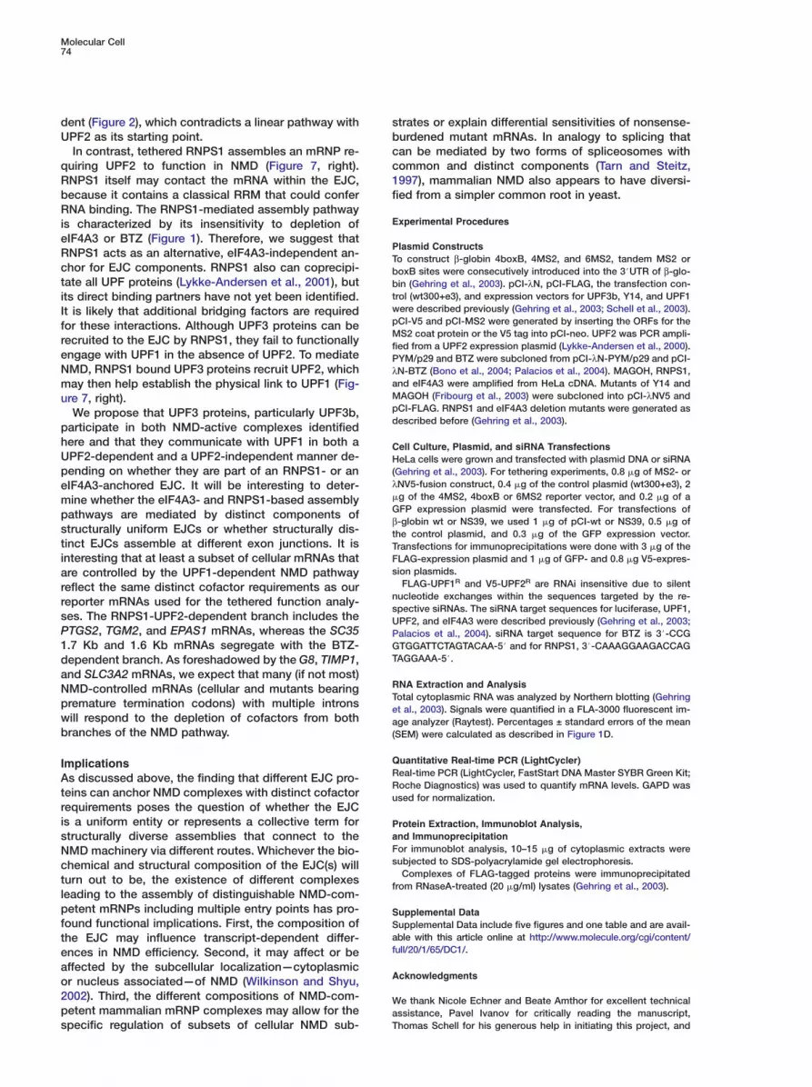
Molecular Cell74
dent (Figure 2), which contradicts a linear pathway withUPF2 as its starting point.
In contrast, tethered RNPS1 assembles an mRNP re-quiring UPF2 to function in NMD (Figure 7, right).RNPS1 itself may contact the mRNA within the EJC,because it contains a classical RRM that could conferRNA binding. The RNPS1-mediated assembly pathwayis characterized by its insensitivity to depletion ofeIF4A3 or BTZ (Figure 1). Therefore, we suggest thatRNPS1 acts as an alternative, eIF4A3-independent an-chor for EJC components. RNPS1 also can coprecipi-tate all UPF proteins (Lykke-Andersen et al., 2001), butits direct binding partners have not yet been identified.It is likely that additional bridging factors are requiredfor these interactions. Although UPF3 proteins can berecruited to the EJC by RNPS1, they fail to functionallyengage with UPF1 in the absence of UPF2. To mediateNMD, RNPS1 bound UPF3 proteins recruit UPF2, whichmay then help establish the physical link to UPF1 (Fig-ure 7, right).
We propose that UPF3 proteins, particularly UPF3b,participate in both NMD-active complexes identifiedhere and that they communicate with UPF1 in both aUPF2-dependent and a UPF2-independent manner de-pending on whether they are part of an RNPS1- or aneIF4A3-anchored EJC. It will be interesting to deter-mine whether the eIF4A3- and RNPS1-based assemblypathways are mediated by distinct components ofstructurally uniform EJCs or whether structurally dis-tinct EJCs assemble at different exon junctions. It isinteresting that at least a subset of cellular mRNAs thatare controlled by the UPF1-dependent NMD pathwayreflect the same distinct cofactor requirements as ourreporter mRNAs used for the tethered function analy-ses. The RNPS1-UPF2-dependent branch includes thePTGS2, TGM2, and EPAS1 mRNAs, whereas the SC351.7 Kb and 1.6 Kb mRNAs segregate with the BTZ-dependent branch. As foreshadowed by the G8, TIMP1,and SLC3A2 mRNAs, we expect that many (if not most)NMD-controlled mRNAs (cellular and mutants bearingpremature termination codons) with multiple intronswill respond to the depletion of cofactors from bothbranches of the NMD pathway.
ImplicationsAs discussed above, the finding that different EJC pro-teins can anchor NMD complexes with distinct cofactorrequirements poses the question of whether the EJCis a uniform entity or represents a collective term forstructurally diverse assemblies that connect to theNMD machinery via different routes. Whichever the bio-chemical and structural composition of the EJC(s) willturn out to be, the existence of different complexesleading to the assembly of distinguishable NMD-com-petent mRNPs including multiple entry points has pro-found functional implications. First, the composition ofthe EJC may influence transcript-dependent differ-ences in NMD efficiency. Second, it may affect or beaffected by the subcellular localization—cytoplasmicor nucleus associated—of NMD (Wilkinson and Shyu,2002). Third, the different compositions of NMD-com-petent mammalian mRNP complexes may allow for thespecific regulation of subsets of cellular NMD sub-
sbcc1f
E
PTbbtwpMfPλaMpd
CH(λ�
GβtTFs
nsUPGT
RTea(
QRRu
PaFs
f
SSaf
A
WaT
trates or explain differential sensitivities of nonsense-urdened mutant mRNAs. In analogy to splicing thatan be mediated by two forms of spliceosomes withommon and distinct components (Tarn and Steitz,997), mammalian NMD also appears to have diversi-ied from a simpler common root in yeast.
xperimental Procedures
lasmid Constructso construct β-globin 4boxB, 4MS2, and 6MS2, tandem MS2 oroxB sites were consecutively introduced into the 3#UTR of β-glo-in (Gehring et al., 2003). pCI-λN, pCI-FLAG, the transfection con-
rol (wt300+e3), and expression vectors for UPF3b, Y14, and UPF1ere described previously (Gehring et al., 2003; Schell et al., 2003).CI-V5 and pCI-MS2 were generated by inserting the ORFs for theS2 coat protein or the V5 tag into pCI-neo. UPF2 was PCR ampli-
ied from a UPF2 expression plasmid (Lykke-Andersen et al., 2000).YM/p29 and BTZ were subcloned from pCI-λN-PYM/p29 and pCI-N-BTZ (Bono et al., 2004; Palacios et al., 2004). MAGOH, RNPS1,nd eIF4A3 were amplified from HeLa cDNA. Mutants of Y14 andAGOH (Fribourg et al., 2003) were subcloned into pCI-λNV5 and
CI-FLAG. RNPS1 and eIF4A3 deletion mutants were generated asescribed before (Gehring et al., 2003).
ell Culture, Plasmid, and siRNA TransfectionseLa cells were grown and transfected with plasmid DNA or siRNA
Gehring et al., 2003). For tethering experiments, 0.8 �g of MS2- orNV5-fusion construct, 0.4 �g of the control plasmid (wt300+e3), 2g of the 4MS2, 4boxB or 6MS2 reporter vector, and 0.2 �g of aFP expression plasmid were transfected. For transfections of-globin wt or NS39, we used 1 �g of pCI-wt or NS39, 0.5 �g ofhe control plasmid, and 0.3 �g of the GFP expression vector.ransfections for immunoprecipitations were done with 3 �g of theLAG-expression plasmid and 1 �g of GFP- and 0.8 �g V5-expres-ion plasmids.FLAG-UPF1R and V5-UPF2R are RNAi insensitive due to silent
ucleotide exchanges within the sequences targeted by the re-pective siRNAs. The siRNA target sequences for luciferase, UPF1,PF2, and eIF4A3 were described previously (Gehring et al., 2003;alacios et al., 2004). siRNA target sequence for BTZ is 3#-CCGTGGATTCTAGTACAA-5# and for RNPS1, 3#-CAAAGGAAGACCAGAGGAAA-5#.
NA Extraction and Analysisotal cytoplasmic RNA was analyzed by Northern blotting (Gehringt al., 2003). Signals were quantified in a FLA-3000 fluorescent im-ge analyzer (Raytest). Percentages ± standard errors of the mean
SEM) were calculated as described in Figure 1D.
uantitative Real-time PCR (LightCycler)eal-time PCR (LightCycler, FastStart DNA Master SYBR Green Kit;oche Diagnostics) was used to quantify mRNA levels. GAPD wassed for normalization.
rotein Extraction, Immunoblot Analysis,nd Immunoprecipitationor immunoblot analysis, 10–15 �g of cytoplasmic extracts wereubjected to SDS-polyacrylamide gel electrophoresis.Complexes of FLAG-tagged proteins were immunoprecipitated
rom RNaseA-treated (20 �g/ml) lysates (Gehring et al., 2003).
upplemental Dataupplemental Data include five figures and one table and are avail-ble with this article online at http://www.molecule.org/cgi/content/ull/20/1/65/DC1/.
cknowledgments
e thank Nicole Echner and Beate Amthor for excellent technicalssistance, Pavel Ivanov for critically reading the manuscript,homas Schell for his generous help in initiating this project, and

Complexes Specifying Mammalian NMD75
Claudia Blumenstock for generating α-MS2 and α-UPF1 antibod-ies. We would also like to acknowledge the following colleaguesfor kindly providing reagents: Elisa Izaurralde and David Gatfield(α-Y14, α-MAGOH, and α-PYM/p29; plasmids for PYM/p29, BTZ,and mutants of Y14-MAGOH), Gideon Dreyfuss (α-Y14 andα-eIF4A3), Catherine Tomasetto and Sebastien Degot (α-MLN51),Jens Lykke-Andersen (α-UPF1, α-UPF2, and α-UPF3b; FLAG-UPF2plasmid), Akila Mayeda (α-RNPS1), and Peter Stockley (α-MS2).This work was funded by grants KU563/7-1 and KU563/8-1 fromthe Deutsche Forschungsgemeinschaft and grant 1999-1076 fromthe Fritz Thyssen Stiftung.
Received: December 20, 2004Revised: June 29, 2005Accepted: August 12, 2005Published: October 6, 2005
References
Baker, K.E., and Parker, R. (2004). Nonsense-mediated mRNA de-cay: terminating erroneous gene expression. Curr. Opin. Cell Biol.16, 293–299.
Bono, F., Ebert, J., Unterholzner, L., Guttler, T., Izaurralde, E., andConti, E. (2004). Molecular insights into the interaction of PYM withthe Mago-Y14 core of the exon junction complex. EMBO Rep. 5,304–310.
Chan, C.C., Dostie, J., Diem, M.D., Feng, W., Mann, M., Rappsilber,J., and Dreyfuss, G. (2004). eIF4A3 is a novel component of theexon junction complex. RNA 10, 200–209.
Czaplinski, K., Ruiz-Echevarria, M.J., Paushkin, S.V., Han, X., Weng,Y., Perlick, H.A., Dietz, H.C., Ter-Avanesyan, M.D., and Peltz, S.W.(1998). The surveillance complex interacts with the translation re-lease factors to enhance termination and degrade aberrantmRNAs. Genes Dev. 12, 1665–1677.
Degot, S., Le Hir, H., Alpy, F., Kedinger, V., Stoll, I., Wendling, C.,Seraphin, B., Rio, M.C., and Tomasetto, C. (2004). Association ofthe breast cancer protein MLN51 with the exon junction complexvia its speckle localizer and RNA binding module. J. Biol. Chem.279, 33702–33715.
Ferraiuolo, M.A., Lee, C.S., Ler, L.W., Hsu, J.L., Costa-Mattioli, M.,Luo, M.J., Reed, R., and Sonenberg, N. (2004). A nuclear transla-tion-like factor eIF4AIII is recruited to the mRNA during splicingand functions in nonsense-mediated decay. Proc. Natl. Acad. Sci.USA 101, 4118–4123.
Fribourg, S., Gatfield, D., Izaurralde, E., and Conti, E. (2003). Anovel mode of RBD-protein recognition in the Y14-Mago complex.Nat. Struct. Biol. 10, 433–439.
Gehring, N.H., Neu-Yilik, G., Schell, T., Hentze, M.W., and Kulozik,A.E. (2003). Y14 and hUpf3b form an NMD-activating complex. Mol.Cell 11, 939–949.
He, F., Li, X., Spatrick, P., Casillo, R., Dong, S., and Jacobson, A.(2003). Genome-wide analysis of mRNAs regulated by the non-sense-mediated and 5# to 3# mRNA decay pathways in yeast. Mol.Cell 12, 1439–1452.
Holbrook, J.A., Neu-Yilik, G., Hentze, M.W., and Kulozik, A.E.(2004). Nonsense-mediated decay approaches the clinic. Nat.Genet. 36, 801–808.
Jackson, A.L., Bartz, S.R., Schelter, J., Kobayashi, S.V., Burchard,J., Mao, M., Li, B., Cavet, G., and Linsley, P.S. (2003). Expressionprofiling reveals off-target gene regulation by RNAi. Nat. Biotech-nol. 21, 635–637.
Kadlec, J., Izaurralde, E., and Cusack, S. (2004). The structural ba-sis for the interaction between nonsense-mediated mRNA decayfactors UPF2 and UPF3. Nat. Struct. Mol. Biol. 11, 330–337.
Kataoka, N., Yong, J., Kim, V.N., Velazquez, F., Perkinson, R.A.,Wang, F., and Dreyfuss, G. (2000). Pre-mRNA splicing imprintsmRNA in the nucleus with a novel RNA-binding protein that persistsin the cytoplasm. Mol. Cell 6, 673–682.
Kataoka, N., Diem, M.D., Kim, V.N., Yong, J., and Dreyfuss, G.(2001). Magoh, a human homolog of Drosophila mago nashi pro-
tein, is a component of the splicing-dependent exon-exon junctioncomplex. EMBO J. 20, 6424–6433.
Kim, V.N., Kataoka, N., and Dreyfuss, G. (2001). Role of the non-sense-mediated decay factor hUpf3 in the splicing-dependentexon-exon junction complex. Science 293, 1832–1836.
Lau, C.K., Diem, M.D., Dreyfuss, G., and Van Duyne, G.D. (2003).Structure of the Y14-Magoh core of the exon junction complex.Curr. Biol. 13, 933–941.
Le Hir, H., Izaurralde, E., Maquat, L.E., and Moore, M.J. (2000). Thespliceosome deposits multiple proteins 20–24 nucleotides up-stream of mRNA exon-exon junctions. EMBO J. 19, 6860–6869.
Le Hir, H., Gatfield, D., Braun, I.C., Forler, D., and Izaurralde, E.(2001a). The protein Mago provides a link between splicing andmRNA localization. EMBO Rep. 2, 1119–1124.
Le Hir, H., Gatfield, D., Izaurralde, E., and Moore, M.J. (2001b). Theexon-exon junction complex provides a binding platform for factorsinvolved in mRNA export and nonsense-mediated mRNA decay.EMBO J. 20, 4987–4997.
Lykke-Andersen, J., Shu, M.D., and Steitz, J.A. (2000). Human Upfproteins target an mRNA for nonsense-mediated decay whenbound downstream of a termination codon. Cell 103, 1121–1131.
Lykke-Andersen, J., Shu, M.D., and Steitz, J.A. (2001). Communica-tion of the position of exon-exon junctions to the mRNA surveil-lance machinery by the protein RNPS1. Science 293, 1836–1839.
Maquat, L.E. (2004). Nonsense-mediated mRNA decay: splicing,translation and mRNP dynamics. Nat. Rev. Mol. Cell Biol. 5, 89–99.
Mayeda, A., Badolato, J., Kobayashi, R., Zhang, M.Q., Gardiner,E.M., and Krainer, A.R. (1999). Purification and characterization ofhuman RNPS1: a general activator of pre-mRNA splicing. EMBO J.18, 4560–4570.
Mendell, J.T., Medghalchi, S.M., Lake, R.G., Noensie, E.N., andDietz, H.C. (2000). Novel Upf2p orthologues suggest a functionallink between translation initiation and nonsense surveillance com-plexes. Mol. Cell. Biol. 20, 8944–8957.
Mendell, J.T., ap Rhys, C.M., and Dietz, H.C. (2002). Separable rolesfor rent1/hUpf1 in altered splicing and decay of nonsense tran-scripts. Science 298, 419–422.
Mendell, J.T., Sharifi, N.A., Meyers, J.L., Martinez-Murillo, F., andDietz, H.C. (2004). Nonsense surveillance regulates expression ofdiverse classes of mammalian transcripts and mutes genomicnoise. Nat. Genet. 36, 1073–1078.
Mitchell, P., and Tollervey, D. (2001). mRNA turnover. Curr. Opin.Cell Biol. 13, 320–325.
Ohnishi, T., Yamashita, A., Kashima, I., Schell, T., Anders, K.R.,Grimson, A., Hachiya, T., Hentze, M.W., Anderson, P., and Ohno,S. (2003). Phosphorylation of hUPF1 induces formation of mRNAsurveillance complexes containing hSMG-5 and hSMG-7. Mol. Cell12, 1187–1200.
Palacios, I.M., Gatfield, D., St Johnston, D., and Izaurralde, E.(2004). An eIF4AIII-containing complex required for mRNA localiza-tion and nonsense-mediated mRNA decay. Nature 427, 753–757.
Schell, T., Kocher, T., Wilm, M., Seraphin, B., Kulozik, A.E., andHentze, M.W. (2003). Complexes between the nonsense-mediatedmRNA decay pathway factor human upf1 (up-frameshift protein 1)and essential nonsense-mediated mRNA decay factors in HeLacells. Biochem. J. 373, 775–783.
Shibuya, T., Tange, T.O., Sonenberg, N., and Moore, M.J. (2004).eIF4AIII binds spliced mRNA in the exon junction complex and isessential for nonsense-mediated decay. Nat. Struct. Mol. Biol. 11,346–351.
Sureau, A., Gattoni, R., Dooghe, Y., Stevenin, J., and Soret, J.(2001). SC35 autoregulates its expression by promoting splicingevents that destabilize its mRNAs. EMBO J. 20, 1785–1796.
Tarn, W.Y., and Steitz, J.A. (1997). Pre-mRNA splicing: the discoveryof a new spliceosome doubles the challenge. Trends Biochem. Sci.22, 132–137.
Wilkinson, M.F., and Shyu, A.B. (2002). RNA surveillance by nuclearscanning? Nat. Cell Biol. 4, E144–E147.
Zhou, Z., Luo, M.J., Straesser, K., Katahira, J., Hurt, E., and Reed,R. (2000). The protein Aly links pre-messenger-RNA splicing tonuclear export in metazoans. Nature 407, 401–405.


![Australasian Pentecostal Studies This Nonsense Must Stop![1] Pentecostal Negotiation of Evil](https://img.pdfslide.net/doc/110x75/635ff90b87d94554380e1f91/australasian-pentecostal-studies-this-nonsense-must-stop1-pentecostal-negotiation.jpg)


