Embed Size (px)
Citation preview

50
Expression Of Oestrogen Progestrone And Androgen Receptors In Salivary Gland Tumours: A Review
Of LiteratureB. Tarakji and o. Kujan
Department of oral Medicine and Diagnostic Sciences, Faculty of Dentistry, Al-Farabi Col-leges, Riyadh, Saudi Arabia
Correspondence: Professor Bassel Tarakji, Head of Department of Oral Medicine and Diagnostic Sciences, Faculty of Dentistry, Al-Farabi Colleges, Riyadh, Kingdom of Saudi Arabia, Email: [email protected]
AbstractHormone therapy is highly recommended
on patients with breast cancer who show positive nuclear staining of the cancer cells. It is not known however whether salivary gland tumours can respond to hormone therapy. Twenty eight studies undertaken between 1980 to 2009 involving different types of salivary gland cancers were evaluated and taken into consideration the type of used antibody and the criteria to assess the intensity staining.
This review has shown that estrogen progesterone and androgen receptors were detected in few cases of salivary gland tumours. Different types of used antibodies were identified, and the criteria of assessment of the staining intensity were different as well.
IntroductionSalivary gland carcinomas accounts for 0.4
% of all cancers and 5% of all head and neck cancers. These tumours occur in major and minor salivary glands, and are comprised of a variety of histological types(1,2). These tumours recur despite combined modality therapy(2) and those recurrences can rarely be avoided with success with the use of chemotherapy treatment (3,4). Therefore, there is a need to develop other treatment options for those recurrent cancers. Salivary gland cancers are histologically similar to certain types of breast cancer (5). The use of hormonal therapy in prostate and breast cancers has been well-known, and the discovery of hormone receptors has given the opportunity to identify other patients who may benefit
The outcome of this review indicated that the growth of those tumours was not dependent on hormone function.
It is recommended that sensitive and specific biochemical methods can be used to determine if estrogen and progesterone receptors can be detected in salivary gland cancers. It is necessary to use one criterion such as positive or negative nuclear staining to determine the existence of estrogen and progesterone receptors and to avoid any bias. The discrepancy in the results reflects a clear need for consensus on a protocol for scoring of immunohistochemical staining.
Keywords oestrogen, progesterone, androgen receptors,
salivary gland tumours.
from such therapy. Steroid hormone receptors are intracellular DNA binding proteins that are found in both the cytosolic and nuclear fractions of a tissue homogenate. They function as growth regulators. Hormone binding causes a conformational change in the receptor followed by transport of the receptor hormone complex to the cell nucleus. In the nucleus the receptor hormone complex binds to specific nucleotide sequences, resulting in transcriptional regulation of the targeted gene. The same hormone may activate different genes in different cells. The sex steroid hormone oestrogen is important in both men and women for a variety of physiological processes. Oestrogen affects growth, differentiation, and function of tissues of the reproductive system, including the uterus, vagina, mammary glands, and ovaries in females, and the testis, epididymis, and prostate in males. It has roles in preventing osteoporosis, and maintaining bone density. In the brain, oestrogen regulates reproductive

G. J. O. Issue 11, 2012
51
behavior, gonadotropin production and its release from the pituitary gland, in addition to mood and behavior (6,7,8). Immunocytochemical techniques have shown nuclear distribution of oestrogen and progesterone receptors in carcinoma of breast cells and benign lobular or ductal epithelial cells, with no specific staining seen in the surrounding connective tissue. Specific oestrogen, progesterone, and androgen immunostaining were found to be very heterogeneous (7). Progesterone and oestrogen positive tumours showed variations in intensity and distribution of staining among cells and this heterogeneity was attributed either to receptor content variations, which were cell cycle dependent, or to variations in the presence of both progesterone and oestrogen response and non-response cells. Immunocytochemistry of oestrogen has been shown to predict response of breast cancer to endocrine therapy (9,10). Studies have shown that sixty-percent of patients having an oestrogen positive tumour will respond to hormonal treatment. Previous studies 6-12) also reported that some tumours other than breast cancer have hormone receptors e.g. melanoma; carcinoid tumours, pancreatic, and renal cancers. It is unknown whether salivary gland cancers are hormone dependent or independent.
The purpose of this study is to review the literature concerning oestrogen progesterone, and androgen receptors in salivary gland cancers and to determine if salivary gland cancers are dependent or independent on endocrine function.
Materials and methodsA literature search using MEDLINE,
accessed via the National Library of Medicine PubMed interface (http://www.ncbi.nlm.nih.gov/pubmed), searching for articles relating to the existence of oestrogen, progesterone and androgen receptors in different types of salivary gland cancers written in English. We used the following search string: (oestrogen receptors, progesterone receptors, androgen receptors) and salivary gland tumours from 1980-2009. We also used the “Related Articles” feature of PubMed to identify further references of interest within the primary search. These references were obtained, and from their bibliographies, pertinent secondary
references were also identified and acquired. The process was repeated until no further new articles could be identified. The abstracted literature was reviewed.
Studies describing case series and miscellaneous clinical reports were retrieved. Twenty-eight studies have been evaluated taking into consideration the specific criteria enumerated below:
1. All the evaluated studies should involve the existence of oestrogen or progesterone, or androgen receptors in the salivary gland tumours. Other sex steroid hormones were excluded;
2. The used scoring criteria to assess the intensity of immunostaining to demonstrate the existence of estrogen progesterone and androgen receptors in the nucleus tumour cells
3. The type of used antibody4. Comparison tissue processing
ResultNo statistical analysis is presented because
the collected data were different and the results cannot be compared. No randomized controlled trials to assess the possibility of using the hormone therapy in the treatment of salivary gland cancers were located in the literature. Our search has identified only twenty-eight studies available in written literature. These were evaluated by two pathologists to determine their agreement with the suggested criteria.
Molteni et al.(13) assessed various head and neck cancers, and an elevated level of estradiol receptor binding protein was detected in normal and neoplastic salivary gland tissues. Therefore, they suggested that some head and neck cancers may be hormone dependent. Dimery et al.(14) have mentioned that 33 cases of salivary gland tissues from a variety of sites including both minor and major glands. 19 samples consisted of normal salivary gland tissue and 14 tissue samples contained tumour. Oestrogen was considered to be present if the receptor protein value exceeded or was equal to 1 femtomoles per milligram of cytosol protein. In the male patients 10 of the 13 (77%) histologically

52
Hormone Receptors In Salivary Gland Tumours, B. Tarakji, et. al.
normal samples and four of the five (80%) tumour samples contained more than 1 fmol/mg of cytosol protein oestrogen. In the female patients, five of the six (83%) normal samples and eight of the nine (88%) tumour samples had observed oestrogen. They established that oestrogen values that exceeded 10 fmol/mg of cytosol protein were considered as positive and therefore hormonally dependent and responsive to hormonal therapy. Values between 3 and 10 fmol/mg of cytosol protein were considered to be intermediate in their response to hormonal therapy. According to their criteria none of the four oestrogen containing tumour samples from male patients were positive, however, of the eight oestrogen containing tumour samples from female patients, four (50%) would be considered positive because the oestrogen values exceeded 10 fmol/mg in these samples. Adenocarcinoma was the predominant histology in the oestrogen containing samples from female patients.
They concluded that oestrogen was identified in the majority of normal and tumour samples from both male and female patients, with the highest levels present in tumour samples obtained from females. These results suggested a possible role for sex steroid hormones in the development of the salivary glands and in promotion of neoplastic growth. Shick et al.(15) have studied oestrogen and progesterone receptors in salivary gland adenoid cystic carcinoma. Their samples consisted of 12 cases of salivary gland adenoid cystic carcinoma and normal salivary glands. The results showed no estrogen staining in either normal or neoplastic groups. Progesterone positive staining was observed in 3 cases out of 12 in normal salivary glands but 6 cases out of 12 showed positive staining in the adenoid cystic carcinomas. These findings were in agreement with Miller et al.(16) who identified progesterone positive and oestrogen negative cells in their study. All the cases in Shick et al.(15) study had aggressive behavior, which was associated with positive progesterone receptor status.
Düe et al.(17) analyzed oestrogen and progesterone receptors in samples of adenoid cystic carcinoma in both breast cancer and salivary gland tissues. No positive staining
was detected for oestrogen or progesterone in any of these cases, though sensitivity with antibodies was uncertain. Dori et al.(18) assessed oestrogen and progesterone receptors in adenoid cystic carcinomas of salivary gland origin. Progesterone receptors were identified in only 2 out of 27 cases but oestrogen was not detected in any of these cases. Their results suggested that the application of hormone therapy to salivary gland was not supported by their study. Dodd & Slevin(19) have studied one hundred and fourteen publications to determine the role of oestrogen and progesterone in salivary gland adenoid cystic carcinoma. They found the role of those receptors to be limited.
Nasser et al.(20) have studied the expression of oestrogen and progesterone in salivary gland tumours where oestrogen and progesterone expressed in only a few cases of salivary gland tumors. Glas et al.(21) have studied the prognostic significance of progesterone receptor and oestrogen receptor in patients with recurrent pleomorphic adenomas, comparing the results in a group of patients with primary adenomas without recurrences during 10 years of follow-up. Oestrogen receptor expression was low in both groups (19% and 17% respectively). Progesterone receptor expression in the recurrent group (96%) was higher compared with progesterone receptor expression in the control group (61%; P < 0.001). Teymoortash et al.(22) have evaluated the steroid hormone receptors in Warthin’s tumour of the parotid gland. Progesterone-positive cells were founded in the epithelial components of Warthin’s tumour. Oestrogen receptor showed negative expression in the epithelial components of Warthin’s tumour.
Jeannon et al.(23) have detected nine oestrogen receptor and six progesterone receptor positive tumours from a sample of 36 salivary tumours. Kolár et al.(24) have indicated that oestrogen receptors, progesterone receptors and some other estrogen-induced proteins were found in the cells of salivary gland carcinomas. Ozono et al.(25) have confirmed the existence of the receptor for progesterone in salivary adenoid cystic carcinomas. Furthermore, Lamey et al.(26) have studied the presence of hormone receptors for

G. J. O. Issue 11, 2012
53
oestrogen and progesterone in the major salivary gland tumours. Eight salivary gland tumours exhibiting varied histology, none showed high affinity receptors for oestrogen or progesterone.
Ito et al.(27) have reported that all cases of
pleomorphic adenomas, Warthin’s tumors, mucoepidermoid carcinomas and adenoid cystic carcinomas of salivary glands were negative for oestrogen and progesterone receptors. Androgen receptor was positive in two cases
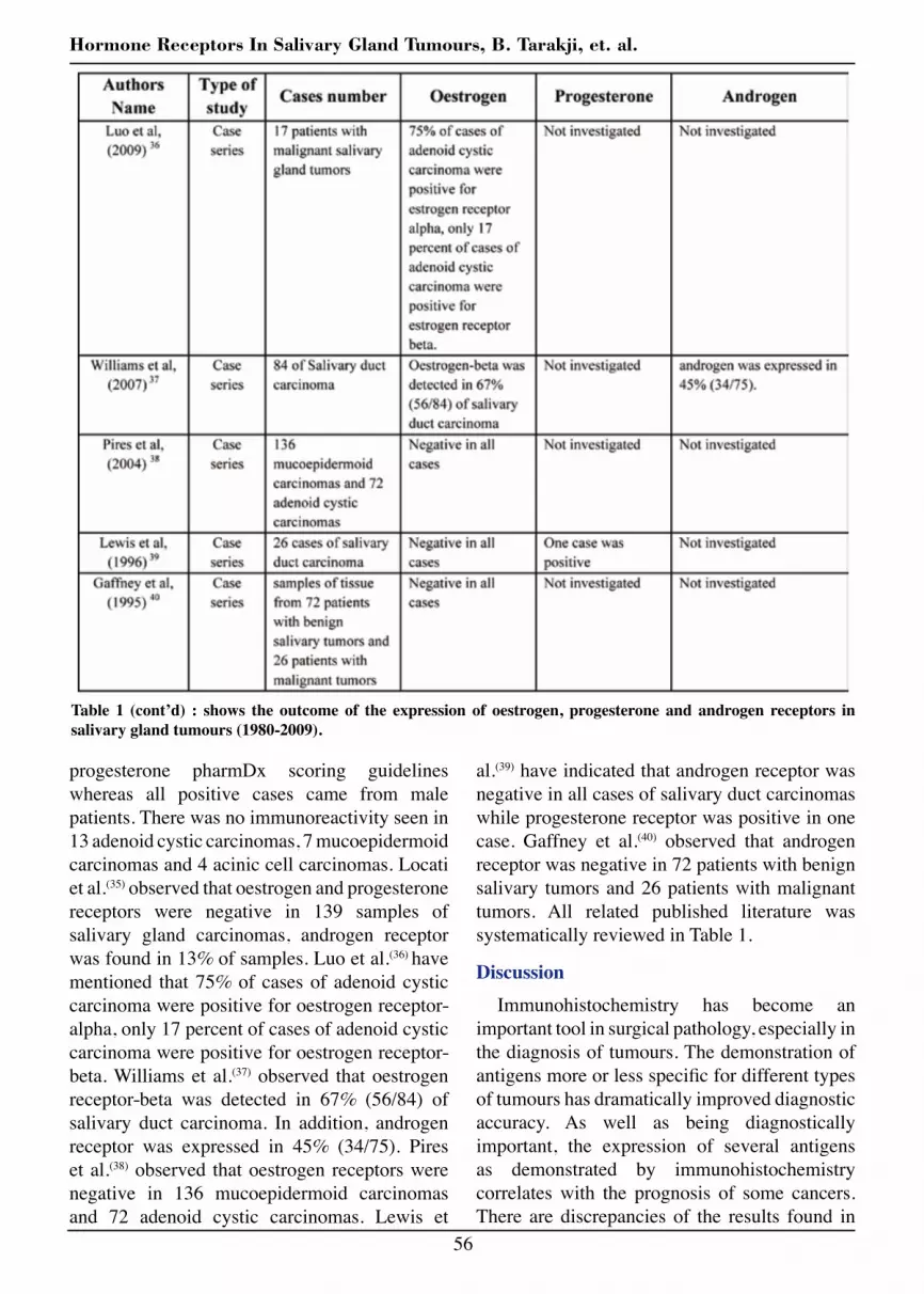
Table 1 : shows the outcome of the expression of oestrogen, progesterone and androgen receptors in salivary gland tumours (1980-2009).

54
Hormone Receptors In Salivary Gland Tumours, B. Tarakji, et. al.
each of pleomorphic adenoma, mucoepidermoid carcinoma and adenoid cystic carcinoma. Tarakji et al.(28) observed that all cases of carcinoma ex pleomorphic adenoma were negative for oestrogen and progesterone receptors.
It is very clear from the presented literature that the expression of oestrogen and progesterone receptors in salivary gland tumors have reported conflicting results. Interestingly, Larbcharoensub et al.(29) have reported that three
Table 1 (cont’d) : shows the outcome of the expression of oestrogen, progesterone and androgen receptors in salivary gland tumours (1980-2009).

G. J. O. Issue 11, 2012
55
cases of metastasizing pleomorphic adenoma shows positive reactivity to progesterone receptor but negative reactivity to oestrogen receptor.
Fan et al.(30) have mentioned that androgen receptor was noticed in 11 of 12 cases of salivary duct carcinoma. Moreover, Kapadia et al.(31) observed that androgen shows positive reactivity in 11 out of 12 of salivary duct carcinoma but oestrogen and progesterone receptors were negative. Likewise, Wick et al.(32) have indicated that oestrogen receptor was negative in salivary duct carcinoma.
Remarkably, Barnes et al.(33) observed that one (8%) of 12 salivary duct carcinomas was positive for oestrogen receptors and none was positive for progesterone receptors. Onitsuka(34)
have observed that estradiol was positive in pleomorphic adenoma. Recently, Sygut et al.(35)
have reported that nuclear immunoreactivity for androgen was demonstrated in 3 of 4 salivary duct carcinomas, 2 of 7 adenocarcinomas and 1 of 2 carcinoma ex pleomorphic adenoma. Expression of androgen was evaluated semi-quantitatively according to DAKO oestrogen/
Table 1 (cont’d) : shows the outcome of the expression of oestrogen, progesterone and androgen receptors in salivary gland tumours (1980-2009).
56
Hormone Receptors In Salivary Gland Tumours, B. Tarakji, et. al.
al.(39) have indicated that androgen receptor was negative in all cases of salivary duct carcinomas while progesterone receptor was positive in one case. Gaffney et al.(40) observed that androgen receptor was negative in 72 patients with benign salivary tumors and 26 patients with malignant tumors. All related published literature was systematically reviewed in Table 1.
DiscussionImmunohistochemistry has become an
important tool in surgical pathology, especially in the diagnosis of tumours. The demonstration of antigens more or less specific for different types of tumours has dramatically improved diagnostic accuracy. As well as being diagnostically important, the expression of several antigens as demonstrated by immunohistochemistry correlates with the prognosis of some cancers. There are discrepancies of the results found in
progesterone pharmDx scoring guidelines whereas all positive cases came from male patients. There was no immunoreactivity seen in 13 adenoid cystic carcinomas, 7 mucoepidermoid carcinomas and 4 acinic cell carcinomas. Locati et al.(35) observed that oestrogen and progesterone receptors were negative in 139 samples of salivary gland carcinomas, androgen receptor was found in 13% of samples. Luo et al.(36) have mentioned that 75% of cases of adenoid cystic carcinoma were positive for oestrogen receptor-alpha, only 17 percent of cases of adenoid cystic carcinoma were positive for oestrogen receptor-beta. Williams et al.(37) observed that oestrogen receptor-beta was detected in 67% (56/84) of salivary duct carcinoma. In addition, androgen receptor was expressed in 45% (34/75). Pires et al.(38) observed that oestrogen receptors were negative in 136 mucoepidermoid carcinomas and 72 adenoid cystic carcinomas. Lewis et
Table 1 (cont’d) : shows the outcome of the expression of oestrogen, progesterone and androgen receptors in salivary gland tumours (1980-2009).

G. J. O. Issue 11, 2012
57
the literature as mentioned because of different methodological problems. First, there may have been selection of patients and differences in treatment of different types of salivary gland tumours. Second, there are often differences in tissue processing from study to study, especially with regard to the type of antibody and the application of antigen retrieval. Also there are several steps in tissue processing that may influence staining patterns and intensity. These include type and duration of fixation, section thickness, antigen retrieval procedures, type and concentrations of primary, second and third step antibodies.
Third, and possibly most important, interpretation of staining and presentation of the results are not standardized resulting in low intra-observer and inter-observer reproducibility. Many authors used different criteria, so the results cannot be compared. This study has shown that different criteria such as (0=negative staining, 1=low, 2= moderate, 3= strong or 0-3= negative and 4= positive or 0-2= negative and 3-4=positive or negative and positive staining) have been used in the literature to assess the existence of oestrogen, progesterone and androgen receptors. Therefore, the use of one criterion such as negative or positive staining only for the assessment of staining will avoid any confusion in the interpretation of the results.
In the presented studies, there was a lack of definition in which part of the tumour the staining had been assessed.
ConclusionThe above mentioned shortcomings are
a very important obstructions for possible clinical applications. There is therefore a clear need for consensus on a protocol for scoring of immunohistochemical staining. The number of studied cases in literature is limited. A small number of salivary gland cancers have shown positive reactivity to the oestrogen, progesterone, and androgen receptors. Also it is recommended that further work involves large series of salivary gland cancers (randomized control trial) to determine if oestrogen, progesterone, and androgen receptors using sensitive and specific biochemical methods can be detected in these tumours. It is necessary to use one criterion such as positive or negative nuclear staining to determine the existence of oestrogen progesterone, and androgen receptors. Interestingly, studies showed that oestrogen, progesterone, and androgen receptors were detected in few cases of salivary gland tumours. This may indicate that the tumourgenesis of salivary gland is not dependent on hormone function.
References1. Speight PM, Barrett AW. Salivary gland tumours.
Oral Dis 2002; 8, 229-40.
2. Fu KK, Leibel SA, Levine ML, et al. Carcinoma of the major and minor salivary glands: analysis of treatment results and sites and causes of failures. Cancer 1977; 40, 2882-90.
3. Tran L, Sadeghi A, Hanson D, et al. Major salivary gland tumors: treatment results and prognostic factors. Laryngoscope 1986, 96, 1139-44.
4. Kaplan MJ, Cantrell ME, Cantrell RW. Chemotherapy for salivary gland cancer. Otolaryngol Head Neck Surg 1986; 95, 165-70.
5. Hui KK, Batsakis JG, Luna MA, et al. . Salivary duct adenocarcinoma: a high grade malignancy. J Laryngol Otol 1986; 100, 105-14.
6. Stedman KE, Moore GE, Morgan RT. Estrogen receptor proteins in diverse human tumors. Arch Surg 1980; 115, 244-8.
7. Fernandez VM, Rua ML, Reyes P, et al. Inhibition of Desulfovibrio gigas hydrogenase with copper salts and other metal ions. Eur J Biochem 1989; 185, 449-54.
8. Jones AS, Niwas S, Tanaka H. Synthesis of two diastereoisomeric p-nitrophenyl phosphodiesters of 2’,3’-secouridine and their affinity for phosphodiesterases. Nucleic Acids Res, 1986; 14, 5409-16.
9. Pertschuk LP, Eisenberg KB, Carter AC, et al. Immunohistologic localization of estrogen receptors in breast cancer with monoclonal antibodies. Correlation with biochemistry and clinical endocrine response. Cancer 1985; 55, 1513-8.
10. Singh Y, Sayami P, Sayami G, et al. Nepalese breast cancer in relation to reproductive factors: comparison between Nepalese and Japanese cases. Anticancer Res 2002; 22, 319-23.
58
Hormone Receptors In Salivary Gland Tumours, B. Tarakji, et. al.
11. Birsak CA, Janssen PJ, van Vroonhoven CC, et al. Sex steroid receptor expression in ‘carcinoid’ tumours of the breast. Breast Cancer Res Treat 1996; 40, 243-9.
12. Schuller DE, Abou-Issa H, Parrish R. Estrogen and progesterone receptors in head and neck cancer. Arch Otolaryngol 1984; 110, 725-7.
13. Molteni A, Warpeha RL, Brizio-Molteni L, et al. Estradiol receptor-binding protein in head and neck neoplastic and normal tissue. Arch Surg 1981, 116, 207-10.
14. Dimery IW, Jones LA, Verjan RP, et al . Estrogen receptors in normal salivary gland and salivary gland carcinoma. Arch Otolaryngol Head Neck Surg 1987; 113, 1082-5.
15. Shick PC, Riordan GP, Foss RD. Estrogen and progesterone receptors in salivary gland adenoid cystic carcinoma. Oral Surg Oral Med Oral Pathol Oral Radiol Endod 1995, 80, 440-4.
16. Miller AS, Hartman GG, Chen SY, et al. Estrogen receptor assay in polymorphous low-grade adenocarcinoma and adenoid cystic carcinoma of salivary gland origin. An immunohistochemical study. Oral Surg Oral Med Oral Pathol Oral Radiol Endod 1994; 77, 36-40.
17. Due W, Herbst WD, Loy V, Stein H . Characterisation of adenoid cystic carcinoma of the breast by immunohistology. J Clin Pathol 1989; 42, 470-6.
18. Dori S, Trougouboff P, David R, Buchner A. Immunohistochemical evaluation of estrogen and progesterone receptors in adenoid cystic carcinoma of salivary gland origin. Oral Oncol 2000; 36, 450-3.
19. Dodd R.L and Slevin N.J. Salivary gland adenoid cystic carcinoma: a review of chemotherapy and molecular therapies. Oral Oncol 2006; 42, 759-69.
20. Nasser SM, Faquin WCDayal, Y. Expression of androgen, estrogen, and progesterone receptors in salivary gland tumors. Frequent expression of androgen receptor in a subset of malignant salivary gland tumors. Am J Clin Pathol 2003; 119, 801-6.
21. Glas AS, Hollema H, Nap RJT, et al. Expression of estrogen receptor, progesterone receptor, and insulin-like growth factor receptor-1 and of MIB-1 in patients with recurrent pleomorphic adenoma of the parotid gland. Cancer 2002; 94, 2211-6.
22. Teymoortash A, Lippert BM, Werner JA. Steroid hormone receptors in parotid gland cystadenolymphoma (Warthin’s tumour). Clin Otolaryngol Allied Sci 2001; 26, 411-6.
23. Jeannon JP, Soames JV, Bell HJA. Immunohistochemical detection of oestrogen and progesterone receptors in salivary tumours. Clin Otolaryngol Allied Sci 1999; 24, 52-4.
24. Kolar Z, Kod’ousek R, Ehrmann JJr, et al. Expression of estrogen receptors and estrogen-induced proteins in tumors of “hormone non-dependent tissues”. Cesk Patol 1994; 30, 12-5.
25. Ozono S, Onozuka M, Sato KY. Immunohistochemical localization of estradiol, progesterone, and progesterone receptor in human salivary glands and salivary adenoid cystic carcinomas. Cell Struct Funct 1992; 17, 169-75.
26. Lamey PJ, Leake RE, Cowan SK, et al. Steroid hormone receptors in human salivary gland tumours. J Clin Pathol 1987; 40, 532-4.
27. Ito FA, Ito K, Coletta RD, et al. Immunohistochemical study of androgen, estrogen and progesterone receptors in salivary gland tumors. Braz Oral Res 2009; 23, 393-8.
28. Tarakji B, Nassani MZ, Sloan P. Immunohistochemical expression of estrogens and progesterone receptors in carcinoma ex pleomorphic adenoma- undifferentiated and adenocarcinoma types. Med Oral Patol Oral Cir Bucal 2009.
29. Larbcharoensub N, Cert PK, Tungkeeratichai J, et al. Expression of hormonal receptor in patients with metastasizing pleomorphic adenoma of the major salivary gland; a clinicopathological report of three cases. J Med Assoc Thai 2009; 92, 1250-5.
30. Fan CY, Melhem MF, Hosal AS, et al. Expression of androgen receptor, epidermal growth factor receptor, and transforming growth factor alpha in salivary duct carcinoma. Arch Otolaryngol Head Neck Surg 2001; 127, 1075-9.
31. Kapadia SB, Barnes L. Expression of androgen receptor, gross cystic disease fluid protein, and CD44 in salivary duct carcinoma. Mod Pathol 1998; 11, 1033-8.
32. Wick MR, Ockner DM, Mills SE, et al. Homologous carcinomas of the breasts, skin, and salivary glands. A histologic and immunohistochemical comparison of ductal mammary carcinoma, ductal sweat gland carcinoma, and salivary duct carcinoma. Am J Clin Pathol 1998; 109, 75-84.
33. Barnes L, Rao U, Contis L, et al. Salivary duct carcinoma. Part II. Immunohistochemical evaluation of 13 cases for estrogen and progesterone receptors, cathepsin D, and c-erbB-2 protein. Oral Surg Oral Med Oral Pathol Oral Radiol Endod 1994; 78, 74-80.
34. Onitsuka T. Sex hormones in papillary carcinoma of thyroid gland and pleomorphic adenoma of parotid gland. Acta Otolaryngo 1994, 114, 218-22.
35. Locati LD, Perrone F, Losa M, et al. Treatment relevant target immunophenotyping of 139 salivary gland carcinomas (SGCs). Oral Oncol 2009; 45, 986-90.

G. J. O. Issue 11, 2012
59
36. Luo SD, Su CY, Chuang HC, et al. Estrogen receptor overexpression in malignant minor salivary gland tumors of the sinonasal tract. Otolaryngol Head Neck Surg 2009; 141, 108-13.
37. Williams MD, Roberts D, Blumenschein GR, et al. Differential expression of hormonal and growth factor receptors in salivary duct carcinomas: biologic significance and potential role in therapeutic stratification of patients. Am J Surg Pathol 2007; 31, 1645-52.
38. Pires FR, da Cruz PDE, de Almeida, et al. Estrogen receptor expression in salivary gland mucoepidermoid carcinoma and adenoid cystic carcinoma. Pathol Oncol Res 2004; 10, 166-8.
39. Lewis JE, McKinney BC, Weiland LH, et al. Salivary duct carcinoma. Clinicopathologic and immunohistochemical review of 26 cases. Cance 1996; 77, 223-30.
40. Gaffney EV, Pinkston JA, Eidson JJ. Estrogen receptors in parotid tumors. Endocr Res 1995; 21, 635-43.
41. Sygut D, Bien S, Ziolkowska MS. Immunohistochemical expression of androgen receptor in salivary gland cancers. Pol J Pathol 2008; 59, 205-10.


