Embed Size (px)
Citation preview

RESEARCH ARTICLE – Pharmaceutics, Drug Delivery and Pharmaceutical Technology
Formulation and Characterization of Atropine Sulfate inAlbumin–Chitosan Microparticles for In Vivo Ocular Drug Delivery
RICHARD T. ADDO,1 KWAME G. YEBOAH,2 RODNEY C. SIWALE,3 ALADIN SIDDIG,4 ALPHIA JONES,5 RUHI V. UBALE,1,6
JANET AKANDE,7 HENRY NETTEY,8 NEIL J. PATEL,1 EVELYN ADDO,9 MARTIN J. D’SOUZA5
1Union University School of Pharmacy, Jackson, Tennessee 38305, USA2College of Pharmacy, Harding University, Searcy, Arkansas 72149, USA3Western New England University College of Pharmacy, Springfield, Massachusetts 01119, USA4University of Charleston School of Pharmacy, Charleston, West Virginia 20304, USA5Mercer University College of Pharmacy and Health Sciences, Atlanta, Georgia 30341, USA6Lake Erie College of Osteopathic Medicine, School of Pharmacy, Bradenton, Florida 34211, USA7Lonza Inc., Alpharetta, Georgia 30004, USA8University of Ghana School of Pharmacy, Legon-Accra, Ghana9Union University College of Education and Human Studies, Jackson, Tennessee 38305, USA
Received 7 August 2014; revised 24 December 2014; accepted 8 January 2015
Published online 4 February 2015 in Wiley Online Library (wileyonlinelibrary.com). DOI 10.1002/jps.24380
ABSTRACT: The overall study goal was to produce a microparticle formulation containing atropine sulfate for ocular administration withimproved efficacy and lower side effects, compared with that of the standard marketed atropine solution. The objective was to prepare anatropine sulfate-loaded bovine serum albumin–chitosan microparticle that would have longer contact time on the eyes as well as bettermydriatic and cycloplegic effect using a rabbit model. The microparticle formulation was prepared by method of spray-drying technique.The percent drug loading and encapsulation efficiency were assessed using a USP (I) dissolution apparatus. The particle sizes and zetapotential were determined using laser scattering technique and the surface morphology of the microparticles was determined using ascanning electron microscope. The product yield was calculated from relative amount of material used. In vitro cytotoxicity and uptake byhuman corneal epithelial cells were examined using AlamarBlue and confocal microscopy. The effects of the microparticle formulation onmydriasis in comparison with the marketed atropine sulfate solution were evaluated in rabbit eyes. The prepared microparticle formulationhad ideal physicochemical characteristics for delivery into the eyes. The in vivo studies showed that the microparticles had superior effectson mydriasis in rabbits than the marketed solutions C© 2015 Wiley Periodicals, Inc. and the American Pharmacists Association J Pharm Sci104:1677–1690, 2015Keywords: microparticles; micropheres; nanoparticles; ocular delivery; albumin; chitosan; atropine; mydriasis
INTRODUCTION
The protective physiological mechanism existing on the pre-cornea area and the anatomical structure of the cornea surfaceputs severe limitations on topical application of drugs into theeye. These limitations that lead to considerable drug loss areserious constraints on the effectiveness of drug delivery sys-tems for topical and transcorneal treatment. These conditionsmake it difficult to provide adequate concentration of drug onthe eye for the proper duration of time in order to achieve therequired bioavailability of the drug needed for its therapeuticeffectiveness. As a result, targeted administration of drugs tothe anterior and posterior segments of the eye remains a sig-nificant challenge in ocular drug delivery.
Within the last decade, drug retention in the cornea has re-ceived great attention with the intent to maximize the residencetime of the drug delivery vehicles on the eyes, thus solving aspecific absorption window issue as well as for localized and
Correspondence to: Richard T. Addo, Associate Professor of Pharmacy(Telephone: +404-358-4518; Fax: +731-661-5980; E-mail: [email protected])
[Correction added after online publication 17 February 2015. An author wasadded and the byline reordered.]
Journal of Pharmaceutical Sciences, Vol. 104, 1677–1690 (2015)C© 2015 Wiley Periodicals, Inc. and the American Pharmacists Association
targeted drug delivery.1 One approach involves the creationof bioadhesive systems that strongly adhere to the mucus orcell surfaces. This has involved the adherence of drug-loadedmicroparticles coated with mucoadhesive polymers or specificligands to the epithelial membrane.2 Since then, a number ofreports have shown that mucoadhesive polymers can increasethe residence time of drugs on the ocular surface.3,4
One of these polymers is chitosan (CSN), which has beenfound to possess favorable biological characteristics suchas biodegradability, biocompatibility, and nontoxicity. Thismakes it suitable for use in biomedical and pharmaceuticalpreparations.5–13 Many researchers have developed differentCSN-based carriers to enhance the transport of drugs acrossintestinal mucosal barriers,14 as artificial kidney membranes,15
as vehicles for compressed tablets,16–18 for gene therapy,19–21 assuture and wound healing materials,22 for vascular grafts,23
and cartilage regeneration.24 In addition, it has also been usedfor its antacid and antiulcer activities that prevent or weakendrug irritation of mucosal surfaces,25 among many other appli-cations such as hydrogels preparation26,27 and as topical oculardrug delivery systems.28
Chitosan is a natural polymer with one primary amino andtwo free hydroxyl groups for each carbon 6 unit. It has beenfound that easy availability of these amino groups gives it a
Addo et al., JOURNAL OF PHARMACEUTICAL SCIENCES 104:1677–1690, 2015 1677

1678 RESEARCH ARTICLE – Pharmaceutics, Drug Delivery and Pharmaceutical Technology
positive charge and thus enables it to easily react with manynegatively charged surfaces.29 This imparts strong bioadhe-sive and particularly mucoadhesive characteristics.30,31 It hasbeen reported that addition of bioadhesive polymers such asCSN as a coating for particulate delivery systems, prolongsthe contact time by significantly increasing the half time of itsclearance.32,33 This makes it a promising candidate for ocular-retentive drug delivery system.34 Furthermore, CSN possessesfilm-forming capacity and has been used for immobilizing en-zymes, living cells, and in ophthalmology.34 As such, it has beenidentified as the biopolymer of choice for the development ofboth soft and hard contact lenses.35 In line with that, it hasalso become an important ingredient in ocular bandage lensesused as a protective device for acutely or chronically trauma-tized eyes.35,36 Moreover, cationic materials, such as CSN, havebeen shown to offer several benefits, including the ability tofacilitate cellular uptake of drug compounds through contactwith the negatively charged cellular membrane.37
One possibility for mucoadhesive systems is the employ-ment of small particles of micron or submicron range calledmicroparticles. Microparticles made from mucoadhesive poly-mers exhibit a prolonged residence time at the site of applica-tion or absorption, facilitate an intimate contact with the un-derlying absorption surface, and thus contribute to improvedtherapeutic performance of drugs. De Campos et al.38 reportedan intense interaction of CSN with ocular surfaces that resultsin uptake and transport across the cell layers when the poly-mer is in microparticulate/nanoparticulate form.35 Micropar-ticles are particularly relevant for ocular drug delivery be-cause for patients’ comfort, it is reported that solid particlesintended for ophthalmic use should not exceed 5–10 : .39 It hasbeen reported that previous work by a number of researchersshowed that poly (alkylcyanoacrylate) nanoparticles40,41 andpoly-g-caprolactone nanocapsules42–44 were able to increase theintraocular penetration of drugs, while reducing their systemicabsorption.38 A more positive result was reported when the mi-croparticles were made of CSN. Genta et al.45 reported an in-creased and prolonged corneal penetration of acyclovir encapsu-lated in CSN microspheres. Furthermore, Felt et al.34 reportedthat the presence of CSN significantly prolonged cornea contacttime of tobramycin following topical instillation to rabbits.38
This improved penetration was attributed to the microparti-cle/nanoparticle sizes of the delivery systems.46
In this project, CSN was combined with bovine serum al-bumin (BSA) to form a copolymer matrix for the atropine sul-fate model drug. Albumin microparticles have been extensivelyinvestigated in controlled-release systems as vehicles for thedelivery of therapeutic agents.47,48 Its exploitable features in-clude its reported biodegradation into natural products, its lackof toxicity and its nonantigenicity49
The model drug for this project was atropine sulfate that iscurrently used for various ophthalmic therapies as a solutionformulation. Atropine is a muscarinic antagonist that is usedtopically as a cycloplegic agent to temporarily paralyze the eye’saccommodation reflex and as a mydriatic to dilate the pupil.50
It acts by blocking the contraction of the circular sphinctermuscle, stimulated by acetylcholine, thereby allowing the ra-dial papillary dilator muscle to contract to dilate the pupil.51
Of all the drug compounds, atropine is reported to provide thegreatest amount of cycloplegia and is considered as the goldstandard for treatment.50 Cycloplegic refraction is reported byophthalmologists as invaluable in the evaluation of patients
with decreased vision and ocular deviation.51 Furthermore, theeyes of today’s children have no time to relax because of read-ing, writing, watching television, using the computer, surfingthe internet, or playing video games. Not surprisingly, abouthalf of school children are reported to be shortsighted, whereasindications for cycloplegic refraction are limited in adults.51 Ithas recently been reported that regular usage of atropine eyedrops can retard the progression or completely stop myopia inchildren.52
However, conventional eye drops typically act transientlyand enter the eye by either diffusion across the cornea orthe sclera.53–56 As a result, atropine is well absorbed into thesystemic circulation and has been reported to exert severesystemic side effects after ocular administration of solutionformulations.51 Serious side effects of the drug include fever,tachycardia, convulsion, and even death, and it is contraindi-cated in patients with Down syndrome and albinism.57–59
Therefore, a sustained-release drug delivery system such asmicroencapsulation that protects and controls the release ofthe drug but having similar or superior pharmacodynamics ef-fect than the standard formulations on the market will be idealin minimizing both the dose and the spread of the drug with itsaccompanying ectopic toxicity after administration.
The goal of this project, therefore, was to formulate a mi-croparticle drug delivery system that has improved efficacy,but with lower side effects than the standard marketed at-ropine solution for delivery into the eyes. The objectives wereto prepare microparticle with BSA and CSN copolymer matrixfor the drug that would have longer contact time, be takenup by human corneal epithelial cells, and have better mydri-atic and cycloplegic effect on the eye using rabbit models. Thecycloplegic and mydriatic effects of atropine sulfate were mea-sured by the novel use of pupil to corneal ratio in rabbit model.The production of the microparticles was by the spray-dryingmethod. Spray drying is robust with high-product yield.48,60–65
Furthermore, the technique avoids the toxic chemicals used insolvent evaporation method that would be of great limitationin ophthalmic application.66
MATERIALS
Chemicals
Bovine serum albumin (Fraction V, DNAase, RNAase, andProtease-free), glutaraldehyde (25% in water), atropine sul-fate powder, formaldehyde, Triton X-100, and sodium bisul-fite were obtained from Fisher Scientific (Norcross, Georgia).Trypsin, sterile deionized water, methanol, and phosphate-buffered saline (1× PBS) pH 7.4 were obtained from Sigma–Aldrich chemicals (St. Louis, Missouri). CSN glutamate was ob-tained from Pronova (Drammen, Norway). The standard 1% at-ropine sulfate ophthalmic solution and natural tear fluid wereobtained from Alcon Pharmaceuticals (Fort Worth, Texas). Hu-man Corneal Epithelial cells were provided by the Ciba Vision(Duluth, Georgia). Ultra culture growth media were obtainedfrom ATCC (Manassas, Virginia). Infra-Red Dye 800 CW wasobtained from Li-cor Biosciences (Lincoln, Nebraska).
Equipment
The Buchi 191 Mini Spray Dryer (used in the spray-dryingprocess) was obtained from Buchi Corporation (Newcastle,Delaware). The Horiba LA920 laser scattering particle size
Addo et al., JOURNAL OF PHARMACEUTICAL SCIENCES 104:1677–1690, 2015 DOI 10.1002/jps.24380
RESEARCH ARTICLE – Pharmaceutics, Drug Delivery and Pharmaceutical Technology 1679
distribution analyzer (used to determine microsphere parti-cle size) was obtained from Horiba Instruments Incorporated(Irvine, California). The Malvern Zetasizer Nano ZS (for zeta-potential analysis) was obtained from Malvern Instruments(Worcestershire, UK). The BioTeK ELx808TM Absorbance Mi-croplate Reader (used for atropine assay analysis) was fromBioTek instruments Inc. (Highland Park, Winooski, Vermont).The JEOL JSM-5800L scanning electron microscope (for sur-face morphology analysis) was obtained from JEOL USA(Peabody, Massachusetts). The Distek dissolution system model2100C, USP Dissolution apparatus I, with a rotating bas-ket (used for the microsphere formulations release studies)was obtained from Distek Inc. (North Brunswick, New Jer-sey). Lambda 4B UV/Vis spectrophotometer (PerkinElmer,Waltham, Massachusetts) was used for reading the releasestudy samples. The Cytofluor R© multi-well plate reader se-ries 4000 (used for the AlamarBlue cytotoxicity assay) wasobtained from PerSeptive Biosystems (Framingham, Mas-sachusetts). Zeiss Confocal microscope LSM410 equipped withargon-krypton laser (Carl Zeiss Micro-Imaging, Thornwood,New York) was used for the uptake studies. The Odyssey infra-red imaging system was from Li-cor Biosciences.
METHODS
Preparation of Atropine Sulfate in Albumin–CSN Microparticles
Bovine serum albumin–CSN solution (1%, w/v) was preparedand cross-linked with 0.75% glutaraldehyde for 24 h. The ex-cess glutaraldehyde was neutralized with sodium bisulphite.Atropine sulfate was added to the cross-linked BSA–CSN ma-trix, to achieve 10% (w/w) drug loading with respect to the con-centration of the polymer matrix. For the blank microparticles,no atropine sulfate was added to the cross-linked BSA–CSNpolymer matrix. The cross-linked solutions were spray driedusing a Buchi 191 Mini Spray Dryer. The settings of the var-ious parameters for the spray dryer were: inlet temperature,110°C; outlet temperature, 80°C; aspirator, 55%; compressionflow rate, 800 psi; pump rate, 2.5% for optimum drug encapsu-lation and yield.
The product yield for the microparticles was calculated usingthe following formula:
Product yield (%)
= Weight of microparticles obtained from spray dryerWeight of the total amount of solid in the feed
×100
Preparation of Standard Atropine Sulfate Solutions forDose–Response Evaluation
Atropine sulfate solutions (1.0%, 0.66%, and 0.33%) were usedfor this study. The 0.33% and 0.65% solutions were prepared bydiluting the 1% solution with normal saline.
Physicochemical Characterization of Microspheres
Particle Size Distribution
The particle size distribution of BSA–CSN microparticles wasmeasured using the Horiba LA920 laser scattering particlesize distribution analyzer. Briefly, the microparticles were sus-pended in Milli-Q pure water (2 mg/mL) containing 0.1% Tween
20. The particle sizes were determined after sonication. The re-sults were calculated from six (n = 6) samples reading.
Zeta Potential
The zeta potential of the particles was measured using aMalvern Zetasizer Nano ZS (Malvern Instruments). The mea-surements were performed on six sample (n = 6) in 0.9% salinesolution (normal saline) at a final microparticle concentrationof 2 mg/mL (pH 6.8).
Surface Morphology
A scanning electron microscope (JEOL JSM 5800LV) was usedto evaluate surface morphology of the microparticles. Micropar-ticles were coated for 1 min under 80 mTorr of vacuum withgold/palladium at 15 mA. The micrographs were obtained us-ing a 3-kV accelerating voltage at a 10,000 magnification.
Release Profile and Mechanism of Drug Release
In vitro release studies of the atropine sulfate-loaded micropar-ticles were carried out at 37°C using natural tear fluid pH7.4 (100 mL) in the modified USP type I dissolution appara-tus. Atropine sulfate (25 mg) microparticles were suspended in3 mL of natural tear fluid inside a dialysis bag with a molecu-lar weight cut off of 12–14 kDa. The dialysis bag was sealed atboth ends, enclosed into the dissolution baskets, and immersedinto the natural tear fluid in the beakers. The dissolution ap-paratus was set at 100 rpm, and samples were taken at prede-termined time intervals from each of the six baskets (n = 6).An equivalent volume of fresh tear fluid was replaced at eachsampling time. The samples were analyzed for atropine sulfateby a Lambda 4B UV/Vis spectrophotometer (PerkinElmer) at220 nm. In order to find out the mechanism of drug release fromthe microparticles, data obtained from in vitro release stud-ies were fitted to various kinetic models. The following kineticmodels were used: zero-order equation, first-order equation,and Higuchi model. Plots of Qt versus t (zero-order model), log(Q0–Qt) versus t (first-order model), and Qt versus �t (Higuchimodel) were made, where Qt is the drug release at time t andQ0 is the initial amount of drug present in the microparticles.
Encapsulation Efficiency
To determine the encapsulation efficiency of the atropine sul-fate in albumin–CSN microparticles, 5 mg of the dry micropar-ticles powder containing the drugs were suspended in 200 :Lof a buffer containing 50 mM Tris–HCl, pH 8.0, and 10 Methylenediaminetetraacetic acid. The suspension was vortexedrepeatedly to generate a homogeneous suspension and incu-bated overnight in a water bath at 37°C. To the above suspen-sion, an equal volume of a buffer containing 200 mM NaOH,1% SDS (w/v), and 100 :g/mL of proteinase K (DNase-free,RNase-free; Roche, Branchburg, New Jersey) was added andvortexed repeatedly. This reaction mixture was further incu-bated at 37°C for an additional period of 5 h. The concentrationof atropine sulfate was determined by absorbance at 220 nmin a Lambda 4B UV/Vis spectrophotometer. Encapsulation ef-ficiency was calculated as per the following formula:
Encapsulation efficiency (%) = Actual drug loadingTheoretical drug loading
×100
DOI 10.1002/jps.24380 Addo et al., JOURNAL OF PHARMACEUTICAL SCIENCES 104:1677–1690, 2015

1680 RESEARCH ARTICLE – Pharmaceutics, Drug Delivery and Pharmaceutical Technology
In Vitro Cytotoxicity, Internalization, and Localization Studies ofAlbumin–CSN Microparticles by Human Corneal Epithelial Cells
Cell Culture
Human corneal epithelial cells were seeded in six-well cul-ture plates at approximately 3.0 × 10⁵ cells/mL in ultraculturegrowth media containing 5% glutamine with no antibiotics. Thehuman corneal epithelial cell cultures were incubated at 37°Cand 5% CO2. The cells were grown until they were 80%–100%confluent.
Determination of Cytotoxicity of Albumin–CSN Microparticles onHuman Corneal Epithelial Cells
To evaluate the cytotoxicity of the formulated albumin–CSNmicroparticles in human corneal epithelial cells, the cells wereexposed to increasing concentrations of microparticles varyingfrom 0.01 to 2 mg/mL for 24 h as previously described by Addoand coworkers.67 The negative control was human corneal ep-ithelial cells in growth media and the positive control was hu-man corneal epithelial cells in media containing 0.005% ben-zalkonium chloride (BAC). BAC is a quaternary ammoniumcationic surface acting agent used as a bactericide/microbicidebecause of its cellular membrane lipid bilayer disruptive prop-erties. After the 24-h incubation, the media were removed, andthe cells were then washed several times with (1× strengthsolution) Dulbecco’s phosphate buffer saline (DPBS; pH 7.4),and incubated with a 1:10 dilution of AlamarBlue in 1× DPBS(pH 7.4) for 2 h. AlamarBlue contains a specific (fluoromet-ric/colorimetric) REDOX indicator that both fluoresces and un-dergoes colorimetric change in response to cellular metabolicreduction. The extent of redox is, therefore, a measure of cel-lular metabolic reductive activity. The plates were then readusing a Cytofluor R© multiwell plate reader at 530 nm excitationand 580 nm emission. The fluorescence was proportional to theconversion of AlamarBlue by viable human corneal epithelialcells.
Uptake Study of Microparticles into Epithelial Cells UsingConfocal Microscopy
Ultra culture growth medium supplemented with 5% glu-tamine was used to suspend FITC-labeled albumin–CSN mi-croparticles at 50 :g/mL. In order to exclude the possibility ofautofluorescence by formaldehyde and nonspecific staining ofcells by free FITC, the growth media supernatant of FITC-labeled BSA–CSN microparticles was vigorously shaken re-peatedly for 2 h and centrifuged. The microparticles were thenincubated with the cells to check for any leakage of dye fromthe microparticles, which might stain cells before they wereused. Human corneal epithelial cells at approximately 1 × 10⁵
cells/plate were seeded and incubated overnight to allow for celladhesion. The washed FITC-labeled microparticles were incu-bated with the cell at a concentration of 10 microparticles percell for 24 h followed by 5× washing with PBS. Cells were thenfixed onto glass slide with formaldehyde. Images were capturedby using a 488-nm fluorescein, and 568-nm rhodamine filters.Differential interference contrast using a Zeiss Confocal micro-scope LSM410 equipped with argon-krypton laser was overlaidto obtain images to determine localization of microparticles in-side the cell compartments.
Confirmatory Studies of the In Vitro Internalization andLocalization by Z-Stacking Method
To ascertain whether the microparticles were actually takeninto the cells and not just on their surfaces, a confirmatorystudy using the Z-stacking method was carried out. In thisstudy, human corneal epithelial cells were exposed for 60 minto 0.5 mg/mL BSA–CSN microparticles containing FITC dye.Confocal serial images along the z-axis of human corneal ep-ithelial cells were taken at 1.5-:m intervals from top to thebottom of the cell monolayer.67
Quantitative Evaluation of the Uptake of Microparticles by theHuman Corneal Epithelial Cells
In these studies, about 1010 BSA–CSN–FITC-labeled micropar-ticles were placed on filters of the transwell plates containingpreviously seeded and confluent human corneal epithelial cells.Total of 3 :m pore size of filter inserts were used. The trans-port epithelial electrical resistance across the cell monolayerwas monitored using an ohmmeter (Millicell ETS) as mark ofmonolayer formation and as an indication of the formation andintegrity of tight junctions between the cells. The FITC-labeledmicroparticles were applied to the apical side of the inserts andmicroparticles transported through the cell layer were collectedin the basolateral compartment by sampling at 0.25, 0.5, 1, 2,4, 8, 12, and 24 h time intervals. The amount of microparticlestaken up and retained in the cells were also determined at thesame time intervals by washing off the unattached micropar-ticles form the cells with PBS and lysing the cells with TritonX (1%, w/v). Samples were analyzed using the fluorescent mi-croplate reader and a cytofluorometer.
Evaluation of the In Vitro Mechanism of Microparticle TransportAcross Human Corneal Epithelial Cells
This study was to determine whether the uptake of the mi-croparticles into the corneal cells was temperature and en-ergy dependent. Confluent corneal cell cultures on transwellplates were exposed to 150 :L of FITC-labeled microparticles(0.5 mg/mL) for predetermined time intervals from 15 min to2 h, at 4°C and 37°C in the presence or absence of 100 mMsodium azide, (a known metabolic inhibitor). The amount ofmicroparticles taken up and retained in the cells was also de-termined at the same time intervals as indicated in sectionQuantitative Evaluation of the Uptake of Microparticles by theHuman Corneal Epithelial Cells above.
In Vivo Evaluation of the Atropine Sulfate Formulation
Standardization of the Novel Procedure for Measuring the Degreeof Mydriasis Using Rabbit Model
The objective of this study was to develop a standardized pro-cedure to measure mydriasis using pupil to corneal ratio of theeye of rabbits. The pupil length and the cornea length of theeyes were measured without the addition of any external drugin the presence or absence of light to determine whether anyconstant parameter could be obtained that would serve as abaseline for comparison. Briefly, the eyes were videotaped froma fixed distance using Panasonic 30× digital camera with an in-built flashlight, with or without light. The video recording wasdownloaded into a window moviemaker. Grids with standarddimensions were also photographed from the same distanceas the eyes. The pictures were then fixed and imported onto
Addo et al., JOURNAL OF PHARMACEUTICAL SCIENCES 104:1677–1690, 2015 DOI 10.1002/jps.24380

RESEARCH ARTICLE – Pharmaceutics, Drug Delivery and Pharmaceutical Technology 1681
Microsoft power point, where the pupil and cornea measure-ments were carried out by superimposing the grids on the pic-tures of the eyes. The ratios of diameter of the pupil to that ofthe corneal were calculated. A graph of pupil to corneal ratioon the y-axis and time on the x-axis was plotted.
Dose–Response Study of Atropine Sulfate Solution Formulationson Mydriasis in Rabbits’ Eye
Atropine sulfate drops (0.33%, 0.66%, and 1%) prepared in tearsolutions (from the marketed 1% solution) were applied to dif-ferent groups of rabbit eyes (n = 12). One eye of each animalreceived the test solution, whereas the other eye served as acontrol and received blank tear solution. Two drops of each so-lution were added to each eye with the aid of a dropper. Eachrabbit’s eye was videotaped with the Panasonic 30× digital cam-era with and without light. The designated time for video tap-ping was 0.25, 0.5, 0.75, 1.0, 2.0, 3.0, 6.0, 8.0, and 24 h. Thevideo recording was downloaded into window moviemaker. Thepictures were then fixed and imported onto Microsoft powerpoint, where the pupil and cornea measurements were carriedout and analyzed as in section Standardization of the NovelProcedure for Measuring the Degree of Mydriasis Using RabbitModel above.
Effect of the Atropine Sulfate Microparticle Formulations onMydriasis in Rabbits Eye
The atropine sulfate microparticles were used to determinetheir effect on mydriasis for comparison with the marketed so-lution formulation. Briefly, the microparticles were suspendedin a tear fluid to produce 0.33% and 0.66% atropine sulfate con-centration, respectively. Two drops of each were administeredin the eyes of different groups of Harlan rabbits (n = 12), withthe aid of a dropper. One eye served as a test and the otheras the control (which received blank tear solution) in each an-imal. Each rabbit’s eye was videotaped using Panasonic 30×digital camera with an in-built flashlight, with and withoutlight. The designated times for video tapping analysis were asindicated in the above sections Standardization of the NovelProcedure for Measuring the Degree of Mydriasis Using RabbitModel and Dose–Response Study of Atropine Sulfate SolutionFormulations on Mydriasis in Rabbits’ Eye. The video recordingwas downloaded into window moviemaker. The pictures werethen fixed and imported onto Microsoft power point, where thepupil and corneal measurements were carried out with the aidof a standardized grid. A graph of pupil to cornea ratio on they-axis and time on the x-axis plotted. Subjects were observedfor any obvious signs of ocular irritations such as redness andlachrymation.
In our initial in vivo rabbit studies, death was observed at ahigher atropine sulfate concentration via ocular administrationbecause of the possibility of tachycardia. We also realized that
the microparticulate formulation resulted into a greater perme-ability and therefore a greater bioavailability. One of the mainaims of this study was to developed an alternative formulationthat could elicit a response similar to the already market formu-lation but at a lower concentration. In other words, the authorsalso wanted to see whether a lower concentration (strength) ofour microparticle encapsulated formulation of atropine sulfatecould produce better pharmacologic effect than the standardmarketed solution formulations. This we believe will lead todecrease in atropine sulfate toxicity. We therefore found it pru-dent not to use the 1% atropine microparticle formulation inthe animal study as described in this section.
Statistical Analysis
Statistical analyses were carried out for both in vitro and in vivowork. All comparisons were made to the control group (mar-keted 1% atropine sulfate ophthalmic solution), and to eachother. Summary of onset of action, duration of action, area un-der the curve (AUC), and maximum effect (pupil–corneal ratio)were calculated. Statistical significance was determined withthe use of ANOVA between groups, and a p value of less than0.05 was considered statistically significant.
RESULTS
Physicochemical Characterization of Microparticles
Particle Size
The atropine–BSA–CSN microparticles with a mean particlesize of approximately 2 :m (Table 1) and a polydispersity in-dex of 0.268 were obtained from spray drying (Fig. 1). Spraydrying has been traditionally used to produce particles with anarrow size distribution. This is evident from the results. In-terestingly, the addition of CSN to the matrix of the micropar-ticles, compared with the atropine–BSA microparticles withoutthe CSN, reduced the particle sizes from 2.40 to 1.99 :m. Thispotentially may not have occurred if the CSN had just beenused to coat preformulated and packaged BSA microparticles.Therefore, incorporation of the CSN as part of the polymericmatrix has a positive effect on particle size reduction. The re-duced size brings with it an extra advantage of large surfaceto volume ratio that is good for controlled-release delivery ofinsoluble drugs.
Zeta Potential
The average zeta potential for the BSA–CSN atropine mi-croparticles was 43.1 mV. A clear inversion of the zeta potentialwas found between BSA microparticles without the CSN andthe BSA–CSN microparticles from a negative value of −38.4 topositive value of 43.1, respectively.
Table 1. Effect of Polymer Type on Particle Size Distribution (:m), Zeta Potential (mV), and Encapsulation Efficiency of Microparticles
Mean size Zeta Potential (mV) Zeta Potential Encapsulation ProductFormulation (:m) Drug-Loaded MS (mV) Blank MS Efficiency (%) Yield (%)
Atropine BSA–CSN MS 1.99 ± 0.13 43.1 ± 2.1 48.3 ± 1.2 98 ± 0.62 86 ± 1.9Atropine BSA MS with no CSN 2.40 ± 0.57 −38.4 ± 2.0 −48.4 ± 2.8 96 ± 0.97 85 ± 1.3Atropine alone NA −2.7 ± 1.6 NA NA NA
The following parameters were kept constant: solvent, stirring speed, and pH.
DOI 10.1002/jps.24380 Addo et al., JOURNAL OF PHARMACEUTICAL SCIENCES 104:1677–1690, 2015

1682 RESEARCH ARTICLE – Pharmaceutics, Drug Delivery and Pharmaceutical Technology
Figure 1. Particle size distribution (mean 1.99 :m) of the BSA–CSN microparticles containing atropine sulfate.
Figure 2. Scanning electron micrograph of atropine sulfate albumin–CSN microparticles. The microparticle samples were coated for 1 minunder 80 mTorr of vacuum with gold/palladium at 15 mA. The mono-graphs were obtained using a scanning electron microscope (JEOL JSM5800LV) with a 3-kV accelerating voltage at 10,000× magnification.
Surface Morphology
Figure 2 shows a scanning electron micrograph of the atropinesulfate BSA–CSN microparticles. The microparticles appeareddimpled or highly porous with a collapsed center and “raisin-like” appearance. They are either round or flat with exter-nal scaffold. This differentiates them from traditional micro-spheres that have dense structure and are spherical in shape.
Release Profile
The percent cumulative release of the atropine sulfate from theBSA–CSN microparticles is shown in Figure 3a. Approximately30% and 72% of the atropine sulfate drug was released in thefirst 5 and 25 h, respectively. However, the release of the drugwas extended throughout the study period showing a patternof extended release. This is an indication of a biphasic pattern
Figure 3. (a) In vitro release of atropine sulfate from BSA–CSN mi-croparticles in tear fluid. (b) Higuchi plot of in vitro release of atropinesulfate-loaded BSA–CSN microparticles in tear fluid.
of release—an initial burst period (first 5 h) and a subsequentmore controlled-released period.
Evaluation of the Mechanism of Drug Release from theFormulation by Higuchi Plot Analysis
The drug release data were fitted to different kinetic models asmentioned in section Release Profile and Mechanism of DrugRelease, and it fitted best in the Higuchi plot answering ourconcern of knowing the release pattern.
Higuchi in 196168 developed an equation for the release ofsolid drugs dispersed in homogeneous matrix dosage systems.The equation indicates that for a release based on a diffusionmechanism, the amount of drug released, Q, is proportional to
Addo et al., JOURNAL OF PHARMACEUTICAL SCIENCES 104:1677–1690, 2015 DOI 10.1002/jps.24380

RESEARCH ARTICLE – Pharmaceutics, Drug Delivery and Pharmaceutical Technology 1683
A, the square root of the total amount of drug in unit volume ofmatrix, D, the diffusion coefficient of the drug, Cs, the solubil-ity of drug in the polymeric matrix, and t, the time of release(Q = 2ADCst)1/2. Because for a given formulation, A, D, and Cs
are constant, for a dissolution and release based on diffusion,a plot of Q, the amount or cumulative percentage of drug re-leased by the square root of them t, should follow a straightline.48
Figure 3b shows the Higuchi plot analysis of the pattern ofrelease of the atropine sulfate from the microparticles for thefirst 25 h. The graph shows that the pattern followed a straightline with an R2 value of 0.9982, thus indicating a primarily dif-fusion controlled release for that time period. Upon incubationof microparticles that follow diffusion controlled release in anaqueous medium, drug molecules located at or near the par-ticle surface are dissolved by the penetrating waterfront anddiffuse out into the surrounding medium within a short pe-riod of time (burst release). In the case of this study, this hap-pened within the first 5 h where 30% of the atropine sulfate wasreleased.
In Vitro Cytotoxicity, Internalization, and Localization Studies ofAlbumin–CSN Microparticles by Human Corneal Epithelial Cells
Cytotoxicity of Albumin–CSN Microparticles in Human CornealEpithelial Cells
In this study, a 2-mg/mL, 10% atropine sulfate-loaded albumin–CSN microparticle suspension in growth media showed no tox-icity to human corneal epithelial cells. Atropine sulfate mi-croparticle cytotoxicity in human corneal epithelial cells at48 h was determined using an AlamarBlue cytotoxicity assay.Cell viability was measured as a percentage of the fluorescenceemitted in the negative control comprising human corneal ep-ithelial cells in growth media without microparticles (n = 6).Figure 4 shows the cell viability in increasing concentrationsof microparticles. There was no significant difference in cell vi-ability as the microparticle concentration increased from 0.01to 2 mg/mL. This is an indication that no inherent toxicity canbe attributed to the microparticles at concentrations as high as2 mg/mL.
Figure 5. Image of the uptake of FITC-labeled BSA–CSN micropar-ticles (50 :g/mL) by human corneal epithelial cells. The cells wereexposed to the microparticles for 24 h. Cells were washed 5× with PBSand fixed onto glass slide with formaldehyde. Image was captured bythe use of a confocal microscope.
Uptake Study of Microparticles into Epithelial Cells UsingConfocal Microscopy
Figure 5 shows the uptake of the fluorescence-labeled BSA–CSN microparticles by the human corneal epithelial cells after24 hours exposure. The possibility of nonspecific staining ofcells by BSA–CSN FITC through leakage from the micropar-ticles had been excluded in the method validation. The figuredemonstrates the uptake of the microparticle by the humancorneal epithelial cells.
Confirmatory Study of the Microparticle Internalization andLocalization by the Z-Stacking Method Using ConfocalMicroscopy
To confirm the internalization of the microparticles, humancorneal epithelial cells were exposed for 60 min to 0.5 mg/mL
Figure 4. Cytotoxic effect of varying concentrations (from 0.01 to 2 mg/mL) of atropine sulfate-loaded BSA–CSN microparticles on humancorneal epithelial cells. The study was carried out for 48 h.
DOI 10.1002/jps.24380 Addo et al., JOURNAL OF PHARMACEUTICAL SCIENCES 104:1677–1690, 2015

1684 RESEARCH ARTICLE – Pharmaceutics, Drug Delivery and Pharmaceutical Technology
Figure 6. Confocal serial images along the z-axis of human corneal epithelial cells. Cells were exposed for 60 min to 0.5 mg/mL FITC-labeledBSA–CSN microparticles. A representative gallery of eleven serial micrographs showing the green fluorescence of the labeled microparticles at1.5 :m intervals along the z-axis, from top to the bottom of the cell monolayer is shown.
of FITC-labeled BSA–CSN microparticles. Figure 6 is a rep-resentative gallery of eleven serial micrographs showing thegreen fluorescence at 1.5-:m intervals along the z-axis, fromtop to bottom of the cell monolayer. The results show thatthe FITC-labeled microparticle was located in the cells andnot on the outside of the cells. This is evident from the factthat there was a gradual increase in the amount of flores-cence from one side of the cell surface at 0 :m to around themiddle of the cell at 7.5–9.0 :m. There onwards, the flores-cence intensity gradually decreased to the other side of the cellat 15.0 :m.
Quantitative Evaluation of the Uptake of Microparticles byHuman Corneal Epithelial Cells
Figure 7a shows the general trend of the percent particlestransported into and across the human corneal epithelial cellswith time. The figure shows that the number of particles takeninto and across the cell layer increased with time. This is ex-pected because the particles had enough contact time with thecells.
A closer quantitative evaluation of the percent uptake atequal time intervals, as shown in Figure 7b, shows time-dependent changes in the rate of particle uptake and transport.There is a burst uptake of the microparticles within the first4 h of incubation. After this time point, the pattern plateausout. This is an indication of a transport system that getssaturated.
Evaluation of the In Vitro Mechanism of Microparticles TransportAcross Human Corneal Epithelial Cells
As shown in Figure 8a, the human corneal epithelial cellsdemonstrated a higher uptake and transport of particles at
37°C than at 4°C (p < 0.05). Transport of particles at 37°C wasmore than double that at 4°C after 60 min.
Figure 8b shows the effect of sodium azide, a metabolic in-hibitor, on the uptake of the microparticles at 37°C. The controlswere without the sodium azide compound. The figure showsthat the presence of the compound significantly inhibited themicroparticle uptake from the first half hour onwards. It can,therefore, be inferred from the study that the transport mech-anism seems to be primarily energy-dependent active transcy-tosis. However, both graphs show a basic level of uptake of themicroparticles at both 4°C and in the presence of the sodiumazide. Therefore, it can be deduced that there is a basic under-lining passive diffusion mechanism taking place. The passivediffusion is boosted by the increase in energy and metabolism.
In Vivo Evaluation of the Atropine Formulation
Standardization of the Procedure for Measuring the Degree ofMydriasis Using Rabbit Model
The objective of this study was to develop a standardized pro-cedure to measure mydriasis using pupil to corneal ratio of theeye of rabbits. The pupil length and the cornea length of theeye were measured without the addition of drugs and in thepresence or absence of light to determine whether a constantparameter could be obtained that will serve as a baseline forcomparison.
The result of the study is shown in Figure 9. It was observedthat the ratio of the pupil length to the corneal length wasconstant in the rabbits. The average ratio came to be 0.527.In addition, it was determined that the pupil to cornea ratiowas more reproducible in the presence of light than in its ab-sence. Plot of the effect of light on the ratio of the pupil to
Addo et al., JOURNAL OF PHARMACEUTICAL SCIENCES 104:1677–1690, 2015 DOI 10.1002/jps.24380

RESEARCH ARTICLE – Pharmaceutics, Drug Delivery and Pharmaceutical Technology 1685
Figure 7. (a) Evaluation of the quantitative uptake of the atropine sulfate BSA–CSN microparticles across human corneal epithelial cells invitro using transwell plates. (b) Evaluation of the quantitative uptake of atropine sulfate BSA–CSN microparticles by human corneal epithelialcells at equal time intervals.
corneal lengths of rabbit eyes showed that the ratio of thesetwo parameters was fairly constant. Hence, for all subsequentstudies, the pupil to cornea ratio was used in the presence oflight (after exposure to light). This is the first time the ratio ofthe pupil to corneal lengths in rabbit eyes has been reported tobe constant.
Mydriatic Effect of the Atropine Sulfate Solutions in Rabbit Model
Figure 10 is the plot of the effect of 1% atropine sulfate standardsolution on the ratio of the pupil to corneal lengths of rabbiteyes. The effect of the drug on ratio was seen after 0.25 h andthe time of maximum effect (Tmax) was seen at 0.5 h with amaximum ratio (Cmax) of 0.596. The effect started to wane fromthen onwards.
Figure 11 is a plot of the effect of the atropine sulfate solu-tions with drug concentrations of 0.33% and 0.66% as comparedwith the 1% standard solution. As expected, there is a well-defined dose–response relationship between concentration andeffect on the ratio of pupil length to cornea length. The Tmax forall three concentrations was 0.5 h.
Comparative Effect of the Atropine Sulfate MicroparticleFormulations on Mydriasis in Rabbit’s Eye
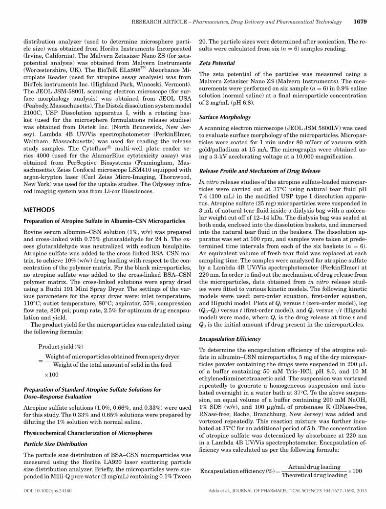
Figure 12 shows the comparative effect of the microparticleformulations with drug loading of 0.33% and 0.66% with thatof the standard 1% solution. First, there was a clear dose–response effect between the microparticle formulations with ahigher response from the 0.66% than the 0.33% drug loading.The results also show that although the recorded onset of actionwas at 0.25 h for the formulations and the standard solution,the Cmax of 0.63 ratio for the microparticles with 0.66% drugloading was at 0.25 h. This was 0.25 h earlier than the standardsolution and 0.33% drug-loaded microparticles.
Table 2 shows the duration of action, AUC, and the Cmax
of the three formulations. All the parameters show that therewas a greater effect from the microparticles with 0.66% drugloading than the 0.33% drug-loaded microparticles and the 1%standard solution (p < 0.05). The 0.66% microparticles had thehighest Cmax of 0.63 as compared with the 0.596 of the standardsolution and 0.51 of the 0.33% microparticles. The 0.66% mi-croparticles had an AUC of 10.67, whereas the 1% standard so-lution and 0.33% microparticles had an AUC of 10.02 and 8.46,
DOI 10.1002/jps.24380 Addo et al., JOURNAL OF PHARMACEUTICAL SCIENCES 104:1677–1690, 2015

1686 RESEARCH ARTICLE – Pharmaceutics, Drug Delivery and Pharmaceutical Technology
Figure 8. (a) Effect of temperature on the uptake of atropine sulfate-loaded BSA–CSN microparticles into corneal epithelial cells. Cell-associated fluorescence after BSA–CSN microparticles exposure at 4°Cor 37°C, expressed as percentage of microparticles (MS) available foruptake was significantly reduced at 4°C at 15, 30, 60, and 120 minincubation time. p � 0.05 calculated by ANOVA comparing the twogroups. (b) Effect of metabolic inhibition with 100 mM sodium azideon the atropine sulfate-loaded BSA–CSN microparticles uptake intohuman corneal epithelial cells. The microparticles uptake was quanti-fied as the fluorescence of cell lysates after microparticles exposure at37°C in the absence or presence of sodium azide. Presence of sodiumazide significantly inhibited the atropine sulfate-loaded BSA–CSN mi-croparticles uptake at 30, 60, and 120 min incubation times. p � 0.05calculated by ANOVA comparing the two groups.
respectively. The duration of action for the 0.66% microparti-cles was 8 h. The effect was 2 h greater than that of 6 h for thestandard solution and 0.33% microparticles.
Figure 9. Plot of the effect of light on the ratio of the pupil to corneallengths of rabbit eyes. The ratio of these two parameters was seen to beconstant for the first time ever at normal conditions. p � 0.05 calculatedby ANOVA comparing the two groups.
Furthermore, the ratio–time curves for the two microparti-cle formulations showed plateau sections not observed in thesolution from 0.25 to 3 h. This is a reflection of the controlled-release properties of the microparticle formulations.
DISCUSSION
The results of this study showed that the BSA–CSN micropar-ticles were an effective delivery system for atropine sulfateadministration into the cornea cells of the eye. The formula-tion had ideal physicochemical characteristics for a controlled-release delivery into the eyes. Spray drying has been tradition-ally used to produce particles with a narrow size distribution.This is evident from particle size of approximately 2 :m ob-tained in this study. Results also showed that the incorporationof the CSN as part of the polymeric matrix reduced the particlessize substantially. It is reported that for effective delivery andpatients’ comfort, solid particles intended for ophthalmic useshould not exceed 5–10 : in diameter, with a narrow size range(low polydispersity index).39,69 The particle sizes of 2.0 :mand polydispersity index of 0.268 obtained in this project are,therefore, ideal for ocular delivery. The small microparticleshave an extra advantage of large surface to volume ratios thatis good for controlled-release delivery of insoluble drugs.
The surface charges of microparticles as represented by thezeta potential are very important for the stability of the formu-lation in suspensions and in their interaction with the mucosalsurfaces. CSN has one primary amino and two free hydroxylgroups for each C6 building unit. The easy availability of thefree amino groups confers a positive charge on the surfacesof CSN and makes it a cationic polysaccharide. The molecularattractive forces formed by electrostatic interaction betweenthe positively charged CSN and negatively charged sialic acidresidue on mucosal surfaces confer mucoadhesive propertieson CSN-coated microparticles.29 CSN has been reported to alsoshow good bioadhesive characteristics and reduce the rate ofclearance of drugs from mucosal surfaces such as the nasal cav-ity, thereby increasing the bioavailability of drugs incorporatedin it.70 Therefore, the positively charged microparticle surfaceis advantageous for the maximum contact time on the eye sur-face. Moreover, adequate stability of microparticles dispersedin aqueous solutions can be achieved by either electrostatic sta-bilization or steric stabilization or by a combination of both.71
Adequately high zeta potential values beyond ±30 mV, as ob-tained in this study, are reported to indicate a stable colloidaldispersion.72 Loss of stability may lead to cloudiness, whichmay cause impairment of vision upon application to the eyes.
The raisin-like and porous appearance of the microparticlesobtained in this study is different from the traditional micro-spheres that have dense structure and are spherical in shape.Compared with traditional microspheres, porous microparticlesshow many unique properties such as large specific surface areafor efficient drug absorption or release and low density.73 Thesecharacteristics may be ideal for ocular delivery as it will leadto large contact surface on the eyes and reduction in patientsdiscomfort because of the low density.
Drug release from the microparticles showed a biphasic pro-file with an initial burst followed by a more controlled period.The second slower phase of release depends on microparticleporosity and hydrophilicity as well as molecular interactionforces between the polymer matrix and drug molecules.48,74 In
Addo et al., JOURNAL OF PHARMACEUTICAL SCIENCES 104:1677–1690, 2015 DOI 10.1002/jps.24380

RESEARCH ARTICLE – Pharmaceutics, Drug Delivery and Pharmaceutical Technology 1687
Figure 10. Plot of the mydriatic effect of 1% atropine sulfate standard solution before and after exposure to light in rabbit eyes. p � 0.05calculated by ANOVA comparing the two groups.
Figure 11. Dose–response studies of the atropine sulfate standard marketed solution showing the extent of mydriasis at three differentconcentrations (0.33%, 0.66%, and 1%) of atropine sulfate solution in rabbit’s eyes. p � 0.05 calculated by ANOVA comparing the groups witheach other.
porous and hydrophilic microparticles (such as that made ofBSA and CSN) or if there is affinity between the drug andpolymer matrix, water penetration into the particles and drugdissolution/diffusion out of the matrix are facilitated. As a re-sult, a second phase of continuous release may follow the burst,leading to a final drug release before the particle erosion setsin or reached an advanced stage—biphasic release pattern.75
Results from this study indicate that the pattern of release ofatropine sulfate from the BSA–CSN microparticles follow thebiphasic release profile with longer phase of extended release.If the bioadhesive characteristic of the CSN is effective in re-ducing the rate of clearance of the microparticles from the eyeand thereby increasing the contact time, then there is a guar-antee of continuous supply of drug into the eye for the durationof contact.
The lack of inherent toxicity of the microparticles confirms asimilar finding reported by De Campos et al.38 The nontoxicity
of the formulation to human corneal epithelial cells coupledwith the antiulcer and nonirritant nature of CSN25,76 makesthe microparticles ideal for topical and transcorneal ophthalmicdrug delivery.
The uptake of the microparticles into the human corneal ep-ithelial cells indicated an intracellular transport system thatgets saturated. This is very important in ocular delivery sys-tems where contact time on the eyes is limited. Prego et al.77
reported that CSN nanoparticles interaction with Caco-2 cellswas saturable in approximately 30 min. However, it was alsoshown by the same group that the presence of mucus in MTX-E12 cell monolayers strongly increased the association with theCSN nanoparticles.77 De Campos et al.38 reported an intenseinteraction of CSN with ocular surfaces (cornea and conjunc-tiva) when the polymer is in microparticulate/nanoparticulateform. This is very relevant in the finding obtained in thisstudy because the fact that more BSA–CSN microparticles were
DOI 10.1002/jps.24380 Addo et al., JOURNAL OF PHARMACEUTICAL SCIENCES 104:1677–1690, 2015
1688 RESEARCH ARTICLE – Pharmaceutics, Drug Delivery and Pharmaceutical Technology
Figure 12. Comparison of the mydriatic effect of BSA–CSN microparticles with 0.33% and 0.66% drug loading with the standard 1% atropinesulfate solution after exposure to light in rabbit’s eyes. p � 0.05 calculated by ANOVA comparing the groups with the control 1% atropine sulfatesolution group.
Table 2. Summary of Comparison of the Onset of Action, Duration of Action, Area Under the Curve (AUC), and Maximum Effect(Pupil–Corneal Ratio) of Microparticles with 0.33% and 0.66% Drug Loading with the Standard 1% Atropine Sulfate Ophthalmic Solution
Formulation Microparticles Onset of Action (h) Duration of Action (h) AUC Maximum Effect (Pupil–Corneal Ratio)
0.33% 0.25 6.00 8.46 0.5110.66% 0.25 8.00 10.67 0.630Standard 1% solution 0.25 6.00 10.02 0.596
transported into the cell up to 4 h shows the important role thepotential mucoadhesive properties of the CSN and microsizes ofthe particles may play in ocular delivery. The study also showedthat the cellular uptake is energy-dependent but with a basicunderlining passive diffusion mechanism. This passive diffu-sion was boosted by the increase in energy and metabolism.
The in vivo studies showed that the microparticles had su-perior effects on mydriasis in rabbits than the marketed so-lutions. The intensity of action as demonstrated by Cmax washigher and about 0.25 h earlier for the microparticles thanthe standard marketed solution. This is important for oculardelivery because of limited contact time. Moreover, one of thereasons adduced against the use of atropine sulfate as a cy-cloplegic agent is the delay in the onset of cycloplegia. It isreported that atropine sulfate in standard marketed formula-tions needs at least 3 h to reach peak effect and, therefore, mustbe used for 3 days to produce complete cycloplegia. As a result,it takes 8–14 days for its effect to wash out from the pupiland ciliary body.51 The earlier onset of effect demonstrated inthis study may, therefore, be more effective in the ocular de-livery of the drug. The result obtained in this study showedthat all the basic pharmacokinetic parameters such as durationof action, AUC, and the Cmax were better in the 0.66% drug-loaded microparticles than the standard marketed 1% solutionformulation.
The results, therefore, demonstrate that our microparticleformulations, though lower in drug concentration than that ofthe standard marketed solution, had superior effect on mydri-asis in rabbit eyes. The 0.66% microparticle formulation may,
therefore, be more effective in delivering the required effect ata shorter onset time, with sustained-release and less drug char-acteristics. This may be effective in ensuring better compliancefrom users.
CONCLUSIONS
The overall results showed that BSA–CSN microparticles arean effective delivery system for atropine sulfate administrationinto the cornea cells of the eyes. The addition of CSN increasedthe contact time and a significantly higher amount of drug wasdelivered in a sustained-release manner into the eye.
As compared with the standard 1% atropine sulfate solutionon the market, the microparticle formulation containing 0.66%atropine sulfate showed superior effect on mydriasis in rabbiteyes.
Bovine serum albumin and CSN are both considered as gen-erally safe materials as they are polymers that possess bio-compatible and biodegradable properties. The combination ofthe polymers in the preparations of microparticles targetingeffective drug delivery into the eyes as shown in this studywill go a long way in addressing the inefficient drug deliveryand safety issues associated with current drug administrationinto the eyes. The in vivo results demonstrated that the at-ropine microparticle formulation, though lower in strength ofatropine sulfate than that of the standard marketed solution,was superior to that of the standard marketed solution and welltolerated in human corneal epithelial cells in vitro.
Addo et al., JOURNAL OF PHARMACEUTICAL SCIENCES 104:1677–1690, 2015 DOI 10.1002/jps.24380

RESEARCH ARTICLE – Pharmaceutics, Drug Delivery and Pharmaceutical Technology 1689
REFERENCES
1. Mathiowitz E, Chickering D, Jacob JS, Santos C. 1999. Bioadhe-sive drug delivery systems. In Encyclopedia of controlled drug delivery;Mathiowitz E, Ed. Vol I. New York: Wiley, pp 9–44.2. Vasir JK, Tambwekar K, Garg S. 2003. Bioadhesive microspheres asa controlled drug delivery system. Int J Pharm 255:13–32.3. Alonso MJ, Sanchez A. 2003. The potential of chitosan in oculardelivery. J Pharm Pharmacol 55:1451–1463.4. Diebold Y, Calonge M. 2010. Applications of nanoparticles in oph-thalmology. Prog Retin Eye Res 29:596–609.5. Chandy T, Sharma CP. 1990. Chitosan—As a biomaterial. Biomate-rials Artif Cells Artif Organs 18(1):1–24.6. Illum L, Jabbal-Gill I, Hinchcliffe M, Fisher AN, Davis SS. 2001.Chitosan as a novel nasal delivery system for vaccines. Adv Drug DelivRev 51:81–96.7. George M, Abraham TE. 2006. Polyionic hydrocolloids for the in-testinal delivery of protein drugs, alginate and chitosan—A review. JControl Release 114 (1):1–14.8. Gan O, Wang T, Cochrane C, McCarron P. 2005. Modulation of sur-face charge particle size and morphological properties of chitosan—TPPnanoparticles intended for gene delivery. Colloids Surf B Biointerfaces44(2–3):65–73.9. Chandy T, Sharma CP. 1992. Chitosan beads and granules fororal sustained delivery of nifedipine: In vitro studies. Biomaterials13(13):949–952.10. Gupta KC, Ravi Kumar MNV. 2000. Drug release behavior of beadsand microgranules of chitosan. Biomaterials 21(11):1115–1119.11. Mi FL, Sung HW, Shyu SS. 2002. Drug release from chitosan–alginate complex beads reinforced by a naturally occurring crosslinkingagent. Carbohydr Polym 48 (1):61–72.12. Lubben MVD, Van Opdorp FAC, Hengeveld MR, Onderwater JJM,Koerten HK, Verhoef JC, Borchard G, Junginger HE. 2002. Transportof chitosan microparticles for mucosal vaccine delivery in a humanintestinal M-cell model. J Drug Target 10 (6):449–456.13. Amidi M, Romeijn SG, Verhoef JC, Junginger HE, Bungener L,Huckriede A, Crommelin D JA, Jiskoot W. 2007. N-trimethyl chitosan(TMC) nanoparticles loaded with influenza subunit antigen for in-tranasal vaccination: Biological properties and immunogenicity in amouse model. Vaccine 25 (1):144–153.14. Shah DN, Rectenwall-Work SM, Anseth KS. 2008. The effect ofbioactive hydrogels on the secretion of extracellular matrix moleculesby valvular intestinal cells. Biomaterials 19:2070–2072.15. Amiji MM. 1995. Permeability and blood compatibility propertiesof chitosan–poly(ethylene oxide) blend membranes for haemodialysis.Biomaterials 16:593–599.16. Kristmundsdottir T, Ingvardottir K, Saemundsdottir G. 1995. Chi-tosan matrix tablets: The influence of excipients on drug release. DrugDev Ind Pharm 21:1591–1598.17. Sabnis S, Rege P, Block LH. 1997. Use of chitosan in compressedtablets of diclofenac sodium: Inhibition of drug release in an acidicenvironment. Pharm Dev Technol 2:243–255.18. Illum L. 1998. Chitosan and its use as a pharmaceutical excipient.Pharm Res 15:1326–1331.19. Danielsen S, Varum KM, Stokke BT. 2004. Structural analysis ofchitosan medicated DNA condensation by AFM: Influence of chitosanmolecular parameters. Biomacromolecules 5(3):928–936.20. Mansouri S, Lavigne P, Corsi K, Benderdour M, Beaumont E,Fernandes JC. 2004. Chitosan–DNA nanoparticles as nonviral vec-tors in gene therapy: Strategies to improve transfection efficacy. Eur JPharm Biopharm 57(1):1–8.21. Wong K, Sun G, Zhang X, Dai H, Liu Y, He C, Leong KW. 2006. PEI-g-chitosan, a novel gene delivery system with transfection efficiencycomparable to polyethylenimine in vitro and after liver administrationin vivo. Bioconjug Chem 17 (1):152–158.22. Iwasaki N, Yamane ST, Majima T, Kasahara Y, Minami A, HaradaK, Nonaka S, Maekawa N, Tamura H, Tokura S, Shiono M, MondeK, Nishimura S. 2004. Feasibility of polysaccharide hybrid materials
for scaffolds in cartilage tissue engineering: Evaluation of chondrocyteadhesion to polyion complex fibres prepared from alginate and chitosan.Biomacromolecules 5(3):828–833.23. Zhu AP, Ming Z, Jian S. 2005. Blood compatibility of chi-tosan/heparin complex surface modified ePTFE vascular graft. ApplSurf Sci 241 (3–4):485–492.24. Zaharoff DA, Rogers CJ, Hance KW, Schlom J, Greiner JW. 2007.Chitosan solution enhances both humoral and cell-medicated im-mune responses to subcutaneous vaccination. Vaccine 25 (11):2085–2094.25. Ito M, Ban A, Ishihara M. 2000. Anti-ulcer effects of chitin andchitosan, healthy foods, in rats. Jpn J Pharmacol 82:218–225.26. Berger J, Reist M, Mayer JM, Felt O, Gurny R. 2004. Structure andinteractions in chitosan hydrogels formed by complexation or aggrega-tions for biomedical applications. Eur J Pharm Biopharm 57 (1):35–52.27. Berger J, Reist M, Mayer JM, Felt O, Peppas NA, Gurny R. 2004.Structure and interactions in covalently and ionically crosslinked chi-tosan hydrogels for biomedical applications. Eur J Pharm Biopharm 57(1):19–34.28. Lehr C-M, Bouwstra JA, Schacht EH, Junginger HE. 1992. In vitroevaluation of mucoadhesive properties of chitosan and some other nat-ural polymers. Int J Pharm 78:43–48.29. Zimmer A, Mutschler E, Lambrecht G, Mayer D, Kreuter J. 1994.Pharmacokinetic and pharmacodynamic aspects of an ophthalmic pilo-carpine nanoparticle-delivery-system. Pharm Res 11 (10):1435–1442.30. He P, Davis SS, Illum L. 1998. In vitro evaluation of the mucoad-hesive properties of chitosan microspheres. Int J Pharm 166 (1):75–88.31. Bernkop-Schnurch A, Humenberger C, Valenta C. 1998. Basic stud-ies on bioadhesive delivery systems for peptide and protein drugs. IntJ Pharm 165 (2):217–225.32. Soane RJ, Frier M, Perkins AC, Jones NS, Davis SS, Illum L. 1999.Evaluation of the clearance characteristics of bioadhesive systems inhumans. Int J Pharm 178:55–65.33. Robinson JR, Mlynek GM. 1995. Bioadhesive and phase-changepolymers for ocular drug delivery. Adv Drug Deliv Rev 16:45–50.34. Felt O, Furrer P, Mayer JM, Plazonnet B, Buri P, Gurny R. 1999.Topical use of chitosan in ophthalmology: Tolerance assessment andevaluation of precorneal retention. Int J Pharm 180:185–193.35. Sinha VR, Singla AK, Wadhawan S, Kaushik R, Kumria R, BansalK, Dhawan S. 2004. Chitosan microspheres as a potential carrier fordrugs. Int J Pharm 274:1–33.36. Markey ML, Bowman LN, Bergamini MVM. 1989. Chitin andchitosan—Source, chemistry, biochemistry, physical properties and ap-plication. London, UK: Elsevier.37. Torchilin VP, Levchenko TS, Rammohan R, Volodina N,Papahadjopoulos-Sternberg B, D’Souza GGM. 2003. Cell transfection invitro and in vivo with nontoxic TATpeptide–liposome–DNA complexes.Proc Natl Acad Sci USA 100:1972–1977.38. De Campos AM, Diebold Y, Carvolho ELS, Sanchez A, Alonso MJ.2004. Chitosan nanoparticles as new ocular drug delivery systems: Invitro stability, in vivo fate, and cellular toxicity. Pharm Res 21(5):803–810.39. Rathore KS, Nema RK. 2009. Insight into ophthalmic drug deliverysystem. Int J Pharm Sci Drug Res 1 (1):1–5.40. Harmia T, Speiser P, Kreuter J. 1986. A solid colloidal drug deliverysystem for the eye: Encapsulation of pilocarpine in nanoparticles. JMicroencapsul 3:3–12.41. Losa C, Calvo P, Castro E, Vila-Jato JL, Alonso MJ. 1991. Im-provement of ocular penetration of amikacin sulphate by association topoly-(butylcyanoacrylate) nanoparticles. J Pharm Pharmacol 43:548–552.42. Losa C, Marchal-Heussler L, Orallo F, Vila-Jato JL, Alonso MJ.1993. Design of new formulations for topical ocular administration:Polymeric nanocapsules containing metipranolol. Pharm Res 10:80–87.43. Calvo P, Sanchez A, Martinez J, Lopez MI, Calonge M, Pastor JC,Alonso MJ. 1996. Polyester nanocapsules as new topical ocular deliverysystems for cyclosporin A. Pharm Res 13:311–315.
DOI 10.1002/jps.24380 Addo et al., JOURNAL OF PHARMACEUTICAL SCIENCES 104:1677–1690, 2015

1690 RESEARCH ARTICLE – Pharmaceutics, Drug Delivery and Pharmaceutical Technology
44. Calvo P, Alonso MJ, Vila-Jato JL, Robinson JR. 1996. Improvedocular bioavailability of indomethacin by novel ocular drug carriers. JPharm Pharmacol 48:1147–1152.45. Genta I, Conti B, Perugini P, Pavanetto F, Spadaro A, Puglisi G.1997. Bioadhesive microspheres for ophthalmic administration of acy-clovir. J Pharm Pharmacol 49:737–742.46. Calvo P, Vila-Jato JL, Alonso MJ. 1996. Comparative in vitroevaluation of several colloidal systems, nanoparticles, nanocapsulesand nanoemulsions as ocular drug carriers. J Pharma Sci 85:530–536.47. Kramer PA. 1974. Albumin microspheres as vehicles forachieving specificity in drug delivery. J Pharm Sci 63(10):1646–1647.48. Yeboah KG, D’Souza MJ. 2009. Evaluation of albumin microspheresas oral delivery system for Mycobacterium tuberculosis vaccines. J Mi-croencapsul 26(2):166–179.49. Thakkar H, Sharma RK, Mishra AK, Chuttani K, Murthy RR. 2005.Albumin microspheres as carriers for the antiarthritic drug celecoxib.AAPS Pharm Sci Tech 6:E65–E73.50. Manny RE, Hussein M, Scheiman M, Kurtz D, Niemann K, ZinzerK, the Comet Study Group. 2001. Tropicamice (1%): An effective cyclo-plegic agent for myopic children. Invest Ophthalmol Vis Sci 42 (8):1728–1735.51. Farhood QK. 2012. Cycloplegic refraction in children with cyclopen-tolate versus atropine. J Clin Exp Ophthalmol 3(7):1–6.52. Chia A, Chua W-H, Wen L, Fong A, Goon YY, Tan D. 2014. Atropinefor the treatment of childhood myopia: Changes after stopping atropine0.01%, 0.1% and 0.5%. Am J Ophthalmol 157(2):451–457.53. Ahmad NN, Dimascio J, Knowlton RG, Tasman WS. 1995. Sticklersyndrome. A mutation in the nonhelical 3′ end of type II procollagengene. Arch Ophthalmol 113(11):1454–1457.54. Krolicki TJ, Tasman W. 1995. Cataract extraction in adultswith retinopathy of prematurity. Arch Ophthalmol 113(2):173–177.55. Ngezahayo A, Lang F, Kolb HA. 1995. Cholecystokinin-octapeptideaffects the fluorescence signal of a single pancreatic acinar cell loadedwith the acrylodan-labelled MARCKS peptide, a protein kinase C sub-strate. Pflugers Arch 429(6):805–808.56. Regillo CD, Brown GC, Savino PJ, Byrnes GA, Benson WE, TasmanWS, Sergott RC. 1995. Diabetic papillopathy. Patient characteristicsand fundus findings. Arch Ophthalmol 113 (7):889–895.57. Dollery C. 1999. Therapeutic drugs. 2nd ed. Edinburgh UK:Churchill Livingstone, section A, pp 40–44.58. Morton RA, Creed RH. 1939. The conversion of carotene to vitaminA(2) by some fresh-water fishes. Biochem J 33(3):318–324.59. Hoefnagel D. 1961. Toxic effects of atropine and homatropine eyedrops in children. N Engl J Med 264:168–171.60. Akande J, Yeboah KG, Addo RT, Siddig A, Oettinger CW, D’SouzaMJ. 2010. Targeted delivery of antigens to the gut-associated lymphoidtissues: 2. Ex vivo evaluation of lectin-labeled albumin microspheresfor targeted delivery of antigens to the M-cells of the Peyer’s patches.J Microencapsul 27 (4):325–336.
61. Yeboah KG, Akande J, Addo RT, Siwale RC, Aninkorah-Yeboah K,Siddig A. 2014. In vitro and ex vivo characterization of lectin-labeledMycobacterium tuberculosis antigen-containing microspheres for en-hanced oral delivery. J Drug Target 22(1):34–47.62. Shastri PN, Ubale RV, D’Souza MJ. 2013. Implementation of mix-ture design for formulation of albumin containing enteric-coated spray-dried microparticles. Drug Dev Ind Pharm 39:164–175.63. Ubale RV, D’Souza MJ, Infield DT, McCarty NA, Zughaier SM.2013. Formulation of meningococcal capsular polysaccharide vaccine-loaded microparticles with robust innate immune recognition. J Mi-croencapsul 30:28–41.64. Patel N, Addo RT, Ubale R, Uddin MN, D’Souza M, Jobe L. 2014.The effect of antisense to NF-6B in an albumin microsphere formula-tion on the progression of left-ventricular remodeling associated withchronic volume overload in rats. J Drug Target 22:796–804.65. Ubale RV, Gala RP, Zughaier SM, D’Souza MJ. 2014. Induction ofdeath receptor CD95 and co-stimulatory molecules CD80 and CD86 bymeningococcal capsular polysaccharide-loaded vaccine nanoparticles.AAPS J 16:986–993.66. Pignatello R, Bucolo C, Spedalieri G, Maltese A, Pugilis G. 2002.Flurbiprofen-loaded acrylate polymer nanosuspensions for ophthalmicapplication. Biomaterials 15:3247–3255.67. Addo RT, Siddig A, Siwale R, Patel NJ, Akande J, Uddin AN,D’Souza MJ. 2010. Formulation, characterization and testing of tetra-caine hydrochloride-loaded albumin–chitosan microparticles for oculardrug delivery. J Microencapsul 27:95–104.68. Huguchi T. 1961. Rate of release of medicaments from ointmentvases containing drugs in suspension. J Pharm Sci 50:874–875.69. Guinedi AS, Mortada ND, Mansour S, Hathout RM. 2005. Prepa-ration and evaluation of reverse-phase evaporation and multilamellarniosomes as ophthalmic carriers of acetazolamide. Int J Pharm 306:71–82.70. Tiyaboonchai W. 2003. Chitosan nanoparticles: A promising systemfor drug delivery. Naresuan Univ J 11(3):51–66.71. Nagarwal RC, Kant S, Singh PN, Maiti P, Pandit JK. 2009. Poly-meric nanoparticulate system: A potential approach for ocular drugdelivery. J Control Release 136:2–13.72. Benita S, Levy MY. 1993. Submicron emulsions as colloidal drugcarriers for intravenous administration: Comprehensive physicochem-cial characteristics. J Pharm Sci 82:1069–1079.73. Cai Y, Chen Y, Hong X, Lui Z, Yuan W. 2013. Porous microsphereand its applications. Int J Nanomed 8:1111–1120.74. Gander B, Johansen P, Nam-Tran H, Merkle HP. 1996. Thermo-dynamic approach to protein microencapsulation into poly(D, L-lactide)by spray drying. Int J Pharm 129:51–61.75. Tamber H, Johansen P, Merkle HP, Gander B. 2005. Formulationaspects of biodegradable polymeric microspheres for antigen delivery.Adv Drug Deliv Rev 57:357–376.76. Miyazaki S, Ishii K, Nadai T. 1981. The use of chitin and chitosanas drug carriers. Chem Pharm Bull 29(10):3067–3069.77. Prego C, Garcia M, Torres D, Alonso MJ. 2005. Transmucosalmacromolecular drug delivery. J Control Release 101:151–162.
Addo et al., JOURNAL OF PHARMACEUTICAL SCIENCES 104:1677–1690, 2015 DOI 10.1002/jps.24380

![[The beginnings of modern biological psychiatry in Hungary: the atropine coma. A historical overview]](https://img.pdfslide.net/doc/110x75/634db87c7b55c75a34073438/the-beginnings-of-modern-biological-psychiatry-in-hungary-the-atropine-coma-a.jpg)


