Embed Size (px)
Citation preview

International Research Journal of Clinical Medicine • Vol 1 • Issue 4 • Apr 2016 23
Fundamentals of In situ Hybridization: A ReviewMohammad Ehtisham1, Firdous Wani2, Iram Wani3, Prabhjot Kaur4, Sheeba Nissar5
1Post-Graduate Student, Department of Oral Pathology and Microbiology, Institute of Dental Studies and Technologies, Kadrabad, Modinagar, Uttar Pradesh, India, 2Post-Graduate Student, Department of Oral Medicine & Radiology, Shree Bankey Bihari Dental College & Research Centre, Ghaziabad, Uttar Pradesh, India, 3Post-Graduate Student, Department of Oral Pathology and Microbiology, Shree Bankey Bihari Dental College & Research Centre, Ghaziabad, Uttar Pradesh, India, 4Post-Graduate Student, Department of Oral Pathology and Microbiology, Swami Devi Dayal Dental College & Hospital, Barwala, Haryana, India, 5Post-Graduate Student, Department of Periodontics & Oral Implantology, Shree Bankey Bihari Dental College & Research Centre, Ghaziabad, Uttar Pradesh, India
Corresponding Author: Dr. Mohammad Ehtisham, B-3, Jamia Apartment, Flat No. 007, Abul Fazal Enclave, Jamia Nagar, Okhla, New Delhi - 110 025, India. Phone: +91-9891194154. E-mail: [email protected]
INTRODUCTIONThe word “in situ” comes from Latin meaning “in position.” By definition it means in the natural or original position.1 Hybridization means production of hybrid by crossbreeding. In molecular cell biology hybridization means pairing of complementary RNA or DNA to produce a double stranded nucleic acid. Hybridization method uses a radio or fluorescent labeled DNA or RNA probe that binds to target DNA or RNA of interest, permitting visualization. In situ hybridization (ISH) detection of specific DNA or RNA sequences in tissue sections or cell preparations using a labeled probe under appropriate conditions this probe will hybridize the target DNA or RNA which will be visualized by radioactive or non-radioactive labels incorporated into the probe. It is a technique used to examine DNA and RNA in their topographic surroundings.2
HISTORYFirst described by almost simultaneously by John et al., Gall and Pradue (1969). The technique originally used
Review Article
ABSTRACTThe word “in situ” derived from Latin word meaning “in position.” It is defined as “in the natural or original position.” Hybridization means the production of hybrid by crossbreeding. In molecular cell biology hybridization means pairing of complementary RNA or DNA to produce a double stranded nucleic acid. It was first described by almost simultaneously by John et al., Gall and Pradue (1969). The technique originally used auto-radiographic labeling to map both repetitive and low-copy DNA sequences. In situ hybridization is the specific annealing of a labeled nucleic acid probe to complementary sequences in fixed tissues followed by visualization of the location of the probe. The development of in situ technologies has provided us with a wealth of information regarding the locations and expression patterns of genes in the cells.Key words: Annealing, Denaturation, DNA hybridization, Fluorescence in situ hybridization, In situ hybridization, Probe
autoradiographic labeling to map both repetitive and low-copy DNA sequences. Although, it was very sensitive but problems associated with this technique include short half-life, safety problem, and long-exposure time which hindered its widespread use in DNA hybridization. To overcome these problems, non-isotopic ISH was developed but its use was limited to animal system. The non-isotopic ISH using biotin labeled DNA probes was first introduced in plant species by Rayburn and Gill (1985). Biotin-labeled repetitive DNA sequences (120 bp) of rye were used as probe to mitotic chromosomes of Triticum aestivum and double ditelosomic lines of B and D genome chromosomes and 4A of Chinese spring. The hybridization sites occurred as brown bands on blue chromosomes. Based on this color difference, the hybridization sites were discriminated from the non-hybridization and Giemsa staining bands. The hybridization pattern revealed that Chinese spring has 7B genome chromosomes and chromosomes 4A, 2D and 5D are from the ditelosomic lines. Durnam et al. (1985) modified the technique using genomic ISH. Further progress in this field, lead to the development of fluorescence in situ hybridization (FISH).3

Ehtisham, et al.: Fundamentals of In situ Hybridization: A Review
International Research Journal of Clinical Medicine • Vol 1 • Issue 4 • Apr 201624
PRINCIPLE OF ISHISH is the specific annealing of a labeled nucleic acid probe to complementary sequences in fixed tissues followed by visualization of the location of the probe. ISH – demonstrates specific nucleic acid sequence in their cellular environment.4
STEPS IN ISH1. Probe preparation2. Pre-treatment of specimens3. Hybridization4. Detection method.2
Probe Preparation
ProbesA probe (a labeled complementary single strand) is incorporated with the DNA/RNA strands of interest. Strands will anneal with complementary nucleotides bonding back together with their homologous partners when cooled. Chances of a probe finding a homologous sequence other than the target sequence decrease as the number of nucleotides in the probe increases (Figure 1).5
These are following types.2
A ‑double strand DNA probeB ‑single strand DNA probeC ‑single strand RNA probe
D ‑oligonucleotide probes
Double stranded DNA probePrepared by nick translation, random primer, PCR in the presence of a labeled nucleotide. Nick translation is a method for incorporating labeled nucleotides into DNA such as an isolated fragment. The method uses a combination of two enzymes, Deoxyribonuclease I which nicks the DNA creating free 3 hydroxyls. DNA polymerase I, which processively adds nucleotides to the 3′ terminal hydroxyl.6 (Figure 2) Random priming is a means of labeling DNA fragments whereby, a mixture of all possible combinations of hexamers, octamers, or nanomers are annealed to denatured DNA. These small oligonucleotides then act as primers that allow for the synthesis of the complementary DNA strand by the Klenow enzyme and incorporation of both labeled and unlabeled nucleotides. More effective when the target is abundant. Less sensitive than single strand probe. Because two strands have a tendency to hybridize to each other, thus reducing the concentration of probe available for hybridization to the target.7
Single stranded DNA probesSingle-stranded DNA probes cover a much larger size range (200-500 bp) than oligonucleotide probes.
They can be prepared by a primer extension on single-stranded templates by reverse transcription-polymerase chain reaction (PCR) of RNA, or an amplified primer extension of a PCR-generated fragment in the presence of a single antisense primer, or chemical synthesis of oligonucleotides. PCR-based methods are much easier and probes can be synthesized from small amounts of starting material. Moreover, PCR allows great flexibility in the choice of probe sequences by the use of appropriate primers.5
RNA probesRNA probes (cRNA probes or riboprobes) are thermostable and are resistant to digestion by RNases. These probes are single-stranded and are the most widely used in ISH. RNA probes are generated by in vitro transcription from a linearized template using a promoter for RNA polymerase. RNA polymerase is used to synthesize RNA complementary to the DNA substrate. Single-stranded probes provide advantages over double-stranded probes such as the probe does not self-anneal in solution, so the probe is not exhausted. Large probe chains are not formed in solution; thus, probe penetration is not affected. If high sensitivity is required, single-stranded probes should be used.5
OligoprobesUsually, shorter 20-40 base pair length. They are produced synthetically by an automated chemical synthesis. These probes are resistant to RNases and are small, thus allowing easy penetration into the cells or tissue of interest. Small size has a disadvantage in that it covers fewer targets. Label should be positioned at the 3′ or the 5′ end. To increase sensitivity one can use a mixture of oligonucleotides that are complementary to different regions of the target molecule. Another advantage of oligonucleotide probes is that they are single stranded, therefore excluding the possibility of renaturation.8
Properties of probes• Probe construct: Oligonucleotide probes are better
than traditional probes because of high specificity, single-stranded, and short probe length (10-50 nucleotides).
• The efficiency of labeling: Labeling by random priming has been reported to be more efficient than nick translation.
• Percentage of G-C base pairs: Higher the content of G-C pairs, the higher the Tm (melting temperature).
• RNA versus DNA probes: Strength of the probe-target bond decreases in the order of RNA-RNA, DNA-RNA, DNA-DNA.
• Probe length: Shorter the probe, the better its penetration into cells.2

International Research Journal of Clinical Medicine • Vol 1 • Issue 4 • Apr 2016 25
Ehtisham, et al.: Fundamentals of In situ Hybridization: A Review
Probes and their choice9
Probes for DNA Probe for RNA
Double‑stranded DNASingle‑stranded DNAOligo‑deoxyribonucleotides
Complementary RNA, a riboprobe
Purification of labeled probesThere are several methods that can be used to test the purification. Here, a list of methods that can be used but it is advisable to follow the manufacturers’ recommendation on their use: Sephadex G-50 column, Sephadex G-50 chromatography, or Selective precipitation.5
Probe concentrationFor DNA probes, the concentration of the probe will be 0.5-2 μg/ml. Oligonucleotide probes can be used with, or without, acetylation. Probes without acetylation pre-treatment of the sample will have a concentration of ~50-200 ng/ml and may provide more intense results with a minimal background. For probes with acetylation pre-treatment, a higher concentration of oligonucleotide probe may be used without incurring non-specific background staining.5
Length of probeOptimal probe size for ISH is small fragments of about 200-300 nucleotides. However, probes may be as small as 20-40 bp or as large as 1000 bp. As probes increase in length, they become more specific. Longer probes may lead to weaker signal (Figure 3). They penetrates less efficiently the cross-linked tissues. The extent of weaker signals and penetration depends also on the nature of the tissue, choice of fixative and whether a pre-treatment has been carried out.7
Pretreatment of Specimens
Tissue sections must adhere well to specially treated glass slides to avoid loss of tissue during the hybridization process. Various “adhesives” are available including poly-l-lysine, gelatin chrome alum, and aminopropyltriethoxysilane.2
FixationMethanol/acetic acid fixation is recommended for metaphase chromosome spreads. Cryostat sections may be fixed with 4% formaldehyde (~30 min), Bouin’s fixative, or paraformaldehyde vapor fixation. This fixation also helps to secure the tissue to the slide. Most commonly, tissue specimens are routinely fixed in 10% buffered formalin, processed overnight in an automatic tissue processor, and embedded in paraffin wax. Fixation time of 8-12 h is optimal.5
Slide/section preparationSections are cut at 4-6 μm on an alcohol-cleaned microtome using positively charged or hand-coated slides. Sections are drained well and then air-dried at room temperature. After
deparaffinization, slides are placed in an alcohol-cleaned staining container of diethyl pyrocarbonate water. The staining container is then placed in the heated water bath at 23-37°C and held until the start of ISH. Gloves must be worn to prevent contamination, and all utensils, such as brushes and forceps, should be cleaned with alcohol and kept within the cleaned area designated for ISH.5
Proteolytic digestionThe use of formaldehyde-based fixatives before paraffin embedding of specimens will mask nucleic acid sequences. Digestion is an important step when performing ISH. Digestion improves probe penetration by increasing cell permeability with minimal tissue degradation.5
Hybridization
Molecular hybridization is the process whereby a single-stranded target sequence is annealed to a complementary single-stranded probe to form a double-stranded hybrid. Before hybridization, both the target and the probe, if double-stranded, must be denatured to render them single-stranded and this can be achieved by heat or alkali treatment. The following denaturation, single-stranded target, and probe sequences are incubated in a hybridization mixture, which provides an optimal environment for re-annealing of single-stranded sequences. Hybridization occurs after denaturation, during cooling, in the presence of a complementary probe, and permits hydrogen bonding of the two strands of nucleic acids.10
Probe must form stable hydrogen bonds with the target. Simultaneously heating the probe and target to high temperatures may increase the consistency and sensitivity of detection. This can only be met if care is taken to precisely control this step of the ISH procedure.10
Post-hybridization washesStringency washes after hybridization aims at decreasing non-specific binding. However, it is preferable to hybridize stringently rather than wash stringently.11
Detection Methods
Various methods are available for visualization of the hybridization. Choice of detection system will be principally determined by the probe label used and second by the ISH procedure type. There are two methods of probe labeling. Detection methods can be either direct method or indirect method.9
Direct method Indirect method
Reporter molecules • Enzyme, • Radioisotope or • Fluorescent markerare directly attached to the DNA or RNA
A hapten 1. Biotin, 2. Digoxigenin, or 3. Fluoresceinare attached to the probe and detected by a labeled binding protein (typically an antibody)

Ehtisham, et al.: Fundamentals of In situ Hybridization: A Review
International Research Journal of Clinical Medicine • Vol 1 • Issue 4 • Apr 201626
Oligonucleotide probe labeling• 5′-end labeling: The 5′ end of DNA or RNA undergoes
direct phosphorylation of the free 5′-terminal OH groups. The free 5′-OH substrates can be labeled using T4 polynucleotide kinase. This method is usually used for radiolabeling. Non-radiolabels use a covalent linker.12
• 3′-end labeling: Terminal dexoxynucleotidyl transferase (TdT) is used to add a labeled residue to the 3′ end of a synthetic oligonucleotide that is approximately 14-100 nucleotides in length. These probes provide excellent specificity but only moderate sensitivity.5
• 3′ tailing: A tail containing labeled nucleotides is added to the free 3′ end of double- or single-stranded DNA using TdT. These probes are more sensitive than the 3′-end labeled versions, but can produce more non-specific background.13
Enzymatic detectionHybridized probes can be detected by enzymatic reactions that produce a colored precipitate at the site of hybridization. The most commonly used enzymes for this application are alkaline phosphatase (AP) Or horseradish peroxidase (HRP) Although these enzymes can be conjugated directly to nucleic acid probes, such enzyme-coupled probes are often inappropriate for ISH to tissue preparations because probe penetration is hampered by the presence of the conjugated enzyme. Therefore, indirect methods are preDNA probe and a target sequence preferred.14
Detected by autoradiographyReactions using radioactive labeled probes are detected by autoradiography. This is based on the emission of fast-electrons or beta-particles from the probe. Beta particles release a large amount of energy when they collide with atoms of an emulsion added to the section on the slide. The excessive energy released reduces ionic silver present in the emulsion to metallic silver. When this happens, a faithful record of the location of the collision between an electron and the silver ions in the emulsion is produced in the form of a latent image. This image, when visualized is the indicator of the probe location in the tissue or cell. Autoradiography for radioactive labels is reputed to be more sensitive than the immunoenzyme systems.2
FluorophoresFluorophores can be associated with nucleic acid probes by chemical conjugation to the nucleic acid
Figure 2: Nick translation
Figure 1: Denaturation of DNA

International Research Journal of Clinical Medicine • Vol 1 • Issue 4 • Apr 2016 27
Ehtisham, et al.: Fundamentals of In situ Hybridization: A Review
OR chemical conjugation of the nucleic acid with a non-fluorescent molecule that can bind fluorescent material after hybridization. The former method is called “direct labeling” and the latter method is called “indirect labeling.” Chemical structures of four common fluorophore classes (A-D) fluoresceins, rhodamines, cyanines, or coumarins.7
Indirect methodIndirectly via incorporation of a nucleotide analog carrying a reactive group and subsequent biotinylation/digoxigenylation. Resulting biotin-labeled probes are then detected using streptavidin (KD = 10-15 M) conjugated with HRP or AP. Digoxigenylation is typically visualized by HRP-or AP modified antibodies.15
Multiple ISHMore than one probe can be applied to the same tissue section to detect different nucleic acid targets. By using
different detection systems with each probe, resulting in different color end products, and visualization of the different nucleic acid targets can be achieve.16
Advantages: Most important advantages of ISH are:1. Simplicity of its methodology2. Specificity of results obtained3. Ease in interpretation of findings
Its applicability on tissue sections (frozen or formalin-fixed, paraffin-embedded) and smears without the need for special specimen collection or processing.2 (Figure 4)
APPLICATIONS OF ISHDetermination of Infective Agent
This is based on the detection of the infective agent’s genome in the tissues or cells studied. Specific typing of infective agents also has important implications for epidemiological surveys and outbreak investigations.17
Localization of Active Infection
The actual cell or cell structures harboring the infective genome can be elucidated by ISH, e.g. hepatitis B virus (HBV) in hepatocytes, parvovirus in cells of the lung.17
Elucidation of mechanism of Virus Dissemination and Transmission
Natural horizontal and vertical transmission routes of viruses can be studied. For example, the presence of Epstein-Barr virus (EBV) in epithelial cells of the oropharynx provides a means for transmission of the virus through saliva.18Figure 3: Hybridization
Figure 4: Identification of infective agents
Ehtisham, et al.: Fundamentals of In situ Hybridization: A Review
International Research Journal of Clinical Medicine • Vol 1 • Issue 4 • Apr 201628
Localization of Persistent Virus Infection
Examples are the persistence of JC virus in oligodendrocytes in progressive multifocal leukoencephalopathy and measles virus in neurons and glia cells in SSPE.19,20
Link between Virus Agents and Carcinogenesis
Etiological role of various viruses in cancers and the mechanisms of malignant transformation of cells. The better known associations are EBV and nasopharyngeal carcinoma and B-cell lymphomas,21 HBV and hepatocellular carcinomas,22 and human papillomavirus and cervical carcinoma.23
Study of cell Development
ISH detection of cell-type specific RNA in cells which do not exhibit morphological differentiation can be applied to identify the cell type.24
Sex Determination
The Y chromosome can be detected through hybridisation.25
Human Gene Mapping26
In situ Hybridisation (ISH) used in human gene mapping.
Interphase Cytogenetics
ISH can be used to detect numerical chromosomal aberrations in interphase nuclei. Probes recognizing highly repetitive sequences in chromosomes 1, 7, 8, 9, 10, 15, 16, 17, 18, X and Y are now available.2
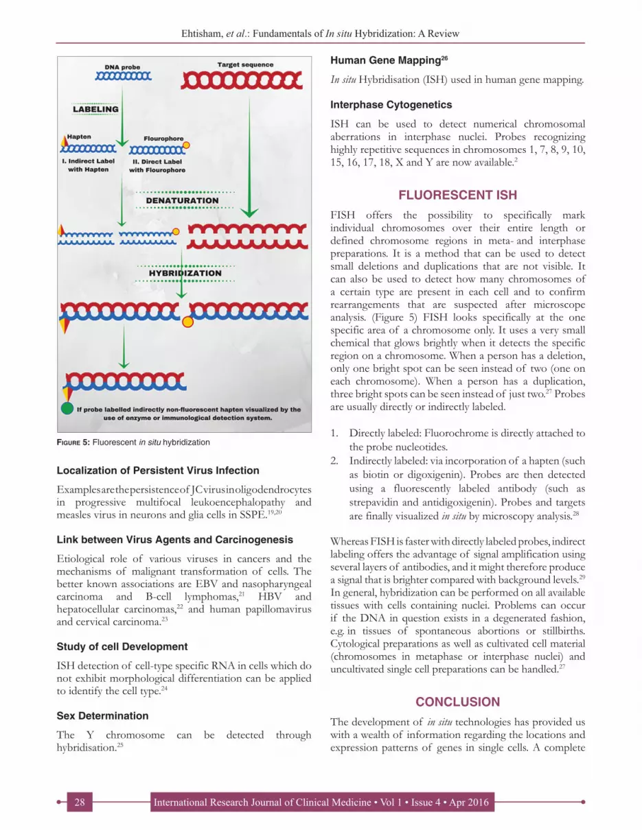
FLUORESCENT ISHFISH offers the possibility to specifically mark individual chromosomes over their entire length or defined chromosome regions in meta- and interphase preparations. It is a method that can be used to detect small deletions and duplications that are not visible. It can also be used to detect how many chromosomes of a certain type are present in each cell and to confirm rearrangements that are suspected after microscope analysis. (Figure 5) FISH looks specifically at the one specific area of a chromosome only. It uses a very small chemical that glows brightly when it detects the specific region on a chromosome. When a person has a deletion, only one bright spot can be seen instead of two (one on each chromosome). When a person has a duplication, three bright spots can be seen instead of just two.27 Probes are usually directly or indirectly labeled.
1. Directly labeled: Fluorochrome is directly attached to the probe nucleotides.
2. Indirectly labeled: via incorporation of a hapten (such as biotin or digoxigenin). Probes are then detected using a fluorescently labeled antibody (such as strepavidin and antidigoxigenin). Probes and targets are finally visualized in situ by microscopy analysis.28
Whereas FISH is faster with directly labeled probes, indirect labeling offers the advantage of signal amplification using several layers of antibodies, and it might therefore produce a signal that is brighter compared with background levels.29 In general, hybridization can be performed on all available tissues with cells containing nuclei. Problems can occur if the DNA in question exists in a degenerated fashion, e.g. in tissues of spontaneous abortions or stillbirths. Cytological preparations as well as cultivated cell material (chromosomes in metaphase or interphase nuclei) and uncultivated single cell preparations can be handled.27
CONCLUSIONThe development of in situ technologies has provided us with a wealth of information regarding the locations and expression patterns of genes in single cells. A complete
Figure 5: Fluorescent in situ hybridization

International Research Journal of Clinical Medicine • Vol 1 • Issue 4 • Apr 2016 29
Ehtisham, et al.: Fundamentals of In situ Hybridization: A Review
gene expression profiles of single cells will provide a new level of insight into the correlation of gene expression patterns with particular cellular phenotypes. This will be particularly important in studies of development and disease progression, where complicated, finely demarcated gene expression programs are in play.
REFERENCES1. Kute JU, Darekar AB, Saudagar RB. In situ gel-novel approach
for nasal delivery. World J Pharm Pharm Sci 2013;3:187-203.2. Looi LM, Cheah PL. In situ hybridisation: Principles and
applications. Malays J Pathol 1992;14:69-76.3. Abbasi FM, Khan MT, Perveen F, Masood R, Inamullah I,
Khan U, et al. Historical perspective of in situ hybridization for the analysis of genomic constitution of plants. Afr J Biotechnol 2015;9:9142-7.
4. Bishop R. Applications of fluorescence in situ hybridization (FISH) in detecting genetic aberrations of medical significance. Biosci Horizons 2010;3:85-95.
5. Bancroft JD, Gamble M. Theory and Practice of Histological Techniques. Amsterdam: Elsevier Health Sciences; 2008.
6. Rittié L, Perbal B. Enzymes used in molecular biology: A useful guide. J Cell Commun Signal 2008;2:25-45.
7. Morrison LE, Ramakrishnan R, Ruffalo TM, Wilber KA. Labeling fluorescence in situ hybridization probes for genomic targets. Methods Mol Biol 2002;204:21-40.
8. Darby IA. In Situ Hybridization Protocols. New York: Springer Science & Business Media; 2004.
9. de Muro MA. Probe design, production, and applications. Molecular Biomethods Handbook. Totowa, NJ: Humana Press; 2008. p. 41-53.
10. Tenover FC. Diagnostic deoxyribonucleic acid probes for infectious diseases. Clin Microbiol Rev 1988;1:82-101.
11. Poulsen L, Søe MJ, Snakenborg D, Møller LB, Dufva M. Multi-stringency wash of partially hybridized 60-mer probes reveals that the stringency along the probe decreases with distance from the microarray surface. Nucleic Acids Res 2008;36:e132.
12. Alberts B, Johnson A, Lewis J, et al., Molecular Biology of the Cell. 4th edition. New York: Garland Science; 2002. Available from: http://www.ncbi.nlm.nih.gov/books/NBK21054/.
13. Hilario E. End labeling procedures: An overview. Mol Biotechnol 2004;28:77-80.
14. Diamandis EP, Christopoulos TK. The biotin-(strept)avidin system: Principles and applications in biotechnology. Clin Chem 1991;37:625-36.
15. Nederlof PM, van der Flier S, Wiegant J, Raap AK, Tanke HJ, Ploem JS, et al. Multiple fluorescence in situ hybridization. Cytometry 1990;11:126-31.
16. Alonso MC, Cano I, Castro D, Perez-Prieto SI, Borrego JJ. Development of an in situ hybridisation procedure for the detection of sole aquabirnavirus in infected fish cell cultures. J Virol Methods 2004;116:133-8.
17. Morey AL, Porter HJ, Keeling JW, Fleming KA. Non-isotopic
How to cite this article: Ehtisham M, Wani F, Wani I, Kaur P, Nissar S. Fundamentals of In situ Hybridization: A Review. Int Res J Cli Med 2016;1(4):23-29.
Source of Support: Nil. Conflict of Interest: None declared.
Month of Submission: 02-2016 Month of Peer Review : 03-2016 Month of Acceptance: 04-2016 Month of Publishing : 04-2016
in situ hybridisation and immunophenotyping of infected cells in the investigation of human fetal parvovirus infection. J Clin Pathol 1992;45:673-8.
18. Rostad SW, Olson K, McDougall J, Shaw CM, Alvord EC Jr. Transsynaptic spread of varicella zoster virus through the visual system: A mechanism of viral dissemination in the central nervous system. Hum Pathol 1989;20:174-9.
19. Ironside JW, Lewis FA, Blythe D, Wakefield EA. The identification of cells containing JC papovavirus DNA in progressive multifocal leukoencephalopathy by combined in situ hybridization and immunocytochemistry. J Pathol 1989;157:291-7.
20. Shapsak P, Tourtellotte WW, Wolman M, Verity N, Verity MA, Schmid P, et al. Search for virus nucleic acid sequences in postmortem human brain tissue using in situ hybridisation technology with cloned probes: Some solutions and results on progressive multifocal leukoencephalopathy and subacute sclerosing panencephalitis tissue. J Neurosci Res 1986;16:281-301.
21. Weiss LM, Movahed LA. In situ demonstration of Epstein-Barr viral genomes in viral-associated B cell lymphoproliferations. Am J Pathol 1989;134:651-9.
22. Simon D, Searls DB, Cao Y, Sun K, Knowles BB. Chromosomal site of hepatitis B virus (HBV) integration in a human hepatocellular carcinoma-derived cell line. Cytogenet Cell Genet 1985;39:116-20.
23. Stoler MH, Rhodes CR, Whitbeck A, Wolinsky SM, Chow LT, Broker TR. Human papillomavirus type 16 and 18 gene expression in cervical neoplasias. Hum Pathol 1992;23:117-28.
24. Martizez Arias A, Baker NE, Ingham PW. Role of segment polarity genes in the definition and maintenance of cell states in the Drosophila embryo. Development 1988;103:157-70.
25. Burns J, Chan VT, Jonasson JA, Fleming KA, Taylor S, McGee JO. Sensitive system for visualising biotinylated DNA probes hybridised in situ: Rapid sex determination of intact cells. J Clin Pathol 1985;38:1085-92.
26. Rowley JD, Diaz MO, Espinosa R 3rd, Patel YD, van Melle E, Ziemin S, et al. Mapping chromosome band 11q23 in human acute leukemia with biotinylated probes: Identification of 11q23 translocation breakpoints with a yeast artificial chromosome. Proc Natl Acad Sci U S A 1990;87:9358-62.
27. Raff R, Schwanitz G. Fluorescence in situ hybridization: General principles and clinical application with special emphasis to interphase diagnostics. Int J Hum Genet 2001;1:65-75.
28. Zhang DY, Brandwein M, Inventors; Mount Sinai School of Medicine, assignee. Detecting target nucleic acids by hybridization with oligonucleotide-ligand probe; forming complex with target, probes and paramagnetic beads; separating and washing complex; circularizing probes forming detectable cluster. United States patent US 5,876,924. March 2; 1999.
29. Speicher MR, Carter NP. The new cytogenetics: blurring the boundaries with molecular biology. Nat Rev Genet 2005;6:782-92.


