Embed Size (px)
Citation preview

J. Exp. Med.
The Rockefeller University Press • 0022-1007/2001/04/803/12 $5.00Volume 193, Number 7, April 2, 2001 803–814http://www.jem.org/cgi/content/full/193/7/803
803
Glucocorticoids Attenuate T Cell Receptor Signaling
By François Van Laethem,
*
Erika Baus,
*
Lesley A. Smyth,
§
Fabienne Andris,
*
Françoise Bex,
‡
Jacques Urbain,
*
Dimitris Kioussis,
§
and Oberdan Leo
*
From the
*
Laboratoire de Physiologie Animale, Institut de Biologie et de Médecine Moléculaires, Université Libre de Bruxelles, 6041 Gosselies, Belgium; the
‡
Laboratoire de Microbiologie, Institut CERIA, Université Libre de Bruxelles, 1070 Bruxelles, Belgium; and the
§
Division of Molecular Immunology, The National Institute of Medical Research, London NW7 1AA, United Kingdom
Abstract
Glucocorticoids (GCs) affect peripheral immune responses by inhibiting T cell immunity atseveral stages of the activation cascade, causing impaired cytokine production and effectorfunction. The recent demonstration that the thymic epithelium and possibly thymocytes them-selves produce steroids suggests that endogenous GCs also play a role in the control of T celldevelopment. As both peripheral responsiveness and thymic differentiation appear to be regu-lated by the quantity and quality of intracellular signals issued by antigen–major histocompati-bility complex-engaged T cell receptor (TCR) complexes, we investigated the effects of GCson the signaling properties of T cells stimulated by anti-CD3 monoclonal antibodies or agonistpeptides. We demonstrate in this work that dexamethasone, a synthetic GC, inhibits the earlysignaling events initiated upon TCR ligation, such as tyrosine phosphorylation of severalTCR-associated substrates including the
z
chain, the ZAP70 kinase, and the transmembraneadapter molecule linker for activation of T cells. Hypophosphorylation was not a consequenceof reduced kinase activity of src protein tyrosine kinases, but was correlated with an altered-membrane compartmentalization of these molecules. These observations indicate that in addi-tion to their well-described ability to interfere with the transcription of molecules involved inperipheral responses, GCs inhibit T cell activation by affecting the early phosphorylating eventsinduced after TCR ligation.
Key words: T lymphocyte • signal transduction • tyrosine kinases • membrane rafts • glycosphingolipid-enriched microdomains
Introduction
T cells undergo a complex and ordered program of pheno-typic changes during both differentiation in the thymus(positive versus negative selection) and after Ag encounterin the periphery (helper subset differentiation, choice be-tween activation and unresponsiveness, or apoptosis). Eachof these developmental transitions appears to be regulatedby the quantity and the quality of intracellular signals issuedby Ag–MHC-engaged TCR complexes (1).
Multiple models have been proposed to explain how asingle receptor can regulate opposing developmental deci-sions. A currently favored model postulates that the“strength” of TCR signaling plays a crucial role in T celldevelopment and differentiation (2). Indeed, numerous
studies have suggested that the TCR does not act as a sim-ple on/off switch, but rather is able to translate subtlechanges in its ligand into unique signaling events leading todifferent phenotypic outcomes (3). This signaling flexibil-ity was first evidenced from the structural and functionalanalysis of immunogenic peptide analogues lacking theability to stimulate some or all T cell effector activities(partial agonists) (4–6). Compared with their immuno-genic counterparts (which induce activation of peripheralcells and cause negative selection in the thymus), thesevariant TCR ligands induce distinct biological responsessuch as T cell anergy in the periphery or positive selectionof developing thymocytes (7–10). Similarly, several studieshave shown that altering the strength of TCR stimulationcan influence the Th1–Th2 differentiation program of na-ive T cells (11–14).
The precise mechanism by which the strength of TCRsignaling is translated into distinct developmental responses
Address correspondence to Oberdan Leo, Laboratoire de Physiologie An-imale, Université Libre de Bruxelles, Rue des Prof. Jeener et Brachet 12,B-6041 Gosselies, Belgium. Phone: 32-2650-9877; Fax: 32-2650-9860;E-mail: [email protected]
on February 19, 2016
jem.rupress.org
Dow
nloaded from
Published March 26, 2001

804
Dexamethasone Inhibits TCR Signaling
is presently unknown. Recently, studies performed on ma-ture T cells stimulated by variant TCR ligands have relatedunique functional responses to a novel pattern of earlyTCR-associated tyrosine phosphorylations. Stimulation ofnaive T cells with a strong agonist peptide induces a fullspectrum of signaling events, including saturated phos-phorylation of the TCR-associated
z
chain, the adaptormolecule linker for activation of T cells (LAT),
1
and the
syk
kinase ZAP70, leading to sustained mobilization of intra-cellular calcium and cytokine gene transcription. Stimula-tion of cells with a low-avidity ligand is followed by an in-complete phosphorylation program, characterized by thepredominance of the p21 phosphorylated form of theTCR-associated
z
chain and the lack of phosphorylation ofZAP70 (15–17). However, studies performed with devel-oping thymocytes suggest that weak TCR ligands knownto induce positive selection do not necessarily induce aunique phosphorylation pattern in these cells (qualitativelydifferent), but rather a gradient of phosphorylation events(quantitatively different; reference 9). In either event, thesestudies suggest that the level of phosphorylation of the re-ceptor-associated immunoreceptor tyrosine-based activa-tion motifs (ITAMs) reflects the intensity of the early sig-nals derived by the TCR and may therefore serve as a firstlevel of regulation for determining signaling thresholds (1).
In most models studied to date, the nature of the signalsissued by the TCR and the resulting functional responseappears to be determined by the overall avidity of theAPC–T cell interaction, which is influenced by the affinityof the Ag–MHC ligand for the TCR (18, 19), the extent ofreceptor–coreceptor aggregation, and the nature of acces-sory signals provided by the APCs (2, 20). However, nu-merous studies have shown that immune responses in vivoare not simply controlled by Ag recognition and cell–cellinteractions between lymphoid cells, but are also sensitiveto the endocrine milieu. Glucocorticoids (GCs) are wellknown immunoregulatory agents widely used for the treat-ment of autoimmune and allergic diseases and to prevent ordelay graft rejection (21). GCs are thought to exert theirantiinflammatory effect by inhibiting (directly or via inter-action with other transcription factors) the transcription ofmolecules (including cytokines) involved in peripheral im-mune responses (22). Recently GCs have been shown toregulate cell fate decisions both in the thymus and in theperiphery (23, 24), suggesting that they may affect TCRsignaling in a more complex manner than previously antic-ipated. The observation that GCs regulate developmentalsteps known to be sensitive to the strength of signals deliv-ered to the TCR prompted us to study the mechanisms bywhich dexamethasone (DEX), a GC analogue, inhibits T
cell activation. We demonstrate in this report that GCs af-fect the signaling properties of the TCR by inhibiting theearly phosphorylating events induced after TCR ligation,possibly through altered membrane compartmentalizationof several key enzymes and substrates. These observationsindicate that the nature of membrane-proximal signals de-livered by the TCR is not only determined by the ligandstructure and avidity, but is also sensitive to the hormonalenvironment, and suggest therefore a novel mechanism bywhich steroids may regulate T cell differentiation and re-sponsiveness.
Materials and Methods
Mice.
Balb/c mice, 6–8 wk old, were purchased fromCharles River Laboratories. Mice transgenic for the
ab
TCR,from the F5 cytotoxic T cell clone, were generated as describedpreviously (25). These mice were crossed onto
b
2-microglobulin(
b
2m)-negative and recombination activating gene (Rag)-1–neg-ative backgrounds and are referred to as F5
b
2m
2
/
2
Rag1
2
/
2
.
Cell Culture, Reagents, and Abs.
The pigeon cytochrome c/IE
k
-specific 3B4.15 murine hybridoma cell line was describedelsewhere (26). The thymic epithelial cell line YO1 (H-2b) wasgenerated as described previously (27). DEX (soluble in ethanol),cycloheximide, saponin, and methyl-
b
-cyclodextrin were pur-chased from Sigma-Aldrich. RU486 was obtained from RousselUCLAF. Cells were cultured in RPMI 1640 (Life Technologies)supplemented with 5% FCS, nonessentials amino acids, 2 mM
L
-glutamine, penicillin-streptomycin, and 5
3
10
2
5
M
b
-mer-captoethanol. Abs used in this study were specific for the follow-ing markers: CD3
e
(hamster mAb 145-2C11, American TypeCulture Collection; and mouse mAb 7D6, reference 28); CD3
z
(hamster mAb H146-968; provided by Dr. R. Kubo, NationalJewish Medical Center, Denver, CO); CD4 (rat mAb GK1.5;American Type Culture Collection); TCR-
b
(hamster mAbH57-95; provided by Dr. R. Kubo); ZAP70 (rabbit serum 1222;provided by Dr. A. Weiss, University of California at San Fran-cisco, San Francisco, CA); LAT (rabbit serum 3023; provided byDr. L. Samelson, National Institutes of Health, Bethesda, MD);Lck (mouse mAb; Santa Cruz Biotechnology, Inc.); Fyn (rabbitserum; Santa Cruz Biotechnology, Inc.); CD90.1 (mouse mAbOX7; provided by D.P. Draper, Academy of Sciences of theCzech Republic, Prague, Czech Republic); phospholipase C(PLC)
g
1 (rabbit serum; Santa Cruz Biotechnology, Inc.); phos-photyrosine (mouse mAb 4G10; Upstate Biotechnology); CD45(rat mAb M1/9.3.4.HL.2; American Type Culture Collection);and CD90 (mouse mAb HO-13.4; American Type Culture Col-lection). When indicated, Abs from ascitic fluids grown in nudemice were purified by anion exchange chromatography (monoQ; Amersham Pharmacia Biotech). F5
b
2m
2
/
2
Rag1
2
/
2
thy-mocytes were stimulated with the following peptides: NP68(from the nucleoprotein of influenza virus A/NT/60/68[NP366–374]) and the control peptide glycosaminoglycan(GAG) (from the GAG protein of the SF2 strain of HIV [390–398]). They were both synthesized on a peptide synthesizer(model 430A; Applied Biosystems).
Cell Stimulation, Immunoprecipitations, and Western Blot Analy-ses.
Thymocytes and T cell hybridomas were cultured for 16 hat 37
8
C in DEX or solvent (ethanol)-supplemented media, pel-leted, and resuspended at 4
3
10
7
–10
8
cells/ml in complete me-dia. Cells were incubated for 5 min with anti-CD3 mAbs (clone7D6, 10
m
g/ml) and then cross-linked with rabbit anti–mouse Ig
1
Abbreviations used in this paper:
b
2m,
b
2-microglobulin; DEX, dexa-methasone; GAG, glycosaminoglycan; GC, glucocorticoid; GEM, glyco-lipid-enriched membrane microdomain; GPI, glycosyl phosphatidylino-sitol; HRP, horseradish peroxidase; ITAM, immunoreceptor tyrosine-basedactivation motif; LAT, linker for activation of T cells; MES, morpholino-ethane sulfonic acid; PLC, phospholipase C; PTK, protein tyrosine ki-nase; Rag, recombination activating gene.
on February 19, 2016
jem.rupress.org
Dow
nloaded from
Published March 26, 2001

805
Van Laethem et al.
(20
m
l serum/ml) for 90 s at 37
8
C. F5
b
2m
2
/
2
Rag1
2
/
2
(5
3
10
7
)thymocytes were centrifuged briefly onto a monolayer of YO1cells that were preincubated with the peptides for 2 h at 37
8
C.After 5 min, thymocytes were removed and rapidly sedimented.Cells were lysed in ice-cold 1% Brij97 lysis buffer (200 mM boricacid, 150 mM NaCl, pH 8.0) containing 5 mM EDTA, 2 mMsodium orthovanadate, 1 mM PMSF, 5 mM sodium fluoride, and1 U/ml aprotinin). After clarification (13,000 rpm for 15 min),cell lysates were subjected to immunoprecipitation with the indi-cated Abs. Immunoprecipitations were performed using mAbs di-rectly coupled to CNBr-activated Sepharose beads (AmershamPharmacia Biotech) or using polyclonal rabbit Abs and proteinA/G–Sepharose beads (Santa Cruz Biotechnology, Inc.). Notethat TCR complex–associated structures were immunoprecipi-tated using the 145-2C11 anti-CD3
e
mAb. The 7D6 anti-CD3mAb used for TCR cross-linking does not compete with the145-2C11 mAb for binding to the TCR complex (28), enabling145-2C11–coupled beads to immunoprecipitate all TCR struc-tures. Immunoprecipitates were resolved on SDS-PAGE (14%gels; Novex) under reducing conditions. Secondary reagents in-cluded horseradish peroxidase (HRP)-coupled protein A (Sigma-Aldrich), HRP-sheep anti–mouse IgG (SAM; Abcam), andHRP-goat anti–rat IgG (GAR; Southern Biotechnology Associ-ates, Inc.). Western blotting and visualization of proteins by en-hanced chemiluminescence (ECL; Amersham Pharmacia Bio-tech) were performed as described previously (26). Densitometricanalysis was performed with the Molecular Analyst
®
software(Bio-Rad Laboratories).
Subcellular Fractionation.
Membrane and cytosol fractionswere obtained as follows. Cells (4
3
10
7
) were pelleted and sus-pended at 10
8
cells/ml in ice-cold lysis buffer containing 50 mMTris, 5 mM EDTA, and a complete, EDTA-free protease inhibi-tor cocktail (Roche Biochemicals). The lysate was homogenizedand centrifuged at 1,600 rpm for 2 min at 4
8
C to remove non-lysed cells and nuclei. The supernatant was ultracentrifuged at22,000 rpm for 15 min at 4
8
C in an AH650 Sorvall rotor. Thesupernatant was collected as the cytosol preparation (10
8
cellequivalent/ml). The pellet was washed, resuspended at 2
3
10
8
cell equivalent/ml in lysis buffer, and used as the membrane prep-aration. Proteins from the samples were then separated by SDS-PAGE as described in the above section.
Kinase Assays.
Kinase immune complexes from 2
3
10
7
hy-bridomas were incubated with 5
m
Ci of adenosine triphosphate([
g
-
32
P]ATP; Amersham Pharmacia Biotech) and the exogenouskinase substrate enolase (Roche Biochemicals) in kinase buffer(Hepes 20 mM, pH 7.4, 100 mM NaCl, 10 mM MgCl
2
, 10 mMMnCl
2
, 50 mM NaF, and 1
m
M ATP) for 2 or 10 min at 30
8
C.Reactions were stopped by adding ice-cold reducing buffer. Im-mune complexes were then separated by SDS-PAGE and ex-posed to x-ray film (Eastman Kodak Co.). One fourth of the im-munoprecipitates were subjected to Western blotting with theindicated Abs.
TCR Downregulation.
Control and DEX-treated T cell hy-bridomas were incubated for 1 h on flat-bottomed 96-well tissueculture plates precoated with 10
m
g/ml purified anti–TCR-
b
inPBS solution. CD3
e
expression was assayed by flow cytometry(FACSort™; Becton Dickinson) using FITC-coupled 145-2C11(produced in our laboratory using standard techniques).
Glycolipid-enriched Membrane Microdomain Isolation.
Purifica-tion of
g
lycolipid-enriched membrane microdomains (GEMs)was performed as described (29, 30). 10
8
/ml T cell hybridomaswere lysed on ice in 1 ml MNE buffer (morpholinoethane sul-fonic acid [MES] 50 mM, pH 6.5, 150 mM NaCl, 5 mM EDTA)
containing 1% Triton X-100 (wt/vol), 1 mM Na
3
VO
4
, 1 mMPMSF, and 1 mM NaF. Lysates were gently sonicated (five burstsof 5 s at 5 W) and cleared at 5,000 rpm for 10 min at 4
8
C. 1 mlsupernatant was mixed with an equivalent volume of 80% sucrosemade with MNE buffer and transferred to a 5-ml ultracentrifugetube. This solution was carefully overlaid with 2 ml of 35% su-crose followed by 1 ml of 5% sucrose (both prepared in MNEbuffer) and the tubes were placed in a cooled AH650 Sorvall ro-tor. The gradients were then ultracentrifuged at 39,000 rpm for16 h at 4
8
C. The GEM-containing fraction was obtained by col-lecting the 5–35% sucrose interface, and the non-GEMs and cy-tosolic fractions were harvested at the bottom of the gradient.Fractions were then subjected to SDS-PAGE under reducingconditions except for CD90 and CD45, which required nonre-ducing conditions for adequate detection. Blots were revealedusing the appropriate primary and HRP secondary reagents anddeveloped by chemiluminescence. GM1 gangliosides were visual-ized using HRP-coupled cholera toxin B subunit (Calbiochem).GEM fractions were concentrated as described (31) and phos-phoproteins were detected by Western blot analysis.
Results
DEX Inhibits TCR-proximal Tyrosine Phosphorylations.
GCs inhibit T cell–dependent immunity by blocking bothcytokine production and cell proliferation (22).
The3B4.15 murine T cell hybridoma (26) was used in thisstudy as a model cell line to gain insight into the mecha-nisms by which GCs antagonize signals delivered via theTCR, as DEX inhibits TCR-induced IL-2 synthesis in thiscell line (Fig. 1 A), without affecting cell viability (as as-sessed by
3-[4,5-dimethylthiazol-2-yl]-2,5-diphenyltetra-zolium bromide [MTT] colorimetric assay; data notshown). 3B4.15 cells were incubated overnight in DEX orsolvent (ethanol)-supplemented media and an equivalentnumber of cells were stimulated with an anti-CD3
e
mAb(clone 7D6) at 37
8
C for 90 s. Whole cell lysates fromcontrol and Ab-stimulated cells were submitted to immu-noprecipitation using anti-CD3
e
–coupled agarose beads(clone 145-2C11) and analyzed for protein tyrosine phos-phorylation by immunoblotting (Fig. 1 B). Stimulation ofcontrol cells led to the tyrosine phosphorylation of severalCD3
e
-associated substrates, including both a p23 and a p21form of phospho-
z
, a TCR-associated phosphoprotein of28–29 kD probably related to the phosphorylated form ofthe CD3
e
subunit, and a molecule of 70 kD that was iden-tified as ZAP70 by immunoblotting (Fig. 1 B). The inten-sity of some of these bands was reduced in extracts frommAb-stimulated DEX-treated cells, suggesting that DEXaffected the early steps of TCR signaling in this cell line.TCR stimulation appeared to be qualitatively affected byGCs, as ZAP70, phospho-CD3
e
, and the high molecularweight form of phospho-
z
(p23) appeared to be selectivelyhypophosphorylated in DEX-treated cells compared withthe p21 form of phospho-
z
(Fig. 1 B). The preferential in-hibition of the p23 high molecular weight form of phos-pho-
z
over the p21 isoform by DEX was further confirmedby scan densitometry analysis of multiple independent ex-periments (Fig. 1 C). Although DEX inhibited the tyrosine
on February 19, 2016
jem.rupress.org
Dow
nloaded from
Published March 26, 2001

806
Dexamethasone Inhibits TCR Signaling
phosphorylation of ZAP70 in T cell hybrids, it did not af-fect its ability to associate with the TCR complex uponCD3 triggering (Fig. 1 B). Accordingly, immunoprecipita-tion studies using anti-ZAP70 Abs revealed that only un-phosphorylated forms of ZAP70 were found to associatewith the TCR complex (Fig. 1 D). Of note, the integralmembrane adaptor molecule LAT is phosphorylated atmultiple tyrosine residues after TCR activation, creatingdocking sites for numerous signaling molecules, includingPLC
g
1, Gads, and Grb2 (32–34). As LAT represents alikely substrate for ZAP70, we investigated the effect ofGCs on its phosphorylation status. As shown in Fig. 1 E,DEX was found to downregulate LAT tyrosine phosphor-ylation induced upon TCR triggering, without affectingLAT protein expression.
Steroid hormones may affect cell function through bothclassical “genomic” effects (requiring binding to intracellu-lar receptors and novel protein synthesis) and “nonge-nomic” rapid (within minutes) effects mediated throughbinding to putative membrane receptors (35). To investi-gate the mechanism by which DEX affects the early waveof intracellular tyrosine phosphorylation, T cell hybridswere incubated in DEX-supplemented media and the ki-netics and the sensitivity to an intracellular GC receptor
antagonist and to a protein synthesis inhibitor were ana-lyzed. As shown in Fig. 2 (and keeping with our own pre-vious observations, reference 26), the inhibitory effects ofDEX on LAT phosphorylation were characterized by de-layed onset (
.
6 h) and required both binding to the GCsreceptor and de novo protein synthesis. Accordingly, DEXdid not inhibit TCR signal transduction in the Jurkat GCreceptor–negative cell line (data not shown).
To establish whether DEX could inhibit signal transduc-tion in murine thymic cells, thymocytes from adult Balb/cmice were treated in vitro with doses of DEX that did notsignificantly affect cell viability or cell surface TCR expres-sion (data not shown) and were stimulated by anti-CD3mAbs as described previously. Whole cell extracts weresubmitted to immunoprecipitation using anti-CD3
e
–cou-pled agarose beads and the phosphorylation status of CD3-associated chains was determined by immunoblotting. Inagreement with published reports (36, 37), the CD3
z
chainwas constitutively phosphorylated in thymocytes, migratingas a single p21 molecular species (Fig. 3 A). TCR stimula-tion led to the appearance of the p23 phospho-
z
isoform,the p29 phospho-CD3
e
chain, and the p70 ZAP70 phos-phoband (see also Fig. 3 B). In keeping with studies per-formed on a monoclonal cell line, treatment with low
Figure 1. DEX causes partial signaling in anti-CD3–stimulated T cell hybridoma cells. (A) T cell hybrids(3 3 104 cells/well) were stimulated by graded doses ofplastic-adsorbed anti-CD3e mAbs in control or 1 mMDEX-supplemented media. Supernatants were col-lected after overnight culture and IL-2 content was de-termined using a standard ELISA. Results are expressedas the mean 6 SD of triplicate determinations. TCRsignaling studies were performed as follows: T cell hy-bridomas (2 3 107 cells/lane) were cultured for 16 hwith 1 mM DEX or solvent (ethanol). An equivalentnumber of cells were left untreated or stimulated byTCR cross-linking for 2 min and lysed in Brij97 lysisbuffer. (B) Lysates were immunoprecipitated (IP) withSepharose beads coupled to anti-CD3e or anti-CD3zmAbs as indicated, resolved by SDS-PAGE, and immu-noblotted (IB) with antiphosphotyrosine mAbs. Theblot was subsequently stripped and reprobed with anmAb against CD3z and a ZAP70-specific rabbit antise-rum. (C) The intensity of phospho-z isoforms resolvedby SDS-PAGE from nine independent experimentswas quantitated by densitometric analysis. Results areexpressed as percent inhibition of the relative intensityof bands in DEX- versus control-treated cells (mean 6SD). (D and E) Lysates were immunoprecipitated withrabbit antisera raised against ZAP70 and LAT. Immu-noprecipitates were resolved by SDS-PAGE and im-munoblotted with antiphosphotyrosine mAbs. Strippedmembranes were reprobed with antisera raised againstZAP70 and LAT (bottom). Similar results were ob-tained in three independent experiments. D, DEX.
on February 19, 2016
jem.rupress.org
Dow
nloaded from
Published March 26, 2001

807 Van Laethem et al.
doses (in the nanomolar range) of DEX inhibited the earlyphosphorylating events secondary to TCR engagement, asshown by reduced intensity of multiple phosphotyrosine-containing bands, including p70 (ZAP70), p29 (CD3e),and the p21 and p23 isoforms of CD3z. Reduction in theintensity of phospho-z was not related to a diminished ex-pression of z, as revealed by immunoblotting with an anti-zchain mAb.
To further investigate the effect of DEX on developingthymocytes, we used an in vitro model in which all T lin-eage cells expressed a single defined TCR. Because of thelack of MHC class I expression, thymocytes from F5 b2m2/2
Rag12/2 mice represent a homogeneous population ofimmature CD41CD81 cells (9). These cells were incubatedovernight in the presence of control or DEX-supple-mented media, washed, and exposed to graded doses of ag-onist (NP68) or irrelevant (GAG) peptides presented by athymic epithelial cell line in the absence of GCs. The phos-phorylation state of TCR-associated substrates was assessedby phosphotyrosine-specific immunoblotting of anti-CD3eimmunoprecipitates. As shown in Fig. 3 B, ligation of theF5 TCR on double positive thymocytes with NP68 re-sulted in the phosphorylation of several substrates, includ-ing ZAP70, CD3e, and the p23 isoform of the z chain.
The unrelated GAG peptide inefficiently stimulated thethymocytes in this assay (reference 9, and data not shown),inducing detectable p23 only when added at high concen-trations (10 mM, Fig. 3 B; data not shown). Consistentwith previous experiments (Figs. 1 B and 3 A), DEX atten-uated TCR signaling, as judged by decreased phosphoryla-tion of several CD3-associated substrates, including CD3eitself, ZAP70, and z. DEX also inhibited the constitutivephosphorylation of the z chain, as shown by reduced inten-
Figure 2. Inhibition of TCR signaling by DEX requires binding to theGC receptor and de novo protein synthesis. (A) T cell hybridomas wereincubated with media or 1 mM DEX for the indicated times before stim-ulation by anti-CD3 mAbs. T cell hybridomas were preincubated for 2 hin the presence of (B) the GC receptor antagonist RU486 (1 mM RU) or(C) the protein synthesis inhibitor cycloheximide (0.25 mg/ml CHX) be-fore addition of 0.1 mM DEX. The signaling properties of these cellswere analyzed after a 16-h incubation period as described in the legendfor Fig. 1. The results are representative of three independent experi-ments. D, DEX. IP, immunoprecipitation.
Figure 3. DEX inhibits the early steps of TCR signaling in thy-mocytes. (A) Balb/c thymocytes were incubated for 16 h in the indicatedconcentrations of DEX or solvent (ethanol). Cultured thymocytes wereleft untreated or stimulated by anti-CD3 mAbs at 378C for 2 min andlysed in Brij97 lysis buffer. Cell lysates (2 3 107 cells/lane) were immu-noprecipitated (IP) with an anti-CD3e mAb directly coupled to beads.The immunoprecipitates were resolved by SDS-PAGE and immunoblot-ted (IB) with antiphosphotyrosine mAbs. The amount of protein wascompared by reblotting the membrane with anti-CD3z mAbs (bottom).(B) F5 b2m2/2Rag12/2 thymocytes were cultured overnight in 1 nMDEX or solvent (ethanol) and left untreated or stimulated with the indi-cated doses of agonist (NP68) or irrelevant (GAG) peptides presented byYO1 thymic epithelial cells. Cell lysates (4 3 107 cells/lane) were immu-noprecipitated with anti-CD3e mAbs and the immunoprecipitates wereresolved by SDS-PAGE and immunoblotted with antiphosphotyrosinemAbs. Membranes were then stripped and blotted with anti-CD3z mAbs(bottom). Similar results were obtained in three independent experi-ments. D, DEX.
on February 19, 2016
jem.rupress.org
Dow
nloaded from
Published March 26, 2001
808 Dexamethasone Inhibits TCR Signaling
sity of the p21 band. A similar decrease in the z chain p21and p23 isoforms was also observed in anti-ZAP70 immu-noprecipitates from DEX-treated, NP68-stimulated thy-mocytes (data not shown).
Collectively, these data demonstrate that DEX affects anearly step of the TCR signaling cascade, resulting in de-creased phosphorylation of several substrates known to playa key role in the recruitment and/or activation of down-stream effectors regulating gene transcription.
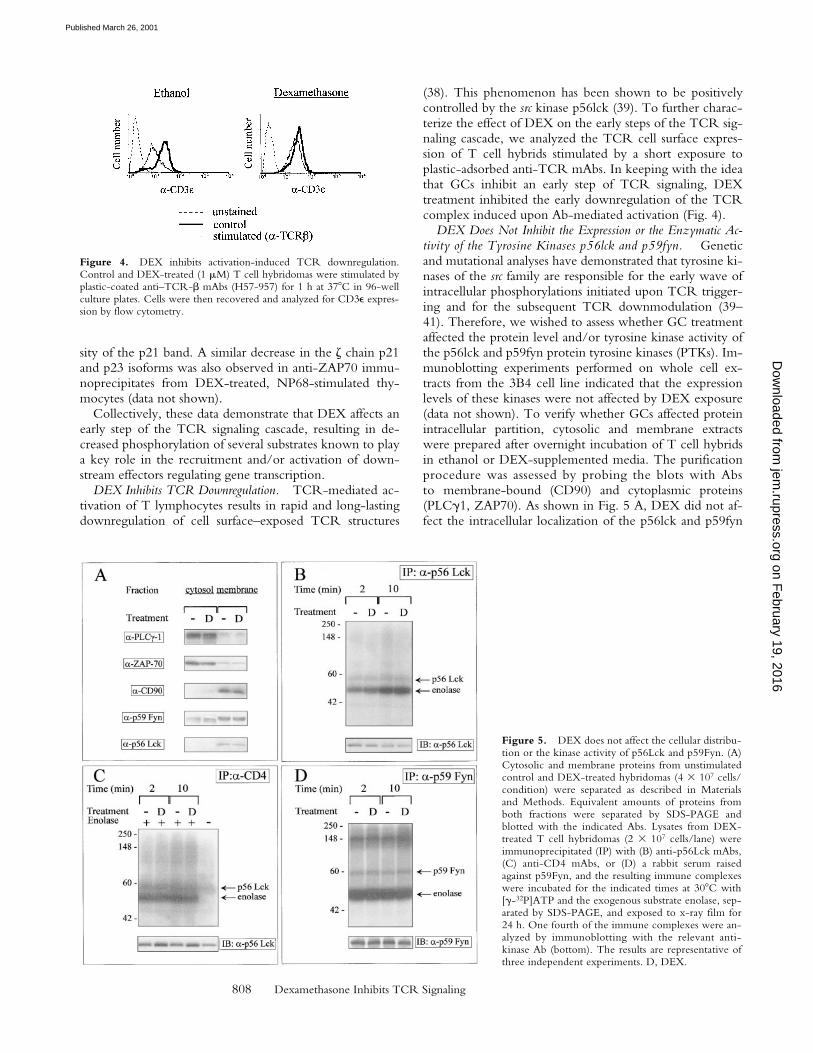
DEX Inhibits TCR Downregulation. TCR-mediated ac-tivation of T lymphocytes results in rapid and long-lastingdownregulation of cell surface–exposed TCR structures
(38). This phenomenon has been shown to be positivelycontrolled by the src kinase p56lck (39). To further charac-terize the effect of DEX on the early steps of the TCR sig-naling cascade, we analyzed the TCR cell surface expres-sion of T cell hybrids stimulated by a short exposure toplastic-adsorbed anti-TCR mAbs. In keeping with the ideathat GCs inhibit an early step of TCR signaling, DEXtreatment inhibited the early downregulation of the TCRcomplex induced upon Ab-mediated activation (Fig. 4).
DEX Does Not Inhibit the Expression or the Enzymatic Ac-tivity of the Tyrosine Kinases p56lck and p59fyn. Geneticand mutational analyses have demonstrated that tyrosine ki-nases of the src family are responsible for the early wave ofintracellular phosphorylations initiated upon TCR trigger-ing and for the subsequent TCR downmodulation (39–41). Therefore, we wished to assess whether GC treatmentaffected the protein level and/or tyrosine kinase activity ofthe p56lck and p59fyn protein tyrosine kinases (PTKs). Im-munoblotting experiments performed on whole cell ex-tracts from the 3B4 cell line indicated that the expressionlevels of these kinases were not affected by DEX exposure(data not shown). To verify whether GCs affected proteinintracellular partition, cytosolic and membrane extractswere prepared after overnight incubation of T cell hybridsin ethanol or DEX-supplemented media. The purificationprocedure was assessed by probing the blots with Absto membrane-bound (CD90) and cytoplasmic proteins(PLCg1, ZAP70). As shown in Fig. 5 A, DEX did not af-fect the intracellular localization of the p56lck and p59fyn
Figure 4. DEX inhibits activation-induced TCR downregulation.Control and DEX-treated (1 mM) T cell hybridomas were stimulated byplastic-coated anti–TCR-b mAbs (H57-957) for 1 h at 378C in 96-wellculture plates. Cells were then recovered and analyzed for CD3e expres-sion by flow cytometry.
Figure 5. DEX does not affect the cellular distribu-tion or the kinase activity of p56Lck and p59Fyn. (A)Cytosolic and membrane proteins from unstimulatedcontrol and DEX-treated hybridomas (4 3 107 cells/condition) were separated as described in Materialsand Methods. Equivalent amounts of proteins fromboth fractions were separated by SDS-PAGE andblotted with the indicated Abs. Lysates from DEX-treated T cell hybridomas (2 3 107 cells/lane) wereimmunoprecipitated (IP) with (B) anti-p56Lck mAbs,(C) anti-CD4 mAbs, or (D) a rabbit serum raisedagainst p59Fyn, and the resulting immune complexeswere incubated for the indicated times at 308C with[g-32P]ATP and the exogenous substrate enolase, sep-arated by SDS-PAGE, and exposed to x-ray film for24 h. One fourth of the immune complexes were an-alyzed by immunoblotting with the relevant anti-kinase Ab (bottom). The results are representative ofthree independent experiments. D, DEX.
on February 19, 2016
jem.rupress.org
Dow
nloaded from
Published March 26, 2001

809 Van Laethem et al.
PTKs. The in vitro enzymatic activity of the p56lck andthe p59fyn PTKs was not significantly affected by over-night incubation in DEX-supplemented media, as assayedon the exogenous substrate enolase (Fig. 5, B and D). Thefaint bands migrating in the 56–60-kD range comigratewith bands revealed by antikinase Abs in Western blotsperformed in parallel gels and probably represent the auto-phosphorylated forms of these kinases (data not shown).DEX did not affect the protein levels and kinase activity ofthe CD4-associated forms of p56lck, as shown by immuno-precipitation studies performed using Abs to CD4 (Fig. 5C). Collectively, these data indicated that DEX exposuredid not affect the expression level or the in vitro enzymaticactivity of the p56lck and p59fyn kinases.
DEX Affects the Membrane Compartmentalization of KeyTransducing Molecules. Recently, the important role ofGEMs or detergent-insoluble lipid rafts in cell signaling hasbeen recognized (42, 43). These GEMs are enriched in gly-cosyl phosphatidylinositol (GPI)-linked glycoproteins andin other lipid-modified proteins such as tyrosine kinases ofthe src family, monomeric and heterotrimeric G proteins,G-coupled protein receptors, and the adaptor protein LAT(30, 42–45). The confinement of signaling molecules tomembrane subdomains suggests that these compartmentsfunction as platforms for the formation of multicomponenttransduction complexes (46, 47). Accordingly, studies per-
formed on T lymphocytes have shown that raft integrity isrequired for effective TCR signal transduction (44, 48–50).To determine the effects of GCs on GEM composition, Tcell hybrids were lysed in a cold buffer containing a non-ionic detergent, and detergent-resistant membrane constit-uents were fractionated by ultracentrifugation in a sucrosegradient (51). The GEMs and the detergent-soluble frac-tion were analyzed by Western blot analysis using Abs andreagents to membrane components and signaling mole-cules. As expected, CD45, a transmembrane proteinknown to be excluded from the GEMs (52), and PLCg1, acytoplasmic protein, were only detected in the soluble frac-tion (Fig. 6 A). DEX did not overtly affect the plasmamembrane microdomains, as GEMs enriched for both theGPI-linked CD90/Thy-1 protein and the gangliosideGM1 could be isolated from both control and DEX-treated cells. However, GCs were found to selectively af-fect the submembrane localization of several key signalingmolecules, since GEMs purified from DEX-treated cellsdisplayed a marked decrease in the amount of p56lck,p59fyn, and LAT, as determined by Western blotting.DEX treatment did not affect the cellular expression levelof these proteins, as judged by Western blots performed onwhole cell extracts (data not shown), membrane fractions(Fig. 5 A), or specific immunoprecipitates (Fig. 1 E andFig. 5, B and D). Noteworthy, since proteins in GEMs
Figure 6. DEX affects membrane signaling complexes.(A) GCs affect protein raft composition. Control andDEX-treated T cell hybridomas (108 cells/condition) weresolubilized in 1% Triton X-100 MES lysis buffer, gentlysonicated, and the subsequent lysates were ultracentrifugedin a sucrose gradient as described in Materials and Meth-ods. Undiluted GEMs and cytoplasmic fractions (1:4 dilu-tion to avoid abnormal migration) from control and DEX-treated unstimulated cells were separated by SDS-PAGEand immunoblotted with Abs to membrane-associated(CD90, CD45, CD3z, p59Fyn, p56Lck, LAT) and cyto-plasmic (PLCg1) proteins. The ganglioside GM1 was de-tected using HRP-coupled cholera toxin B subunit. (B)Reduced phospho-LAT in the GEM fraction of DEX-treated cells. Control and DEX-treated hybridomas wereleft untreated or stimulated by CD3 cross-linking for 2 minand lysed in 1% Triton X-100 MES buffer. Lysates wereultracentrifuged as described previously and GEM fractionswere concentrated as described in Materials and Methods,separated by SDS-PAGE, and immunoblotted (IB) withantiphosphotyrosine mAbs. Similar results were obtainedin five independent experiments. (C) Coimmunoprecipita-tion of LAT with CD90 requires membrane cholesterol. Tcell hybridomas (107 cells/condition) were pretreated with10 mM methyl-b-cyclodextrin (MCD) for 30 min at 378Cor with 0.2% saponin for 10 min at 48C in PBS before lysisin 1% Triton X-100 MES buffer. After sonication, lysateswere subjected to immunoprecipitation (IP) with anti-CD90 (clone HO-13.4)–coupled Sepharose beads. Immu-noprecipitates were resolved by SDS-PAGE and immuno-blotted with the indicated Abs. (D) DEX affects signalingcomplexes. T cell hybridomas were incubated in controland 1 mM DEX-supplemented media before lysis and im-munoprecipitation with anti-CD90 mAbs. Proteins in totalextracts (2 3 105 cells/lane) or anti-CD90 immunoprecip-itates (from 107 cells/lane) were revealed by immunoblot-ting with the indicated Abs. D, DEX.
on February 19, 2016
jem.rupress.org
Dow
nloaded from
Published March 26, 2001

810 Dexamethasone Inhibits TCR Signaling
only represent a minor fraction of the total cellular proteincontent (z1–2%), loss of a given protein from the GEMsdoes not always lead to a detectable increase in the deter-gent-soluble fraction. Consistent with previous publica-tions (44, 45), TCR activation led to the tyrosine phos-phorylation of GEM-associated proteins such as LAT (Fig.6 B). In keeping with observations performed on immuno-precipitates, DEX inhibited the amount of phospho-LATin the GEM fraction (Fig. 6 B), possibly as a consequenceof altered membrane compartmentalization and/or reducedaccess to the appropriate kinase.
The existence of signaling complexes at the cell mem-brane can also be demonstrated by coimmunoprecipitationstudies. In particular, the GPI-anchored Thy-1 molecule isknown to associate with elements of the intracellular sig-naling machinery (53, 54). Noteworthy, these molecularassociations require membrane cholesterol, suggesting thatsuch linkages may occur by virtue of colocalization tomembrane subdomains (54). To further document the ef-fects of GCs on these signaling complexes, postnuclear su-pernatants obtained after extraction of 3B4 cells in 1% Tri-ton X-100 were immunoprecipitated using an anti–Thy-1mAb and analyzed by immunoprecipitation. In keepingwith previous observations, signaling proteins were foundto coimmunoprecipitate with Thy-1 under these experi-mental conditions, whereas transmembrane (CD3z) andcytoplasmic (PLCg1) proteins were excluded from thesecomplexes (Fig. 6 D). Noteworthy, cholesterol depletionby methyl-b-cyclodextrin or saponin prevented these mo-lecular associations (including Thy-1/LAT coimmunopre-cipitation, shown as an example in Fig. 6 C), suggestingthat coimmunoprecipitation required membrane subdo-main integrity. In agreement with our previous observa-tions, anti–Thy-1 immunoprecipitates from DEX-treatedcells displayed reduced amounts of p59fyn and LAT pro-teins, suggesting that GCs affected the association of severalsignaling molecules with membrane microdomains en-riched in GPI-anchored proteins and cholesterol. Collec-tively, the observations reported in Fig. 6 strongly suggestthat DEX inhibits the early steps of TCR signaling by af-fecting the submembrane localization of important signal-ing molecules.
DiscussionGCs are potent immunosuppressive agents able to inhibit
the expression of several proteins involved in inflammatoryimmune responses. In particular, their ability to inhibit Tcell responses is thought to be related to the modulation ofgene expression induced upon TCR triggering. Negativeregulation of gene transcription in activated T lymphocytescan occur directly by competition for DNA binding sites inthe promoter region of several cytokine-encoding genes,or indirectly, by sequestration/binding to transcription fac-tors such as activator protein-1, nuclear factor kB, cAMPresponse element–binding protein (CREB), octamer bind-ing factor 1, and signal transducer and activator of tran-scription 5 (55, and references therein). Previously, we
have shown that in addition to their effect on gene tran-scription, GCs can inhibit an early step of TCR-initiatedsignal transduction, causing impaired calcium flux in acti-vated murine T lymphocytes and T cell hybrids (26). Re-cently, numerous reports have underlined the importanceof the early signaling events in determining cell fate deci-sion in the immune system, leading us to further explorethe mechanisms by which GCs antagonize TCR-issuedsignals.
In this study we provide evidence that DEX, a syntheticGC analogue, affects the response of T cells to a givenTCR ligand by interfering with a membrane-proximalphosphorylative event, thus identifying a novel mechanismby which GCs exert their antiinflammatory properties.TCR stimulation of DEX-treated cells led to a defectivephosphorylation of CD3-associated substrates, includingthe z chain. In keeping with numerous observations sug-gesting a key role for z in transducing activation signals (3,56), cells expressing reduced amounts of phospho-z alsodisplayed reduced phosphorylation of other moleculesknown to play a major role in relaying activation signals todownstream effectors, including the ZAP70 kinase and theadaptor molecule LAT. The pattern of phosphorylativeevents evoked in DEX-treated cells suggests that GCs mayaffect T responses in a partial agonist fashion, as TCR oc-cupancy in a DEX-treated T cell hybridoma only evoked asubset of the signaling events observed in control-stimu-lated cells. In particular, the recruitment of ZAP70 to theTCR complex and the accumulation of p21 phospho-zwere not or were only weakly affected by GCs in activatedcells, whereas other intracellular events (phosphorylation ofCD3z p23 form, ZAP70, LAT, PLCg1, and sustained cal-cium influx; this study and reference 26) were inhibited.Similarly, the profile of phosphorylative events observed inDEX-treated thymocytes was reminiscent of the pattern ofphosphoproteins induced by altered peptide ligands in de-veloping thymocytes. Indeed, in the F5 TCR transgenicmodel, peptides known to favor positive selection in vitrowere shown to reduce z chain phosphorylations in a morequantitative that qualitative fashion (9). Previous studieshave suggested that the ordered phosphorylation of CD3zestablishes the threshold for T cell activation (56). Thesestudies identified the CD3-associated ITAM modules ascritical signaling thresholds detectors, able to integrategraded signals such as TCR signal strength (15–17), co-stimulation (57–59), and CTLA4 engagement (60) into all-or-none developmental decisions. This study demonstratesthat CD3z is also sensitive to the hormonal environment,suggesting an important and complex role for CD3-associ-ated ITAMs in relaying external cues to the interior of thecell.
The mechanism by which DEX affects the early steps ofTCR signaling was further studied using a T cell hybridknown to resist the proapoptotic effects of GCs (26). In thiscell line, DEX did not affect the expression or the in vitroPTK activity of p56lck and p59fyn, which have beenshown to positively regulate the early steps of signalingevents (such as phosphorylation of CD3z and TCR down-
on February 19, 2016
jem.rupress.org
Dow
nloaded from
Published March 26, 2001

811 Van Laethem et al.
modulation) inhibited by GCs. This observation suggeststhat PTK activity is not the primary target of DEX, arguingagainst a role for Csk (a tyrosine kinase known to inhibitthe enzymatic activity of p56lck; reference 61), CD45, orsrc homology 2 domain–bearing protein tyrosine phos-phatase 1 (SHP-1) (hematopoietic tyrosine phosphatasesable to downregulate Lck kinase activity; references62, 63).
Analysis of membrane subdomains revealed a possiblemechanism by which DEX may modulate TCR signaling.Recently it has been suggested that src PTKs and other im-portant signaling molecules including LAT are confinedinto specific detergent-resistant plasma membrane domains(GEMs) enriched in sphingolipids and GPI-anchored pro-teins (30, 43, 44). The fact that components of the TCR–CD3 complex are recruited into the GEMs upon stimula-tion and phosphorylated on tyrosine residues within thiscompartment argues for an important role of these mem-brane domains in signal transduction (46). Accordingly,numerous experiments indicate that disruption of GEMs(and the consequent dispersion of their content into thedetergent-soluble membrane fraction) inhibits TCR signaltransduction (43, 44, 47, 48). The present report suggeststhat DEX affects the submembrane compartmentalizationof several molecules associated with the inner (cytoplasmic)lipid layer of the GEMs, including the src PTKs, p56lckand p59fyn. Based on the current literature, displacementof Src family kinases and LAT from the GEMs might bebecause of modification of the GEM lipid content (such aslower cholesterol content and/or the phospholipid compo-sition; references 47–49) or by altered protein acylation.Indeed, studies using Lck and LAT mutant proteins lackingacylation sites have shown that these proteins are excludedfrom the GEMs in transfected T cells and are unable totransduce signals issued from the TCR (30, 64, 65). How-ever, note that an Lck mutant that cannot be palmitoylatedis unable to associate with membranes (65), whereas DEXfailed to affect stable membrane interaction of Lck (Fig. 5A). Moreover, no effect of DEX on membrane-associatedcholesterol was evidenced by filipin staining (unpublishedobservations). In any event, the results reported in thisstudy are consistent with the view that disruption of GEMsmay be the primary cause of partial signaling in DEX-treated T cells. Dispersion of signaling molecules such asLck or Fyn in the soluble membrane fraction may affecttheir enzymatic activity, as it has been recently recognizedthat these kinases exhibit optimal activity only when associ-ated with cholesterol-enriched domains (66). Moreover,altered membrane localization may impede an adequatejuxtaposition of PTKs with their substrate upon TCRstimulation or cause their sequestration in ineffective sig-naling complexes, resulting in a novel pattern of phosphor-ylating events and altered biological response. Of interest,it has been recently demonstrated that inhibition of CD4association with the Ag–MHC-engaged TCR leads to par-tial signaling (predominant accumulation of the p21 iso-form of phospho-z) in response to an agonist (67). Collec-tively, these data strongly suggest that partial signaling may
be a consequence of ineffective clustering of TCR subunitswith the relevant membrane-associated src PTKs. Theanalysis of phospho-z p21 isoform levels in unstimulatedcells is in agreement with this hypothesis. Reduced levelsof p21 in these cells (Fig. 1 B) appear to be the conse-quence of altered membrane localization of PTKs (Fig. 6A), as both CD3z submembrane compartmentalization(Fig. 6 A) and PTK enzymatic activity (Fig. 5) were unaf-fected by DEX treatment.
The interest in the phenomenon reported here lies in itspossible in vivo relevance. Thymic selection, a processwhereby autoreactive thymocytes are induced to die (nega-tive selection) while thymocytes displaying self-MHC–restricted potential are allowed to differentiate into matureT cells (positive selection), is thought to depend on subtledifferences in the signals issued by the TCR in response toself-peptide–MHC complexes expressed in the thymus (1,68, 69). Recently, it has been demonstrated that the thymicepithelium and possibly thymocytes themselves producesteroids, suggesting a potential role of GCs in the control ofT cell development and repertoire development (23, 70).Accordingly, downregulation of GC receptors on thy-mocytes and/or pharmacological inhibition of GC synthe-sis have been shown to affect thymic differentiation in vivo(71, 72). These authors have proposed that GCs producedin the thymus antagonize TCR signals, thus shifting thepositive selection window towards higher TCR aviditiesfor self-Ag–MHC complexes. The work presented hereprovides a sound basis for explaining these observations.We propose that GCs may affect thymocyte developmentby modifying the early signals issued by the TCR, convert-ing an agonist signal (causing negative selection) into a pos-itively selecting, partial agonist–like signal. Similarly, GCsmay cause cell death by the neglect of cells expressingTCRs with very low self-reactivity. Therefore, it is tempt-ing to speculate that by lowering TCR sensitivity, GCsmay shift the range of positively selected TCRs towards ahigher self-reactivity, possibly ensuring that T cells surviv-ing positive selection display a sufficient reactivity to self-MHC in the periphery. Noteworthy, this hypothesis pos-tulates that lack of thymic GC receptor expression wouldlead to subtle changes in repertoire selection, with no ma-jor alteration in overall thymic cellularity. In keeping withthis hypothesis, lack of GC responsiveness led to the spe-cific loss of T cells bearing a TCR specific for a given Ag(73), with no major impact on thymic cell numbers andphenotype (74).
Signal strength has also been proposed to regulate thedevelopment of helper subsets in the periphery. Numerousstudies have suggested that “weak” and/or partial signalingin mature lymphocytes favors Th2 cell development. Prim-ing of naive T cells with optimal doses of an agonist pep-tide will induce Th1 development, whereas very low dosesof an agonist peptide or stimulation by a low-avidity al-tered peptide ligand will favor Th2 differentiation (11, 75).GCs in vivo and in vitro have been shown to favor Th2cell differentiation in response to Ag (24, 76, 77), suggest-ing again that by reducing TCR signal strength, GCs may
on February 19, 2016
jem.rupress.org
Dow
nloaded from
Published March 26, 2001

812 Dexamethasone Inhibits TCR Signaling
affect cell fate decisions in the periphery. Noteworthy, weand others have proposed previously that GCs may favorTh2 development by affecting both survival of dendriticcells and IL-12 production (78–80). These observationssuggest that by affecting both APC function and TCR sig-naling, endogenous GCs produced during acute infectionmay favor the development of Th2-like cells with antiin-flammatory properties.
In conclusion, our study illustrates a novel aspect of thesignaling flexibility of the TCR, revealing that in additionto avidity for Ag–MHC and costimulation, environmentalfactors such as GC hormones determine the nature of intra-cellular signals induced by the TCR. Further studies are re-quired to better understand how all these influences aretranslated in vivo into distinct functional responses.
We would like to thank Drs. L. Samelson, R. Kubo, P. Draper, andA. Weiss for providing reagents; Drs. H.T. He, O. Williams, andD. Nolan for interesting discussions and comments on the manu-script; Dr. M. Moser for careful review of the manuscript; and G.Dewasme, F. Thielemans, M. Swaenepoel, and P. Veirman fortechnical assistance.
This work was funded by the Belgian Program in InteruniversityPoles of Attraction initiated by the Belgian State, Prime Minister’sOffice, Science Policy Programming, and by a Research ConcertedAction of the Communauté Française de Belgique. The scientificresponsibility is assumed by its authors. F. Van Laethem is sup-ported by the Fonds pour la Formation à la Recherche dans l’In-dustrie et dans l’Agriculture; and E. Baus is supported by the FondsNational de la Recherche Scientifique.
Submitted: 6 April 2000Revised: 14 February 2001Accepted: 16 February 2001
References1. Germain, R.N., and I. Stefanova. 1999. The dynamics of T
cell receptor signaling: complex orchestration and the keyroles of tempo and cooperation. Annu. Rev. Immunol. 17:467–522.
2. Constant, S.L., and K. Bottomly. 1997. Induction of Th1 andTh2 CD41 T cell responses: the alternative approaches.Annu. Rev. Immunol. 15:297–322.
3. Kersh, E.N., A.S. Shaw, and P.M. Allen. 1998. Fidelity of Tcell activation through multistep T cell receptor z phosphor-ylation. Science. 281:572–575.
4. Evavold, B.D., J. Sloan-Lancaster, and P.M. Allen. 1993.Tickling the TCR: selective T-cell functions stimulated byaltered peptide ligands. Immunol. Today. 14:602–609.
5. Sloan-Lancaster, J., and P.M. Allen. 1996. Altered peptideligand-induced partial T cell activation: molecular mecha-nisms and role in T cell biology. Annu. Rev. Immunol. 14:1–27.
6. Jameson, S.C., and M.J. Bevan. 1995. T cell receptor antago-nists and partial agonists. Immunity. 2:1–11.
7. Sloan-Lancaster, J., B.D. Evavold, and P.M. Allen. 1993. In-duction of T-cell anergy by altered T-cell-receptor ligand onlive antigen-presenting cells. Nature. 363:156–159.
8. Sebzda, E., T.M. Kundig, C.T. Thomson, K. Aoki, S.Y.Mak, J.P. Mayer, T. Zamborelli, S.G. Nathenson, and P.S.Ohashi. 1996. Mature T cell reactivity altered by peptide ag-
onist that induces positive selection. J. Exp. Med. 183:1093–1104.
9. Smyth, L.A., O. Williams, R.D. Huby, T. Norton, O.Acuto, S.C. Ley, and D. Kioussis. 1998. Altered peptideligands induce quantitatively but not qualitatively differentintracellular signals in primary thymocytes. Proc. Natl. Acad.Sci. USA. 95:8193–8198.
10. Hogquist, K.A., S.C. Jameson, W.R. Heath, J.L. Howard,M.J. Bevan, and F.R. Carbone. 1994. T cell receptor antago-nist peptides induce positive selection. Cell. 14:17–27.
11. Constant, S., C. Pfeiffer, A. Woodard, T. Pasqualini, and K.Bottomly. 1995. Extent of T cell receptor ligation can deter-mine the functional differentiation of naive CD41 T cells. J.Exp. Med. 182:1591–1596.
12. Hosken, N.A., K. Shibuya, A.W. Heath, K.M. Murphy, andA. O’Garra. 1995. The effect of antigen dose on CD41 Thelper cell phenotype development in a T cell receptor-ab–transgenic model. J. Exp. Med. 182:1579–1584.
13. Tao, X., C. Grant, S. Constant, and K. Bottomly. 1994. In-duction of IL-4-producing CD41 T cells by antigenic pep-tides altered for TCR binding. J. Immunol. 158:4237–4244.
14. Smith, J.A., Q. Tang, and J.A. Bluestone. 1998. Partial TCRsignals delivered by FcR-nonbinding anti-CD3 monoclonalantibodies differentially regulate individual Th subsets. J. Im-munol. 160:4841–4849.
15. Sloan-Lancaster, J., A.S. Shaw, J.B. Rothbard, and P.M.Allen. 1994. Partial T cell signaling: altered phospho-z andlack of zap70 recruitment in APL-induced T cell anergy.Cell. 79:913–922.
16. Madrenas, J., R.L. Wange, J.L. Wang, N. Isakov, L.E.Samelson, and R.N. Germain. 1995. z phosphorylation with-out ZAP-70 activation induced by TCR antagonists or par-tial agonists. Science. 267:515–518.
17. Reis é Sousa, C., E.H. Levine, and R.N. Germain. 1996.Partial signaling by CD81 T cells in response to antagonistligands. J. Exp. Med. 184:149–157.
18. Alam, S.M., P.J. Travers, J.L. Wung, W. Nasholds, S. Red-path, S.C. Jameson, and N.R. Gascoigne. 1996. T-cell-receptor affinity and thymocyte positive selection. Nature.381:616–620.
19. Allen, P.M. 1994. Peptides in positive and negative selection:a delicate balance. Cell. 76:593–596.
20. Madrenas, J., L.A. Chau, J. Smith, J.A. Bluestone, and R.N.Germain. 1997. The efficiency of CD4 recruitment toligand-engaged TCR controls the agonist/partial agonistproperties of peptide-MHC molecule ligands. J. Exp. Med.185:219–229.
21. Cato, A.C., and E. Wade. 1996. Molecular mechanisms ofanti-inflammatory action of glucocorticoids. Bioessays. 18:371–378.
22. Ashwell, J.D., F.W. Lu, and M.S. Vacchio. 2000. Glucocor-ticoids in T cell development and function. Annu. Rev. Im-munol. 18:309–345.
23. Vacchio, M.S., V. Papadopoulos, and J.D. Ashwell. 1994.Steroid production in the thymus: implications for thymocyteselection. J. Exp. Med. 179:1835–1846.
24. Daynes, R.A., and B. Araneo. 1989. Contrasting effects ofglucocorticoids on the capacity of T cells to produce thegrowth factors interleukin 2 and interleukin 4. Eur. J. Immu-nol. 19:2319–2325.
25. Mamalaki, C., T. Norton, Y. Tanaka, A.R. Townsend, P.Chandler, E. Simpson, and D. Kioussis. 1992. Thymic deple-tion and peripheral activation of class I major histocompati-
on February 19, 2016
jem.rupress.org
Dow
nloaded from
Published March 26, 2001

813 Van Laethem et al.
bility complex-restricted T cells by soluble peptide in T-cellreceptor transgenic mice. Proc. Natl. Acad. Sci. USA. 89:11342–11346.
26. Baus, E., F. Andris, P.M. Dubois, J. Urbain, and O. Leo.1996. Dexamethasone inhibits the early steps of antigen re-ceptor signaling in activated T lymphocytes. J. Immunol. 156:4555–4561.
27. Tanaka, Y., O. Williams, R. Tarazona, A. Wack, T. Norton,and D. Kioussis. 1997. In vitro positive selection of ab TCRtransgenic thymocytes by a conditionally immortalized corti-cal epithelial clone. Int. Immunol. 9:381–393.
28. Coulie, P.G., C. Uyttenhove, P. Wauters, N. Manolios,R.D. Klausner, L.E. Samelson, and J. Van Snick. 1991. Iden-tification of a murine monoclonal antibody specific for an al-lotypic determinant on mouse CD3. Eur. J. Immunol. 21:1703–1709.
29. Nolan, D.P., D.G. Jackson, M.J. Biggs, E.D. Brabazon, A.Pays, F. Van Laethem, F. Paturiaux-Hanocq, J.F. Elliot, H.P.Voorheis, and E. Pays. 2000. Characterization of a novel ala-nine-rich protein located in surface microdomains in Trypa-nosoma brucei. J. Biol. Chem. 275:4072–4080.
30. Zhang, W., R.P. Trible, and L.E. Samelson. 1998. LATpalmitoylation: its essential role in membrane microdomaintargeting and tyrosine phosphorylation during T cell activa-tion. Immunity. 9:239–246.
31. Wessel, D., and U.I. Flugge. 1984. A method for the quanti-tative recovery of protein in dilute solution in the presence ofdetergents and lipids. Anal. Biochem. 138:141–143.
32. Zhang, W., J. Sloan-Lancaster, J. Kitchen, R.P. Trible, andL.E. Samelson. 1998. LAT: the ZAP-70 tyrosine kinase sub-strate that links T cell receptor to cellular activation. Cell. 92:83–92.
33. Weber, J.R., S. Orstavik, K.M. Torgersen, N.C. Danbolt,S.F. Berg, J.C. Ryan, K. Tasken, J.B. Imboden, and J.T.Vaage. 1998. Molecular cloning of the cDNA encodingpp36, a tyrosine-phosphorylated adaptor protein selectivelyexpressed by T cells and natural killer cells. J. Exp. Med. 187:1157–1161.
34. Finco, T.S., T. Kadlecek, W. Zhang, L.E. Samelson, and A.Weiss. 1998. LAT is required for TCR-mediated activationof PLCg1 and the Ras pathway. Immunity. 9:617–625.
35. Wehling, M. 1997. Specific, nongenomic actions of steroidhormones. Annu. Rev. Physiol. 59:365–393.
36. van Oers, N.S., N. Killeen, and A. Weiss. 1994. ZAP-70 isconstitutively associated with tyrosine-phosphorylated TCRz in murine thymocytes and lymph node T cells. Immunity.1:675–685.
37. Wiest, D.L., J.M. Ashe, R. Abe, J.B. Bolen, and A. Singer.1996. TCR activation of ZAP70 is impaired in CD41CD81
thymocytes as a consequence of intrathymic interactions thatdiminish available p56lck. Immunity. 4:495–504.
38. Valitutti, S., S. Muller, M. Cella, E. Padovan, and A. Lanza-vecchia. 1995. Serial triggering of many T-cell receptors by afew peptide-MHC complexes. Nature. 375:148–151.
39. D’Oro, U., M.S. Vacchio, A.S. Weissman, and J.D. Ashwell.1997. Activation of Lck tyrosine kinase targets cell surface Tcell antigen receptors for lysosomal degradation. Immunity.7:619–628.
40. van Oers, N.S., N. Killeen, and A. Weiss. 1996. Lck targetsthe tyrosine phosphorylation of the T cell receptor subunitsand ZAP-70 in murine thymocytes. J. Exp. Med. 183:1053–1062.
41. Veillette, A., M.A. Bookman, E.M. Hork, L.E. Samelson,
and J.B. Bolen. 1989. Signal transduction through the CD4receptor involves the activation of the internal membrane ty-rosine kinase p56Lck. Nature. 338:257–259.
42. Brown, D.A., and E. London. 1998. Function of lipid rafts inbiological membranes. Annu. Rev. Cell Dev. Biol. 14:111–136.
43. Simons, K., and D. Toomre. 2000. Lipid rafts and signaltransduction. Nat. Rev. (Mol. Cell. Biol.). 1:31–39.
44. Xavier, R., T. Brennan, Q. Li, C. McCormack, and B. Seed.1998. Membrane compartmentation is required for efficientT cell activation. Immunity. 8:723–732.
45. Montixi, C., C. Langlet, A.M. Bernard, J. Thimonier, C.Dubois, M.A. Wurbel, J.P. Chauvin, M. Pierres, and H.T.He. 1998. Engagement of T cell receptor triggers its recruit-ment to low-density detergent-insoluble membrane domains.EMBO (Eur. Mol. Biol. Organ.) J. 17:5334–5348.
46. Viola, A., and A. Lanzavecchia. 1999. T-cell activation andthe dynamic world of rafts. APMIS. 107:615–623.
47. Ilangumaran, S., H.T. He, and D.C. Hoessli. 2000. Micro-domains in lymphocyte signaling: beyond GPI-anchoredproteins. Immunol. Today. 21:2–7.
48. Stulnig, T.M., M. Berger, T. Sigmund, H. Stockinger, V.Horesji, and W. Waldhausl. 1997. Signal transduction viaglycosyl phosphatidyl-anchored proteins in T cells is inhib-ited by lowering cellular cholesterol. J. Biol. Chem. 272:19242–19247.
49. Stulnig, T.M., M. Berger, T. Sigmund, H. Stockinger, V.Horesji, and W. Waldhausl. 1998. Polyunsaturated fatty acidsinhibit T cell signaling transduction by modification of deter-gent-insoluble membrane domains. J. Cell Biol. 143:637–644.
50. Webb, Y., L. Hermida-Matsumoto, and M.D. Resh. 2000.Inhibition of protein palmitoylation, raft localization, and Tcell signaling by 2-bromopalmitate and polyunsaturated fattyacids. J. Biol. Chem. 275:261–270.
51. Brown, D.A., and J.K. Rose. 1992. Sorting of GPI-anchoredproteins to glycolipid-enriched membrane subdomains dur-ing transport to the apical cell surface. Cell. 68:533–544.
52. Rodgers, W., and J.K. Rose. 1996. Exclusion of CD45 in-hibits activity of p56lck associated with glycolipid-enrichedmembrane domains. J. Cell Biol. 135:1515–1523.
53. Thomas, P.M., and L.E. Samelson. 1992. The glycophos-phatidylinositol-anchored Thy-1 molecule interacts with thep60fyn protein tyrosine kinase in T cells. J. Biol. Chem. 267:12317–12322.
54. Draberova, L., M. Amoui, and P. Draber. 1996. Thy-1-mediated activation of rat mast cells: the role of Thy-1 mem-brane microdomains. Immunology. 87:141–148.
55. De Bosscher, K., M.L. Schmitz, W. Vanden Berghe, S. Plai-sance, W. Fiers, and G. Haegeman. 1997. Glucocorticoid-mediated repression of nuclear factor-kB-dependent tran-scription involves direct interference with transactivation.Proc. Natl. Acad. Sci. USA. 94:13504–13509.
56. Irving, B.A., A.N. Chan, and A. Weiss. 1993. Functionalcharacterization of a signal transducing motif present in the Tcell antigen receptor z chain. J. Exp. Med. 177:1093–1103.
57. Tuosto, L., and O. Acuto. 1998. CD28 affects the earliestsignaling events generated by TCR engagement. Eur. J. Im-munol. 28:2131–2142.
58. Boussiotis, V.A., D.L. Barber, B.J. Lee, J.G. Gribben, G.J.Freeman, and N.M. Nadler. 1996. Differential association ofprotein tyrosine kinases with the T cell receptor is linked tothe induction of anergy and its prevention by B7 family-
on February 19, 2016
jem.rupress.org
Dow
nloaded from
Published March 26, 2001

814 Dexamethasone Inhibits TCR Signaling
mediated costimulation. J. Exp. Med. 184:365–376.59. Moran, M., and M.C. Miceli. 1998. Engagement of GPI-
linked CD48 contributes to TCR signals and cytoskeletal re-organization: a role for lipid rafts in T cell activation. Immu-nity. 9:787–796.
60. Lee, K.M., E. Chuang, M. Griffin, R. Khattri, D.K. Hong,W. Zhang, D. Straus, L.E. Samelson, C.B. Thompson, andJ.A. Bluestone. 1998. Molecular basis of T cell inactivationby CTLA-4. Science. 282:2256–2263.
61. Chow, L.M., M. Fournel, D. Davidson, and A. Veillette.1993. Negative regulation of T-cell receptor signaling by ty-rosine protein kinase p50csk. Nature. 365:156–160.
62. D’Oro, U., and J.D. Ashwell. 1999. The CD45 tyrosinephosphatase is an inhibitor of Lck activity in thymocytes. J.Immunol. 162:1879–1883.
63. Dittel, B.N., I. Stefanova, R.N. Germain, and C.A. Janeway,Jr. 1999. Cross-antagonism of a T cell clone expressing twodistinct T cell receptors. Immunity. 11:289–298.
64. Melkonian, K.A., A.G. Ostermeyer, J.Z. Chen, M.G. Roth,and D. Brown. 1999. Role of lipid modifications in targetingproteins to detergent-resistant membrane rafts. Many raftproteins are acylated, while few are prenylated. J. Biol. Chem.274:3910–3917.
65. Kabouridis, P.S., A.L. Magee, and S.C. Ley. 1997. S-acyla-tion of LCK protein tyrosine kinase is essential for its signal-ing function in T lymphocytes. EMBO (Eur. Mol. Biol. Or-gan.) J. 16:4983–4998.
66. Ilangumaran, S., S. Arni, G. van Echten-Deckert, B. Borisch,and D.C. Hoessli. 1999. Microdomain-dependent regulationof Lck and Fyn protein-tyrosine kinases in T lymphocyteplasma membranes. Mol. Biol. Cell. 10:891–905.
67. Madrenas, J., L.A. Chau, J. Smith, J.A. Bluestone, and R.N.Germain. 1997. The efficiency of CD4 recruitment toligand-engaged TCR controls the agonist/partial agonistproperties of peptide-MHC molecule ligands. J. Exp. Med.185:219–229.
68. Goldrath, A.W., and M.J. Bevan. 1999. Selecting and main-taining a diverse T-cell repertoire. Nature. 402:255–262.
69. Mariathasan, S., R.G. Jones, and P.S. Ohashi. 1999. Signalsinvolved in thymocyte positive and negative selection. Semin.Immunol. 11:263–272.
70. Lechner, O., G.J. Wiegers, A.J. Oliveira-Dos-Santos, H. Diet-
rich, H. Recheis, M. Waterman, R. Boyd, and G. Wick.2000. Glucocorticoid production in the murine thymus. Eur.J. Immunol. 30:337–346.
71. Vacchio, M.S., and J.D. Ashwell. 1997. Thymus-derivedglucocorticoids regulate antigen-specific positive selection. J.Exp. Med. 185:2033–2038.
72. Vacchio, M.S., J.Y. Lee, and J.D. Ashwell. 1999. Thymus-derived glucocorticoids set the thresholds for thymocyte se-lection by inhibiting TCR-mediated thymocyte activation. J.Immunol. 163:1327–1333.
73. Lu, F.W., K. Yasutomo, G.B. Goodman, L.J. McHeyzer-Williams, M.G. McHeyzer-Williams, R.N. Germain, andJ.D. Ashwell. 2000. Thymocyte resistance to glucocorticoidsleads to antigen-specific unresponsiveness due to “holes” inthe T cell repertoire. Immunity. 12:183–192.
74. Purton, J.F., R.L. Boyd, T.J. Cole, and D.I. Godfrey. 2000.Intrathymic T cell development and selection proceeds nor-mally in the absence of glucocorticoid receptor signaling. Im-munity. 13:179–186.
75. Leitenberg, D., and K. Bottomly. 1999. Regulation of naiveT cell differentiation by varying the potency of TCR signaltransduction. Semin. Immunol. 11:283–292.
76. Ramirez, F., D.J. Fowell, M. Puklavec, S. Simmonds, and D.Mason. 1996. Glucocorticoids promote a Th2 cytokine re-sponse by CD41 T cells in vitro. J. Immunol. 156:2406–2412.
77. Dozmorov, I.M., and R.A. Miller. 1998. Generation of anti-gen-specific Th2 cells from unprimed mice in vitro: effects ofdexamethasone and anti-IL-10 antibody. J. Immunol. 160:2700–2705.
78. Moser, M., T. De Smedt, T. Sornasse, F. Tielemans, A.A.Chentoufi, E. Muraille, M. Van Mechelen, J. Urbain, and O.Leo. 1995. Glucocorticoids down-regulate dendritic cellfunction in vitro and in vivo. Eur. J. Immunol. 25:2818–2824.
79. Vieira, P.L., P. Kalinski, E.A. Wierenga, M.L. Kapsenberg,and E.C. de Jong. 1998. Glucocorticoids inhibit bioactiveIL-12p70 production by in vitro-generated human dendriticcells without affecting their T cell stimulatory potential. J.Immunol. 161:5245–5251.
80. DeKruyff, R.H., Y. Fang, and D.T. Umetsu. 1998. Corti-costeroids enhance the capacity of macrophages to induceTh2 cytokine synthesis in CD41 lymphocytes by inhibitingIL-12 production. J. Immunol. 160:2231–2237.
on February 19, 2016
jem.rupress.org
Dow
nloaded from
Published March 26, 2001


