Embed Size (px)
Citation preview

Glucocorticoids Play a Key Rolein Circadian Cell Cycle RhythmsThomas Dickmeis
1, Kajori Lahiri
1, Gabriela Nica
2, Daniela Vallone
1, Cristina Santoriello
1¤, Carl J. Neumann
3,
Matthias Hammerschmidt2
, Nicholas S. Foulkes1*
1 Max-Planck-Institut fur Entwicklungsbiologie, Tubingen, Germany, 2 Max-Planck-Institut fur Immunbiologie, Freiburg, Germany, 3 European Molecular Biology Laboratory
Heidelberg, Heidelberg, Germany
Clock output pathways play a pivotal role by relaying timing information from the circadian clock to a diversity ofphysiological systems. Both cell-autonomous and systemic mechanisms have been implicated as clock outputs;however, the relative importance and interplay between these mechanisms are poorly understood. The cell cyclerepresents a highly conserved regulatory target of the circadian timing system. Previously, we have demonstrated thatin zebrafish, the circadian clock has the capacity to generate daily rhythms of S phase by a cell-autonomousmechanism in vitro. Here, by studying a panel of zebrafish mutants, we reveal that the pituitary–adrenal axis also playsan essential role in establishing these rhythms in the whole animal. Mutants with a reduction or a complete absence ofcorticotrope pituitary cells show attenuated cell-proliferation rhythms, whereas expression of circadian clock genes isnot affected. We show that the corticotrope deficiency is associated with reduced cortisol levels, implicatingglucocorticoids as a component of a systemic signaling pathway required for circadian cell cycle rhythmicity. Strikingly,high-amplitude rhythms can be rescued by exposing mutant larvae to a tonic concentration of a glucocorticoid agonist.Our work suggests that cell-autonomous clock mechanisms are not sufficient to establish circadian cell cycle rhythmsat the whole-animal level. Instead, they act in concert with a systemic signaling environment of which glucocorticoidsare an essential part.
Citation: Dickmeis T, Lahiri K, Nica G, Vallone D, Santoriello C, et al. (2007) Glucocorticoids play a key role in circadian cell cycle rhythms. PLoS Biol 5(4): e78. doi:10.1371/journal.pbio.0050078
Introduction
The physiology of most plants and animals changessignificantly between day and night. These daily rhythmsare generated by endogenous clocks or pacemakers, andpersist under constant conditions with a period length ofapproximately 24 h (hence, they are termed circadian). Invertebrates, cell-autonomous circadian clocks are present inmost cell types, and are termed peripheral clocks. In addition,a limited number of specialized central pacemakers such asthe suprachiasmatic nucleus (SCN) of the hypothalamus [1,2]appear to play a key role in coordinating the function ofperipheral clocks. Although it is known that ocular photo-reception synchronizes the SCN pacemaker with the environ-ment, the identity of the pathways that subsequently transmittiming information to the peripheral clocks remains elusive.Current models implicate multiple humoral signals thatresult indirectly from the SCN circadian control of systemicfunction, such as feeding behavior [3].
Both cell-autonomous and systemic regulatory mechanismshave been implicated in clock output pathways that relaytiming information from the clock to physiological systems.Circadian E box enhancers represent key regulatory elementswithin the core transcription–translation feedback loop ofthe vertebrate clock. These promoter elements directcircadian rhythms of transcription of clock genes by actingas binding sites for the clock components CLOCK andBMAL1. Circadian E boxes are also encountered in thepromoters of many non-clock genes (so-called clock-con-trolled genes, e.g., see [4]). Via such target genes and theirdownstream effectors, peripheral circadian clock compo-nents directly regulate many aspects of cell physiology, such
as membrane trafficking, detoxification, nutrient metabolism,and the cell cycle [4]. The central SCN pacemaker, in contrast,has been documented to influence systemic functions rangingfrom locomotor activity rhythms and the sleep–wake cycle toendocrine activity. Thus, the circulating levels of manyhormones are under circadian control and so exert theireffects only during specific times of day. A major unexploredissue is the relative contribution of cell-autonomous andsystemic factors in directing circadian clock outputs. Docertain clock outputs rely solely upon direct peripheral clockregulation, or do they require input from systemic factors,acting either upstream or downstream of the peripheralclocks? Are other outputs driven solely by circadian oscil-lations of systemic signals?A particularly interesting clock output is the timing of cell
proliferation. Daily rhythms of cell division are conservedacross huge evolutionary distances, from cyanobacteria tohumans [5,6]. This property has been proposed as a strategyfor minimizing the ultraviolet damaging effects of sunlight
Academic Editor: Ueli Schibler, University of Geneva, Switzerland
Received May 19, 2006; Accepted January 16, 2007; Published March 20, 2007
Copyright: � 2007 Dickmeis et al. This is an open-access article distributed underthe terms of the Creative Commons Attribution License, which permits unrestricteduse, distribution, and reproduction in any medium, provided the original authorand source are credited.
Abbreviations: BrdU, bromodeoxyuridine; CT, circadian time; DD, constant dark;dpf, day post-fertilization; GR, glucocorticoid receptor; LD, light–dark; RPA, RNAseprotection assay; SCN, suprachiasmatic nucleus; ZT, zeitgeber time
* To whom correspondence should be addressed. E-mail: [email protected]
¤ Current address: Istituto Firc di Oncologia Molecolare, Milan, Italy
PLoS Biology | www.plosbiology.org April 2007 | Volume 5 | Issue 4 | e780854
PLoS BIOLOGY
during critical steps of the cell proliferation. In vertebrates,circadian gating of certain cell cycle steps also occurs in celllines [7,8]. Furthermore, clock components have beenimplicated in controlling the transcription of cell cycleregulatory genes [9–12]. These observations imply that thecircadian clock may regulate cell cycle progression via cell-autonomous mechanisms. However, given that systemicfactors such as hormones are well-known regulators of cellproliferation [13,14], one important question is whether cell-autonomous regulatory mechanisms are sufficient to directcircadian cell cycle rhythms at the whole-animal level.
The zebrafish represents a valuable model for exploring thevertebrate circadian clock and its regulation of cell cycletiming. Robust daily S-phase rhythms are observed in larvaeraised under light–dark (LD) cycles [7]. The persistence ofthese rhythms following transfer of the larvae to constantdarkness (DD) conditions demonstrates that they are undercontrol of the circadian clock. Furthermore, consistent withother clock outputs [15], exposure to a LD cycle is essentialfor the establishment of these rhythms because they areabsent in larvae raised in DD. Circadian rhythms of S phase,albeit with lower amplitude, are also observed in zebrafishprimary cell lines, implicating cell-autonomous regulation byperipheral clock mechanisms [7]. Interestingly, peripheralclocks in this species can be entrained by direct exposure toLD cycles [16]. However, zebrafish also possess centralpacemakers: a structural counterpart of the SCN and aphotosensitive pineal complex where nighttime synthesis ofthe hormone melatonin is directed by an endogenous clock[15,17–20].
Extensive panels of zebrafish mutants that show specificdevelopmental defects in a range of organ systems have beenassembled, thanks to large-scale screening efforts [21]. Theseanimals represent potentially powerful tools to dissect thefunctional contribution of specific organs and tissues to thegeneration of clock outputs at the whole-animal level. Here,by studying a set of blind mutants, we have demonstrated thatocular photoreception is not required to establish circadiancell cycle rhythms during early larval development. Incontrast, a severe attenuation of cell cycle rhythms isobserved in mutants that exhibit a reduction or absence of
the corticotrope cell lineage in the pituitary gland. Impor-tantly, high-amplitude circadian cell cycle rhythms can berescued by exposing corticotrope-deficient larvae to tonicconcentrations of the glucocorticoid receptor (GR) agonistdexamethasone. Our work reveals the contribution ofsystemic factors to establishing circadian cell cycle rhythmsat the whole-animal level.
Results
Ocular Photoreception Is Not Required for ZebrafishCircadian Cell Cycle RhythmsWe have previously demonstrated that exposure to a LD
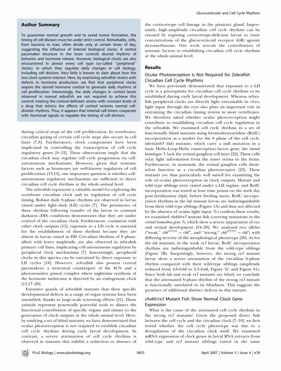
cycle is a prerequisite for circadian cell cycle rhythms to beestablished during early larval development. Whereas zebra-fish peripheral clocks are directly light entrainable in vitro,light input through the eyes also plays an important role inentraining the circadian timing system in most vertebrates.We therefore asked whether ocular photoreception mightcontribute to establishing circadian cell cycle regulation inthe zebrafish. We examined cell cycle rhythms in a set offunctionally blind mutants using bromodeoxyuridine (BrdU)incorporation as a marker for the S phase of the cell cycle.lakritz/ath5 (lak) mutants, which carry a null mutation in abasic Helix-Loop-Helix transcription factor gene, the atonalhomologue 5, lack the retinal ganglion cell layer [22]. These cellsrelay light information from the inner retina to the brain.Furthermore, in mammals, the retinal ganglion cells them-selves function as a circadian photoreceptor [23]. Thesemutants are thus particularly well suited for examining therole of ocular photoreception in clock outputs. Mutant andwild-type siblings were raised under a LD regime, and BrdUincorporation was tested at four time points on the sixth daypost-fertilization (dpf), before feeding starts. BrdU incorpo-ration rhythms in the lak mutant larvae are indistinguishablefrom their wild-type siblings (Figure 1A) and thus not affectedby the absence of ocular light input. To confirm these results,we examined chokh/rx3 mutant fish (carrying mutations in theretinal homeobox gene 3), which show a severe impairment of eyeand retinal development [24–26]. We analyzed two alleles(‘‘weak,’’ chkt25181 ¼ chkw, and ‘‘strong,’’ chkt25327 ¼ chks) withdiffering severity of the morphological phenotype [26]. As forthe lak mutants, in the weak rx3 larvae, BrdU incorporationrhythms are indistinguishable from the wild-type siblings(Figure 1B). Surprisingly, however, the strong rx3 mutantlarvae show a severe attenuation of the circadian S-phaserhythm compared with their wild-type siblings (amplitudereduced from 4.0-fold to 2.3-fold, Figure 1C and Figure S1).Since both lak and weak rx3 mutants are blind, we concludethat the attenuated S-phase rhythm of the strong rx3 mutantis functionally unrelated to its blindness. This suggests thepresence of additional distinct defects in this mutant.
chokh/rx3 Mutant Fish Show Normal Clock GeneExpressionWhat is the cause of the attenuated cell cycle rhythms in
the strong rx3 mutants? Given the proposed direct linkbetween the cell cycle and the circadian clock [7–10], we firsttested whether the cell cycle phenotype was due to aderegulation of the circadian clock itself. We examinedmRNA expression of clock genes in larval RNA extracts fromwild-type and rx3 mutant siblings raised in the same
PLoS Biology | www.plosbiology.org April 2007 | Volume 5 | Issue 4 | e780855
Glucocorticoids and Cell Cycle Rhythms
Author Summary
To guarantee normal growth and to avoid tumor formation, thetiming of cell division must be under strict control. Remarkably, cells,from bacteria to man, often divide only at certain times of day,suggesting the influence of internal biological clocks. A centralpacemaker structure in the brain controls diurnal rhythms ofbehavior and hormone release. However, biological clocks are alsoencountered in almost every cell type (so-called ‘‘peripheral’’clocks), in which they regulate daily changes in cell biology,including cell division. Very little is known to date about how thetwo clock systems interact. Here, by examining zebrafish strains withdefects in hormone production, we find that peripheral clocksrequire the steroid hormone cortisol to generate daily rhythms ofcell proliferation. Interestingly, the daily changes in cortisol levelsobserved in normal zebrafish are not required to achieve thiscontrol; treating the cortisol-deficient strains with constant levels ofa drug that mimics the effects of cortisol restores normal cell-division rhythms. Thus, it appears that internal cell timers cooperatewith hormonal signals to regulate the timing of cell division.
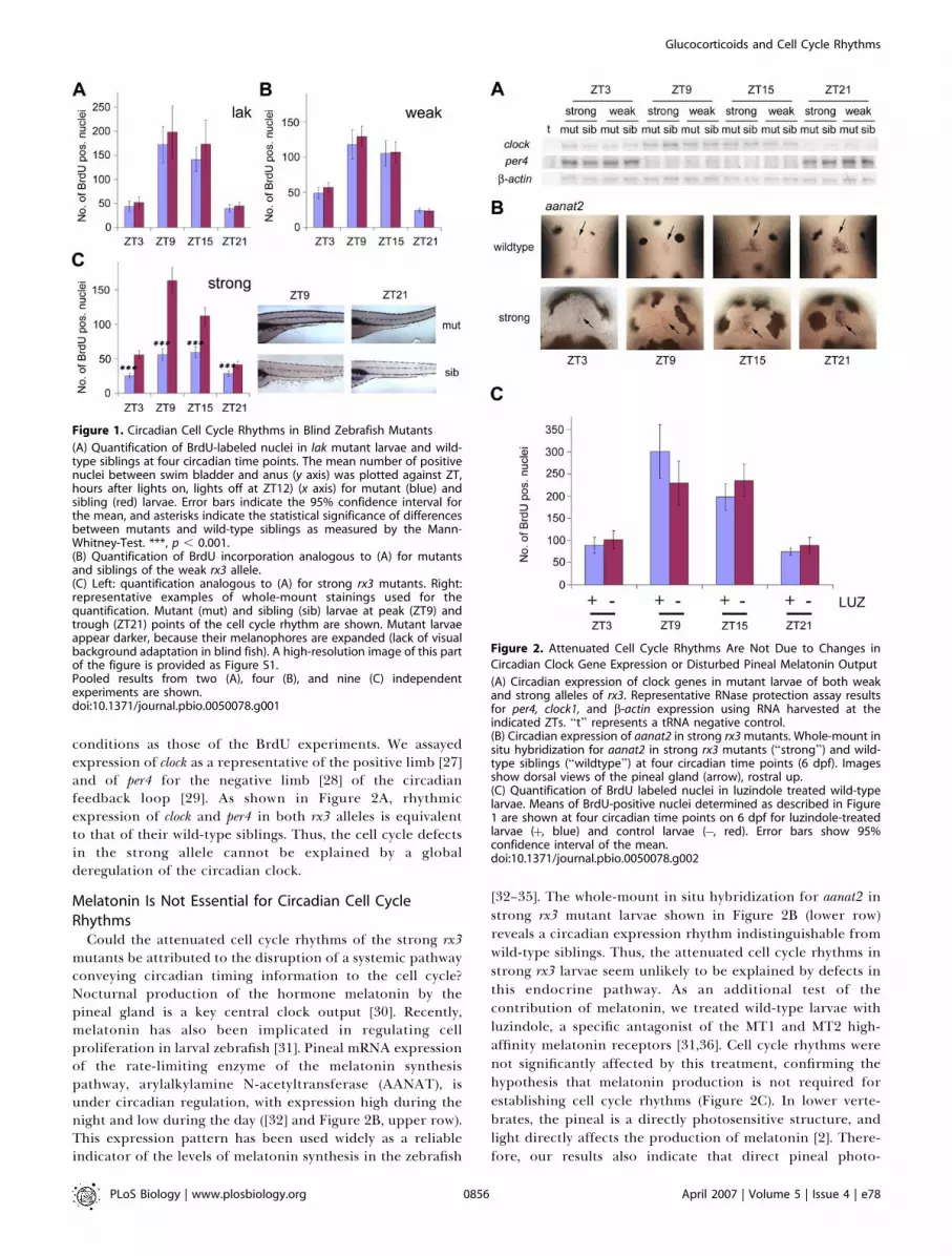
conditions as those of the BrdU experiments. We assayedexpression of clock as a representative of the positive limb [27]and of per4 for the negative limb [28] of the circadianfeedback loop [29]. As shown in Figure 2A, rhythmicexpression of clock and per4 in both rx3 alleles is equivalentto that of their wild-type siblings. Thus, the cell cycle defectsin the strong allele cannot be explained by a globalderegulation of the circadian clock.
Melatonin Is Not Essential for Circadian Cell CycleRhythms
Could the attenuated cell cycle rhythms of the strong rx3mutants be attributed to the disruption of a systemic pathwayconveying circadian timing information to the cell cycle?Nocturnal production of the hormone melatonin by thepineal gland is a key central clock output [30]. Recently,melatonin has also been implicated in regulating cellproliferation in larval zebrafish [31]. Pineal mRNA expressionof the rate-limiting enzyme of the melatonin synthesispathway, arylalkylamine N-acetyltransferase (AANAT), isunder circadian regulation, with expression high during thenight and low during the day ([32] and Figure 2B, upper row).This expression pattern has been used widely as a reliableindicator of the levels of melatonin synthesis in the zebrafish
[32–35]. The whole-mount in situ hybridization for aanat2 instrong rx3 mutant larvae shown in Figure 2B (lower row)reveals a circadian expression rhythm indistinguishable fromwild-type siblings. Thus, the attenuated cell cycle rhythms instrong rx3 larvae seem unlikely to be explained by defects inthis endocrine pathway. As an additional test of thecontribution of melatonin, we treated wild-type larvae withluzindole, a specific antagonist of the MT1 and MT2 high-affinity melatonin receptors [31,36]. Cell cycle rhythms werenot significantly affected by this treatment, confirming thehypothesis that melatonin production is not required forestablishing cell cycle rhythms (Figure 2C). In lower verte-brates, the pineal is a directly photosensitive structure, andlight directly affects the production of melatonin [2]. There-fore, our results also indicate that direct pineal photo-
Figure 1. Circadian Cell Cycle Rhythms in Blind Zebrafish Mutants
(A) Quantification of BrdU-labeled nuclei in lak mutant larvae and wild-type siblings at four circadian time points. The mean number of positivenuclei between swim bladder and anus (y axis) was plotted against ZT,hours after lights on, lights off at ZT12) (x axis) for mutant (blue) andsibling (red) larvae. Error bars indicate the 95% confidence interval forthe mean, and asterisks indicate the statistical significance of differencesbetween mutants and wild-type siblings as measured by the Mann-Whitney-Test. ***, p , 0.001.(B) Quantification of BrdU incorporation analogous to (A) for mutantsand siblings of the weak rx3 allele.(C) Left: quantification analogous to (A) for strong rx3 mutants. Right:representative examples of whole-mount stainings used for thequantification. Mutant (mut) and sibling (sib) larvae at peak (ZT9) andtrough (ZT21) points of the cell cycle rhythm are shown. Mutant larvaeappear darker, because their melanophores are expanded (lack of visualbackground adaptation in blind fish). A high-resolution image of this partof the figure is provided as Figure S1.Pooled results from two (A), four (B), and nine (C) independentexperiments are shown.doi:10.1371/journal.pbio.0050078.g001
Figure 2. Attenuated Cell Cycle Rhythms Are Not Due to Changes in
Circadian Clock Gene Expression or Disturbed Pineal Melatonin Output
(A) Circadian expression of clock genes in mutant larvae of both weakand strong alleles of rx3. Representative RNase protection assay resultsfor per4, clock1, and b-actin expression using RNA harvested at theindicated ZTs. ‘‘t’’ represents a tRNA negative control.(B) Circadian expression of aanat2 in strong rx3 mutants. Whole-mount insitu hybridization for aanat2 in strong rx3 mutants (‘‘strong’’) and wild-type siblings (‘‘wildtype’’) at four circadian time points (6 dpf). Imagesshow dorsal views of the pineal gland (arrow), rostral up.(C) Quantification of BrdU labeled nuclei in luzindole treated wild-typelarvae. Means of BrdU-positive nuclei determined as described in Figure1 are shown at four circadian time points on 6 dpf for luzindole-treatedlarvae (þ, blue) and control larvae (�, red). Error bars show 95%confidence interval of the mean.doi:10.1371/journal.pbio.0050078.g002
PLoS Biology | www.plosbiology.org April 2007 | Volume 5 | Issue 4 | e780856
Glucocorticoids and Cell Cycle Rhythms

reception is unlikely to contribute to circadian cell cyclerhythms.
chokh/rx3 Mutant Fish Show Pituitary DefectsThe hypothalamic–pituitary axis is another endocrine
pathway with a crucial role in the control of cell proliferation
that shows circadian variations of activity [37]. We examinedexpression of a set of specific pituitary cell-lineage markers inthe rx3 mutants: The transcription factor pit1 [38], growthhormone, gh [39], prolactin, prl [39], and glycoproteinhormone alpha subunit, a-gsu [38]. Expression of thesemarkers is equivalent in rx3 mutants of both alleles whencompared with their wild-type siblings (Figure 3A). Thus, thesomatotrope (gh), lactotrope (prl), and gonadotrope/thyro-trope (a-gsu) lineages appear to be normally formed in thestrong rx3 mutants. However, for the corticotrope/melano-trope lineage marker proopiomelanocortin (pomc, [39]), twoexpression domains show a marked reduction in strong allelerx3 mutants. The anterior pituitary domain is stronglyreduced (arrowhead), and the expression corresponding tothe b-endorphin/MSHa synthesizing cells of the arcuatenucleus ([40], arrow) is essentially absent, whereas theposterior pituitary expression domain (asterisk) appearsnormal. All these domains have a wild-type–like appearancein the weak allele mutant larvae (Figure 3A).To test whether these differences reflect a general
disorganization of the diencephalon, we examined theexpression of a number of hypothalamic markers (somatosta-tin3, isotocin, and corticotropin releasing factor; for details, seeFigure S2). The structures labeled by these markers arepresent in both mutant alleles and show no major disruption,despite the lack of normal eyecups. Thus, the strong rx3mutation specifically seems to affect the pomc-expressing cellsin the anterior pituitary and the arcuate nucleus. Previousstudies have established that the anterior pituitary expressiondomain of pomc consists mainly of cells of the corticotropelineage, whereas the posterior domain also contains melano-trope cells [41]. Furthermore, the arcuate nucleus has beenimplicated in regulation of the corticotrope axis in mammals[42]. Thus, the reduced number of corticotrope cells in thestrong rx3 mutants might additionally lack normal hypotha-lamic control. These findings implicate the corticotropelineage in circadian cell cycle regulation.
Corticotrope Deficiency Attenuates Cell Cycle RhythmsGiven the pituitary defect in the strong rx3 mutant, we
asked whether disruption of the hypothalamic–pituitary axiswould cause similar circadian cell cycle defects to those seenin the strong rx3 mutants. To address this issue, we examinedrhythms of BrdU incorporation in a series of zebrafishmutants that lack either the entire pituitary or specificsubsets of pituitary lineages (Figure 3B–3E) [38,40,43–46]. Thefibroblast growth factor 3mutant lia/fgf3 (two alleles, [43]) and theproneural basic Helix-Loop-Helix transcription factor achaetescute-complex like 1amutant pia/ascl1a [46], which lack the entirepituitary, show severely attenuated rhythms (Figure 3B andunpublished data). Thus, genetic ablation of the pituitarycreates a circadian cell cycle phenotype highly similar to thatobserved for the strong rx3 mutant. Since lia and pia mutantsshow normal pomc expression in the arcuate nucleus, we canalso exclude a non-pituitary–mediated contribution of b-endorphin/MSHa–expressing arcuate nucleus neurones tocell cycle rhythm generation.To pinpoint the precise pituitary cell type responsible for
the establishment of normal circadian cell cycle rhythms, weexamined BrdU incorporation rhythms in two other pituitarymutants that lack subsets of the pituitary lineages (Figure 3E):The protein tyrosine phosphatase eyes absent 1 mutant aal/eya1
Figure 3. Reduction or Absence of the Pituitary Corticotrope Lineage
Results in Attenuated Cell Cycle Rhythms
(A) Expression of pituitary markers in rx3 mutants. Whole-mount in situhybridizations for pit1, growth hormone (gh), prolactin (prl), theglycoprotein hormone alpha subunit (a-gsu), and proopiomelanocortin(pomc) (6 dpf), with ventral views through the jaw cartilages, rostral up.Domains of pomc expression are indicated by arrows (arcuate nucleus),arrowheads (anterior pituitary), and asterisks (posterior pituitary). strong,strong rx3 mutants; weak, weak rx3 mutants; wildtype, wild-type siblings.(B–D) Quantification of BrdU incorporation in the lia (B), aal (C), and pit1(D) mutants. Mean numbers of BrdU-positive nuclei determined asdescribed in Figure 1 are indicated for each time point for mutants (blue)and siblings (red). Error bars show the 95% confidence interval of themean; asterisks indicate statistical differences between mutant and wild-type values as determined by the Mann-Whitney test: *, p , 0.05; **, p ,0.01; ***, p , 0.001. Pooled results from two (B), five (C), and four (D)independent experiments are shown.(E) Scheme illustrating the pituitary lineages that are lacking (bracketed)in different pituitary mutants: lia and pia mutants lack all pituitarylineages. aal retains the lactotropes and lacks all other lineages. In pit1,only gonadotropes and corticotropes/melanotropes are present, andlactotropes, thyrotropes, and somatotropes are absent.doi:10.1371/journal.pbio.0050078.g003
PLoS Biology | www.plosbiology.org April 2007 | Volume 5 | Issue 4 | e780857
Glucocorticoids and Cell Cycle Rhythms
[44,45], which possesses only the lactotropes, and the POU-domain transcription factor pit1 mutant [38], in which onlythe corticotropes/melanotropes and the gonadotropes arepresent. The aal mutants show a similar phenotype to the lia,
pia, and the strong rx3 mutants (Figure 3C), demonstratingthat the lactotrope lineage alone is not sufficient forestablishing circadian cell cycle rhythms. In contrast, thepit1 mutants are indistinguishable from their wild-typesiblings (Figure 3D). Thus, the presence of only the cortico-trope/melanotrope and gonadotrope lineages is sufficient toestablish wild-type circadian cell cycle rhythmicity. Togetherwith the reduced number of corticotropes observed in the rx3mutant embryos, this result strongly suggests that thecorticotropes are required for the establishment of thecircadian cell cycle rhythms.
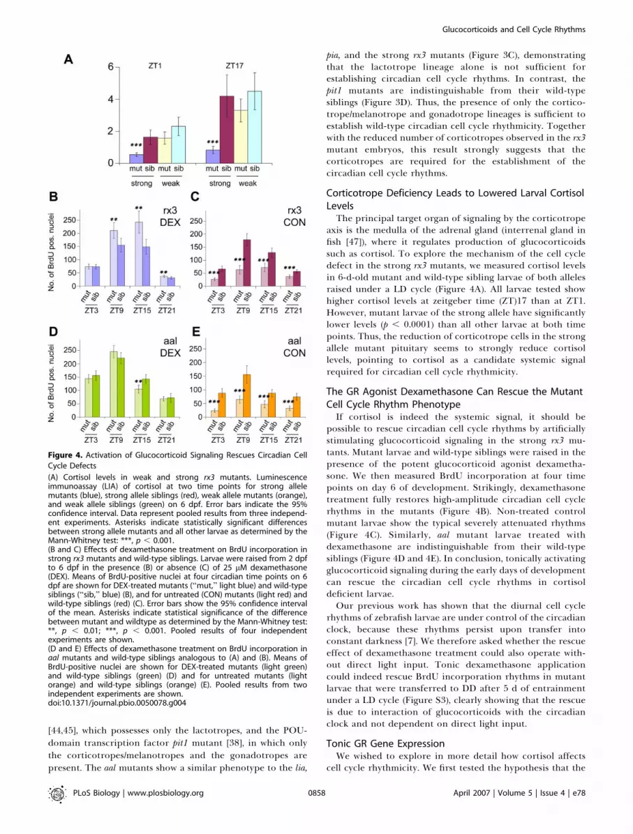
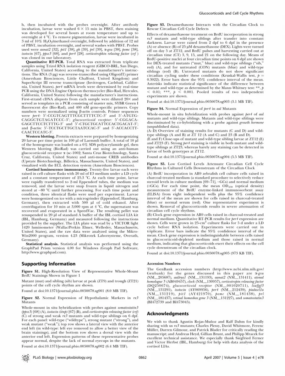
Corticotrope Deficiency Leads to Lowered Larval CortisolLevelsThe principal target organ of signaling by the corticotrope
axis is the medulla of the adrenal gland (interrenal gland infish [47]), where it regulates production of glucocorticoidssuch as cortisol. To explore the mechanism of the cell cycledefect in the strong rx3 mutants, we measured cortisol levelsin 6-d-old mutant and wild-type sibling larvae of both allelesraised under a LD cycle (Figure 4A). All larvae tested showhigher cortisol levels at zeitgeber time (ZT)17 than at ZT1.However, mutant larvae of the strong allele have significantlylower levels (p , 0.0001) than all other larvae at both timepoints. Thus, the reduction of corticotrope cells in the strongallele mutant pituitary seems to strongly reduce cortisollevels, pointing to cortisol as a candidate systemic signalrequired for circadian cell cycle rhythmicity.
The GR Agonist Dexamethasone Can Rescue the MutantCell Cycle Rhythm PhenotypeIf cortisol is indeed the systemic signal, it should be
possible to rescue circadian cell cycle rhythms by artificiallystimulating glucocorticoid signaling in the strong rx3 mu-tants. Mutant larvae and wild-type siblings were raised in thepresence of the potent glucocorticoid agonist dexametha-sone. We then measured BrdU incorporation at four timepoints on day 6 of development. Strikingly, dexamethasonetreatment fully restores high-amplitude circadian cell cyclerhythms in the mutants (Figure 4B). Non-treated controlmutant larvae show the typical severely attenuated rhythms(Figure 4C). Similarly, aal mutant larvae treated withdexamethasone are indistinguishable from their wild-typesiblings (Figure 4D and 4E). In conclusion, tonically activatingglucocorticoid signaling during the early days of developmentcan rescue the circadian cell cycle rhythms in cortisoldeficient larvae.Our previous work has shown that the diurnal cell cycle
rhythms of zebrafish larvae are under control of the circadianclock, because these rhythms persist upon transfer intoconstant darkness [7]. We therefore asked whether the rescueeffect of dexamethasone treatment could also operate with-out direct light input. Tonic dexamethasone applicationcould indeed rescue BrdU incorporation rhythms in mutantlarvae that were transferred to DD after 5 d of entrainmentunder a LD cycle (Figure S3), clearly showing that the rescueis due to interaction of glucocorticoids with the circadianclock and not dependent on direct light input.
Tonic GR Gene ExpressionWe wished to explore in more detail how cortisol affects
cell cycle rhythmicity. We first tested the hypothesis that the
Figure 4. Activation of Glucocorticoid Signaling Rescues Circadian Cell
Cycle Defects
(A) Cortisol levels in weak and strong rx3 mutants. Luminescenceimmunoassay (LIA) of cortisol at two time points for strong allelemutants (blue), strong allele siblings (red), weak allele mutants (orange),and weak allele siblings (green) on 6 dpf. Error bars indicate the 95%confidence interval. Data represent pooled results from three independ-ent experiments. Asterisks indicate statistically significant differencesbetween strong allele mutants and all other larvae as determined by theMann-Whitney test: ***, p , 0.001.(B and C) Effects of dexamethasone treatment on BrdU incorporation instrong rx3 mutants and wild-type siblings. Larvae were raised from 2 dpfto 6 dpf in the presence (B) or absence (C) of 25 lM dexamethasone(DEX). Means of BrdU-positive nuclei at four circadian time points on 6dpf are shown for DEX-treated mutants (‘‘mut,’’ light blue) and wild-typesiblings (‘‘sib,’’ blue) (B), and for untreated (CON) mutants (light red) andwild-type siblings (red) (C). Error bars show the 95% confidence intervalof the mean. Asterisks indicate statistical significance of the differencebetween mutant and wildtype as determined by the Mann-Whitney test:**, p , 0.01; ***, p , 0.001. Pooled results of four independentexperiments are shown.(D and E) Effects of dexamethasone treatment on BrdU incorporation inaal mutants and wild-type siblings analogous to (A) and (B). Means ofBrdU-positive nuclei are shown for DEX-treated mutants (light green)and wild-type siblings (green) (D) and for untreated mutants (lightorange) and wild-type siblings (orange) (E). Pooled results from twoindependent experiments are shown.doi:10.1371/journal.pbio.0050078.g004
PLoS Biology | www.plosbiology.org April 2007 | Volume 5 | Issue 4 | e780858
Glucocorticoids and Cell Cycle Rhythms

circadian clock might regulate expression levels of theglucocorticoid receptor gene (GR) and thereby confer acircadian rhythm of sensitivity to the receptor ligand. Such amechanism would enable even tonic levels of the ligand toactivate GR signaling pathways with a circadian rhythm.Furthermore, recent reports have highlighted that manymembers of the nuclear receptor superfamily show circadiancycling of transcript levels [48]. We thus prepared a timecourse of total RNA and protein extracts from wild-typesibling larvae during one LD cycle. Quantitative real-timePCR analysis failed to detect any significant change in GRmRNA levels during the course of the LD cycle (Figure 5A).Consistently, levels of an 82-kDa immunoreactive proteincorresponding to the zebrafish GR also did not cycle asdetermined by Western blotting analysis (Figure 5B). Theseresults indicate that the circadian clock does not simply affectglobal expression levels of the GR.
Shorter Dexamethasone Treatments Also Rescue CellCycle RhythmsMany studies have documented the functional complexity
of the glucocorticoid signaling pathway in vivo [49]. Lack ofcortisol during development and larval growth mightgenerally alter larval physiology and thereby also indirectlyaffect cell cycle rhythms. Rescue of high-amplitude cell cyclerhythms in rx3 mutant larvae by continuous exposure todexamethasone from early development onwards could actvia rescuing developmental defects as well as by affecting cellsmore directly. In order to address this point, we systemati-cally tested the effect of reducing the duration of exposure todexamethasone on cell cycle rhythms. We supplemented themedium with dexamethasone at progressively later stagesbefore harvesting on day 6 of larval development (Figure 5Cand unpublished data). Addition of dexamethasone as late asthe night before sampling (day 5) still resulted in a significantincrease in cell cycle amplitude. Dexamethasone delivered atlater time points failed to rescue the rhythm. Given that allthe major organ systems have developed and are functional atthis freely feeding larval stage [50], this would exclude a majorrole for indirect developmental mechanisms.
Discussion
The zebrafish represents an attractive model to explorehow circadian clock outputs are regulated at the whole-animal level. We have previously implicated a contribution ofdirectly light-entrainable peripheral clocks in the cell-autonomous control of circadian rhythms of S phase. Here,by studying panels of zebrafish mutants affecting develop-ment of the eye and hypothalamic–pituitary axis, we havebeen able to define the regulatory contribution of thesestructures to establishing circadian cell cycle rhythms. Thisstudy illustrates the power of using complementary sets ofzebrafish mutants in the genetic dissection of physiologicalpathways in vivo.We have shown that ocular photoreception is dispensable
for establishing this clock output function, potentiallyreinforcing the notion that cell-autonomous light sensingplays a key role in cell cycle entrainment in the zebrafish.However, in common with most lower vertebrates, zebrafishpossess additional extraocular specialized photoreceptortissues: the pineal complex [2] and also the so-called deep
Figure 5. Shorter-Term Treatment with Dexamethasone Also Rescues
Cell Cycle Rhythms in Strong rx3 Mutants
(A and B) Expression analysis of the zebrafish GR. (A) GR mRNAexpression as determined by quantitative RT-PCR in wild-type larvae atfour circadian time points. No significant oscillation is apparent (one-wayANOVA [analysis of variance], p ¼ 0.4231). Error bars show the 95%confidence interval of the mean from three independent experiments. Asa control, per4 mRNA levels were assayed in the same samples andshown to exhibit a high-amplitude rhythm (unpublished data; see alsoFigure2A). (B) Representative Western blot showing expression levels ofthe GR protein (upper) and of CREB (lower) as a loading control.Consistent with mRNA expression levels, no overt oscillation can beobserved.(C) Upper panel: experimental design to define a minimum period ofexposure to the tonic dexamethasone (DEX) signal required to rescuecell cycle rhythms in the rx3 mutants. Black and white bars represent thelight and dark periods of day 5 and 6 post-fertilization. Above: times ofDEX addition to the medium. Red arrows and lines indicate treatmentsthat are able to increase the cycling amplitude of BrdU incorporation instrong rx3 mutant larvae, whereas black arrows and lines indicate thosethat fail to do so. Below: times for BrdU labeling (as 15-min pulses) andharvesting on day 6 (indicated as ZT time points).Lower panel: quantification of BrdU incorporation in strong rx3 mutantsfollowing the treatments outlined in the upper panel. Peak (ZT9, darkcolor) and trough (ZT21, light color) mean values on day 6 are shown foruntreated controls (CON) and the four times of DEX addition on day 5(þDEX). Red bars: treatments that result in a significant differencebetween peak and trough values (Mann-Whitney-Test; ***, p , 0.001),black bars: no significant differences observed between peak and troughvalues. Error bars indicate the 95% confidence interval of the mean. Dataare pooled from six independent experiments.doi:10.1371/journal.pbio.0050078.g005
PLoS Biology | www.plosbiology.org April 2007 | Volume 5 | Issue 4 | e780859
Glucocorticoids and Cell Cycle Rhythms

brain photoreceptors that line the third ventricle of thediencephalon [51,52]. Since no zebrafish mutants are avail-able to date that specifically lack these photoreceptors, it isproblematic to assess their contribution. We used analternative pharmacological approach to interfere with themelatonin signal, the major output of the pineal gland, andthereby tested whether photoreception through the pinealcomplex might affect cell cycle rhythms. Because treatment oflarvae with the melatonin receptor antagonist luzindole didnot change circadian cell cycle rhythms, pineal lightreception is not strictly required for the timing of circadiancell cycle progression. However, it is still conceivable thatlight input from the pineal conveyed via neuronal pathwaysmay contribute to the timing of the cell cycle [53–55]. Also,one type of dedicated photoreceptor might be able tosubstitute for lack of input from the other types, leading tofunctional redundancy of inputs from, e.g., the eye and thepineal complex. Finally, the direct peripheral light receptionalone might also be sufficient to time this clock output in thecontext of the whole animal. Ultimately, mutant zebrafishthat lack all specialized photoreceptor cells will be requiredto assess the relative contribution of directly light-sensingperipheral clocks to entraining the circadian cell cycle.
By studying pituitary mutants with overlapping cell-lineagedefects, and by our demonstration that pituitary cortico-tropes are severely reduced in strong allele rx3 mutant larvae,we have implicated this pituitary cell lineage in the establish-ment of circadian cell cycle rhythms. Furthermore, levels ofcortisol in strong rx3mutants are reduced and, importantly, aGR agonist rescues the circadian cell cycle defects in bothstrong rx3 and pituitary mutants. These findings point toglucocorticoids as a requirement for high-amplitude cellcycle rhythms.
Previously, glucocorticoids have been implicated in theentrainment of peripheral circadian clocks in mammals.Injection of dexamethasone into mice can reset the phase ofperipheral oscillators [56]. However, mice lacking the GRshow normal clock gene expression rhythms in the liver [56].Furthermore, here we observe normal circadian clock genecycling in the strong rx3 mutant larvae (Figure 2A), despitetheir low levels of cortisol. Consistently, also in the aalmutants, per4 exhibits normal circadian rhythms of expres-sion (Figure S4). Finally, zebrafish cells grown in cortisol-depleted medium still show normal clock expression under aLD cycle (Figure S5B, see below). Thus, glucocorticoidsignaling is not an absolute requirement for the regulationof the peripheral pacemakers themselves. Our data directlyimplicate glucocorticoids in the regulation of clock output.
Glucocorticoids are well known to influence cell prolifer-ation, both in vitro and in vivo. For example, dexamethasonecan stimulate myoblast proliferation [57] and enhance themitogenic response of fibroblasts to epidermal growth factor(EGF) [58], whereas it inhibits cell division in a lymphosarco-ma cell line [59]. In addition, at the whole-animal level,corticosteroids have been reported to affect the capacity ofthe liver to regenerate after hepatectomy in rats [60]. Ourresults indicate that part of the effect of corticosteroids oncell proliferation might be brought about by cooperationwith circadian clock output pathways.
At which level of organization might this interaction occur?Given the wealth of physiological targets of glucocorticoids,they might function through indirect, systemic pathways. For
example, in zebrafish larvae, they might be involved in thematuration of organ systems and their physiological func-tions during development. However, our finding thatdexamethasone treatment starting 10 h before sampling stillrescues cell cycle rhythms in rx3 mutants makes a long-termeffect on development unlikely. Nevertheless, other indirectsystemic pathways might act within this time frame. Forexample, glucocorticoids could stimulate the release ofmitogens from neighboring tissues or influence the releaseof other hormones (e.g., see [61–63]). Alternatively, they mightact directly at the level of the proliferating cells themselves.We tested a potential cell-autonomous role for glucocorti-coids in circadian cell cycle rhythms by examining zebrafishcell cultures grown in charcoal-treated (and thereby steroid-depleted) medium (Figure S5). Whereas circadian clock geneexpression is normal in these cultures (Figure S5B), circadiancell cycle rhythms are severely attenuated (Figure S5A), thusmimicking the situation in cortisol-deficient larvae. However,in this cell culture system, treatment with dexamethasonedoes not rescue the attenuation (unpublished data). Oneexplanation for this result could be that glucocorticoidsmight need to act synergistically with other substances thatare also depleted from the medium by charcoal treatment.Taken together, although there are some hints for a directcell-autonomous action of glucocorticoids on circadian cellcycle rhythms, more indirect systemic effects certainly cannotbe excluded. Indeed, given the multifaceted actions ofglucocorticoids, confinement of their effects on cell cycleprogression to a single level would be rather surprising. In theanimal, glucocorticoids might help to create a systemicsignaling environment, within which they could also exertmore direct effects.What is the relative importance of systemic and peripheral
circadian clock mechanisms in glucocorticoid-mediatedcircadian cell cycle regulation? Circadian rhythms of circu-lating glucocorticoids have long been recognized as a clockoutput in various vertebrates [37]. However, here we showthat the role played by glucocorticoids in circadian cell cyclerhythms does not necessarily involve conveying timinginformation via changes in circulating levels. Rather, aconstant glucocorticoid signal can rescue rhythms of cellcycle in cortisol-deficient mutant larvae. In the animal, thetiming information might stem from another cycling systemicsignal, or it might be provided by peripheral circadian clocks.Circadian changes in glucocorticoid levels might thenreinforce the peripheral timing information in cell cyclecontrol, or they might be required for other physiologicalfunctions.How can a constant glucocorticoid signal lead to a
rhythmic output? We propose a working model in which acertain level of glucocorticoid signaling may be permissive(Figure 6A, green arrow) for peripheral circadian clockregulation of genes involved in cell proliferation (Figure 6A,red arrow). Alternatively, the clock could gate responsivenessto the glucocorticoid signal, which would then act to regulatecell proliferation (Figure 6A, blue arrow). In either scenario,in corticotrope-deficient mutants, GR signaling is attenuated,and then peripheral clock input alone would be insufficientto generate full cell cycle rhythms (Figure 6B). Furthermore,in the rescue experiments, tonic dexamethasone treatmentactivates GR signaling and would restore cell cycle rhythms inthe mutants with timing information being provided by the
PLoS Biology | www.plosbiology.org April 2007 | Volume 5 | Issue 4 | e780860
Glucocorticoids and Cell Cycle Rhythms

peripheral circadian clock (Figure 6C). In this model, cyclingcortisol levels are not required for, but they might reinforcecell cycle rhythmicity.
An attractive mechanism for gating responsiveness toglucocorticoids would involve circadian clock regulation ofexpression of the GR itself. Expression of many nuclearreceptor transcripts is under circadian clock control indifferent peripheral tissues [48]. However, here we show thatthe levels of the GR transcript as well as the protein are notsubject to significant day–night variation. Thus, a simplescenario in which transcription of the GR gene is a regulatorytarget of peripheral clock components, e.g., via E boxelements, appears unlikely. One can speculate that if indeedthe receptor is under clock control, then this may occur at thepost-translational level. Alternatively, circadian clock controlmight operate at various levels on other elements of theglucocorticoid signaling pathway, perhaps downstream of thereceptor itself [64,65].
Glucocorticoids can increase the amplitude of cell cyclerhythms in strong rx3 mutants even when they are deliveredthe night before assaying BrdU incorporation. Specifically,they have to be present at least before ZT17, or 16 h beforethe peak of BrdU incorporation at ZT9 is reached. Addingdexamethasone at ZT21 (12 h before the expected peak) is notsufficient. This might reflect either a minimum time needed
to exert downstream effects on the cell cycle, or a require-ment for the presence of glucocorticoids at a certain time ofday. Interestingly, the last time point with rescue capabilitycoincides with the natural peak in cortisol levels in wild-typelarvae (Figure 4A). Experiments involving a precisely con-trolled temporal activation and inactivation of the glucocor-ticoid signaling pathway will be needed to decide betweenthese possibilities.In summary, our results call for a re-evaluation of existing
models that account for control of circadian cell cycle timingpurely via the direct regulation of gene expression byperipheral clock components. We reveal a requirement forendocrine regulation involving glucocorticoids that operatesdownstream of the clock mechanism itself. It is tempting tospeculate that many other clock outputs may involve similarcontributions from cell-autonomous and systemic controlelements.
Materials and Methods
Fish care, RNase protection assay, BrdU labeling, and dexametha-sone treatment. Fish were raised and bred according to standardprocedures [50]. RNase protection analysis was carried out asdescribed previously [27], and the per4, clock1, and b-actin probeshave been described [27,28,66]. The raising of larvae under controlledlighting and temperature conditions, and the BrdU labelingprocedure have been described in [7]. Briefly, ten mutant and tenwild-type sibling larvae each were sorted into cell culture flasks onday 2 post-fertilization and raised at 25 8C under a 12-h light:12-hdark cycle until day 6 of development. Three hours after ‘‘lights on’’(ZT¼ 3), the larvae of one flask were incubated for 20 min with BrdUbefore fixation in 4% paraformaldehyde/PBS, and this procedure wasrepeated at three additional time points at 6-h intervals. The cellculture BrdU incorporation experiments (AB.9 cells, ATCC) werecarried out as described in [7]. Cells were raised in L15 mediumsupplemented with gentamycin, streptomycin, penicillin [16], andeither 15% fetal bovine serum (FBS) or 15% charcoal-treated FBS(both Biochrom, Berlin, Germany).
The pituitary and lak mutants are not morphologically distinguish-able from wild-type siblings early in development. Thus, two flaskswith 25 larvae each from a cross of heterozygous carriers were sortedand processed as described above to ensure the presence of asufficient number of mutants per time point. lakmutant larvae can berecognized by their expanded melanophore phenotype later indevelopment. To identify the pituitary mutants, larvae were stainedfor growth hormone expression by whole-mount in situ hybridization(see below) and then stained for BrdU incorporation.
Dexamethasone treatment was carried out essentially as describedby [41]. Dexamethasone (Sigma, St. Louis, Missouri, United States) wasdissolved in distilled water at 1 mM as a stock solution, then dilutedfurther in E3 medium to a final concentration of 25 lM. Ten strongallele rx3 mutants and wild-type siblings were sorted into each cellculture flask on day 2 of development and raised in the presence orabsence of dexamethasone until BrdU labeling on day 6 as describedabove. Luzindole treatments were performed similarly, with a stocksolution of 0.01 M luzindole (Sigma) in ethanol diluted further in E3to a final concentration of 0.00001 M [31]. Twenty larvae were raisedin 25 ml of E3 at this luzindole concentration from day 2 ofdevelopment, and on day 4, an additional 30 ll of luzindole stocksolution were added to compensate for potential degradation of thecompound. Control larvae were treated with equivalent solvent(ethanol) concentrations only.
In situ hybridization. In situ hybridization was carried outessentially as described [50], with the following modifications: The4% paraformaldehyde fixation step after rehydration was omitted.Larvae were washed twice in PBSþ0.1% Tween-20 (PBST), then rinsedfor 2 min in distilled water, incubated for 7 min in pre-cooledacetone at �20 8C, passed through distilled water for 2 min at roomtemperature, and washed 335 min in PBST. Then, larvae weredigested with 1 mg/ml of collagenase P (Roche, Basel, Switzerland) inPBS with 1% BSA and 1% DMSO at room temperature for 45 min,before fixation for 20 min at 4 8C in 4% paraformaldehyde. After fivewashes with PBST, larvae were prehybridized with HYB buffer for 4–6
Figure 6. Working Model for Glucocorticoid Action on Circadian Cell
Cycle Rhythms
(A) In wild-type larvae, corticotrope function is associated with normalcortisol levels (that exhibit circadian cycling). In target cells, cortisolactivates GR signaling (green arrow) that may be permissive for theperipheral clock regulation of cell cycle. In addition, cycling cortisol levelsmay confer circadian rhythms of GR signaling that potentially reinforcecell cycle rhythmicity. The red arrow indicates the peripheral circadianclock control of cell cycle, and the blue arrow symbolizes the potentialgating of GR signaling by the peripheral pacemaker. Cooperationbetween the peripheral clock mechanism and the GR signaling pathwaygenerates a circadian rhythm of cell cycle.(B) In corticotrope-deficient mutants, circulating cortisol levels aresignificantly reduced. In the absence of normal GR signaling, peripheralclock input is not sufficient to generate cell cycle rhythms. Dashedarrows indicate attenuated pathways.(C) Cell cycle rhythms are rescued in corticotrope-deficient mutants bytonic dexamethasone (DEX) treatment. Thus timing information isprovided by peripheral circadian clocks and is not reliant on cyclingcortisol levels.doi:10.1371/journal.pbio.0050078.g006
PLoS Biology | www.plosbiology.org April 2007 | Volume 5 | Issue 4 | e780861
Glucocorticoids and Cell Cycle Rhythms
h, then incubated with the probes overnight. After antibodyincubation, larvae were washed 6 3 15 min in PBST, then stainingwas developed for several hours at room temperature and up toovernight at 4 8C. To remove pigmentation, larvae were incubated in3 ml of 10% H2O2/methanol overnight, followed by addition of 12 mlof PBST, incubation overnight, and several washes with PBST. Probesused were aanat2 [32], pit1 [38], gh [39], prl [39], a-gsu [38], pomc [38],isotocin [67], ppss3 [68], and per4 [28]. corticotrophin relasing factor (crf )was cloned in our laboratory.
Quantitative RT-PCR. Total RNA was extracted from triplicatesamples using Trizol RNA isolation reagent (GIBCO-BRL, San Diego,California, United States) according to the manufacturer’s instruc-tions. The RNA (3 lg) was reverse-transcribed using Oligo(dT) primer(Amersham Biosciences, Little Chalfont, United Kingdom) andSuperScript III reverse transcriptase (Invitrogen, Carlsbad, Califor-nia, United States). per4 mRNA levels were determined by real-timePCR using the DNA Engine Opticon thermocycler (Bio-Rad, Hercules,California, United States) following the manufacturer’s instructions.First-strand cDNA aliquots from each sample were diluted 203 andserved as templates in a PCR consisting of master mix, SYBR Green Ifluorescent dye (Bio-Rad), and 400 nM gene-specific primers. Copynumbers were normalized using b-actin controls. Primer sequenceswere per4: 59-CCGTCAGTTTCGCTTTTCTC-39 and 59-ATGTG-CAGGCTGTAGATCCC-39; glucocorticoid receptor: 59-CGGACA-GAGCTTCCTCTTTG-39 and 59-CTGCTGCATTCCACTGACAT-39;and b-actin: 59-TCCTGCTTGCTAATCCAC-39 and 59-ACCACCTT-CAACTCCATC-39.
Western blotting. Protein extracts were prepared by homogenizing20 larvae per time point in 100 ll of Laemmli buffer. A total of 10 llof the homogenate was loaded on a 6% SDS polyacrylamide gel, thenWestern blotting (BioRad) was carried out using an anti-humanglucocorticoid receptor a (sc-1002; Santa Cruz Biotechnology, SantaCruz, California, United States) and anti-mouse CREB antibodies(Upstate Biotechnology, Billerica, Massachusetts, United States), andvisualized with the ECL detection system (Amersham Biosciences).
Cortisol luminescence immunoassay. Twenty-five larvae each wereraised in cell culture flasks with 20 ml of E3 medium under a LD cycleand a constant temperature of 25.3 8C. At each time point, larvaewere rapidly transferred to 2-ml Eppendorf tubes, the medium wasremoved, and the larvae were snap frozen in liquid nitrogen andstored at �80 8C until further processing. For each time point andcondition, three identical flasks were used per experiment. Larvaewere homogenized on ice with a microgrinder (Eppendorf, Hamburg,Germany), then extracted with 500 ll of cold ethanol. Aftercentrifugation for 10 min at 3,000 rpm at 4 8C, the supernatant wasrecovered and evaporated in a SpeedVac. The resulting pellet wasresuspended in 20 ll of standard A buffer of the IBL cortisol LIA kit(IBL, Hamburg, Germany) and measured following the instructionsprovided by the supplier. The LIA plate was read by a VICTOR light1420 luminometer (Wallac/Perkin Elmer, Wellesley, Massachusetts,United States), and the raw data were analyzed using the Mikro-Win2000 program, version 4.23 (Mikrotek Laborsysteme, Overath,Germany).
Statistical analysis. Statistical analysis was performed using theGraphPad Prism version 4.00 for Windows (Graph Pad Software,http://www.graphpad.com).
Supporting Information
Figure S1. High-Resolution View of Representative Whole-MountBrdU Stainings Shown in Figure 1
Mutant (mut) and sibling (sib) larvae at peak (ZT9) and trough (ZT21)points of the cell cycle rhythm are shown.
Found at doi:10.1371/journal.pbio.0050078.sg001 (8.0 MB TIF).
Figure S2. Normal Expression of Hypothalamic Markers in rx3Mutants
Whole-mount in situ hybridization with probes against somatostatin3(ppss3) [68] (A), isotocin (itnp) [67] (B), and corticotropin releasing factor (crf)(C) of strong and weak rx3 mutants and wild-type siblings on 6 dpf.For each panel: wild-type (‘‘wildtype’’), strong mutant (‘‘strong’’), andweak mutant (‘‘weak’’); top row shows a lateral view with the anteriorend left (in wild-type: left eye removed to allow a better view of thebrain stainings), and the bottom row shows a dorsal view with theanterior end left. Expression patterns of these representative probesappear normal, despite the lack of normal eyecups in the mutants.
Found at doi:10.1371/journal.pbio.0050078.sg002 (6.4 MB TIF).
Figure S3. Dexamethasone Interacts with the Circadian Clock toRescue Circadian Cell Cycle Defects
Effects of dexamethasone treatment on BrdU incorporation in strongrx3 mutants and wild-type siblings after transfer into constantdarkness. Larvae were raised from 2 dpf to 6 dpf in the presence(A) or absence (B) of 25 lM dexamethasone (DEX). Lights were turnedoff on day 5 at ZT12, and BrdU pulses and harvesting carried out atcircadian time (CT) 3, 9, 15, and 21 on the following day. Means ofBrdU-positive nuclei at four circadian time points on 6 dpf are shownfor DEX-treated mutants (‘‘mut,’’ blue) and wild-type siblings (‘‘sib,’’red) (A), and for untreated (CON) mutants (blue) and wild-typesiblings (red) (B). Untreated mutants do not show significantcircadian cycling under these conditions (Kruskal-Wallis test, p ¼0.3022). Error bars show the 95% confidence interval of the mean.Asterisks indicate statistical significance of the difference betweenmutant and wild-type as determined by the Mann-Whitney test: **, p, 0.01; ***, p , 0.001. Pooled results of two independentexperiments are shown.
Found at doi:10.1371/journal.pbio.0050078.sg003 (3.1 MB TIF).
Figure S4. Normal Expression of per4 in aal Mutants
Whole-mount in situ hybridization with probes against per4 of aalmutants and wild-type siblings. Mutants and wild-type siblings werediscriminated by co-hybridizing with a probe against growth hormone(unpublished data).(A–D) Overview of staining results for mutants (C and D) and wild-type siblings (A and B) at ZT 12 (A and C) and 23 (B and D).(E and F) Close-ups of mutant and wild-type sibling larvae at ZT12 (E)and ZT23 (F). Strong per4 staining is visible in both mutant and wild-type siblings at ZT23, whereas barely any staining can be detected inlarvae of both genotypes at ZT12.
Found at doi:10.1371/journal.pbio.0050078.sg004 (5.5 MB TIF).
Figure S5. Low Cortisol Levels Attenuate Circadian Cell CycleRhythms in Cultured Cells Downstream of the Circadian Clock
(A) BrdU incorporation in AB9 zebrafish cell culture cells raised incharcoal-treated medium (a standard procedure to selectively reducesteroid levels in culture medium [69–71],�GCs) and normal medium(þGCs). For each time point, the mean OD450 (optical density)measurement of the BrdU enzyme-linked immunosorbent assay(ELISA) from eight independent wells plus the 95% confidenceinterval of the mean are shown for cells raised in charcoal-treated(blue) or normal serum (red). One representative experiment isshown. Removal of glucocorticoids results in severe attenuation ofcircadian cell cycle rhythms.(B) Clock gene expression in AB9 cells raised in charcoal-treated andnormal medium. Quantitative RT-PCR results for per4 expression areshown. Cells were grown in 25-cm2 culture flasks for 5 d under a LDcycle before RNA isolation. Experiments were carried out intriplicate. Error bars indicate the 95% confidence interval of themean. Clock gene expression is indistinguishable between cells raisedin glucocorticoid-depleted medium and those raised in normalmedium, indicating that glucocorticoids exert their effects on the cellcycle downstream of the circadian clock.
Found at doi:10.1371/journal.pbio.0050078.sg005 (973 KB TIF).
Accession Numbers
The GenBank accession numbers (http://www.ncbi.nlm.nih.gov/Genbank) for the genes discussed in this paper are a-gsu(NM_205687), aal/eya1 (NM_131193), aanat2 (NM_131411), atonalhomologue 5 (AB049457), clock (NM_130957), corticotropin releasing factor(DQ250674), glucocorticoid receptor (NM_001020711), lia/fgf3(NM_131291), isotocin (AY069956), per4 (NM_212439), pia/ascl1a(NM_131219), pit1 (AY421970), pomc (NM_181438), prl(NM_181437), retinal homeobox gene 3 (NM_131227), and somatostatin3(BI472739 and BI473045).
Acknowledgments
We wish to thank Agustin Rojas-Munoz and Ralf Dahm for kindlysharing with us rx3 mutants; Charles Plessy, David Whitmore, FerencMuller, Darren Gilmour, and Patrick Blader for critically reading themanuscript; and Andreas Heyd, Gillian Brunt, and Philipp Mracek forexcellent technical assistance. We especially thank Siegfried Fernerand Victor Herbst (IBL, Hamburg) for help with data analysis of thecortisol LIAs.
PLoS Biology | www.plosbiology.org April 2007 | Volume 5 | Issue 4 | e780862
Glucocorticoids and Cell Cycle Rhythms

Author contributions. TD and NSF conceived and designed theexperiments. TD, KL, DV, CS, and NSF performed the experiments.TD and NSF analyzed the data. TD, GN, CJN, and MH contributedreagents/materials/analysis tools. TD, DV, and NSF wrote the paper.
Funding.We acknowledge funding by the Max-Planck-Gesellschaft,
Centre National de la Recherche Scientifique (NSF), DeutscheForschungsgemeinschaft (TD), and European Molecular BiologyOrganization (TD).
Competing interests. The authors have declared that no competinginterests exist.
References1. Klein DC, Moore RY, Reppert SM, editors (1991) Suprachiasmatic nucleus:
The mind’s clock. New York: Oxford University Press. 467 p.2. Menaker M, Moreira LF, Tosini G (1997) Evolution of circadian
organization in vertebrates. Braz J Med Biol Res 30: 305–313.3. Schibler U, Ripperger J, Brown SA (2003) Peripheral circadian oscillators in
mammals: Time and food. J Biol Rhythms 18: 250–260.4. Panda S, Antoch MP, Miller BH, Su AI, Schook AB, et al. (2002)
Coordinated transcription of key pathways in the mouse by the circadianclock. Cell 109: 307–320.
5. Bjarnason GA, Jordan R (2000) Circadian variation of cell proliferation andcell cycle protein expression in man: Clinical implications. Prog Cell CycleRes 4: 193–206.
6. Mori T, Johnson CH (2000) Circadian control of cell division in unicellularorganisms. Prog Cell Cycle Res 4: 185–192.
7. Dekens MP, Santoriello C, Vallone D, Grassi G, Whitmore D, et al. (2003)Light regulates the cell cycle in zebrafish. Curr Biol 13: 2051–2057.
8. Nagoshi E, Saini C, Bauer C, Laroche T, Naef F, et al. (2004) Circadian geneexpression in individual fibroblasts: Cell-autonomous and self-sustainedoscillators pass time to daughter cells. Cell 119: 693–705.
9. Fu L, Pelicano H, Liu J, Huang P, Lee C (2002) The circadian gene Period2plays an important role in tumor suppression and DNA damage responsein vivo. Cell 111: 41–50.
10. Matsuo T, Yamaguchi S, Mitsui S, Emi A, Shimoda F, et al. (2003) Controlmechanism of the circadian clock for timing of cell division in vivo. Science302: 255–259.
11. Fu L, Patel MS, Bradley A, Wagner EF, Karsenty G (2005) The molecularclock mediates leptin-regulated bone formation. Cell 122: 803–815.
12. Gery S, Komatsu N, Baldjyan L, Yu A, Koo D, et al. (2006) The circadiangene per1 plays an important role in cell growth and DNA damage controlin human cancer cells. Mol Cell 22: 375–382.
13. Felig P, Baxter J, Broadus A, Frohman LA, editors (1987) Endocrinologyand metabolism. New York: McGraw-Hill. 1855 p.
14. Ganong WF (2001) Review of medical physiology. New York: Lange MedicalBooks/McGraw-Hill. 870 p.
15. Cahill GM (2002) Clock mechanisms in zebrafish. Cell Tissue Res 309: 27–34.
16. Whitmore D, Foulkes NS, Sassone-Corsi P (2000) Light acts directly onorgans and cells in culture to set the vertebrate circadian clock. Nature 404:87–91.
17. Wullimann M, Rupp B, Reichert H (1996) Neuroanatomy of the zebrafishbrain: A topological atlas. Basel: Birkhauser Verlag. 144 p.
18. Diaz ML, Becerra M, Manso MJ, Anadon R (2002) Distribution ofthyrotropin-releasing hormone (TRH) immunoreactivity in the brain ofthe zebrafish (Danio rerio). J Comp Neurol 450: 45–60.
19. Burrill JD, Easter SS Jr (1994) Development of the retinofugal projectionsin the embryonic and larval zebrafish (Brachydanio rerio). J Comp Neurol 346:583–600.
20. Arenzana FJ, Arevalo R, Sanchez-Gonzalez R, Clemente D, Aijon J, et al.(2006) Tyrosine hydroxylase immunoreactivity in the developing visualpathway of the zebrafish. Anat Embryol (Berl) 211: 323–324.
21. Amsterdam A, Hopkins N (2006) Mutagenesis strategies in zebrafish foridentifying genes involved in development and disease. Trends Genet 22:473–478.
22. Kay JN, Finger-Baier KC, Roeser T, Staub W, Baier H (2001) Retinalganglion cell genesis requires lakritz, a Zebrafish atonal Homolog. Neuron30: 725–736.
23. Berson DM (2003) Strange vision: Ganglion cells as circadian photo-receptors. Trends Neurosci 26: 314–320.
24. Loosli F, Staub W, Finger-Baier KC, Ober EA, Verkade H, et al. (2003) Lossof eyes in zebrafish caused by mutation of chokh/rx3. EMBO Rep 4: 894–899.
25. Kennedy BN, Stearns GW, Smyth VA, Ramamurthy V, van Eeden F, et al.(2004) Zebrafish rx3 and mab21l2 are required during eye morphogenesis.Dev Biol 270: 336–349.
26. Rojas-Munoz A, Dahm R, Nusslein-Volhard C (2005) chokh/rx3 specifies theretinal pigment epithelium fate independently of eye morphogenesis. DevBiol 288: 348–362.
27. Whitmore D, Foulkes NS, Strahle U, Sassone-Corsi P (1998) Zebrafish Clockrhythmic expression reveals independent peripheral circadian oscillators.Nat Neurosci 1: 701–707.
28. Vallone D, Gondi SB, Whitmore D, Foulkes NS (2004) E-box function in aperiod gene repressed by light. Proc Natl Acad Sci U S A 101: 4106–4111.
29. Reppert SM, Weaver DR (2002) Coordination of circadian timing inmammals. Nature 418: 935–941.
30. Arendt J (2005) Melatonin: Characteristics, concerns, and prospects. J BiolRhythms 20: 291–303.
31. Danilova N, Krupnik VE, Sugden D, Zhdanova IV (2004) Melatoninstimulates cell proliferation in zebrafish embryo and accelerates itsdevelopment. FASEB J 18: 751–753.
32. Gothilf Y, Coon SL, Toyama R, Chitnis A, Namboodiri MA, et al. (1999)Zebrafish serotonin N-acetyltransferase-2: Marker for development ofpineal photoreceptors and circadian clock function. Endocrinology 140:4895–4903.
33. Falcon J, Gothilf Y, Coon SL, Boeuf G, Klein DC (2003) Genetic, temporaland developmental differences between melatonin rhythm generatingsystems in the teleost fish pineal organ and retina. J Neuroendocrinol 15:378–382.
34. Ziv L, Levkovitz S, Toyama R, Falcon J, Gothilf Y (2005) Functionaldevelopment of the zebrafish pineal gland: Light-induced expression ofperiod2 is required for onset of the circadian clock. J Neuroendocrinol 17:314–320.
35. Vuilleumier R, Besseau L, Boeuf G, Piparelli A, Gothilf Y, et al. (2006)Starting the zebrafish pineal circadian clock with a single photic transition.Endocrinology 147: 2273–2279.
36. Dubocovich ML (1988) Luzindole (N-0774): A novel melatonin receptorantagonist. J Pharmacol Exp Ther 246: 902–910.
37. Aschoff J (1979) Circadian rhythms: General features and endocrinologicalaspects. In: Krieger D, editor. Endocrine rhythms. New York: Raven Press.pp. 1–63.
38. Nica G, Herzog W, Sonntag C, Hammerschmidt M (2004) Zebrafish pit1mutants lack three pituitary cell types and develop severe dwarfism. MolEndocrinol 18: 1196–1209.
39. Herzog W, Zeng X, Lele Z, Sonntag C, Ting JW, et al. (2003)Adenohypophysis formation in the zebrafish and its dependence on sonichedgehog. Dev Biol 254: 36–49.
40. Herzog W, Sonntag C, Walderich B, Odenthal J, Maischein HM, et al. (2004)Genetic analysis of adenohypophysis formation in zebrafish. Mol Endo-crinol 18: 1185–1195.
41. Liu NA, Huang H, Yang Z, Herzog W, Hammerschmidt M, et al. (2003)Pituitary corticotroph ontogeny and regulation in transgenic zebrafish.Mol Endocrinol 17: 959–966.
42. Bell ME, Bhatnagar S, Akana SF, Choi S, Dallman MF (2000) Disruption ofarcuate/paraventricular nucleus connections changes body energy balanceand response to acute stress. J Neurosci 20: 6707–6713.
43. Herzog W, Sonntag C, von der Hardt S, Roehl HH, Varga ZM, et al. (2004)Fgf3 signaling from the ventral diencephalon is required for earlyspecification and subsequent survival of the zebrafish adenohypophysis.Development 131: 3681–3692.
44. Kozlowski DJ, Whitfield TT, Hukriede NA, Lam WK, Weinberg ES (2005)The zebrafish dog-eared mutation disrupts eya1, a gene required for cellsurvival and differentiation in the inner ear and lateral line. Dev Biol 277:27–41.
45. Nica G, Herzog W, Sonntag C, Nowak M, Schwarz H, et al. (2006) Eya1 isrequired for lineage-specific differentiation, but not for cell survival in thezebrafish adenohypophysis. Dev Biol 292: 189–204.
46. Pogoda HM, von der Hardt S, Herzog W, Kramer C, Schwarz H, et al. (2006)The proneural gene ascl1a is required for endocrine differentiation andcell survival in the zebrafish adenohypophysis. Development 133: 1079–1089.
47. Bone Q, Marshall NB, Blaxter JHS (1995) Biology of fishes. London: BlackieAcademic & Professional. 332 p.
48. Yang X, Downes M, Yu RT, Bookout AL, He W, et al. (2006) Nuclearreceptor expression links the circadian clock to metabolism. Cell 126: 801–810.
49. Wintermantel TM, Berger S, Greiner EF, Schutz G (2005) Evaluation ofsteroid receptor function by gene targeting in mice. J Steroid Biochem MolBiol 93: 107–112.
50. Nusslein-Volhard C, Dahm R, editors (2002) Zebrafish: A practicalapproach. Oxford: Oxford University Press. 303 p.
51. Foster RG, Grace MS, Provencio I, Degrip WJ, Garcia-Fernandez JM (1994)Identification of vertebrate deep brain photoreceptors. Neurosci BiobehavRev 18: 541–546.
52. Kojima D, Mano H, Fukada Y (2000) Vertebrate ancient-long opsin: Agreen-sensitive photoreceptive molecule present in zebrafish deep brainand retinal horizontal cells. J Neurosci 20: 2845–2851.
53. Yanez J, Anadon R (1996) Afferent and efferent connections of thehabenula in the rainbow trout (Oncorhynchus mykiss): An indocarbocyaninedye (DiI) study. J Comp Neurol 372: 529–543.
54. Yanez J, Pombal MA, Anadon R (1999) Afferent and efferent connections ofthe parapineal organ in lampreys: A tract tracing and immunocytochemicalstudy. J Comp Neurol 403: 171–189.
55. Mandado M, Molist P, Anadon R, Yanez J (2001) A DiI-tracing study of theneural connections of the pineal organ in two elasmobranchs (Scyliorhinus
PLoS Biology | www.plosbiology.org April 2007 | Volume 5 | Issue 4 | e780863
Glucocorticoids and Cell Cycle Rhythms

canicula and Raja montagui) suggests a pineal projection to the midbrainGnRH-immunoreactive nucleus. Cell Tissue Res 303: 391–401.
56. Balsalobre A, Brown SA, Marcacci L, Tronche F, Kellendonk C, et al. (2000)Resetting of circadian time in peripheral tissues by glucocorticoidsignaling. Science 289: 2344–2347.
57. Guerriero V Jr, Florini JR (1980) Dexamethasone effects on myoblastproliferation and differentiation. Endocrinology 106: 1198–1202.
58. Baker JB, Barsh GS, Carney DH, Cunningham DD (1978) Dexamethasonemodulates binding and action of epidermal growth factor in serum-freecell culture. Proc Natl Acad Sci U S A 75: 1882–1886.
59. Thompson EA Jr (1980) Properties of a cell-culture line derived fromlymphosarcoma P1798. Mol Cell Endocrinol 17: 95–102.
60. Hemingway JT, Cater DB (1958) Effects of pituitary hormones andcortisone upon liver regeneration in the hypophysectomized rat. Nature181: 1065–1066.
61. Buckingham JC, Solito E, John C, Tierney T, Taylor A, et al. (2003) Annexin1: A paracrine/juxtacrine mediator of glucorticoid action in the neuro-endocrine system. Cell Biochem Funct 21: 217–221.
62. Bolkenius U, Hahn D, Gressner AM, Breitkopf K, Dooley S, et al. (2004)Glucocorticoids decrease the bioavailability of TGF-beta which leads to areduced TGF-beta signaling in hepatic stellate cells. Biochem Biophys ResCommun 325: 1264–1270.
63. Tan JT, Patel BK, Kaplan LM, Koenig JI, Hooi SC (1998) Regulation of
leptin expression and secretion by corticosteroids and insulin. Implicationsfor body weight. Endocrine 8: 85–92.
64. Chrousos GP, Kino T (2005) Intracellular glucocorticoid signaling: Aformerly simple system turns stochastic. Sci STKE 2005: pe48.
65. Lu NZ, Cidlowski JA (2006) Glucocorticoid receptor isoforms generatetranscription specificity. Trends Cell Biol 16: 301–307.
66. Lahiri K, Vallone D, Gondi SB, Santoriello C, Dickmeis T, et al. (2005)Temperature regulates transcription in the zebrafish circadian clock. PLoSBiol 3: e351. doi:10.1371/journal.pbio.0030351
67. Unger JL, Glasgow E (2003) Expression of isotocin-neurophysin mRNA indeveloping zebrafish. Gene Expr Patterns 3: 105–108.
68. Devos N, Deflorian G, Biemar F, Bortolussi M, Martial JA, et al. (2002)Differential expression of two somatostatin genes during zebrafishembryonic development. Mech Dev 115: 133–137.
69. Strahle U, Klock G, Schutz G (1987) A DNA sequence of 15 base pairs issufficient to mediate both glucocorticoid and progesterone induction ofgene expression. Proc Natl Acad Sci U S A 84: 7871–7875.
70. Danesch U, Gloss B, Schmid W, Schutz G, Schule R, et al. (1987)Glucocorticoid induction of the rat tryptophan oxygenase gene is mediatedby two widely separated glucocorticoid-responsive elements. EMBO J 6:625–630.
71. Westley B, Rochefort H (1980) A secreted glycoprotein induced by estrogenin human breast cancer cell lines. Cell 20: 353–362.
PLoS Biology | www.plosbiology.org April 2007 | Volume 5 | Issue 4 | e780864
Glucocorticoids and Cell Cycle Rhythms


![[Mood, mood fluctuations and depression: role of the circadian rhythms]](https://img.pdfslide.net/doc/110x75/6347136162473b71ec01100b/mood-mood-fluctuations-and-depression-role-of-the-circadian-rhythms.jpg)