Embed Size (px)
Citation preview

Original Article
Human Skin Cell Fractions Fail to Self-OrganizeWithin a Gellan Gum/Hyaluronic Acid Matrixbut Positively Influence Early Wound Healing
Mariana T. Cerqueira, PhD,1,2 Lucılia P. da Silva, MSC,1,2 Tırcia C. Santos, PhD,1,2 Rogerio P. Pirraco, PhD,1,2
Vitor M. Correlo, PhD,1,2 Alexandra P. Marques, PhD,1,2 and Rui L. Reis, PhD1,2
Split-thickness autografts still are the current gold standard to treat skin, upon severe injuries. Nonetheless,autografts are dependent on donor site availability and often associated to poor quality neoskin. The generationof dermal–epidermal substitutes by tissue engineering is seen as a promising strategy to overcome this prob-lematic. However, solutions that can be safely and conveniently transplanted in one single surgical interventionare still very challenging as their production normally requires long culture time, and graft survival is manytimes compromised by delayed vascularization upon transplantation. This work intended to propose a strategythat circumvents the prolonged and laborious preparation period of skin substitutes and allows skin cells self-organization toward improved healing. Human dermal/epidermal cell fractions were entrapped directly fromisolation within a gellan gum/hyaluronic acid (GG-HA) spongy-like hydrogel formed from an off-the-shelfdried polymeric network. Upon transplantation into full-thickness mice wounds, the proposed constructs ac-celerated the wound closure rate and re-epithelialization, as well as tissue neovascularization. A synergisticeffect of the GG-HA matrix and the transplanted cells over those processes was demonstrated at early timepoints. Despite the human-derived and chimeric blood vessels found, the proposed matrix did not succeed inprolonging cells residence time and in sustaining the self-organization of transplanted human cells possibly dueto primitive degradation. Despite this, the herein proposed approach open the opportunity to tackle woundhealing at early stages contributing to re-epithelialization and neovascularization.
Introduction
Despite the wide range of commercially available skinsubstitutes,1–4 along with the effort to generate new
skin tissue-engineered solutions,5–7 split-thickness auto-grafts remain the gold standard for skin replacement.8,9
These autologous skin grafts, combining epidermis but onlypart of the dermis, are however dependent on donor siteavailability requiring several surgeries,10 are associated tosome pain and discomfort due to tissue harvesting,8 and fre-quently lead to hypertrophic scarring11 or keloid formation.12
It is believed that improved quality neoskin can beachieved using tissue-engineered autologous dermal–epidermalsubstitutes, which can be securely and conveniently trans-planted with minimal scarring in a single surgical inter-vention. However, artificial skin models that completelyreplicate normal and uninjured skin are yet to be developed,and only few off-the-shelf bilayered dermal–epidermalsubstitutes have been engineered.3 Additionally, despite thecomplex and rather sophisticated alternatives to design and
develop improved dermal–epidermal substitutes, a keyquestion relying on the importance of having a full-differ-entiated model has recently emerged.13 Is the mimicking ofskin complex structure in vitro necessary or is a substitutewith minimum level of differentiation and organization thatwould be more rapidly produced and yet capable of inducingan active and efficient regeneration in vivo sufficient?Considering inherent costs and time of production, tissue-engineered constructs with the exact biomimicry of adultskin and a defined extent of differentiation are likely toencompass limited clinical usefulness. Sun et al.14 demon-strated that human skin cells have the ability to self-organizein vitro within a three-dimensional (3D) environment de-prived from any biochemical cues, underestimating the ne-cessity of having a cellular preorganization within a scaffoldfor skin tissue engineering. Moreover, Lee et al.13 exploredthe concept of using dissociated mouse epidermal and der-mal cells, demonstrating their ability to interact and self-organize exhibiting a typical topological organization withadditional formation of a high number of hair follicles. The
13B’s Research Group—Biomaterials, Biodegradables and Biomimetics, University of Minho, Headquarters of the European Institute ofExcellence on Tissue Engineering and Regenerative Medicine, Guimaraes, Portugal.
2ICVS/3B’s—PT Government Associate Laboratory, Braga/Guimaraes, Portugal.
TISSUE ENGINEERING: Part AVolume 00, Number 00, 2014ª Mary Ann Liebert, Inc.DOI: 10.1089/ten.tea.2013.0460
1

importance of paracrine cues between the different celltypes is highlighted,14 in agreement with previous worksthat showed the cross talk between fibroblasts and kerati-nocytes to be essential to attain a functional skin substi-tute15,16 and mesenchymal–epidermal interactions crucialfor hair follicle formation.17
Minimizing the time of preparation of skin substitutesin vitro, reducing production/maintenance costs, adds to thechallenge of efficient in situ cell delivery with high cellsurvival percentage. Numerous polymeric matrices derivedfrom both natural2,18 and synthetic19–21 sources have beenused as cellular supports; nonetheless, the particular need toavoid skin contraction during the regeneration process pla-ces a significant role over the properties of that scaffoldingmatrix. Currently developed matrices intend to be less asimple carrier for cell delivery, and more and more ECManalogues that would help in the remodeling process leadingto satisfactory skin regeneration. In this context, hydrogels,due to their hygroscopic nature and soft tissue-like me-chanical performance, have been proposed for woundhealing applications.22–25 However, hydrogels lack naturalcell adhesion sites26 and hamper the maximization of theirpotential for cell niche recreation.
This work proposes a cell-adhesive gellan gum (GG) andhyaluronic acid (HA) spongy-like hydrogel,27 entrappingdissociated human epidermal and dermal cellular fractions,as a dermal–epidermal substitute to target full-thicknessexcisional wound regeneration. We hypothesized that theisolated cellular fractions include early differentiationstage keratinocytes, fibroblasts, and endothelial cells,which, together with the proposed biomatrix, would becapable to survive and self-organize to improve neo-vascularization and to promote a rapid re-epithelialization.Overall, our approach offers the combination of an off-the-shelf matrix with a short time culture of cellular derivativesas an alternative to skin 3D dermal–epidermal substitutesthat need fastidious and complex cell isolation procedures,as well as extensive in vitro cell culture periods to begenerated.
Materials and Methods
Spongy-like hydrogels preparation
GG-HA spongy-like hydrogels were prepared as de-scribed previously.27 In brief, a 0.75% (m/V) hyaluronate(1.5 MDa; LifeCore Biomedical) and 1.25% (m/V) gelzan(Sigma-Aldrich) solution was prepared at 90�C and castedonto 24-well plates. Cross-linking was performed with theaddition of divalent cations, such as Ca2 + ,28 and the hy-drogels formed were left to fully hydrate in a phosphate-buffered saline (PBS; Sigma) solution for 48 h. Followingthat, hydrogels were frozen overnight at - 80�C and freeze-dried (Telstar) for 3 days to obtain the dried polymericnetworks. Spongy-like hydrogels were achieved by hydra-tion of dried polymeric structures with the culture mediumat the time of cell seeding.
Cellular fractions isolation
Epidermal and dermal cellular fractions were isolatedfrom human skin samples, obtained from the Hospital daPrelada (Porto, Portugal), after patient’s informed consent
and under a collaboration protocol with 3B’s ResearchGroup approved by the ethical committees of both insti-tutions. After washing with PBS, skin specimens were cutinto small fragments and incubated overnight in Dispase(2.4 m/mL; BD Biosciences) at 4�C. Epidermis was thenpeeled off and kept in PBS at 4�C, whereas dermis wasdigested with 0.1% collagenase type I (Sigma) for 3 h at37�C under agitation. After that, dermal cellular fractionwas obtained through the mechanical dissociation of theenzymatically digested dermis that was then filteredthrough a sterile 100-mm cell strainer (BD Biosciences),centrifuged, and resuspended in the Endothelial GrowthMedium (EGM2; Lonza). Likewise, epidermal cellularfraction was isolated through enzymatic digestion of theepidermis at 37�C for 10 min using 0.05% Trypsin-EDTA(Invitrogen). Digested epidermis was then filtered througha sterile 100-mm cell strainer, centrifuged, and finallyresuspended in the keratinocyte serum-free medium(KSFM) (Invitrogen) with 1% Antibiotic/Antimycotic(Invitrogen).
Constructs preparation
To prepare GG-HA spongy-like hydrogel constructs, aratio of 1:5 epidermal:dermal cellular fractions was used, aspreviously suggested.13 Briefly, a combined suspension of2.5 · 106/mL epidermal and 1.25 · 107/mL dermal cells wasprepared, and 100 mL was added to the GG-HA dried net-work structures (1.2 cm of diameter and 0.3 cm high).Constructs were incubated in a humidified incubator at 37�Cand 5% CO2 for 30 min to enhance cell entrapment. Afterthat time, 2 mL per well of KSFM and EGM2 (1:1) wasadded, and constructs were cultured in vitro for 2 days be-fore in vivo implantation. Individual cellular fractions wereplated in tissue culture polystyrene (TCPS) coverslips andcultured under the same conditions for 2 days, for parallelcell characterization.
Implantation in mice excisional wounds
In vivo experimental protocol was approved by the Di-reccao-Geral de Alimentacao e Veterinaria (DGAV) andthe Portuguese National Authority for Animal Health, andall the surgical and necropsy procedures were performedaccording to the applicable national regulations respectinginternational animal welfare rules. Twenty-four Swiss Nu/Nu male mice (Charles River Laboratories) were randomlydivided in two groups comprising the GG-HA spongy-likehydrogel construct (experimental) and the control wherethe animals were left with an empty wound (control). Fouranimals were used per condition and per time point (3, 7,14, and 21 days). Mice were anesthetized with a mixture ofImalgene (ketamine) (75 mg/kg) (Merial Portuguesa) plusDomitor (medetomidine) (1 mg/kg) (Esteve Farma, LDA).A 1.2-cm-diameter full-thickness excision was performedin each mouse at *0.5 cm caudal to the intrascapular re-gion. After placing spongy-like hydrogel constructs,wounds were covered with transparent dressing Hydrofilm(Hartmann) and a final set of bandages was used to avoidthe dislocation of the transparent dressing and to protectthe whole treatment set. Control wounds left empty weredressed likewise. A subcutaneous injection of Depomedrol
2 CERQUEIRA ET AL.

(20 mg/kg BW) (Pfizer) was applied to the animals at days0, 7, and 14 to delay wound healing.29 The animals werekept separately and at the established endpoints were eu-thanized by CO2 inhalation, and explants were processedfor histological analysis.
Histology
Skin explants were collected at days 3, 7, 14, and 21 andfixed in 10% formalin (Sigma) and paraffin embedded.Representative sections were stained with routine protocolsof hematoxylin and eosin (H&E) and Masson’s trichrome.Sections were analyzed with an Axioplan Imager Z1m mi-croscope (Zeiss), and images were acquired and processedwith AxioVision V.4.8 software (Zeiss).
Immunolabeling
Immunostaining of both dermal and epidermal cellularfractions in coverslips and in replicates of GG-HA spongy-like hydrogel constructs maintained in vitro was performedafter fixation with 3.7% (v/v) buffered formalin, permeabi-lization with 0.2% TritonX-100, and blocking with 3% bo-vine serum albumin (Sigma). For paraffin-embeddedsamples, sections were re-hydrated and incubated withAntigen retrieval solution (Tris-EDTA buffer—10 mM TrisBase, 1 mM EDTA solution, 0.05% Tween 20, pH 9.0) (allSigma) before permeabilization and blocking. Primary an-tibodies CD31 (mouse anti-human, 1:30; Dako), von Will-ebrand factor (vWF) (rabbit anti-human/mouse, 1:200;Dako), and keratin 5 (K5) (rabbit anti-human/mouse; Cov-ance), K14 (rabbit-anti-human/mouse, 1:800; Covance),K10 (mouse anti-human/mouse, 1:800; Covance), and CD31(rabbit anti-human/mice, 1:20; Abcam), and tagged sec-ondary antibodies, rabbit anti-mouse Alexafluor 594 anddonkey anti-rabbit Alexafluor 488 (1:500; Invitrogen), wereused. For the detection of human/mice CD31, VECTA-STAIN Elite ABC Kit (Vector Labs) was used according tothe manufacturer’s instructions. Nuclei were stained eitherwith DAPI (Invitrogen) or Mayer’s hematoxylin. Cytoske-leton F-actin fibers in GG-HA spongy-like hydrogel con-structs were stained with Phalloidin-TRITC (Sigma). All thesamples were examined under AXIOIMAGER Z1M mi-croscope, using AxioVision software 4.1, with the exceptionof in vitro spongy-like hydrogel constructs that were ob-served by confocal microscopy, under Olympus FluoviewFV1000.
Chromogenic in situ hybridization
Histological sections were deparaffinized, dehydrated,air-dried, and heated in a boiling water bath at 95�C inpretreatment 1% MES buffer (Sigma) for 10 min. Enzy-matic digestion was performed using 4 mg/mL pepsinbuffer (Worthington) for 40 min at 37�C in a humidchamber. After dehydration, histological slides were air-dried and positive DNA-Biotin-labeled probe (Pan Path)was applied. Denaturation was performed at 95�C for5 min, and hybridization was achieved at 37�C overnight.After that, samples were incubated for 10 min at 37�Cwith a stringency wash buffer (Pan Path). Streptavidin–peroxidase complex (Vector Labs) was used for detection,and DAB chromogen substrate system (Vector Labs) was
used for revelation. Mayer’s hematoxylin was used forcounterstaining.
Image analysis
Image analysis for the quantification of wound closure,vessels density, epidermal thickness, and number of hairfollicles was performed using Image J image analysis soft-ware (Wayne Rasband, NIH) under the conditions detailedin each of the subsections below.
Planimetric digital images were taken on the day of sur-gery and at days 3, 7, 14, and 21 postimplantation for allfour animals, per condition and time point. The percentageof wound closure was then calculated as:
% of Wound Closure¼Area of Original Wound� Area of Actual Wound
Area of Original Wound
· 100
Wound was considered completely closed when the ac-tual wound area was equal to zero.30
Vessels density quantification. The number of vesselswas quantified at days 7 and 14 postoperative in the CD31-stained samples. Quantification and measurement wereperformed in randomly selected five high-power fields offive nonconsecutive tissue sections per time point and peranimal within each group. Only vessels with a diameter of< 50 mm31 were considered. Mean results of the number ofvessels per field are expressed as vessels density.
Epidermal thickness measurement. Epidermal thick-ness of the formed neoskin was evaluated on H&E-stainedhistological sections of both the experimental and controlgroups explanted at day 21. Four tissue sections peranimal within each group were analyzed by randomlyselecting five high-power fields in each section and per-forming five measurements of the epidermal thickness perfield.
Quantification of hair follicles number. The number ofhair follicles formed in the neotissue at day 21 was countedon five randomly selected high-magnification fields ofH&E-stained histological sections per animal within theexperimental and control groups samples explanted.
Statistic analysis
Four animals (n = 4) were used in each group and for eachtime point. Statistical analysis of wound closure and thenumber of hair follicles were performed using two-wayanalysis of variance (ANOVA) with Bonferroni post-tests.Data of vessels density were analyzed by one-way ANOVAwith Tukey’s post-tests, whereas for epidermal thicknessdata, statistic analysis was carried out with Kruskal–Wallistest, with Dunn’s post-test (GraphPad Prism 4.02). Sig-nificance levels, determined using post-tests, between thegroups were set at p < 0.05.
SKIN CELLS IN A GELLAN GUM/HYALURONIC ACID MATRIX 3
Results
GG-HA spongy-like hydrogels constructs properties
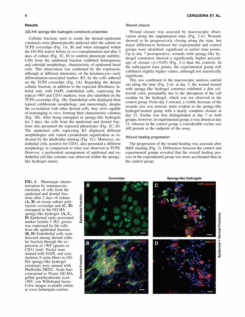
Cellular fractions used to create the dermal–epidermalconstructs were phenotypically analyzed after the culture onTCPS coverslips (Fig. 1A, B) and when entrapped withinthe GG-HA matrix before in vivo transplantation and after 2days of culture (Fig. 1C, D) to confirm phenotype stability.Cells from the epidermal fraction exhibited homogenousand cuboidal morphology, characteristic of epidermal basalcells. This observation was confirmed by the expression,although at different intensities, of the keratinocytes earlydifferentiation-associated marker, K5, by the cells adheredon the TCPS coverslips (Fig. 1A). Regarding the dermalcellular fraction, in addition to the expected fibroblasts, la-beled only with DAPI, endothelial cells, expressing thetypical vWF and CD31 markers, were also identified on theTCPS coverslips (Fig. 1B). Endothelial cells displayed theirtypical cobblestone morphology, and interestingly, despitethe co-isolation with other dermal cells, they were capableof rearranging in vitro, forming their characteristic colonies(Fig. 1B). After being entrapped in spongy-like hydrogelsfor 2 days, the cells from the epidermal and dermal frac-tions also presented the expected phenotypes (Fig. 1C, D).The epidermal cells expressing K5 displayed differentmorphologies and varied cytoskeleton organization as in-dicated by the phalloidin staining (Fig. 1C). Moreover, en-dothelial cells, positive for CD31, also presented a differentmorphology in comparison to what was observed in TCPS.However, a preferential arrangement of epidermal and en-dothelial cell-like colonies was observed within the spongy-like hydrogel matrix.
Wound closure
Wound closure was assessed by macroscopic obser-vation along the implantation time (Fig. 2-ii). Woundsshowed to be progressively closing along the time, butmajor differences between the experimental and controlgroups were identified, significant at earlier time points.Up to day 7 postoperative, wounds with spongy-like hy-drogel constructs showed a significantly higher percent-age of closure ( p < 0.05) (Fig. 2-i) than the controls. Inthe subsequent time points, the experimental group stillexhibited slightly higher values, although not statisticallysignificant.
This was confirmed in the macroscopic analysis carriedout along the time (Fig. 2-ii); at day 3, the wound treatedwith spongy-like hydrogel construct exhibited a dim yel-lowish color, presumably due to the absorption of the cellexudate by the hydrogel, which was not observed in thecontrol group. From day 3 onward, a visible decrease of thewounds size was noticed, more evident in the spongy-likehydrogel-treated group with a nearly complete closure atday 21. Eschar was first distinguished at day 7 in bothgroups; however, in experimental group, it was absent at day21, whereas in the control group, a considerable eschar wasstill present at the endpoint of the assay.
Wound healing progression
The progression of the wound healing was assessed afterH&E staining (Fig. 3). Differences between the control andexperimental groups revealed that the overall healing pro-cess in the experimental group was more accelerated than inthe control group.
FIG. 1. Phenotypic charac-terization by immunocyto-chemistry of cells from theepidermal and dermal frac-tions after 2 days of culture(A, B) on tissue culture poly-styrene coverslips and (C, D)entrapped in the GG-HAspongy-like hydrogel. (A, C,D) Epidermal early-associatedmarker keratin 5 (K5, green)was expressed by the cellsfrom the epidermal fraction.(B, D) Endothelial cells weredetected among dermal cellu-lar fraction through the ex-pression of vWF (green) orCD31 (red). Nuclei werestained with DAPI, and cyto-skeleton F-actin fibers in GG-HA spongy-like hydrogelconstructs were stained withPhalloidin-TRITC. Scale barscorrespond to 50 mm. GG-HA,gellan gum/hyaluronic acid;vWF, von Willebrand factor.Color images available onlineat www.liebertpub.com/tea
4 CERQUEIRA ET AL.

At day 3, granulation tissue formation was almost absentin the control group (Fig. 3A) but was observed in signifi-cant amounts in the experimental group (Fig. 3E). In fact, byday 7, granulation tissue was detected in the control group(Fig. 3B), whereas in the experimental group, neoepidermis(Fig. 3F), identified by the expression of K5 (Fig. 4-i, B),was observed underneath the eschar tissue. Similar findingswere observed in the control group (Fig. 3C) at day 14,although an exuberant eschar was still present. A completere-epithelialization of the wounds was observed at day 14for the experimental group (Fig. 3G) and at day 21 for thecontrol group (Fig. 3D). Nonetheless, epidermal thicknessmeasured at day 21 (Fig. 4E) did not show significant dif-ferences between the experimental and control groups.
The immunohistochemical assessment of K5 and K10expression along the implantation time revealed the pres-ence of K5, but not K10, positive cells both within thespongy-like hydrogel and at the material–wound bed inter-face 3 days after implantation (Fig. 4-i, A), correspondingto the entrapped human cells from the epidermal fraction(Fig. 5). Moreover, the neoepidermis found in the experi-mental group after 7 days of implantation was formed bycells strongly expressing K5 in the basal layer, with somecells expressing K10 on the top layers (Fig. 4-i, B). Similarobservations were found at day 14 (Fig. 4C), although with astronger expression of K10. A fully differentiated epidermisand the presence of newly formed hair follicles, expressingcharacteristic high levels of K5, were identified at day 21
(Fig. 4D). In fact, at day 21, a significantly higher number ofhair follicles ( p < 0.05) were observed in the experimentalgroup compared to the control group (Fig. 4D, F).
The identification of the transplanted human cells fromthe dermal fraction revealed that while cells expressing fi-broblast surface protein were found both within the spongy-like hydrogel and at the material–wound bed interface 3days after implantation (Fig. 5A, B, E), endothelial cells,positive for CD31, were only present within the hydrogel(Fig. 5A, B, F).
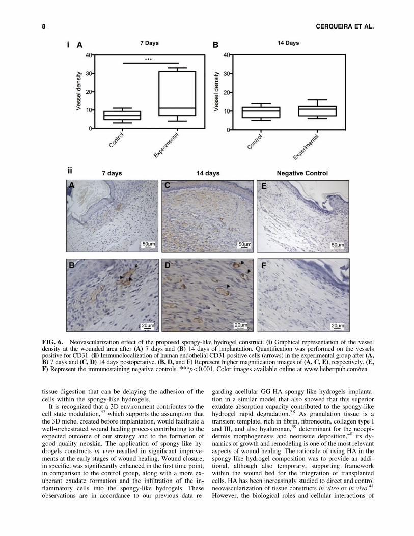
In fact, spongy-like hydrogel constructs promoted a no-torious effect on early neovascularization. A significantlyhigher vessel density ( p < 0.05) was observed at day 7 forthe experimental group in relation to the control group (Fig.6-i, A), which was not detected at day 14 in which bothconditions presented equivalent results regarding the num-ber of vessels in the neotissue (Fig. 6-i, B). At both timepoints, human endothelial cells were detected within theneoformed vasculature forming both human-derived andchimeric vessels (Fig. 6A–D). Moreover, some of thoseneovessels were perfused, as observed by the presence oferythrocytes in their lumen (Fig. 6C, D).
Discussion
An effective approach to rapidly cover wounds and ac-tively accelerate healing, shortening the extensive cellculture and respective substitute preparation period, which
FIG. 2. Evaluation of theeffect of spongy-like hydro-gel constructs over woundclosure. (i) Representation ofthe percentage of woundclosure in the experimentaland control groups after 3, 7,14, and 21 days. (ii) Re-presentative macroscopicimages of the wounds alongthe implantation time.**p < 0.01. Color imagesavailable online at www.liebertpub.com/tea
SKIN CELLS IN A GELLAN GUM/HYALURONIC ACID MATRIX 5

commonly leads to patient complications and decrease ofthe functionality of implanted cells, still is a major clinicaldemand.8 Hence, instead of a full 3D skin biomimeticmodel, we proposed the use of a 3D system based on a cell-adhesive GG-HA spongy-like hydrogel, as an integratedin vitro niche for the self-organization of human epidermaland dermal cellular fractions, used directly from isolation.GG-HA spongy-like hydrogel was chosen, based both onthe importance of HA as main skin ECM component, withan active role in skin remodeling,32,33 and on angiogene-sis.31,34–36 Moreover, spongy-like hydrogels resulted fromthe hydration of off-the-shelf dried networks at the time ofcell seeding, which open the possibility of a ready-to-useapplication. These spongy-like hydrogels were also shownto be easily manipulated upon transplantation and to pos-
sess high ability to shape adaptability, but more impor-tantly, present attractive cell adhesive features thatdistinguish them from traditional hydrogels.27 In fact,spongy-like hydrogels have shown to support the adhesionof different cell types, including human keratinocytes,endothelial cells, and fibroblastic cells.27 Interestingly, thecells obtained from both the epidermal and dermal frac-tions, and entrapped within the GG-HA spongy-like hy-drogel, did not depict the expected morphology after 2days of culture. This behavior can be related to the fact thatcells were entrapped directly from the isolation, withoutany subculture step that permits the selection of the line-ages of interest by adhesion to TCPS under specific cultureconditions. Therefore, the cellular fractions directly fromthe isolation contain other components resultant from the
FIG. 3. Representative images of he-matoxylin and eosin (H&E)-stainedsections of (A–D) the control and(E–H) experimental group explants, atdays 3, 7, 14, and 21 postoperative, re-vealing the wound healing progressionat the centre of the wound. Scale barcorresponds to 50mm. Arrow heads in-dicate the hair-follicles formation. GT,granulation tissue; ES, eschar; NE,neoepidermis. Color images availableonline at www.liebertpub.com/tea
6 CERQUEIRA ET AL.

FIG. 4. Epidermal mor-phogenesis. (i) Expressionprofile of K5 (green) and K10(red) detected by immuno-histochemistry in the experi-mental group at days (A) 3,(B) 7, (C) 14, and (D) 21postimplantation. (A) Thepresence of K5, but not K10,positive cells within thespongy-like hydrogel ( + ) 3days after implantation. (B)Early epidermal formation atday 7 (limited by dashedlines) underneath the eschartissue (#) with (C–D) subse-quent expression of K10 insuprabasal layers and (D) hairfollicles formation (arrow-heads) at the end of the timepoint. (ii) Analysis of neoe-pidermis maturation on boththe control and experimentalgroups, based on H&E-stained samples. Representa-tion of (E) the epidermalthickness and (F) the numberof hair follicles at days 14and 21 postimplantation.**p < 0.01. Nuclei werecounterstained with DAPI,and scale bars correspond to50mm. Color images avail-able online at www.liebertpub.com/tea
FIG. 5. (A, B) Identification of transplanted human cells within spongy-like hydrogel ( + ) by chromogenic in situhybridization at day 3 postoperative. (B) Represents higher magnification of A showing brown nuclei corresponding tohuman cells both inside the spongy-like hydrogel ( + ) and in the tissue that is newly forming (#). (C) Illustrates the negativecontrol of the assay after hematoxylin counterstaining. (D–F) Immunohistochemistry representative images confirming theepidermal (K5-positive cells), endothelial (CD31 expressing cells), and fibroblastic cells positive for fibroblast surfaceprotein (FSP) phenotype of the cells (arrowheads) within the spongy-like hydrogel matrix. Nuclei were stained with DAPI.Color images available online at www.liebertpub.com/tea
7
tissue digestion that can be delaying the adhesion of thecells within the spongy-like hydrogels.
It is recognized that a 3D environment contributes to thecell state modulation,37 which supports the assumption thatthe 3D niche, created before implantation, would facilitate awell-orchestrated wound healing process contributing to theexpected outcome of our strategy and to the formation ofgood quality neoskin. The application of spongy-like hy-drogels constructs in vivo resulted in significant improve-ments at the early stages of wound healing. Wound closure,in specific, was significantly enhanced in the first time point,in comparison to the control group, along with a more ex-uberant exudate formation and the infiltration of the in-flammatory cells into the spongy-like hydrogels. Theseobservations are in accordance to our previous data re-
garding acellular GG-HA spongy-like hydrogels implanta-tion in a similar model that also showed that this superiorexudate absorption capacity contributed to the spongy-likehydrogel rapid degradation.38 As granulation tissue is atransient template, rich in fibrin, fibronectin, collagen type Iand III, and also hyaluronan,39 determinant for the neoepi-dermis morphogenesis and neotissue deposition,40 its dy-namics of growth and remodeling is one of the most relevantaspects of wound healing. The rationale of using HA in thespongy-like hydrogel composition was to provide an addi-tional, although also temporary, supporting frameworkwithin the wound bed for the integration of transplantedcells. HA has been increasingly studied to direct and controlneovascularization of tissue constructs in vitro or in vivo.41
However, the biological roles and cellular interactions of
FIG. 6. Neovascularization effect of the proposed spongy-like hydrogel construct. (i) Graphical representation of the vesseldensity at the wounded area after (A) 7 days and (B) 14 days of implantation. Quantification was performed on the vesselspositive for CD31. (ii) Immunolocalization of human endothelial CD31-positive cells (arrows) in the experimental group after (A,B) 7 days and (C, D) 14 days postoperative. (B, D, and F) Represent higher magnification images of (A, C, E), respectively. (E,F) Represent the immunostaining negative controls. ***p < 0.001. Color images available online at www.liebertpub.com/tea
8 CERQUEIRA ET AL.

HA with cells are HA chain length-dependent. While long-chain HA serves to maintain a highly hydrated environmentand regulate osmotic balance, acting as a lubricant,33,41
small HA oligomers induce inflammatory cytokine releaseby inflammatory cells, which then modulate later woundhealing phases cascade of events.32–36,42 The transplantedhuman cells were detected at early time points, not onlyinside the spongy-like hydrogel but also in the neotissueforming underneath, suggesting their contribution to woundclosure. Moreover, wounds treated with spongy-like hy-drogel constructs showed improved neovascularization atearly time points. After 7 days, the number of vesselspresent in the wounded area was significantly higher in theexperimental condition than in the controls, and more im-portantly, human-derived and chimeric vessels were de-tected at days 7 and 14, showing the direct contribution ofthe transplanted endothelial cells.
Despite the observed results regarding the enhancedeffect over wound closure, re-epithelialization, hair-follicleformation, and neovascularization/angiogenesis in thepresence of the GG-HA spongy-like hydrogels, the hy-pothesis herein proposed of using this as a template to in-duce self-organization of human skin cells, turned out to beunrealistic. In fact, the human transplanted cells were onlydetected at early time points; therefore, we consider thatGG-HA spongy-like hydrogel failed in providing an ade-quate framework for that purpose. The rapid degradation ofthe implanted constructs, as described in our previouswork,38 did not allow to fully take advantage of the type ofcells within both the epidermal and dermal fractions and ofthe multiple communication routes known to occur betweenthem.43 Nonetheless, our observations meet the intermedi-ate formation of epidermis and later hair follicle detection,as observed by Lee et al.,13 but, in addition to the useof newborn mice cells instead of human cells, no dataregarding the origin of the organized structures formedwere reported.
Thus, the herein proposed approach failed in providingthe necessary conditions to allow dissociated human epi-dermal and dermal cellular fractions to self-organize alongthe wound healing process. Nonetheless, the GG-HAspongy-like hydrogel acted as a suitable supporting matrixfor the transplanted cells during the early time points al-lowing them to contribute to the observed early wounds re-epithelialization and neovascularization.
Our strategy proposes a combination of a GG-HA spon-gy-like hydrogel and freshly dissociated skin relevant line-ages for the promotion of cells self-organization in vivo. Thegenerated construct was able to accelerate the wound heal-ing process, successfully promoting earlier re-epithelializa-tion, increased formation of hair follicle appendages, andan overall enhanced re-vascularization at early time pointsthat assured the nourishment of neoformed tissue. However,this system did not sustain the hypothesized self-organiza-tion of entrapped cells, possibly due to the suggested rapiddegradation.
Acknowledgments
We thank the Hospital da Prelada (Porto), in particularDr. Paulo Costa for lipoaspirates collection and to financialsupport by Skingineering (PTDC/SAU-OSM/099422/2008),
Portuguese Foundation for Science and Technology (FCT)-funded project. The research leading to these results has alsoreceived funding from the European Union’s Seventh Fra-mework Programme (FP7/2007–2013) under grant agree-ment no. REGPOT-CT2012-316331-POLARIS.
Disclosure Statement
No competing financial interests exist.
References
1. Schiestl, C., Stiefel, D., and Meuli, M. Giant naevus, giantexcision, eleg(i)ant closure? Reconstructive surgery withIntegra Artificial Skin to treat giant congenital melanocyticnaevi in children. J Plast Reconstr Aesthet Surg 63, 610,2009.
2. Haslik, W., Kamolz, L.P., Nathschlager, G., Andel, H.,Meissl, G., and Frey, M. First experiences with the colla-gen-elastin matrix Matriderm as a dermal substitute in se-vere burn injuries of the hand. Burns 33, 364, 2007.
3. Waymack, P., Duff, R.G., and Sabolinski, M. The effect ofa tissue engineered bilayered living skin analog, over me-shed split-thickness autografts on the healing of excisedburn wounds. The Apligraf Burn Study Group. Burns 26,609, 2000.
4. Marston, W.A., Hanft, J., Norwood, P., and Pollak, R. Theefficacy and safety of Dermagraft in improving the healingof chronic diabetic foot ulcers: results of a prospectiverandomized trial. Diabetes Care 26, 1701, 2003.
5. Butler, C.E., and Orgill, D.P. Simultaneous in vivo regen-eration of neodermis, epidermis, and basement membrane.Adv Biochem Eng Biotechnol 94, 23, 2005.
6. Supp, D.M., and Boyce, S.T. Engineered skin substitutes:practices and potentials. Clin Dermatol 23, 403, 2005.
7. Supp, D.M., Wilson-Landy, K., and Boyce, S.T. Humandermal microvascular endothelial cells form vascular ana-logs in cultured skin substitutes after grafting to athymicmice. FASEB J 16, 797, 2002.
8. Bottcher-Haberzeth, S., Biedermann, T., and Reichmann,E. Tissue engineering of skin. Burns 36, 450, 2009.
9. Enoch, S., Roshan, A., and Shah, M. Emergency and earlymanagement of burns and scalds. BMJ 338, b1037, 2009.
10. Horch, R.E., Kopp, J., Kneser, U., Beier, J., and Bach, A.D.Tissue engineering of cultured skin substitutes. J Cell MolMed 9, 592, 2005.
11. Gurtner, G.C., Werner, S., Barrandon, Y., and Longaker,M.T. Wound repair and regeneration. Nature 453, 314, 2008.
12. Aarabi, S., Longaker, M.T., and Gurtner, G.C. Hyper-trophic scar formation following burns and trauma: newapproaches to treatment. PLoS Med 4, e234, 2007.
13. Lee, L.F., Jiang, T.X., Garner, W., and Chuong, C.M. Asimplified procedure to reconstitute hair-producing skin.Tissue Eng Part C Methods 17, 391, 2010.
14. Sun, T., Mai, S., Norton, D., Haycock, J.W., Ryan, A.J.,and MacNeil, S. Self-organization of skin cells in three-dimensional electrospun polystyrene scaffolds. Tissue Eng11, 1023, 2005.
15. Delvoye, P., Pierard, D., Noel, A., Nusgens, B., Foidart,J.M., and Lapiere, C.M. Fibroblasts induce the assembly ofthe macromolecules of the basement membrane. J InvestDermatol 90, 276, 1988.
16. Konig, A., and Bruckner-Tuderman, L. Epithelial-mesen-chymal interactions enhance expression of collagen VIIin vitro. J Invest Dermatol 96, 803, 1991.
SKIN CELLS IN A GELLAN GUM/HYALURONIC ACID MATRIX 9

17. Havlickova, B., Biro, T., Mescalchin, A., Tschirschmann,M., Mollenkopf, H., Bettermann, A., et al. A human folli-culoid microsphere assay for exploring epithelial- mesen-chymal interactions in the human hair follicle. J InvestDermatol 129, 972, 2009.
18. Ahmed, T.A., Dare, E.V., and Hincke, M. Fibrin: a versatilescaffold for tissue engineering applications. Tissue EngPart B Rev 14, 199, 2008.
19. Sanders, J.E., Stiles, C.E., and Hayes, C.L. Tissue responseto single-polymer fibers of varying diameters: evaluation offibrous encapsulation and macrophage density. J BiomedMater Res 52, 231, 2000.
20. Ng, K.W., Hutmacher, D.W., Schantz, J.T., Ng, C.S., Too,H.P., Lim, T.C., et al. Evaluation of ultra-thin poly(epsilon-caprolactone) films for tissue-engineered skin. Tissue Eng7, 441, 2001.
21. van Dorp, A.G., Verhoeven, M.C., Koerten, H.K., vanBlitterswijk, C.A., and Ponec, M. Bilayered biodegradablepoly(ethylene glycol)/poly(butylene terephthalate) copoly-mer (Polyactive) as substrate for human fibroblasts andkeratinocytes. J Biomed Mater Res 47, 292, 1999.
22. Wong, V.W., Rustad, K.C., Galvez, M.G., Neofytou, E.,Glotzbach, J.P., Januszyk, M., et al. Engineered pullulan-collagen composite dermal hydrogels improve early cuta-neous wound healing. Tissue Eng Part A 17, 631, 2010.
23. Tran, N.Q., Joung, Y.K., Lih, E., and Park, K.D. In situforming and rutin-releasing chitosan hydrogels as injectabledressings for dermal wound healing. Biomacromolecules12, 2872, 2011.
24. Sun, G., Zhang, X., Shen, Y.I., Sebastian, R., Dickinson,L.E., Fox-Talbot, K., et al. Dextran hydrogel scaffoldsenhance angiogenic responses and promote complete skinregeneration during burn wound healing. Proc Natl AcadSci U S A 108, 20976, 2011.
25. Luo, Y., Diao, H., Xia, S., Dong, L., Chen, J., and Zhang,J. A physiologically active polysaccharide hydrogelpromotes wound healing. J Biomed Mater Res A 94, 193,2010.
26. Khademhosseini, A., and Langer, R. Microengineered hy-drogels for tissue engineering. Biomaterials 28, 5087, 2007.
27. da Silva, L.C., Cerqueira, M.T., Marques, A.P., Correlo,V.M., Sousa, R.A., and Reis, R.L. Gellan Gum-basedspongy-like hydrogels: methods and biomedical applica-tions thereof. Portugal. Provisional Patent 106890. 2013.
28. Oliveira, J.T., Martins, L., Picciochi, R., Malafaya, P.B.,Sousa, R.A., Neves, N.M., et al. Gellan gum: a new bio-material for cartilage tissue engineering applications. JBiomed Mater Res A 93, 852, 2009.
29. Wicke, C., Halliday, B., Allen, D., Roche, N.S., Scheuen-stuhl, H., Spencer, M.M., et al. Effects of steroids andretinoids on wound healing. Arch Surg 135, 1265, 2000.
30. Ananta, M., Brown, R.A., and Mudera, V. A rapid fabri-cated living dermal equivalent for skin tissue engineering:an in vivo evaluation in an acute wound model. Tissue EngPart A 18, 353, 2011.
31. Liu, S., Zhang, H., Zhang, X., Lu, W., Huang, X., Xie, H.,et al. Synergistic angiogenesis promoting effects ofextracellular matrix scaffolds and adipose-derived stemcells during wound repair. Tissue Eng Part A 17, 725,2011.
32. Price, R.D., Myers, S., Leigh, I.M., and Navsaria, H.A. Therole of hyaluronic acid in wound healing: assessment ofclinical evidence. Am J Clin Dermatol 6, 393, 2005.
33. Kirker, K.R., Luo, Y., Nielson, J.H., Shelby, J., and Prestwich,G.D. Glycosaminoglycan hydrogel films as bio-interactivedressings for wound healing. Biomaterials 23, 3661, 2002.
34. Tonello, C., Vindigni, V., Zavan, B., Abatangelo, S.,Abatangelo, G., Brun, P., et al. In vitro reconstruction of anendothelialized skin substitute provided with a micro-capillary network using biopolymer scaffolds. FASEB J 19,1546, 2005.
35. Gao, F., Liu, Y., He, Y., Yang, C., Wang, Y., Shi, X., et al.Hyaluronan oligosaccharides promote excisional woundhealing through enhanced angiogenesis. Matrix Biol 29,107, 2010.
36. Ibrahim, S., and Ramamurthi, A. Hyaluronic acid cues forfunctional endothelialization of vascular constructs. J Tis-sue Eng Regen Med 2, 22, 2008.
37. Green, J.A., and Yamada, K.M. Three-dimensional micro-environments modulate fibroblast signaling responses. AdvDrug Deliv Rev 59, 1293, 2007.
38. Cerqueira, M., da Silva, L.P., Santos, T.C., Pirraco, R.P.,Martins, A.R., Correlo, V.M., Marques, A.P., and Reis,R.L. Pre-vascularized Gellan Gum-Hyaluronic AcidSpongy-like Hydrogels improve Skin wound healing. Pre-sented at the Annual Meeting of the Society For Bioma-terials 2013, Boston, USA.
39. Witte, M.B., and Barbul, A. General principles of woundhealing. Surg Clin North Am 77, 509, 1997.
40. Singer, A.J., and Clark, R.A. Cutaneous wound healing. NEngl J Med 341, 738, 1999.
41. Pardue, E.L., Ibrahim, S., and Ramamurthi, A. Role ofhyaluronan in angiogenesis and its utility to angiogenictissue engineering. Organogenesis 4, 203, 2008.
42. Noble, P.W. Hyaluronan and its catabolic products in tissueinjury and repair. Matrix Biol 21, 25, 2002.
43. Lugo, L.M., Lei, P., and Andreadis, S.T. Vascularization ofthe dermal support enhances wound re-epithelialization byin situ delivery of epidermal keratinocytes. Tissue Eng PartA 17, 665, 2010.
Address correspondence to:Alexandra P. Marques, PhD
3B’s Research Group—Biomaterials,Biodegradables and Biomimetics
University of MinhoHeadquarters of the European Institute of Excellence
on Tissue Engineering and Regenerative MedicineAvePark
Zona Industrial da GandraS. Claudio do Barco
4806-909 Caldas das TaipasGuimaraes
Portugal
E-mail: [email protected]
Received: July 26, 2013Accepted: November 25, 2013
Online Publication Date: January 16, 2014
10 CERQUEIRA ET AL.


