Embed Size (px)
Citation preview

13
OMICS A Journal of Integrative Biology Volume 9, Number 1, 2005© Mary Ann Liebert, Inc.
Identification of Putative Sulfurtransferase Genes in theExtremophilic Acidithiobacillus ferrooxidans ATCC
23270 Genome: Structural and Functional Characterization of the Proteins
MAURICIO ACOSTA, SIMON BEARD, JOSE PONCE, MARIO VERA, JUAN C. MOBAREC, and CARLOS A. JEREZ
ABSTRACT
Eight nucleotide sequences containing a single rhodanese domain were found in theAcidithiobacillus ferrooxidans ATCC 23270 genome: p11, p14, p14.3, p15, p16, p16.2, p21,and p28. Amino acids sequence comparisons allowed us to identify the potentially catalyticCys residues and other highly conserved rhodanese family features in all eight proteins. Thegenomic contexts of some of the rhodanese-like genes and the determination of their ex-pression at the mRNA level by using macroarrays suggested their implication in sulfur ox-idation and metabolism, formation of Fe-S clusters or detoxification mechanisms. Several ofthe putative rhodanese genes were successfully isolated, cloned and overexpressed in E. coliand their thiosulfate:cyanide sulfurtransferase (TST) and 3-mercaptopyruvate/cyanide sul-furtransferase (MST) activities were determined. Based on their sulfurtransferase activitiesand on structural comparisons of catalytic sites and electrostatic potentials between homol-ogy-modeled A. ferrooxidans rhodaneses and the reported crystal structures of E. coli GlpE(TST) and SseA (MST) proteins, two of the rhodanese-like proteins (P15 and P16.2) couldclearly be defined as TSTs, and P14 and P16 could possibly correspond to MSTs. Never-theless, several of the eight A. ferrooxidans rhodanese-like proteins may have some differ-ent functional activities yet to be discovered.
INTRODUCTION
SULFURTRANSFERASES ARE ALMOST UBIQUITOUS ENZYMES capable of catalyzing in vitro the transfer of asulfane sulfur atom from a donor to an appropriate sulfur acceptor. According to the sulfur donor, these
enzymes belong to the thiosulfate:cyanide sulfurtransferase family (TSTs) or the 3-mercaptopyruvate sul-furtransferases (MSTs) family (Spallarossa et al., 2004; Bordo and Bork, 2002). The rhodanese type pro-teins are composed of a single domain or in combination with distinct protein domains (Koonin et al., 2000).An increasing number of reports indicate that rhodanese modules are versatile sulfur carriers that have
Laboratory of Molecular Microbiology and Biotechnology, and Millennium Institute for Advanced Studies in CellBiology and Biotechnology (CBB), Department of Biology, Faculty of Sciences, University of Chile, Santiago, Chile.

adapted their function to fulfill the need for reactive sulfane sulfur in distinct metabolic and regulatory path-ways. Rhodaneses together with the Cdc25 family of protein tyrosine phosphatases form a superfamily(Bordo and Bork, 2002) in which most of the proteins contain the predicted active cysteine that covalentlybinds the transferred anion in both rhodanese and tyrosine phosphatases (Spallarossa et al., 2001; Bordoand Bork, 2002). Many of these proteins have been biochemically and structurally characterized, and sev-eral functional roles have been proposed for them, such as cyanide detoxification, sulfur insertion in thebiosynthesis and/or repair of iron-sulfur clusters, involvement in sulfur metabolism, and interaction withthioredoxin (Westley, 1981; Bonomi et al., 1985; Pagani et al., 1984; Nandi and Westley, 1998). Recentevidence indicates a physiological role of a rhodanase-like protein in the biosynthesis of the molybdenumcofactor in humans (Matthies et al., 2004). However, the identity of true biological substrates—proteins orsmall molecules that interact with the rhodanese domains—is still an open question.
Acidithiobacillus ferrooxidans is a chemolithoautotrophic bacterium that obtains its energy from the ox-idation of ferrous iron, elemental sulfur, or partially oxidized sulfur compounds (Rohwerder et al., 2003;Rawlings, 2002; Olson et al., 2003). The ability of this and other microorganisms present in its habitat tosolubilize metal sulfides is succesfully applied in industrial biomining operations in many countries (Ro-hwerder et al., 2003; Rawlings, 2002; Olson et al., 2003). This biotechnology is especially important inChile (Gentina and Acevedo, 1985; Acevedo et al., 1993), since this country is the principal copper pro-ducer in the world, generating about 400,000 ton of copper per year by using biomining. It is expectedthat the process could be improved by knowing the mechanisms by which the microorganisms attack andsolubilize the ores. Recently, it has been proposed that pyrite (FeS2) and other metal sulfides are degradedby an indirect mechanism generating thiosulfate as the main intermediate (Schipers and Sand, 1999). Iron(III) ions are exclusively the oxidizing agents for the dissolution. Thiosulfate would be consequently de-graded in a cyclic process to sulfate, with elemental sulfur being a side product. This explains why onlyFe(II) ion–oxidizing bacteria are capable of oxidizing these metal sulfides (Schipers and Sand, 1999). Inaddition, enzymes for thiosulfate or sulfite oxidation from A. ferrooxidans or A. thiooxidans may suc-cessfully compete with the chemical reactions with iron (III) ions as an oxidizing agent (Schipers andSand, 1999).
A rhodanese activity has been previously reported in A. ferrooxidans (Tabita et al., 1969). This enzymeis a TST, which breaks the S-S bond present in thiosulfate, generating sulfur and sulfite. Other enzymesmay also participate in the mechanism proposed by Schipers and Sand (1999), such as the thiosulfate-oxi-dizing enzyme of A. ferrooxidans (Silver and Lundgren, 1968).
By proteomic analysis, we have previously studied the global changes in gene expression of A. ferrooxi-dans when the microorganism was grown under different conditions (Ramírez et al., 2002, 2004). We iden-tified an exported rhodanese-like protein (P21) whose levels are increased when A. ferrooxidans is grownon metal sulfides and different sulfur compounds but is almost entirely absent during growth on ferrousiron (Ramírez et al., 2002, 2004). Unlike cytoplasmic rhodaneses, P21 was located in the periphery of A.ferrooxidans cells and was regulated depending on the oxidizable substrate. By using the available genomicsequence of A. ferrooxidans ATCC 23270, the genomic context around gene p21 showed the presence ofother ORFs corresponding to proteins such as sulfate-thiosulfate binding proteins, clearly suggesting the in-volvement of P21 in inorganic sulfur metabolism in A. ferrooxidans (Ramírez et al., 2002, 2004). Here, weextend our analysis and define seven new rhodanese-like proteins in the genome of A. ferrooxidans. Sev-eral of the genes coding for these proteins were isolated, and after their cloning and expression in E. coli,their functional sulfurtransferase activity was determined. The results showed that both TSTs and MSTsproteins are present in A. ferrooxidans, suggesting that the rhodanese domain may be of great significancefor the chemolithoautotrophic metabolism of this acidophilic bacterium.
MATERIALS AND METHODS
Bacterial strains and growth conditions
A. ferrooxidans strains ATCC 19859 or ATCC 23270 were grown in ferrous iron–containing modified9K medium, or in sulfur or thiosulfate as described before (Ramirez at al., 2002, 2004). E. coli strain
ACOSTA ET AL.
14

BL21(DE3) containing plasmid pGZ105 with the glpE insert coding for the E. coli GlpE rhodanese (Rayet al., 2000) was a kind gift of T. Larson. E. coli strains BL21(DE3), DH5-� and derivatives were grownin LB medium (Sambrook and Russel, 2001).
Primer design and PCR amplification
The oligonucleotide primer sequences were deduced from the ORFs found in the available DNA genomicsequence of A. ferrooxidans strain ATCC 23270 (www.tigr.org), and the primers were made by Genset Cor-poration. Primers used to amplify the DNA fragments to be used in macroarrays were chosen to have melt-ing temperatures (Tm) of 50–56°C and were designed with the program Web-Primer (http://seq.yeastgenome.org/cgi-bin/web-primer). The PCR products obtained were between 230 and 780 base pairslong. The amplification and reamplification conditions were similar for all the genes. They consisted of 30cycles of 30 sec at 94°C, 30 sec at 50–54°C, 1 min at 72°C followed by 3 min at 72°C. The reamplifica-tion reaction was done with a 1/50 dilution of the first PCR product as template. All PCR products result-ing from the reamplification were purified by gel electrophoresis followed by Ultra-Free DNA purificationcolumns (Millipore). The products were diluted to a final concentration of 5 ng/�L in 40% DMSO.
The oligonucleotide primers for molecular cloning of the rhodanese-like genes were as follows:
P11NTER-NdeI (5�-CATATgTACggATTTCAggAAATC-3�)P11CTER (5�-TCAACTggACAACTggACCAAg-3�)P14NTER-NdeI (5�-gTTTTTAgTCATATggggAAggTCATgg-3�)P14CTER-XhoIHT (5�-TAggCTCCggCTCTCgAgggAAACgAC-3�)P15NTER-NdeI (5�-CATATgACAAAgTCggTAATAAACC-3�)P15CTER (5�-TTATATTTgCTCCCAgggTAgTC-3�)P16NTER-NdeI (5�-CATATggACCATAAAATTCTTTTCTT-3�)P16CTER (5�-TCACTTCTCTACCggCAgACC-3�)P16.2NTER-NdeI (5�-CATATgAgTACCgAAACgATTTTg-3�)P16.2CTER (5�-CTACTTgTTCTTCCAgggCAgg-3�)
To amplify the rhodanese-like genes, we used a two-step HotPCR protocol: 3 min at 95°C followed by20 cycles at 95°C for 30 sec, 52–60°C for 30 sec and 45 sec at 72°C and 72°C for 3 min.
Gene cloning and expression
Restriction enzyme digestions, T4 DNA ligase and recombinant DNA techniques were carried out ac-cording to standard laboratory procedures (Sambrook and Russel, 2001). DNA fragments were automati-cally sequenced by means of a 3100-Avant Genetic Analyzer (Applied Biosystems).
The genes of interest were obtained by PCR using the primers corresponding to the N-terminal and C-terminal end sequences of each of the rhodanese-like proteins and were directly cloned in the plasmidpCRT7/NT-TOPO (Invitrogen), except in the case of p14, which was cloned using pGEM-T (Promega) andthe pET System (Novagen). The reaction products were used to transform E. coli DH5-�. The recombinantclones were analyzed by using colony PCR and the corresponding plasmids with inserts in the correct ori-entations were purified and used to transform E. coli BL21 (DE3) for expression of the rhodanese-like pro-teins from A. ferrooxidans. The recombinant clones were selected on LB solid medium supplemented withampicillin (100 �g/mL). The induction/expression analysis was done in the presence or absence of 1 mMIPTG, added when the cultures reached an OD600 of 0.6, and the expressions of all the recombinant cloneswere analyzed by SDS-PAGE.
Macroarray expression analysis
Membranes were prepared by spotting the DNA probes manually by using the Colony copier VP381(V&P Scientific). The PCR products were printed onto Inmobilon-NY+ membranes (Millipore). A DNAfragment coding for human tau1 protein was used as a heterologous control DNA. Each spot was printedin quadruplicate three times, to a final concentration of 15 ng of DNA per spot. The membranes were air-dried, and treated with denaturation solution (0.5 M NaOH, 1.5 M NaCl) for 7 min. After this treatment,
PUTATIVE SULFURTRANSFERASE GENES
15

the membranes were incubated twice with neutralization solution (0.5 M Tris–HCl, pH 8, 1.5 M NaCl, 1mM EDTA) for 3 min each, and the DNA was UV-crosslinked to the membranes.
Total RNA was prepared from A. ferrooxidans cultures by a hot phenol method essentially as describedby Guiliani et al. (1997). Radioactively labeled cDNAs were generated by reverse transcription of 25 �gof total RNA using ImPromII reverse transcriptase (Promega), a mixture of specific oligonucleotides and�-[32P]dCTP. In vitro transcribed “spiked” control RNA (exp1 from Prunus persica) was added as a con-trol labeling reaction. The RNA template was degraded by alkaline treatment and the probe was purifiedusing Microspin S-200 columns (Amersham Pharmacia Biotech). The hybridization was done overnight inhybridization solution (5 � SSPE, 2% SDS, 1 � Dendhardt’s reagent and 100 µg of sheared salmon spermDNA/mL) with 3 � 106 cpm/mL of freshly heat-denatured probe, after a 2-h prehybridization in the samebuffer at 65°C. Stringent washes were performed with 0.5 � SSPE, 0.2% SDS at 65°C. Exposed Phos-phorImager screens were scanned on a PhosphorImager (Molecular Imager FX™ Sistems, BioRad) at aresolution of 50 �m/pixel. The analysis and quantification of the spots signals was done with VersArray1.0 software (BioRad). The results were very reproducible from membrane to membrane, and spots in gen-eral upregulated or downregulated at least threefold were considered valid changes.
Bioinformatic and structural analysis
Identity/similarity searching in databases was done by using the BLASTp program (Altschul et al., 1997)from NCBI (www.ncbi.nlm.nih.gov) and from the A. ferrooxidans ATCC 23270 genome site (www.tigr.org).Multiple alignments, molecular masses, and isoelectric points of ORFs, the presence of transmembrane do-mains in the analyzed ORFs, and the putative functions and predicted subcellular locations of the proteinscoded by the different ORFs were analyzed as described before (Ramirez et al., 2004).
Homology models construction, evaluation, and energy minimization: nine models were built for eachof the eight A. ferrooxidans rhodanese-like sequences with the MODELLER program (Sali and Blundell,1993). The GlpE protein structure from E. coli, Protein DataBank (PDB; Berman et al., 2000) entry 1GN0,determined by x-ray diffraction at 1.8 Å (Spallarossa et al., 2001) was used as template for the modelingof P11, P15, P16, P16.2, P21 and P28. The chain B of polysulfide-sulfur transferase (Sud) from Wolinellasuccinogens (PDB; Berman et al., 2000) entry 1QXN, determined by NMR (Lin et al., 2004) was used astemplate for P14 modeling. The structure of AT5G6040, the putative senescence-associated family proteinfrom A. thaliana (PDB; Berman et al., 2000) entry 1TQ1, determined by NMR was used as template forP14.3.
All the models and their respective templates were evaluated with the VERIFY-3D program (Lüthy etal., 1992). The model selection criterion was based on the highest model profile score that best matched tothe template profile. Since homology modeling puts emphasis on backbone atoms, side chains were relaxedto a lower energy state to avoid clashes of their atoms that could distort the model. Thus, for each protein,the model with similar profile score to that of the template was selected for energy minimization with ver-sion 3.0 of the GROMACS molecular dynamics simulation package (Lindahl et al., 2001). Each model wasimmersed in the center of a triclinic box, with explicit water molecules. Ions were added to ensure systemelectroneutrality, and energy minimized with a steepest descent algorithm. The energy-minimized modelswere evaluated again with VERIFY-3D and showed similar scoring profiles to those of the respective tem-plates (data not shown). The models for each protein showed only positive values in their evaluation, sug-gesting a good three-dimensional fold. Later, these models were used for analysis, and some are shown inthe figures.
Electrostatic potential projected over the solvent-accessible surface was computed with the linearizedPoisson-Boltzmann equation from Swiss PDB viewer (Guex and Peitsch, 1997) and was computed takinginto account protein partial charges. Dielectric constants for water and protein interior were set to 80 and4, respectively. Ionic strength was set to a default value of zero. A color gradient from blue to white to redis used to color the molecular surface, where blue, red, and white are for positive, negative, and neutral po-tentials, respectively, according to the given cutoff values of 1.8 kT/e for blue, �1.8 kT/e for red, and 0.0kT/e for white. Since Figure 4 (below) is printed in black and white, blue appears as dark gray and red aslight gray.
ACOSTA ET AL.
16

Determination of sulfurtransferase activity
Thiosulfate/cyanide sulfurtransferase (TST, EC 2.8.1.1) activity was assayed in crude enzyme extractsfrom all E. coli recombinant clones or in cell-free extracts from A. ferrooxidans grown under different con-ditions. As controls, we used the recombinant rhodanese GlpE from E. coli (Ray et al., 2000) and the E.coli cells containing the plasmid vector without the DNA insert. Assays contained 100 mM Tris-acetate(pH 8.6), 50 mM ammonium thiosulfate, 50 mM KCN, and 100–150 �g of protein enzyme extract in a fi-nal volume of 0.5 mL. Reactions were initiated by the addition of KCN and terminated, after 0.5–2 min,by the addition of 0.25 mL of 15% formaldehyde. Color was developed by the addition of 0.75 mL of fer-ric nitrate reagent [100 g of Fe(NO3)3�9H2O and 200 mL of 65% HNO3 per 1,500 mL]. Assays were clar-ified by centrifugation, and the absorbance at 460 nm was determined (Westley, 1981). One unit of enzymewas defined as the amount that catalyzes the production of 1 �mol of thiocyanate per min.
3-Mercaptopyruvate/cyanide sulfurtransferase (MST, EC 2.8.1.2) activity was measured by the rate ofpyruvate formation by using the method of Jarabak (1981). The assay mixture contained 5 mM sodium 3-mercaptopyruvate, 25 mM mercaptoethanol, 225 mM 2-methyl-2-aminopropanediol buffer, pH 9.55, andenzyme solution in a final volume of 1.5 mL. The absorbance at 460 nm was measured 35 min after theaddition of the 2,4-dinitrophenyl hydrazine reagent. One unit of activity was defined as the amount of en-zyme that catalyzes the production of 1 �mol of pyruvate per min in this assay system.
Nuclotide sequence accession numbers
The nuclotide sequences of the p15 and p16.2 genes are available in the GenBank database under the accession numbers AY863107 and AY863108, respectively.
RESULTS
Search in the genome sequence of A. ferrooxidans for putative rhodanese genes and their genomic contexts
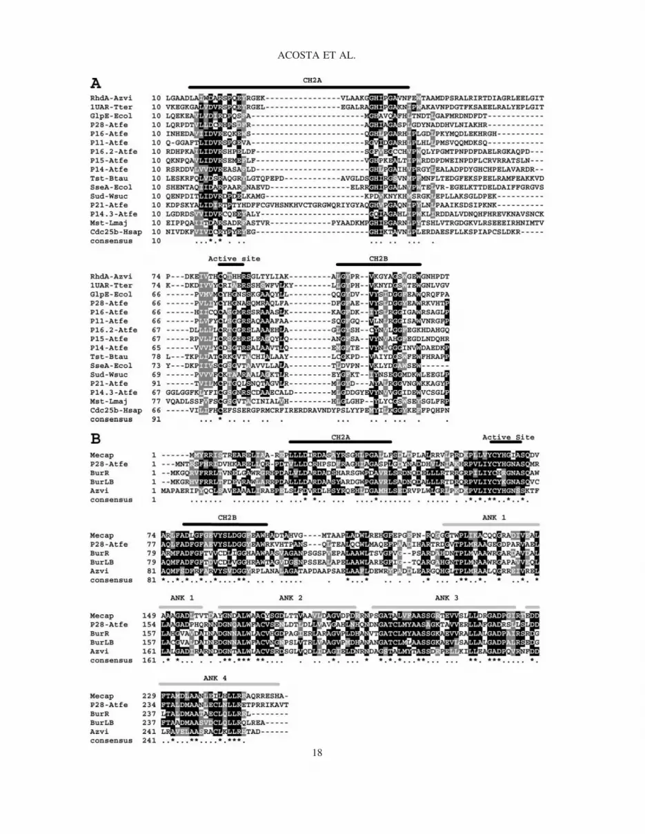
In the A. ferrooxidans genome sequence, we found eight nucleotide sequences coding for putative pro-teins with similarities to rhodanese-like domains: P11, P14, P14.3, P15, P16, P16.2, P28, and the previ-ously described P21 (Ramirez et al., 2002). Figure 1 shows the amino acid sequence alignments of all theseputative rhodanese-like proteins with several known thiosulfate-sulfur transferases whose activity has beendemonstrated in vitro. All the proteins showed a significant similarity, containing the highly conserved struc-tural domains CH2A, CH2B (Fauman et al., 1998) and a catalytic site with a Cys, typical of thiosulfate-sulfur transferases (Fig. 1A). Two other amino acid residues, Asp19 and Arg21 (GlpE numbering is used),which are conserved in all these enzymes and in most other rhodanese domains (Bordo et al., 2000), werealso present (not shown). In all known rhodanese and Cdc25 phosphatase structures, these residues form ahydrogen-bonded salt bridge, an interaction that may contribute substantially to the overall free energy ofthe rhodanese-like domain (Spallarossa et al., 2001).
P28 protein was the largest A. ferrooxidans rhodanese and contained, in addition to the rhodanese do-main, the ankyrin (ANK) repeat, a common protein sequence motif which allows the proteins containingthem to have inter- and/or intramolecular contacts with different macromolecular targets (Sedgwick andSmerdon, 1999). Figure 1B shows an alignment between the amino acid sequences of P28 from A. fer-rooxidans and the only four other proteins found in the database to contain both the rhodanese domain andthe ANK repeats. All of the proteins shown in Figure 1B contained four ANK repeats in the C-terminalportion of their sequences, in agreement with the numbers of repeats described for many other proteins(Sedgwick and Smerdon, 1999). The role of ANK repeats in mediating protein–protein interactions has beenwell documented, and their presence is often interpreted as an indicator of a similar function in otherwiseuncharacterized systems (Sedgwick and Smerdon, 1999). Unfortunately, the proteins with amino acid se-quences similar to P28 in Figure 1B are all hypothetical proteins with unknown functions.
Some properties of eight putative rhodaneses from A. ferrooxidans are shown in Table 1. The molecularmasses ranged from 11.7 to 28.6 kDa, and the isolectric points were very basic in P11, P16, and P21. The
PUTATIVE SULFURTRANSFERASE GENES
17
ACOSTA ET AL.
18

other rhodanese-like proteins had isoelectric points ranging between 4.75 and 6.49. Only P16 and P21showed transmembrane domains, suggesting their insertion in the cytoplasmic membrane (P16) or periplas-mic location, anchored on the periplasmic side of the cytoplasmic membrane, as we suggested before forP21 (Ramirez et al., 2002). The rest of the proteins are expected to be located in the cytoplasm. All pro-teins contained the rhodanese domain, and as already mentioned, P28 contained in addition the ANK re-peats (Sedgwick and Smerdon, 1999).
At least two polar (often charged) residues are observed within the six-residue active site loop of TSTenzymes (Spallarossa et al., 2001). In contrast, Bordo and Bork (2002) note that no charged residues areobserved in biochemically characterized MSTs. Applying these criteria, one could predict that P15, P16.2,and P28 could be TSTs. On the other hand, P11, P14, P14.3, P16, and P21 could be putative MSTs (Table1). However, the amino acid sequences of their active site loops do not match entirely with the highly con-served CG(S/T)GVT sequence found in the known MSTs (Spallarossa et al., 2004).
As pointed out by Spallarossa et al. (2001), the use of bioinformatic approaches based on recurring in-stances of neighboring genes in distinct genomes will likely provide a more definitive answer to the bio-logical roles and substrates of the rhodanese protein superfamily.
Figure 2 shows the genomic analysis of the regions surrounding all the A. ferrooxidans rhodanese-likegenes. Upstream of p11, a molybdenum cofactor biosynthesis enzyme (moaA) was located in the same ori-entation. Downstream of p11, five ORFs that could be forming a cluster were present: a predicted redoxprotein, regulator of disulfide bond formation (sirA), an uncharacterized conserved protein (hyp1), and threeheterodisulfide reductase subunits, subunit C (hdrC), subunit B (hdrB), and subunit A (hdrA). It is knownthat the precursor Z of molybdopterin (MPT) is synthesized by MoaA, MoaB, and MoaC proteins (Hänzel-mann and Schindelin, 2004). MPT forms part of the catalytic site of several redox enzymes such as poly-
PUTATIVE SULFURTRANSFERASE GENES
19
FIG. 1. (A) Partial amino acid sequence alignments of the rhodanese-like proteins of Acidithiobacillus ferrooxidanswith rhodaneses of known structures. The active site and two conserved structural motifs, designated CH2A and CH2B,are labeled with thick black lines on the top of the alignments. RhdA-Azvi, Azotobacter vinelandii; 1UAR-Tter, rho-danese from Thermus thermophilus; GlpE-Ecol, TST from E. coli; Tst-Btau, TST from Bos taurus; SseA-Ecol, MSTfrom E. coli; Sud-Wsuc, polysulfide sulfurtransferase (PST) from Wolinella succinogenes; Mst-Lmaj, MST from Leish-mania major; Cdc25b-Hsap, CdcC25 phosphatase from Homo sapiens; rhodanese-like proteins of A. ferrooxidans areP11–Atfe, P14-Atfe, P14.3-Atfe, P15-Atfe, P16-Atfe, P16.2-Atfe, P21-Atfe, and P28-Atfe. (B) Sequence alignments ofP28 from A. ferrooxidans with proteins possessing similar domains organizations. The P28 protein sequence containeda rhodanese domain and 4 ANK repeats, indicated by light gray thick lines on the top of the alignments. BurLB, Burk-holderia fungorum LB400; Mecap, Methylococcus capsulatus str. Bath; Azvi, Azotobacter vinelandii; BurR, Burk-holderia cepacia R1808.
TABLE 1. PROPERTIES OF THE PUTATIVE RHODANESE-LIKE PROTEINS
FOUND IN THE ACIDITHIOBACILLUS FERROOXIDANS GENOME
PredictedMW transmembrane Domains Active site Predicted
Protein (kDa) I domain present loop activity Predicted subcellular location
P11 11.7 9.10 No Rhod CLSGGR ? CytoplasmicP14 14.5 4.84 No Rhod CDSGTR ? CytoplasmicP14.3 14.3 4.75 No Rhod CGSGNR ? CytoplasmicP15 15.2 6.18 No Rhod CRSGHR TST CytoplasmicP16 15.6 9.26 Yes Rhod CASGMR MST? Inner membraneP16.2 16.3 6.49 No Rhod CRTGGR TST CytoplasmicP21 23.8 9.36 Yes Rhod CPTGQL MST? Periplasmic/inner membrane boundP28 28.6 5.80 No Rhod�Ank CYHGNA TST? Cytoplasmic

sulfide reductase and sulfite oxidase. Although right now it is only speculative, it may be possible that P11is involved in the biosynthesis of molybdenum cofactor in A. ferrooxidans. In this regard, very recently,evidence was presented for the physiological role of a rhodanese-like protein for the biosynthesis of themolybdenum cofactor in humans (Matthies et al., 2004). In some phototrophic sulfur microorganisms, asulfide carrier system transporting sulfides or polysulfanes from the periplasmic space to the cytoplasm hasbeen proposed (Pott and Dahl, 1998). This carrier system includes several components, among which a het-erodisulfide reductase on the cytoplasmic side of the membrane is present. The presence of at least threegenes coding for subunits of a putative heterodisulfide reductase in A. ferrooxidans suggests that a similarsystem may function in this microorganism, too.
The p14 gene product shows higher sequence similarity to a polysulfide-sulfurtransferase (Sud) domainthan to a rhodanese domain. Despite this, the active-site environment resembles that of rhodanase RhdAfrom Azotobacter vinelandii (Lin et al., 2004). Downstream of p14, a carbohydrate kinase gene (pfkB) fromthe PfkB family, was located in a divergent direction. Upstream of p14, we found in the same orientationan ORF (hyp2) corresponding to a hypothetical protein required for attachment to host cells in other mi-croorganisms. This putative gene was followed in the opposite direction by a putative carbohydrate phos-phorylase (glgP).
The p15 genomic context (Fig. 2A) showed downstream of it a putative spermidine/spermine synthase(speE), a protein related to amino acid transport and metabolism (Hashimoto et al., 1998). In the same ori-entation, there was an ORF coding for a putative DNA binding protein from the BRO family (Bideshi etal., 2003). Upstream of p15, we found in the same orientation a putative gene (imp2) and next to it, but inopposite direction, the putative gene imp1. Both of these putative genes code for potential integral mem-brane proteins of unknown functions.
Downstream of p16 (Fig. 2A) and with the same orientation, we found ORFs coding for putatives glutare-doxin (grx), a thioredoxin (thi) (Holmgren, 1989), an N-acetylmuramoyl-L-alanine amidase (amiC) (Berhardt
ACOSTA ET AL.
20
FIG. 2. Genomic contexts of the rhodanese-like genes present in Acidithiobacillus ferrooxidans. (A) The eight puta-tive rhodanese genes are shown with their genomic contexts. The names and putative functions of each of the putativegenes are described in the text. (B) Comparison of the genomic contexts of p28 from A. ferrooxidans and a p28-likegene from Azotobacter vinelandii. A. vinelandii 1, genomic region containing the genes involved in the formation ofFe-S centers; A. ferrooxidans, genomic region of A. ferrooxidans containing p28; A. vinelandii 2, genomic region ofA. vinelandii containing a p28-like gene.

and De Boer, 2003), and imp3, a predicted integral membrane protein that can be a hemolysin III homolog(Baida and Kuzmin, 1995). Upstream of the putative rhodanese-like p16, we found with the same orienta-tion a small ORF (hyp3) coding for a putative unknown protein. Continuing upstream of the p16 genomiccontext, there were three ORFs (acrB, acrA, tolC) coding for AcrB and AcrA, both cation/multidrug effluxpumps involved in defense mechanisms, and TolC, an outer membrane efflux protein in gram-negative bac-teria (Andersen, 2003). Finally, an ORF (arsR) was also forming part of the p16, context. ArsR is an ar-senical resistance operon repressor similar to the prokaryotic metal-regulated homodimeric type repressors.These proteins appear to dissociate from DNA in the presence of metal ions (San Francisco et al., 1990).The putative genes surrounding p16 strongly suggest that these genes could form part of a defense systemagainst stressing conditions, such as high toxic metals concentrations, a condition that this biomining bac-terium often encounters. It is expected therefore to possess detoxification mechanisms to cope with its en-vironment.
The rhodanese-like putative protein P16.2 context showed only one close ORF (nei) downstream of itand in opposite direction. The putative protein Nei is a formamidopyrimidine–DNA glycosylase, possiblyinvolved in DNA replication, recombination, and repair (Zhang et al., 2000).
Several putative ORFs related to sulfur metabolism were deduced in the context of p21 (Ramirez et al.,2002, 2004): downstream of p21, a p14.3 rhodanese domain-containing protein and a putative gene ahpCcoding for a protein from the AhpC-TSA family were located. This family contains proteins related to alkylhydroperoxide reductase (AhpC) and thiol-specific antioxidant (TSA) (Chae et al., 1994). Upstream of p21,several ORFs apparently forming a cluster with the same orientation were present: doxDA, coding for asubunit of the terminal quinol oxidase present in the plasma membrane of Acidianus ambivalens (Müller etal., 2004); modA, coding for a putative ABC-type periplasmic molybdate transport system (previously de-scribed as a thiosulfate/sulfate binding protein in Ramirez et al., 2002); hyp4, an unknown protein; and tehA,a putative C4-dicarboxylate transporter (Janausch et al., 2002). Finally, araJ, an arabinose efflux permease(Reeder and Schleif, 1991), was oriented in a divergent way from the p21 cluster.
p28 (Fig. 2A) was at the end of a possible cluster in which the following ORFs were found: hesB (alsoknown as iscN), which may be involved in nitrogen fixation, since this gene is expressed only under ni-trogen fixation conditions (Dombrecht et al., 2002), nifU (also known as iscU), and nifS (iscS). The NifSand NifU proteins from Azotobacter vinelandii are required for the full activation of nitrogenase (Yu-vaniyama et al., 2000; Frazzon et al., 2002). NifS is a cysteine desulfurase that supplies the inorganic sul-fur necessary for the formation of the Fe-S clusters contained within the nitrogenase component proteins.NifU has been suggested to complement NifS either by mobilizing the Fe necessary for nitrogenase Fe-Scluster formation or by providing an intermediate Fe-S cluster assembly site (Yuvaniyama et al., 2000).Since these two genes are found in non-nitrogen fixing organisms, it is possible that NifU provides a gen-eral mechanism for the maturation of Fe-S clusters contained in proteins. Next in this cluster, leuA waspresent, coding for an isopropylmalate/homocitrate/citramalate synthase, a protein related with aminoacidtransport and metabolism (Evans et al., 1991); cysE (nifV), a putative gene coding for a serine acetyltrans-ferase; nifW, a gene coding for a putative nitrogen fixation protein which may be an accessory protein fornitrogenase activity. Finally, next to p28, an unknown putative gene was present (hyp5). The genomic con-text of the rhodanese-like protein P28 supports one of the general functional roles that have been proposedfor rhodaneses: involvement in the biosynthesis and/or repair of iron sulfur clusters (Pagani et al., 1984).It is known that some strains of A. ferrooxidans are capable of fixing nitrogen (Mackintosh, 1978). Re-cently, theoretical work based on genome analysis of A. ferrooxidans resulted in a speculative regulatoryscheme for sulfur assimilation and its connection with nitrogen fixation and the formation of Fe-S centers(Valdes et al., 2003). In this scheme, the authors found in the genome three copies of clusters involved innitrogen fixation. One of them corresponds to the genomic context where P28 belongs, although Valdes etal. (2003) did not report the presence of this rhodanese-like gene.
In Figure 2B, we compare the genomic context of a group of genes from A. vinelandii known to partic-ipate in the formation of Fe-S clusters with the genomic context of p28. Six out of the eight genes from A.ferrooxidans had the same organization and orientation. The percentages of identity for these genes are in-dicated in parenthesis: hesB (40%), nifU (56%), nifS (66%), leuA (60%), cysE (66%), nifW (54%), hyp3(44%), and p28 (41%). The A. vinelandii genes hyp6, hyp7, nifZ, rot, clpX, and fldA were not present in
PUTATIVE SULFURTRANSFERASE GENES
21

similar contexts in A. ferrooxidans. A p28-like gene was present in a separate genome section from A.vinelandii, A vinelandii 2, a second group of genes in which p28-like (Fig. 2B) and hyp4 genes were pres-ent in the same organization and close to a nifB gene. This strongly suggests that this group of genes, in-cluding p28, are involved in the generation and/or repair of Fe-S centers in A. ferrooxidans.
Determination of thiosulfate-sulfur transferase and mercaptopyruvate-sulfur transferaseactivities in cell-free extracts from A. ferrooxidans and recombinant E. coli strains
We isolated and cloned several of the rhodanese-like genes from A. ferrooxidans in E. coli. Table 2 showsthe TST activity measured in vitro by the transfer of the sulfane sulfur from thiosulfate to cyanide usingthe cell-free extracts from the E. coli transformants and A. ferrooxidans. The TST values obtained for A.ferrooxidans cell extracts in our conditions are in the range of those reported by Tabita et al. (1969) forthis microorganism (0.250 moles SCN�/min/mg of protein).
The cell-free extracts from the E. coli strains overexpressing p15 and p16.2 showed the highest TST ac-tivity values (1.45 and 3.0 U/mg of protein, respectively). These values were 5- and 10-fold higher com-pared with the control strain. Although the TST activity observed was smaller than that seen in E. coli over-expressing GlpE from a plasmid (Ray et al., 2000), these results clearly indicate that genes p15 and p16.2code for functional proteins with TST activity. Most likely, these proteins could be responsible for the rho-danese activity that was detected in the crude extracts from A. ferrooxidans grown in thiosulfate (Table 2).Cell extracts containing P16 and P14 showed rather low values of TST activity.
We could not find in vitro TST activity in the rhodanese-like P21 protein that we recently described(Ramirez et al., 2002). The lack of rhodanase activity of P21 may be due to the need of additional polypep-tides required for it to be active, as it occurs with the thiosulfate-oxidizing complex from Paracoccus ver-sutus (Friedrich, 1998). Alternatively, the regulated exported P21 may have a different role during sulfurmetabolism.
When the MTS activity was measured in the same cell extracts, by using mercaptopyruvate as sulfurdonor, the highest activities were obtained with proteins P14 and P16 (not shown). Therefore, in A. fer-rooxidans, at least two rhodanese-like proteins appeared to be TSTs and two possibly MSTs. These resultsare in very good agreement with the prediction made in Table 2, based on the active site loop amino acidsequences, at least for the TST proteins.
Use of a macroarray expression system to study the levels of transcription of some of therhodanese-like genes from A. ferrooxidans
The genome sequence of A. ferrooxidans was completed by TIGR (www.tigr.org), but it is not yet an-notated. To start analyzing the relative variations in mRNA abundance of the rhodanese-like genes in thisbacterium grown in different oxidizable substrates, we performed a preliminary pilot macroarray formed
ACOSTA ET AL.
22
TABLE 2. TST ACTIVITY IN CELL-FREE EXTRACTS FROM A. FERROOXIDANS AND RECOMBINANT E. COLI CELLS
OVEREXPRESSING SOME OF THE RHODANESE-LIKE PROTEINS FROM A. FERROOXIDANSa
Extract from TST activity (U/mg protein)
A. ferrooxidans ATCC 23270 grown in thiosulfate 0.32 � 0.12E. coli expressing
p15 1.45 � 0.072p16 0.57 � 0.184p16.2 3.03 � 0.185p14 0.43 � 0.134plasmid with no insert 0.28 � 0.024glpE 11.89 � 0.231
aOne unit of activity is the amount of enzyme generating 1 �mol of SCN/min. The values reported are means of tripli-cate measurements � SD.

with 70 different genes. We have previously shown, by proteomic analysis (Ramirez et al., 2002, 2004;Vera et al., 2003), that the proteins coded in several of these genes change their expression in growth con-ditions as the ones used here and follow the same patterns of induction/repression that we observed withthe macroarrays. Figure 3 shows an example of macroarrays portions and the type and quality of the repli-cated spots obtained. In these experiments, the signals obtained were highly reproducible in different mem-branes from the same kind of condition. p11, modA, sirA, and hyp4 were all expressed in higher levels whenthe cells were grown in sulfur compounds (Fig. 3). The normalized quantitation of the expression ratiosS°/Fe (elemental sulfur/ferrous iron) and T/Fe (thiosulfate/ferrous iron) obtained after growth of A. fer-rooxidans in the corresponding oxidizable substrates is shown in Table 3. This table also shows that p14was expressed in much higher levels when A. ferrooxidans was grown in elemental sulfur or thiosulfate.
In the case of p21, this gene and all the putative genes upstream of it that could be forming a clusterwere highly expressed when the cells were grown in sulfur compounds compared to the levels seen in fer-rous iron. This clearly supports our previous proposal based on proteomic analysis that the rhodanese-likegene p21 forms part of a group of genes related with sulfur oxidation (Ramirez et al., 2002). In addition,the macroarray results obtained for p21 were validated by our previous RT-PCR studies, both indicatingthe same behavior of induction of expression by growth in sulfur compounds for both p21 and the proteinP21 (Ramirez et al., 2002, 2004). A DNA fragment corresponding to p14.3 was also present in the array,but it produced no detectable signal. It is possible that the set of primers for this ORF did not work prop-erly or that this ORF is not cotranscribed with the rest of the neighboring genes.
In the case of p16 and some of the genes present in its context, most of them were expressed but at verylow levels, except for acrB. This group of genes is interesting, since they are possibly related to the re-sponse of microorganisms to stressing conditions generated by toxic elements such as heavy metals. It ispossible that they are induced differentially only when the inducing stress is present, as it occurs in the rho-danese-like protein PspE from E. coli and its operon, which are induced only in the presence of the stresscaused by the phage infection, high temperatures, or the presence of some organic solvents (Brissette et al.,1991).
Structural comparison of the putative rhodanese proteins from A. ferrooxidans with known sulfurtransferases
Recently, Spallarossa et al. (2001) compared the crystalline structure of the GlpE, the TST protein fromE. coli, with that of other rhodaneses and described that the catalytic site for the thiosulfate sulfur trans-ferase activity is formed by six amino acids containing a Cys-65. The loops forming part of the site con-
PUTATIVE SULFURTRANSFERASE GENES
23
FIG. 3. Example of a DNA-macroarray expression membrane. Shown is a section of a membrane with DNA spotshybridized with the A. ferrooxidans genes tehA, hyp4, modA, doxDA, p21, and p14.3. The spots shown at the right cor-respond to the exp-1 gene from Prunus persica used as “spiked” DNA that allowed the normalization of the results ob-tained with different membranes. The cDNA probes were obtained from A. ferrooxidans ATCC 23270 grown on fer-rous iron (FeII), elemental sulfur (S°), or thiosulfate (S2O3
2�) as indicated.

tain the �D sheet and the D �-helix previously described (Fauman et al., 1998). The three-dimensional struc-ture of E. coli GlpE (PDB: 1GMX) has been considered the prototype sulfurtransferase for the single-domainrhodanese homology superfamily (Spallarosa et al., 2002), since this structure shows enhanced similarity tosingle-domain rhodaneses compared with the previous structures of bovine rhodanese (PDB: 1RHD; Ploeg-man et al., 1978) and the rhodanese from A. vinelandii (PDB: 1E0C; Bordo et al., 2000). Bacterial rhodane-ses have shorter loops than eukaryotic rhodaneses. This difference affects the size of the binding pocket.
Since the structure conservation is higher than the sequence conservation in the rhodanese family, weperformed a homology modeling on A. ferrooxidans rhodaneses, to get insight into the structural charac-teristics not evident from sequence comparisons, which could help to identify their biological function.
When we compared the structure of GlpE with the putative structures of the two proteins that showedthe highest TST activities in our in vitro assays, P15 and P16.2, we found very similar overall three-di-mensional structures with a great conservation in the residues of the active sites where thiosulfate wouldbe bound, as shown in Figure 4. Other structural signatures such as the salt bridge formed between Asp25and Arg27 (GlpE numbering) (Spallarossa et al., 2001) were also conserved in P16.2 (Asp26–Arg28), inP15 (Asp27–Arg 29) and in P16 (Asp25–Arg27) (not shown).
Positively charged residues were present in the three loop structures and appeared to be a signature of TSTs.In spite of the observed amino acid variability, the active site loop conformation was similar in all the pro-teins compared in the upper row of Figure 4. At least three polar (often charged) residues are observed at thesesix sites in rhodanese enzymes, contributing to the buildup of a positive electrostatic field, expected to lowerthe pKa of the catalytic Cys residue. Figure 4 also shows the structural conservation of a positive electrosta-tic potential field on the surface of all the three complete TST proteins (Fig. 4, middle row). The highly pos-itive charge around the active sites of the three proteins (dark gray areas enclosed by circles) was clearly vis-ible in a surrounding surface with a negative electrostatic potential (light gray areas).
ACOSTA ET AL.
24
TABLE 3. NORMALIZED QUANTIFICATIONS OF SIGNALS OBTAINED WITH MACROARRAYS
FOR SOME OF THE RHODANESE-LIKE PUTATIVE GENES AND THEIR NEIGHBORING
GENES IN A. FERROXIDANSa
Genetic contextb ORF Ratio T/Fe Ratio S°/Fe
p11 moaA 0.4 16.2p11 34.0 53.0sirA 5.4 8.8hyp1 54.7 23.4
p14 hyp2 0.2 0.2p14 2.0 5.5
p14.3/p21 tehA 3.9 19.9hyp4 4.9 19.0modA 107.4 431.4doxDA 3.3 12.0p21 35.3 132.2p14.3 NDc ND
p16 tolC 0.3 0.6acrA 0.7 0.9acrB 4.8 9.0hyp3 0.7 2.4p16 ND 1.0grx 0.2 1.6thi 0.2 1.8
aThe total RNA was isolated from A. ferrooxidans grown in elemental sulfur (S°), thiosulfate (T),or ferrous iron (Fe).
bGenetic contexts and ORFs names are according to Figure 2.cND, not detected.

Structural comparisons between SseA, an MST from Leishmania major (Alphey et al., 2003), and P16,one of the A. ferrooxidans proteins that showed some in vitro MST activity, also indicated a great similar-ity at the level of the active site loops (Fig. 4, bottom row of structures). The loops had a lower number ofcharged residues compared with those in TSTs, in agreement with the lack of positive residues being a sig-nature for MSTs. The loop of P16 contained an Arg106 in the position Cys�5. However, this positivelycharged residue points away from the active site pocket. Therefore, it is possible that this Arg does not di-rectly affect the interaction of the substrate with the active site.
At the level of the electrostatic potentials distribution on the surface of each of the A. ferrooxidans pos-sible MST molecules, we found very different patterns compared with SseA or the Leishmania MST (notshown), suggesting that some of the rhodanase-like proteins from A. ferrooxidans with an MST activity
PUTATIVE SULFURTRANSFERASE GENES
25
FIG. 4. Active-site loops and surface electrostatic potentials for some sulfurtransferases. (Upper row) Catalytic loopstructures of GlpE from E. coli, and P16.2 and P15, putative TSTs from A. ferrooxidans. Amino acid residues are shownin sticks and colored by CPK. A sodium cation at the center of the GlpE active-site structure is shown in spacefill andgrey color. (Middle row) Electrostatic potentials projected over the accessible surface of GlpE, P16.2, and P15 com-plete structures. Negative potentials are in light gray, positives in dark gray, and neutrals in white. The positive regionconserved in the active-site loops present in the three structures is indicated with a circle. (Bottom row) Catalytic loopstructures of SseA from E. coli in “open” conformation, the putative MST protein P16 from A. ferrooxidans and MSTfrom Leishmania (LmMST).

may have different overall structures or, alternatively, that these proteins could have different unknown sub-strates and cellular functions.
DISCUSSION
An abundance of potentially functional rhodanese-like proteins has been observed in several genomes(Koonin, 2000; Bordo and Bork, 2002). We have observed eight such proteins in the genome of A. fer-rooxidans ATCC 23270. The same number of ORFs coding for proteins consisting of (or containing) a rho-danese domain have been identified in the E. coli K-12 genome (Spallarossa et al., 2001). As pointed outby these authors, the amino acid variability observed for the putative active-site loops in all the identifiedhomologs suggests a diversification of substrate specificity, while keeping the enzymatic activities relatedto the formation, interconversion, and transport of compounds containing sulfane sulfur atoms. In E. coli,for example, in addition to the GlpE and PspE TSTs, and the SseA MST, a ThiI protein has been describedthat contains a C-terminal rhodanese homology domain fused to an N-terminal domain that is involved inbiosynthesis of the thiazole ring of thiamin and 4-thiouridine of tRNAs (Wolfe et al., 2004). Furthermore,one of the eight sulfurtransferases from E. coli is a YbbB protein, which is a selenophosphate-dependenttRNA 2-selenouridine synthase (Wolfe et al., 2004). Proteins with similarity to these sulfurtransferases fromE. coli with newly discovered functions were not found in the genome of A. ferrooxidans. It is possible thatproteins with these functions are not present in the acidophile microorganism. The eight rhodanese-like pro-teins that we describe here belong to the ubiquitous rhodanese protein superfamily, and may have impor-tant roles in sulfur metabolism and/or acquisition by A. ferrooxidans. The existence in A. ferrooxidans ofan exported protein P21 similar to a thiosulfate-sulfur-transferase, and which is regulated depending on theoxidizable substrate, is very interesting, considering the proposal that the oxidation of pyrite generates thio-sulfate as one of the main intermediates (Schippers and Sand, 1999).
If P21 and some of the proteins coded by its adjacent genes are involved in thiosulfate metabolism, oneshould expect an increased expression of these proteins when the cells are grown in pyrite, thiosulfate, orsulfur, as we have observed by proteomics (Ramirez et al., 2002) and by mRNA expression by macroar-rays as shown here. Protein P21 may not be a periplasmic rhodanese enzyme but rather part of a possiblecomplex in charge of thiosulfate oxidation. This putative complex could be different from the Sox modelproposed for sulfur oxidation in many bacteria (Friedrich, 1998), since we did not find any sox-like genesin the genome of A. ferrooxidans.
The studies on a small rhodanese-like protein from Wolinella succinogenes showed that it acts as aperiplasmic sulfide dehydrogenase (Sud) and uses the same catalytic cysteine involved in anion transferaseand hydrolase activity (Kreis-Kleinschmidt et al., 1995). This suggests a possible redox function for rho-danese-like proteins similar to that of the thioredoxin proteins. This is supported by the presence at the C-terminal end of P21, of a cysteine motif Cys-XX-Trp-XX-Cys known to bind iron-sulfur clusters in elec-tron transport complexes (Berks et al., 1995). It is also possible that P21 from A. ferrooxidans has adithiol-disulfide redox activity analogous to the one in W. succinogenes. The rhodanese-like protein P14from A. ferrooxidans showed the highest similarity to the Sud protein, possessing a polysulfide transferasemotif rather than a thiosulfate transferase one. Since P14 did not show a signal peptide, it may not be aperiplasmic protein like Sud, and therefore we do not know what role it might have in A. ferrooxidans.
Although we did not measure the expression of all the rhodanese-like proteins in this work, some ofthem, specially those having TST activity, such as P15 and P16.2, may well be rather constitutive, beingexpressed at similar levels in cells grown in ferrous iron or sulfur compounds, since cell-free extracts fromcells grown in both conditions gave similar in vitro TST activity values (not shown).
Recently, Gardner and Rawlings (2000) detected thiosulfate-sulfur transferase activity in whole cells andcrude extracts from A. ferrooxidans, A. thiooxidans, and A. caldus, whereas this activity was absent fromLeptospirillum ferrooxidans, since this microorganism is only capable of oxidizing ferrous iron or the ironcontained in pyrite, but not its sulfur moiety (Schippers and Sand, 1999). These results support the idea ofrhodaneses being involved during in vivo sulfur oxidation in these acidophilic microorganisms. Most likely,the TST activity detected in cell-free extracts from A. ferrooxidans (Gardner and Rawlings, 2000; Tabitaet al., 1969) was due to proteins P15 and P16.2 described here.
ACOSTA ET AL.
26

Finally, our results support the idea that the A. ferrooxidans proteins containing rhodanese domains mayhave crucial roles in metal sulfide oxidation and environmental adaptation in this biomining bacterium.
CONCLUSION
1. We have found eight genes coding for rhodanase-like proteins in A. ferrooxidans. The genomic contextof some of these genes strongly suggest the involvement of these proteins in sulfur metabolism and/oroxidation in this bacterium. Several of these protein genes are transcribed preferentially when A. fer-rooxidans grows in sulfur compounds, as detected by DNA macroarray assays, suggesting an importantfunction for this chemolithoautotrophic microorganism.
2. Proteins P15 and P16.2 are TSTs based on their in vitro measured activity and the characteristics of theirputative active site structures. Preliminary activity measurements suggest that proteins P14 and P16 couldbe MSTs.
3. Due to the lack of an appropriate workable genetic system to perform functional genomics in A. fer-rooxidans, at this point it is not possible to assign definitive roles to these sulfurtransferase proteins insulfur metabolism or in other non-defined functions in this bacterium.
ACKNOWLEDGMENTS
This work was supported in part by grant 1030767 from FONDECYT and ICM-P99-031-F. M.A. wasthe recipient of a DAAD and M.V. of a CONICYT Ph.D. scholarships. “Spiked” RNA exp1 from Prunuspersica was a kind gift of M. Gonzalez, from INTA, and the tau protein DNA from R. Maccioni, Univer-sity of Chile. Sequence data for the A. ferrooxidans strain 23270 was obtained from The Institute for Ge-nomic Research website at www.tigr.org.
REFERENCES
ACEVEDO, F., GENTINA, J.C., and BUSTOS, S. (1993). Bioleaching of minerals—a valid alternative for develop-ing countries. J Biotechnol 31, 115–123.
ALPHEY, M.S., WILLIAMS, R.A.M., MOTTRAM, J.C., et al. (2003). The crystal structure of Leishmania major 3-mercaptopyruvate sulfurtransferase. J Biol Chem 278, 48219–48227.
ALTSCHUL, S.F., MADDEN, T.L., SCHÄFFER, A.A., et al. (1997). Gapped BLAST and PSI-BLAST: a new gen-eration of protein database search programs. Nucleic Acids Res 25, 3389–3402.
ANDERSEN, C. (2003). Channel-tunnels: outer membrane components of type I secretion systems and multidrug ef-flux pumps of Gram-negative bacteria. Rev Physiol Biochem Pharmacol 147, 122–165.
BAIDA, G.E., and KUZMIN, N.P. (1995). Cloning and primary structure of a new hemolysin gene from Bacillus cereus.Biochim Biophys Acta 1264, 151–154.
BERKS, B.C., FERGUSON, S.J., MOIR, J.W.B., et al. (1995). Enzymes and associated electron transport systems thatcatalyze the respiratory reduction of nitrogen oxides and oxyanions. Biochim Biophys Acta 1232, 97–173.
BERMAN, H.M., WESTBROOK, J., FENG, Z., et al. (2000). The protein data bank. Nucleic Acids Res 28, 235–242.BERNHARDT, T.G., and DE BOER, P.A. (2003). The Escherichia coli amidase AmiC is a periplasmic septal ring
component exported via the twin-arginine transport pathway. Mol Microbiol 48, 1171–1182.BIDESHI, D.K., RENAULT, S., STASIAK, K., et al. (2003). Phylogenetic analysis and possible function of bro-like
genes, a multigene family widespread among large double-stranded DNA viruses of invertebrates and bacteria. J GenVirol 84, 2531–2544.
BONOMI, F., PAGANI, S., and KURTZ, D.M.J. (1985). Enzymic synthesis of the 4Fe-4S clusters of Clostridium pas-teurianum ferredoxin. Eur J Biochem 148, 67–73.
BORDO, D., DERIU, D. COLNAGHI, R., et al. (2000). The crystal structure of a sulfurtransferase from Azotobactervinelandii highlights the evolutionary relationship between the rhodanese and phosphatase enzyme families. J MolBiol 298, 691–704.
BORDO, D., and BORK, P. (2002). The rhodanese/Cdc25 phosphatase superfamily. Sequence–structure–function re-lations. EMBO Rep 3, 741–746.
PUTATIVE SULFURTRANSFERASE GENES
27

BRISSETTE, J.L., WEINER, L., RIPMASTER, T.L., et al. (1991). Characterization and sequence of the Escherichiacoli stress-induced psp operon. J Mol Biol 220, 35–48.
CHAE, H.Z., ROBISON, K., POOLE, L.B., et al. (1994). Cloning and sequencing of thiol-specific antioxidant frommammalian brain: alkyl hydroperoxide reductase and thiol-specific antioxidant define a large family of antioxidantenzymes. Proc Natl Acad Sci USA 91, 7017–7021.
DOMBRECHT, B., TESFAY, M.Z., VERRETH, C., et al. (2002). The Rhizobium etli gene iscN is highly expressedin bacteroids and required for nitrogen fixation. Mol Genet Genomics 267, 820–828.
EVANS, D.J., JONES, R., WOODLEY, P.R., et al. (1991). Nucleotide sequence and genetic analysis of the Azoto-bacter chroococcum nifUSVWZM gene cluster, including a new gene (nifP) which encodes a serine acetyltrans-ferase. J Bacteriol 173, 5457–5469.
FAUMAN, E.B., COGSWELL, J.P., LOVEJOY, B., et al. (1998). Crystal structure of the catalytic domain of the hu-man cell cycle control phosphatase, Cdc25A. Cell 93, 617–625.
FRAZZON, J., FICK, J.R., and DEAN, D.R. (2002). Biosynthesis of iron-sulphur clusters is a complex and highly con-served process. Biochem Soc Trans 30, 680–685.
FRIEDRICH, C.G. (1998). Physiology and genetics of sulfur-oxidizing bacteria. Adv Microb Physiol 39, 235–289.GARDNER, M.N., and RAWLINGS, D.E. (2000). Production of rhodanese by bacteria present in bio-oxidation plants
used to recover gold from arsenopyrite concentrates. J Appl Microbiol 89, 185–190.GENTINA, J.C., and ACEVEDO, F. (1985). Microbial ore leaching in developing countries. Trends Biotechnol 3, 86–89.GUEX, N., and PEITSCH, M.C. (1997). Swiss-model and the Swiss-PdbViewer: an environment for comparative pro-
tein modeling. Electrophoresis 18, 2714–2723.GUILIANI, N., BENGRINE, A., BORNE, F., et al. (1997). Alanyl-tRNA synthetase gene of the extreme acidophilic
chemolithoautotrophic Thiobacillus ferrooxidans is highly homologous to alaS genes from all living kingdoms butcannot be transcribed from its promoter in Escherichia coli. Microbiology 143, 2179–2187.
HÄNZELMANN, P., and SCHINDELIN, H. (2004). Crystal structure of the S-adenosylmethionine dependent enzymeMoaA and its implications for molybdenum cofactor deficiency in humans. Proc Natl Acad Sci USA 101,12870–12875.
HASHIMOTO, T., TAMAKI, K., SUZUKI, K., et al. (1998). Molecular cloning of plant spermidine synthases. PlantCell Physiol 39, 73–79.
HOLMGREN, A. (1989). Thioredoxin and glutaredoxin systems. J Biol Chem 264, 13963–13966.JANAUSCH, I.G., ZIENTZ, E., TRAN, Q.H., et al. (2002). C4-dicarboxylate carriers and sensors in bacteria. Biochim
Biophys Acta 1553, 39–56.JARABAK, R. (1981). 3-Mercatopyruvate sulfurtransferase. Methods Enzymol 77, 291–297.KOONIN, E.V., ARAVIND, L., and GALPERIN, M.Y. (2000). A comparative-genomic view of the microbial stress-
response. In Bacterial Stress Responses. G. Storz and R. Hengge-Aronis, eds. (ASM Press, Washington, DC), pp.417–444.
KREIS-KLEINSCHMIDT, V., FAHRENHOLZ, F., KOJRO, E., et al. (1995). Periplasmic sulphide dehydrogenase(Sud) from Wolinella succinogenes: isolation, nucleotide sequence of the sud gene and its expression in Escherichiacoli. Eur J Biochem 227, 137–142.
LIN, Y.J., DANCEA, F., LÖHR, F., et al. (2004). Solution structure of the 30 Kda polysulfide-sulfur transferase ho-modimer from Wolinella succinogens. Biochemistry 43, 1418–1424.
LINDAHL, E., HESS, B., and VAN DER SPOEL, D. (2001). GROMACS 3.0: A package for molecular simulationand trajectory analysis. J Mol Mod 7, 306–317.
LUTHY, R., BOWIE, J.U., and EISENBERG, D. (1992). Assessment of protein models with three-dimensional pro-files. Nature 356, 83–85.
MACKINTOSH, M.E. (1978). Nitrogen fixation by Thiobacillus ferrooxidans. J Gen Microbiol 105, 215–218.MATTHIES, A., RAJAGOPALAN, K.V., MENDEL, R.R., et al. (2004). Evidence for the physiological role of a rho-
danese-like protein for the biosynthesis of the molybdenum cofactor in humans. Proc Natl Acad Sci USA 101,5946–5951.
MÜLLER, F.H., BANDEIRAS, T.M., URICH, T., et al. (2004). Coupling of the pathway of sulphur oxidation to dioxy-gen reduction: characterization of a novel membrane-bound thiosulphate:quinone oxidoreductase. Mol Microbiol 53,1147–1160.
NANDI, D.L., and WESTLEY, J. (1998). Reduced thioredoxin as sulfur-acceptor substrate for rhodanese. Int J BiochemCell Biol 30, 973–977.
OLSON, G.J., BRIERLEY, J.A., and BRIERLEY, C.L. (2003). Bioleaching review part B: progress in bioleaching:applications of microbial processes by the minerals industries. Appl Microbiol Biotechnol 63, 249–257.
PAGANI, S., BONOMI, F., and CERLETTI, P. (1984). Enzymic synthesis of the iron-sulfur cluster of spinach ferre-doxin. Eur J Biochem 142, 361–366.
ACOSTA ET AL.
28

PLOEGMAN, J.H., DRENT, G., KALK, K.H., et al. (1978). Structure of bovine liver rhodanese. I. Structure determinationat 2.5 A resolution and a comparison of the conformation and sequence of its two domains. J Mol Biol 123, 557–594.
POTT, A.S., and DAHL, C. (1998). Sirohaem sulfite reductase and other proteins encoded by genes at the dsr locusof Chromatium vinosum are involved in the oxidation of intracellular sulfur. Microbiology 144, 1881–1894.
RAMIREZ, P., TOLEDO, H., GUILIANI, N., et al. (2002). An exported rhodanese-like protein is induced during growthof Acidithiobacillus ferrooxidans in metal sulfides and different sulfur compounds. Appl Environ Microbiol 68,1837–1845.
RAMIREZ, P., GUILIANI, N., VALENZUELA, L., et al. (2004). Differential protein expression during growth ofAcidithiobacillus ferrooxidans on ferrous iron, sulfur compounds, or metal sulfides. Appl Environ Microbiol 68,1837–1845.
RAWLINGS, D.E. (2002). Heavy metal mining using microbes. Annu Rev Microbiol 56, 65–91.RAY, W.K., ZENG, G., POTTERS, M.B., et al. (2000). Characterization of a 12–kilodalton rhodanese encoded by glpE
of Escherichia coli and its interaction with thioredoxin. J Bacteriol 182, 2277–2284.REEDER, T., and SCHLEIF, R. (1991). Mapping, sequence, and apparent lack of function of araJ, a gene of the
Escherichia coli arabinose regulon. J Bacteriol 73, 7765–7771.ROHWERDER, T., GEHRKE, T., KINZLER, K., et al. (2003). Bioleaching review part A: progress in bioleaching:
fundamentals and mechanisms of bacterial metal sulfide oxidation. Appl Microbiol Biotechnol 63, 239–248.SALI, A., and BLUNDELL, T.L. (1993). Comparative protein modelling by satisfaction of spatial restraints. J. Mol
Biol 234, 779–815.SAMBROOK, J., and RUSSELL, D.W. (2001). Molecular Cloning: A Laboratory Manual (Cold Spring Harbor Lab-
oratory Press, Cold Spring Harbor, NY).SAN FRANCISCO, M.J.D., HOPE, C.L., OWOLABI, J.B. et al. (1990). Identification of the metalloregulatory ele-
ment of the plasmid-encoded arsenical resistance operon. Nucleic Acid Res 18, 619–624.SCHIPPERS, A., and SAND, W. (1999). Bacterial leaching of metal sulfides proceeds by two indirect mechanisms via
thiosulfate or via polysulfides and sulfur. Appl Environ Microbiol 65, 319–321.SEDGWICK, S.G., and SMERDON, S.J. (1999). The ankyrin repeat: a diversity of interactions on a common struc-
tural framework. Trends Biochem Sci 24, 311–316.SILVER, M., and LUNDGREN, D.G. (1968). The thiosulfate-oxidizing enzyme of Ferrobacillus ferrooxidans
(Thiobacillus ferrooxidans). Can J Biochem 46, 1215–1220.SPALLAROSSA, A., DONAHUE, J.L., LARSON, T.J., et al. (2001). Escherichia coli GlpE is a prototype sulfur-
transferase for the single-domain rhodanese homology superfamily. Structure 9, 1117–1125.SPALLAROSSA, A., FORLANI, A., CARPEN, A., et al. (2004). The “rhodanese” fold and catalytic mechanism of 3-
mercaptopyruvate sulfurtransferase: crystal structure of SseA from Escherichia coli. J Mol Biol 335, 583–593.TABITA, R., SILVER, M., and LUNDGREN, D.G. (1969). The rhodanese enzyme of Ferrobacillus ferrooxidans
(Thiobacillus ferrooxidans). Can J Biochem 47, 1141–1145.VALDES, J., VELOSO, F., JEDLICKI, E., et al. (2003). Metabolic reconstruction of sulfur assimilation in the ex-
tremophile Acidithiobacillus ferrooxidans based on genome analysis. BMC Genomics 4, 51–57.VERA, M., GUILIANI, N., and JEREZ, C.A. (2003). Proteomic and genomic analysis of the phosphate starvation re-
sponse of Acidithiobacillus ferrooxidans. Hydrometallurgy 71, 125–132.WESTLEY, J. (1981). Thiosulfate:cyanide sulfurtransferase (rhodanese). Methods Enzymol 77, 285–291.WOLFE, M.D., AHMED, F., LACOURCIERE, G.M., et al. (2004). Functional diversity of the rhodanese homology
domain. J Biol Chem 279, 1801–1809.YUVANIYAMA, P., AGAR, J.N., CASH, V.L., et al. (2000). NifS-directed assembly of a transient [2Fe-2S] cluster
within the NifU protein. Proc Natl Acad Sci USA 97, 599–604.ZHANG, Q.M., MIYABE, I., MATSUMOTO, Y., et al. (2000). Identification of repair enzymes for 5-formyluracil in
DNA. Nth, Nei, and MutM proteins of Escherichia coli. J Biol Chem 275, 35471–35477.
Address reprint requests to:Dr. Carlos A. Jerez
Departamento de BiologíaFacultad de CienciasUniversidad de Chile
Santiago 1, Casilla 653Santiago, Chile
E-mail: [email protected]
PUTATIVE SULFURTRANSFERASE GENES
29


