Embed Size (px)
Citation preview

CLINICALTRIAL ARTICLEpublished: 03 March 2014
doi: 10.3389/fimmu.2014.00082
Immune responses to AAV-vectors, the Glybera examplefrom bench to bedsideValerie Ferreira*, Harald Petry and Florence Salmon
Research and Development, uniQure B.V., Amsterdam, Netherlands
Edited by:Federico Mingozzi, Université Pierreet Marie Curie, France; Genethon,France
Reviewed by:Zejing Wang, Fred Hutchinson CancerResearch Center, USARoland W. Herzog, University ofFlorida, USALouise R. Rodino-Klapac, TheResearch Institute at NationwideChildren’s Hospital, USA
*Correspondence:Valerie Ferreira, uniQure B.V.,Meibergdreef 61, Amsterdam 1105BA, Netherlandse-mail: [email protected]
Alipogene tiparvovec (Glybera®) is an adeno-associated virus serotype 1 (AAV1)-basedgene therapy that has been developed for the treatment of patients with lipoprotein lipase(LPL) deficiency. Alipogene tiparvovec contains the human LPL naturally occurring genevariant LPLS447X in a non-replicating viral vector based on AAV1. Such virus-derived vec-tors administered to humans elicit immune responses against the viral capsid protein andimmune responses, especially cellular, mounted against the protein expressed from theadministered gene have been linked to attenuated transgene expression and loss of effi-cacy. Therefore, a potential concern about the use of AAV-based vectors for gene therapyis that they may induce humoral and cellular immune responses in the recipient that mayimpact on efficacy and safety. In this paper, we review the current understanding of immuneresponses against AAV-based vectors and their impact on clinical efficacy and safety. In par-ticular, the immunogenicity findings from the clinical development of alipogene tiparvovecup to licensing in Europe will be discussed demonstrating that systemic and local immuneresponses induced by intra-muscular injection of alipogene tiparvovec have no deleteriouseffects on clinical efficacy and safety.These findings show that muscle-directed AAV-basedgene therapy remains a promising approach for the treatment of human diseases.
Keywords: adeno-associated viral vectors, gene therapy, alipogene tiparvovec, immune responses, clinical safety,clinical efficacy
INTRODUCTIONAdeno-associated virus (AAV) is a naturally occurring virus thatis known to infect humans and other primates. It is expected tointeract at multiple levels with the innate and adaptive immunesystem and elicit immune responses when injected in man. AAV-based vectors are nowadays often chosen for the development ofnew, promising gene therapy approaches because of several inter-esting features such as their inability to self-replicate. However, apotential concern about the use of such virus-derived vectors isthe potential to induce humoral and cellular immune responses inthe recipient that may impact on efficacy and safety. In this report,we review the current understanding of immune responses againstAAV-based vectors and their impact on clinical efficacy and safetyusing alipogene tiparvovec (Glybera®) as an example. Glybera®has received marketing authorization under exceptional circum-stances in Europe in 2012. Alipogene tiparvovec is an AAV-basedgene therapy vector that has been developed for the treatment ofpatients with lipoprotein lipase deficiency (LPLD). It contains thegene of the naturally occurring gain-of-function variant LPLS447X
of the human lipoprotein lipase (LPL) in a non-replicating viralvector based on adeno-associated virus serotype 1 (AAV1). Theimmunogenicity findings from the clinical development of alipo-gene tiparvovec will be discussed, demonstrating that systemic andlocal immune responses induced by intra-muscular injection ofalipogene tiparvovec have not shown deleterious effects on clin-ical efficacy and safety. Alipogene tiparvovec is an example thatmuscle-directed AAV-based gene therapy is a promising approachfor the treatment of human diseases.
PARTICULAR FEATURES OF AAV AND AAV-VECTORSWild-type AAV is not associated with any known disease or pathol-ogy in humans (1, 2). In addition, the virus is naturally replication-defective and requires a helper virus such as adenovirus to replicate(3). Wild-type AAV also has been shown in vitro to have the abil-ity to stably integrate into the host cell genome at a specific site(designated AAVS1) in the human chromosome 19 with mini-mal risk for random incorporations into the genome. For thesereasons, AAV has attracted considerable interest because of itspotential as a gene therapy vector. The use of AAV as gene ther-apy vectors has required the elimination of the rep gene from thevector, since it is coding for the protein that is involved in replica-tion of the viral DNA and site-specific integration. In the vectorgenome, the rep and cap genes are replaced by the transgene, inthe case of alipogene tiparvovec the gene for LPL, together witha promoter that is necessary to drive transcription. This cassetteis flanked by inverted terminal repeats (ITRs) that are necessaryfor the formation of so-called concatemers in the nucleus afterthe single-stranded vector DNA is converted by host cell DNApolymerase complexes into double-stranded DNA. These episo-mal concatemers remain intact in the nucleus of non-dividinghost cells. Hence, transferred genomes tend to persist inside thecells mainly in this episomal, non-integrated form (4, 5). The gen-eration of AAV-vectors currently used for gene therapy in humanshas strongly reduced the risk of insertional mutagenesis (6–8). Asa result, AAV-vectors are among the simplest gene therapy vec-tors, containing only the transgene expression cassette flanked bytwo non-coding viral ITRs, and enclosed in a capsid composed of
www.frontiersin.org March 2014 | Volume 5 | Article 82 | 1

Ferreira et al. Immunosafety of AAV1 gene therapy in muscle
three structural proteins, VP1, 2, and 3 (9). Alipogene tiparvovecindeed is such an AAV-vector and contains the transgene codingfor LPLS447X.
Another important feature of AAV and also of AAV-based vec-tors is their very low immunogenic potential. Immune responsesagainst AAV in general seem restricted and mainly consist in thegeneration of neutralizing antibodies, while well-defined cytotoxicresponses seem minimal (10). This feature, along with the abilityto infect quiescent cells, is another important advantage for AAVfor their use as vectors for human gene therapy. Presumably severalfeatures of AAV contribute to this low immunogenicity, includingthe simplicity of AAV-vectors and their low efficiency in transduc-ing professional antigen presenting cells such as macrophages ordendritic cells, and their lacking capacity to express viral proteins(11, 12).
NON-CLINICAL INVESTIGATIONS ON THE IMMUNOGENICITY OFAAV-VECTORSA large number of studies in various animal species have demon-strated the potential of AAV-vectors as a therapeutic platform forgene delivery (13–22). However, the AAV capsid protein as wellas the transgene product can interact at multiple levels with theinnate and adaptive immune system. Consistent with current con-cepts in immunology, the immune response can vary substantiallydepending upon the tissue which is targeted, with outcomes rang-ing from almost unresponsiveness (gene transfer in the eye or inthe brain) to responsiveness (gene transfer in the muscle, liver, orlung). Humoral immune responses to AAV capsid proteins werereported in all animal studies in which AAV-vectors were usedto target muscle or liver. While cellular and humoral immuneresponses to AAV were reported to be modest in intensity inmouse models (23–25), cytotoxic T-cell responses to AAV-vectorand transgene product in muscle of large animal models have beenrecently reported, which emphasizes the importance of appropri-ate animal models to address safety and efficacy of the approachand predict clinical outcomes (26).
CLINICAL STUDIES WITH AAV-GENE THERAPY VECTORS IN HUMANSOver the last two decades, numerous clinical studies were per-formed using AAV to deliver therapeutic genes to different organsand tissues including muscle, liver, and the CNS. Those studiesincluded hundreds of patients and indicate an excellent safetyrecord of AAV-vectors for gene therapy in humans. The differ-ent safety aspects of AAV for the use in humans have recently beensummarized elsewhere [see for review Ref. (23, 26, 27)].
Immune responses have been assessed in clinical trials by mea-suring systemic and local cytotoxic reactions as well as (neutral-izing) antibodies against AAV and/or the expressed therapeuticprotein (24, 25, 28–37). Results from these measurements in theseclinical studies indicate that the immune responses measuredcould impact on the efficacy of the product rather than the overallsafety profile. The immunogenicity data reported so far show thatimmune responses against AAV capsid proteins can vary widelyand amongst others are influenced by the target organ, route ofdelivery, and dosing schedule.
The eye and central nervous system are known to be immune-privileged compartments of the body due to adaptations that
limit immune responses. Delivery of AAV-vectors directly intothe brain has been tested in a number of studies (31, 38–40).Similarly, subretinal vector delivery has been performed in a num-ber of clinical studies (28, 33). In all these studies, AAV-vectoradministration was associated with little or no detectable immuneresponse to the capsid or the transgene protein in serum andperipheral blood mononuclear cell (PBMC). In contrast, humoralimmune responses to AAV capsid proteins were reported in alltrials targeting AAV-based vectors to muscle or liver.
Cellular immune responses against the AAV-vector have beenfound in only some of the clinical trials performed. The firstobservation of a cellular immune response induced by AAV-genetherapy to our knowledge was in patients with hemophilia B whowere treated with an AAV-vector to deliver human coagulationfactor IX (24, 27, 41, 42). In this study, a cell-mediated immuneresponse to AAV2 capsid was reported, which was measured inparallel with a loss of transgene expression. In a more recent clin-ical study in patients with hemophilia B, using the capsid of AAV8to deliver FIX to the liver, similar reactions were observed in someof the patients treated with the highest dose (37). Whereas bothstudies in patients with hemophilia B indicate a direct correla-tion between the induction of a CD8 T-cell response toward theAAV capsid proteins and a loss of transgene expression, this seemsnot to be the case after intra-muscular administration of an AAV-vector. In a clinical study in patients with α-1 antitrypsin (AAT)deficiency in which the gene for AAT was delivered by an AAV1capsid, cellular immune responses were found against the capsidproteins from day 14 in all subjects. However, the influence ofthose T-cells is not clear since the expression of the transgene wassustained at sub-therapeutic levels in all subjects. These data sug-gest that the cellular immune responses to the AAV capsid did noteliminate the transduced cells (25). Similarly, systemic and localcellular immune responses induced by intra-muscular injection ofalipogene tiparvovec did not appear to have an impact on safetyand did not prevent clinical efficacy (43). However, cellular hostimmune responses to both AAV capsid and transgene productshave been shown in the context of muscular dystrophy (26).
CLINICAL STUDIES WITH ALIPOGENE TIPARVOVECAlipogene tiparvovec (called AAV1–LPLS447X in the early phasesof clinical development) is an AAV1 vector expressing a natu-rally occurring variant of the LPL transgene, LPLS447X, associatedwith improved lipid profile and carried by approximately 20%of the general population (44). Building on successful proof-of-concept studies in animal models (45, 46), three interventionalclinical studies have been conducted with alipogene tiparvovecin patients with LPL-deficiency (CT-AMT-010, CT-AMT-011-01,CT-AMT-011-02) (Figure 1). In all studies, alipogene tiparvovecwas administered via multiple direct intra-muscular injectionsinto the lower extremities in the subjects with LPL-deficiency.Alipogene tiparvovec was administered with a 22 gage needle asmultiple injections of 0.5 ml volume (maximum) with a distanceof 2.5–3 cm between each site. The total number of injections wasdivided equally between the vastus lateralis and vastus medialis ofboth the left and right musculus femoralis. The calf muscles werealso injected if the number of injections exceeded 40 injection sites.Both the muscle and major blood vessels were identified prior to
Frontiers in Immunology | Microbial Immunology March 2014 | Volume 5 | Article 82 | 2

Ferreira et al. Immunosafety of AAV1 gene therapy in muscle
FIGURE 1 | Summary of the clinical studies with alipogene tiparvovec.
injection using ultrasound to ensure intra-muscular administra-tion, and to avoid intra-vascular injection. Two injection sites werelabeled with permanent skin tattoos so that injection sites couldbe identified for subsequent biopsy. The total dose delivered tosubjects was calculated based upon the subject’s body weight andhis/her allocation to a specific dosing cohort.
CLINICAL DEVELOPMENT PROGRAMStudy CT-AMT-010Eight patients with LPL-deficiency were first monitored for sev-eral months, and then treated once with multiple intra-muscularinjections of AAV1–LPLS447X (predecessor of alipogene tiparvoveccontaining the same construct but manufactured in another cellsystem). The patients did not receive immunosuppressants pre- orpost-exposure to AAV1–LPLS447X, and were followed-up for up to18 months after administration in the active phase of the study.
Study CT-AMT-011-01After an observation period of a few months, 14 patients with LPL-deficiency were treated with alipogene tiparvovec. Twelve of thesepatients received immunosuppressants. The immunosuppressantregimen consisted of cyclosporine A at a dose of 3 mg/kg/day andmycophenolate mofetil at a dose of 2 g/day and was maintaineduntil 12 weeks after administration of alipogene tiparvovec. Thepatients were followed-up for 5 years after administration.
Study CT-AMT-011-02After a run-in phase of a few months, five patients with LPL-deficiency were treated with alipogene tiparvovec. All patients havereceived immunosuppressants, starting shortly before exposure toalipogene tiparvovec. The immunosuppressant regimen consistedof 3 mg/kg/day cyclosporine A, 2 g/day mycophenolate mofetil,
and a bolus injection of methylprednisolone was given 30 minprior to alipogene tiparvovec administration. The immunosup-pressant regimen with cyclosporine A and mycophenolate mofetilwas maintained until 12 weeks after exposure. The patients werefollowed-up for a year after administration in the active phase ofthe study.
A summary of the clinical analyses schedule with alipogenetiparvovec is given in Figure 2. The follow-up scheme includedroutine hematology and biochemistry up to 3 months, immunol-ogy monitoring up to 1.5 years, and a biopsy of the injected muscle.No hematology visits were planned after week 12. Antibodies aswell as cellular responses against AAV1 and LPL were monitoredin the long-term follow-up at 2, 3, 4, and 5 years after administra-tion of alipogene tiparvovec. Blood samples were obtained fromall subjects enrolled in the clinical trials pre- and up to 5-yearpost-administration of alipogene tiparvovec, and analyzed for thepresence of total antibodies against AAV1 capsid proteins and LPLby ELISA-based assay. In addition, all blood samples were testedfor presence of T-cells specific for AAV with an enzyme-linkedimmunospot (ELISpot) assay. Of note, patients with pre-existingtotal antibodies against AAV1 were included in the clinical trials.Biopsy specimens of the injected muscle as well as specimensfrom the non-injected muscle (control) were taken between 10and 52 weeks after injection for immunohistochemical studies.The specimens were analyzed for the presence and the natureof any cellular infiltrates. In addition, blood samples were testedat regular intervals for inflammation markers such as C-reactiveprotein (CRP) and neutrophil counts, as well as for parametersreflecting local (inflammatory) muscle damage such as creatinephosphokinase (CPK).
IMMUNOLOGICAL MEASUREMENTS IN THE CLINICAL PROGRAM OFALIPOGENE TIPARVOVECAntibodies against AAV1 capsid proteinsHumoral (total antibodies) responses against AAV1 capsid pro-teins were measured with an ELISA assay. Briefly, AAV1 capsidproteins were fixed to polystyrene ELISA plates and incubatedwith the serum samples to be tested. Bound antibodies weredetected by a subsequent incubation with conjugated antibodiesagainst human immunoglobulins. The ELISA did not discriminatebetween IgG subclass antibodies. To discriminate between sampleswith normal and with elevated levels of anti-AAV antibodies, acut-off level was established using serum samples from 30 healthyvolunteers. The test results of the samples to be tested were scoredby comparison with those of the negative control, which was aserum sample from a healthy human control. To this end, algo-rithms were developed to convey the optical density results into asemi-quantitative scoring system. Based on the algorithms, sam-ples were said to be strongly positive (++), weakly positive (+),or negative (−) for AAV1 total antibodies.
Antibodies against LPLAntibody responses against LPLS447X were assessed by measuringtotal antibody levels in pre- and post-exposure serum sampleswith an ELISA assay. This ELISA was similar to that describedabove for antibodies against AAV1 proteins, except that recom-binant LPL was used to coat plates. The recombinant LPLS447X
www.frontiersin.org March 2014 | Volume 5 | Article 82 | 3

Ferreira et al. Immunosafety of AAV1 gene therapy in muscle
FIGURE 2 | Clinical analyses schedule of the studies with alipogene tiparvovec until 1.5 years after drug delivery.
was isolated from medium of stably transfected CHO cells thatexpress LPLS447X. The ELISA did not discriminate between IgGsubclass. A cut-off level of the assay was established in a similarway as described in the previous paragraph for anti-AAV1 anti-body ELISA. Also for this ELISA, an algorithm was developed toconvey the optical density results into a semi-quantitative scoringsystem. Based on the algorithms, samples were said to be positive(+) or negative (−) for anti-LPL antibodies.
Assay for AAV1-specific T-lymphocytesIn order to monitor the T-cell-mediated immune response in thepatients treated with alipogene tiparvovec, an ELISpot assay wasdeveloped. This assay is based on the detection and quantifica-tion of interferon gamma (IFN-γ) secreting cells upon stimulationwith AAV1 capsid antigens. To this end, PBMCs were obtainedfrom the patients and incubated with AAV1 capsid antigens. Aone color ELISpot assay was used for this purpose as previouslydescribed (23, 24) and further validated by a contract research lab-oratory, SeraCare Life sciences (Milford, MA, USA), according topredefined QA/QC specifications.
Two criteria are widely used to evaluate test results of ELISpotassays, which are the number of spot forming unit (SFU) per mil-lion cells upon stimulation with antigen and the increase of SFUnumber compared to that in medium only. Generally, samples aresaid to be positive for T-cells when upon stimulation with antigenthey contain >50 SFU per million cells, and when that number isat least threefold higher than that of the medium control. Thesecriteria were also used to assess T-cells against AAV1.
Immunohistochemical analysisOpen muscle biopsies were collected between 10 and 52 weeks afterintra-muscular injection of AAV1–LPL from both an injected (tat-tooed) muscle (vastus lateralis) and a non-injected control musclesite (not tattooed musculus tibialis anterior). The specimens wereanalyzed according to routine evaluations, including muscle his-tology and immunohistochemical characterization of infiltratingcells when present. The biopsy procedures were performed at
variable time after injection mainly dependent on the availabil-ity of the patients. The predefined criteria for the collection oftissue specimens in the protocol were 14 weeks for the first biopsyand 52 weeks for the follow-up biopsy, independently of any clin-ical indication. However, due to the availability of the patients,deviations from the protocol occurred and the real time of col-lection of the biopsy specimens is reported for each patient inFigure 3. Specimens from the injected muscle were compared tothose from the contralateral non-injected muscles from each sub-ject. The histological assessments were carried out according toroutine procedures at the Department of Neuropathology, Aca-demic Medical Centre (AMC), Amsterdam, The Netherlands, byDr. Dirk Troost and Dr. Eleonora Aronica, both specialized in thehistopathology of human muscle. Tissues’ scoring was expressedas negative to 3+ positive.
ANALYSIS OF THE TREATMENT-EMERGENT IMMUNERESPONSES IN THE PATIENTS TREATED WITH ALIPOGENETIPARVOVECAn overview of the systemic as well as local humoral and cellu-lar immune responses observed in all patients participating in theclinical studies is shown in Figures 3A–C. It should be noted thatonly 19 patients gave their consent to have muscle biopsies taken, 7patients from study CT-AMT-010, 7 from study CT-AMT-011-01,and 5 from study CT-AMT-011-02.
HUMORAL IMMUNE RESPONSES UPON TREATMENT WITH ALIPOGENETIPARVOVECFifteen of the 26 subject had pre-existing antibodies against AAV1.Among the 19 patients of whom a post-exposure biopsy wasavailable, 11 had pre-existing anti-AAV antibodies. No apparentrelationship was found between pre-existing AAV1 antibodies andLPL-expression after administration of alipogene tiparvovec: 7 ofthe 11 patients with pre-existing anti-AAV1 antibodies had LPL-expression in the biopsy versus 4 of the 7 patients with no suchantibodies (Figure 3). As expected, and in line with published dataobserved with other AAV-vectors all subjects, whether exhibiting
Frontiers in Immunology | Microbial Immunology March 2014 | Volume 5 | Article 82 | 4

Ferreira et al. Immunosafety of AAV1 gene therapy in muscle
pre-existing antibodies or not, showed a treatment-emergent anti-AAV1 total antibody response, which became detectable at 1 or2 weeks after the admginistration of alipogene tiparvovec. Theanti-AAV1 total antibody titers measured at those early timepoints remained stable over the whole observation period. Theresponses of circulating antibodies against AAV1 observed in theclinical development program with alipogene tiparvovec are con-sistent with data reported for other published gene therapy trialswith AAV-based vectors [among others, Ref. (25, 41, 42, 47)].In each patient, an increase in the level of anti-AAV antibody
titers was observed upon administration of alipogene tiparvovec,which was sustained overtime. There was no apparent differencein anti-AAV1 antibody response between studies and dose cohortssuggestive that the dose and/or the immunosuppressive regimedid not influence anti-AAV1 total antibody formation.
None of the patients had anti-LPL antibodies prior to theadministration of alipogene tiparvovec nor developed those afterdelivery of the product. The baseline levels of anti-LPL antibod-ies were below the cut-off level of detection in all patients prior toadministration of alipogene tiparvovec. Post-administration levels
FIGURE 3 | Continued
www.frontiersin.org March 2014 | Volume 5 | Article 82 | 5

Ferreira et al. Immunosafety of AAV1 gene therapy in muscle
FIGURE 3 | A per-patient summary of the immunological data obtainedin study CT-AMT-010 (A), CT-AMT-011-01 (B), and CT-AMT-011-02 (C).Biopsies: the scoring of LPL-expression in injected muscle tissue reflect thenumber of cells positive for lipid deposits, as identified using oil red O stainingof cross-sections: 0, −, 1+, rare; 2+, moderate; 3+, high number. The scoringof T-cells infiltrates in injected muscle tissue reflect the number of infiltratesobserved, as identified using staining of cross-sections with cell-specificmarkers: 0, −, 1+, rare; 2+, moderate; 3+, high number. It should be notedthat the scores given are arbitrary, simply providing a semi-quantitative orrelative means to distinguish between patients in terms of amount ofinflammatory cells observed. As such, a score of 3+ represents the highestscore observed in the study. Systemic T-cells responses: samples were saidto be positive for T-cells when upon stimulation with antigen they contained
>50 SFU per million cells, and when that number was at least threefoldhigher than that of the medium control. A T-cell response to the antigen wasreported transient positive (transient) when at least two consecutive samplingtime points were measured positive in the ELISpot assay. The patientreported “sporadic” present recurrent non-consecutive T-cell response overtime. When none or only one sampling time was measured positive, the T-cellresponse was reported negative (−). Antibodies: the test results of thesamples were scored by comparison with those of a negative control (aserum sample from a healthy human control). To this end, algorithms weredeveloped to convey the optical density results into a semi-quantitativescoring system. Based on the algorithms, samples were said to be stronglypositive (++), weakly positive (+), or negative for AAV1 antibodies andpositive (+) or negative (−) for anti-LPL antibodies.
Frontiers in Immunology | Microbial Immunology March 2014 | Volume 5 | Article 82 | 6
Ferreira et al. Immunosafety of AAV1 gene therapy in muscle
remained below the cut-off level, demonstrating that no antibodyresponses were mounted against the expressed LPL protein afteradministration of alipogene tiparvovec even in the long-term.
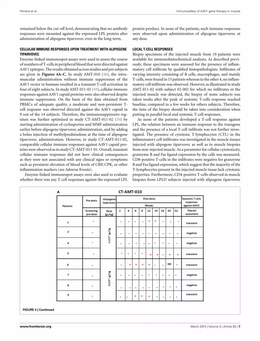
CELLULAR IMMUNE RESPONSES UPON TREATMENT WITH ALIPOGENETIPARVOVECEnzyme-linked immunospot assays were used to assess the courseof numbers of T-cells in peripheral blood that were directed againstAAV1 epitopes. The results obtained across studies and per subjectsare given in Figures 4A–C. In study AMT-010 (48), the intra-muscular administration without immune suppression of theAAV1 vector in humans resulted in a transient T-cell activation infour of eight subjects. In study AMT-011-01 (49), cellular immuneresponses against AAV1 capsid proteins were also observed despiteimmune suppression. On the basis of the data obtained fromPBMCs of adequate quality, a moderate and non-persistent T-cell response was observed directed against the AAV1 capsid in9 out of the 14 subjects. Therefore, the immunosuppressive reg-imen was further optimized in study CT-AMT-011-02 (50) bystarting administration of cyclosporine and MMF administrationearlier before alipogene tiparvovec administration, and by addinga bolus injection of methylprednisolone at the time of alipogenetiparvovec administration. However, in study CT-AMT-011-02,comparable cellular immune responses against AAV1 capsid pro-teins were observed as in study CT-AMT-011-01. Overall, transientcellular immune responses did not have clinical consequencesas they were not associated with any clinical signs or symptomssuch as persistent elevation of blood levels of CRP, CPK, or otherinflammation markers (see Adverse Events).
Enzyme-linked immunospot assays were also used to evaluatewhether there was any T-cell responses against the expressed LPL
protein product. In none of the patients, such immune responseswere observed upon administration of alipogene tiparvovec atany dose.
LOCAL T-CELL RESPONSESBiopsy specimens of the injected muscle from 19 patients wereavailable for immunohistochemical analyses. As described previ-ously, these specimens were assessed for the presence of inflam-matory cell infiltrate by qualified histopathologists. Infiltrates ofvarying intensity consisting of B cells, macrophages, and mainlyT-cells, were found in 15 patients whereas in the other 4, no inflam-matory cell infiltrate was observed. However, as illustrated in studyAMT-011-02 with subject 02-002 for which no infiltrates in theinjected muscle was detected, the biopsy of some subjects wastaken weeks after the peak of systemic T-cells response reachedbaseline, compared to a few weeks for others subjects. Therefore,the time of the biopsy should be taken into consideration whenputting in parallel local and systemic T-cell responses.
As none of the patients developed a T-cell response againstLPL, the relation between an immune response to the transgeneand the presence of a local T-cell infiltrate was not further inves-tigated. The presence of cytotoxic T-lymphocytes (CTL) in theinflammatory cell infiltrates was investigated in the muscle tissuesinjected with alipogene tiparvovec as well as in muscle biopsiesfrom non-injected muscle. As a parameter for cellular cytotoxicity,granzyme B and Fas ligand expression by the cells was measured.CD8-positive T-cells in the infiltrates were negative for granzymeB and Fas ligand expression, which suggest that the majority of theT-lymphocytes present in the injected muscle tissue lack cytotoxicproperties. Furthermore, CD4-positive T-cells observed in musclebiopsies from LPLD subjects injected with alipogene tiparvovec
FIGURE 4 | Continued
www.frontiersin.org March 2014 | Volume 5 | Article 82 | 7

Ferreira et al. Immunosafety of AAV1 gene therapy in muscle
FIGURE 4 | Systemic cellular immune responses followingalipogene tiparvovec administration. The tables below providesan overview of the individual patient systemic T-cell responseagainst AAV1 overtime for study CT-AMT-010 (A), study AMT-011-01(B), and study AMT-011-02 (C). We considered that a subject
developed a T-cell-mediated immune response to AAV1 capsidproteins when at least two of the eight to nine sampling timepoints were measured positive (+) in the ELISpot assay. When onlyone sampling time was reported positive (+), the T-cell responsewas considered negative.
Frontiers in Immunology | Microbial Immunology March 2014 | Volume 5 | Article 82 | 8

Ferreira et al. Immunosafety of AAV1 gene therapy in muscle
were further assessed for the expression of the transcription factorFoxP3, as a marker for regulatory T-cells. FoxP3-positive/CD4-positive T-cells were also found in the infiltrates suggesting thatmultiple mechanisms contribute to the local immune tolerance toalipogene tiparvovec administration (50).
IMMUNOGENICITY OF ALIPOGENE TIPARVOVEC ANDEFFICACYThe presence of LPL protein in the muscle biopsies and theimproved clearance of post-prandial chylomicrons levels in plasmawere used to measure local and systemic activity of alipogene tipar-vovec and were considered as efficacy markers (49, 51). However,the use of muscle biopsies has several hindrances. At first, out ofthe 27 patients treated with alipogene tiparvovec, 19 patients gavetheir consent for a muscle biopsy once. Only one patient allowedthe procedure to be done twice. Second, the results were influencedby the limited spread of the product in the muscle tissue and thevariability in the procedure, as not all biopsies were consistentlytaken in close proximity to the actual injection site. Therefore, thecirculating chylomicrons plasma levels measurement was devel-oped in parallel to the clinical study CT-AMT-011-01 and usedas a primary end point only in the study CT-AMT-011-02, as themost reliable endpoint to determine the systemic activity of theLPL enzyme.
Fifteen of the 26 patients, with registered data for presence ofpre-administration anti-AAV1 antibodies, had pre-existing anti-bodies against AAV1. Among the 15 patients with pre-existingantibodies against AAV1, biopsies were obtained from 11 patients.Among those 11 patients, 7 had LPL-expression in the biopsy.In comparison, from the 11 patients without pre-existing anti-bodies, 8 muscle biopsies were obtained; and from those, 5 werestained positive for LPL-expression. This distribution was verysimilar across the three studies CT-AMT-010, CT-AMT-011-01,and CT-AMT-011-02. Our results strongly indicate that there wasno apparent relationship between the presence of pre-existingAAV1 antibodies in LPLD patients and LPL-expression afteradministration of alipogene tiparvovec.
After the administration of alipogene tiparvovec in all 27patients, the development of treatment-emergent antibodiesagainst AAV1 capsid proteins was observed, independently ofwhether pre-existing antibodies were present or not. In rela-tion with efficacy, those treatment-emergent antibody responsesagainst the AAV1 capsid proteins upon treatment with alipogenetiparvovec, did not seem to affect expression of the transgene.
A similar conclusion as drawn for the presence and develop-ment of AAV1-specific antibodies can be drawn for the develop-ment and presence of AAV-specific T-cells after administration ofalipogene tiparvovec. The percentage of patients with treatment-emergent T-cell response across studies and among the three dosegroups was 50% (2/4) of the subjects treated with 1× 10¹¹ gc/kghaving a positive response, 40% (4/10) of the patients treatedwith 3× 10¹¹ gc/kg, and 69% (9/13) of the patients treated with1× 1012 gc/kg, which suggest an AAV1-dose-dependent kineticsof T-cell response appearance (48–50). Of the 19 patients whoconsented to the biopsy procedure, 10 developed AAV1-specific T-cells. Of those 10 patients, 5 were tested positive for the expressionof LPL in the biopsies. Of the 9 patients with no detectable T-cell
response against AAV1, 7 had detectable LPL-expression in theirbiopsy. In relation with efficacy, those transient T-cell responsesagainst the AAV1 capsid proteins upon treatment with alipogenetiparvovec, did not seem to directly influence the expression of thetransgene.
However, as mentioned previously, the variability of the biopsyprocedure and by consequence, the difficulty for quantificationand comparison between patients has to be considered in theinterpretation of the data. The differences in the results obtainedwith the biopsies of patient 01-001 (in study CT-AMT-011-02),collected at 18 and 52 weeks after administration of alipogenetiparvovec, illustrate this issue. The biopsy taken at week 18 yieldedno detectable LPL-expression, whereas the biopsy taken at week52 yielded a strong expression of LPL.
The administration of alipogene tiparvovec resulted in func-tional LPL activity levels sufficient to achieve a beneficial clinicaleffect in patients with LPLD. This conclusion is supported by theevidence that levels of plasma TG decreased in LPLD patients afteradministration of alipogene tiparvovec. The data obtained in stud-ies CT-AMT-010, without immunosuppression, and CT-AMT-011-01 and AMT-011-02, with immunosuppression, are consid-ered comparable in terms of the decrease in TG levels (Figure 5).However,plasma TG levels subsequently increased in most patientsaround 12–14 weeks post-administration of alipogene tiparvovec.This was at a time interval when immune suppression had alreadybeen discontinued. This phenomenon was observed across thethree studies and showed that fasting TG levels are not a sufficientlysensitive marker to monitor the long-term therapeutic effects ofalipogene tiparvovec. Post-prandial chylomicron clearance kinet-ics has been recognized as the most relevant biological marker forthe systemic activity of LPL during the clinical development of ali-pogene tiparvovec (50, 51). However, post-prandial chylomicronlevel measurements were included as endpoint only in the last ofthe three interventional studies, CT-AMT-011-02. The results havebeen reported (51) and show that the post-prandial chylomicronplasma levels are significantly reduced in all patients included inthe study, independently of the presence of humoral (all patients)or cellular systemic (two on five patients) or local (three on fivepatients) immune responses against AAV1 (Figure 3), thus indi-cating that these responses had no influence on the efficacy ofalipogene tiparvovec.
Furthermore, an ongoing study has shown a reduction in acutepancreatitis events in a series of more than 25 affected subjects(43). The analysis for a treatment-effect of Glybera taking intoaccount exposure time demonstrated a significant and clinicallyrelevant reduction in the risk of definite acute pancreatitis duringvarious periods ranging from 2.5 to 10 years pre-treatment topost-treatment (median 2.9 years) (52).
EFFECT OF IMMUNOSUPPRESSANTS ONTREATMENT-EMERGENT IMMUNE RESPONSESStudy CT-AMT-010, the first clinical study with AAV1–LPLS447X,was performed without treating the patients with immunosup-pressants. In this study, no antibody or T-cells responses againstLPL were found. However, antibodies against AAV1 capsid epi-topes were observed in all patients whereas a T-cell responseagainst AAV1 was detected only in four of the eight subjects.
www.frontiersin.org March 2014 | Volume 5 | Article 82 | 9

Ferreira et al. Immunosafety of AAV1 gene therapy in muscle
FIGURE 5 |TG responder status in relation with humoral andsystemicT-cell response against AAV. The table below provides anoverview of the individual patient cellular and humoral immune responsesin relation with the clinical end point (fasting) total plasma TG. A T-cellresponse to the antigen was reported transient positive (transient) when
at least two consecutive sampling time points were measured positive inthe ELISpot assay. The patient reported “sporadic” present recurrentnon-consecutive T-cell response over time. When none, or only onesampling time was reported positive, the T-cell response was reportednegative (−).
As discussed, none of these immune responses raised specificsafety concerns. However, they triggered more a concern aboutthe efficacy of the product; especially the development of anyAAV-specific T-cells (48) that were thought at the time to pos-sibly hamper the expression of LPL. A similar T-cell responsewas observed in gene therapy trial for hemophilia B, in whichan AAV2 vector was used to deliver the human coagulation fac-tor IX (24, 41, 42). In this trial, two patients developed a T-cellresponse to AAV2 capsid proteins, which was not predicted frompre-clinical studies. In those two patients, transgene expressiondeclined subsequently to pre-treatment levels. Based on theseobservations, it was concluded by the investigators that a cyto-toxic T-lymphocyte response to the capsid may have contributedto a loss of transgene-expressing cells.
These discussions heavily influenced the decision to include animmunosuppressant regimen in the clinical study protocols forCT-AMT-011-01 as well as CT-AMT-011-02.
As a result of these discussions, 17 patients (12 in CT-AMT-011-01 and 5 in CT-AMT-011-02) treated with alipogene tiparvovec
received a concomitant administration of immunosuppres-sants. Treatment-emergent anti-AAV1 antibody responses wereobserved in all the patients and were not affected by the immuno-suppressants, neither during the time of administration nor aftercessation of the administration. The AAV1–LPLS447X vector usedcannot induce expression of viral proteins in host cells. Hence,AAV1 capsid proteins are expected to be only transiently presentedto the immune system of the recipient after injection of AAV1–LPLS447X (3, 19, 53). Therefore, the immune suppressants drugswere given for a period of 12 weeks. A 12-week-period was con-sidered to be sufficient for prevention of capsid immunogenicity,based on observations in monkeys (19) and the investigators earlyobservations in humans that indicated TG levels started to rise afteran initial decrease usually at some time between 4 and 12 weekspost-dosing. A combination of cyclosporine and MMF was cho-sen because this combination is widely used to prevent cytotoxicT-cell responses in transplant rejection. The doses of the immunesuppressants proposed to co-administer with alipogene tiparvovecwere according to approved doses for transplant rejection.
Frontiers in Immunology | Microbial Immunology March 2014 | Volume 5 | Article 82 | 10

Ferreira et al. Immunosafety of AAV1 gene therapy in muscle
FIGURE 6 | Effect of immune suppression on LPL-expression andimmune responses upon administration of alipogene tiparvovec.
In study CT-AMT-011-01, humoral and cellular immuneresponses against AAV1 capsid proteins were observed. Theseresponses were similar to those observed in study CT-AMT-010although in study CT-AMT-011-01, a higher dose of AAV1–LPLS447X was used (Figure 4). Therefore, the immunosuppres-sant regimen was further optimized in study CT-AMT-011-02 bystarting administration of cyclosporine and MMF administrationearlier before alipogene tiparvovec administration, and by addinga bolus injection of methylprednisolone at the time of alipogenetiparvovec administration. However, in study CT-AMT-011-02,comparable humoral and cellular immune responses againstAAV1 capsid proteins were observed as in study CT-AMT-011-01(Figure 4).
The effect of immunosuppressants on LPL-expression andimmune responses upon administration of alipogene tiparvovecis summarized in Figure 6. In the clinical development of ali-pogene tiparvovec, no untoward side effects were observed thatcould be assigned to the use of prednisolone or one of the otherimmunosuppressants.
CLINICAL DATACHEMISTRY AND HEMATOLOGICAL VALUESPatients were monitored for up to 12 weeks post alipogenetiparvovec administration regarding routine hematology andbiochemistry including CPK and CRP, increases of which areexpected in case of local inflammatory damage at the injectionsite. In addition, other inflammatory markers such as neutrophilcounts were also determined at several pre- and post-exposureoccasions in the patients. None of the patients had neutrophilcounts outside the normal range. A per-patient summary of theCRP and CPK data is given in Figure 7. The majority of thepatients had normal CRP and CPK levels pre- and post-exposure
to alipogene tiparvovec, suggesting that inflammatory reactionsin the injected muscle were mild, if present, and of little clinicalsignificance.
ADVERSE EVENTSAlipogene tiparvovec was administered via a one-time set of intra-muscular injections in the lower limbs. No consistent change inany laboratory parameter, linked to the administered product, wasobserved, including CPK. Injections were well tolerated with mild–moderate local injection site reactions lasting for a few days relatingto the injection sites, and no change in muscle function. None ofthe patients showed clinical signs of persistent local inflamma-tion at injection sites such as redness, swelling, warmth, pain, ordysfunction, upon administration of alipogene tiparvovec. Thereare to date no reports of muscle dysfunction in LPLD patientsadministered with alipogene tiparvovec.
The adverse reactions observed during the clinical developmentof alipogene tiparvovec are summarized in Figure S1 in Supple-mentary Material. Most of the adverse reactions were related tothe administration procedure. All were of transient nature andresolved within days after the administration of the product. Onlyone serious adverse event involving muscle was seen, that wasconsidered to be at least possibly related to alipogene tiparvovecby the Investigator (#01-002 in CT-AMT-011-02). In this subject,at 15 weeks post-administration, a transient rise in CPK, accom-panied by a rise in CRP (Figure 7), was correlated with a lowpositive AAV cellular response associated with no LPL-related cel-lular or humoral significant response was seen in a complex ofclinical symptoms and signs indicated as “polyarthralgia of impre-cise origin.” The subject show sporadic systemic T-cell responsesagainst AAV across the observation period at weeks 2, 8, 14, 39.Despite those T-cell responses, the muscle biopsy of this subjectwhich was taken 30 weeks post alipogene tiparvovec showed robustLPL-expression. In addition, the subject did not show any anti-LPLcellular or humoral response (50). No adverse effect was observedthat could have been related to the immune responses discussedin the previous paragraphs.
From the clinical data obtained across studies, we conclude thatthere is no clinical untoward impact of the delivery of alipogenetiparvovec.
DISCUSSIONTo evaluate the immunogenicity of alipogene tiparvovec, an exten-sive testing program was performed that included the analyses ofantibody and T-cell responses to LPL as well as to AAV1. Antibodyand cellular responses were measured pre-exposure to alipogenetiparvovec, and at various occasions post-exposure. In addition,biopsies were taken from the injected muscle and non-injectedmuscle as control, to evaluate local immune and inflammatoryprocesses. Finally, CPK and CRP levels and neutrophil countswere measured, and patients were clinically evaluated for localand systemic symptoms indicative for inflammatory or immunereactions. Expression of LPL in the injected muscle as well as theimprovement of post-prandial chylomicrons clearance in plasmawas used as biochemical markers for efficacy.
The success of in vivo gene therapy not only depends on theability to control the immune response toward the vector, but
www.frontiersin.org March 2014 | Volume 5 | Article 82 | 11

Ferreira et al. Immunosafety of AAV1 gene therapy in muscle
FIGURE 7 | A per-patient summary of CRP and CPK values pre- andpost-exposure to alipogene tiparvovec. Scheduled visits pre-treatment andpost-treatment have been included. The post-treatment visits were at day 1and weeks 1, 2, 4, 8, and 12 for studies CT-AMT-010 and CT-AMT-011-01.Weeks 14, 26, 39, and 52 were added for study CT-AMT-011-02. In the study
AMT-011-01, the isolated elevation of CRP for subject 014 was concomitantwith a transient medical condition. In the study AMT-011-02, the isolatedelevation of CRP for subject 01-001 was concomitant with a transient medicalcondition. The isolated elevation of CRP and CPK for subject 01-002 wasrelated to the general clinical condition of the subject.
also to monitor any potential reaction to the therapeutic proteinexpressed from the transgene. The data obtained from patientswith LPL-deficiency who received a single treatment with multipleinjections of alipogene tiparvovec, support the initial expecta-tion that the protein product is minimally immunogenic, if atall: neither treatment-emergent antibody responses against LPLnor T-cells responses against LPL were found. Thus, the expressedprotein itself appears to be non-immunogenic.
Concerning anti-AAV immunity, the majority of healthy indi-viduals are exposed to AAV during lifetime (2, 54). Hence it isnot surprising that 15 of 27 patients had pre-existing antibod-ies against AAV1. There was no difference among patients with orwithout these antibodies with regard to detectable LPL-expressionin the biopsies or improvement of post-prandial chylomicronclearance, suggesting that pre-existing anti-AAV1 antibodies didnot appear to prevent LPL transgene expression and clinical effi-cacy. Therefore, pre-existing anti-AAV1 antibodies likely have noeffect on the efficacy of alipogene tiparvovec following intra-muscular administration. All patients showed treatment-emergentantibody responses to AAV1 and there was no difference inanti-AAV1 antibody response between the various dosing cohorts.
The markers that were used to demonstrate LPL-expressionand functionality after the delivery of alipogene tiparvovec arecomplex. Therefore, the present report is focusing on LPL-expression in muscle biopsies and post-prandial chylomicronclearance as marker for systemic LPL activity. For the directmeasurement of LPL-expression, we made use of biopsies thatwere obtained at different time points up to 12 months afterdrug administration. However, and as described above, not allpatients gave their consent to this procedure which can presentsome variability in the execution. Nineteen patients providedtheir consent and in total 20 biopsies were obtained, which allwere examined for LPL-expression. In 12 of the 20 biopsies, LPL-expression was found irrespective of the presence of antibodiesagainst AAV1. The second biochemical marker for successful genedelivery in this review is the level of clearance of post-prandialchylomicrons in plasma, which reflect the systemic activity ofLPL (49). This marker was however, included only in the studyCT-AMT-011-02. The post-prandial chylomicrons plasma lev-els were shown to be significantly reduced in all five patientsincluded in this study, independently of the presence of anti-AAV1antibodies.
Frontiers in Immunology | Microbial Immunology March 2014 | Volume 5 | Article 82 | 12
Ferreira et al. Immunosafety of AAV1 gene therapy in muscle
Altogether, these observations demonstrate that anti-AAV1antibody responses did not exclude sustained transgene expressionnor did impair the systemic biological activity of the expressedprotein. Our results further support that treatment-emergentanti-AAV1 antibody responses do not necessarily have anyinfluence on the long-term efficacy and safety of AAV-basedgene therapy.
Treatment-emergent T-cell responses against AAV1 capsidswere measured with the ELISpot assay and were observed in 18of the 27 patients. However, it has to be noted that the patientstreated with the higher dose, 1× 10¹² gc/kg, were more promptto develop an AAV1-specific T-cells responses. Somewhat higherT-cell responses were noticed in some patients upon cessationof the immunosuppressants, pointing to a suppressive effect onthe T-cell responses. It is therefore not possible to make defini-tive conclusions regarding the effect of immunosuppressants onT-cell responses to AAV1. The immunosuppressants did not affectantibody levels as was expected since the regimen was mainly aim-ing at reducing potential T-cell responses. MMF and cyclosporineare both described to be effective in suppressing cytotoxic T-cellresponses. MMF has an effect on B-cell proliferation, because itinhibits de novo guanosine nucleotide synthesis, a pathway com-monly required for both T- and B-cell proliferation. Cyclosporinespecifically inhibits T-cell activation by inhibiting IL-2 productionand exerts limited, if any, effect on B-cell proliferation. Thus, theincreased anti-AAV1 titers are not surprising. Similar observationswere made in a study in monkeys (52) when administrating AAV8-hFIX together with immunosuppressants consisting of MMF andtacrolimus. In the third study of the three interventional studies foralipogene tiparvovec, CT-AMT-011-02, prednisolone was admin-istered in the form of a bolus injection to prevent the release ofsubstances that mediate inflammation and to enhance the potencyof the other immunosuppressants used.
Our studies should be considered in the context of the grow-ing body of clinical and pre-clinical studies evaluating the roleof capsid-specific T-cells in AAV gene. The immunogenicity datafound in the clinical studies conducted with AAV-based vectorsin human, show that immune responses against AAV capsid pro-teins can vary widely and amongst others are influenced by thetarget organ, route of delivery, and dosing schedule. When tar-geting the muscle in humans, T-cell responses directed to thecapsid antigen were documented in AAT-deficient subjects receiv-ing intra-muscular injection of an AAV1–AAT vector (25, 55), instudy on AAV1-α-sarcoglycan in limb-girdle muscular dystrophysubjects (35) and in our own LPL-deficient subjects. The contro-versial aspect of the capsid-specific T-cell hypothesis is whetherthe vector sensitizes transduced cells to become targets for CTL-mediated clearance by virtue of MHC presentation of peptidesfrom the input capsid protein. Also, immune modulation wasused which could have impacted on the AAV-specific immuneresponses, our study provides the most direct and extensive testof this hypothesis because we observed transgene expression until52 weeks (long-term follow-up) after injection of AAV1–LPLS447X
in the muscle, despite the detection of circulating T-cell specificfor AAV capsid peptides in some subjects and persistent focal infil-trates in all subjects for whom transgene expression was detected.These data clearly demonstrate that transgene expression can
persist, despite the presence of capsid-specific T-cells and cellu-lar infiltrates. Sustained transgene expression in the presence ofT-lymphocyte responses have been reported in the literature inexperimental animals and in different tissues (56, 57) and humans(25, 35, 37). However, whereas attention has been focused initiallyon the AAV capsid as target of an undesirable T-cell response (24,41, 42), observations made by the groups of Dr C. Walker (58) andsupported by observations of others (59) suggest that loss of func-tion and programed death by most tissue-infiltrating AAV-primedT-cells seem to argue against their direct participation in clearanceof AAV-vector-transduced target cells. It has been described thatT-cells in AAV-vectors related infiltrates present characteristics ofanergy (55). Such T-cell infiltrates are therefore generally consid-ered as unable to initiate cellular self-destruction and therefore donot impact on efficacy of transgene expression.
Another mechanism via which T-cells may affect LPL-expression is stimulating the proliferation and differentiation ofB cells that subsequently form antibodies against AAV1. How-ever, as is discussed above, anti-AAV1 antibodies seem to haveno impact on LPL-expression and there is no evidence whetherantibody-dependent cell-mediated cytotoxicity played a role. Asmentioned before, a sustained long-term transgene expression wasobserved after intra-muscular injection, despite the presence ofcirculating antibodies directed against the AAV1 capsid peptides.The results obtained with alipogene tiparvovec demonstrate thatthe presence of the anti-AAV1 humoral immune response hadno apparent influence on the long-term efficacy of the therapy.The same observation has been reported in other clinical studies(25, 35, 37, 47, 55).
Multiple intra-muscular injections of the vector and supposedinflammatory and immune reactions ensuing at the injection sitesraise concerns about inflammatory damage in the injected muscle.However, except for transient mild local procedural symptoms atthe injection sites, no clinical symptoms such as swelling, pain,or dysfunction pointing at inflammatory damage were observedin the patients. In addition, serial monitoring of CRP and CPKrevealed normal levels of these markers in most patients. Occa-sional elevations of CRP and CPK were seen in two and in onepatient, respectively, without any clinical correlation. In addition,though a mild mononuclear infiltrate was observed in 14 of the 19patients of whom a biopsy was obtained, this infiltrate lacked sub-stantial cytotoxic T-cells activity. Hence, no clinical, biochemical,histochemical, or immunological evidence for inflammatory mus-cle damage at the injections sites was found.
All together, the data collected on systemic and local immuneresponses induced by intra-muscular injection of alipogene tipar-vovec demonstrate the absence of impact on safety and did notcompromise LPL transgene expression. These findings indicatethat muscle-directed AAV-based gene therapy through the intra-vascular route remains a promising approach for the treatment ofhuman diseases.
REGULATORY PERSPECTIVEDuring the assessment of the Glybera marketing authorizationdossier, the fact that no responses had been seen against theexpressed LPL protein was considered as a positive safety assetand no material concerns were expressed.
www.frontiersin.org March 2014 | Volume 5 | Article 82 | 13

Ferreira et al. Immunosafety of AAV1 gene therapy in muscle
The necessity of using immunosuppressants was not provenduring the clinical development of Glybera since the regimenseemed not to improve efficacy whilst having a major negativeimpact on the safety aspects. It remains questionable whether itmakes clinical sense to co-administer immunosuppressants withany AAV-based vector. However, in the case of Glybera, since LPLDis such a rare disease, it will not be possible to further assesslong-term clinical efficacy in absence of any immunosuppressantswithin the present indication.
During the regulatory review by the European MedicinesAgency, multiple questions arose on whether the cellular responsesto the viral capsid proteins could have any meaningful negativeeffect on the long-term safety or efficacy of alipogene tiparvovec.Our data clearly demonstrate that transgene expression can persist,despite the presence of capsid-specific T-cells and cellular infil-trates and without apparent toxicity or attenuation of transgeneexpression. Furthermore, the purity of the vector preparations (interms of total amount of viral proteins injected versus dose ingenome copies) and impurities profile of the vectors used in thevarious clinical trials described in the literature may be very diver-gent and therefore may lead to very different immune responses.Nonetheless, the scientific debate has been powerfully influencedby previous findings with other vectors and by the hypothesis thatthe vector sensitizes transduced cells to become targets for CTL-mediated clearance. Therefore, since the safety data on alipogenetiparvovec have been collected in a small number of patients, theirclinical relevance and possible interpretations were considered notfully unequivocal and further data collection has been requested bythe European Medicine Agency post-approval of Glybera®. Suchdata will be collected from all treated patients in future in a LPLDregistry, thus allowing for long-term data analysis.
SUPPLEMENTARY MATERIALThe Supplementary Material for this article can be found online athttp://www.frontiersin.org/Journal/10.3389/fimmu.2014.00082/abstract
REFERENCES1. Gao GP, Alvira MR, Wang L, Calcedo R, Johnston J, Wilson JM. Novel adeno-
associated viruses from rhesus monkeys as vectors for human gene therapy. ProcNatl Acad Sci U S A (2002) 99:11854–9. doi:10.1073/pnas.182412299
2. Calcedo R, Vandenberghe LH, Gao G, Lin J, Wilson JM. Worldwide epidemiol-ogy of neutralizing antibodies to adeno-associated viruses. J Infect Dis (2009)199:381–90. doi:10.1086/595830
3. Xiao W, Warrington KH Jr, Hearing P, Hughes J, Muzyczka N. Adenovirus-facilitated nuclear translocation of adeno-associated virus type 2. J Virol (2002)76:11505–17. doi:10.1128/JVI.76.22.11505-11517.2002
4. Clark KR, Penaud-Budloo M. Evaluation of the fate of rAAV genomes followingin vivo administration. Methods Mol Biol (2011) 807:239–58. doi:10.1007/978-1-61779-370-7_10
5. Léger A, Le Guiner C, Nickerson ML, McGee Im K, Ferry N, Moullier P, et al.Adeno-associated viral vector-mediated transgene expression is independentof DNA methylation in primate liver and skeletal muscle. PLoS One (2011)6:e20881. doi:10.1371/journal.pone.0020881
6. Duan D, Sharma P, Yang J, Yue Y, Dudus L, Zhang Y, et al. Circular intermediatesof recombinant adeno-associated virus have defined structural characteristicsresponsible for long term episomal persistence in muscle tissue. J Virol (1998)72:8568–77.
7. Nakai H, Yant SR, Storm TA, Fuess S, Meuse L, Kay MA. Extrachromosomalrecombinant adeno-associated virus vector genomes are primarily responsible
for stable liver transduction in vivo. J Virol (2001) 75:6969–76. doi:10.1128/JVI.75.15.6969-6976.2001
8. Kaeppel C, Beattie SG, Fronza R, van Logtenstein R, Salmon F, Schmidt S, et al.A largely random AAV integration profile after LPLD gene therapy. Nat Med(2013) 19:889–91. doi:10.1038/nm.3230
9. Samulski RJ, Berns KI, Tan M, Muzyczka N. Cloning of adeno-associated virusinto pBR322: rescue of intact virus from the recombinant plasmid in humancells. Proc Natl Acad Sci U S A (1982) 79:2077–81. doi:10.1073/pnas.79.6.2077
10. Wu Z, Asokan A, Samulski RJ. Adeno-associated virus serotypes: vector toolkitfor human gene therapy. Mol Ther (2006) 14:316–27. doi:10.1016/j.ymthe.2006.05.009
11. Jooss K, Yang Y, Fisher KJ, Wilson JM. Transduction of dendritic cells by DNAviral vectors direct the immune response to transgene products in muscle fibers.J Virol (1998) 72:4212–23.
12. Zaiss AK, Liu Q, Bowen GP, Wong NC, Bartlett JS, Muruve DA. Differential acti-vation of innate immune responses by adenovirus and adeno-associated virusvectors. J Virol (2002) 76:4580–90. doi:10.1128/JVI.76.9.4580-4590.2002
13. Acland GM, Aguirre GD, Ray J, Zhang Q, Aleman TS, Cideciyan AV, et al. Genetherapy restores vision in a canine model of childhood blindness. Nat Genet(2001) 28:92–5. doi:10.1038/ng0501-92
14. Adriaansen J, Tas SW, Klarenbeek PL, Bakker AC, Apparailly F, Firestein GS, et al.Enhanced transfer to arthritic joints using adeno-associated virus type 5: impli-cations for intra-articular gene therapy. Ann Rheum Dis (2005) 64:1677–84.doi:10.1136/ard.2004.035063
15. Flotte TR. Recent developments in recombinant AAV-mediated gene ther-apy for lung diseases. Curr Gene Ther (2005) 5(3):361–6. doi:10.2174/1566523054064986
16. Ghosh A, Yue Y, Long C, Bostick B, Duan D. Efficient whole body transductionwith trans-splicing adeno-associated viral vectors. Mol Ther (2007) 15:750–5.doi:10.1038/sj.mt.6300153
17. Goyenvalle A, Vulin A, Fougerousse F, Leturcq F, Kaplan JC, Garcia L, et al. Res-cue of dystrophic muscle through U7 snRNA mediated exon skipping. Science(2004) 306(5702):1796–9. doi:10.1126/science.1104297
18. Herzog RW, Hagstrom JN, Kung SH, Tai SJ, Wilson JM, Fisher KJ, et al. Stablegene transfer and expression of human blood coagulation factor IX after intra-muscular injection of recombinant adeno-associated virus. Proc Natl Acad SciU S A (1997) 94(11):5804–9. doi:10.1073/pnas.94.11.5804
19. Jiang H, Couto LB, Patarroyo-White S, Liu T, Nagy D, Vargas JA, et al. Effectsof transient immunosuppression on adenoassociated, virus-mediated, liver-directed gene transfer in rhesus macaques and implications for human genetherapy. Blood (2006) 108:3321–8. doi:10.1182/blood-2006-04-017913
20. Mas A, Montané J, Anguela XM, Muñoz S, Douar AM, Riu E, et al. Reversalof type I diabetes by engineering a glucose sensor in skeletal muscle. Diabetes(2006) 55:1546–53. doi:10.2337/db05-1615
21. Mount JD, Herzog RW, Tillson DM, Goodman SA, Robinson N, McClelandML, et al. Sustained phenotypic correction of hemophilia B dogs with a factorIX null mutation by liver directed gene therapy. Blood (2002) 99(8):2670–6.doi:10.1182/blood.V99.8.2670
22. Song S, Morgan M, Ellis T, Poirier A, Chesnut K, Wang J, et al. Sustainedsecretion of human alpha-1-antitrypsin from murine muscle transduced withadeno-associated virus vectors. Proc Natl Acad Sci U S A (1998) 95(24):14384–8.doi:10.1073/pnas.95.24.14384
23. Mingozzi F, High KA. Immune responses to AAV vectors: overcoming barriersto successful gene therapy. Blood (2013) 122:23–36. doi:10.1182/blood-2013-01-306647
24. Manno CS, Pierce GF, Arruda VR, Glader B, Ragni M, Rasko JJ, et al.Successful transduction of liver in hemophilia by AAV-factor IX and limita-tions imposed by the host immune response. Nat Med (2006) 12(3):342–7.doi:10.1038/nm1358
25. Brantly ML, Chulay JD, Wang L, Mueller C, Humphries M, Spencer LT, et al. Sus-tained transgene expression despite T lymphocyte responses in a clinical trialof rAAV1-AAT gene therapy. Proc Natl Acad Sci U S A (2009) 106(38):16363–8.doi:10.1073/pnas.0904514106
26. Wang Z, Tapscott SJ, Chamberlain JS, Storb R. Immunity and AAV-mediatedgene therapy for muscular dystrophies in large animal models and human trials.Front Immunol (2011) 2:201. doi:10.3389/fmicb.2011.00201
27. Mueller C, Flotte TR. Clinical gene therapy using recombinant adeno associatedvirus vectors. Gene Ther (2008) 15:858–63. doi:10.1038/gt.2008.68
Frontiers in Immunology | Microbial Immunology March 2014 | Volume 5 | Article 82 | 14

Ferreira et al. Immunosafety of AAV1 gene therapy in muscle
28. Bainbridge JW, Smith AJ, Barker SS, Robbie S, Henderson R, Balaggan K, et al.Effect of gene therapy on visual function in Leber’s congenital amaurosis. N EnglJ Med (2008) 358(21):2231–9. doi:10.1056/NEJMoa0802268
29. Cideciyan AV, Aleman TS, Boye SL, Schwartz SB, Kaushal S, Roman AJ, et al.Human gene therapy for RPE65 isomerase deficiency activates the retinoidcycle of vision but with slow rod kinetics. Proc Natl Acad Sci U S A (2008)105(39):15112–7. doi:10.1073/pnas.0807027105
30. Jaski BE, Jessup ML, Mancini DM, Cappola TP, Pauly DF, Greenberg B, et al.Calcium upregulation by percutaneous administration of gene therapy in car-diac disease (CUPID Trial), a first-in-human phase 1/2 clinical trial. J Card Fail(2009) 15(3):171–81. doi:10.1016/j.cardfail.2009.01.013
31. Kaplitt MG, Feigin A, Tang C, Fitzsimons HL, Mattis P, Lawlor PA, et al. Safetyand tolerability of gene therapy with an adenoassociated virus (AAV) borneGAD gene for Parkinson’s disease: an open label, phase I trial. Lancet (2007)369(9579):2097–105. doi:10.1016/S0140-6736(07)60982-9
32. Kay MA, Manno CS, Ragni MV, Larson PJ, Couto LB, McClelland A, et al.Evidence for gene transfer and expression of factor IX in haemophilia Bpatients treated with an AAV vector. Nat Genet (2000) 24(3):257–61. doi:10.1038/73464
33. Maguire AM, Simonelli F, Pierce EA, Pugh EN Jr, Mingozzi F, Bennicelli J, et al.Safety and efficacy of gene transfer for Leber’s congenital amaurosis. N Engl JMed (2008) 358(21):2240–8. doi:10.1056/NEJMoa0802315
34. Maguire AM, High KA, Auricchio A, Wright JF, Pierce EA, Testa F, et al.Age dependent effects of RPE65 gene therapy for Leber’s congenital amau-rosis: a phase 1 dose escalation trial. Lancet (2009) 374(9701):1597–605.doi:10.1016/S0140-6736(09)61836-5
35. Mendell JR, Rodino-Klapac LR, Rosales-Quintero X, Kota J, Coley BD,Galloway G, et al. Limb-girdle muscular dystrophy type 2D gene therapyrestores alpha-sarcoglycan and associated proteins. Ann Neurol (2009) 66(3):290–7. doi:10.1002/ana.21732
36. Stroes ES, Nierman MC, Meulenberg JJ, Franssen R, Twisk J, Henny CP, et al.Intramuscular administration of AAV1-lipoprotein lipase S447X lowers triglyc-erides in lipoprotein lipase-deficient patients. Arterioscler Thromb Vasc Biol(2008) 28(12):2303–4. doi:10.1161/ATVBAHA.108.175620
37. Nathwani AC, Tuddenham EG, Rangarajan S, Rosales C, McIntosh J, Linch DC,et al. Adenovirus-associated virus vector-mediated gene transfer in hemophiliaB. N Engl J Med (2011) 365(25):2357–65. doi:10.1056/NEJMoa1108046
38. Marks WJ Jr, Bartus RT, Siffert J, Davis CS, Lozano A, Boulis N, et al. Gene deliv-ery of AAV2-neurturin for Parkinson’s disease: a double-blind, randomised, con-trolled trial. Lancet Neurol (2010) 9(12):1164–72. doi:10.1016/S1474-4422(10)70254-4
39. Christine CW, Starr PA, Larson PS, Eberling JL, Jagust WJ, Hawkins RA, et al.Safety and tolerability of putaminal AADC gene therapy for Parkinson disease.Neurology (2009) 73(20):1662–9. doi:10.1212/WNL.0b013e3181c29356
40. McPhee SW, Janson CG, Li C, Samulski RJ, Camp AS, Francis J, et al. Immuneresponses to AAV in a phase I study for canavan disease. J Gene Med (2006)8:577–88. doi:10.1002/jgm.885
41. Mingozzi F, Maus MV, Hui DJ, Sabatino DE, Murphy SL, Rasko JE, et al. CD8+T-cell responses to adeno-associated virus capsid in humans. Nat Med (2007)13:419–22. doi:10.1038/nm1549
42. Mingozzi F, Hasbrouck NC, Basner-Tschakarjan E, Edmonson SA, Hui DJ,Sabatino DE, et al. Modulation of tolerance to the transgene product in a non-human primate model of AAV-mediated gene transfer to liver. Blood (2007)110(7):2334–41. doi:10.1182/blood-2007-03-080093
43. Gaudet D, de Wal J, Tremblay K, Déry S, van Deventer S, Freidig A, et al.Review of the clinical development of alipogene tiparvovec gene therapy forlipoprotein lipase deficiency. Atheroscler Suppl (2010) 11:55–60. doi:10.1016/j.atherosclerosissup.2010.03.004
44. Rip J, Nierman MC, Sierts JA, Petersen W, Van den Oever K, Van Raalte D, et al.Gene therapy for lipoprotein lipase deficiency: working toward clinical applica-tion. Hum Gene Ther (2005) 16:1276–86. doi:10.1089/hum.2005.16.1276
45. Ross CJ, Twisk J, Meulenberg JM, Liu G, van den Oever K, Moraal E, et al. Correc-tion of feline lipoprotein lipase deficiency with adeno-associated virus serotype1-mediated gene transfer of the lipoprotein lipase S447X beneficial mutation.Hum Gene Ther (2004) 15(9):906–19. doi:10.1089/hum.2004.15.906
46. Ross CJ, Twisk J, Bakker AC, Miao F, Verbart D, Rip J, et al. Long-term correc-tion of murine lipoprotein lipase deficiency with AAV1-mediated gene transferof the naturally occurring LPL(S447X) beneficial mutation. Hum Gene Ther(2006) 17(5):487–99. doi:10.1089/hum.2006.17.487
47. Brantly ML, Spencer T, Humphries M, Conlon TJ, Spencer CT, Poirier A, et al.Phase I trial of intramuscular injection of a recombinant adeno-associated virusserotype 2 alpha 1-antitrypsin (AAT) vector in AAT-deficient adults. Hum GeneTher (2006) 17:1177–86. doi:10.1089/hum.2006.17.1177
48. Mingozzi F, Meulenberg JJ, Hui DJ, Basner-Tschakarjan E, Hasbrouck NC,Edmonson SA, et al. AAV-1-mediated gene transfer to skeletal muscle in humansresults in dose-dependent activation of capsid-specific T cells. Blood (2009)114(10):2077–86. doi:10.1182/blood-2008-07-167510
49. Gaudet D, Méthot J, Déry S, Brisson D, Essiembre C, Tremblay G, et al. Effi-cacy and long-term safety of alipogene tiparvovec (AAV1-LPLS447X) gene ther-apy for lipoprotein lipase deficiency: an open-label trial. Gene Ther (2013)20(4):361–9. doi:10.1038/gt.2012.43
50. Ferreira V, Twisk J, Kwikkers KL, Aronica E, Brisson D, Methot J, et al. Immuneresponses to intramuscular administration of alipogene tiparvovec (AAV1-LPLS447X) in a phase II clinical trial of lipoprotein lipase deficiency (LPLD)gene therapy. Hum Gene Ther (2013). doi:10.1089/hum.2013.169
51. Carpentier AC, Frisch F, Labbé SM, Gagnon R, de Wal J, Greentree S, et al.Effect of alipogene tiparvovec (AAV1-LPL(S447X)) on postprandial chylomi-cron metabolism in lipoprotein lipase-deficient patients. J Clin Endocrinol Metab(2012) 97:1635–44. doi:10.1210/jc.2011-3002
52. Bruno MJ, Deakin M, Ruszniewski PB, Bulk NV, de Wal J, Camozzi CR,et al. Alipogene tiparvovec gene therapy reduces the risk of acute pancre-atitis in patients with lipoprotein lipase deficiency. Gastroenterology (2012)142(S1):S112.
53. Lux K, Goerlitz N, Schlemminger S, Perabo L, Goldnau D, Endell J, et al. Greenfluorescent protein-tagged adeno-associated virus particles allow the study ofcytosolic and nuclear trafficking. J Virol (2005) 79:11776–87. doi:10.1128/JVI.79.18.11776-11787.2005
54. Boutin S, Monteilhet V, Veron P, Leborgne C, Benveniste O, Montus MF, et al.Prevalence of serum IgG and neutralizing factors against adeno-associatedvirus (AAV) types 1, 2, 5, 6, 8, and 9 in the healthy population: implicationsfor gene therapy using AAV vectors. Hum Gene Ther (2010) 21(6):704–12.doi:10.1089/hum.2009.182
55. Rodino-Klapac LR, Lee JS, Mulligan RC, Clark KR, Mendell JR. Lack oftoxicity of alpha-sarcoglycan overexpression supports clinical gene transfertrial in LGMD2D. Neurology (2008) 71:240–7. doi:10.1212/01.wnl.0000306309.85301.e2
56. Wang L, Dobrzynski E, Schlachterman A, Cao O, Herzog RW. Systemic proteindelivery by muscle-gene transfer is limited by a local immune response. Blood(2005) 105:4226–34. doi:10.1182/blood-2004-03-0848
57. Wang Z, Allen JM, Riddell SR, Gregorevic P, Storb R, Tapscott SJ, et al. Immu-nity to adeno-associated virus-mediated gene transfer in a random-bred caninemodel of Duchenne muscular dystrophy. Hum Gene Ther (2007) 18:18–26.doi:10.1089/hum.2006.093
58. Velazquez VM, Bowen DG, Walker CM. Silencing of T lymphocytes byantigen programmed death in recombinant adeno-associated virus vector-mediated gene therapy. Gene Ther (2009) 113:538–45. doi:10.1182/blood-2008-01-131375
59. Haurigot V, Mingozzi F, Buchlis G, Hui DJ, Chen Y, Basner-Tschakarjan E, et al.Safety of AAV factor IX peripheral transvenular gene delivery to muscle in hemo-philia B dogs. Mol Ther (2010) 18:1–12. doi:10.1038/mt.2010.73
Conflict of Interest Statement: The funding body (uniQure) was involved in allaspects of the study. The authors (Valerie Ferreira, Florence Salmon, and HaraldPetry) are employes of uniQure.
Received: 09 September 2013; accepted: 16 February 2014; published online: 03 March2014.Citation: Ferreira V, Petry H and Salmon F (2014) Immune responses to AAV-vectors, the Glybera example from bench to bedside. Front. Immunol. 5:82. doi:10.3389/fimmu.2014.00082This article was submitted to Microbial Immunology, a section of the journal Frontiersin Immunology.Copyright © 2014 Ferreira, Petry and Salmon. This is an open-access article distributedunder the terms of the Creative Commons Attribution License (CC BY). The use, dis-tribution or reproduction in other forums is permitted, provided the original author(s)or licensor are credited and that the original publication in this journal is cited, inaccordance with accepted academic practice. No use, distribution or reproduction ispermitted which does not comply with these terms.
www.frontiersin.org March 2014 | Volume 5 | Article 82 | 15