Embed Size (px)
Citation preview

Summary. Alcoholism in humans is a chronic andprogressive disease, characterized by loss of ethanolconsumption control. Previous studies have reported thatprolonged exposure to ethanol was responsible foralterations in glandular tissues of human and rodents.However, the interrelationship between ethanol and theglandular system is still the subject of numerousinvestigations, including the possible resistance of thesubmandibular gland (SG). In the present study, weinvestigated whether chronic ethanol exposure duringadolescence may affect the parotid gland (PG) and SG infemale rats. Female rats (n=16) were treated withdistilled water or ethanol (dose of 6.5 g/kg/day, 22.5%w/v) through gavage for 55 days. Glands were collected,weighed and submitted to histological processing.Morphometric analysis was assessed by parenchymaland stromal area measurements. Smooth muscle actin(α-SMA), cytokeratin-19 (CK19) and apoptotic caspase-3 (CAS) were measured using ImageJ® software.Chronic ethanol administration did not alter the bodyweight of rats after treatment, although it increasedglandular weight (p<0.001), reduced the parenchymaarea (p<0.001) and decreased CK19 and α-SMAimmunostainning in the PG. Besides, ethanol inducedCK19 and CAS overexpression, and the occurrence ofduct-like structures in SG. These results suggest that
ethanol induces histological and morphometric changesin salivary glands of female rats intoxicated with ethanolduring adolescence. Furthermore, the mechanismunderlying these alterations needs to be investigated butmay be not related to the inflammatory process.Key words: Ehanol, Salivary glands, Immunohisto-chemistry, Adolescence
Introduction
Alcoholism is a chronic and progressive psychiatricdisorder characterized by the loss of control of ethanolconsumption (Friedlander et al., 2003). The WorldHealth Organization (WHO) estimates that ethanol abuseis responsible for approximately 3 million prematuredeaths worldwide each year and approximately 4% ofglobal morbidity (Schuckit, 2009). The use of the drughas increased in a heavy binge-drinking way (higherdoses and longer period of time), among youngerwomen, highlighting the vulnerability of this group andemerging as a public health emergency (Schuckit, 2009;INPAD, 2013).
The effects of chronic ethanol abuse are associatedto damage in the tissues and organs in humans andanimals, as a result of morphological and functionalchanges (Molina et al., 2002; Owczarek et al., 2005).Studies have pointed to several organs and systemsaffected by chronic alcohol exposure in rodents, such asupper digestive tract (Mascres et al., 1984; Bagyánszkiet al., 2010); liver, lungs, heart, and skeletal muscle(Molina et al., 2002; Owczarek et al., 2005); central
Immunohistochemical changes and atrophy afterchronic ethanol intoxication in rat salivary glandsLuanna Melo Pereira Fernandes1, Francisco Bruno Teixeira2, Sergio Melo Alves Junior2, João de Jesus Viana Pinheiro2, Cristiane Socorro Ferraz Maia3 and Rafael Rodrigues Lima11Laboratory of Functional and Structural Biology, Institute of Biological Sciences, 2School of Dentistry and 3Laboratory ofPharmacology of Inflammation and Behaviour, Faculty of Pharmacy, Institute of Health Science, Federal University of Pará, CampusGuamá, Belém, Pará, Brazil
Histol Histopathol (2015) 30: 1069-1078
http://www.hh.um.esHistology andHistopathologyCellular and Molecular Biology
Offprint requests to: Cristiane do Socorro Ferraz Maia, PhD, Laboratoryof Pharmacology of Inflammation and Behaviour, Institute of HealthSciences, Federal University of Pará, Street Augusto Corrêa N. 1,Campus do Guamá, Belém-Pará 66075-900, Brazil. e-mail:[email protected], [email protected]: 10.14670/HH-11-604

nervous system (Oliveira et al., 2014; Teixeira et al.,2014); pancreas (Orabi et al., 2011); and salivary glands(Maier et al., 1986, 1988a,b; Banderas et al., 1992;Carrard et al., 2007; Nör et al., 2013). Studies in humansrevealed damage to liver, lungs, heart, skeletal muscleand salivary glands (Scott et al., 1987, 1988; Ferraris etal., 1995, 1999; Bohl et al., 2008). The latter was one ofthe first structures affected by chronic ethanolconsumption (Mandel and Baurmash, 1971).
Parotid (PG) and submandibular (SG) glands areclassified as major salivary glands, and both aremicroscopically similar in architecture. The glandularparenchyma consists of continuous-secreting acini cells,which leads to a system of ducts that empty into the oralcavity (Actis et al., 2002). In rodents, the PG is locatedbehind and below the ear, caudally bordering the SG.Microscopically, the PG consists of pure serous acini.On the other side, SG is a mixed gland that includes bothserous and mucous acini (Amano et al., 2012). Boththese glands contribute to 95% of the total salivarysecretion produced and secreted into oral cavity, whichthe SG contributes 60-70% (Faustino and Stipp, 2003;Katchburian and Arana, 2004).
Ethanol rapidly diffuses into saliva and oral tissues(Gifford et al., 2008). Immediately after alcoholexposure, high concentrations of this drug are detectedin the salivary glands (Waszkiewicz et al., 2013b). Thus,chronic alcohol exposure may promote effects on saliva,including a reduction in the salivary flow rate associatedto gland atrophy, and reduced electrolytes, salivaryproteins and glycoproteins (e.g. amylase, immuno-globulin A, lysozyme, lactoferrin) which modifies themorphology and physiology of the salivary glands(Lieber, 1991; Faustino and Stipp, 2003; Riedel et al.,2003; Waszkiewicz et al., 2013a,b).
However, there are insufficient studies in theliterature in order to clarify these structural alterations,as well as in the morphometry and in the structure of itscellular components. Thus, our protocol aims toinvestigate the morphometric and immunohistochemicalchanges of the PG and SG in female rats exposed toheavy alcohol exposure during adolescence.Materials and methods
Animals
A total of 16 adolescent female Wistar rats, 35 daysold at the beginning of the experiments (50-60 g) wereobtained from the Federal University of Pará (UFPA)and kept in collective cages (a maximum of five animalsper cage). Animals were maintained in a climate-controlled room on a 12-h reverse light/dark cycle (7:00AM lights on), with food and water ad libitum. Allprocedures were approved by the Ethics Committee onExperimental Animals of the Federal University of Paráunder license number BIO-043-12 and followed theguidelines suggested by the NIH Guide for the Care andUse of Laboratory Animals.
Experimental groups
Animals were initially allocated to the followinggroups: distilled water (n=8) or ethanol (6.5 g/kg/day,22.5% w/v) (n=8). Every day, rats were administeredthrough orogastric cannula over a period of 55 days (i.e.,until the 90th day of life) according to a procedurepreviously described (Livy et al., 2001; Maier and West,2001; Maia et al., 2009; Oliveira et al., 2014; Teixeira etal., 2014). The current ethanol administration protocolwas based on previous studies from our group (Maia etal., 2009; Teixeira et al., 2014; Oliveira et al., 2014)showing that ethanol intoxication (6.5 g/kg/day) in thedeveloping central nervous system may induce long-lasting neurobehavioral impairments in rats. In order toinvestigate putative effects of ethanol exposure onoverall poor nutrition levels, which may directly affectthe quality of the tissues, were noted the animals’ bodyweight, behavioral dysfunctions, loss or changes in haircoat, diarrhea, and edema during the entire period ofethanol administration (Campana et al., 1975).
Collection and preparation of tissue
After 7.5 hours of the last administration of alcohol,which is the period related to non-detectable amounts ofethanol in the blood (Livy et al., 2003), animals wereanaesthetised with a mixture of ketamine hydrochloride(72 mg/kg, i.p.) and xylazine (9 mg/kg, i.p.) and thenperfused with heparinized saline followed byparaformaldehyde 4%. The PG and SG were removedand weighed by analytical balance (FA 2104 N, EletronicBalance Bioprecisa, Shanghai, China). The relativeglandular weight (RGW) was calculated by the formula:RGW=gland weight x 100/final body weight.
After weighing, glands were post-fixed in formalinsolution (10%), and submitted to histological process bydehydration in increasing alcohol battery and xylene,and finally embedded in Paraplast® resin.Histomorphometry
For histochemistry procedures, 7-µm samples werecut from the region corresponding to the largest cross-sectional area of the organ with a microtome (RM2255,Leica Microsystems, Nussloch, Germany), depara-ffinized in xylene, rehydrated in decreasingconcentrations of alcohol and water, and finally stainedwith hematoxylin/eosin (HE). Thereafter, formorphometric analysis, the mean percentage equivalentto the glandular parenchyma and stroma region wasevaluated.
The area of the samples was evaluated by countingpoint planimetry method. The sections were stained byHE and illustrative images from all experimental groupswere obtained with a digital camera (Cyber-shot DSC-W230 4X optical zoom, Sony, Tokyo, Japan) attached toa stereo microscope (1.5x; Eclipse E200, Nikon, Tokyo,
1070Ethanol intoxication in salivary glands

Japan). Subsequently, we used five random sections peranimal of both PG and SG computerized images, whichwere plotted in the computer monitor followed byoverlapping of the grid of planimetry, which measures21x16 cm and contains 2,852 points equidistant of 0.5cm. Coincident points with parenchymal elements andstromal glandular were counted, and the percentageobtained of each region was calculated by the formulas:Morphometric Parenchymal Area (%MPA) = number ofcoincident points in the parenquimal x 100/ number totalof points on the grid; and, Morphometric Stromal Area(%MSA) = number of coincident points in the stroma x100/ number total of points on the grid (Borgers et al.,1993).Immunohistochemistry
Immunohistochemical studies were performed onparaffin-embedded tissues using streptavidin (Springbioscience, Pleasanton, USA) and 3,3-diaminobenzidine(DAB; Sigma-Aldrich, St. Louis, USA) methods.Briefly, 3-µm sections were deparaffinized andrehydrated in decreasing concentration of alcoholbattery. After antigen retrieval in chamber Pascal(S2800, Dako, Carpinteria, USA) and blocking ofendogenous peroxidase activity, sections were incubatedwith the primary antibody anti-α smooth muscle actin(α-SMA) (1:50 dilution, Dako, Carpinteria, USA), acytoplasmic myofilaments marker (Takahashi et al.2001); anti-cytokeratin 19 (CK19) (1:100 dilution,Bioss, Boston, USA), a marker of intermediate filamentsin the cytoplasm cells ductal (Fradette et al., 1998); andanti-caspase 3 (CAS) (1:250 dilution, Promega,Madison, USA), which reacts with the active form ofcaspase 3 and is associated with induction of apoptosis(Takahashi et al., 2004), in 0.1 M phosphate buffer saline(PBS). After, sections were submitted to streptavidin andrevealed by DAB and counter-stained by Mayerhematoxylin methods.Digital image analysis
Evaluation of immunostaining was performed by thepercentage of labelled area of six regions randomlyselected of each gland by the operator. Briefly, imagesselected were obtained by microscope (Axio Scope, CarlZeiss, Jena, Germany) attached to the camera (AxioCamHRC, Carl Zeiss, Jena, Germany) with a magnificationof 400x.
Digital images were prepared for analyses usingImageJ® software [Bethesda, Maryland, USA,(http://rsb.info.nih.gov/ij/download.html)]. According toRuifrok and Johnston (2001), each color images wereselected and separated from the rest of the field usingimage segmentation tool. CK19, CAS and α-SMApositive area (brown stain) and total gland area in theanalysed field were automatically measured using colordeconvolution plugin by calculating the contribution ofeach stain based on the stain-specific RGB absorption. In
our protocol, color deconvolution was used to separateDAB stain (positive areas) from hematoxylin (total areaof the glands). Each image was changed to black/whitecolor image. The measurements button was calibrated tocalculate the area of the field (total gland area) and thearea fraction (black color representing DAB stain).Finally, scores for CK19, CAS and α-SMA expression inthe analysed field were calculated by the positivelystained area over the total gland area. Statistical analysis
All values are expressed as means + S.E.M. (n=8animals per group). Statistical comparison wasperformed using Mann-Whitney test. The accepted levelof significance was p≤0.05. All tests were performedusing the GraphPad Prism software version 5.0 (SanDiego, USA).Results
Body weight of female rats in adulthood was not affectedby chronic ethanol exposure during adolescence
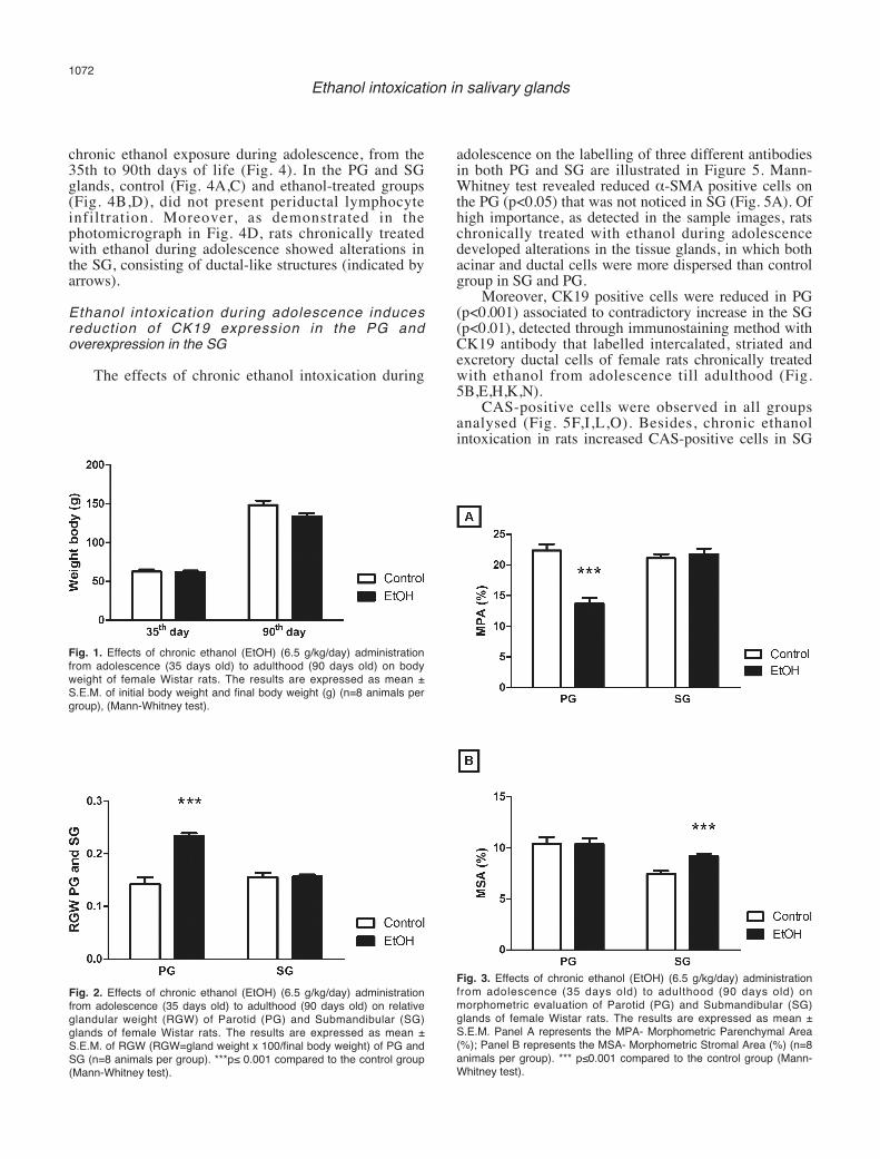
As illustrated in Fig. 1, the chronic ethanol (6.5g/kg/day) administration over a period of 55 days (i.e.,from the 35th day until the 90th day of life) did not alterthe final body weight of ethanol-treated rats.Ethanol exposure increases PG weight, but does notchange SG weight
The chronic ethanol (6.5 g/kg/day) administrationover a period of 55 days (i.e., from the 35th day until the90th day of life) induced a significant increase in theRGW of PG of female rats from the 35th to 90th days oflife (p<0.001). However, the total SG weight betweencontrol- and ethanol-treated rats after the period of 55days of treatment did not differ statistically (Fig. 2).Chronic ethanol exposure induces reduction and atrophyof PG glandular parenchyma and increases SGglandular stroma area
The histomorphometric results evaluated in thesalivary glands are summarised in Fig. 3. Mann-Whitneytest revealed that chronic ethanol exposure reduced the percentage of the parenchymal area of the PG(p<0.001) that was not observed in the SG (Fig. 3A).
In addition, chronic ethanol exposure increasedstromal area of SG (p<0.001) that was not noticed in thePG (Fig. 3B).Chronic ethanol intoxication during adolescence doesnot induce inflammatory infiltration in PG and SG
The HE method did not reveal inflammatoryprocesses in the salivary glands of female rats after
1071Ethanol intoxication in salivary glands
chronic ethanol exposure during adolescence, from the35th to 90th days of life (Fig. 4). In the PG and SGglands, control (Fig. 4A,C) and ethanol-treated groups(Fig. 4B,D), did not present periductal lymphocyteinfiltration. Moreover, as demonstrated in thephotomicrograph in Fig. 4D, rats chronically treatedwith ethanol during adolescence showed alterations inthe SG, consisting of ductal-like structures (indicated byarrows). Ethanol intoxication during adolescence inducesreduction of CK19 expression in the PG andoverexpression in the SG
The effects of chronic ethanol intoxication during
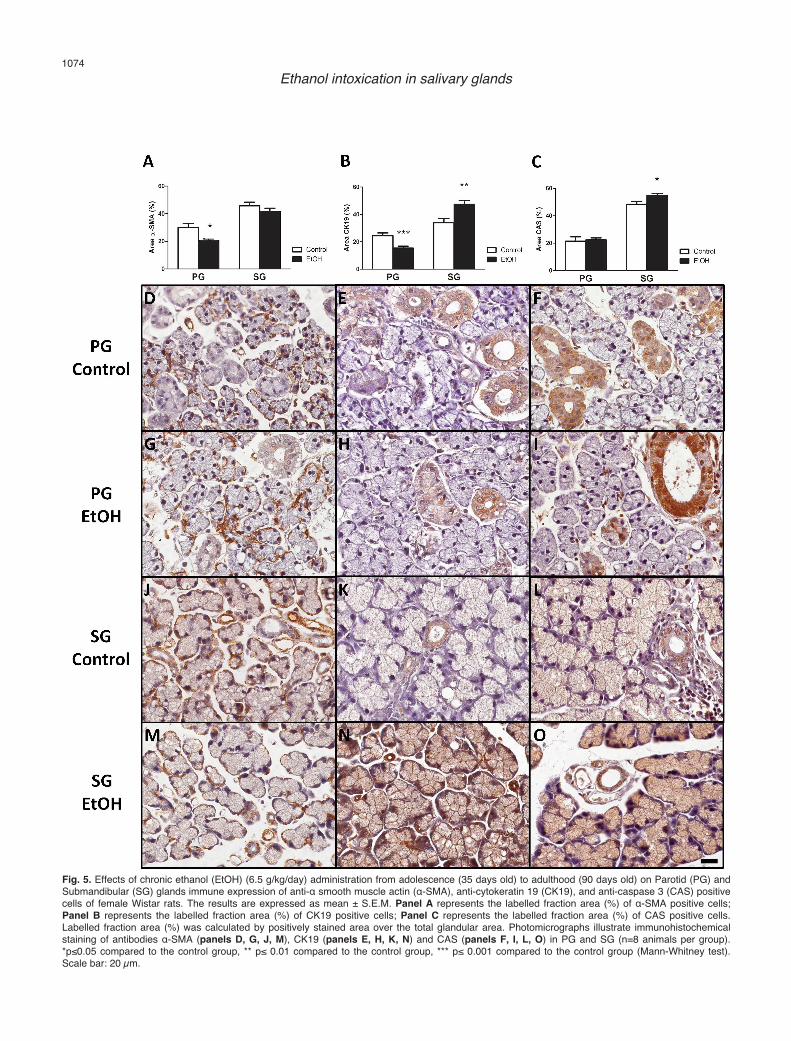
adolescence on the labelling of three different antibodiesin both PG and SG are illustrated in Figure 5. Mann-Whitney test revealed reduced α-SMA positive cells onthe PG (p<0.05) that was not noticed in SG (Fig. 5A). Ofhigh importance, as detected in the sample images, ratschronically treated with ethanol during adolescencedeveloped alterations in the tissue glands, in which bothacinar and ductal cells were more dispersed than controlgroup in SG and PG.
Moreover, CK19 positive cells were reduced in PG(p<0.001) associated to contradictory increase in the SG(p<0.01), detected through immunostaining method withCK19 antibody that labelled intercalated, striated andexcretory ductal cells of female rats chronically treatedwith ethanol from adolescence till adulthood (Fig.5B,E,H,K,N).
CAS-positive cells were observed in all groupsanalysed (Fig. 5F,I,L,O). Besides, chronic ethanolintoxication in rats increased CAS-positive cells in SG
1072Ethanol intoxication in salivary glands
Fig. 1. Effects of chronic ethanol (EtOH) (6.5 g/kg/day) administrationfrom adolescence (35 days old) to adulthood (90 days old) on bodyweight of female Wistar rats. The results are expressed as mean ±S.E.M. of initial body weight and final body weight (g) (n=8 animals pergroup), (Mann-Whitney test).
Fig. 2. Effects of chronic ethanol (EtOH) (6.5 g/kg/day) administrationfrom adolescence (35 days old) to adulthood (90 days old) on relativeglandular weight (RGW) of Parotid (PG) and Submandibular (SG)glands of female Wistar rats. The results are expressed as mean ±S.E.M. of RGW (RGW=gland weight x 100/final body weight) of PG andSG (n=8 animals per group). ***p≤ 0.001 compared to the control group(Mann-Whitney test).
Fig. 3. Effects of chronic ethanol (EtOH) (6.5 g/kg/day) administrationfrom adolescence (35 days old) to adulthood (90 days old) onmorphometric evaluation of Parotid (PG) and Submandibular (SG)glands of female Wistar rats. The results are expressed as mean ±S.E.M. Panel A represents the MPA- Morphometric Parenchymal Area(%); Panel B represents the MSA- Morphometric Stromal Area (%) (n=8animals per group). *** p≤0.001 compared to the control group (Mann-Whitney test).

(p<0.05) but not in PG (Fig. 5C).Discussion
In the present study, we investigated whether chronicethanol exposure during adolescence affects the salivaryglands in female rats. The current findings demonstrate,for the first time, that chronic ethanol exposure duringadolescence induces parenchyma atrophy related togland weight increases and reduction in ductal andmyoepithelial cell population in the PG as evaluated inthe histomorphometry and immunohistochemicalanalysis. Moreover, histological evaluation revealed thatour alcohol exposure altered cell population in the SG,appearing ductal-type structures and inducing apoptosis.Heavy binge-drinking (i.e., high doses and long periodof time) has increased worldwide mainly among
adolescents (Schuckit, 2009; INPAD, 2013). Therefore,our group has studied the effects of chronic ethanolexposure in body tissues in adolescent rats. Heavychronic ethanol intoxication (6.5 g/kg/day) duringadolescence induces long-lasting neurobehavioralimpairments as well as brain histochemical damage(Oliveira et al., 2014; Teixeira et al., 2014). In this sense,our group also observed that chronic ethanolconsumption from adolescence till adulthood alters thehomeostasis of the oral cavity promoting alveolar boneloss in rats (Bannach et al., 2015).
It has been shown that chronic ethanol consumptioninduces glandular atrophy and reduces salivary flow rate,salivary protein and glycoprotein levels and electrolytespromoting, among alcoholics, worse dental state andsignificantly worse periodontal state (Waszkiewicz et al.,2013a,b). Whereas saliva ensures the homeostasis of the
1073Ethanol intoxication in salivary glands
Fig. 4. Effects of chronic ethanol (EtOH) (6.5 g/kg/day) administration from adolescence (35 days old) to adulthood (90 days old) on histologicalsections of Parotid (PG) and Submandibular (SG) glands stained by hematoxylin/eosin (HE) of female Wistar rats. Panel A represents the PG of thecontrol group; Panel B represents the PG of ethanol-treated group; Panel C represents the SG of the control group; Panel D represents the SG ofethanol-treated group. Arrows indicate ductal-like structures. Scale bar: 20 µm.
1074Ethanol intoxication in salivary glands
Fig. 5. Effects of chronic ethanol (EtOH) (6.5 g/kg/day) administration from adolescence (35 days old) to adulthood (90 days old) on Parotid (PG) andSubmandibular (SG) glands immune expression of anti-α smooth muscle actin (α-SMA), anti-cytokeratin 19 (CK19), and anti-caspase 3 (CAS) positivecells of female Wistar rats. The results are expressed as mean ± S.E.M. Panel A represents the labelled fraction area (%) of α-SMA positive cells;Panel B represents the labelled fraction area (%) of CK19 positive cells; Panel C represents the labelled fraction area (%) of CAS positive cells.Labelled fraction area (%) was calculated by positively stained area over the total glandular area. Photomicrographs illustrate immunohistochemicalstaining of antibodies α-SMA (panels D, G, J, M), CK19 (panels E, H, K, N) and CAS (panels F, I, L, O) in PG and SG (n=8 animals per group).*p≤0.05 compared to the control group, ** p≤ 0.01 compared to the control group, *** p≤ 0.001 compared to the control group (Mann-Whitney test).Scale bar: 20 µm.

oral environment, defence of the oral cavity, oropharynxand upper region of the gastrointestinal tract, andprotection of hard and soft tissues of the oral cavity aswell as of the teeth and periodont (Pronko et al., 2002;Prestifilippo et al., 2009), saliva flow rate reduced mayinduce oral cavity vulnerability. Thereby, investigationsare necessary about alterations that chronic ethanolconsumption induce on salivary gland tissues.
Studies have demonstrated that alcohol exposureaffects salivary glands, inducing fat accumulation, acinicell swelling and reduction of protein content in the PG.Besides, salivary flow rate was reduced related to glandatrophy of the secretory parenchyma (Scott et al., 1988;Maier et al., 1990; Banderas et al., 1992; Riedel et al.,2003; Carranza et al., 2005). However, the effects on thegland size after ethanol exposure remain unclear(Mandel and Baurmash, 1971; Maier et al., 1986; Scottet al., 1988; Campos et al., 2005; Mandel et al., 2005).
Ethanol intoxication protocol used in our studyrevealed weight increase of PG (but not SG). Similarresults were found in Campos and colleagues study(Campos et al., 2005). On the contrary, Maier et al.(1986) related that chronic ethanol exposure reduced thePG weight, not affecting SG.
The effects of heavy alcohol consumption onnutrition are well recognized and may contribute to poordevelopment of organs (Campos et al., 2005). Themalnutrition status may alter the physiology andmorphology of salivary glands (Nör et al., 2013) whichin rodents is characterized by behavioral dysfunction,loss or changes in hair coat, diarrhea, and edema, apartfrom remarkable weight loss (Campana et al., 1975). Infact, in the present study was not reveal any ofmalnutrition characteristics in the female rats in theadulthood, as previously observed in Oliveira el al.(2014) study. However, our data demonstrated PGatrophy (observed in the parenchyma area reduced)associated to weight increased displayed by alcoholexposure as previously reported by Campos andcolleagues (2005) that noticed a weight gain and salivarygland atrophy in PG (Riedel et al., 2003; Maier et al.,1986). Moreover, glandular atrophy depends on the doseand duration of ethanol consumption (Scott et al., 1988),but accurate data related to the minimum period requiredfor atrophy is poorly understood.
Besides, our results also reveal that the stroma areaof SG was increased which was not followed byinflammatory process, since there was not noticedinflammatory infiltration in the glandular stroma. Ourresults are contradictory to other studies that claim thatchronic ethanol intoxication increased periductallymphocyte infiltration (Dale, 2001; Carda et al., 2005).However, it is well reported that chronic alcoholism isone of the primary causes of sialadenosis, a non-inflammatory pathology that induces glandularenlargement and alters gland weight (Mandiy andSuraffanon, 2002), which could partially explain ourfindings.
Furthermore, the glandular atrophy produced by
alcohol exposure is associated with increasedproliferative activity of the oral mucosa epithelium thatmay be caused by ethanol cytotoxic effect (Riedel et al.,2003). Recent study in rats (Nör et al., 2013)demonstrated that chronic ethanol exposure duringadulthood associated to SG partially removed inducedcell proliferative profile and enhanced convoluted ductsthat are a source of growth factors and are present in theSG but not PG (Amano et al., 2012), at the 7th post-surgical day. In the present study, chronic ethanolintoxication during 55 days showed increased expressionof CK19 and appearing ductal-type structures in the SG,however, reduction in CK19 expression in PG after aharmful stimulus.
Cytokeratins are epithelium-specific intermediatefilament proteins that support cellular integrity,contributing to cell attachments (Fradette et al., 1998).However, previous studies affirm that CK19overexpression by suprabasal cells of the oral mucosaindicates cell dysfunction and probable premalignantchanges (Michel et al., 1996; Zhong et al., 2007). On theother hand, long-term glandular atrophy leads to areduction in ductal cells in PG (Walker and Gobé, 1987). Considering that ductal cells in the total SG volume isabout 20% compared to PG (5%) and these cells exhibitcharacteristics of resistance, we suggest that SG may beless susceptible to stressor factors (i.e., alcoholexposure) than PG, since the SG presents resistancefactors in higher proportion than PG (Dehaye andTurner, 1991; Burgess and Dardick, 1998). However, thepresent results cannot provide direct evidence to accountfor these changes or the mechanisms responsible forgland alteration.
The immunohistochemistry performed to identifymyoepithelial cells showed reduced labelling intensity inthe samples from the ethanol group in PG but not SG.According to studies with experimental induction ofductal atrophy by other mechanisms (i.e., ligature), thepersistence of experimental induction of ductal atrophypromotes reduced population of myoepithelial cells(Burgess et al., 1996; Burgess and Dardick, 1998). Inthis sense, our study showed that myoepithelial cellpopulation of the glandular parenchyma in PG wasreduced by chronic stress caused by ethanol intakeduring adolescence.
The caspase 3-positive cells found in the glandswere presented in ductal and acini cells. It has also beenreported that apoptosis participates in progressive andregressive processes which occurs both in developmentand regeneration process of salivary gland (Takahashi etal., 2004). Apoptosis contributes to the deletion ofterminal tubule cells (Hecht et al., 2000) and lumenformation of ducts (Jaskoll and Melnick, 1999). In ourstudy, after alcohol intoxication during adolescence,apoptotic cell immunostaining in the treated group wasincreased in SG but not in PG. However, themechanisms that induce caspase-3 levels in the SG, butnot PG are not understood. Campos et al. (2005)reported that the biochemical mechanisms of both glands
1075Ethanol intoxication in salivary glands

are diverse, PG have a predominantly aerobicbiochemical mechanism, while SG have a predominantlyanaerobic mechanism. Such differences may indicatethat the events of cell death in PG (more susceptible tooxidative damage) differ from occurring in SG, whichthis could be more susceptible to apoptotic events
For the first time, our study performed immunohisto-chemical analyses by three different antibodies in orderto investigate the cell-type and mechanisms involved inthe damage of chronic ethanol intoxication duringadolescence. The anti CK19 e anti α-SMA are specificmarkers of glandular salivary cells that label ductal andmyoepithelial cells respectively, which are veryimportant to salivary gland morphology and physiologystudies. Indeed, α-SMA is the marker of myoepithelialcells more indicated to studies in glandular tissue andwas performed in this study (Takahashi et al., 1999).Therefore, we investigated whether ethanol exposurealters specific proteins and/or induces apoptosis in theparenchyma glandular cells. It is well noticed thatprevious immunohistochemical studies that assessatrophy in salivar glands are related to identification thediagnosis, or in experimental conditions, are caused byductal ligation as noxious stimulus (Burgess et al., 1996;Burgess and Dardick, 1998; Takahashi et al., 1998;Safadi et al., 2010; Laco et al., 2012). Thereby, our studyemerges as a relevant work that highlight theimmunohistochemical evaluation in salivary glands afterdamage produced by chronic chemical stimulus.
In conclusion, our results provide new evidence thatethanol exposure during adolescence induces PGatrophy, but not SG. Paradoxically, alcohol exposureinduced the occurrence of increased duct-like cellsrelated to caspase-3 overexpression in SG. Ofsignificance are the current findings indicating thatdifferences between both PG and SG have beendemonstrated. The exact mechanisms involved in theobserved harmful alcohol effects in PG, as well asproliferating capacity of duct cells in SG should beinvestigated in future research, but the present dataprovide evidence that alcohol during adolescencedamages salivary glands. Acknowledgements. Luanna de Melo Pereira Fernandes was supportedby Research Support Foundation of the State of Pará (FAPESPA). Also,we would like to thank FAPESPA for providing financial support. Theauthors have no financial or personal conflicts of interest related to thiswork.
References
Actis A.B., Lampe P.D and Eynard A.R. (2002). Cellular basis andclinical implications of biological markers in salivary tissues: theirtopological distribution in murine submandibular gland. Oral Oncol.38, 441-449.
Amano O., Mizobe K., Bando Y. and Sakiyama K. (2012). Anatomy andhistology of rodent and human major salivary glands. ActaHistochem. Cytochem. 45, 241–250.
Bagyánszki M., Krecsmarik M., De Winter B.Y., De Man J.G., Fekete E.,Pelckmans P.A., Adriaensen D., Kroese A., Nassauw L.V. andTimmermans J.P. (2010). Chronic alcohol consumption affectsgastrointestinal motility and reduces the proportion of neuronalNOS-immunoreactive myenteric neurons in the murine jejunum.Anat. Rec. (Hoboken). 293, 1536-1542.
Banderas J.A., Gaitan L.A., Portilla J. and Aguirre, A. (1992). Effects ofchronic ethanol consumption on the rat parotid gland. Arch. OralBiol. 37, 69-72.
Bannach S.V., Teixeira F.B., Fernandes L.M., Ferreira R.O., SantanaL.N., Fontes-Júnior E.A., Oliveira G.B. and Prediger R.D. (2015).Alveolar bone loss induced by chronic ethanol consumption fromadolescence to adulthood in Wistar rats. Indian J. Exp. Biol. 53, 93-97.
Bohl L., Merlo C., Carda C., Gómez de Ferraris M.E. and Carranza M.(2008). Morphometric analysis of the parotid gland affected byalcoholic sialosis. J. Oral Pathol. Med. 37, 499-503.
Borgers M., Thoné F., Wouters L., Ausma J., Shivalkar B. and FlamengW. (1993). Structural correlates of regional myocardial dysfunction inpatients with critical coronary artery stenosis: Chronic hibernation?Cardiovasc. Pathol. 2, 237-245.
Burgess K.L. and Dardick I. (1998). Cell population changes duringatrophy and regeneration of rat parotid gland. Oral Surg. Oral Med.Oral Pathol. Oral Radiol. Endod. 85, 699-706
Burgess K.L., Dardick I., Cummins M.M, Burford-Mason A.P., Bassett R.and Brown D.H. (1996). Myoepithelial cells actively proliferate duringatrophy of rat parotid gland. Oral Surg. Oral Med. Oral Pathol. OralRadiol. Endod. 82, 674-680.
Campana A.O., Burini R.C., Outa A.Y. and DeCamargo J.L. (1975).Experimental protein deficiency in adult rats. Rev. Bras. Pesqui.Med. Biol. 8, 221-226.
Campos S.C.G., Moreira D.A.C., Nunes T.D.S., Colepicolo P. andBrigagão M.R.P.L. (2005). Oxidative stress in alcohol-induced ratparotid sialadenosis. Arch. Oral Biol. 50, 661-668.
Carda C., Carranza M., Arriaga A., Díaz A., Peydró A. and Gomez deFerraris M. (2005). Structural differences between alcoholic anddiabetic parotid sialosis. Med. Oral Patol. Oral Cir. Bucal. 10, 309-314.
Carranza M., Ferraris M.E. and Galizzi M. (2005). Structural andmorphometrical study in glandular parenchyma from alcoholicsialosis. J. Oral Pathol. Med. 34, 374-379.
Carrard V.C., Mendez M., Nolde J., Fossati A.C.M. and Filho M.S.(2007). Influência do consumo de etanol nas glândulas salivares.Sci. Med. 17, 87-92.
Dale A.C. (2001). Histologia bucal: desenvolvimento, estrutura e função.Rio de Janeiro. Guanabara Koogan.
Dehaye J.P. and Turner R.J. (1991). Isolation and characterization of ratsubmandibular intralobular ducts. Am. J. Physiol. 261, C490-496.
Faustino S.E.S. and Stipp A.C.M. (2003). Efeitos do alcoolismo crônicoe da desintoxicação alcóolica sobre a glândula submandibular deratos. Estudo morfométrico. J. Appl. Oral Sci. 11, 21-26.
Ferraris M.E., Carranza M., Ferraris R. and Fili T. (1995). Variacionesestructurales en glándulas salivales de alcohólicos crónicos. Rev.Fac. Odontol. Univ. Nac. Colomb. 20, 59-68.
Ferraris M., Arriaga A., Busso C. and Carranza M. (1999). Histologicalstudy of parotid, submaxillary and von Ebner salivary glands inchronic alcoholics. Acta Odontol. Latinoam. 12, 97-102.
Fradette J., Germain L., Seshaiah P. and Coulombe P.A. (1998). Thetype I keratin 19 possesses distinct and context-dependent
1076Ethanol intoxication in salivary glands

assembly properties. J. Biol. Chem. 276, 35176-35184.Friedlander A.H., Marder S.R., Pisegna J.R. and Yagiela J.A. (2003).
Structural variations in salivary glands of chronic alcoholic patients.J. Am. Dent. Assoc. 134, 731-740.
Gifford A.N., Espaillat M.P. and Gatley S.J. (2008). Biodistribution ofradiolabeled ethanol in rodents. Drug Metab. Dispos. 36, 1853-1858.
Hecht R., Connelly M., Marchetti L., Ball W.D. and Hand A.R. (2000).Cell death during development of intercalated ducts in the ratsubmandibular gland. Anat. Rec. (Hoboken). 258, 349-358.
INPAD-Instituto Nacional de Ciência e Tecnologia para PolíticasPúblicas do Álcool e Outras Drogas (2013). II LevantamentoNacional de Álcool e Drogas (LENAD). http://inpad.org.br/lenad/alcool/resultados-preliminares. Accessed in: 25/08/2013.
Jaskoll T. and Melnick M. (1999). Submandibular gland morphogenesis:stage-specific expression of TGF-alpha/EGF, IGF, TGF-beta, TNF,and IL-6 signal transduction in normal embryonic mice and thephenotypic effects of TGF-beta2, TGF-beta3, and EGF-r nullmutations. Anat. Rec. (Hoboken). 256, 252-268.
Katchburian E. and Arana V. (2004). Histologia e embriologia oral. In:Texto, atlas, correlações clínicas. 2nd ed. Rio de Janeiro.Guanabara Koogan S.A.
Laco J., Kamarádová K., Vítková P., Sehnálková E., Dvořáková S.,Václavíková E., Sýkorová V., Kašpírková J., Skálová A. and RyškaA. (2012). Cribriform adenocarcinoma of minor salivary glands mayexpress galectin-3, cytokeratin 19, and HBME-1 and containspolymorphisms of RET and H-RAS proto-oncogenes. VirchowsArch. 461, 531-540.
Lieber C.S. (1991). Hepatic, matabolic and toxic effects of ethanol.Alcohol Clin. Exp. Res. 15, 573-592.
Livy D.J., Maier S.E. and West J.R. (2001). Fetal alcohol exposure andtemporal vulnerability: Effects of binge-like alcohol exposure on theventrolateral nucleus of the thalamus. Alcohol Clin. Exp. Res. 25,774-780.
Livy D.J., Parnell S.E. and West J.R. (2003). Blood ethanolconcentration profiles: a comparison between rats and mice.Alcoholism 29, 165-171.
Maia C.S.F., Lucena G.M.R.S., Corrêa P.B.F., Serra R.B., MatosR.W.M., Menezes F.C., Santos S.N., Sousa J.B., Costa E.T. andFerreira V.M.M. (2009). Interference of ethanol and methylmercuryin the developing central nervous system. Neurotoxicology 30, 23-30.
Maier S.E. and West J.R. (2001). Regional differences in cell lossassociated with binge-like alcohol exposure during the first twotrimesters equivalent in the rat. Alcoholism 23, 49-57.
Maier H., Born I.A., Veith S., Adler D. and Seitz H.K. (1986). The effectof chronic ethanol consumption on salivary gland morphology andfunction in the rat. Alcohol Clin. Exp. Res. 10, 425-427.
Maier H., Born I.A. and Mall G. (1988a). Effect of chronic ethanol andnicotine consumption on the function and morphology of the salivaryglands. Klin. Wochenschr. 66, 140-150.
Maier H., Born I.A. and Veith S. (1988b). The effect of ethanolconsumption on salivary gland morphology and function in the rat.Alcohol Clin. Exp. Res. 10, 425-429.
Maier H., Seitz H.K., Mayer B., Adler D., Mall G. and Born I.A. (1990).Lipomatous atrophy of the parotid gland in chronic alcoholconsumption. Laryngorhinootologie 69, 600-604.
Mandel L. and Baurmash H. (1971). Parotid enlargement due toalcoholism. J. Am. Dent. Assoc. 82, 369-373.
Mandel L., Vakkas J. and Saqi A. (2005). Alcoholic (Beer) sialosis. J.
Oral Maxillofac. Surg. 63, 402-405.Mandiy L. and Suraffanon F. (2002). Bilateral parotid swelling: a review.
Oral Surg. Oral Med. Oral Pathol. Oral Radiol. Endod. 93, 221-237.Mascres C., Ming-Wen F. and Joly J.G. (1984). Morphologic changes of
the esophageal mucosa in the rat after chronic alcohol ingestion.Exp. Pathol. 25, 147-153.
Michel M., Török N., Godbout M.J., Lussier M., Gaudreau P., Royal A.and Germain L. (1996). Keratin 19 as a biochemical marker of skinstem cells in vivo and in vitro: keratin 19 expressing cells aredifferentially localized in function of anatomic sites, and their numbervaries with donor age and culture stage. J. Cell Sci. 109, 1017-28.
Molina P.E., McClain C., Valla D., Guidot D., Diehl A.M., Lang C.H. andNeuman M. (2002). Molecular pathology and clinical aspects ofalcohol induced tissue injury. Alcohol Clin. Exp. Res. 26, 120-128.
Nör F., Hartmann M.D., Slongo P.R., Lamers R.L. and Fossati A.C.(2013). Chronic alcohol consumption promotes alterations onsalivary gland regeneration process. Microsc. Res. Tech. 76, 1125-1130.
Oliveira G.B., Fontes E. Jr, de Carvalho S., da Silva J.B., FernandesL.M., Oliveira M.C., Prediger R.D., Gomes-Leal W., Rodrigues L.R.and Maia C.S. (2014). Minocycline mitigates motor impairments andcortical neuronal loss induced by focal ischemia in rats chronicallyexposed to ethanol during adolescence. Brain Res. 1561, 23-34.
Orabi A.I., Shah A.U., Muili K., Luo Y., Mahmood S.M., Ahmad A., ReedA. and Husain S.Z. (2011). Ethanol enhances carbachol-inducedprotease activation and accelerates Ca2+ waves in isolated ratpancreatic acini. J. Biol. Chem. 286, 14090-14097.
Owczarek J., Jasinska M. and Orszulak-Michalak D. (2005). Drug-induced myopathies. An overview of the possible mechanisms.Pharmacol. Rep. 57, 23-34.
Prestifilippo J.P., Fernández-Solari J., Medina V., Rettori V. andElverdin J.C. (2009). Pharmacology and cell metabolism role of theendocannabinoid system in ethanol-induced inhibition of salivarysecretion. Alcohol Alcohol. 44, 443–448.
Pronko P., Bardina L., Satanovskaya V., Kuzmich A. and Zimatkin S.(2002). Effect of chronic alcohol consumption on the ethanol- andacetaldehyde-metabolizing systems in the rat gastrointestinal tract.Alcohol Alcohol. 37, 229-335.
Riedel F., Goessler U. and Hörmann K. (2003). Alcohol-related diseasesof the mouth and throat. Best Pract. Res. Clin. Gastroenterol. 17,543-555.
Ruifrok A.C. and Johnston D.A. (2001). Quantification of histochemicalstaining by color deconvolution. Anal. Quant. Cytol. Histol. 23, 291-299.
Safadi R.A., Musleh A.S., Al-Khateeb T.H. and Hamasha A.A. (2010).Analysis of immunohistochemical expression of k19 in oral epithelialdysplasia and oral squamous cell carcinoma using colordeconvolution-image analysis method. Head Neck Pathol. 4, 282-289.
Schuckit M.A. (2009). Alcohol-use disorders. Lancet 373, 492-501.Scott J., Flower E.A. and Burns J. (1987). A quantitative study of
histological changes in the human parotid gland occurring with adultage. J. Oral Pathol. 16, 505-510.
Scott J., Burns J. and Flower E. (1988). Histological analysis of parotidand submandibular glands in chronic alcohol abuse: a necropsystudy. J. Clin. Pathol. 41, 837-840.
Takahashi S., Schoch E. and Walker N.I. (1998). Origin of acinar cellregeneration after atrophy of the rat parotid induced by ductobstruction. Int. J. Exp. Pathol. 79, 293-301.
1077Ethanol intoxication in salivary glands

Takahashi S., Nakamura S., Suzuki R., Domon T., Yamamoto T.,Wakita M. (1999). Changing myoepithelial cell distribution duringregeneration of rat parotid glands. Int. J. Exp. Pathol. 80, 283-290.
Takahashi S., Nakamura S., Shinzato K., Domon T., Yamamoto T. andWakita M. (2001). Apoptosis and proliferation of myoepithelial cellsin atrophic rat submandibular glands. J. Histochem. Cytochem. 49,1557-1563.
Takahashi S., Shinzato K., Nakamura S., Domon T., Yamamoto T.,Wakita M. (2004). Cell death and cell proliferation in theregeneration of atrophied rat submandibular glands after ductligation. J. Oral Pathol. Med. 33, 23-29.
Teixeira F.B., Santana L.N., Bezerra F.R., De Carvalho S., Fontes-Júnior E.A., Prediger R.D., Crespo-López M.E., Maia C.S. and LimaR.R. (2014). Chronic ethanol exposure during adolescence in ratsinduces motor impairments and cerebral cortex damage associatedwith oxidative stress. PLoS One 9, e101074.
Walker N.I. and Gobé G.C. (1987). Cell death and cell proliferation
during atrophy of the rat parotid gland induced by duct obstruction.J. Pathol. 153, 333-344.
Waszkiewicz N., Chojnowska S., Zalewska A., Zwierz K., Szulc A. andSzajda S.D. (2013a). Salivary hexosaminidase in smoking alcoholicswith bad periodontal and dental states. Drug Alcohol Depend. 129,33-40.
Waszkiewicz N., Zalewska-Szajda B., Chojnowska S., Szajda S.D.,Zalewska A., Konarzewska B., Szulc A., Wojtulewska-Supron A.,Kwpka A., Knás M., Aadny J.R., Milewski R. and Zwierz K. (2013b).The salivary�-HEX A% index as an excellent marker of periodontitisin smoking alcohol-dependent persons. Dis. Markers 35, 457-463.
Zhong B., Strnad P., Toivola D.M., Tao G.Z., Ji X., Greenberg H.B. andOmary M.B. (2007). Reg-II is an exocrine pancreas injury-responseproduct that is up-regulated by keratin absence or mutation. Mol.Biol. Cell. 18, 4969-4978.
Accepted March 12, 2015
1078Ethanol intoxication in salivary glands


