Embed Size (px)
Citation preview

BioMed CentralParticle and Fibre Toxicology
ss
Open AcceResearchIn vitro cytotoxicity of Manville Code 100 glass fibers: Effect of fiber length on human alveolar macrophagesPatti C Zeidler-Erdely1, William J Calhoun2, Bill T Ameredes2, Melissa P Clark2, Gregory J Deye3, Paul Baron3, William Jones4, Terri Blake1 and Vincent Castranova*1Address: 1Health Effects Laboratory Division, National Institute for Occupational Safety and Health, Morgantown, WV, USA, 2AAARC, Division of Pulmonary, Allergy, and Critical Care Medicine, University of Pittsburgh, Pittsburgh, PA, USA, 3Division of Applied Research and Technology, National Institute for Occupational Safety and Health, Cincinnati, OH, USA and 4Division of Respiratory Disease Studies, National Institute for Occupational Safety and Health, Morgantown, WV, USA
Email: Patti C Zeidler-Erdely - [email protected]; William J Calhoun - [email protected]; Bill T Ameredes - [email protected]; Melissa P Clark - [email protected]; Gregory J Deye - [email protected]; Paul Baron - [email protected]; William Jones - [email protected]; Terri Blake - [email protected]; Vincent Castranova* - [email protected]
* Corresponding author
AbstractBackground: Synthetic vitreous fibers (SVFs) are inorganic noncrystalline materials widely used in residentialand industrial settings for insulation, filtration, and reinforcement purposes. SVFs conventionally include threemajor categories: fibrous glass, rock/slag/stone (mineral) wool, and ceramic fibers. Previous in vitro studies fromour laboratory demonstrated length-dependent cytotoxic effects of glass fibers on rat alveolar macrophageswhich were possibly associated with incomplete phagocytosis of fibers ≥ 17 µm in length. The purpose of thisstudy was to examine the influence of fiber length on primary human alveolar macrophages, which are larger indiameter than rat macrophages, using length-classified Manville Code 100 glass fibers (8, 10, 16, and 20 µm). Itwas hypothesized that complete engulfment of fibers by human alveolar macrophages could decrease fibercytotoxicity; i.e. shorter fibers that can be completely engulfed might not be as cytotoxic as longer fibers. Humanalveolar macrophages, obtained by segmental bronchoalveolar lavage of healthy, non-smoking volunteers, weretreated with three different concentrations (determined by fiber number) of the sized fibers in vitro. Cytotoxicitywas assessed by monitoring cytosolic lactate dehydrogenase release and loss of function as indicated by a decreasein zymosan-stimulated chemiluminescence.
Results: Microscopic analysis indicated that human alveolar macrophages completely engulfed glass fibers of the20 µm length. All fiber length fractions tested exhibited equal cytotoxicity on a per fiber basis, i.e. increasinglactate dehydrogenase and decreasing chemiluminescence in the same concentration-dependent fashion.
Conclusion: The data suggest that due to the larger diameter of human alveolar macrophages, compared to ratalveolar macrophages, complete phagocytosis of longer fibers can occur with the human cells. Neither incompletephagocytosis nor length-dependent toxicity was observed in fiber-exposed human macrophage cultures. Incontrast, rat macrophages exhibited both incomplete phagocytosis of long fibers and length-dependent toxicity.The results of the human and rat cell studies suggest that incomplete engulfment may enhance cytotoxicity of fiberglass. However, the possibility should not be ruled out that differences between human versus rat macrophagesother than cell diameter could account for differences in fiber effects.
Published: 28 March 2006
Particle and Fibre Toxicology2006, 3:5 doi:10.1186/1743-8977-3-5
Received: 05 December 2005Accepted: 28 March 2006
This article is available from: http://www.particleandfibretoxicology.com/content/3/1/5
© 2006Zeidler-Erdely et al; licensee BioMed Central Ltd.This is an Open Access article distributed under the terms of the Creative Commons Attribution License (http://creativecommons.org/licenses/by/2.0), which permits unrestricted use, distribution, and reproduction in any medium, provided the original work is properly cited.
Page 1 of 7(page number not for citation purposes)

Particle and Fibre Toxicology 2006, 3:5 http://www.particleandfibretoxicology.com/content/3/1/5
BackgroundSynthetic vitreous fibers (SVFs) are inorganic noncrystal-line materials widely used in residential and industrial set-tings for insulation, filtration, and reinforcementpurposes. SVFs conventionally include three major cate-gories: fibrous glass, rock/slag/stone (mineral) wool, andceramic fibers [1]. The chemical composition of fibrousmaterials is known to play a role in fiber-induced toxicityas fiber biodurability directly correlates with pathogenicpotential in rodents [2], but it has also been suggested thatfiber length is an important factor. In the past, the studyof fiber length as a cause of toxicity has been complicatedby the inability to obtain pure size-selected fiber samples.However, the development of the dielectrophoretic classi-fier by Baron and colleagues has aided in the study ofmonodisperse size-selected fiber samples on lung cell acti-vation and toxicity [3]. This classifier separates fibers bylength using dielectrophoresis that involves the move-ment of neutral particles in a gradient electric field [3,4].Rodent macrophage toxicity and activation have previ-ously been demonstrated in vitro in our laboratory usingthese length-classified fibers and indeed, fiber length wasan important determinant [5-7].
Frustrated or incomplete phagocytosis has been impli-cated as a mechanism of fiber-induced cytotoxicity. Thisprocess involves repeated attempts by a phagocyte toengulf a fiber longer than its diameter, thereby possiblyenhancing its respiratory burst activity [8]. In comparisonto short fibers that are fully engulfed, longer fibers maycause sustained cellular activation and increase phagocyterecruitment into the airspace, subsequently increasinglung oxidant burden [9-11]. Indeed, several in vivo and invitro rodent studies suggest longer fibers are more patho-genic than short fibers [12-14]. However, macrophagesize is relevant when investigating fiber toxicity becausehuman alveolar macrophages are larger in size than ratalveolar macrophages, approximately 18 and 13 µm inaverage diameter, respectively [15]. Therefore, the pur-pose of this study was to examine the influence of fiberlength on isolated primary human alveolar macrophages,which are larger in diameter than rat macrophages, usinglength-classified Manville Code 100 (JM-100) glass fibers(8, 10, 16, and 20 µm). Respiratory burst activity and leak-age of cytosolic lactate dehydrogenase (LDH) were used asparameters of activation and toxicity, respectively. Micro-scopic analysis was also conducted to determine if frus-trated phagocytosis had occurred. A comparison to resultsobtained using the rat alveolar macrophage is made. Sincethis investigation employed a static rather than a flow sys-tem, issues of fiber solubility were not addressed.
ResultsGlass fiber induced LDHFigure 1 shows glass fiber-induced cytotoxicity on humanalveolar macrophages as measured by the LDH assay 18hours post-treatment in vitro. The fiber lengths tested (8,10, 16, and 20 µm) all exhibited equal cytotoxic effects.These effects were not significantly different among fiberlength groups of the same concentration, i.e., fiber lengthdid not affect cytotoxicity over the range of lengths evalu-ated. A trend toward a concentration-response wasobserved although this did not achieve statistical signifi-cance. Table 1 depicts the effects of glass fiber length onrat alveolar macrophages as reported previously [7]. Sig-nificant toxicity, measured as % of total LDH, occurredwith the long (17 µm) glass fiber, but a shorter fiber(7µm) showed little or no toxic effect.
Glass fiber induced inhibition of zymosan-stimulated chemiluminescenceZymosan-stimulated chemiluminescence, reported as %control, is depicted in Figure 2. Zymosan, a particulate β-glucan from yeast cell walls, was used to stimulate reactivespecies production in the human alveolar macrophagesexposed to glass fibers for 18 hours in vitro. Data show aconcentration-dependent decrease in macrophage activa-tion (i.e. reactive species production) with increasing fiberconcentration. Fiber length did not significantly affecthuman alveolar macrophage function. It is of interest tonote the lowest concentration of fibers caused a priming(71% increase in activation) of the human cells to releasereactive species in response to zymosan while the highest
Lactate dehydrogenase release from primary human alveolar macrophages (1 × 10⁵ cells/well) following exposure to JM-100 glass fibers for 18 hoursFigure 1Lactate dehydrogenase release from primary human alveolar macrophages (1 × 10⁵ cells/well) following exposure to JM-100 glass fibers for 18 hours. Data are presented as percent control from 100%. Bars represent mean values ± S.E. of three independent experiments. No significant difference was found among fiber length groups of a given concentration.
Page 2 of 7(page number not for citation purposes)

Particle and Fibre Toxicology 2006, 3:5 http://www.particleandfibretoxicology.com/content/3/1/5
concentration caused an 84% loss of activation. Table 2,adapted from Blake et al., represents the percent decreasedactivation versus control observed in rat macrophages fol-lowing glass fiber exposure for 18 hours [7]. In contrast tothe human cells, the rat macrophages show distinctlydecreased oxidative function with increasing fiber length,with the 17 µm fiber exhibiting a stronger effect (100%loss of activation) than the 7µm fiber (15% reduction ofactivation). In addition, no cellular priming was observedin the rat macrophages as compared to the human.
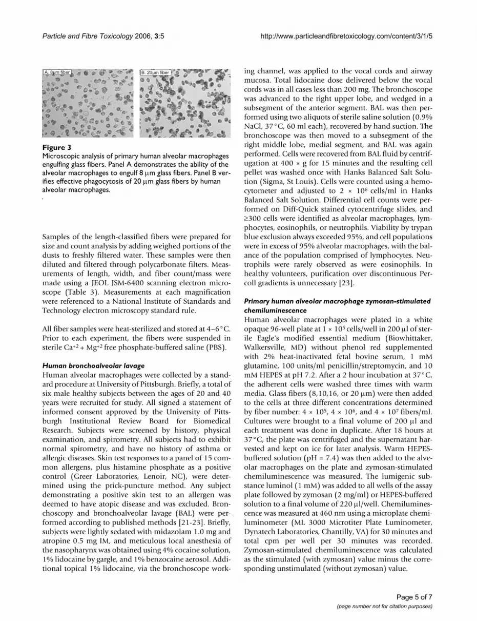
Microscopic examination of primary human alveolar macrophagesFigure 3, panel A, demonstrates the ability of human mac-rophages to engulf 8 µm glass fibers in vitro. Figure 3,panel B, demonstrates that human macrophages can suc-cessfully engulf 20 µm glass fibers. Data from Blake et al.revealed incomplete phagocytosis by rat alveolar macro-phages occurred with glass fibers ≥ 17 µm in length [7].These results suggest human alveolar macrophages cancompletely phagocytize fiber lengths that rat alveolarmacrophages can not. Therefore, frustrated phagocytosisand its possible effects do not appear to be a factor withhuman alveolar macrophages exposed to fibers ≤ 20 µmin length.
DiscussionThe present study employed an in vitro system to exposeprimary human alveolar macrophages to monodisperseJM-100 glass fibers of different target lengths. Using thissystem, human alveolar macrophages were assessed forlength-dependent cellular effects and results were com-pared to previous data obtained with rat alveolar macro-phages. The data presented here reveal differences in theresponses of rat versus human macrophages to glass fibersof various lengths.
Previous studies have shown that rat alveolar and mouseperitoneal macrophages attempt to engulf glass fibers andthat successful phagocytosis is influenced by fiber length[5,7]. Short (7 µm) glass fibers were completely engulfed,
whereas long (17 µm) glass fibers were only partiallyengulfed. Human alveolar macrophages have a largerdiameter than the rat counterpart; therefore, it washypothesized that these phagocytes would completelyengulf longer fibers and the absence of frustrated phago-cytosis would attenuate cytotoxicity. Overall, the data sup-ported this hypothesis.
Human alveolar macrophage function, measured aszymosan-stimulated chemiluminescence, was signifi-cantly affected by fiber concentration but not fiber lengthover the range of 8–20 µm (Figure 2). The lowest of threefiber concentrations (4 × 10⁵ fibers/ml or 1:1 fiber:cellratio) primed the cellular response to zymosan while thehighest concentration (4 × 10⁷fibers/ml or 80:1 fiber:cellratio) decreased this response from control. The activationlevel of cells exposed to the intermediate fiber concentra-tion (4 × 10⁶ fibers/ml or 8:1 fiber:cell ratio) was similarto that of controls). In contrast, our previous findingsshowed no increased priming effect of JM-100 glass fibersin rat alveolar macrophages at any concentration. This dif-ferential response between human and rat cells may bethe result of several possible factors. First, fiber to cell ratiohas been shown in our laboratory to be important in theactivation of rodent macrophages. Prior studies revealed afiber to cell ratio of at least 5:1 is required for significant
Inhibition of zymosan-stimulated primary human alveolar macrophage chemiluminescence following an18 hour expo-sure to JM-100 glass fibersFigure 2Inhibition of zymosan-stimulated primary human alveolar macrophage chemiluminescence following an18 hour expo-sure to JM-100 glass fibers. Data are presented as percent control from 100%. Bars represent mean values ± S.E of three independent experiments. * - indicates a significant dif-ference between the lowest (4 × 10⁵ fibers/ml or 1:1 fiber:cell) and intermediate (4 × 10⁶ fibers/ml or 8:1 fiber:cell) fiber concentrations, †- between the lowest and highest (4 × 10⁷ fibers/ml or 80:1 fiber:cell) fiber concentrations, and $ - between the intermediate and highest fiber concentrations of the same fiber length (p ≤ 0.05). Fiber length did not affect human alveolar macrophage function.
Table 1: Relative Toxicity (% of total LDH) of Rat Alveolar Macrophages Exposed to Length Classified Glass Fibers
Fiber Length (µm) Log (fiber #/ml)
106 (1:2.5 fiber:cell) 107 (4:1 fiber:cell)
3 ND ND4 ND ND7 ND 2%17 19%* 45%*
Note: Adapted from Blake et al., 1998; ND is not detected ; LDH is lactate dehydrogenase; * represents a significant difference from control
Page 3 of 7(page number not for citation purposes)

Particle and Fibre Toxicology 2006, 3:5 http://www.particleandfibretoxicology.com/content/3/1/5
tumor necrosis factor-α (TNF-α) production in mousemacrophages [5]. The effective fiber to cell ratio found toelicit human macrophage cellular priming in this studywas 1:1. Approximately a 1:2.5 fiber to cell ratio (106 fib-ers/ml) was used as a low concentration in the rat macro-phage studies reported by Blake et al., which may not havebeen high enough to prime the cellular response tozymosan above control following short fiber phagocytosis[7]. Secondly, luminol was used as the light enhancer inthe present study, but lucigenin was used in the rat cellstudy. Luminol detects multiple reactive species com-pared to lucigenin, which is a superoxide specific lightenhancer [16-18]. Most likely, superoxide is not the soleoxidant produced in glass fiber-exposed cells. Therefore,this experimental system may have an enhanced detectionlevel compared to the past system. Finally, the result mayreflect inherent differences in the oxidant production ofrat and human alveolar macrophages. Human alveolarmacrophages are reportedly more active than the rat in theproduction of reactive species following particulate expo-sure [19]. Whether this is the case with glass fibers wouldneed further investigation.
It was found that no fiber length or concentration inducedsignificant cellular membrane damage as assessed bycytosolic LDH release into the cellular supernatantalthough a concentration-dependent trend was apparent(Figure 1). Castranova et al. reported effects of metallicions on particle-stimulated oxygen consumption andchemiluminescence in alveolar macrophages, i.e., func-tional assays, occurs at concentrations that do not affectthe integrity of the cellular membrane [20]. Therefore, areason for the lack of a significant LDH concentration-response may be that the cellular function is typicallyaffected before membrane damage making the chemilu-minescence response a more sensitive measure of cyto-toxic effects.
In summary, this study reported no effects of fiber lengthover the range of 8–20 µm on the human alveolar macro-phage response. This conflicts with previous data where
increased glass fiber concentration decreased chemilumi-nescence and increased LDH in rat alveolar macrophagesbut major length-dependent effects were evident. The rea-son for this discrepancy most likely is due to the largerdiameter of human alveolar macrophages, resulting in thelack of frustrated phagocytosis in the present experimentalsystem. Microscopic analysis verified these cells were ableto completely engulf even the longest fiber sample tested(20 µm) (Figure 3B). Blake et al. demonstrated cellulareffects on rat alveolar macrophages (diameter ~13 µm)using 17 and 33 µm glass fibers, approximately 4–20 µmlonger than the cells (multiple rat macrophages wereoften observed adhered to a long fiber with the fiberappearing to protrude through the cellular membrane)[7]. In the present study, the longest fiber sample used was20 µm, only 2 µm longer than the human alveolar macro-phage cellular diameter; therefore not a sufficient lengthto cause frustrated phagocytosis. Consequently, furtherstudies are needed using fiber samples over a range of 20–40 µm in length. Assuming similar mechanisms areinvolved in human and rat fiber phagocytosis, resultsmost likely would agree with those reported by Blake andcolleagues.
ConclusionThis study showed: 1.) an absence of length-associatedcytotoxicity in primary human alveolar macrophages thatwas previously observed in rat alveolar macrophagestreated in vitro with length-classified glass fibers 2.) a pos-sible mechanism for this absence of length-dependentcytotoxicity may be the lack of frustrated phagocytosis inthe human macrophages versus the rat 3.) human alveolarmacrophages appear to be activated by glass fibers ofmonodisperse lengths at a fiber to cell ratio of approxi-mately 1:1, while significant cytotoxicity was observedonly at an excessively high fiber:cell ratio of 80:1. In con-clusion, the use of monodisperse length-classified fibersamples will aid in the determination of specific fiberlengths that macrophages can engulf. In addition, thesepreliminary data may aid in the design of future in vivoexperiments using fibrous particles.
MethodsFiber samplesBulk samples of JM-100 glass (Manville code 100 sup-plied by John Mansville Corporation) were first milled,aerosolized, and separated into length categories usingdielectrophoresis as previously described [3,4]. The die-lectrophoretic classifier was operated in a differentialmode so that fibers with narrow length distributions wereextracted in an air suspension at the end of the classifier.These length-classified fiber samples were collected onpolycarbonate (Nuclepore) filters at rates up to 1 mg/day.Fibers were scraped off the filters for microscopic analysisand for biological experiments.
Table 2: Relative Toxicity (% Decreased Activation vs. Control) of Rat Alveolar Macrophages Exposed to Length Classified Glass Fibers
Fiber Length (µm) Log (fiber #/ml)
106 (1:2.5 fiber:cell) 107 (4:1 fiber:cell)
3 ND ND4 ND ND7 ND 15%17 53%* 100%*
Note: Adapted from Blake et al., 1998; ND is not detected;* represents a significant decrease from control
Page 4 of 7(page number not for citation purposes)
Particle and Fibre Toxicology 2006, 3:5 http://www.particleandfibretoxicology.com/content/3/1/5
Samples of the length-classified fibers were prepared forsize and count analysis by adding weighed portions of thedusts to freshly filtered water. These samples were thendiluted and filtered through polycarbonate filters. Meas-urements of length, width, and fiber count/mass weremade using a JEOL JSM-6400 scanning electron micro-scope (Table 3). Measurements at each magnificationwere referenced to a National Institute of Standards andTechnology electron microscopy standard rule.
All fiber samples were heat-sterilized and stored at 4–6°C.Prior to each experiment, the fibers were suspended insterile Ca+2 + Mg+2 free phosphate-buffered saline (PBS).
Human bronchoalveolar lavageHuman alveolar macrophages were collected by a stand-ard procedure at University of Pittsburgh. Briefly, a total ofsix male healthy subjects between the ages of 20 and 40years were recruited for study. All signed a statement ofinformed consent approved by the University of Pitts-burgh Institutional Review Board for BiomedicalResearch. Subjects were screened by history, physicalexamination, and spirometry. All subjects had to exhibitnormal spirometry, and have no history of asthma orallergic diseases. Skin test responses to a panel of 15 com-mon allergens, plus histamine phosphate as a positivecontrol (Greer Laboratories, Lenoir, NC), were deter-mined using the prick-puncture method. Any subjectdemonstrating a positive skin test to an allergen wasdeemed to have atopic disease and was excluded. Bron-choscopy and bronchoalveolar lavage (BAL) were per-formed according to published methods [21-23]. Briefly,subjects were lightly sedated with midazolam 1.0 mg andatropine 0.5 mg IM, and meticulous local anesthesia ofthe nasopharynx was obtained using 4% cocaine solution,1% lidocaine by gargle, and 1% benzocaine aerosol. Addi-tional topical 1% lidocaine, via the bronchoscope work-
ing channel, was applied to the vocal cords and airwaymucosa. Total lidocaine dose delivered below the vocalcords was in all cases less than 200 mg. The bronchoscopewas advanced to the right upper lobe, and wedged in asubsegment of the anterior segment. BAL was then per-formed using two aliquots of sterile saline solution (0.9%NaCl, 37°C, 60 ml each), recovered by hand suction. Thebronchoscope was then moved to a subsegment of theright middle lobe, medial segment, and BAL was againperformed. Cells were recovered from BAL fluid by centrif-ugation at 400 × g for 15 minutes and the resulting cellpellet was washed once with Hanks Balanced Salt Solu-tion (Sigma, St Louis). Cells were counted using a hemo-cytometer and adjusted to 2 × 10⁶ cells/ml in HanksBalanced Salt Solution. Differential cell counts were per-formed on Diff-Quick stained cytocentrifuge slides, and≥300 cells were identified as alveolar macrophages, lym-phocytes, eosinophils, or neutrophils. Viability by trypanblue exclusion always exceeded 95%, and cell populationswere in excess of 95% alveolar macrophages, with the bal-ance of the population comprised of lymphocytes. Neu-trophils were rarely observed as were eosinophils. Inhealthy volunteers, purification over discontinuous Per-coll gradients is unnecessary [23].
Primary human alveolar macrophage zymosan-stimulated chemiluminescenceHuman alveolar macrophages were plated in a whiteopaque 96-well plate at 1 × 10⁵ cells/well in 200 µl of ster-ile Eagle's modified essential medium (Biowhittaker,Walkersville, MD) without phenol red supplementedwith 2% heat-inactivated fetal bovine serum, 1 mMglutamine, 100 units/ml penicillin/streptomycin, and 10mM HEPES at pH 7.2. After a 2 hour incubation at 37°C,the adherent cells were washed three times with warmmedia. Glass fibers (8,10,16, or 20 µm) were then addedto the cells at three different concentrations determinedby fiber number: 4 × 10⁵, 4 × 10⁶, and 4 × 10⁷ fibers/ml.Cultures were brought to a final volume of 200 µl andeach treatment was done in duplicate. After 18 hours at37°C, the plate was centrifuged and the supernatant har-vested and kept on ice for later analysis. Warm HEPES-buffered solution (pH = 7.4) was then added to the alve-olar macrophages on the plate and zymosan-stimulatedchemiluminescence was measured. The lumigenic sub-stance luminol (1 mM) was added to all wells of the assayplate followed by zymosan (2 mg/ml) or HEPES-bufferedsolution to a final volume of 220 µl/well. Chemilumines-cence was measured at 460 nm using a microplate chemi-luminometer (ML 3000 Microtiter Plate Luminometer,Dynatech Laboratories, Chantilly, VA) for 30 minutes andtotal cpm per well per 30 minutes was recorded.Zymosan-stimulated chemiluminescence was calculatedas the stimulated (with zymosan) value minus the corre-sponding unstimulated (without zymosan) value.
Microscopic analysis of primary human alveolar macrophages engulfing glass fibersFigure 3Microscopic analysis of primary human alveolar macrophages engulfing glass fibers. Panel A demonstrates the ability of the alveolar macrophages to engulf 8 µm glass fibers. Panel B ver-ifies effective phagocytosis of 20 µm glass fibers by human alveolar macrophages.
Page 5 of 7(page number not for citation purposes)

Particle and Fibre Toxicology 2006, 3:5 http://www.particleandfibretoxicology.com/content/3/1/5
Lactate dehydrogenase activityLDH activity was determined in the culture supernatantby monitoring the LDH catalyzed oxidation of pyruvatecoupled with the reduction of NAD at 340 nm using acommercial kit and a Cobas Mira Plus Transfer Analyzer(Roche Diagnostics Systems, Montclair, NJ).
Microscopic analysisPhotographs of human alveolar macrophages were takenusing an Olympus IX70 inverted light microscopeequipped with a Dage camera.
Data analysisAll data were analyzed using a one-way analysis of vari-ance model (ANOVA) with a significant differencedefined as p ≤ 0.05. Pairwise differences were assessedthrough appropriate contrasts.
AbbreviationsBAL Bronchoalveolar Lavage
JM-100 Manville Code 100 Glass Fibers
LDH Lactate Dehydrogenase
ND Not Detected
PBS Phosphate Buffered Saline
SEM Scanning Electron Microscope
SVFs Synthetic Vitreous Fibers
Competing interestsThe author(s) declare that they have no competing inter-ests.
Authors' contributionsPCZE carried out the in vitro cytotoxicity studies, analyzedthe data, and drafted the manuscript. WJC, BTA, and MPCprovided the human macrophages and provided assist-ance in study design and coordination. GJD and PB pro-
vided the length-classified fiber samples and aided in fiberanalysis. WJ performed the fiber analysis and providedassistance in the study design. TB conducted studies withthe rat alveolar macrophages. VC conceived of the study,participated in its design and coordination and helped inmanuscript preparation. All authors read and approvedthe final manuscript.
DisclaimerThe findings and conclusions in this report are those ofthe authors and do not necessarily represent the views ofthe National Institute for Occupational Safety and Health.
References1. Hesterberg TW, Hart GA: Synthetic vitreous fibers: a review of
toxicology research and its impact on hazard classification.Crit Rev Toxicol 2001, 31(1):1-53.
2. Hesterberg TW, Hart GA, Chevalier J, Miller WC, Hamilton RD,Bauer J, Thevenaz P: The importance of fiber biopersistenceand lung dose in determining the chronic inhalation effectsof X607, RCF1, and chrysotile asbestos in rats. Toxicol ApplPharmacol 1998, 153:68-82.
3. Baron PA, Deye GJ, Fernback J: Length separation of fibers. Aer-osol Sci Technol 1994, 21:179-192.
4. Deye GJ, Gao P, Baron PA, Fernback J: Performance evaluation ofa fiber length classifier. Aerosol Sci Technol 1999, 30:420-437.
5. Ye J, Shi X, Jones W, Rojanasakul Y, Cheng N, Schwegler-Berry D,Baron P, Deye GJ, Li C, Castranova V: Critical role of glass fiberlength in TNF-alpha production and transcription factoractivation in macrophages. Am J Physiol 1999, 276:L426-434.
6. Ye J, Zeidler P, Young S-H, Martinez A, Robinson VA, Jones W, BaronP, Shi X, Castranova V: Activation of mitogen-activated proteinkinase p38 and extracellular signal-regulated kinase isinvolved in glass fiber-induced tumor necrosis factor-α pro-duction in macrophages. J Biol Chem 2001, 276:5360-5367.
7. Blake T, Castranova V, Schwegler-Berry D, Baron P, Deye GJ, Li C,Jones W: Effect of fiber length on glass microfiber cytotoxic-ity. J Toxicol Environ Health A 1998, 54:243-259.
8. Archer VE: Carcinogenicity of fibers and films: a theory. MedHypotheses 1979, 5:1257-1262.
9. Dorger M, Munzing S, Allmeling A-M, Messmer K, Krombach F: Dif-ferential responses of rat alveolar and peritoneal macro-phages to man-made vitreous fibers in vitro. Environ Res 2001,85:207-214.
10. Oberdorster G: Toxicokinetics and effects of fibrous and non-fibrous particles. Inhal Toxicol 2002, 14:29-56.
11. Vallyathan V, Mega JF, Shi X, Dalal NS: Enhanced generation offree radicals from phagocytes induced by mineral dusts. AmJ Respir Cell Mol Biol 1992, 6:404-413.
12. Hart GA, Kathman LM, Hesterberg TW: In vitro cytotoxicity ofasbestos and man-made vitreous fibers: roles of fiber length,diameter and composition. Carcinogenesis 1994, 15:971-977.
Table 3: Physical Characteristics of JM-100 Glass Fibers Determined by SEM
Measurement Length Classified Sample
8 µm 10 µm 16 µm 20 µm
Lengtha (µm) 8.38 ± 2.89 10.40 ± 4.63 16.22 ± 1.90 18.90 ± 2.64Width (µm) 0.69 ± 0.29 0.70 ± 0.29 1.08 ± 0.43 0.97 ± 0.47Fiber #/mg 11.4 × 10⁷ 8.95 × 10⁷ 2.2 × 10⁷ 2.92 × 10⁷
aLength measurements reported for the 16 and 20 µm fiber samples exclude short fiber populations that occasionally adhered to the long fibers during classification. Prior to use, fiber suspensions were gently vortexed rather than sonicated to minimize release of these short fibers. Inclusion of short fibers in the analysis slightly lowered the mean fiber length in the distribution for the 16 and 20 µm fiber samples. Note: Values for length and width are means ± SD
Page 6 of 7(page number not for citation purposes)

Particle and Fibre Toxicology 2006, 3:5 http://www.particleandfibretoxicology.com/content/3/1/5
Publish with BioMed Central and every scientist can read your work free of charge
"BioMed Central will be the most significant development for disseminating the results of biomedical research in our lifetime."
Sir Paul Nurse, Cancer Research UK
Your research papers will be:
available free of charge to the entire biomedical community
peer reviewed and published immediately upon acceptance
cited in PubMed and archived on PubMed Central
yours — you keep the copyright
Submit your manuscript here:http://www.biomedcentral.com/info/publishing_adv.asp
BioMedcentral
13. Hesterberg TW, Barrett JC: Dependence of asbestos- and min-eral dust-induced transformation of mammalian cells in cul-ture on fiber dimension. Cancer Res 1984, 44:2170-2180.
14. Miller BG, Searl A, Davis JMG, Donaldson K, Cullen RT, Bolton RE,Duncan B, Soutar CA: Influence of fibre length, dissolution, andbiopersistence on the production of mesothelioma in the ratperitoneal cavity. Am Occup Hyg 1999, 43:155-166.
15. Lapp NL, Lewis D, Schwegler-Berry D, Abrons H, Castranova V:Bronchoalveolar lavage in asymptomatic underground coalminers. Proceedings of the 3rd Symposium on Respirable Dust in theMineral Industries Society for Mining, Metallurgy and Exploration: 1991;Colorado 1991:159-169.
16. DeChatelet LR, Long GD, Shirley PS, Bass DA, Thomas MJ, Hender-son FW, Cohen MS: Mechanism of the luminol-dependentchemiluminescence of human neutrophils. J Immunol 1982,129:1589-1593.
17. Gyllenhammar H: Lucigenin chemiluminescence in the assess-ment of neutrophil superoxide production. J Immunol Meth1987, 97:209-213.
18. Ischiropoulos H, Kumae T, Kikkawa Y: Effect of interferon induc-ers on superoxide anion generation from rat liver micro-somes detected by lucigenin chemiluminescence. BiochemBiophys Res Commun 1989, 161:1042-1048.
19. Rahman Q, Norwood J, Hatch G: Evidence that exposure of par-ticulate air pollutants to human and rat alveolar macro-phages leads to differential oxidative response. BiochemBiophys Res Commun 1997, 240:669-672.
20. Castranova V, Bowman L, Reasor MJ, Miles PR: Effects of heavymetal ions on selected oxidative metabolic processes in ratalveolar macrophages. Toxicol Appl Pharmacol 1980, 53:14-23.
21. Calhoun WJ, Dick EC, Schwartz LB, Busse WW: A common coldvirus, rhinovirus 16, potentiates airway inflammation aftersegmental antigen bronchoprovocation in allergic subjects. JClin Invest 1994, 94:2200-2008.
22. Calhoun WJ, Jarjour NN: Macrophages and macrophage diver-sity. In Asthma and Rhinitis Edited by: Busse W, Holgate S. Cambridge:Blackwell Science; 1995.
23. Calhoun WJ, Reed HE, Moest DR, Stevens CA: Enhanced superox-ide production by alveolar macrophages and airspace cells,airway inflammation, and alveolar macrophage densitychanges follow segmental antigen bronchoprovocation inallergic subjects. Am Rev Respir Dis 1992, 145:317-325.
Page 7 of 7(page number not for citation purposes)


