Embed Size (px)
Citation preview
ANTIMICROBIAL AGENTS AND CHEMOTHERAPY, Sept. 2010, p. 3834–3841 Vol. 54, No. 90066-4804/10/$12.00 doi:10.1128/AAC.00125-10Copyright © 2010, American Society for Microbiology. All Rights Reserved.
Stable Synthetic Cationic Bacteriochlorins as SelectiveAntimicrobial Photosensitizers�§
Liyi Huang,1,2,3 Ying-Ying Huang,1,2,4 Pawel Mroz,1,2 George P. Tegos,1,2† Timur Zhiyentayev,1,5
Sulbha K. Sharma,1 Zongshun Lu,1,6 Thiagarajan Balasubramanian,7 Michael Krayer,8Christian Ruzie,8 Eunkyung Yang,9 Hooi Ling Kee,9 Christine Kirmaier,9
James R. Diers,10 David F. Bocian,10 Dewey Holten,9Jonathan S. Lindsey,8 and Michael R. Hamblin1,2,11*
Wellman Center for Photomedicine, Massachusetts General Hospital, Boston, Massachusetts1; Department of Dermatology,Harvard Medical School, Boston, Massachusetts2; Department of Infectious Diseases, First Affiliated College & Hospital,
Guangxi Medical University, Nanning, China3; Aesthetic and Plastic Center of Guangxi Medical University, Nanning,China4; Chemistry Department, Massachusetts Institute of Technology, Cambridge, Massachusetts5; Department ofGastroenterology, Tianjin Medical University General Hospital, Tianjin, China6; NIRvana Pharmaceuticals, Inc.,
Raleigh, North Carolina7; Department of Chemistry, North Carolina State University, Raleigh, North Carolina8;Department of Chemistry, Washington University, St. Louis, Missouri9; Department of Chemistry,
University of California Riverside, Riverside, California10; and Harvard-MIT Division ofHealth Sciences and Technology, Cambridge, Massachusetts11
Received 28 January 2010/Returned for modification 10 March 2010/Accepted 26 May 2010
Photodynamic inactivation is a rapidly developing antimicrobial treatment that employs a nontoxic photo-activatable dye or photosensitizer in combination with harmless visible light to generate reactive oxygen speciesthat are toxic to cells. Tetrapyrroles (e.g., porphyrins, chlorins, bacteriochlorins) are a class of photosensitizersthat exhibit promising characteristics to serve as broad-spectrum antimicrobials. In order to bind to andefficiently penetrate into all classes of microbial cells, tetrapyrroles should have structures that contain (i) oneor more cationic charge(s) or (ii) a basic group. In this report, we investigate the use of new stable syntheticbacteriochlorins that have a strong absorption band in the range 720 to 740 nm, which is in the near-infraredspectral region. Four bacteriochlorins with 2, 4, or 6 quaternized ammonium groups or 2 basic amine groupswere compared for light-mediated killing against a Gram-positive bacterium (Staphylococcus aureus), a Gram-negative bacterium (Escherichia coli), and a dimorphic fungal yeast (Candida albicans). Selectivity was assessedby determining phototoxicity against human HeLa cancer cells under the same conditions. All four compoundswere highly active (6 logs of killing at 1 �M or less) against S. aureus and showed selectivity for bacteria overhuman cells. Increasing the cationic charge increased activity against E. coli. Only the compound with basicgroups was highly active against C. albicans. Supporting photochemical and theoretical characterizationstudies indicate that (i) the four bacteriochlorins have comparable photophysical features in homogeneoussolution and (ii) the anticipated redox characteristics do not correlate with cell-killing ability. These resultssupport the interpretation that the disparate biological activities observed stem from cellular binding andlocalization effects rather than intrinsic electronic properties. These findings further establish cationicbacteriochlorins as extremely active and selective near-infrared activated antimicrobial photosensitizers,and the results provide fundamental information on structure-activity relationships for antimicrobialphotosensitizers.
Photodynamic therapy (PDT) employs a nontoxic dyetermed a photosensitizer and low-intensity visible light, whichin the presence of molecular oxygen produces reactive oxygenspecies, such as singlet oxygen, superoxide, and hydroxyl rad-icals (15). PDT has the advantage of dual selectivity in that thephotosensitizer can be targeted to a destination cell or tissue,
and in addition the illumination can be spatially directed to thelesion (7, 48). PDT has its origins over a hundred years ago inthe discovery of light-mediated killing of microorganisms (35)but since then has been principally developed as a treatmentfor cancer (8) and age-related macular degeneration (58). Pho-todynamic inactivation (PDI) is the term used to describe theuse of PDT to inactivate an unwanted entity such as a micro-bial cell.
There has been a relentless rise in antibiotic resistance overmany years in most regions of the world and in many differentclasses of microbial cells (41). In recent times the phenomenonhas become even more worrying, with concerns that hithertofairly trivial infections could again become untreatable as inthe days before antibiotics were discovered (3). In fact, thepresent time has been termed the “end of the antibiotic era”(1). The rise in multidrug resistance among microbial patho-
* Corresponding author. Mailing address: BAR414, 40 BlossomStreet, Wellman Center for Photomedicine, Massachusetts GeneralHospital, Boston, MA 02114-2696. Phone and fax: (617) 726-6182.E-mail: [email protected].
† Present address: Department of Pathology, University of NewMexico School of Medicine, 2325 Camino de Salud, Albuquerque, NM87131.
§ Supplemental material for this article may be found at http://aac.asm.org/.
� Published ahead of print on 12 July 2010.
3834
gens has motivated an international search for alternative an-timicrobial strategies, particularly those which could be appliedto infections in wounds and burns (32).
PDI has attracted attention as a possible alternative treat-ment for localized infections (14, 19, 27). In this treatment, thephotosensitizer is topically or locally applied to the infectedtissue and, after a relatively short time interval, light is deliv-ered to the area. Depending on the effectiveness of the anti-microbial photosensitizer, up to three logs of bacterial or fun-gal cells can be killed without causing unacceptable damage tothe host tissue (11). PDI is thought to be equally effectiveagainst multidrug-resistant and naïve species (49), and in ad-dition the PDI treatment itself is unlikely to cause resistance toarise (26). It should be noted that the lack of development ofresistance after PDT is generally difficult to prove experimen-tally but can be shown in particular instances.
Gram-negative bacteria are resistant to PDI with many com-monly used photosensitizers that readily lead to phototoxicityfor Gram-positive species (29). On the other hand, photosen-sitizers bearing a cationic charge (31, 33, 37) or the use ofagents that increase the permeability of the outer membrane(38) are known to increase the efficacy of killing of Gram-negative organisms. The ideal photosensitizer for killing bac-teria should possess an overall cationic charge and preferablymultiple cationic charges (16, 52).
Photosensitizers based on the bacteriochlorin backbonehave been studied as potential PDT agents for cancer andnononcological applications (10, 43, 46, 55). The large absorp-tion feature in the near-infrared spectral region, which is char-acteristic of bacteriochlorins, is considered to be ideal for max-imizing light penetration through tissue. This is so becauseboth absorption and scattering of light in the 700- to 800-nm
region are minimal (40, 42). However, in addition to goodoptical properties, it is necessary for a photosensitizer mole-cule to possess the appropriate structural characteristics thatwill optimize the binding to and penetration into microbialcells. For antimicrobial applications, the effective molecularfeatures are likely to include the presence of positively chargedsubstituents such as quaternized ammonium groups.
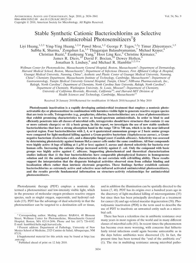
A de novo synthetic pathway to bacteriochlorins that containa geminal dimethyl group in each pyrroline ring has beendeveloped recently (22). This structural attribute blocks adven-titious dehydrogenation (to form the chlorin) and thereby af-fords a stable macrocycle. This synthetic route has provided anumber of bacteriochlorin building blocks, which providedmodular access to bacteriochlorins 1 to 4 (Fig. 1). The fourmolecules were designed to allow investigation of the struc-ture-activity relationship among differently charged bacterio-chlorins. Bacteriochlorin 1 is a neutral species with two basicamino groups; bacteriochlorins 2 to 4 contain 2, 4, or 6 cationiccharges, respectively. The synthesis of bacteriochlorins 1 to 3has been reported (44), and the synthesis of bacteriochlorin 4is described herein (see the supplemental material) making useof known routes to bacteriochlorin (44) and tetraaminoalkane(34) building blocks. The photophysical and molecular orbitalcharacteristics of all four bacteriochlorins have been investi-gated as part of this study. The goals of the present study wereto (i) test bacteriochlorins 1 to 4 as antimicrobial photosensi-tizers against a panel of human pathogens of different taxo-nomic classifications and (ii) determine selectivity of the fourbacteriochlorins for killing microbial cells versus mammalian(human cancer) cells using the same incubation time and otherexperimental conditions.
MATERIALS AND METHODS
Determination of logP values. A mixture of 2 ml of octanol and 2 ml of waterin a 20-ml scintillation vial was stirred at room temperature for 3 h. Then, lessthan 0.5 mg of bacteriochlorin was introduced. Stirring was continued at roomtemperature at 100 to 200 rpm for 24 h. The mixture was allowed to stand for 30min to allow separation of the phases. A 30-�l aliquot of each phase was placedin 3.0 ml of dimethyl sulfoxide (DMSO), and the absorption spectrum of eachphase was measured. The ratio of the peak intensity of the near-infrared feature,the Qy(0,0) band (Fig. 2), for the two phases (octanol/water) was calculated.When there was no detectable amount of bacteriochlorin in a given phase, thenoise level (A � 0.001) was used as the limiting reading, and the logP value was
FIG. 1. Bacteriochlorin photosensitizers.
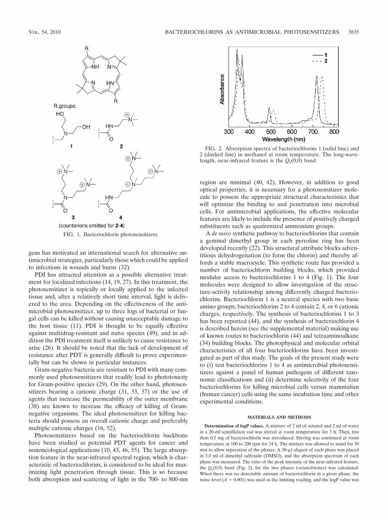
FIG. 2. Absorption spectra of bacteriochlorins 1 (solid line) and2 (dashed line) in methanol at room temperature. The long-wave-length, near-infrared feature is the Qy(0,0) band.
VOL. 54, 2010 BACTERIOCHLORINS AS ANTIMICROBIAL PHOTOSENSITIZERS 3835
bounded accordingly; these measured quantities are here denoted mLogP values.The values were also calculated based simply on the bacteriochlorin structureusing ACD Labs (Toronto, Canada) V11.01 software; these calculated quantitiesare here denoted cLogP values. The cLogP and mLogP values are presented inTable 1. A positive versus negative logP value reflects preferential solubilizationin the octanol versus water phases, respectively.
Photophysical measurements. Photophysical measurements were preformedas described previously (20). Measurement of the fluorescence (�f) and tripletexcited state quantum yields (�isc) and singlet (�S) and triplet (�T) lifetimesutilized Ar-purged solutions (methanol or 2-methyltetrahydrofuran) except thatthe �T values for bacteriochlorin 2 in methanol and bacteriochlorin 3 in ethanolutilized rigorously degassed (by freeze-pump-thaw) solutions. The �f values weredetermined with respect to 8,8,18,18-tetramethylbacteriochlorin (50) in Ar-purged toluene, for which �f � 0.125 was established with respect to chlorophylla in benzene (�f � 0.325 [56]) and free base tetraphenylporphyrin in toluene(�f � 0.09 [12]) using Soret and Qx excitation. Triplet yields were determinedusing a reference technique to facilitate comparisons (20). First, a value of �isc �0.57 was measured for bacteriochlorin 3. This value, along with the �f and �S
values for this compound (Table 1), gives a value of kic � (11.4 ns)�1 for the rateconstant for internal conversion of the lowest singlet excited state to the groundstate via the expression kic � (�S)�1[1 � �f � �isc]. This value is in goodagreement with the average value of kic � (10 ns)�1 obtained for a number ofanalogous 3,13-substituted synthetic bacteriochlorins (unpublished work). Thiskic value was used to obtain the triplet yield for each of the bacteriochlorins usingthe expression �isc
ref � 1 � �f � kic � �S (Table 1).Molecular orbital calculations. Density-functional-theory (DFT) calculations
were performed with Spartan ’06 for Windows (Wavefunction, Inc.) using thehybrid B3LYP functional, 6-31G* basis set, and equilibrium geometries werefully optimized using the default program parameters (24).
Microorganisms and culture conditions. Staphylococcus aureus 8325–4 andEscherichia coli K-12 (both wild type) as well as the DAY286 reference strain ofCandida albicans (39) were employed. Planktonic bacterial cells were cultured inbrain heart infusion (BHI) broth with aeration at 37°C in mid-log-growth phase(unless otherwise stated) (39). Yeasts were cultured in yeast-peptone-dextrose(YPD) broth with aeration at 30°C. The cell number was assessed with a hema-cytometer (47).
In vitro PDI and viability assessment/determination. A cell suspension con-sisting of 108 cells/ml for bacteria (107 cells/ml for Candida albicans [6]) wasincubated with various concentrations of the bacteriochlorins for 30 min at roomtemperature in the dark. Aliquots (1 ml) were transferred to a 24-well plate andilluminated at room temperature with a 732-nm laser source (732/6 Diode Laser,Pharmacyclics, Sunnyvale, CA) and a lens adjusted to give a uniform spot of 2.5cm in diameter with an irradiance of 130 mW/cm2 as measured with a powermeter (model DMM 199 with 201 standard head; Coherent, Santa Clara, CA). Afluence of 10 J/cm2 was delivered over a period of 77 s. Cells treated withbacteriochlorin in the dark were incubated covered with aluminum foil for thesame time as the PDT groups (30 min).
At the completion of illumination (or dark incubation), the contents of thewells were mixed before sampling. Aliquots (100 �l) were taken from each wellto determine CFU. The aliquots were serially diluted 10-fold in phosphate-
buffered saline (PBS) to give dilutions of 10�1 to 10�5 times in addition to theoriginal concentration; then, 10-�l aliquots of each of the dilutions were streakedhorizontally on square BHI or YPD (for Candida) plates by the method of Jettand colleagues (18). Plates were streaked in triplicate and incubated for 12 to36 h at 30°C or 37°C in the dark to allow colony formation. A control group ofcells treated with light alone (no bacteriochlorin added) showed the same num-ber of CFU as the absolute control (data not shown). Survival fractions wereroutinely expressed as ratios of CFU of microbial cells treated with light andbacteriochlorin (or bacteriochlorin in the absence of light) to CFU of microbestreated with neither.
Confocal microscopy of Candida. C. albicans cells (107 cells/ml) were incubatedwith bacteriochlorin 1 or 2 at a concentration of 100 �M for 30 min in PBS (pH7.4) at room temperature. Cells were washed in PBS, pelleted, and resuspendedin 200 �l PBS, and 10 �l was placed on a microscope slide and covered with acoverslip. An Olympus Fluoview 1000-MPE multiphoton confocal microscope(Olympus Corporation, Tokyo, Japan) was used to image the cells at a resolutionof 1,024 � 1,024 pixels with a 100 � 1.4-numerical aperture (NA) oil immersionlens. The microscope used excitation with a 488-nm argon laser and emissionbandpass filter (525 � 10 nm) for green autofluorescence and excitation with a405-nm violet diode laser and a 655- to 755-nm bandpass filter for near-infraredbacteriochlorin fluorescence (false-colored red). Images were acquired usingFluoview 10-ASW software (version 2.0; Olympus Corporation, Tokyo, Japan).
PDT killing of mammalian cells. A human cervical cancer cell line, HeLa, wasobtained from ATCC (Manassas, VA). The cells were cultured in RPMI-1640medium with L-glutamine and NaHCO3 (Gibco-Invitrogen, Carlsbad, CA) sup-plemented with 10% heat-inactivated fetal bovine serum and penicillin (100U/ml) (Sigma, St. Louis, MO) at 37°C in 5% CO2-humidified atmosphere in75-cm2 flasks (Falcon-Invitrogen, Carlsbad, CA). When the cells reached 80%confluence, they were washed with PBS and harvested with 2 ml of 0.25%trypsin-EDTA solution (Sigma). Cells were then centrifuged and counted intrypan blue to ensure viability and plated at a density of 5,000/well in flat-bottom96-well plates (Fisher Scientific, Pittsburgh, PA). On the following day, dilutionsof bacteriochlorins 1 to 4 were prepared in two different kinds of medium: (i)complete growth medium with 10% serum and (ii) serum-free medium. Thesedilutions (0.01 to 10 �M concentrations of bacteriochlorins) were added to thecells for 30 min incubation. The dimethyl sulfoxide concentration in the mediumdid not exceed 0.2%. The medium was replaced, and 10 J/cm2 of illumination wasdelivered. The light spot covered four wells, which were considered one exper-imental group illuminated at the same time. Control groups were as follows: notreatment, light alone, and medium with the same bacteriochlorin dilutionsdescribed above. Following PDT treatment the cells were returned to the incu-bator overnight. Then a 4-h MTT assay (4) was carried out the next day and readat 562 nm using a microplate spectrophotometer (Spectra Max 340 PC; Molec-ular Devices, Sunnyvale, CA). Each experiment was repeated three times.
RESULTS
PDI studies against Gram-positive S. aureus. The best wayto compare the phototoxicity of a group of photosensitizers
TABLE 1. Chemical and photophysical properties of bacteriochlorinsa
Compound
Partition coefficient Qy absorptionb Qy fluorescenceb
�fc �S
d
(ns) �iscrefe �T
f
(�s)
Orbital energy
mLogPg cLogPh �(nm)
fwhm(nm)
�(nm)
fwhm(nm)
HOMO(eV)
LUMO(eV)
1 2.3 4.8 � 1.5 718 18 724 23 0.095 3.8 0.53 190i �4.42 �2.192 �0.5 �1.1 � 1.7 742 23 750 25 0.13 4.0 0.48 77j �4.72 �3.343 �1.4 �5.3 � 1.7 729 19 735 24 0.12 3.5 0.53 54k �5.05 �3.894 �1.7 �5.8 � 1.7 740 24 750 25 �4.56 �3.92
a All data measured for compounds at room temperature.b Peak wavelength (�) and full width at half maximum (fwhm) of spectral feature for compound in aerated methanol.c Fluorescence quantum yield for compound in Ar-purged methanol.d Lifetime of the lowest singlet excited state for compound in Ar-purged methanol determined using fluorescence detection.e Yield of the lowest triplet excited state determined using the expresssion �
iscref � 1 � �f � kic � �S, with kIC � (10 ns)�1 as described in the text.
f Lifetime of the lowest triplet excited state.g Measured logP.h Calculated logP.i In Ar-purged 2-methyltetrahydrofuran.j In freeze-pump-thaw-degassed methanol.k In freeze-pump-thaw-degassed ethanol.
3836 HUANG ET AL. ANTIMICROB. AGENTS CHEMOTHER.
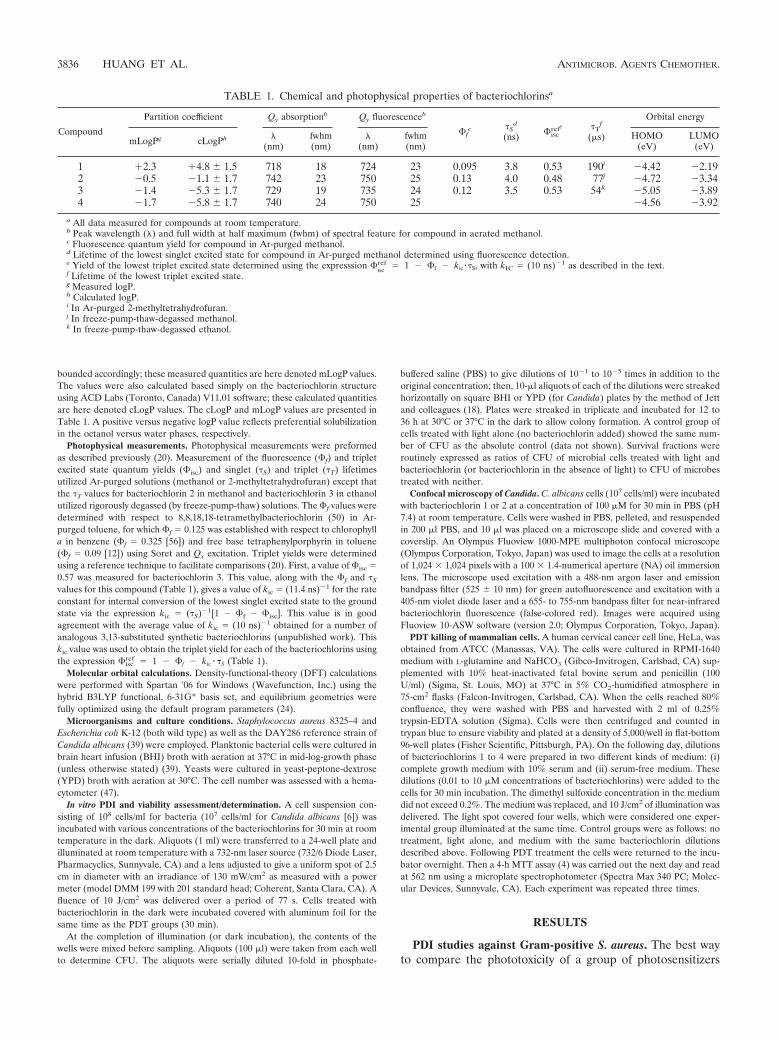
with very different potencies is to vary the concentration overseveral orders of magnitude and determine the survival frac-tion with and without (dark toxicity) a single light dose. Fig-ure 3 displays the survival fraction curves obtained against theGram-positive bacterium S. aureus incubated for 30 min usingbacteriochlorins 1 to 4 with and without illumination (10 J/cm2
732-nm laser light). The noncationic bacteriochlorin 1 pro-duces 1 log of killing at 100 nM and almost 6 logs at 1 �M, andit eliminates the cells at higher concentrations (Fig. 3A). Themost effective compound is the bis-cationic bacteriochlorin 2,which kills a remarkable 5 logs at 100 nM and eliminates thepopulation at 1 �M (Fig. 3B). No dark toxicity is observed.Less effective than bacteriochlorin 2 are the tetrakis-cationicbacteriochlorin 3 (Fig. 3C) and the hexakis-cationic bacterio-chlorin 4 (Fig. 3D), both of which have a small degree ofphototoxicity at 100 nM and kill 5 logs at 1 �M. Again no darktoxicity is seen.
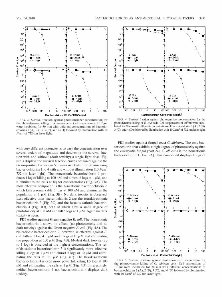
PDI studies against Gram-negative E. coli. The noncationicbacteriochlorin 1 shows no effects (no phototoxicity and nodark toxicity) against the Gram-negative E. coli (Fig. 4A). Thebis-cationic bacteriochlorin 2, however, is effective against E.coli, killing 1 log at 1 �M and 3 logs at 10 �M and eliminatingthe population at 100 �M (Fig. 4B). Modest dark toxicity (upto 1 log) is observed at the highest concentrations. The tet-rakis-cationic bacteriochlorin 3 is significantly more effective,killing 4 logs at 1 �M and almost 6 logs at 10 �M and elimi-nating the cells at 100 �M (Fig. 4C). The hexakis-cationicbacteriochlorin 4 is even more powerful, killing 1.5 logs at 100nM and eliminating the cells at 1 �M (Fig. 4D). Interestingly,neither bacteriochlorin 3 nor bacteriochlorin 4 displays darktoxicity.
PDI studies against fungal yeast C. albicans. The only bac-teriochlorin that exhibits a high degree of phototoxicity againstthe eukaryotic fungal yeast cell C. albicans is the noncationicbacteriochlorin 1 (Fig. 5A). This compound displays 4 logs of
FIG. 5. Survival fraction against photosensitizer concentration forthe photodynamic killing of C. albicans cells. Cell suspensions of107/ml were incubated for 30 min with different concentrations ofbacteriochlorins 1 (A), 2 (B), 3 (C), and 4 (D) followed by illuminationwith 10 J/cm2 of 732-nm laser light.
FIG. 3. Survival fraction against photosensitizer concentration forthe photodynamic killing of S. aureus cells. Cell suspensions of 108/mlwere incubated for 30 min with different concentrations of bacterio-chlorins 1 (A), 2 (B), 3 (C), and 4 (D) followed by illumination with 10J/cm2 of 732-nm laser light.
FIG. 4. Survival fraction against photosensitizer concentration for thephotodynamic killing of E. coli cells. Cell suspensions of 108/ml were incu-bated for 30 min with different concentrations of bacteriochlorins 1 (A), 2 (B),3 (C), and 4 (D) followed by illumination with 10 J/cm2 of 732-nm laser light.
VOL. 54, 2010 BACTERIOCHLORINS AS ANTIMICROBIAL PHOTOSENSITIZERS 3837
killing at 10 �M and gives total elimination (6 logs killing) at100 �M. A much lower degree of phototoxicity is observedwith the bis-cationic bacteriochlorin 2, with 1 to 2 logs of killingat 10 to 100 �M (Fig. 5B). Neither the tetrakis-cationic bac-teriochlorin 3 (Fig. 5C) nor the hexakis-cationic bacterio-chlorin 4 (Fig. 5D) show any PDI effect whatsoever. The onlydark toxicity toward C. albicans is 2 logs in the case of bacte-riochlorin 1 at 100 �M (Fig. 5A).
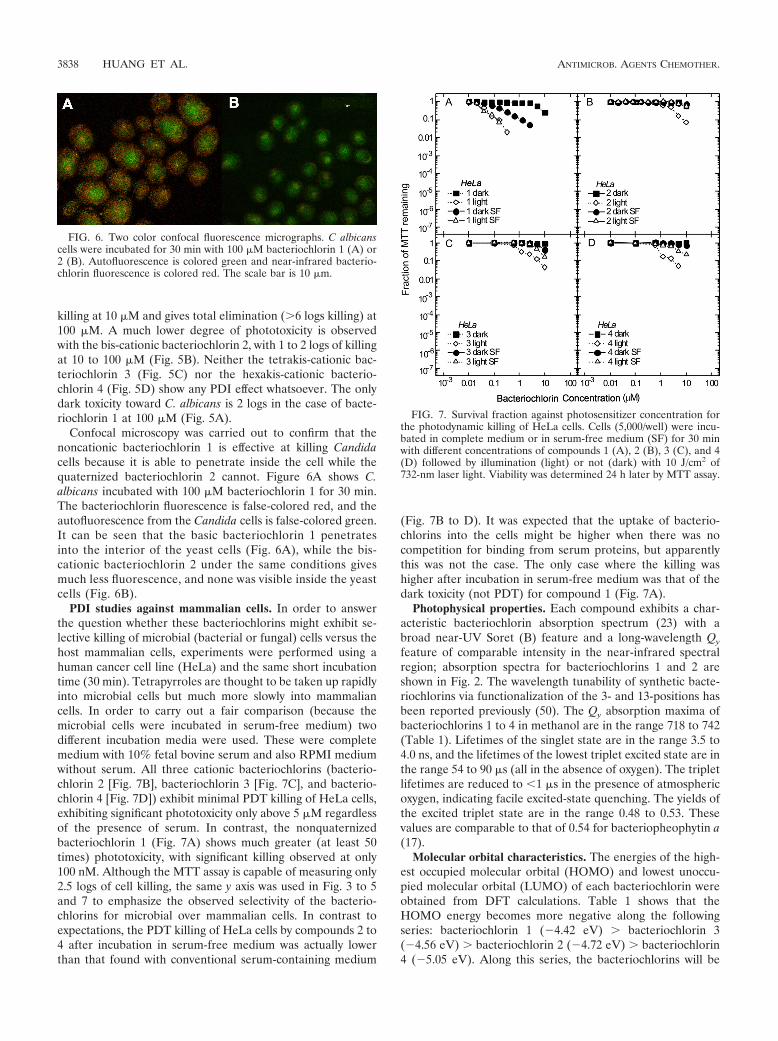
Confocal microscopy was carried out to confirm that thenoncationic bacteriochlorin 1 is effective at killing Candidacells because it is able to penetrate inside the cell while thequaternized bacteriochlorin 2 cannot. Figure 6A shows C.albicans incubated with 100 �M bacteriochlorin 1 for 30 min.The bacteriochlorin fluorescence is false-colored red, and theautofluorescence from the Candida cells is false-colored green.It can be seen that the basic bacteriochlorin 1 penetratesinto the interior of the yeast cells (Fig. 6A), while the bis-cationic bacteriochlorin 2 under the same conditions givesmuch less fluorescence, and none was visible inside the yeastcells (Fig. 6B).
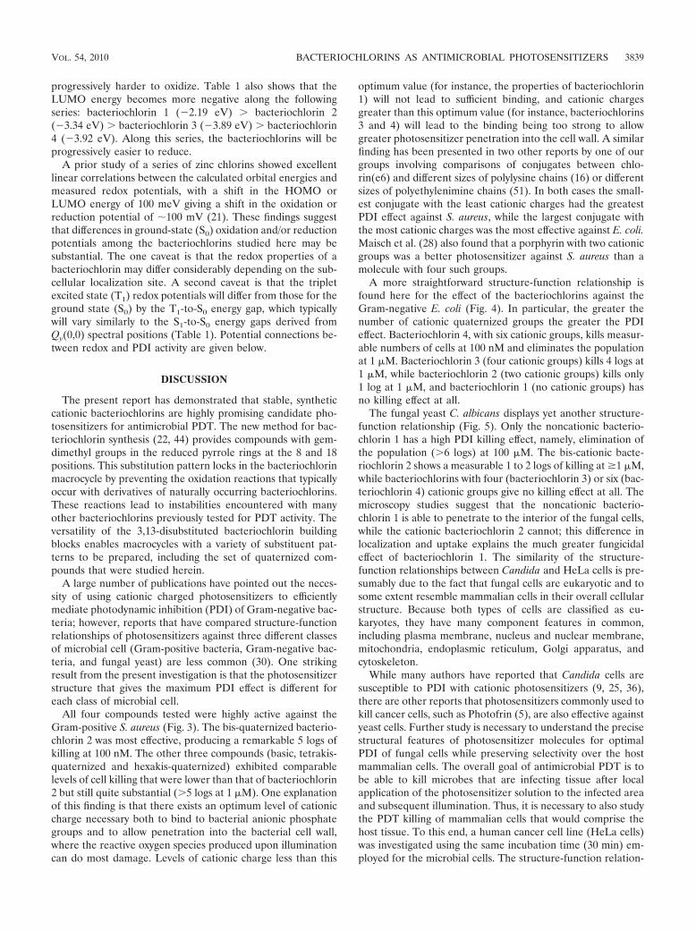
PDI studies against mammalian cells. In order to answerthe question whether these bacteriochlorins might exhibit se-lective killing of microbial (bacterial or fungal) cells versus thehost mammalian cells, experiments were performed using ahuman cancer cell line (HeLa) and the same short incubationtime (30 min). Tetrapyrroles are thought to be taken up rapidlyinto microbial cells but much more slowly into mammaliancells. In order to carry out a fair comparison (because themicrobial cells were incubated in serum-free medium) twodifferent incubation media were used. These were completemedium with 10% fetal bovine serum and also RPMI mediumwithout serum. All three cationic bacteriochlorins (bacterio-chlorin 2 [Fig. 7B], bacteriochlorin 3 [Fig. 7C], and bacterio-chlorin 4 [Fig. 7D]) exhibit minimal PDT killing of HeLa cells,exhibiting significant phototoxicity only above 5 �M regardlessof the presence of serum. In contrast, the nonquaternizedbacteriochlorin 1 (Fig. 7A) shows much greater (at least 50times) phototoxicity, with significant killing observed at only100 nM. Although the MTT assay is capable of measuring only2.5 logs of cell killing, the same y axis was used in Fig. 3 to 5and 7 to emphasize the observed selectivity of the bacterio-chlorins for microbial over mammalian cells. In contrast toexpectations, the PDT killing of HeLa cells by compounds 2 to4 after incubation in serum-free medium was actually lowerthan that found with conventional serum-containing medium
(Fig. 7B to D). It was expected that the uptake of bacterio-chlorins into the cells might be higher when there was nocompetition for binding from serum proteins, but apparentlythis was not the case. The only case where the killing washigher after incubation in serum-free medium was that of thedark toxicity (not PDT) for compound 1 (Fig. 7A).
Photophysical properties. Each compound exhibits a char-acteristic bacteriochlorin absorption spectrum (23) with abroad near-UV Soret (B) feature and a long-wavelength Qy
feature of comparable intensity in the near-infrared spectralregion; absorption spectra for bacteriochlorins 1 and 2 areshown in Fig. 2. The wavelength tunability of synthetic bacte-riochlorins via functionalization of the 3- and 13-positions hasbeen reported previously (50). The Qy absorption maxima ofbacteriochlorins 1 to 4 in methanol are in the range 718 to 742(Table 1). Lifetimes of the singlet state are in the range 3.5 to4.0 ns, and the lifetimes of the lowest triplet excited state are inthe range 54 to 90 �s (all in the absence of oxygen). The tripletlifetimes are reduced to �1 �s in the presence of atmosphericoxygen, indicating facile excited-state quenching. The yields ofthe excited triplet state are in the range 0.48 to 0.53. Thesevalues are comparable to that of 0.54 for bacteriopheophytin a(17).
Molecular orbital characteristics. The energies of the high-est occupied molecular orbital (HOMO) and lowest unoccu-pied molecular orbital (LUMO) of each bacteriochlorin wereobtained from DFT calculations. Table 1 shows that theHOMO energy becomes more negative along the followingseries: bacteriochlorin 1 (�4.42 eV) bacteriochlorin 3(�4.56 eV) bacteriochlorin 2 (�4.72 eV) bacteriochlorin4 (�5.05 eV). Along this series, the bacteriochlorins will be
FIG. 6. Two color confocal fluorescence micrographs. C albicanscells were incubated for 30 min with 100 �M bacteriochlorin 1 (A) or2 (B). Autofluorescence is colored green and near-infrared bacterio-chlorin fluorescence is colored red. The scale bar is 10 �m.
FIG. 7. Survival fraction against photosensitizer concentration forthe photodynamic killing of HeLa cells. Cells (5,000/well) were incu-bated in complete medium or in serum-free medium (SF) for 30 minwith different concentrations of compounds 1 (A), 2 (B), 3 (C), and 4(D) followed by illumination (light) or not (dark) with 10 J/cm2 of732-nm laser light. Viability was determined 24 h later by MTT assay.
3838 HUANG ET AL. ANTIMICROB. AGENTS CHEMOTHER.
progressively harder to oxidize. Table 1 also shows that theLUMO energy becomes more negative along the followingseries: bacteriochlorin 1 (�2.19 eV) bacteriochlorin 2(�3.34 eV) bacteriochlorin 3 (�3.89 eV) bacteriochlorin4 (�3.92 eV). Along this series, the bacteriochlorins will beprogressively easier to reduce.
A prior study of a series of zinc chlorins showed excellentlinear correlations between the calculated orbital energies andmeasured redox potentials, with a shift in the HOMO orLUMO energy of 100 meV giving a shift in the oxidation orreduction potential of �100 mV (21). These findings suggestthat differences in ground-state (S0) oxidation and/or reductionpotentials among the bacteriochlorins studied here may besubstantial. The one caveat is that the redox properties of abacteriochlorin may differ considerably depending on the sub-cellular localization site. A second caveat is that the tripletexcited state (T1) redox potentials will differ from those for theground state (S0) by the T1-to-S0 energy gap, which typicallywill vary similarly to the S1-to-S0 energy gaps derived fromQy(0,0) spectral positions (Table 1). Potential connections be-tween redox and PDI activity are given below.
DISCUSSION
The present report has demonstrated that stable, syntheticcationic bacteriochlorins are highly promising candidate pho-tosensitizers for antimicrobial PDT. The new method for bac-teriochlorin synthesis (22, 44) provides compounds with gem-dimethyl groups in the reduced pyrrole rings at the 8 and 18positions. This substitution pattern locks in the bacteriochlorinmacrocycle by preventing the oxidation reactions that typicallyoccur with derivatives of naturally occurring bacteriochlorins.These reactions lead to instabilities encountered with manyother bacteriochlorins previously tested for PDT activity. Theversatility of the 3,13-disubstituted bacteriochlorin buildingblocks enables macrocycles with a variety of substituent pat-terns to be prepared, including the set of quaternized com-pounds that were studied herein.
A large number of publications have pointed out the neces-sity of using cationic charged photosensitizers to efficientlymediate photodynamic inhibition (PDI) of Gram-negative bac-teria; however, reports that have compared structure-functionrelationships of photosensitizers against three different classesof microbial cell (Gram-positive bacteria, Gram-negative bac-teria, and fungal yeast) are less common (30). One strikingresult from the present investigation is that the photosensitizerstructure that gives the maximum PDI effect is different foreach class of microbial cell.
All four compounds tested were highly active against theGram-positive S. aureus (Fig. 3). The bis-quaternized bacterio-chlorin 2 was most effective, producing a remarkable 5 logs ofkilling at 100 nM. The other three compounds (basic, tetrakis-quaternized and hexakis-quaternized) exhibited comparablelevels of cell killing that were lower than that of bacteriochlorin2 but still quite substantial (5 logs at 1 �M). One explanationof this finding is that there exists an optimum level of cationiccharge necessary both to bind to bacterial anionic phosphategroups and to allow penetration into the bacterial cell wall,where the reactive oxygen species produced upon illuminationcan do most damage. Levels of cationic charge less than this
optimum value (for instance, the properties of bacteriochlorin1) will not lead to sufficient binding, and cationic chargesgreater than this optimum value (for instance, bacteriochlorins3 and 4) will lead to the binding being too strong to allowgreater photosensitizer penetration into the cell wall. A similarfinding has been presented in two other reports by one of ourgroups involving comparisons of conjugates between chlo-rin(e6) and different sizes of polylysine chains (16) or differentsizes of polyethylenimine chains (51). In both cases the small-est conjugate with the least cationic charges had the greatestPDI effect against S. aureus, while the largest conjugate withthe most cationic charges was the most effective against E. coli.Maisch et al. (28) also found that a porphyrin with two cationicgroups was a better photosensitizer against S. aureus than amolecule with four such groups.
A more straightforward structure-function relationship isfound here for the effect of the bacteriochlorins against theGram-negative E. coli (Fig. 4). In particular, the greater thenumber of cationic quaternized groups the greater the PDIeffect. Bacteriochlorin 4, with six cationic groups, kills measur-able numbers of cells at 100 nM and eliminates the populationat 1 �M. Bacteriochlorin 3 (four cationic groups) kills 4 logs at1 �M, while bacteriochlorin 2 (two cationic groups) kills only1 log at 1 �M, and bacteriochlorin 1 (no cationic groups) hasno killing effect at all.
The fungal yeast C. albicans displays yet another structure-function relationship (Fig. 5). Only the noncationic bacterio-chlorin 1 has a high PDI killing effect, namely, elimination ofthe population (6 logs) at 100 �M. The bis-cationic bacte-riochlorin 2 shows a measurable 1 to 2 logs of killing at �1 �M,while bacteriochlorins with four (bacteriochlorin 3) or six (bac-teriochlorin 4) cationic groups give no killing effect at all. Themicroscopy studies suggest that the noncationic bacterio-chlorin 1 is able to penetrate to the interior of the fungal cells,while the cationic bacteriochlorin 2 cannot; this difference inlocalization and uptake explains the much greater fungicidaleffect of bacteriochlorin 1. The similarity of the structure-function relationships between Candida and HeLa cells is pre-sumably due to the fact that fungal cells are eukaryotic and tosome extent resemble mammalian cells in their overall cellularstructure. Because both types of cells are classified as eu-karyotes, they have many component features in common,including plasma membrane, nucleus and nuclear membrane,mitochondria, endoplasmic reticulum, Golgi apparatus, andcytoskeleton.
While many authors have reported that Candida cells aresusceptible to PDI with cationic photosensitizers (9, 25, 36),there are other reports that photosensitizers commonly used tokill cancer cells, such as Photofrin (5), are also effective againstyeast cells. Further study is necessary to understand the precisestructural features of photosensitizer molecules for optimalPDI of fungal cells while preserving selectivity over the hostmammalian cells. The overall goal of antimicrobial PDT is tobe able to kill microbes that are infecting tissue after localapplication of the photosensitizer solution to the infected areaand subsequent illumination. Thus, it is necessary to also studythe PDT killing of mammalian cells that would comprise thehost tissue. To this end, a human cancer cell line (HeLa cells)was investigated using the same incubation time (30 min) em-ployed for the microbial cells. The structure-function relation-
VOL. 54, 2010 BACTERIOCHLORINS AS ANTIMICROBIAL PHOTOSENSITIZERS 3839
ship was to some extent similar to that found for C. albicans,with only the basic bacteriochlorin (bacteriochlorin 1) givingany significant level of killing at concentrations lower than 1�M. Therefore, selective PDT killing of bacteria (and to alesser extent fungal cells) compared to that of mammalian cellsis accomplished with quaternized bacteriochlorins, with thebis-cationic compound giving the highest selectivity for S. au-reus and the hexakis-cationic compound giving the highest se-lectivity for E. coli.
To our knowledge there has been only one prior investiga-tion of bacteriochlorins as antimicrobial photosensitizers.Schastak et al. (45) compared the photodynamic killing of S.aureus, methicillin-resistant S. aureus (MRSA), E. coli, andPseudomonas aeruginosa using a meso-substituted tetramethyl-pyridinium bacteriochlorin with that with a chorin(e6) deriva-tive called Photolon. The cationic bacteriochlorin was able tokill both Gram-positive and Gram-negative bacteria, while theanionic Photolon was only able to kill Gram-positive species.Several groups have studied bacteriochlorins to kill cancer cellsand to treat tumors in vivo. The long-wavelength light between700 and 800 nm that is absorbed by bacteriochlorins is believedto be ideally suited to penetrate living tissue due to reducedabsorption by tissue chromophores and reduced Mie scattering(53). The large extinction coefficient (100,000 M�1cm�1)typical of the bacteriochlorin Qy band is also advantageous forstrong absorption of near-infrared light by the photosensitizer.The Pd-containing bacteriochlorins known as TOOKAD (13,57) and Stakel (2) have been extensively investigated in labo-ratory studies, and, in addition, TOOKAD has been studied inclinical trials of PDT for prostate cancer (54).
The photophysical studies and DFT calculations indicatethat the activity differences observed among bacteriochlorins 1to 4 must stem from cellular binding and localization effectsrather than photochemical properties. Indeed, the yield of thetriplet excited state (from which the reactive oxygen species isproduced) is essentially identical (0.48 to 0.53) for the fourbacteriochlorins, and in each case the lifetime is reduced to �1�s in the presence of atmospheric oxygen, indicating facileexcited-state quenching. Moreover, there is no specific corre-lation between the anticipated differences in redox properties(based on the molecular orbital energies) for the four bacte-riochlorins and their PDI activities against any of the organ-isms studied. The only broad trend is that bacteriochlorins 1and 2 are typically more active than bacteriochlorins 3 and 4,which, all other things being equal, would favor a mechanismof activity that involves reduction rather than oxidation of thephotoexcited bacteriochlorin to the extent that electron trans-fer is involved.
In conclusion, bacteriochlorins with constitutive cationiccharges provided by quaternized ammonium groups are highlyactive antibacterial photosensitizers. The hexakis-cationic bac-teriochlorin 4 is capable of eliminating (6 logs killing) bothGram-positive (S. aureus) and Gram-negative (E. coli) bacteriaat the remarkably low concentration of 1 �M. Good selectivity(4 to 5 logs) for bacteria over mammalian cells is observed.Only the nonquaternized bacteriochlorin 1 shows good PDTkilling of the yeast (C. albicans), and selectivity over mamma-lian cells is lower in this case because both cell types areeukaryotic organisms.
ACKNOWLEDGMENTS
This work was supported by grants from the NIH (R01AI050875 toM.R.H. and R01GM36238 to J.S.L.), a Burroughs-Wellcome fellow-ship (to M.K.), and the JimmyV NCSU Cancer Therapeutics TrainingProgram. L.H., Y.-Y.H. and T.B. were supported by a grant(R41AI072854) from the National Institute of Allergy and InfectiousDiseases to NIRvana Pharmaceuticals, Inc. P.M. was partly supportedby a Genzyme-Partners Translational Research Grant. G.P.T waspartly supported by a Massachusetts Technology Transfer CenterAward. Characterization of the photophysical and redox properties ofthe bacteriochlorins described herein was initially motivated by solar-energy studies and supported by grants from the Division of ChemicalSciences, Geosciences and Biosciences Division, Office of Basic En-ergy Sciences of the U.S. Department of Energy, to D.F.B. (DE-FG02-05ER15660) and D.H. (DE-FG02-05ER15661).
We thank Aaron Mitchell, Department of Microbiology, ColumbiaUniversity, New York, NY, for the gift of DAY286 reference strain ofC. albicans. We thank Jie (Jenny) Zhao and Margaret E. Sherwood,Wellman Center for Photomedicine, Massachusetts General Hospitalfor help with confocal microscopy.
REFERENCES
1. Bell, S. G. 2003. Antibiotic resistance: is the end of an era near? NeonatalNetw. 22:47–54.
2. Berdugo, M., R. A. Bejjani, F. Valamanesh, M. Savoldelli, J. C. Jeanny, D.Blanc, H. Ficheux, A. Scherz, Y. Salomon, D. BenEzra, and F. Behar-Cohen.2008. Evaluation of the new photosensitizer Stakel (WST-11) for photody-namic choroidal vessel occlusion in rabbit and rat eyes. Invest. Ophthalmol.Vis. Sci. 49:1633–1644.
3. Carmeli, Y. 2008. Strategies for managing today’s infections. Clin. Microbiol.Infect. 14(Suppl. 3):22–31.
4. Carmichael, J., W. G. DeGraff, A. F. Gazdar, J. D. Minna, and J. B. Mitchell.1987. Evaluation of a tetrazolium-based semiautomated colorimetric assay:assessment of chemosensitivity testing. Cancer Res. 47:936–942.
5. Chabrier-Rosello, Y., T. H. Foster, N. Perez-Nazario, S. Mitra, and C. G.Haidaris. 2005. Sensitivity of Candida albicans germ tubes and biofilms tophotofrin-mediated phototoxicity. Antimicrob. Agents Chemother. 49:4288–4295.
6. Demidova, T. N., and M. R. Hamblin. 2005. Effect of cell-photosensitizerbinding and cell density on microbial photoinactivation. Antimicrob. AgentsChemother. 49:2329–2335.
7. Demidova, T. N., and M. R. Hamblin. 2004. Photodynamic therapy targetedto pathogens. Int. J. Immunopathol. Pharmacol. 17:245–254.
8. Dolmans, D. E., D. Fukumura, and R. K. Jain. 2003. Photodynamic therapyfor cancer. Nat. Rev. Cancer 3:380–387.
9. Foley, J. W., X. Song, T. N. Demidova, F. Jilal, and M. R. Hamblin. 2006.Synthesis and properties of benzo[a]phenoxazinium chalcogen analogues asnovel broad-spectrum antimicrobial photosensitizers. J. Med. Chem. 49:5291–5299.
10. Fukuzumi, S., K. Ohkubo, X. Zheng, Y. Chen, R. K. Pandey, R. Zhan, andK. M. Kadish. 2008. Metal bacteriochlorins which act as dual singlet oxygenand superoxide generators. J. Phys. Chem. B. 112:2738–2746.
11. Gad, F., T. Zahra, K. P. Francis, T. Hasan, and M. R. Hamblin. 2004.Targeted photodynamic therapy of established soft-tissue infections in mice.Photochem. Photobiol. Sci. 3:451–458.
12. Gradyushko, A. T., A. N. Sevchenko, K. N. Solovyov, and M. P. Tsvirko. 1970.Energetics of photophysical processes in chlorophyll-like molecules. Photo-chem. Photobiol. 11:387–400.
13. Gross, S., A. Gilead, A. Scherz, M. Neeman, and Y. Salomon. 2003. Moni-toring photodynamic therapy of solid tumors online by BOLD-contrast MRI.Nat. Med. 9:1327–1331.
14. Hamblin, M. R., and T. Hasan. 2004. Photodynamic therapy: a new antimi-crobial approach to infectious disease? Photochem. Photobiol. Sci. 3:436–450.
15. Hamblin, M. R., and P. Mroz. 2008. Advances in photodynamic therapy:basic, translational and clinical. Artech House, Boston, MA.
16. Hamblin, M. R., D. A. O’Donnell, N. Murthy, K. Rajagopalan, N. Michaud,M. E. Sherwood, and T. Hasan. 2002. Polycationic photosensitizer conju-gates: effects of chain length and Gram classification on the photodynamicinactivation of bacteria. J. Antimicrob. Chemother. 49:941–951.
17. Holten, D., M. Gouterman, W. W. Parson, M. W. Windsor, and M. G.Rockley. 1976. Electron transfer from photoexcited singlet and triplet bac-teriopheophytin. Photochem. Photobiol. 23:415–420.
18. Jett, B. D., K. L. Hatter, M. M. Huycke, and M. S. Gilmore. 1997. Simplifiedagar plate method for quantifying viable bacteria. Biotechniques 23:648–650.
19. Jori, G., C. Fabris, M. Soncin, S. Ferro, O. Coppellotti, D. Dei, L. Fantetti,G. Chiti, and G. Roncucci. 2006. Photodynamic therapy in the treatment ofmicrobial infections: basic principles and perspective applications. LasersSurg. Med. 38:468–481.
3840 HUANG ET AL. ANTIMICROB. AGENTS CHEMOTHER.
20. Kee, H. L., J. Bhaumik, J. R. Diers, P. Mroz, M. R. Hamblin, D. F. Bocian,J. S. Lindsey, and D. Holten. 2008. Photophysical characterization of imid-azolium-substituted Pd(II), In(III), and Zn(II) porphyrins as photosensitiz-ers for photodynamic therapy. J. Photochem. Photobiol. A 200:346–355.
21. Kee, H. L., C. Kirmaier, Q. Tang, J. R. Diers, C. Muthiah, M. Taniguchi,J. K. Laha, M. Ptaszek, J. S. Lindsey, D. F. Bocian, and D. Holten. 2007.Effects of substituents on synthetic analogs of chlorophylls. Part 2: Redoxproperties, optical spectra and electronic structure. Photochem. Photobiol.83:1125–1143.
22. Kim, H. J., and J. S. Lindsey. 2005. De novo synthesis of stable tetrahydro-porphyrinic macrocycles: bacteriochlorins and a tetradehydrocorrin. J. Org.Chem. 70:5475–5486.
23. Kobayashi, M., M. Akiyama, H. Kano, and H. Kise. 2006. Chlorophylls andbacteriochlorophylls: biochemistry, biophysics, functions and applications, p.79–94. In B. Grimm, R. J. Porra, W. Rudiger, and H. Scheer (ed.), Advancesin photosynthesis and respiration, vol. 25. Springer, Dordrecht, Netherlands.
24. Kong, J., C. A. White, A. I. Krylov, D. Sherrill, R. D. Adamson, T. R. Furlani,M. S. Lee, A. M. Lee, S. R. Gwaltney, T. R. Adams, C. Ochsenfeld, A. T. B.Gilbert, G. S. Kedziora, V. A. Rassolov, D. R. Maurice, N. Nair, Y. Shao,N. A. Besley, P. E. Maslen, J. P. Dombroski, H. Daschel, W. Zhang, P. P.Korambath, J. Baker, E. F. C. Byrd, T. Van Voorhis, M. Oumi, S. Hirata,C.-P. Hsu, N. Ishikawa, J. Florian, A. Warshel, B. G. Johnson, P. M. W. Gill,M. Head-Gordon, and J. A. Pople. 2000. Q-Chem. 2.0: a high-performanceab initio electronic structure program package. J. Comput. Chem. 21:1532–1548.
25. Lambrechts, S. A., M. C. Aalders, and J. Van Marle. 2005. Mechanistic studyof the photodynamic inactivation of Candida albicans by a cationic porphy-rin. Antimicrob. Agents Chemother. 49:2026–2034.
26. Lauro, F. M., P. Pretto, L. Covolo, G. Jori, and G. Bertoloni. 2002. Photo-inactivation of bacterial strains involved in periodontal diseases sensitized byporphycene-polylysine conjugates. Photochem. Photobiol. Sci. 1:468–470.
27. Maisch, T. 2007. Anti-microbial photodynamic therapy: useful in the future?Lasers Med. Sci. 22:83–91.
28. Maisch, T., C. Bosl, R. M. Szeimies, N. Lehn, and C. Abels. 2005. Photody-namic effects of novel XF porphyrin derivatives on prokaryotic and eukary-otic cells. Antimicrob. Agents Chemother. 49:1542–1552.
29. Malik, Z., H. Ladan, and Y. Nitzan. 1992. Photodynamic inactivation ofGram-negative bacteria: problems and possible solutions. J. Photochem.Photobiol. B. 14:262–266.
30. Mantareva, V., V. Kussovski, I. Angelov, E. Borisova, L. Avramov, G.Schnurpfeil, and D. Wohrle. 2007. Photodynamic activity of water-solublephthalocyanine zinc(II) complexes against pathogenic microorganisms.Bioorg. Med. Chem. 15:4829–4835.
31. Merchat, M., G. Bertolini, P. Giacomini, A. Villanueva, and G. Jori. 1996.Meso-substituted cationic porphyrins as efficient photosensitizers of gram-positive and gram-negative bacteria. J. Photochem. Photobiol. B. 32:153–157.
32. Michel, M., and L. Gutmann. 1997. Methicillin-resistant Staphylococcusaureus and vancomycin-resistant enterococci: therapeutic realities and pos-sibilities. Lancet 349:1901–1906.
33. Minnock, A., D. I. Vernon, J. Schofield, J. Griffiths, J. H. Parish, and S. B.Brown. 2000. Mechanism of uptake of a cationic water-soluble pyridiniumzinc phthalocyanine across the outer membrane of Escherichia coli. Antimi-crob. Agents Chemother. 44:522–527.
34. Mizzoni, R. H., M. A. Hennessey, and C. R. Scholz. 1954. Polyamine saltswith autonomic blocking properties. J. Am. Chem. Soc. 76:2414–2417.
35. Moan, J., and Q. Peng. 2003. An outline of the hundred-year history of PDT.Anticancer Res. 23:3591–3600.
36. Munin, E., L. M. Giroldo, L. P. Alves, and M. S. Costa. 2007. Study of germtube formation by Candida albicans after photodynamic antimicrobial che-motherapy (PACT). J. Photochem. Photobiol. B. 88:16–20.
37. Nitzan, Y., R. Dror, H. Ladan, Z. Malik, S. Kimel, and V. Gottfried. 1995.Structure-activity relationship of porphines for photoinactivation of bacteria.Photochem. Photobiol. 62:342–347.
38. Nitzan, Y., M. Gutterman, Z. Malik, and B. Ehrenberg. 1992. Inactivation ofgram-negative bacteria by photosensitized porphyrins. Photochem. Photo-biol. 55:89–96.
39. Nobile, C. J., and A. P. Mitchell. 2005. Regulation of cell-surface genes and
biofilm formation by the C. albicans transcription factor Bcr1p. Curr. Biol.15:1150–1155.
40. Oertel, M., S. I. Schastak, A. Tannapfel, R. Hermann, U. Sack, J. Mossner,and F. Berr. 2003. Novel bacteriochlorine for high tissue-penetration: pho-todynamic properties in human biliary tract cancer cells in vitro and in amouse tumour model. J. Photochem. Photobiol. B. 71:1–10.
41. Owens, R. C., Jr. 2008. Antimicrobial stewardship: concepts and strategies inthe 21st century. Diagn. Microbiol. Infect. Dis. 61:110–128.
42. Rovers, J. P., M. L. de Jode, and M. F. Grahn. 2000. Significantly increasedlesion size by using the near-infrared photosensitizer 5,10,15,20-tetrakis (m-hydroxyphenyl)bacteriochlorin in interstitial photodynamic therapy of nor-mal rat liver tissue. Lasers Surg. Med. 27:235–240.
43. Rovers, J. P., M. L. de Jode, H. Rezzoug, and M. F. Grahn. 2000. In vivophotodynamic characteristics of the near-infrared photosensitizer 5,10,15,20-tetrakis(M-hydroxyphenyl) bacteriochlorin. Photochem. Photobiol. 72:358–364.
44. Ruzie, C., M. Krayer, T. Balasubramanian, and J. S. Lindsey. 2008. Tailor-ing a bacteriochlorin building block with cationic, amphipathic, or lipophilicsubstituents. J. Org. Chem. 73:5806–5820.
45. Schastak, S., B. Gitter, R. Handzel, R. Hermann, and P. Wiedemann. 2008.Improved photoinactivation of gram-negative and gram-positive methicillin-resistant bacterial strains using a new near-infrared absorbing meso-tetrahy-droporphyrin: a comparative study with a chlorine e6 photosensitizer pho-tolon. Methods Find. Exp. Clin. Pharmacol. 30:129–133.
46. Schuitmaker, J. J., J. A. van Best, J. L. van Delft, T. M. Dubbelman, J. A.Oosterhuis, and D. de Wolff-Rouendaal. 1990. Bacteriochlorin a, a newphotosensitizer in photodynamic therapy. In vivo results. Invest. Ophthal-mol. Vis. Sci. 31:1444–1450.
47. Sherman, F. 1991. Getting started with yeast. Methods Enzymol. 194:3–21.48. Solban, N., I. Rizvi, and T. Hasan. 2006. Targeted photodynamic therapy.
Lasers Surg. Med. 38:522–531.49. Tang, H. M., M. R. Hamblin, and C. M. Yow. 2007. A comparative in vitro
photoinactivation study of clinical isolates of multidrug-resistant pathogens.J. Infect. Chemother. 13:87–91.
50. Taniguchi, M., D. L. Cramer, A. D. Bhise, H. L. Kee, D. F. Bocian, D. Holten,and J. S. Lindsey. 2008. Accessing the near-infrared spectral region withstable, synthetic, wavelength-tunable bacteriochlorins. New J. Chem. 32:947–958.
51. Tegos, G. P., M. Anbe, C. Yang, T. N. Demidova, M. Satti, P. Mroz, S.Janjua, F. Gad, and M. R. Hamblin. 2006. Protease-stable polycationicphotosensitizer conjugates between polyethyleneimine and chlorin(e6) forbroad-spectrum antimicrobial photoinactivation. Antimicrob. Agents Che-mother. 50:1402–1410.
52. Tegos, G. P., T. N. Demidova, D. Arcila-Lopez, H. Lee, T. Wharton, H. Gali,and M. R. Hamblin. 2005. Cationic fullerenes are effective and selectiveantimicrobial photosensitizers. Chem. Biol. 12:1127–1135.
53. Torricelli, A., A. Pifferi, P. Taroni, E. Giambattistelli, and R. Cubeddu. 2001.In vivo optical characterization of human tissues from 610 to 1010 nm bytime-resolved reflectance spectroscopy. Phys. Med. Biol. 46:2227–2237.
54. Trachtenberg, J., R. A. Weersink, S. R. Davidson, M. A. Haider, A. Bogaards,M. R. Gertner, A. Evans, A. Scherz, J. Savard, J. L. Chin, B. C. Wilson, andM. Elhilali. 2008. Vascular-targeted photodynamic therapy (padoporfin,WST09) for recurrent prostate cancer after failure of external beam radio-therapy: a study of escalating light doses. BJU Int. 102:556–562.
55. van Duijnhoven, F. H., J. P. Rovers, K. Engelmann, Z. Krajina, S. F.Purkiss, F. A. Zoetmulder, T. J. Vogl, and O. T. Terpstra. 2005. Photody-namic therapy with 5,10,15,20-tetrakis(m-hydroxyphenyl) bacteriochlorin forcolorectal liver metastases is safe and feasible: results from a phase I study.Ann. Surg. Oncol. 12:808–816.
56. Weber, G., and F. W. J. Teale. 1957. Determination of the absolute quantumyield of fluorescent solutions. Trans. Faraday Soc. 53:646–655.
57. Woodhams, J. H., A. J. MacRobert, M. Novelli, and S. G. Bown. 2006.Photodynamic therapy with WST09 (Tookad): quantitative studies in normalcolon and transplanted tumours. Int. J. Cancer 118:477–482.
58. Wormald, R., J. Evans, L. Smeeth, and K. Henshaw. 2005. Photodynamictherapy for neovascular age-related macular degeneration. Cochrane Data-base Syst. Rev. 4:CD002030.
VOL. 54, 2010 BACTERIOCHLORINS AS ANTIMICROBIAL PHOTOSENSITIZERS 3841


![Synthesis and Properties of Benzo[ a ]phenoxazinium Chalcogen Analogues as Novel Broad-Spectrum Antimicrobial Photosensitizers](https://img.pdfslide.net/doc/110x75/6355492c25e9052f090c9d2c/synthesis-and-properties-of-benzo-a-phenoxazinium-chalcogen-analogues-as-novel.jpg)


