Embed Size (px)
Citation preview

Histol Histopathol (2000) 15: 1-6
http://www.ehu.es/histol-histopathol
Histology and Histopathology Cellular and Molecular Biology
. \ lntermediate filament protein expression .- -
7- \
i - and sugar moieties in normal canine placenta P.E. Fernándezl, C.G. Barbeito112, E.L. Portianskyl and E.J. Gimen01 'Institute of Pathology, "Prof. Dr. Bernardo Epstein" and
2Departrnent of Histology and Ernbryology, School of Veterinary Sciences, National University of La Plata, La Plata, Argentina
Summary. In the female dog, the placenta is considered zonal, endotheliochorial and labyrinthic. The distribution of the intermediate filaments as well as the surface glycoproteins in the canine placenta are still unknown. The aim of the present study was to provide this information for further understanding of pathological conditions in the bitch. Samples were obtained from normal uterine horns at the end of gestation. Tissues were routinely fixed and stained. Monoclonal antibodies against cytokeratins, vimentin and desmin were used for immunohistochemical staining. UEA-1, PNA, RCA-1, SBA, DBA, WGA and ConA were used for the lectin histochemical staining. A computer morphometrical analysis was made. Statistical analysis was then accomplished. The results showed the maximun immunohistochemical percentage for vimentin in the supraglandular connective tissue, for pancytokeratin in the spongy zone and for desmin in miometrium. SBA showed the highest staining percentage in the gland cells of the spongy zone, while ConA was the highest in the syncytiotrophoblastic cells and gland cells of the deep glandular zone. The results obtained indicate that the lectin binding pattern is partially different from other animal species. On the contrary, the intermediate filament data coincide with similar observations from other mammals.
Key words: Bitch, Placenta, Lectins, Immunohisto- chemistry, Glycoconjugates
lntroduction
The chorioalantoic placenta is a complex and transitory organ in mammals. In spite of having similar functions in al1 the animals, this organ displays wide structural differences among the species. The placenta of
Offprint requests to: Dr. P.E. Fernández, Institute of Pathology, "Prof. Dr. Bernardo Epstein", School of Veterinary Sciences, National University of La Plata, Calle 60 y 118, 1900 La Plata, Argentina. Fax: 54-221-425 3286. e-mail: [email protected]
the bitch is described as zonal and belt-like in Strahl's classification and endotheliocorial. in Grosser 's histological classification (Anderson, 1969; ~jorkma;, 1970).
Áccording to the morphology of its chorionic villi, the canine placenta at term is considered labyrinthic, where the following areas can be defined: a ) the chorioalantoic membrane; b) the labyrinth, with the chorionic villi contacting maternal blood vessels (in this region the cytotrophoblastic and syncytiotrophoblastic cells are elements of foetal origin and with the aid of electron microscopy a third kind of cell, the decidual cell, can be identified. These cells, of maternal origin, are similar to those observed in humans, rodents and female cats); c) the spongy zone, which represents the foetal maternal separation area during partum (this area corresponds to the endometrial glands, maternal blood vessels and terminal segments of the chorionic villi); d) the supraglandular layer, formed by connective tissue; and e) the deep glandular layer (Dellman, 1993).
The interest in and the study of the histology of the placenta in the canine female have extended over a considerable period of time, especially in the sixties and seventies (Anderson, 1969; Bjorkman, 1973; Barrau et al., 1976). Nowadays, some descriptions about the placental ultrastructural and histological organization are available (Grether and Friess, 1993). Nevertheless, the expression of intermediate filament proteins and descriptions of the carbohydrate moieties on different areas of this tissue have not been described yet. In a previous work we demonstrated the importance of the application of these techniques for a better under- standing of the pathogenesis of the subinvolution of the placental sites in the bitch (Fernández et al., 1998). Now, in view of the insufficient research on the canine placenta, we have considered it of interest to deal with new aspects of the structure of the normal placenta in this species. The purpose of the present study is to provide quantitative data on the intermediate filament (1F) expression at the different zones, as well as on sugar moieties of syncytiotrophoblastic cells and endometrial glands in normal canine placenta.

Normal canine placenta
Materials and methods
Tissue samples
Ten normal uterine horns from non-breed bitches, obtained by histerectomy were examined after 5513 days pregnancy.
Tissues were fixed in 10% neutral formaldehyde, dehydrated in graded alcohols and embedded in paraffin. Representative 5 p m sections were stained with haematoxylin and eosin for histology.
Paraffin-embedded sections mounted on slides coated with Poly-L-Lisine (Sigma Diagnostics, St. Louis, MO, USA) were deparaffinized with xylene, incubated with 0.03% methanolic hydrogen peroxide for 30 minutes at room temperature to inhibit endogenous peroxidase activity, passed through graded alcohols and rinsed three times in deionized water and PBS.
lmmunohistochemical staining
The following commercial antibodies were used: polyclonal rabbit anti-cow pancytokeratins (1:300); monoclonal anti-swine desmin clone DE-R-11 (1:100); and monoclonal anti-swine vimentin clone V9 (prediluted) (Dako, Carpinteria, CA, USA). The ABC method was applied according to the manufacturer's instructions (Vector, Burlingame, CA, USA). The horseradish peroxidase was activated by incubation during 4-10 minutes with a buffered Tris-HC1 solution (0.05M, pH 7.6) containing 0.02% 3,3'-diamino- benzidine tetrahydrochloride (DAB) and 0.05% H202.
Positively-stained cells were demonstrated by the dark golden brown DAB-H202 reaction product.
Lectin histochemical analysis
Lectin histochemistry was conducted as previously described (Gimeno et al., 1995). Paraffin sections were deparaffinized with xylene, and endogenous peroxidase was quenched. They were then hydrated, washed in PBS, and incubated with biotinylated lectins for 1 hour, followed by incubation using the avidin-biotin-complex (ABC) technique (Vector, Burlingame, CA, USA.) The horseradish peroxidase was activated as described in the
previous section. Al1 the preparations were counter- stained with Meyer's haematoxylin. Table 1 lists the seven lectins (Vector Burlingame, Ca, USA) used in this study as well as their acronyms, their concentration and their major sugar specificities (Goldstein and Hayes, 1978). Both the immunohistochemical and lectin histochemical techniques followed the protocol previously used in our Institute (Gimeno et al., 1995).
lmage processing and analysis
Histological images were obtained from a microscope (Olympus BX50 system microscope, Tokyo, Japan) with an objective magnification of x40, through an attached video camera (Sony DXC 151 A CCD color video camera, Tokyo, Japan) and digitized in a 24 bit true color TIFF format in a PC Pentium 11 266 MHz, 64 Mb RAM, Flashpoint 128 frame grabber (Integral Technologies Inc., Indianapolis, IN, USA) software: Image Pro Plus for Windows v3.01 (Media Cybernetics, Silver Spring, MA, USA). The grid matrix of each of the images was calibrated so as to give a yield of 0.32 pmlpixel. The background correction operation was performed on each image in order to meaningfully compare the optical density (OD) of the different slides. To separate the immunostain (brown stain) from the hematoxylin stain (blue stain) the cube-based method of the color segmentation operation was applied. The brownish stain was selected with a sensitivity of 4 (maximum 5). A mask was then applied in order to make the separation of colors permanent. The images were then transformed to an 8 bit gray scale TIFF format. After spatial and intensity of light calibration of the images, the immunohistochemically-stained area and the optical density (OD) of the labelled reaction defined by the antigen-antibody complex (Wells et al., 1993) was obtained. Values registered from the histograms obtained from at least 25 images of each slide were exported to a spreadsheet in order to perform the statistical analysis.
Image processing and analysis were done for IF expression in al1 the placental areas, whereas for the lectin histochemical technique just the images corresponding to the syncytiotrophoblastic cells and endometrial gland cells from the deep glandular and spongy zones were examined.
Table 1. Lectins used for identifying carbohydrate residues in paraffin-embedded tissue sections
LECTIN ACRONYM CONCENTRATION (ml/rnl) SPEClFlClTY a
Ulex europaeus-l Arachis hypogaea Ricinus communis-l Glycine maximus Dolichos biflorus Triticum vulgaris Canavalia ensiformis
UEA-I PNA RCA-I S BA DBA WGA ConA
a-L-Fuc B-D-Gal(B1-3) D-GalNAc B-GaL a-D-GalNAc; B-D-GalNAc a-D-GalNAc B-D-GlcNAc; NeuNAc a-D-Man; a-D-Glc
a: Goldstein and Hayes (1978). Fuc: fucose; Gal: galactose; GalNAc: N-acetyl galactosamine; Glc: glucose; GlcNAc: N-acetyl glucosamine; Man: mannose; NeuNAc: acetyl neurarninic acid (sialic acid).

Normal canine placenta
Statktical analysis among the muscular fascicles.
The variance analysis was used to evaluate differences among the samples. Tuckey's method (Zar, 1984) was used as a post hoc test. Significant differences were defined as those with an error probability of c0.05. Highly significant differences were defined as those with a P value of c0.01.
Results
lmmunohistochemistry
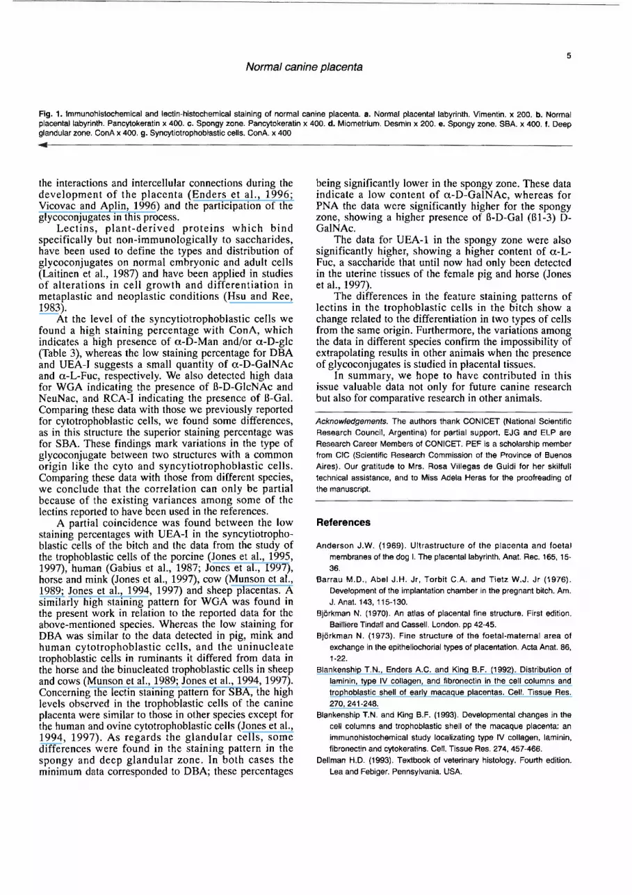
As can be seen in Table 2, the different zones of the placenta present remarkable discrepancies in the feature patterns using antibodies against different IF. The chorioalantoic membrane displayed a low immuno- histochemical percentage (IP) with the different monoclonal antibodies applied, this being more evident in the case of desmin. Data of the placental labyrinth were similar for vimentin and pancytokeratins, and significantly lower for desmin. Vimentin was visualised especially in the blood vessels (Fig. la) and cytokeratins in the trophoblastic cells (Fig. lb).
The IP of the spongy zone was significantly higher for pancytokeratins. The glands and cytotrophoblastic cells were positive to pancytokeratins (Fig. lc) whereas vimentin was found in the blood vessels and connective tissue. Desmin was very low in the area previously described. The supraglandular connective tissue also had a higher IP for vimentin. The deep glandular zone also had a higher expression of vimentin, especially in the interglandular connective tissue.
The IP of the miometrium was higher for vimentin. Desmin was detected in the muscular fibers (Fig. Id) whereas vimentin was found in the connective tissue,
Lectins
Table 3 summarizes the lectin binding pattern of the different zones of the normal canine placenta. The maximum staining percentage corresponded to SBA (Fig.le) in the gland cells of the spongy zone, while the minimun staining percentage to DBA, which was significantly lower compared to al1 the other lectins applied.
The maximum staining percentage of the cells in the deep glandular zone (Fig. l f ) and syncytiotrophoblast (Fig. lg) corresponded to ConA and the lowest to DBA. These differences were also statistically significant.
Discussion
The results of the present work provide quantitative data about the distribution of the IF in the different areas of the normal canine placenta. As no previous papers about the expression of the IF in the normal canine placenta are available, it is only possible to relate our data to those from other species. The presence of cytokeratins in the syncytiotrophoblastic cells coincides with our observations in cytotrophoblastic-like cells (Fernández et al., 1998) and data in the trophoblastic cells of human placentas (Khong et al., 1986; Mülhauser et al., 1995), macaque placentas (Blankenship et al., 1992, Blankenship and King, 1993) and baboons (Pijnenborg et al., 1996). In the rest of the stmctures, the feature staining patterns display keratin in the epithelia, vimentin in connective tissue and blood vessels and desmin in the muscle tissue.
It is interesting to interpret the feature staining pattern of the glycoconjugates due to the importance of
Table 2. lmmunohistochemical staining percentage of the diirent zones of the normal canine placenta.
Chorioalantoic membrane 7.5420.39 Placental labyrinth 28.8712.28 Spongy zone 18.73t1.31 Supraglandular connective tissue 44.99i4.31 Deep glandular zone 37.00i2.90 Miomehium 27.95i2.06
Numbers express the percentage of immunohistochemically stained area (iSD) of the different zones of the normal canine placenta.
Table 3. Lectin histochemical staining percentage of the dierent cells of the normal canine placenta.
Con A DBA PNA RCA-I SBA UEA-I WGA
Gland cells of the spongy zone. 67.4814.45 9.4710.49 58.90k3.47 67.43i4.98 69.08S.14 63.64*4.90 49.8313.83 Gland cells of the deep glandular zone. 81.03k6.07 32.4712.20 33.5652.01 45.9512.89 57.0215.01 40.01 52.76 65.62M.59 Syncytiotrophoblastic cells. 65.3413.78 18.7511.23 29.7511.75 55.29t4.31 49.7913.93 23.4411.42 55.5613.66
Numbers express the lectin-histochemical staining percentage (ISD) of the dierent cells of the normal canine placenta.

5
Normal canine placenta
Fig. 1. Immunohistochemical and lectin-histochemical staining of normal canine placenta. a. Normal placental labyrinth. Virnentin. x 200. b. Normal placental labyrinth. Pancytokeratin x 400. c. Spongy zone. Pancytokeratin x 400. d. Miometrium. Desmin x 200. e. Spongy zone. SBA. x 400. f. Deep glandular zone. ConA x 400. g. Syncytiotrophoblastic cells. ConA. x 400
the interactions and intercellular connections during the development of the placenta (Enders et al., 1996; Vicovac and Aplin, 1996) and the participation of the glycoconjugates in this process.
Lectins, plant-derived proteins which bind specifically but non-immunologically to saccharides, have been used to define the types and distribution of glycoconjugates on normal embryonic and adult cells (Laitinen et al., 1987) and have been applied in studies of alterations in cell growth and differentiation in metaplastic and neoplastic conditions (Hsu and Ree, 1983).
At the level of the syncytiotrophoblastic cells we found a high staining percentage with ConA, which indicates a high presence of a-D-Man andlor a-D-glc (Table 3), whereas the low staining percentage for DBA and UEA-1 suggests a small quantity of a-D-GalNAc and a-L-Fuc, respectively. We also detected high data for WGA indicating the presence of B-D-GlcNAc and NeuNac, and RCA-1 indicating the presence of B-Gal. Comparing these data with those we previously reported for cytotrophoblastic cells, we found some differences, as in this structure the superior staining percentage was for SBA. These findings mark variations in the type of glycoconjugate between two structures with a common origin like the cyto and syncytiotrophoblastic cells. Comparing these data with those from different species, we conclude that the correlation can only be partial because of the existing variances among some of the lectins reported to have been used in the references.
A partial coincidence was found between the low staining percentages with UEA-1 in the syncytiotropho- blastic cells of the bitch and the data from the study of the trophoblastic cells of the porcine (Jones et al., 1995, 1997), human (Gabius et al., 1987; Jones et al., 1997), horse and mink (Jones et al., 1997), cow (Munson et al., 1989; Jones et al., 1994, 1997) and sheep placentas. A similarly high staining pattern for WGA was found in the present work in relation to the reported data for the above-mentioned species. Whereas the low staining for DBA was similar to the data detected in pig, mink and human cytotrophoblastic cells, and the uninucleate trophoblastic cells in ruminants it differed from data in the horse and the binucleated trophoblastic cells in sheep and cows (Munson et al., 1989; Jones et al., 1994, 1997). Concerning the lectin staining pattern for SBA, the high levels observed in the trophoblastic cells of the canine placenta were similar to those in other species except for the human and ovine cytotrophoblastic cells (Jones et al., 1994, 1997). As regards the glandular cells, some differences were found in the staining pattern in the spongy and deep glandular zone. In both cases the minimum data corresponded to DBA; these percentages
being significantly lower in the spongy zone. These data indicate a low content of a-D-GalNAc, whereas for PNA the data were significantly higher for the spongy zone, showing a higher presence of B-D-Gal (Bl-3) D- GalNAc.
The data for UEA-1 in the spongy zone were also significantly higher, showing a higher content of a-L- Fuc, a saccharide that until now had only been detected in the uterine tissues of the female pig and horse (Jones et al., 1997).
The differences in the feature staining patterns of lectins in the trophoblastic cells in the bitch show a change related to the differentiation in two types of cells from the same origin. Furthermore, the variations among the data in different species confirm the impossibility of extrapolating results in other animals when the presence of glycoconjugates is studied in placental tissues.
In summary, we hope to have contributed in this issue valuable data not only for future canine research but also for comparative research in other animals.
Acknowledgements. The authors thank CONICET (National Scientific Research Council, Argentina) for partial support. EJG and ELP are Research Career Members of CONICET. PEF is a scholarship member from CIC (Scientific Research Cornmission of the Province of Buenos Aires). Our gratitud0 to Mrs. Rosa Villegas de Guidi for her skilfull technical assistance, and to Miss Adela Heras for the proofreading of the manuscript.
References
Anderson J.W. (1969). Ultrastructure of the placenta and foetal membranes of the dog l. The placental labyrinth. Anat. Rec. 165, 15- 36.
Barrau M.D., Abel J.H. Jr, Torbit C.A. and Tietz W.J. Jr (1976). Development of the implantation chamber in the pregnant bitch. Am. J. Anat. 143, 11 5-130.
Bjorkman N. (1970). An atlas of placental fíne structure. First edition. Bailliere Tindall and Cassell. London. pp 42-45.
Bjorkman N. (1973). Fine structure of the foetal-maternal area of exchange in the epitheliochorial types of placentation. Acta Anat. 86, 1-22.
Blankenship T.N., Enders A.C. and King B.F. (1992). Distribution of laminin, type IV collagen, and fibronectin in the cell columns and trophoblastic shell of early macaque placentas. Cell. Tissue Res. 270, 241 -248.
Blankenship T.N. and King B.F. (1993). Developmental changes in the cell columns and trophoblastic shell of the macaque placenta: an irnmunohistochemical study localizating type IV collagen, laminin, fibronectin and cyiokeratins. Cell. Tissue Res. 274. 457-466.
Dellman H.D. (1993). Textbook of veterinary histology. Fouith edition. Lea and Febiger. Pennsylvania. USA.

Normal canine placenta
Enders A.C., Lantz K.C. and Schlafke S. (1996). Preference of invasive cytotrophoblast for maternal vessels in early implantation in the macaque. Acta Anat. 155, 145-162.
Fernández P.E., Portiansky E.L., Barbeito C.G. and Gimeno E.J. (1998). Characterisation of cytotrophoblastic-like cells present in subinvolutioned placental cites of the bitch. Histol. Histopathol. 13, 995-1 000.
Gabius H.J., Debbage P.L., Engelhardt R., Osmers R. and Lange W. (1987). ldentification of endogenous sugar-binding proteins (lectins) in human placental by histochemical localisation and biochemical characterisation. Eur. J. Cell Biol. 44, 265-272.
Gimeno E.J., Massone A.R., Marino F.P. and ldiart J.R. (1995). lntermediate filament expression and lectin histochemical features of canine transmissible venereal tumour. APMlS 103, 645-650.
Goldstein I.J. and Hayes C.G. (1978). The lectins: Carbohydrate binding proteins of plants and animals. Adv. Carbohydr. Chem. Biochem. 35, 127-340.
Grether B.M. and Friess A.E. (1993). The glandular compartments of the canine placenta - A scanning electron microscopic study. Schweiz. Arch. Tierheilkd. 9, 272-278.
Hsu S.M. and Ree H.J. (1983). Histochemical studies on lectin binding in reactive lymphoid tissues. J. Histochem. Cytochem. 31.538-546.
Jones C.J.P., Dantzer V., Leiser R.. Krebs C. and Stoddart R.W. (1997). Localisation of glycans in the placenta: a comparative study of epitheliochorial, endotheliochorial and haernochorial placentation. Micros. Res. Tech. 38, 100-114.
Jones C.J.P., Dantzer V. and Stoddart R.W. (1995). Changes in glycan distribution within the porcine interhaemal barrier during gestation. Cell Tissue Res. 279,551 -564.
Jones C.J.P.. Koob B., Stoddart R.W., Hoffmann B. and Leiser R.
(1994). Lectin-histochemical analysis of glycans in ovine and bovine near-term placental binucleate cells. Cell Tissue Res. 278, 601-610.
Khong T.Y., Lane E.B. and Robertson W.B. (1986). An immunocyto- chemical study of foetal cells at the maternal-placental interface using monoclonal antibodies to keratins, vimentin and desmin. Cell Tissue Res. 246, 189-1 95.
Laitinen L., Virtanen l. and Saxen L. (1987). Changes in the glycosilation pattern during embryonic developrnent of mouse kidney as revealed with lectin conjugates. J. Histochem. Cytochem. 35, 55- 65.
Mülhauser J., Crescimanno C., Kasper M., Zaccheo D. and Castellucci M. (1995). Differentiation of human trophoblast populations involves alterations in cytokeratin patterns. J. Histochem. Cytochem. 43, 579- 589.
Munson L.. Kao J.J. and Schafler D.H. (1989). Characterisation of glycoconjugates in the bovine endometrium and chorion by lectin histochemistry. J. Reprod. Fert. 87, 509-51 7.
Pijnenborg R.. Hooghe T.D., Vercruysse L. and Bambra C. (1996). Evaluation of trophoblast invasion in placental bed biopsies of the baboon. with immunohistochemical localisation of cytokeratin, fibronectin and laminin. J. Med. Primatol. 25, 272-281.
Vicovac L. and Aplin J.D. (1996). Epithelial-mesenchymal transition during trophoblast differentiation. Acta Anat. 156, 202-216.
Wells W.A., Rainer R.O. and Memoli V.A. (1993). Equipment, standardization, and application of image processing. Am. J. Clin. Pathol. 99, 48-56.
Zar J.H. (1984). Multiple comparisons. In: Biostatistical analysis. Zar J.H. (ed). Englewood Cliffs Eds. Prentice Hall, NYJ. pp 185-205.
Accepted June 1,1999


