Embed Size (px)
Citation preview
http://jdr.sagepub.com/Journal of Dental Research
http://jdr.sagepub.com/content/24/3/143The online version of this article can be found at:
DOI: 10.1177/00220345450240030501
1945 24: 143J DENT RESAnna Morse
Teeth and Bones for Sectioning in ParaffinFormic Acid-Sodium Citrate Decalcification and Butyl Alcohol Dehydration of
Published by:
http://www.sagepublications.com
On behalf of:
International and American Associations for Dental Research
can be found at:Journal of Dental ResearchAdditional services and information for
http://jdr.sagepub.com/cgi/alertsEmail Alerts:
http://jdr.sagepub.com/subscriptionsSubscriptions:
http://www.sagepub.com/journalsReprints.navReprints:
http://www.sagepub.com/journalsPermissions.navPermissions:
http://jdr.sagepub.com/content/24/3/143.refs.htmlCitations:
What is This?
- Jun 1, 1945Version of Record >>
at PENNSYLVANIA STATE UNIV on March 3, 2014 For personal use only. No other uses without permission.jdr.sagepub.comDownloaded from at PENNSYLVANIA STATE UNIV on March 3, 2014 For personal use only. No other uses without permission.jdr.sagepub.comDownloaded from
FORMIC ACID-SODIUM CITRATE DECALCIFICATION AND BUTYLALCOHOL DEHYDRATION OF TEETH AND BONES FOR
SECTIONING IN PARAFFIN'
ANNA MORSE, M.S.'Department of Oral Pathology, Tufts College Dental School, Boston, Mass.
Oral calcified tissue, which includes extracted teeth, teeth in situ, and alveolarbone, is usually prepared for histological examination by the general method ofnitric acid decalcification, ethyl alcohol dehydration, and celloidin embedding.This paper describes a different method of tissue preparation which consists offormic acid-sodium citrate decalcification, butyl alcohol dehydration, andparaffin or celloidin embedding. The technic, described here in detail, wasarrived at after 10 years of experimentation. During the last 3 years of thisperiod, approximately 500 pieces of tissue were prepared and from these some30,000 slides. An analysis of the various problems involved in the preparation oforal calcified tissue are presented in an effort to demonstrate the advantages ofthe proposed technic of preparation.For purposes of clarity, the outline of the technic is followed by an analysis of
each step of the method in sequence.
METHOD1. Fix in 10% aqueous formaldehyde solution.2. Wash in running tap water or several changes of distilled water for 2-5 hours.3. Decalcify in formic acid-sodium citrate reagent until chemical test is negative for the
presence of calcium.Formic acid-sodium citrate reagent:
Solution A: 90% formic acid C.P. 1 partDistilled water 1 part
Solution B: Sodium citrate C.P. 20 gramsDistilled water 100 c.c.
Just before use combine equal portions of Solution A and Solution B. Use largevolumes of the reagent and change daily until test is negative for calcium.
Test: To 5 c.c. of the used decalcifying reagent, add 1 c.c. of concentrated ammoniumhydroxide, mix thoroughly, then add 0.1 c.c. of a saturated aqueous solution of am-monium oxalate. A precipitate will form when calcium is present. Repeat the addi-tions of 0.1 c.c. amounts of ammonium oxalate at intervals of 15-20 minutes until atotal of 0.4 c.c. has been added. If a precipitate fails to form after the addition of 0.4c.c. of ammonium oxalate, leave the tissue in the same decalcifying reagent for atleast 48 hours longer and repeat the test. When the test remains negative for 3 daysin the case of a single tooth and for 1 week in the case of larger tissues, proceed withdehydration and infiltration.
1 Received for publication March 2, 1945.2Now at Harvard University, School of Dental Medicine.
143
at PENNSYLVANIA STATE UNIV on March 3, 2014 For personal use only. No other uses without permission.jdr.sagepub.comDownloaded from
ANNA MORSE
4. Dehydrate and infiltrate as follows:A. For paraffin embedding:
1. Wash in running tap water or several changes of distilled water for 5-24 hours.2. 30%, 50%, 70% ethyl alcohol each for at least 24 hours.3. Solutions 1,i 2, 3, 4, 5, and 6 of butyl alcohol (see Table I) each for at least 48
hours.4. Infiltrate with paraffin of 56o-58oC. melting point for 5-24 hours.
B. For celloidin embedding:1. Wash in running tap water or several changes of distilled water for 5-24 hours.2. 30%, 50%, 70%, 80%, 95% ethyl alcohol, and equal portions of 95% ethyl alcohol
and ether, each for at least 24 hours.3. Infiltrate with 2%, 4%, 6%, 10%, 14%, and 25% celloidin for at least one week in
each.
Rapid fixation of all tissue elements, which are of paramount importance incritical histological studies, is difficult when calcified oral tissues are involved be-cause the penetration of the fixing fluid through dense alveolar bone, enamel,and dentin is a slow process. Post-mortem changes occur in the center of thespecimen before the fixing agent can penetrate the periphery. Perhaps, the mostseriously affected tissue of this type is the pulp.Many methods have been proposed to facilitate the penetration of fixing agents
into the pulpal tissue. Cutting of holes or slots, and stripping of the dentin aresome of these practices. Hill (1) believed "satisfactory fixation of dental pulps"is obtained "by immediate grinding of the side of the tooth under water until thecoronal pulp was freely exposed" and then placing it in the fixative. Willman(2) advocated cutting holes in the side of the tooth or cutting off the apex. Cow-dry (3), on the other hand, says that these practices are "apt to disturb the posi-tion of the pulp and should be avoided." One undesirable result is the disturb-ance due to the trauma and heat subjacent to the cut. Certainly, the reductionof the depth through which the fixing fluid has to pass will reduce the time in-volved. However, there is some doubt in the author's mind whether the so-called fixation artifacts are eliminated by these methods.The problem of excellent, rapid, cytological fixation of oral calcified tissues is
still unsolved. It must be noted, also, that subsequent treatment can influenceprofoundly the primary fixation image.
Choice of a fixing reagent is dependent upon the tissue itself and the purposefor which it is to be preserved. Special reagents prepare the tissue for study ofspecified elements or reactions and chemically prepare for the application ofselective stains. Formaldehyde solution is an excellent general fixing reagentand permits the subsequent use of a large variety of staining methods. Manyquestions may be raised as to the advisability of its use. However, it is an agentwhich may be handled by anyone not acquainted with the details of the specialagents. Its use in our laboratory is due to the fact that most of the tissues comefrom other departments or from the outside and take varying lengths of time toreach the laboratory. Under these circumstances, the use of special reagentsrequiring a few hours or days for fixation is precluded. Furthermore, autopsymaterial has already been subjected to the influence of formaldehyde in the em-
144
at PENNSYLVANIA STATE UNIV on March 3, 2014 For personal use only. No other uses without permission.jdr.sagepub.comDownloaded from
DECALCIFICATION METHOD: TEETH AND BONES
balming fluids. Tissues may be left in formaldehyde solution indefinitely al-though there is some indication that after a year the stainability of the tissue isdecreased.
Following fixation, tissues should be washed in running water in order to re-move accumulations of debris and as much of the fixing fluid as possible. If thereis any danger of diatoms, rust, etc. in the water supply or if the tap water ischemically impure, it is preferable to wash in several changes of distilled water.
Decalcification is most important in the preparation of oral calcified tissues formicroscopic examination. It is important from two standpoints: first, sectionsof teeth, bone, and surrounding tissues are difficult to obtain without removal ofthe calcium; and second, the effect of the various chemical decalcifiers upon thetissue components differ.
This second important item is the greatest problem of the whole technic. Theprimary fixation picture can be changed by subsequent treatment. Sectionsmust be carefully studied so that correct conclusions are drawn as to the effectof the decalcifier upon the cellular elements. Distortion and macerations canbe demonstrated by the use of chemicals following fixation. Grossly, the tissuemay appear unchanged but closer study reveals that the cellular elements ex-hibit swelling, shrinkage, vacuolization, disruption, and fraying not attributableto pathological conditions. Short periods of action by the decalcifier upon thetissue do not necessarily result in these changes. However, continued action tobring about complete decalcification very often results in distortion and actualdestruction of the tissue. This is the reason that small pieces of tissue measuring2 mm. x 2 cm. x 2 cm. are recommended when decalcification is necessary.Single teeth are larger and pieces of tissue containing up to 6 teeth are muchlarger and must remain in the decalcifier for long periods of time. Therefore,the results of the longer action must be evaluated carefully before any decalcifieris used upon oral tissue for histological examination.
Nitric acid is unfortunately, the most common decalcifying reagent used todecalcify oral tissues. It is used in aqueous (2,4, 5, 6, 7, 8) or alcoholic solution,with or without the addition of a preservative such as phloroglucin. Smallpieces of tissue can be decalcified quickly and well in 5% to 10% aqueous nitricacid. Single teeth decalcify fairly well but larger specimens exhibit many dis-tortions and destructions. In order to remove the calcium from the center of apiece of tissue, the periphery is subjected to the action of the acid for too long aperiod (over-decalcification). Maceration of the exterior is common and distor-tion takes place for some distance into the specimen on all sides. The stainabilityis markedly decreased with a resultant diffuse eosin stain. For these reasons,nitric acid as a decalcifying reagent is undesirable for blocks of tissue larger than2 mm. x 2 cm. x 2 cm. When the tissue is thin in at least one dimension, the acidpenetrates quickly and the decalcification is complete before too much harm isdone. Cytological studies on any nitric acid decalcified material are open to agreat deal of question.
Inorganic acids, other than nitric, have been used for decalcifying agents,among which are hydrochloric, sulphurous, and sulfuric acids. Each has a place
145
at PENNSYLVANIA STATE UNIV on March 3, 2014 For personal use only. No other uses without permission.jdr.sagepub.comDownloaded from
ANNA MORSE
in decalcification under certain circumstances, but they have been rejected for useon human oral tissue for varying reasons. Of the organic acids, acetic and tri-chloracetic acids are those most commonly used, and, although decalcification israpid and the preservation good, swelling occurs. Jaff6 (9) states that "in gen-eral, it may be said that mineral acids possess greater decalcifying power, but aremost injurious to the nuclei, while organic acids produce the greatest swelling ofthe fibrils [of bone] but less injury to the nuclei, if exposure is not too pro-longed."Some time ago, in the search for a decalcifying fluid which could be employed
upon oral tissue of all types and sizes and would eliminate or diminish distortionwe were introduced by the Department of Pathology at Tufts College MedicalSchool to a reagent made up of equal portions of 45% aqueous formic acid and20% aqueous sodium citrate, which was being used upon bone with excellentpreservation of the cellular elements. The original source of the reagent isunknown. The preliminary results of the use of the mixture upon teeth and bonesubstantiated the observations upon bone as to the cellular preservation. Thisbrought about the continuation of its employment and observations of the actionof the reagent upon tissues from the oral structures.Formic acid has been used as a decalcifying agent in oral histology and path-
ology usually as a strong aqueous solution of 33% or combined with formol.In 1930, Evans and Krajian (10) introduced "a new method of decalcification"which was a combination of equal parts of 85%o aqueous formic acid and 20%aqueous sodium citrate. Glickman and Wood (11) are the first to report the useof our reagent (45% aqueous formic acid and 20% aqueous sodium citrate) Pponoral calcified tissue. Lillie (12) has studied combinations of formic acid andsodium citrate in relation to the successful staining of bone marrow.The objection to the use of formic acid is the swelling action it has upon tis-
sues. Jaff6 (9) said that it causes great swelling of the collagenous fibers. Evansand Krajian (10) stated that with their solution "the most prominent point isthat the cellular elements are practically unaffected and take the stain apparentlyas perfectly as tissues that are not subjected to decalcification." These authorssuggested that the citrate counteracts the swelling tendency of the formic acid.
In order to study more fully the effect of the combinations of formic acid andsodium citrate upon extracted teeth, normal teeth from the same patient werefixed simultaneously in 10% formaldehyde. Then one half of the number of theteeth were decalcified in equal portions of 85% formic acid and 20%o sodium ci-trate; the other half were decalcified in equal portions of 45% formic acid and20% sodium citrate. During the decalcification period, the teeth in the formerreagent gradually became more glassy. Following dehydration and embeddingin paraffin according to the method proposed in the article, it was found that thefirst group was very difficult to section. The second group sectioned easily.A second series of studies were instituted to learn the effect of the components
of the 45% formic acid and 20% sodium citrate reagent upon the cellular ele-ments of normal tissue. Pieces of the same kidney and liver of a normal rat fol-lowing fixation in 10% formol were subjected to the influence of the following
146
at PENNSYLVANIA STATE UNIV on March 3, 2014 For personal use only. No other uses without permission.jdr.sagepub.comDownloaded from
DECALCIFICATION METHOD: TEETH AND BONES
solutions: (1) 45% aqueous fQrmic acid, (2) 20% aqueous sodium citrate, and(3) equal portions of 45% formic acid and 20% sodium citrate. The remainderof the technic was identical with the proposed method. The material was al-lowed to remain in the various solutions for 7 days.The results of this study brought to light some very interesting facts. Those
tissues passed through the formic acid alone exhibited great swelling of all tissueelements so that the structure of the organ was barely discernible. The nucleiwere very poorly stained. The tissues passed through sodium citrate aloneshowed shrinkage of the cytoplasm throughout with excellent staining of thenuclei. Tissues run through the combination showed only slight swelling of thecytoplasm and collagenous fibers and the nuclei stained more brilliantly thanthose of similar untreated material.As a result of these preliminary tests, the combination of equal portions of 45%
aqueous formic acid and 20% aqueous sodium citrate is now used routinely fordecalcification. The solutions are kept separate until needed. A crystal ofthymol added to the sodium citrate aids in keeping down bacterial and fungalgrowth. Tissues to be decalcified are placed in 10 times their volume of thereagent, which is changed every day until decalcification is complete.
Testing chemically for the end-point in decalcification is the only accuratemethod for determination of the complete removal of the calcium. The methodsof needle-testing, flexibility, and x-ray have been, in our experience, extremelyvariable in results. Williams (4) used the production of ammonium phospho-molybdate as an indicator with nitric acid decalcification. The chemical testoutlined in the method is that published by Arnim (13).The description of the test is clear and its application simple. However, some
annotations are needed for better interpretation of the results. Testing beginson the 2nd or 3rd day of decalcification when the addition of 0.1 c.c.of ammonium oxalate to the reagent plus the ammonia causes a heavy precipi-tate. The end-point is approaching when a precipitate fails to appear upon theaddition of 0.3 c.c. of ammonium oxalate and only a slight precipitate upon theaddition of the fourth 0.1 c.c. When no precipitate appears upon the addition of0.4 c.c. of ammonium oxalate, the tissues must be left in the same fluid another3 days and tested again. This interval of 3 days is necessary because thedensity of the dentin and bone impedes the penetration of the reagent into themiddle of the specimen. Judicious use of slight negative pressure at this time isrecommended. Longer action of the acid and the negative pressure may cause aprecipitate upon the addition of only 0.1 c.c. of ammonium oxalate. Largerspecimens of tissue are more apt to react in this fashion than single teeth. Ex-perience with this chemical method enables one to evaluate the results.Towards the end of the decalcifying period, the tooth or larger specimen should
be trimmed in the plane of cutting. This is done with a razor blade and islimited by the structures desired in the section. Two results are obtained bythis practice: 1, a flat surface is provided for easier embedding; and 2, a largerarea is exposed to the further action of the decalcifier.The length of time required to completely decalcify tissues naturally varies
,147
at PENNSYLVANIA STATE UNIV on March 3, 2014 For personal use only. No other uses without permission.jdr.sagepub.comDownloaded from

ANNA MORSE
with the size of the specimen. A singleextracted toothsubjected to the action ofthe reagent requires about 10 days Nor complete decalcification. As the speci-men increases in size from a single tooth to a block of 5 to 6 teeth in situ, the de-calcifying period extends into weeks and months.
Single extracted teeth ......................... 7-24 daysOne tooth with surrounding tissue ......................... 14-30 daysTwo teeth in situ ................................................. 30-40 days3, 4, 5, 6 teeth.................................................. 1-2 monthsRat mandible.................................................. 1-2 daysRat head in toto ........... 3-5 daysDog blocks of 2-3 teeth ................ 2-3 months
The extraction of the calcium takes slightly longer with the formic acid-sodiumcitrate reagent than with some of the other agents, but the results are well worththe time involved. Apparently, a long continued subjection to the reagent isnot detrimental to the cellular and histological elements of the specimen. Over-decalcification is almost entirely eliminated as a hazard. This is a distinct ad-vantage in many ways but perhaps most advantageous in the flexibility of thetechnic. Laboratory hours and intervals of absence do not cause the loss ofvaluable material.
Immediately following the negative test for the presence of calcium, the tissueis washed in running water or several changes of distilled water for 5 to 24 hours.Usually, tissues are transferred from formic acid directly into alcohol in order tocounteract swelling. However, the addition of the sodium citrate to the reagentresults in an elimination of swelling during this step. Both methods, washingand direct transfer to alcohol, seem to have very little difference in their effectupon the subsequent histological picture. Washing for 5 to 24 hours eliminatesfrom the tissue any reagent which would tend to interfere with the subsequentstaining.
After washing, the schedule of dehydration proposed above is followed. Thetime noted for the first part of the series is "at least 24 hours" and for the secondpart is "at least 48 hours." Our experience has been that a single extractedtooth can be moved up in the series this fast, but no faster without poor dehydra-tion of the pulp. The density of the dentin obstructs the equilibration of the 2adjacent solutions in the substance of the tooth. We prefer to use still longerperiods of time for each member of the series even for a single tooth and increasethe time as the block increases in size.Butyl alcohol as a dehydration-infiltration chemical has long been used in
botanical technic and has only recently come into use in animal technic. Stiles(14) believed this is because there has been little experience with it. Lang (15)considered butyl alcohol the most satisfactory agent and least likely of alldehy-dration reagents to "exercise deleterious effects upon theprimary fixation image."Stowell (16) recommended tertiary butyl alcohol in his TBEA series afterJohansen as giving the "most satisfactory results in general micro-technieworkboth in ease of sectioning and in final microscopic quality of tissue."The use of butyl alcohol dehydration in oral histological and pathological
148
at PENNSYLVANIA STATE UNIV on March 3, 2014 For personal use only. No other uses without permission.jdr.sagepub.comDownloaded from
DECALCIFICATION METHOD: TEETH AND BONES
technic is almost unknown. Fish (17) mentioned its use. Williams (4) notedthat butyl alcohol followed by paraffin brought no success. However, she doesnot mention the series used or the technic followed. Gairns (18) said butylalcohol used for clearing resulted in no noticeable difference.
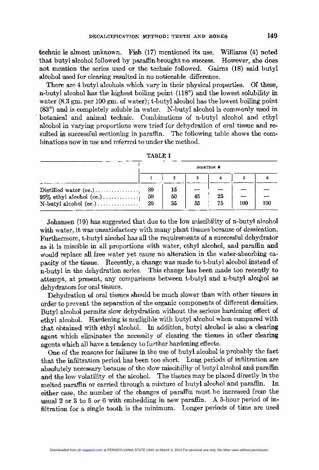
There are 4 butyl alcohols which vary in their physical properties. Of these,n-butyl alcohol has the highest boiling point (1180) and the lowest solubility inwater (8.3 gm. per 100 gm. of water); t-butyl alcohol has the lowest boiling point(83°) and is completely soluble in water. N-butyl alcohol is commonly used inbotanical and animal technic. Combinations of n-butyl alcohol and ethylalcohol in varying proportions were tried for dehydration of oral tissue and re-sulted in successful sectioning in paraffin. The following table shows the com-binations now in use and referred to under the method.
TABLE I
SOLUTION %
1 2 3 4 5 6
Distilled water (cc.) ................ 30 15 -_ _95% ethyl alcohol (cc.) ............. 50 50 45 25 -N-butyl alcohol (cc.) ............... 20 35 55 75 100 100
Johansen (19) has suggested that due to the low miscibility of n-butyl alcoholwith water, it was unsatisfactory with many plant tissues because of dessication.Furthermore, t-butyl alcohol has all the requirements of a successful dehydratoras it is miscible in all proportions with water, ethyl alcohol, and paraffin andwould replace all free water yet cause no alteration in the water-absorbing ca-pacity of the tissue. Recently, a change was made to t-butyl alcohol instead ofn-butyl in the dehydration series. This change has been made too recently toattempt, at present, any comparisons between t-butyl and n-butyl alcohol asdehydrators for oral tissues.
Dehydration of oral tissues should be much slower than with other tissues inorder to prevent the separation of the organic components of different densities.Butyl alcohol permits slow dehydration without the serious hardening effect ofethyl alcohol. Hardening is negligible with butyl alcohol when compared withthat obtained with ethyl alcohol. In addition, butyl alcohol is also a clearingagent which eliminates the necessity of clearing the tissues in other clearingagents which all have a tendency to further hardening effects.One of the reasons for failures in the use of butyl alcohol is probably the fact
that the infiltration period has been too short. Long periods of infiltration are
absolutely necessary because of the slow miscibility of butyl alcohol and paraffinand the low volatility of the alcohol. The tissues may be placed directly in themelted paraffin or carried through a mixture of butyl alcohol and paraffin. Ineither case, the number of the changes of paraffin must be increased from theusual 2 or 3 to 5 or 6 with embedding in new paraffin. A 5-hour period of in-
filtration for a single tooth is the minimum. Longer periods of time are used
149
at PENNSYLVANIA STATE UNIV on March 3, 2014 For personal use only. No other uses without permission.jdr.sagepub.comDownloaded from
ANNA MORSE
without the hardening experienced with the usual reagents. Single teeth sectioneasily after 24 hours infiltration with paraffin of 56°-58° C. melting point andspecimens as large as 3 teeth have been left in the oven for 3 days with excellentresults. As Stiles (14) said, "it would seem that it is not the HOT paraffin whichcauses the serious hardening of tissues during infiltration but rather hot paraffinfollowing certain types of dehydration and clearing."A paraffin of high melting point should be used for bones and teeth. Soft
tissues such as kidney and liver can be sectioned successfully at 5-6 micron usingparawax as an embedding material. Hard tissues such as bones and teeth canbe successfully sectioned only with a paraffin of 560-580C. melting point whichgives a more solid matrix.
Subsequent to embedding, the block is trimmed to size and mounted for sec-tioning on a rotary microtome. Trim the block roughly to size and mount it onthe cross-cut end of a rectangular piece of pine wood impregnated with paraffin.This mounted block is then placed in the carrier of the microtome and thereaccurately trimmed with 2 parallel sides by means of a Book block trimmer (20).The Book block trimmer is essentially a single edge razor blade set on a piece ofwood or metal at an angle of 78°. This is the easiest trimmer to use and thesimplest to construct of any that have been proposed. An added advantage isthe resulting accurate alinement of the 2 surfaces of the block and the knife edge.With 2 motions, the block edges are parallel to the knife edge. Other blocktrimmers, more elaborate in construction, give parallel edged blocks but theseare obtained away from the microtome. Then the block must be oriented in thecarrier of the microtome so that the edges are parallel to the knife edge. At bestthis is a difficult manipulation. The Book trimmer accomplishes these 2 opera-tions in 1, and much better than can be done manually.
Sectioning of the material is just as important to success with the technic asadherence to the schedule through the embedding. A few notes should be in-cluded on the sectioning procedure in order to aid in reproducing the results ob-tained.
First, the microtome knife, which is a very important tool, should be mostcarefully sharpened. Many excellent discussions on the technic of sharpeninghave been written so there is no need for repetition here. Reference may bemade to Richards (21) and Bailey (22) for aid in obtaining the knife edge require-ments for bone and teeth. These requirements are (1) an edge free from serra-tions at better than 100 diameters, and (2) a small bevel angle. The clamps forholding the knife in position should be placed as near to the paraffin block asallowable without interference with the block. All the set screws must betightened to prevent any vibrations of the knife, carrier, or block during section-ing. The knife, when set in the microtome holder, is tilted until the angle be-tween the surface of the block and the surface of the knife facet (the clearanceangle) is that best suited for the material.
Second, the surface of the block should be exposed to water from time to timeduring cutting. After the initiation of the complete technic, difficulty was ex-perienced in obtaining a good ribbon. All possible remedies, such as changing
150
at PENNSYLVANIA STATE UNIV on March 3, 2014 For personal use only. No other uses without permission.jdr.sagepub.comDownloaded from
DECALCIFICATION METHOD: TEETH AND BONES
the knife angle, critical embedding and blocking, resharpening of the knife, etc.were tried with no success. An article by David (23) in which she stated that thecut end of a block of bone should be exposed to water before cutting in order toobtain uniform sections gave a hint for remedy of the difficulty. Gairns (18)has recently suggested exposure to water of blocks of cat teeth and bone. Ex-posure of paraffin embedded teeth to water overnight resulted in good sectioning,but only to the depth to which the water had penetrated. Modifications fol-lowed these first experiments until at the present time the method used is asfollows: cut the block until the desired orientation is obtained, then cover thesurface of the block with absorbent cotton wet with cold water, remove the cot-ton and eliminate the excess water on the knife and block, cut about 15 sections,replace the wet cotton while these sections are mounted on slides, and repeat theprocess until the block has been sectioned. A little practice with the methodenables one to determine the length of time necessary for the action of the waterupon the next 15 sections. As we spread our sections upon hot water and thenmount, the time involved in spreading, separation, and mounting of 15 sectionsis just about enough time for the softening action of the water upon the next setof sections.The only limits to the use of paraffin as an embedding medium are not those
imposed by the technic, but rather those imposed by the mechanical constructionof the microtome. When tissues exceed in size a specimen containing 3 teeth insitu, the microtome knife cannot be made rigid enough to successfully cut themevenly. In addition, the microtome is so constructed that the strain of cuttingsuch large sections is too great.The solution to this problem is found in the employment of celloidin as an
embedding material. The schedule of dehydration and embedding differs in noway from that usually used. Unfortunately, no way has been found in which todehydrate with butyl alcohol and embed in celloidin, for celloidin is not soluble inbutyl alcohol.
Serial sections of celloidin material are more tedious to obtain than serial sec-tions of paraffin material. Different ways of maintaining the series have beenproposed and all are of value. The simplest method and the one which we useis to remove the sections from the knife with a small piece of onion skin paper andplace the sections with the paper in a pile. The pile is then wrapped in a largerlabelled piece of paper and placed in a j-ar of 80% alcohol for future staining.Just before staining, each section in series is removed, one corner of the celloidinblotted, and a number stamped upon it. For this purpose, an ordinary datestamp, a used stamp pad, and some mimeograph ink are all that is needed. The2 rows of numbers in the center of the stamp are left in place and rest is cut off.A stamp pad which has been used until the ink needs renewing is the best.Mimeograph ink is applied to the surface of the pad until enough is added toimpregnate the pad. It will be found that numbers applied in this way willremain visible for some time if the sections are NOT LEFT TOO LONG in theclearing fluid. Sections mounted with numbers applied in this way 2 years agostill have numbers showing.
1'51
at PENNSYLVANIA STATE UNIV on March 3, 2014 For personal use only. No other uses without permission.jdr.sagepub.comDownloaded from
ANNA MORSE
The proposed paraffin technic has been used during the past 3 years on ap-proximately 500 pieces of tissue including rat and dog jaws, human single ex-tracted teeth, and human autopsy material with 1 to 6 teeth. The single teethwere cut for student laboratory examination and from each tooth an average of100 sections at 7 micron were obtained. Blocks of autopsy material containing,1, 2, and 3 teeth were cut either bucco-lingually or mesio-distally with great suc-cess. Tissues from experimental rats were the most successful and those fromdogs the least successful.Although oral calcified tissue has been the chief object of the study, other
calcified tissues of the body may be just as successfully prepared in this manner.Tissues such as rib, vertebra, and long bones sectioned extremely well followingparaffin embedding.
SUMMARY
A technic for the preparation of oral calcified tissue for histological examina-tion is presented. This technic differs from other available methods in com-bining 3 steps which are (1) decalcification by means of a formic acid-sodiumcitrate reagent, (2) dehydration with n-butyl alcohol, and (3) embedding inparaffin. Excellent sections at 7 microns were obtained following employmentof the method on rat, dog, and human jaws. Other calcified tissue has been suc-cessfully treated in the same manner. Celloidin embedding is used only when thepieces of human or dog tissue exceed 3 teeth in situ.
REFERENCES1. HILL, T. J., Pathology of the Dental Pulp, J. A. D. A., 21: 820, 1934.2. WILLMAN, M., A Technique for the Preparation of Histologic Sections Through Teeth
and Jaws for Teaching and Research, J. D. Res., 16: 183, 1937.3. COWDRY, E. V., Microscopic Technique in Biology and Medicine (Baltimore, Maryland,
Williams & Wilkins Co., 1943), p. 188.4. WILLIAMS, A., The Preparation of Combined Hard and Soft Tissues for Histological
Study, D. Cosmos, 69: 715, 1927.5. ORBAN, B., The Development of the Dentin, J. A. D. A., 16:1547, 1929.6. LOGAN, W. H. G., AND KRONFELD, R., Development of the Human Jaws and Surround-
ing Structures from Birth to the Age of Fifteen Years, J. A. D. A., 20: 379, 1933.7. JAMES, W. W., AND COUNSELL, A., A Histological Investigation into "so-called Pyorrhea
Alveolaris," Brit. D. J., 48: 1237, 1927.8. CHASE, S. W., Histogenesis of the Enamel, J. A. D. A., 19: 1275, 1932.9. JAFFJ, H. L., Methods for the Histologic Study of Normal and Diseased Bone, Arch.
Path., 8: 817, 1929.10. EVANS, N., AND KRAJIAN, A., New Method of Decalcification, Arch. Path., 10: 447,
1930.11. GLICKMAN, I., AND WOOD, H., Bone Histology in Periodontal Disease, J. D. Res., 21:
35, 1942.12. LILLIE, R. D., Studies on the Decalcification of Bone, Am. J. Path., 20: 291, 1944.14. ARNIM, S. S., A Method for Preparation of Serial Sections of Teeth and Surrounding
Structures of the Rat, Anat. Rec., 62: 321, 1935.14. STILES, K. A., Normal Butyl Alcohol Technic forAnimal Tissues, with Special Reference
to Insects, Stain Techn., 9: 97, 1934.15. LANG, A. G., The Use of N-butyl Alcohol in the Paraffin Method, Stain Techn., 12: 113,
1937.
152
at PENNSYLVANIA STATE UNIV on March 3, 2014 For personal use only. No other uses without permission.jdr.sagepub.comDownloaded from
DECALCIFICATION METHOD: TEETH AND BONES 153
16. STOWELL, R. E., Effect on Tissue Volume of Various Methods of Fixation, Dehydration,and Embedding, Stain Techn., 16: 67, 1941.
17. FISH, E. W., The Pathology of the Dentine and the Dental Pulp, Brit. D. J., 53: 351,1932 (July-Dec.).
18. GAIRNs, F. W., Preparing Specimens of Bone and Teeth for Cutting by the ParaffinMethod, Stain Techn., 19: 127,1944.
19. JOHANSEN, D. A., Dehydration and Infiltration, Science, 82: 253, 1935.20. BooK, M. H., An Inexpensive Trimmer for Paraffin Blocks, Stain Techn., 18: 25,1943.21. RIcHARDs. 0. W., The Effective Use and Proper Care of the Microtome (Buffalo, Spen-
cer Lens Co.: 1942), p. 18.22. BAILEY, A. J., Precision Sectioning of Wood, Stain Techn., 12: 159, 1937.23. DAVID, L. T., A Method of Preparing Paraffin Sections of Bone, Science, 82:179, 1935.
at PENNSYLVANIA STATE UNIV on March 3, 2014 For personal use only. No other uses without permission.jdr.sagepub.comDownloaded from