Embed Size (px)
Citation preview
BULLETIN OF MARINE SCIENCE. 56(2\t 569-577. 1995 CORAL REEF PAPER
MORPHOLOGY AND SYSTEMATICS OF THE ENIGMATICVOLUTID PLICOLM ZELINDAE (PETUCH, 1979)
(MOLLUSCA: GASTROPODA)
Josd H. Leal and M. G. Harasewvch
A B S T R A C T
The morphology of the living animal, anterior alimentary system and nervous system ofPlicoliva zelindae is described. Presence of paired siphonal appendages, broad cephalic shieldand lateral lappets, as well as a long connective between the supraesophageal and right pleuralganglia confirm the assignment of this taxon to the family Volutidae. The subfamily Plico-livinae, characterized by its multicuspid lateral teeth, distinctive sabot-like rachidian teeth,and a gland of Leiblein with greatly enlarged terminal bulb, shares some anatomical featureswith the volutid subfamilies Haliinae and Volutinae. With certain members of the former(e.g., Amoria) it shares a distinctive external pigmentation pattern and similar siphonal ap-pendages and rachidian teeth. Comparisons of Plicolivinae with published data on variousspecies of Volutinae reveal similar shell morphology and the presence of a gland of Leibleinwith an enlarged terminal bulb in both subfamilies.
The genus Plicoliva Petuch, 7979, contains two shallow-water, geographicallyrestricted species with broadly disjunct ranges. Plicoliva z.elindae (Petuch, 1979),the type species, is endemic to the vicinity of the Abrolhos Reef Complex andfrom off Espirito Santo State in eastern Brazil, while P. ryalli Bouchet, 1990 isknown only from off Ghana, in western Africa.
To date, only shell and radular characters have been used to infer the systematicallocation of Plicoliva. Petuch (1979) originally described Plicoliva zelindae inthe family Olividae, erecting Plicoliva as a subgenus of Oliva. While noting thatP. zelindae "resembles volutes in the genera Lyria or Enaeta more than typicalOliva or Olivella" (Petuch, 1979: 521), he argued that the shell shape, high gloss,and number of columellar folds favored the inclusion of this taxon in the Olividae.Later, Petuch and Sargent (1986: 16) elevated Plicoliva to generic rank and trans-ferred it to the Volutidae without further discussion. More recently, Petuch (1987:140) reduced Plicoliva to a subgenus of Lyria, also without discussion.
Bouchet (1990) described a second species of Plicoliva, P. ryalli, from 240m depth off the coast of western Africa. Based on an examination of the radulaeof both species, he concluded that the peculiar combination of conchological andradular features warranted the introduction of the new subfamily Plicolivinae,within the Volutidae, "although future anatomical work may disprove this place-ment" (Bouchet, 1990:9). In this paper we present additional data on the mor-phology of the animal of Plicoliva zelindae and discuss the implications of thesefindings on the suprageneric systematics of Plicoliva.
M,q.rsnraI- AND METHoDS
Protoconch and ultrastructure data are fiom the holotype of Plic'oliva zelintlae (USNM 780655)from the south side of Guaratibas Reefs, Abrolhos Reef Complex, Bahia State, Brazil (17'25'5,039"08'W), 1 m depth, in shell gravel in a tide pool. Live animal observations are based on a singlejuvenile specimen (MNHN) collected by Bouchet, M6tivier and Leal off Espirito Santo State, Brazil(21'31'5,040"18'W) in 37 m (WV MenroN-DUFREsNE MD55-Br6si l Cruise, stat ion DC15, 05/1987).The shell and radula of this specimen are figured in Bouchet (1990: figs. 1,2 and 14-16 respectively).Anatomical and additional radular data are based on the incomplete soft parts of a single, femalespecimen collected off Guarapari, Espirito Santo State, Brazil, by J. Coltro and M. V. Coltro, in May7992, and sent by E. C. Rios.
569
570 BULLETIN OF MARINE SCIENCE. VOL.56. NO.2. 1995
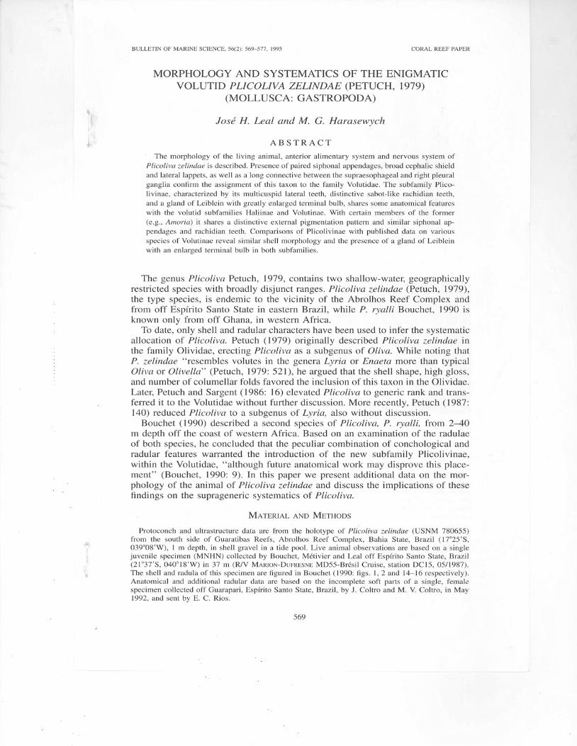
Figures 1--4. Plicoliva zelindae, Abrolhos Reef Complex, Brazil, USNM 780655, holotype. 1. Shell.Scale bar : 5 mm; 2. Shell ultrastructure, collabral section at outer lip. Scale bar : 100 p,m. 3, 4.Protoconch, lateral and apical views. Scale bars : 2 mm, Abbreviations: cl, cross-lamellar layer; pl,prismatic layer; ex, external shell surface; tr, transition from protoconch to teleoconch.
Toluidine blue stain was used to provide contrast during dissections. The distal tip of the probosciswith everted radula was dehydrated through a standard alcoholic series, and critical point dried usingcarbon dioxide as the transitional fluid (Lewis and Nemanic, 1973). Critical point dried tissue, radularpreparations, and shell fragments were coated with carbon and gold-palladium, and examined underHitachi 5-570, or Cambridge Stereoscan 250 Mk2 scanning electron microscopes. The protoconch ofthe holotype was examined uncoated under SEM. Tissues for histological examination were excised,paraffin-embedded, sectioned at 6 pm, and stained with Harris haematoxylin and eosin (Humason,1979).
We add to the conchological data provided by Petuch (1979) and Bouchet (1990) by including briefdescriptions and SEM micrographs of the protoconch and shell ultrastructure. These authors shouldbe consulted for full descriotions of shell characters.
RrsulrsShell Morphology.-Protoconch (Figs. 3, 4) mammillated, smooth, reddish brown,increasing in diameter from 0.85 mm to 1.3 mm in l1/z whorls. Transition to
LEAL AND HARASEWYCH: MORPHOLOGY AND SYSTEMATICS OF PLICOLIVA ZELINDAE
e i
asgd s g
571
7 mdolbsl cgsin
p n/ rbg
@lorm
dag
s g
rpgrcg
spg panosn bg
p ls
p o
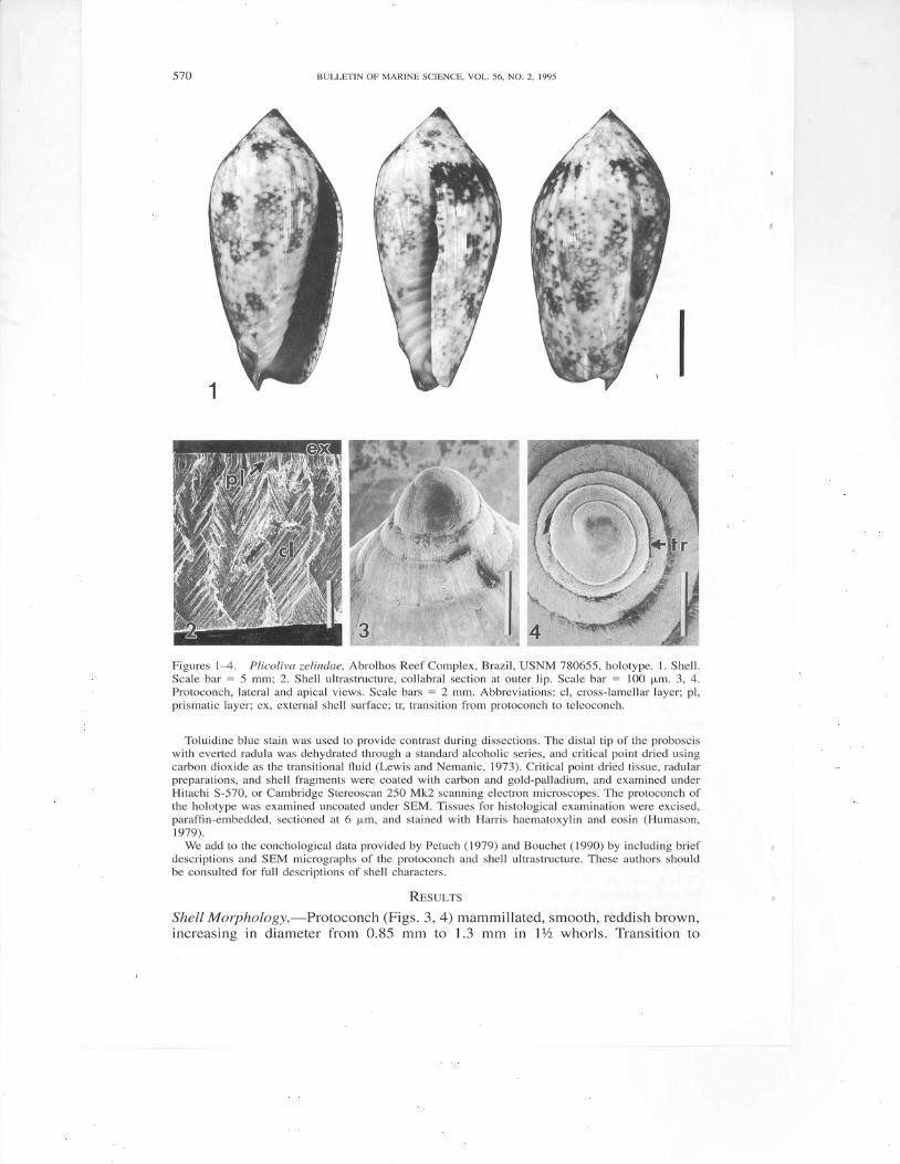
Figures 5-8. Plicoliva zelindae. Diagrammatic renditions of: 5. Dissected mantle cavity and cephalichemocoel; 6. Head; 7. Dissected anterior alimentary system with proboscis rotated 90" in counter-clockwise direction; 8. Terminal bulb of gland of Leiblein. Dashed lines indicate positions of histo-logical sections in Figures 15-20. Scale bars : 2 mm. Abbreviations: ao,'anterior esophagus; asg,accessory salivary gland; ct, ctenidium; dag, duct of accessory salivary gland; dsg, duct of salivarygland; e, eye; fhl, frontal head lobe; gL, gland of Leiblein; in, integumentary nerve; lbg, left buccalganglion; lcg, left cerebral ganglion; 11, lateral lappet; lsa, left siphonal appendage; mdo, mid-esoph-agus; mo, mouth; nr, circumesophageal nerve ring; orm, odontophore retractor muscle; os, osphradium;osn, osphradial nerve; pan, pallial nerve; plg, pleural ganglion; po, posterior esophagus; pn, proboscisnerve; pr, proboscis; rbg, right buccal ganglion; rcg, right cerebral ganglion; rpg, right pedal ganglion;rs, radular sac; rsa, right siphonal appendage; sg, salivary gland; si, siphon; sbg, subesophageal gan-glion; sin, siphonal nerve; spg, supraesophageal ganglion; tb, terminal bulb of gland of Leiblein; te,tentacle: vsn. visceral nerve.
teleoconch (Fig. a, tr) abrupt, demarcated by slight inflation of whorl and changein surface texture of shell, otherwise almost imperceptible.
A chip from the thin (350 pm), immature outer lip of holotype (Fig. 2) iscomposed of two layers, a thin (68 p,m), outer prismatic layer (Fig. 2, pl), andthicker (280 pm) inner layer of collabrally oriented, crossed-lamellar aragonite(Fig. 2, cl). At least one additional layer is present on the inner surface furtherwithin the aperture.
General External Morphology.-Head and foot of living animal dark brownishred, with numerous, closely spaced, creamy white, circular spots that produce anet-like pattern. Along the middle and posterior portions of the foot, the white
I g L
8
./ Rr-= ' !t:,w6ft\ r u o :
5'72 BULLETIN OF MARINE SCIENCE. VOL.56. NO. 2. 1995
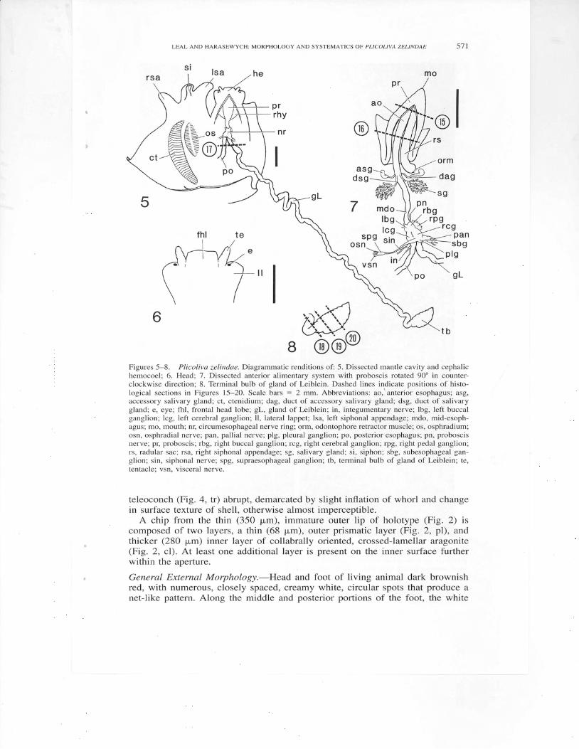
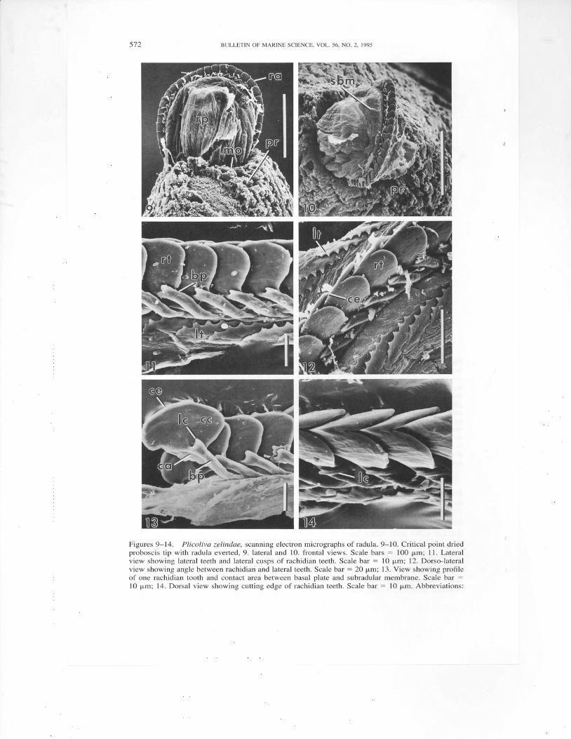
Figures 9-14. Plicoliva zelindae, scanning electron micrographs of radula. 9-10. Critical point driedproboscis tip with radula everted, 9. lateral and 10. frontal views. Scale bars : 100 p,m; 11. Lateralview showing lateral teeth and lateral cusps of rachidian teeth. Scale bar : l0 p,m; 12. Dorso-lateralview showing angle between rachidian and lateral teeth. Scale bar = 20 pm; 13. View showing prolileof one rachidian tooth and contact area between basal plate and subradular membrane. Scale bar :
10 pm; 14. Dorsal view showing cutting edge of rachidian teeth. Scale bar : 10 p,m. Abbreviations:
LEAL AND HARASEWYCH: MORPHOLOGY AND SYSTEMATICS OF PLICOLIVA ZELINDAE 5t3
spots give way to bright-yellow spots each circled by a white ring. Mantle thin,whitish, semi-transparent, almost completely enveloping shell when extended. Ex-tended foot slightly larger than shell, bilobed anteriody, rounded posteriorly.Operculum absent. Head (trigs. 5, he; 6) broad, flattened, with short, cylindricaltentacles (Fig. 6, te) and truncated, squarish frontal lobe (Fig. 6, fhl) that lacks amedian cephalic furrow. Lateral lappets (Fig. 6, ll) rounded anteriorly, taperingposteriorly. Eyes (Fig. 6, e) situated on lateral lappets near their junction withtentacles. Siphon (Fig. 5, si) well developed. Siphonal appendages (Fig. 5, rsa,lsa) equal in size, wider and longer than siphon. Mantle cavity shallow, broad,with large osphradium (Fig. 5, os), ctenidium (Fig. 5, ct) as wide and twice aslong as osphradium. Specimen lacking remaining portion of mantle cavity.
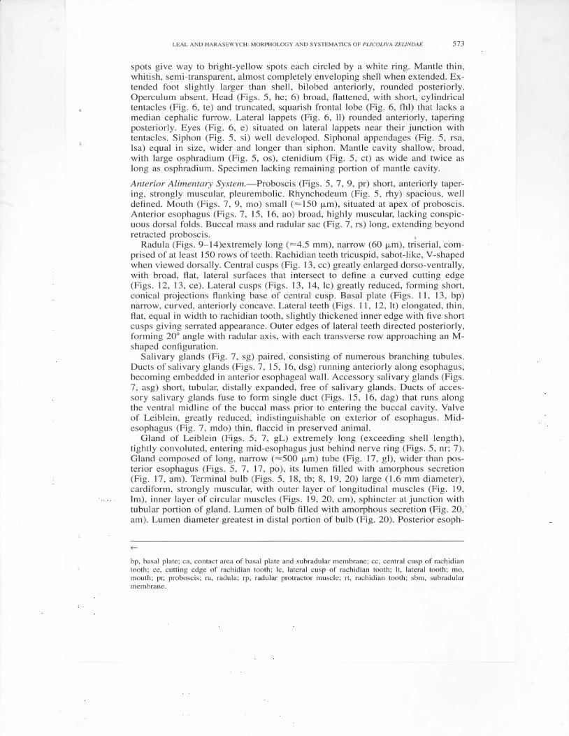
Anterior Alimentary System.-Proboscis (Figs. 5, 7, 9, pr) short, anteriorly taper-ing, strongly muscular, pleurembolic. Rhynchodeum (Fig. 5, rhy) spacious, welldefined. Mouth (Figs.7,9, mo) small (:150 pm), situated at apex of proboscis.Anterior esophagus (Figs. 7, 15, 16, ao) broad, highly muscular, lacking conspic-uous dorsal folds. Buccal mass and radular sac (Fig. 7, rs) long, extending beyondretracted proboscis.
Radula (Figs. 9-14)extremely long (:4.5 mm), narrow (60 pm), triserial, com-prised of at least 150 rows of teeth. Rachidian teeth tricuspid, sabot-like, V-shapedwhen viewed dorsally. Central cusps (Fig. 13, cc) greatly enlarged dorso-ventrally,with broad, flat, lateral surfaces that intersect to define a curved cutting edge(Figs. 12, 13, ce). Lateral cusps (Figs. 13, 14,1c) greatly reduced, forming short,conical projections flanking base ofcentral cusp. Basal plate (Figs. l l , 13, bp)narrow, curved, anteriorly concave. Lateral teeth (Figs. 11,12, lt) elongated, thin,flat, equal in width to rachidian tooth, slightly thickened inner edge with five shortcusps giving serrated appearance. Outer edges of lateral teeth directed posteriorly,forming 20" angle with radular axis, with each transverse row approaching an M-shaped configuration.
Salivary glands (Fig. 7, sg) paired, consisting of numerous branching tubules.Ducts of salivary glands (Figs.7, 15, 16, dsg) running anteriorly along esophagus,becoming embedded in anterior esophageal wall. Accessory salivary glands (Figs.7, asg) short, tubular, distally expanded, free of salivary glands. Ducts of acces-sory salivary glands fuse to form single duct (Figs. 15, 16, dag) that runs alongthe ventral midline of the buccal mass prior to entering the buccal cavity. Valveof Leiblein, greatly reduced, indistinguishable on exterior of esophagus. Mid-esophagus (Fig. 7, mdo) thin, flaccid in preserved animal.
Gland of Leiblein (Figs. 5, 1, gL) extremely long (exceeding shell length),t ightly convoluted, entering mid-esophagus just behind nerve ring (Figs. 5, nr 7).Gland composed of long, narrow (-500 pm) tube (Fig. 17, gl), wider than pos-terior esophagus (Figs. 5,7, 17, po), its lumen fi l led with amorphous secretion(Fig. 17, am). Terminal bulb (F igs.5, 18, tb ; 8,19,20) large (1.6 mm diameter) ,cardiform, strongly muscular, with outer layer of longitudinal muscles (Fig. 19,lm), inner layer of circular muscles (Figs. 19, 20, cm), sphincter at junction withtubular portion of gland. Lumen of bulb filled with amorphous secretion (Fig. 20,am). Lumen diameter greatest in distal portion of bulb (Fig. 20). Posterior esoph-
<-
bp, basal plate; ca, contact area of basal plate and subradular membrane; cc, central cusp of rachidiantooth; ce, cutting edge of rachidian tooth; lc, lateral cusp of rachidian tooth; lt, lateral tooth; mo,mouth; pq proboscis; ra, radula; rp, radular protractor muscle; rt, rachidian tooth; sbm, subradularmembrane.
BULLETIN OF MARINE SCIENCE. VOL. 56. NO.2. 1995
dsg $ l ; :
r z s l 8 brFigures 15-20. Plicoliva zelindae, histological sections of anterior alimentary system (see Figs. 5,7, 8 for position of sections); l5-Anterior third of proboscis; 16-Posterior third of proboscis; 17-Esophagus and gland of Leiblein posteriorly to point of separation; 18-Gland of Leiblein and terminalbulb at junction; l9-Proximal third of terminal bulb of gland of Leiblein; 2O-Terminal bulb ofgland of Leiblein at region of maximum diameter. All scale bars = 0.5 mm. Abbreviations: ao, anterioresophagus; am, amorphous secretion; cm, circular muscle; dag, duct of accessory salivary gland; dsg,duct of salivary gland; gL, gland of Leiblein; lm, longitudinal muscle; po, posterior.esophagus; ra,radula; rb, radular bolster; rp, radular protractor muscle; rr, radular retractor muscle; rsh, radular sheath;tb. terminal bulb of eland of Leiblein.
agus (Figs. 5,-7, 17, po) lined with longitudinal ridges. Remainder of alimentarysystem lacking in specimen examined.
Nervous System.-Circumesophageal nerve ring (Fig. 5, nr) highly concentrated,except for supraesophageal ganglion (Fig. 7, spg), which is widely separated fromright pleural ganglion (Fie.7, plg). Supraesophageal ganglion situated beneathepithelium of mantle cavity, near osphradium, giving rise to visceral (Fig. 7, vsn)and osphradial nerves (Fig. 7, osn).
DrscussroN
Although the overall shell morphology of immature specimens of Plicolivazelindae resemblesthatof thegenus Oliva,shell morphologyof adult P.ryall i is
LEAL AND HARASEWYCH: MORPHOLOGY AND SYSTEMATICS OF PLICOLM ZELINDAE 575
recognizably volutid, strongly resembling Enaeta barnesii (Gray, 1825) in overallshape, gloss, axial sculpture, columellar dentition and color pattern (Poppe andGoto, 1992:- pl. 18). Plicoliva zelindae also lacks such conchological and anatom-ical apomorphies of Olividae as: a channelled suture; pedal lobes; crescent-shapedpropodium with longitudinal cleft; posterior mantle lobe; anterior and posteriormantle tentacles (Kantor, I99l). Absence of these characters precludes the inclu-sion of Plicoliva in Olividae.
A broad head with a hood-like frontal lobe and lateral lappets, as well as long,paired siphonal appendages, are characters that are unique to Volutidae, but notubiquitous within the family. The presence of these characters in Plicoliva zelin-dae supports its inclusion within Volutidae, as does the morphology of its radula.The sabot-like rachidian teeth are very similar to those of Amoria canaliculata(Weaver and du Pont, 1970: fig.33a), but could conceivably be derived by ventralenlargement of the central cusp of the rachidian tooth of Enaeta cylleniformis(Sowerby, 1844) (Bouchet, 1990: figs.2I-22). Unlike Bouchet (1990: 8), we donot regard these teeth to be of the "wishbone" type found in Volutomitra andScaphella, as the central cusps in these genera are pointed rather thqn having abroad cutting edge, and likely function by piercing rather than incising prey tis-sues. Although triserial radulae have been reported within several volutid subfam-ilies (Aiken and Fuller, 1986: 39), the multicuspid lateral teeth of Plicoliva areunique within Volutidae, and a principal diagnostic feature of the subfamily Pli-colivinae. Similar multicuspid lateral teeth also occur in Fasciolariidae (Maes,1967: figs. l-7) and certain Mitridae (Cernohorsky,l99l: pl. 38, fig. 5), and wereregarded as primitive features within the Neogastropoda (Ponder, 1973: 308).
The arrangement of the lateral teeth, with their outer edges directed posteriorly,is nearly perpendicular to the usual orientation of these teeth in neogastropods.Similar orientations of monocuspid lateral teeth have been illustrated for Volu-tomitra (Arnaud and van Mol, 1979: fig. l2B, C), and the volutids Volutocorbis(Aiken and Fuller, 1986: 39a) and Callipara (Aiken and Fuller, 1986: 39f).
The salivary glands of Plicoliva zelindae differ from those of most neogastro-pods in that the branching structure of the individual tubules is readily apparent.Within the Volutidae, branching salivary glands have been reported only in thegenus Voluta (Clench and Turner, 1964: 134). The accessory salivary glands areshort and do not intertwine with the salivary glands as in some volutids (Clenchand Turner, 1964: pl.82).
The external morphology of the gland of Leiblein of Plicoliva zelindae, witha long, convoluted duct associated with a large, terminal muscular bulb, is mostsimilar to those of some specialized marginellids and toxoglossans. As in thesetaxa, the reduction or loss of the valve of Leiblein in Plicoliva zelindae is cor-related with the modification of the gland of Leiblein into a long, tubular "poisongland." Although the gland of Leiblein of most volutids consists of a convolutedtube of approximately constant diameter, those of Voluta musica (Pace, l9O2: 23;Clench and Turner, 1964: pl. 82, fi5. 18B), Voluta ebraea Linnaeus, 1758 (pers.observ.), and Ampulla priamus (Poirier, 1885: pl. 3, fig. 2; Pace, I9O2: 29) havebeen shown to have enlarged, terminal muscular bulbs.
Among rachiglossan neogastropods, the presence of a long connective betweenthe supraesophageal and right pleural ganglia, such as occurs in P. zelindae, hasbeen reported only in Muricidae (Marcus and Marcus, 1959: I49) and certainmembers of the family Volutidae (Ponder, 1973: 322). Within the Volutidae, thistype of nervous system (Type 1 following the terminology of Ponder, l97O: 159)has been reported only in the Volutinae, Haliinae, and eastern Atlantic Yetinae.
BULLETIN OF MARINE SCIENCE. VOL.56, NO.2, 1995
In Athletinae, Calliotectinae, Zidoninae, and Indo-Pacific Yetinae, these two gan-glia are closely fused together (Type 2 nervous system).
Of the nine other Recent subfamilies of Volutidae (Ponder and War6n, 1988:306), Plicolivinae shares a number of features with the Volutinae and some mem-bers of the Haliinae. It shares a Type I nervous system and salivary glands withbranching ducts with both these subfamilies. Like various species of Volutinae,Plicolivinae has shells with pronounced axial sculpture and numerous (>5) col-umellar folds, as well as a gland of Leiblein with an enlarged terminal bulb.
Plicolivinae shares the following similarities with species of Amoria: glazedshell exterior enveloped by mantle lobes, long siphonal appendages of equallength, rachidian morphology, and a similar pigmentation pattern of white spotscontaining centered bright-yellow spots on the foot [e.g., Arnoria canaliculata(McCoy, 1869), see Weaver and du Pont, 1970: pl. 69, topl. Although Arnoria isincluded in the subfamily Haliinae, of which the better known name Scaphellinaeis a synonym, both Plicoliva and Amoria differ from Scaphella in rachidian mor*phology, foot pigmentation, siphonal appendage morphology (Scaphella has asingle appendage on right side), and lack the median cephalic furrow present inScaphella (Clench and Turner, 1964:135).
It is premature to provide a cladistic analysis of the family Volutidae in orderto determine the relationships of Plicolivinae, as the literature on the family con-tains insufficient anatomical data to yield a resolved phylogenetic hypothesis.
AcrNowr-slcMENTS
E. de C. Rios (Museu OceanogrSfico, Rio Grande, Brazil) kindly presented the soft parts examinedin this study, which were collected and preserved by J. and M. V. Coltro. P. Bouchet (Mus6um Nationald'Histoire Naturelle, Paris) provided additional information on Plicoliva and, along with two anony-mous reviewers, criticalty read the manuscript. J. B. Wise (George Washington University, Washing-ton) and the late R. S. Houbrick (National Museum of Natural History, Smithsonian Institution) helpedduring different stages of this work. We thank staff members at the National Museum of NaturalHistory: S. G. Braden for assistance with scanning electron microscopy and V. Krantz, who preparedthe negatives used in Figures 1-3. The efforts of J.H.L. in this project were supported by a SmithsonianPostdoctoral Fellowship, Smithsonian Institution Office of Fellowships and Grants, and facilitated bya Conchologists of America 1992 Grant Award.
LrrsnRruns CIrso
Aiken, D. W and K. J. Fuller. 1986. The living Volutes of Africa. Sea Gifts, Cape Town. 41 pp.Arnaud, P and J.-J. van Mol. 1979. Anatomy, ecology and distribution of the Volutidae and Volu-
tomitridae of the southern Indian Ocean (Castropoda: Prosobranchia). The Veliger 22: 19-31.Bouchet, P 1990. Systematics of Plicoliva with description of a new subfamily (Gastropoda: Volu-
to idea). Arch. Mol luskenk. 120(1989)( l i3) : 1-10.Cernohorsky, W. O. 1991. The Mitridae of the World. Part 2. The subfamily Mitrinae concluded and
subfamilies Imbricariinae and Cylindromitrinae. Monogr. Mar. Moll. (4): l-164.Clench, W. J. and R. D. Turner. 1964. The subfamilies Volutinae, Zidoninae, Odontocymbiolinae and
Calliotectinae in the western Atlantic. Johnsonia 4(43): 129-I8O.Gray, J. E. 1825. Monograph on the Cypraeidae. Zool. J. l: 511-512 (note on Errata).Humason, G. L. 1979. Animal tissue techniques. Fourth edition. W. H. Freeman, San Francisco.
661 pp.Kantor, Y. 1991. On the morphology and relationships of some oliviform gastropods. Ruthenica 1:
17-52.Lewis, E. R. and M. K. Nemanic. 1973. Ctitical point drying techniques. SEM 1973, IIT Research
Institute, Chicago. Pp. 7 67 *77 4.Maes, V. O. 1967. Radulae of two species of Pleuroploca (Fasciolariidae) from the Indo-Pacific.
Nautilus 8l:48*54.Marcus, E. and E. Marcus. 1959. Studies on "Olividae." Bolm Fac. Filos. Ci6nc. Univ. S. Paulo
(232), (Zool.) (22): 99-188.McCov. F. 1869. On a new volute. Ann. Mag. Nat. Hist. 4(4): 34.
LEAL AND HARASEWYCH: MORPHOLOGY AND SYSTEMATICS OF PLICOLM ZELINDAE 577
Pace, S. 1902. On the anatomy and relationships of Voluta musica, Linn.; with notes upon certainother supposed members of the Volutidae. Proc. Malac. Soc. Lond. 5: 21-31.
Petuch, E. J. 1979. New gastropods from the Abrolhos Archipelago and reef complex, Brazil. Proc.Biol . Soc. Wash. 92: 510-526.
1987. New Caribbean molluscan faunas. Coast. Educ. Res. Found. Charlottesville. 154 -l
A-4 pp---:-and D. Sargent. 1986. Atlas of the living olive shells of the world. Coast. Educ. Res. Found.
Charlottesville. 253 pp.Poirier, M. J. 1885. Recherches anatomiques sur Halia priamus (Risso). Bull. Soc. Malac. Fr. 2: l7-5O.Ponder, W. F 1970. The morphology of Alcithoe arabica (Gastropoda: Volutidae). Malac. Rev. 3:
127-165.1973. The origin and evolution of the Neogastropoda. Malacologia 12: 295-338.
and A. War6n. 1988. Classification of the Caenogastropoda and Heterostropha-a list offamily-group names and higher taxa. Malac. Rev., suppl. 4: 288-328.
Poppe, G. and Y. Goto. 1992. Volutes. I-llnformatore Piceno, Ancona. 348 pp., 107 pls.Sowerby, G. B., lst. 1844. Description of six new species of volutes. Proc. Zool. Soc. Lond. (12):
149-152.Weaver, C. S. and J. E. du Pont. 1970. The living volutes. A monograph of the Recent Volutidae of
the World. Delaware Museum of Natural History, Greenville. xv + 375 pp.
DlrE Accppreo: November 8, 1993. I
Aoonsss: Department of Invertebrate Zoology, National Museum of Natural History, SmithsonianInstitution, Washington, D.C. 20560. PRESENT ADDRESS: (J.H.L.) Rosenstiel School of Marine andAtmosphere Science, 4600 Rickenbacker Causeway, Miami, Florida 33149-1098.