Embed Size (px)
Citation preview
Neuron
Article
Multisensory Mechanisms in Temporo-Parietal CortexSupport Self-Location and First-Person PerspectiveSilvio Ionta,1,5 Lukas Heydrich,1,4,5 Bigna Lenggenhager,1 Michael Mouthon,1 Eleonora Fornari,3 Dominique Chapuis,2,6
Roger Gassert,2,6 and Olaf Blanke1,4,*1Laboratory of Cognitive Neuroscience2Robotic Systems LaboratoryEcole Polytechnique Federale de Lausanne (EPFL), Lausanne, 1015, Switzerland3Department of Radiology, CIBM-CHUV unit, Centre Hospitalier Universitaire Vaudois and University of Lausanne, Lausanne, 1011,
Switzerland4Department of Neurology, University Hospital, Geneva, 1211, Switzerland5These authors contributed equally to this work6Present address: Rehabilitation Engineering Laboratory, Eidgenossische Technische Hochschule Zurich (ETHZ), LEO B 9.1,
Leonhardstrasse 27, 8092 Zurich, Switzerland
*Correspondence: [email protected] 10.1016/j.neuron.2011.03.009
SUMMARY
Self-consciousness has mostly been approached byphilosophical enquiry and not by empirical neurosci-entific study, leading to an overabundance ofdiverging theories and an absence of data-driventheories. Using robotic technology, we achievedspecific bodily conflicts and induced predictablechanges in a fundamental aspect of self-conscious-ness by altering where healthy subjects experiencedthemselves to be (self-location). Functional magneticresonance imaging revealed that temporo-parietaljunction (TPJ) activity reflected experimentalchanges in self-location that also depended on thefirst-person perspective due to visuo-tactile andvisuo-vestibular conflicts. Moreover, in a large lesionanalysis study of neurological patients with a well-defined state of abnormal self-location, braindamage was also localized at TPJ, providing causalevidence that TPJ encodes self-location. Our find-ings reveal that multisensory integration at the TPJreflects one of the most fundamental subjective feel-ings of humans: the feeling of being an entity local-ized at a position in space and perceiving the worldfrom this position and perspective.
INTRODUCTION
How can a human brain develop self-consciousness? What are
the brain mechanisms involved in this process? Extending earlier
data from neurological patients (Critchley, 1953; Hecaen and
Ajuriaguerra, 1952; Schilder, 1935), recent neurological theories
stress the importance of bodily processing for the self and
self-consciousness. These theories highlight the importance of
interoceptive, proprioceptive, and motor signals and their multi-
sensory and sensorimotor integration with other bodily signals
(Damasio, 1999; Frith, 2005; Gallagher, 2000; Jeannerod,
2003), but do not indicate how such integration induces key
subjective states such as self-location (‘‘Where am I in space?’’)
and the first-person perspective (‘‘From where do I perceive the
world?’’) and which neural mechanisms are involved (Blanke and
Metzinger, 2009). Data from neurological patients suffering from
out-of-body experiences (OBEs) provide such evidence,
showing that focal brain damage may lead to pathological
changes of the first-person perspective and self-location (Blanke
et al., 2002; De Ridder et al., 2007), due to interference with the
integration of multisensory bodily information at the TPJ. It was
argued that such changes in first-person perspective and self-
location are due to a double disintegration of bodily signals,
a disintegration between somatosensory (proprioceptive and
tactile) and visual signals combined with an additional visuo-
vestibular disintegration (Blanke et al., 2004; Lopez et al.,
2008); yet this has not been tested experimentally. Moreover,
there is a low number of investigated cases, and OBEs have
been associated with many different brain structures: the right
and left TPJ (Blanke et al., 2002, 2004; Brandt et al., 2005; Mail-
lard et al., 2004) and several structures within the TPJ (Blanke
et al., 2002, 2005; Heydrich et al., 2011; Brandt et al., 2005;
De Ridder et al., 2007; Maillard et al., 2004), precuneus
(De Ridder et al., 2007), and fronto-temporal cortex (Devinsky
et al., 1989). Accordingly, it is not clear which of these structures
are involved in abnormal conscious states of first-person
perspective and self-location and the significance of these
clinical findings for self-consciousness under normal conditions.
Recent behavioral and physiological work, using video-projec-
tion and various visuo-tactile conflicts, showed that self-location
can also be manipulated experimentally in healthy participants
(Ehrsson, 2007; Lenggenhager et al., 2007). Thus, synchronous
stroking of the participant’s back and the back of a visually pre-
sentedvirtual body led tochanges in self-location (toward a virtual
body at a position outside the participant’s bodily borders) and
self-identification with the virtual body (Lenggenhager et al.,
2007). So far, these experimental findings and techniques have
not been integrated with neuroimaging, such as fMRI, probably
because the above-mentioned experimental setups require
Neuron 70, 363–374, April 28, 2011 ª2011 Elsevier Inc. 363
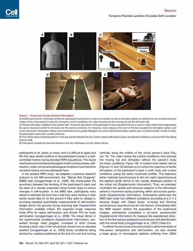
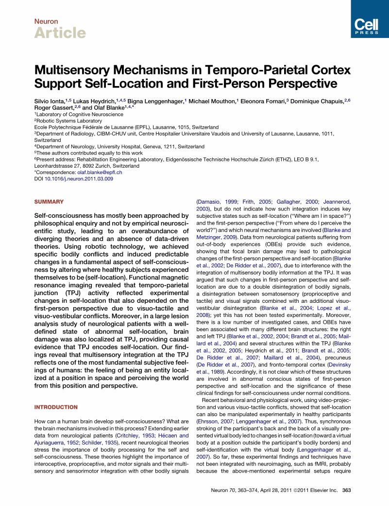
Figure 1. Visual and Tactile-Robotic Stimulation
(A and B) Visual stimuli. In the body conditions, participants were shown a video of a wooden rod with a stimulation sphere (in red) that moved vertically along the
midline of the virtual person’s back (A). During the control conditions, the video showed only the moving rod and the stimulator (B).
(C) Robotic stimulator installed on the scanner bed. The tactile stimulation of the participant’s back was performed by a custom-made robotic device generating
the same movement profile for the body and the control conditions. An ultrasonic motor placed at the level of the feet actuated the stimulation sphere over
a rack-and-pinion mechanism. Motion was transmitted over a guided fiberglass rod, which held the stimulation sphere over a compliant blade in order to follow
the participant’s back with constant pressure.
(D) The robotic device (stroking sphere in red) was placed between the two custom-made mattresses (in gray; the standard mattress is removed from the sliding
scanner bed).
(E) Participant (outside the scanner) placed on the two mattresses and the robotic device.
Neuron
Temporo-Parietal Junction Encodes Self-Location
participants to sit, stand, or move, and it is difficult to apply and
film the visuo-tactile conflicts on the participant’s body in a well-
controlled manner during standard fMRI acquisitions. The neural
mechanismsof a fundamental aspect of self-consciousness, self-
location, under normal andpathological conditionshave therefore
remained elusive and are addressed here.
In the present fMRI study, we adapted a previous research
protocol to the MR-environment: the ‘‘Mental Ball Dropping’’
(MBD) task (Lenggenhager et al., 2009). We manipulated the
synchrony between the stroking of the participant’s back and
the back of a visually presented virtual human body to induce
changes in self-location. In the MBD task, participants were
asked to estimate the time that a ball they were holding in their
hands would take to hit the ground if they were to release it,
providing repeated quantifiable measurements of self-location
(height above the ground) during scanning (see Supplemental
Information available online). We expected longer response
times (RTs) for higher self-location and shorter RTs for lower
self-location (Lenggenhager et al., 2009). The visual stimuli in
the experimental conditions (Supplemental Information), pre-
sented through video goggles, consisted of short movies
showing a back view of the virtual body filmed from an elevated
position (Lenggenhager et al., 2009) (body conditions) being
stroked by a sphere positioned at the end of a rod and moving
364 Neuron 70, 363–374, April 28, 2011 ª2011 Elsevier Inc.
vertically along the midline of the virtual person’s back (Fig-
ure 1A). The video during the control conditions only showed
the moving rod and stimulator without the person’s body
(no-body conditions; Figure 1B). A custom-built robotic device
(Figures 1C and 1D) allowed us to control the trajectory of tactile
stimulation of the participant’s back in both body and control
conditions (using the same movement profile). This trajectory
either matched (synchronous) or did not match (asynchronous)
the applied tactile stimuli to the visually displayed position of
the virtual rod (Supplemental Information). Thus, we precisely
controlled the spatial and temporal aspects of the stimulation
sphere’s movement during scanning within and across partici-
pants (Supplemental Information). Participants performed the
MBD task under four different conditions according to a 2 3 2
factorial design with Object (body; no-body) and Stroking
(synchronous; asynchronous) as main factors. Immediately after
the fMRI session (before the acquisition of the anatomical
images), participants completed a six-items questionnaire
(Supplemental Information) to measure the experienced direc-
tion of the first-person perspective and illusory self-identification
with the virtual body (Lenggenhager et al., 2007) (Table S1).
To define the structures that are involved in abnormal states of
first-person perspective and self-location, we also studied
a large group of neurological patients suffering from OBEs
Table 1. Free Reports during Robotic Visuo-Tactile Stroking
A Condition Up-Group
S3 S ‘‘This time the only thing that made me doubt that
the filmed body was not me, is that I could not see
the hands. Indeed I had the clear impression
of floating even if I knew I was not moving.’’
AS ‘‘Always well relaxed but the fact that I could not
feel the same thing that I was watching disturbed me.’’
S5 S ‘‘When I focused to estimate the timing,
it was as if I did not feel anymore what was
happening on my back, as if I was only watching
the video in front of me.’’
AS ‘‘It was clear that I was watching a movie
unrelated to my experience.’’
S8 S ‘‘I felt rising in a strange way towards the roof.’’
AS ‘‘I had the impression of watching a video in the
rewind mode.’’
S12 S ‘‘I did not have any particular sensation despite
a general, but nevertheless mild, elevation.’’
AS ‘‘Not even elevation.’’
B Condition Down-Group
S4 S ‘‘I was looking at my own body from above. The
perception of being apart from my body was a bit
weak but still there. I saw the stick moving onto
my back and I perceived it to be somehow at odds
with what I was looking at.’’
AS ‘‘This time what I felt on my back did not correspond
at all to what I saw. I had the impression of being
very far from the real me.’’
S9 S ‘‘I felt myself a bit floating but in a descendent
direction. On the contrary of the reality I had the
impression that my body was thicker as if front
and back were not as close as before the stick
touched my back.’’
AS ‘‘I felt like I was watching someone else’s body
from above, while someone was rubbing my chest
with a stick. I also felt as being above the body
I was watching at. I felt I was physically located
above the body I was watching.’’
S11 S ‘‘I asked myself: if the one that I see in the movie is
me, how can they move the mattress up and down?’’
AS ‘‘I felt as if I was floating high and I did not know
where I was.’’
S17 S ‘‘At the beginning I was expecting to feel the stick
on my front side, but then I realized that it was
touching my back. I felt as if I was laying on myself,
face to face.’’
AS ‘‘I felt as if I was floating, very light, without weight.
I had the impression of feeling the impact of
a surface on my back as if I was touching the roof.’’
Participants were asked to write down what they experienced during
synchronous (S) and asynchronous (AS) visuo-tactile stroking conditions.
Selected responses are listed for participants from the Up-group (A) and
the Down-group (B).
See also Table S4.
Neuron
Temporo-Parietal Junction Encodes Self-Location
(Blanke et al., 2002, 2004; Heydrich et al., 2011; Devinsky et al.,
1989; Maillard et al., 2004). We performed quantitative lesion
analysis (Rorden et al., 2007a) and compared the distribution
of brain lesions in nine OBE-patients with those of eight other
patients showing complex hallucinations involving people or
faces, but without abnormal self-location, self-identification, or
first-person perspective (control group; Table S3). This allowed
us to determine the anatomical sub-regions of maximal lesion
overlap and to perform statistical comparisons contrasting the
lesions of OBE and control patients (voxel-based lesion
symptom mapping; VLSM) (Bates et al., 2003a). Based on
previous data in patients with OBEs, we predicted to find
maximal involvement of the TPJ. Based on these clinical data,
we also predicted that the BOLD response of this structure in
healthy subjects would reflect changes in self-location that are
dependent on the experimental factors Stroking and Object.
Importantly, we further predicted that TPJ activity should also
reflect changes in self-location that depend on the direction of
the first-person perspective because (1) such changes are
a key element of OBEs and because (2) we were able to manip-
ulate the experienced direction of the first-person perspective
and its influence on self-location with our robotic stroking setup
(interaction between Stroking, Object, and Perspective; see next
section).
RESULTS
Robotically-Induced Changes in the Directionof the First-Person PerspectiveEarlier pilot questionnaire data revealed that, next to self-loca-
tion and self-identification, we were also able to manipulate the
experienced direction of the first-person perspective. In the pilot
study, several participants mentioned spontaneously that they
felt as if they were looking down at the virtual body (even though
they were physically in a supine position and facing upward).
Thus, for the present study, we added a related question (ques-
tion 1; Q1) to the questionnaire (Table S1). To answer Q1, while
being still within the MR-scanner, our participants were asked
to indicate the direction of their experienced first-person
perspective by placing a cursor on one out of three possible
answers (up, not sure, down). After the fMRI session, all partici-
pants were, in addition, asked to write a free report about their
experience during the stroking (Table 1; Table S4). With respect
to Q1, participants who chose the ‘‘not sure’’ response were also
interviewed after the experiment and asked to estimate which
perspective they used most of the time. On the basis of both
written free reports and interviews, the most frequent perspec-
tive across conditions was determined for these participants
and allowed us to assign all participants to either the Up- or
the Down-group. As in the pilot study, in the present study we
found that many participants reported looking always upward
(n = 10) or looking for most of the time upward (n = 1) at the virtual
body located above them (i.e., congruent with their physical
perspective: Up-group, n = 11). Selected experiences of the
Up-group participants during the synchronous and asynchro-
nous body conditions are listed in Table 1A. The remaining
participants reported that they had the impression that they
were always looking down (n = 6) or were for most of the time
Neuron 70, 363–374, April 28, 2011 ª2011 Elsevier Inc. 365
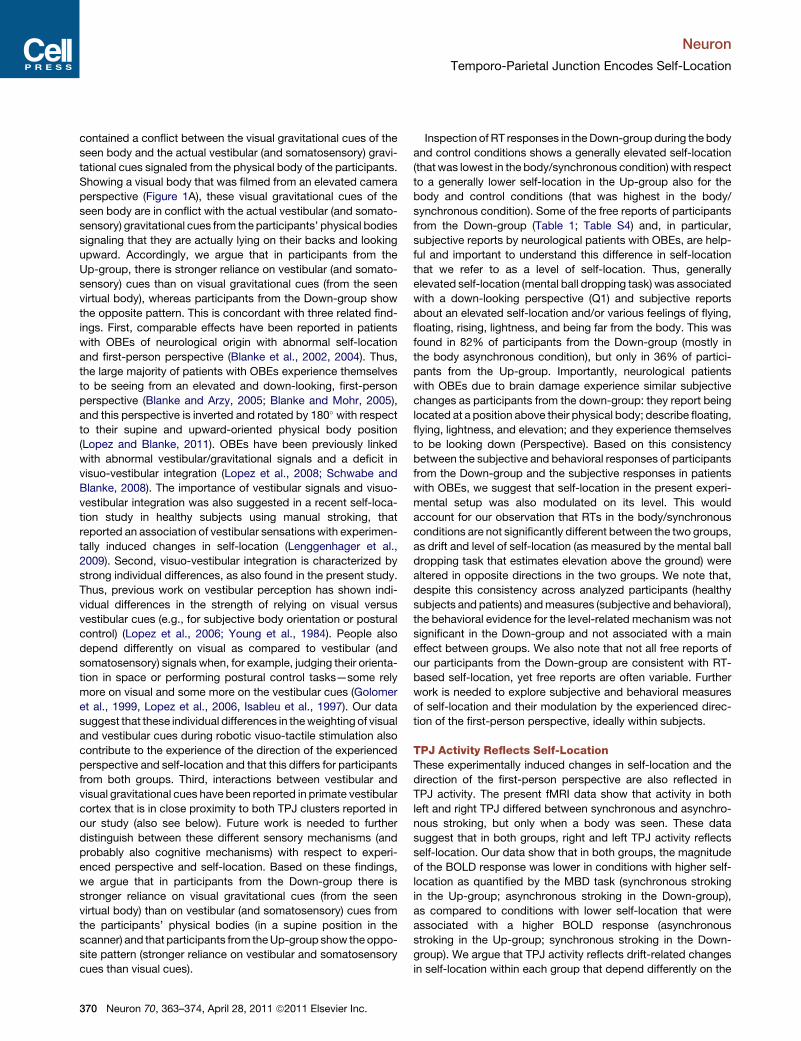
Figure 2. Self-Location Manipulation
Graphic representation of the experimentally induced changes in self-location and perspective in the Up- and Down-group. The position of the human bodies
represents the experienced position as indicated by the self-location task (mental ball dropping). The labels on the trousers indicate the experimental conditions.
The direction of the experienced first-person perspective (asmeasured through questionnaires) is represented by the direction of the feet and nose, as well as the
black arrows (pointing upward or downward). In both perspective groups, the body/synchronous condition leads to a drift in self-location toward the virtual body,
but in opposite directions depending on the experienced perspective.
(A) Thus, participants that had the impression of looking upward at the virtual body (Up-group) had increased response times (RTs) in the MBD task during the
synchronous as compared to the asynchronous stroking condition (represented by a blue line), indicating an elevation of self-location.
(B) Participants that had the impression of looking downward at the virtual body from an elevated perspective (Down-group) had decreased RTs in the MBD task
during the synchronous as compared to the asynchronous stroking condition (represented by a blue line) indicating a lowering of self-location. The drift in
self-location occurred in the direction of the experienced perspective (black arrows). RTs for the MBD task are plotted for each group as a function of the factors
Object and Stroking. Orange bars represent the synchronous stroking conditions and the red bars the asynchronous stroking conditions. Asterisks indicate
significant differences. Error bars indicate standard error. Note the differences between synchronous and asynchronous stroking conditions only in the body
conditions (not in the control conditions) and in opposite directions between Up- and Down-group. The MR-scanner is depicted only for illustration purposes
(participants were not asked to estimate the position of the scanner bed).
Neuron
Temporo-Parietal Junction Encodes Self-Location
looking down (n = 5) at the virtual body located below them
(i.e., incongruent with their physical perspective: Down-group,
n = 11). Selected experiences of the Down-group participants
during the synchronous and asynchronous body conditions are
listed in Table 1B. In summary, whereas several participants
felt as if they were looking upward at the virtual body ‘‘above
them’’ (Up-group), the remaining participants had the impression
that they were looking down at the virtual body ‘‘below them’’
(Down-group). This was found despite somatosensory, motor,
and cognitive cues from our participants about their body posi-
tion (they were lying on their back, facing upward, and were
head-constrained in the headcoil; Figure 1E; Supplemental Infor-
mation). Based on these findings, we carried out data analysis
considering each group of participants. This led to a 2 3 2 3 2
factorial design with Perspective (up; down) as in-between
factor, and Object (body; no-body) and Stroking (synchronous;
asynchronous) as within factors that were applied to the analysis
of self-location, self-identification, and the fMRI data.
Robotically-Induced Changes in Self-Locationand Self-IdentificationStatistical analysis of RTs in theMBD task showed that self-loca-
tion depended on Object, Stroking, and Perspective [significant
three-way interaction; F(1,20) = 4.4; p < 0.05]. Post hoc compar-
isons showed that in the body conditions, the participants of the
Up-group (participants experiencing themselves to be looking
upward at the visually presented body) estimated self-location
as higher (longer RTs) during the synchronous (1071 ms)
comparedwith the asynchronous stroking (991ms; p < 0.01; Fig-
ure 2A). The opposite pattern was found in the Down-group
(participants experiencing that they were looking downward at
the visually presented body): lower self-location and shorter
366 Neuron 70, 363–374, April 28, 2011 ª2011 Elsevier Inc.
RTs during the synchronous stroking (1047 ms) with respect to
the asynchronous stroking while viewing the body (1138 ms;
p < 0.03; Figure 2B). No significant differences were found
between synchronous and asynchronous stroking in the control
conditions in both groups (all p > 0.2; see Figures 2A and 2B).
Notably, RTs in the body conditions are modulated, within
each group, as a function of stroking and the experienced direc-
tion of the first-person perspective. Thus, self-location changes
for the Up-group were characterized by a generally lower self-
location that was further modulated by stroking in the upward
direction (toward the seen virtual body), whereas self-location
changes for the Down-group were characterized by a generally
higher self-location that was further modulated by stroking in
the downward direction (toward the seen virtual body) (see Fig-
ure 2). For other effects see Supplemental Information.
Our questionnaire results showed that predictable changes in
self-identification and illusory touch, depending on the factors
Object and Stroking, can be induced using robotic stroking in
the fMRI environment. As predicted, and in accordance with
previous work (Ehrsson, 2007; Lenggenhager et al., 2007,
2009), statistical analysis of the questionnaires (Supplemental
Information) showed that, regardless of Perspective, responses
to Q3 (‘‘How strong was the feeling that the body you saw was
you?’’) indicated stronger self-identification [F(4,80) = 13.5;
p<0.01]with the virtual bodyduring synchronous (4.1) thanasyn-
chronous stroking (2.3), and that responses to Q5 (‘‘How strong
was the feeling that the touch you felt was located where you
saw the stroking?’’) indicated stronger illusory touch [F(4,80) =
13.5; p < 0.001] during the synchronous (8.1) than the asynchro-
nous stroking (2.8; Figure 3; Supplemental Information).
To summarize these findings, participants from the Up-group
experienced themselves to be looking up at the body above
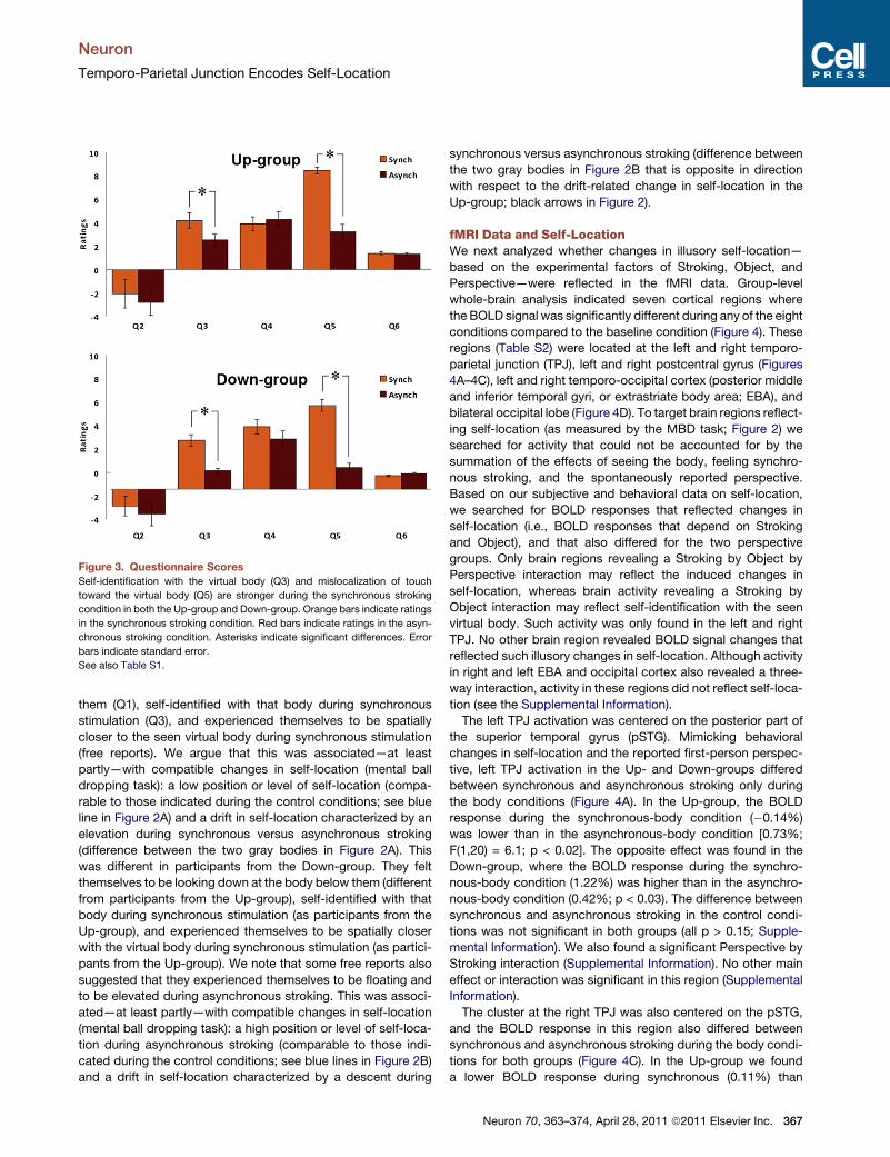
Figure 3. Questionnaire Scores
Self-identification with the virtual body (Q3) and mislocalization of touch
toward the virtual body (Q5) are stronger during the synchronous stroking
condition in both the Up-group and Down-group. Orange bars indicate ratings
in the synchronous stroking condition. Red bars indicate ratings in the asyn-
chronous stroking condition. Asterisks indicate significant differences. Error
bars indicate standard error.
See also Table S1.
Neuron
Temporo-Parietal Junction Encodes Self-Location
them (Q1), self-identified with that body during synchronous
stimulation (Q3), and experienced themselves to be spatially
closer to the seen virtual body during synchronous stimulation
(free reports). We argue that this was associated—at least
partly—with compatible changes in self-location (mental ball
dropping task): a low position or level of self-location (compa-
rable to those indicated during the control conditions; see blue
line in Figure 2A) and a drift in self-location characterized by an
elevation during synchronous versus asynchronous stroking
(difference between the two gray bodies in Figure 2A). This
was different in participants from the Down-group. They felt
themselves to be looking down at the body below them (different
from participants from the Up-group), self-identified with that
body during synchronous stimulation (as participants from the
Up-group), and experienced themselves to be spatially closer
with the virtual body during synchronous stimulation (as partici-
pants from the Up-group). We note that some free reports also
suggested that they experienced themselves to be floating and
to be elevated during asynchronous stroking. This was associ-
ated—at least partly—with compatible changes in self-location
(mental ball dropping task): a high position or level of self-loca-
tion during asynchronous stroking (comparable to those indi-
cated during the control conditions; see blue lines in Figure 2B)
and a drift in self-location characterized by a descent during
synchronous versus asynchronous stroking (difference between
the two gray bodies in Figure 2B that is opposite in direction
with respect to the drift-related change in self-location in the
Up-group; black arrows in Figure 2).
fMRI Data and Self-LocationWe next analyzed whether changes in illusory self-location—
based on the experimental factors of Stroking, Object, and
Perspective—were reflected in the fMRI data. Group-level
whole-brain analysis indicated seven cortical regions where
the BOLD signal was significantly different during any of the eight
conditions compared to the baseline condition (Figure 4). These
regions (Table S2) were located at the left and right temporo-
parietal junction (TPJ), left and right postcentral gyrus (Figures
4A–4C), left and right temporo-occipital cortex (posterior middle
and inferior temporal gyri, or extrastriate body area; EBA), and
bilateral occipital lobe (Figure 4D). To target brain regions reflect-
ing self-location (as measured by the MBD task; Figure 2) we
searched for activity that could not be accounted for by the
summation of the effects of seeing the body, feeling synchro-
nous stroking, and the spontaneously reported perspective.
Based on our subjective and behavioral data on self-location,
we searched for BOLD responses that reflected changes in
self-location (i.e., BOLD responses that depend on Stroking
and Object), and that also differed for the two perspective
groups. Only brain regions revealing a Stroking by Object by
Perspective interaction may reflect the induced changes in
self-location, whereas brain activity revealing a Stroking by
Object interaction may reflect self-identification with the seen
virtual body. Such activity was only found in the left and right
TPJ. No other brain region revealed BOLD signal changes that
reflected such illusory changes in self-location. Although activity
in right and left EBA and occipital cortex also revealed a three-
way interaction, activity in these regions did not reflect self-loca-
tion (see the Supplemental Information).
The left TPJ activation was centered on the posterior part of
the superior temporal gyrus (pSTG). Mimicking behavioral
changes in self-location and the reported first-person perspec-
tive, left TPJ activation in the Up- and Down-groups differed
between synchronous and asynchronous stroking only during
the body conditions (Figure 4A). In the Up-group, the BOLD
response during the synchronous-body condition (�0.14%)
was lower than in the asynchronous-body condition [0.73%;
F(1,20) = 6.1; p < 0.02]. The opposite effect was found in the
Down-group, where the BOLD response during the synchro-
nous-body condition (1.22%) was higher than in the asynchro-
nous-body condition (0.42%; p < 0.03). The difference between
synchronous and asynchronous stroking in the control condi-
tions was not significant in both groups (all p > 0.15; Supple-
mental Information). We also found a significant Perspective by
Stroking interaction (Supplemental Information). No other main
effect or interaction was significant in this region (Supplemental
Information).
The cluster at the right TPJ was also centered on the pSTG,
and the BOLD response in this region also differed between
synchronous and asynchronous stroking during the body condi-
tions for both groups (Figure 4C). In the Up-group we found
a lower BOLD response during synchronous (0.11%) than
Neuron 70, 363–374, April 28, 2011 ª2011 Elsevier Inc. 367
Figure 4. TPJ Activity and Self-Location
TPJ activity in the body conditions is shown for the Up- and the Down-group. In both groups, the magnitude of the BOLD response was lower in conditions with
high self-location as quantified by the MBD task (synchronous stroking in the Up-group; asynchronous stroking in the Down-group) as compared to conditions
with lower estimated self-location (asynchronous stroking in the Up-group; synchronous stroking in the Down-group).
(A) (higher panel) Left TPJ activation centered on pSTG. The lower panel shows the BOLD response at the left TPJ as a function of Perspective and Stroking during
the body conditions.
(B) Activation in left and right TPJ and left superior postcentral gyrus.
(C) (higher panel) Right TPJ activation centered on pSTG. The lower panel shows the BOLD response at the right TPJ as a function of Perspective and Stroking
during the body conditions. The pattern of the left TPJ BOLD response was the same as found at the right TPJ.
(D) Activation of left and right posterior middle and inferior temporal gyri.
See also Table S2. Asterisks indicate significant differences. Error bars indicate standard error.
Neuron
Temporo-Parietal Junction Encodes Self-Location
asynchronous stroking [1.14%; F(1, 20) = 7; p < 0.016], whereas
in the Down-group we found the opposite trend with a higher
BOLD response during the synchronous (1.03%) than the asyn-
chronous stroking condition (0.34%; p = 0.09). The BOLD
response was not significantly different between synchronous
and asynchronous stroking in the control conditions in both
groups (all p > 0.32). No other main effect or interaction was
significant in this region (Supplemental Information).
Other fMRI DataTo target brain regions reflecting self-identification (as measured
by the questionnaire; question Q3; Figure 3) we searched for
activity that could not be accounted for by the summation of
the effects of seeing the body and feeling synchronous stroking.
To this aim, we searched for brain regions showing an interaction
between Object and Stroking characterized by a difference
between the two body conditions, but not the control conditions.
Such activity was only found in the right EBA. The ANOVA per-
formed on the BOLD signal change in right EBA (Supplemental
Information) showed a significant two-way interaction between
Object and Stroking [F(1,20) = 6.56; p < 0.02], accounted for
by the higher BOLD response in the body/asynchronous condi-
tion (1.2%) with respect to the body/synchronous (0.47%) and
the no-body/asynchronous conditions (0.72%; all p < 0.05).
Yet right EBA activity in the body/synchronous condition (strong
self-identification) did not differ from any of the two no-body
control conditions (all p > 0.14). No other brain region revealed
BOLD signal changes that reflected changes in self-identification
368 Neuron 70, 363–374, April 28, 2011 ª2011 Elsevier Inc.
with the seen virtual body (Supplemental Information). Finally,
only activity in the cluster centered at the right postcentral gyrus
revealed a main effect of Stroking [F(1,20) = 24.02; p < 0.001]
revealing a lower BOLD in the synchronous (�0.51%) with
respect to the asynchronous conditions (0.13%). For other
fMRI data descriptions, see the Supplemental Information.
Lesion AnalysisWe found that in eight out of nine OBE-patients, brain damage
affected the right temporal and/or parietal cortex, most often
at the TPJ (Table S3). Lesion analysis revealed maximal lesion
overlap at the right angular gyrus, pSTG, and middle temporal
gyrus in seven out of eight OBE-patients (Figure 5A). This was
confirmed by VLSM showing maximal involvement of the right
TPJ (MNI: 54,�52,26; Z-score = 3.53; p < 0.01, FDR-corrected),
centered at the angular gyrus and posterior STG (32% of the
voxels were within the pSTG, 27% within the middle temporal
gyrus, 26% within the angular gyrus, and 6% within the supra-
marginal gyrus; Figure 5B).
DISCUSSION
Self-Location Depends on the Direction of the First-Person Perspective and StrokingUsing robotic technology, the present data show that, in the
noisy and physically constraining MR-environment, we were
able to manipulate two key aspects of self-consciousness:
self-location and the first-person perspective. We induced
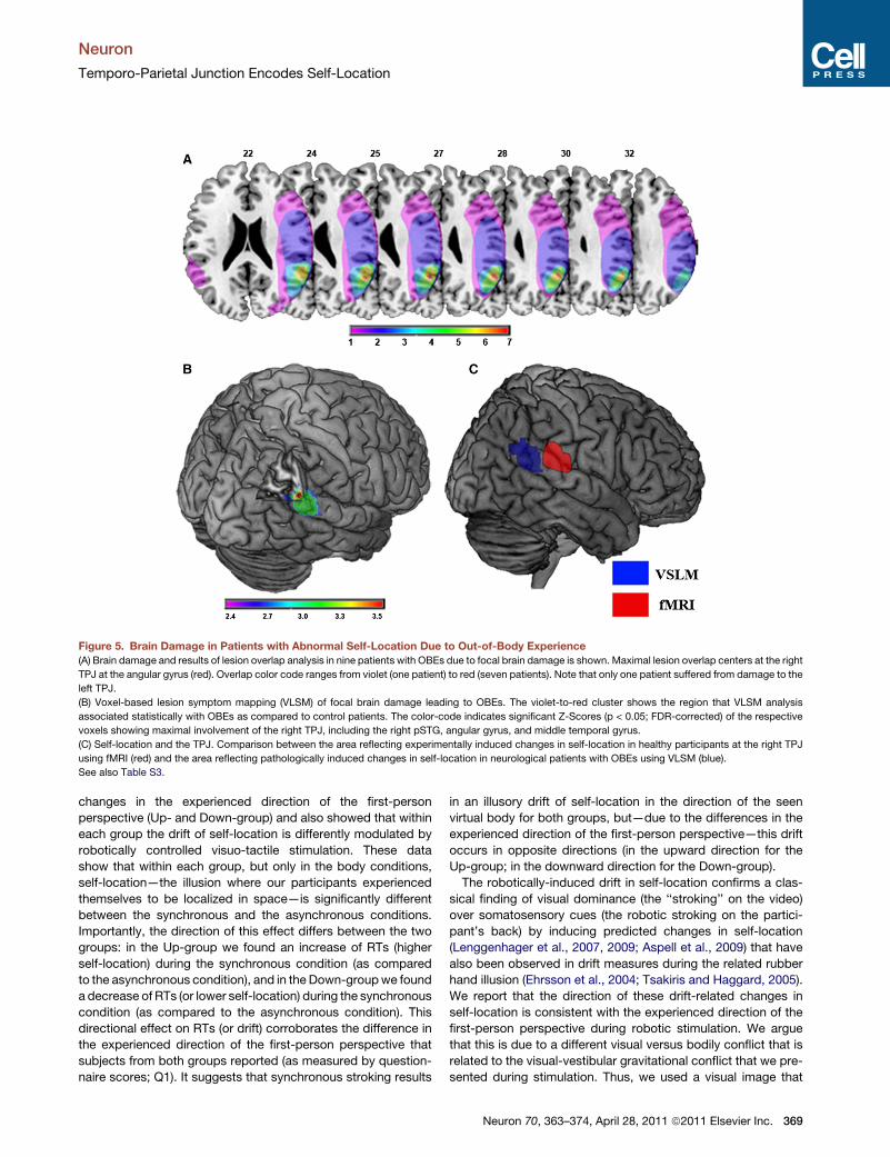
Figure 5. Brain Damage in Patients with Abnormal Self-Location Due to Out-of-Body Experience
(A) Brain damage and results of lesion overlap analysis in nine patients with OBEs due to focal brain damage is shown. Maximal lesion overlap centers at the right
TPJ at the angular gyrus (red). Overlap color code ranges from violet (one patient) to red (seven patients). Note that only one patient suffered from damage to the
left TPJ.
(B) Voxel-based lesion symptom mapping (VLSM) of focal brain damage leading to OBEs. The violet-to-red cluster shows the region that VLSM analysis
associated statistically with OBEs as compared to control patients. The color-code indicates significant Z-Scores (p < 0.05; FDR-corrected) of the respective
voxels showing maximal involvement of the right TPJ, including the right pSTG, angular gyrus, and middle temporal gyrus.
(C) Self-location and the TPJ. Comparison between the area reflecting experimentally induced changes in self-location in healthy participants at the right TPJ
using fMRI (red) and the area reflecting pathologically induced changes in self-location in neurological patients with OBEs using VLSM (blue).
See also Table S3.
Neuron
Temporo-Parietal Junction Encodes Self-Location
changes in the experienced direction of the first-person
perspective (Up- and Down-group) and also showed that within
each group the drift of self-location is differently modulated by
robotically controlled visuo-tactile stimulation. These data
show that within each group, but only in the body conditions,
self-location—the illusion where our participants experienced
themselves to be localized in space—is significantly different
between the synchronous and the asynchronous conditions.
Importantly, the direction of this effect differs between the two
groups: in the Up-group we found an increase of RTs (higher
self-location) during the synchronous condition (as compared
to the asynchronous condition), and in the Down-groupwe found
a decrease of RTs (or lower self-location) during the synchronous
condition (as compared to the asynchronous condition). This
directional effect on RTs (or drift) corroborates the difference in
the experienced direction of the first-person perspective that
subjects from both groups reported (as measured by question-
naire scores; Q1). It suggests that synchronous stroking results
in an illusory drift of self-location in the direction of the seen
virtual body for both groups, but—due to the differences in the
experienced direction of the first-person perspective—this drift
occurs in opposite directions (in the upward direction for the
Up-group; in the downward direction for the Down-group).
The robotically-induced drift in self-location confirms a clas-
sical finding of visual dominance (the ‘‘stroking’’ on the video)
over somatosensory cues (the robotic stroking on the partici-
pant’s back) by inducing predicted changes in self-location
(Lenggenhager et al., 2007, 2009; Aspell et al., 2009) that have
also been observed in drift measures during the related rubber
hand illusion (Ehrsson et al., 2004; Tsakiris and Haggard, 2005).
We report that the direction of these drift-related changes in
self-location is consistent with the experienced direction of the
first-person perspective during robotic stimulation. We argue
that this is due to a different visual versus bodily conflict that is
related to the visual-vestibular gravitational conflict that we pre-
sented during stimulation. Thus, we used a visual image that
Neuron 70, 363–374, April 28, 2011 ª2011 Elsevier Inc. 369
Neuron
Temporo-Parietal Junction Encodes Self-Location
contained a conflict between the visual gravitational cues of the
seen body and the actual vestibular (and somatosensory) gravi-
tational cues signaled from the physical body of the participants.
Showing a visual body that was filmed from an elevated camera
perspective (Figure 1A), these visual gravitational cues of the
seen body are in conflict with the actual vestibular (and somato-
sensory) gravitational cues from the participants’ physical bodies
signaling that they are actually lying on their backs and looking
upward. Accordingly, we argue that in participants from the
Up-group, there is stronger reliance on vestibular (and somato-
sensory) cues than on visual gravitational cues (from the seen
virtual body), whereas participants from the Down-group show
the opposite pattern. This is concordant with three related find-
ings. First, comparable effects have been reported in patients
with OBEs of neurological origin with abnormal self-location
and first-person perspective (Blanke et al., 2002, 2004). Thus,
the large majority of patients with OBEs experience themselves
to be seeing from an elevated and down-looking, first-person
perspective (Blanke and Arzy, 2005; Blanke and Mohr, 2005),
and this perspective is inverted and rotated by 180� with respect
to their supine and upward-oriented physical body position
(Lopez and Blanke, 2011). OBEs have been previously linked
with abnormal vestibular/gravitational signals and a deficit in
visuo-vestibular integration (Lopez et al., 2008; Schwabe and
Blanke, 2008). The importance of vestibular signals and visuo-
vestibular integration was also suggested in a recent self-loca-
tion study in healthy subjects using manual stroking, that
reported an association of vestibular sensations with experimen-
tally induced changes in self-location (Lenggenhager et al.,
2009). Second, visuo-vestibular integration is characterized by
strong individual differences, as also found in the present study.
Thus, previous work on vestibular perception has shown indi-
vidual differences in the strength of relying on visual versus
vestibular cues (e.g., for subjective body orientation or postural
control) (Lopez et al., 2006; Young et al., 1984). People also
depend differently on visual as compared to vestibular (and
somatosensory) signals when, for example, judging their orienta-
tion in space or performing postural control tasks—some rely
more on visual and some more on the vestibular cues (Golomer
et al., 1999, Lopez et al., 2006, Isableu et al., 1997). Our data
suggest that these individual differences in theweighting of visual
and vestibular cues during robotic visuo-tactile stimulation also
contribute to the experience of the direction of the experienced
perspective and self-location and that this differs for participants
from both groups. Third, interactions between vestibular and
visual gravitational cues have been reported in primate vestibular
cortex that is in close proximity to both TPJ clusters reported in
our study (also see below). Future work is needed to further
distinguish between these different sensory mechanisms (and
probably also cognitive mechanisms) with respect to experi-
enced perspective and self-location. Based on these findings,
we argue that in participants from the Down-group there is
stronger reliance on visual gravitational cues (from the seen
virtual body) than on vestibular (and somatosensory) cues from
the participants’ physical bodies (in a supine position in the
scanner) and that participants from theUp-group show theoppo-
site pattern (stronger reliance on vestibular and somatosensory
cues than visual cues).
370 Neuron 70, 363–374, April 28, 2011 ª2011 Elsevier Inc.
Inspection of RT responses in theDown-group during the body
and control conditions shows a generally elevated self-location
(that was lowest in the body/synchronous condition) with respect
to a generally lower self-location in the Up-group also for the
body and control conditions (that was highest in the body/
synchronous condition). Some of the free reports of participants
from the Down-group (Table 1; Table S4) and, in particular,
subjective reports by neurological patients with OBEs, are help-
ful and important to understand this difference in self-location
that we refer to as a level of self-location. Thus, generally
elevated self-location (mental ball dropping task) was associated
with a down-looking perspective (Q1) and subjective reports
about an elevated self-location and/or various feelings of flying,
floating, rising, lightness, and being far from the body. This was
found in 82% of participants from the Down-group (mostly in
the body asynchronous condition), but only in 36% of partici-
pants from the Up-group. Importantly, neurological patients
with OBEs due to brain damage experience similar subjective
changes as participants from the down-group: they report being
located at a position above their physical body; describe floating,
flying, lightness, and elevation; and they experience themselves
to be looking down (Perspective). Based on this consistency
between the subjective and behavioral responses of participants
from the Down-group and the subjective responses in patients
with OBEs, we suggest that self-location in the present experi-
mental setup was also modulated on its level. This would
account for our observation that RTs in the body/synchronous
conditions are not significantly different between the two groups,
as drift and level of self-location (as measured by the mental ball
dropping task that estimates elevation above the ground) were
altered in opposite directions in the two groups. We note that,
despite this consistency across analyzed participants (healthy
subjects and patients) andmeasures (subjective and behavioral),
the behavioral evidence for the level-related mechanism was not
significant in the Down-group and not associated with a main
effect between groups. We also note that not all free reports of
our participants from the Down-group are consistent with RT-
based self-location, yet free reports are often variable. Further
work is needed to explore subjective and behavioral measures
of self-location and their modulation by the experienced direc-
tion of the first-person perspective, ideally within subjects.
TPJ Activity Reflects Self-LocationThese experimentally induced changes in self-location and the
direction of the first-person perspective are also reflected in
TPJ activity. The present fMRI data show that activity in both
left and right TPJ differed between synchronous and asynchro-
nous stroking, but only when a body was seen. These data
suggest that in both groups, right and left TPJ activity reflects
self-location. Our data show that in both groups, the magnitude
of the BOLD response was lower in conditions with higher self-
location as quantified by the MBD task (synchronous stroking
in the Up-group; asynchronous stroking in the Down-group),
as compared to conditions with lower self-location that were
associated with a higher BOLD response (asynchronous
stroking in the Up-group; synchronous stroking in the Down-
group). We argue that TPJ activity reflects drift-related changes
in self-location within each group that depend differently on the
Neuron
Temporo-Parietal Junction Encodes Self-Location
experienced direction of the first-person perspective. This is
compatible with prominent differences for the direction of the
first-person perspective that were measured through question-
naire data, participants’ free reports, and drift-related RTs in
both groups. These changes are also compatible with subjective
data from OBE patients suffering from TPJ damage (see next
section). Alternatively, TPJ activity may reflect stroking-related
changes in self-location with respect to the participants’ physical
body position in both groups, but based on the questionnaire,
free report, and RT data in healthy participants and the subjec-
tive reports by OBE patients, this account is less likely. More
work in healthy subjects is needed to describe TPJ activity
with respect to self-location and the first-person perspective.
Out-of-Body Experiences and TPJThe above-mentioned account of TPJ activity is also corrobo-
rated by classically reported changes in self-location and the
direction of the first-person perspective in patients with OBEs
suffering from TPJ damage: such patients report an elevated
perspective that is distanced from the body and down-looking
(i.e., comparable to participants from the Down-group in the
asynchronous body condition). The present lesion data from
a group of OBE-patients put previous anecdotal data about
abnormal self-location and first-person perspective on solid
grounds. They also show that the detailed analysis of such clin-
ical neuroanatomical data on self-consciousness translate to
functional neuroimaging data on self-consciousness in healthy
participants, highlighting collectively the significance of the
TPJ as an important brain structure for self-consciousness
related to self-location and the first-person perspective (Fig-
ure 5C). There are only a few carefully analyzed case studies in
neurological patients with OBEs due to focal brain damage or
electrical brain stimulation. In addition, previous work has asso-
ciated OBEs with many different brain structures, such as the
right and left TPJ (Blanke et al., 2002, 2004; Brandt et al.,
2005; Maillard et al., 2004), and several structures within the
TPJ: posterior superior temporal gyrus (Blanke et al., 2004),
angular gyrus (Blanke et al., 2002; Brandt et al., 2005; Heydrich
et al., 2011), and supramarginal gyrus (De Ridder et al., 2007;
Maillard et al., 2004), but also the precuneus (De Ridder et al.,
2007) and fronto-temporal cortex (Devinsky et al., 1989).
Here we lateralized and localized brain damage in OBE-
patients to the right TPJ. The right TPJ is the classical lesion
site and side associated with visuo-spatial neglect (Halligan
et al., 2003; Karnath et al., 2001), a clinical condition shown to
disturb the patient’s egocentric spatial relationship with extrap-
ersonal space, visuo-spatial perspective taking (Farrell and
Robertson, 2000), and own body perception such as somatopar-
aphrenia (Vallar and Ronchi, 2009). A bilateral, but right lateral-
ized, implication of the TPJ has also been observed during
egocentric visuo-spatial perspective taking (Maguire et al.,
1998; Ruby and Decety, 2001), multisensory integration, as well
as imagined changes in self-location (Arzy et al., 2006; Blanke
et al., 2005; Schwabe et al., 2009) in healthy subjects. Despite
the present strongly right-lateralized lesion data, our fMRI data
reveal that self-location and first-person perspective likely
depends on cortical processing in both TPJs. One of our patients
suffered from OBEs due to left TPJ involvement. It may thus be
that OBEs following interference with the left TPJ may be less
reported by patients, potentially due to interference with the
language cortex at the left TPJ. More data in larger patient
samples in patients with OBEs will be necessary to clarify this.
Self-Consciousness and Multisensory Integrationat the TPJThe TPJ is an excellent candidate for self-consciousness. TPJ
has been implicated in cognitive manipulations of the first-
person perspective (Ruby and Decety, 2001; Vogeley and Fink,
2003; Vogeley et al., 2004) as well as self-other discriminations
based on perceptual, cognitive, and motor cues (Farrer et al.,
2003; Frith, 2005). Neurons in the primate TPJ (and function-
ally-related regions in the posterior parietal cortex) encode the
seen and felt position of one’s body and such neurons discharge
when the trunk or face is touched or when an approaching stim-
ulus is seen close to the body (Bremmer et al., 2002; Grusser
et al., 1990). The receptive fields are most often large and bilat-
eral, may encompass the face, trunk, hemibody, or entire body,
and have bimodal visuo-tactile receptive fields that are anchored
to the body (Bremmer et al., 2002; Duhamel et al., 1998; Grusser
et al., 1990). It may be argued that TPJ activity reflects a match-
ing between visual and tactile signals from the participant’s body
and the seen body through multisensory correlation and thus is
compatible with related findings on hand ownership that have
been reported for bimodal visuo-tactile neurons in the premotor
and intraparietal sulcus region that are anchored to the hand
(Graziano et al., 2000; Iriki et al., 1996; Maravita and Iriki,
2004). Yet, in the present study, TPJ activity was not only modu-
lated by the visuo-tactile synchrony of stroking, but was also
differently influenced by the modulation of self-location depend-
ing on the experienced direction of the first-person perspective.
This excludes the possibility that mere multisensory correlations
(a matching between visual and tactile signals from the partici-
pant’s body and the seen body (Graziano et al., 2000; Iriki
et al., 1996; Maravita and Iriki, 2004) alone account for TPJ
activity. The present data suggest that TPJ activity also reflects
visuo-vestibular effects on self-location and first-person
perspective. This is compatible with neurological data (Blanke
et al., 2004; Kahane et al., 2003) that were based on a compara-
tive analysis betweenOBEs and the related experiences of heau-
toscopy and autoscopic hallucinations (Brugger et al., 1994;
Brugger, 2002). These clinical data suggest that remapping of
self-location and first-person perspective from the physical
body position to an elevated and distanced position and first-
person perspective in extrapersonal space at the TPJ is based
on a double disintegration of bodily signals, including disintegra-
tion between visual and vestibular signals. Our fMRI findings
corroborate and extend these data and suggest that the magni-
tude of TPJ activity reflects drift- and perspective-related
changes in self-location that depend on visuo-tactile and vi-
suo-vestibular conflicts respectively. This is compatible with
the tuning of TPJ neurons to vestibular stimuli (Grusser et al.,
1990; Guldin and Grusser, 1998); the presence of trimodal
neurons in this region integrating somatosensory, visual, and
vestibular signals (Bremmer et al., 2002; Schlack et al., 2002);
and the location of human vestibular cortex in close proximity
to the TPJ (Brandt and Dieterich, 1999; Kahane et al., 2003;
Neuron 70, 363–374, April 28, 2011 ª2011 Elsevier Inc. 371
Neuron
Temporo-Parietal Junction Encodes Self-Location
Lobel et al., 1998). Although the exact location of the human
vestibular cortex is still under debate (for review see Guldin
and Grusser, 1998; Lopez et al., 2008; Lopez and Blanke,
2011), fMRI work consistently identified the vestibular cortex in
the parietal operculum (Eickhoff et al., 2006; Fasold et al.,
2002) and the posterior insula (Bucher et al., 1998; Fasold
et al., 2002; Vitte et al., 1996). Earlier lesion work also associated
vestibular deficits with damage of the posterior insula (Brandt
and Dieterich, 1999). Although none of these regions were signif-
icantly activated in our fMRI study, the proximity of the present
fMRI and lesion TPJ locations to vestibular cortex suggests
a potential involvement of vestibular cortex or adjacent multisen-
sory cortex (integrating visual, vestibular, and somatosensory
signals) in self-location and the first-person perspective.
Extrastriate Body Area and Self-IdentificationOur questionnaire data (Q3) show that participants from both
groups self-identified more strongly with the virtual body when
the tactile stroking was applied synchronously with the visual
stroking (Aspell et al., 2009; Lenggenhager et al., 2007). Our fMRI
analysisdetectedanactivation in the rightmiddle-inferior temporal
cortex thatmay partly reflect changes in self-identificationwith the
seen virtual body. This activationwas found to bepartially overlap-
pingwith the stereotaxic location of the right extrastriate bodyarea
(EBA). Yet, although right EBA activity showed a body-specific
differencebetweensynchronousversusasynchronousstimulation
inbothgroups (Supplemental Information) thatarecompatiblewith
EBA’s involvement in self-identification, EBA activity in the body/
synchronous conditions was not significantly different from those
in the control conditions, where no self-identification occurs
(Supplemental Information). Accordingly, we are cautious to inter-
pret this activity as related to self-identification, also because
related changes concerning self-attribution of a fake or virtual
hand (during the rubber hand illusion)were associatedwith activity
increases (not decreases as in our right EBAdata) in lateral premo-
tor and frontal opercular regions (Ehrsson et al., 2004). We note
however, that this finding of a potential implication of right EBA in
self-identification with a full body extends previous notions that
the EBA is involved in the processing of human bodies (Downing
et al., 2001; Grossman and Blake, 2002; Astafiev et al., 2004)
and human body form recognition (Urgesi et al., 2007). The
synchrony-related differences in the right EBA activity during the
visual presentation of a human body are also of interest as they
are concordant with higher consistency (Downing et al., 2001)
and selectivity (Downing et al., 2006a, 2006b) of the right versus
left EBA. Finally, other studies have revealed the role of the EBA
in the perception (Downing et al., 2001; Grossman and Blake,
2002; Urgesi et al., 2007),mental imagery (Arzy et al., 2006; Blanke
et al., 2010), and sensorimotor coding of human bodies (Astafiev
et al., 2004) and EBA damage leads to deficits in body, but not
face, recognition (Moro et al., 2008).
ConclusionIn conclusion, our results illustrate the power of merging technol-
ogies fromengineeringwith those ofMRI for the understanding of
the nature of one of the greatest mysteries of the human mind:
self-consciousness and its neural mechanisms. Using roboti-
cally-controlled multisensory conflicts, we induced changes in
372 Neuron 70, 363–374, April 28, 2011 ª2011 Elsevier Inc.
two fundamental aspects of self-consciousness—self-location
and the first-person perspective—that selectively depended on
the timing between the tactile stroking and the ‘‘visual’’ stroking
of a seen virtual body and on the subjects’ spontaneously adop-
ted first-person perspective that wasmanipulated through visuo-
vestibular conflict. These subjective changes about the location
and perspective of the self were reflected in TPJ activity and
causally linked toTPJdamage in a groupof neurological patients.
Based on fMRI and lesion data, we argue that the magnitude of
TPJ activity as manipulated through visuo-tactile and visuo-
vestibular conflicts reflects the drift-related changes in self-loca-
tion that depend on the experienced direction of the first-person
perspective. TPJ activity thus reflects the conscious experience
of being localized at a position with a perspective in space and
was manipulated here through specific bodily conflicts high-
lighting the importance of multisensory bodily signals for self-
consciousness (Blanke and Metzinger, 2009). We also show
that the daily ‘‘inside-body-experience’’ of humans depends on
bilateral TPJ. These findings on experimentally and pathologi-
cally induced altered states of self-consciousness present
a powerful research technology and reveal that TPJ activity
reflects one of the most fundamental subjective feelings of
humans: the feeling that ‘‘I’’ amanentity that is localized at a posi-
tion in space and that ‘‘I’’ perceive the world from here.
EXPERIMENTAL PROCEDURES
MR-Compatible Robotics
The device was built entirely fromMR-compatible materials (wood, aluminum,
and brass for the grounded parts; polymers and fiberglass for the moving
parts) and was mounted on a flexible wooden board that could be placed on
the scanner bed and adapted to its shape (Gassert et al., 2008). The motor
actuated a stimulation sphere over a polymer rack and pinion mechanism.
To ensure a constant pressure against the participant’s back, the sphere
was attached to a compliant blade, which was translated over a guided fiber-
glass rod (Figure 1C). To ensure MR-compatibility, a commercial MR-compat-
ible traveling wave ultrasonic motor was used (USR 60; Shinsei Corp.; Japan)
(Gassert et al., 2006). The actuator and rod were embedded within two
custom-designed mattresses to provide a comfortable support for the partic-
ipant (Figure 1D) and to define the distance between the participant’s back and
the stroking rod (i.e., a paramedian position, 3 cm to the right of the partici-
pant’s spine, with a maximal vertical stroke of 20 cm for the application of
the tactile stimulation during the experiment) (Supplemental Information).
fMRI Data Analysis
All MR images were collected using a Siemens Trio 3T scanner with a standard
head birdcage-coil operating at the CHUV (Centre Hospitalier Universitaire
Vaudois, Lausanne, Switzerland) in collaboration with the ‘‘Centre d’Imagerie
BioMedicale’’ (CIBM) (Supplemental Information). Functional images were
preprocessed with SPM8 (Wellcome Department of Cognitive Neurology,
Institute of Neurology, UCL, London, UK), and subsequently analyzed at
a single subject level using a first-level fixed effects analysis (Supplemental
Information). According to a 2 3 2 design with Object (body; no-body) and
Stroking (synchronous; asynchronous) as main factors, four contrast images
representing the estimated amplitude of the hemodynamic response in the
‘‘synchronous’’ and ‘‘asynchronous’’ stroking for the ‘‘body’’ and ‘‘no-body’’
conditions relative to the ‘‘baseline’’ condition, were computed for each partic-
ipant. Contrast images were then entered into a second-level random-effect
analysis with nonsphericity correction as implemented in SPM8 (Worsley
and Friston, 1995), in order to identify regions where the effect of any of these
contrasts was significant (p < 0.05; FDR corrected). For each identified cluster,
the BOLD percent signal change in each condition (relative to baseline) was
computed for each participant and analyzed by means of a three-way
Neuron
Temporo-Parietal Junction Encodes Self-Location
ANOVA with the in-between factor Perspective (up; down), and the two within
factors Object (body; no-body) and Stroking (synchronous; asynchronous)
(Supplemental Information). Post hoc comparison for significant main effects
and interactions were carried out using a Fisher Least Significant Difference
(LSD), thresholded at p < 0.05. To localize and visualize the activated clusters
we used the BrainShow software (Galati et al., 2008) implemented in Matlab
(MathWorks Inc., MA). The BrainShow software was also used to project group
activations onto the cortical surface of the PALS atlas, to superimpose them to
the standard cerebral cortex, and to automatically assign anatomical labels
(Tzourio-Mazoyer et al., 2002).
Lesion Analysis
The group of neurological patients with OBEs due to focal brain damage
consisted of nine patients (Table S3). The control group comprised eight
patients (Supplemental Information). Normalization of each patient’s lesion
into the common MNI (Montreal Neurological Institute) reference space
permitted voxel-wise algebraic comparisons within and between patient
groups (Supplemental Information). Statistical lesion overlap comparison
was carried out, contrasting the lesions of the OBEs-patients with those
from the control group using voxel-based lesion symptom mapping (VLSM;
Bates et al., 2003a). For VLSMwe only included patients suffering from lesions
on the right hemisphere (predominantly affected, as confirmed by the binomial
test we applied; Supplemental Information).
SUPPLEMENTAL INFORMATION
Supplemental Information includes four tables and Supplemental Experi-
mental Procedures and can be found with this article online at doi:10.1016/j.
neuron.2011.03.009.
ACKNOWLEDGMENTS
The authors thank R. Frackowiak and C. Lopez for their critical comments on
an earlier version of themanuscript. This work was supported by the Stoicescu
Foundation, the Swiss Science Foundation (Sinergia grant Balancing Body
and Self), the Centre d’Imagerie BioMedicale (CIBM) of the University of Lau-
sanne (UNIL), the Swiss Federal Institute of Technology Lausanne (EPFL), the
University of Geneva (UniGe), the Centre Hospitalier Universitaire Vaudois
(CHUV), the Hopitaux Universitaires de Geneve (HUG), and the Leenaards
and the Jeantet Foundations. LH is supported by the Swiss National Science
Foundation (SNSF, grant 323530-123718). The authors are supported by the
Swiss National Foundation (SINERGIA CRSII1-125135/1).
Accepted: February 16, 2011
Published: April 27, 2011
REFERENCES
Arzy, S., Thut, G., Mohr, C., Michel, C.M., and Blanke, O. (2006). Neural basis
of embodiment: distinct contributions of temporoparietal junction and extras-
triate body area. J. Neurosci. 26, 8074–8081.
Aspell, J.E., Lenggenhager, B., and Blanke, O. (2009). Keeping in touch with
one’s self: multisensory mechanisms of self-consciousness. PLoS ONE 4,
e6488.
Astafiev, S.V., Stanley, C.M., Shulman, G.L., and Corbetta, M. (2004).
Extrastriate body area in human occipital cortex responds to the performance
of motor actions. Nat. Neurosci. 7, 542–548.
Bates, E., Wilson, S.M., Saygin, A.P., Dick, F., Sereno, M.I., Knight, R.T., and
Dronkers, N.F. (2003a). Voxel-based lesion-symptommapping. Nat. Neurosci.
6, 448–450.
Blanke, O., and Arzy, S. (2005). The out-of-body experience: disturbed
self-processing at the temporo-parietal junction. Neuroscientist 11, 16–24.
Blanke, O., and Metzinger, T. (2009). Full-body illusions and minimal phenom-
enal selfhood. Trends Cogn. Sci. 13, 7–13.
Blanke, O., and Mohr, C. (2005). Out-of-body experience, heautoscopy, and
autoscopic hallucination of neurological origin: Implications for neurocognitive
mechanisms of corporeal awareness and self-consciousness. Brain Res.
Brain Res. Rev. 50, 184–199.
Blanke, O., Ortigue, S., Landis, T., and Seeck, M. (2002). Stimulating illusory
own-body perceptions. Nature 419, 269–270.
Blanke, O., Landis, T., Spinelli, L., and Seeck, M. (2004). Out-of-body experi-
ence and autoscopy of neurological origin. Brain 127, 243–258.
Blanke, O., Mohr, C., Michel, C.M., Pascual-Leone, A., Brugger, P., Seeck, M.,
Landis, T., and Thut, G. (2005). Linking out-of-body experience and self pro-
cessing to mental own-body imagery at the temporoparietal junction.
J. Neurosci. 25, 550–557.
Blanke, O., Ionta, S., Fornari, E., Mohr, C., and Maeder, P. (2010). Mental
imagery for full and upper human bodies: common right hemisphere activa-
tions and distinct extrastriate activations. Brain Topogr. 23, 321–332.
Brandt, T., and Dieterich, M. (1999). The vestibular cortex. Its locations, func-
tions, and disorders. Ann. N Y Acad. Sci. 871, 293–312.
Brandt, C., Brechtelsbauer, D., Bien, C.G., and Reiners, K. (2005). Nervenarzt
76, 1259, 1261–1262.
Bremmer, F., Klam, F., Duhamel, J.R., Ben Hamed, S., and Graf, W. (2002).
Visual-vestibular interactive responses in the macaque ventral intraparietal
area (VIP). Eur. J. Neurosci. 16, 1569–1586.
Brugger, P., Agosti, R., Regard, M., Wieser, H.G., and Landis, T. (1994).
Heautoscopy, epilepsy, and suicide. J. Neurol. Neurosurg. Psychiatry 57,
838–839.
Brugger, P. (2002). Reflective mirrors: perspective-taking in autoscopic
phenomena. Cogn. Neuropsychiatry 7, 179–194.
Bucher, S.F., Dieterich, M., Wiesmann, M., Weiss, A., Zink, R., Yousry, T.A.,
and Brandt, T. (1998). Cerebral functional magnetic resonance imaging of
vestibular, auditory, and nociceptive areas during galvanic stimulation. Ann.
Neurol. 44, 120–125.
Critchley, M. (1953). The Parietal Lobes (New York, London: Hafner Publishing
Company).
Damasio, A.R. (1999). How the brain creates the mind. Sci. Am. 281, 112–117.
De Ridder, D., Van Laere, K., Dupont, P., Menovsky, T., and Van deHeyning, P.
(2007). Visualizing out-of-body experience in the brain. N. Engl. J. Med. 357,
1829–1833.
Devinsky, O., Feldmann, E., Burrowes, K., and Bromfield, E. (1989).
Autoscopic phenomena with seizures. Arch. Neurol. 46, 1080–1088.
Downing, P.E., Jiang, Y., Shuman, M., and Kanwisher, N. (2001). A cortical
area selective for visual processing of the human body. Science 293,
2470–2473.
Downing, P.E., Chan, A.W., Peelen, M.V., Dodds, C.M., and Kanwisher, N.
(2006a). Domain specificity in visual cortex. Cereb. Cortex 16, 1453–1461.
Downing, P.E., Peelen, M.V., Wiggett, A.J., and Tew, B.D. (2006b). The role of
the extrastriate body area in action perception. Soc. Neurosci. 1, 52–62.
Duhamel, J.R., Colby, C.L., and Goldberg, M.E. (1998). Ventral intraparietal
area of the macaque: congruent visual and somatic response properties.
J. Neurophysiol. 79, 126–136.
Ehrsson, H.H. (2007). The experimental induction of out-of-body experiences.
Science 317, 1048.
Ehrsson, H.H., Spence, C., and Passingham, R.E. (2004). That’s my hand!
Activity in premotor cortex reflects feeling of ownership of a limb. Science
305, 875–877.
Eickhoff, S.B., Weiss, P.H., Amunts, K., Fink, G.R., and Zilles, K. (2006).
Identifying human parieto-insular vestibular cortex using fMRI and cytoarchi-
tectonic mapping. Hum. Brain Mapp. 27, 611–621.
Farrell, M.J., and Robertson, I.H. (2000). The automatic updating of egocentric
spatial relationships and its impairment due to right posterior cortical lesions.
Neuropsychologia 38, 585–595.
Neuron 70, 363–374, April 28, 2011 ª2011 Elsevier Inc. 373
Neuron
Temporo-Parietal Junction Encodes Self-Location
Farrer, C., Franck, N., Georgieff, N., Frith, C.D., Decety, J., and Jeannerod, M.
(2003). Modulating the experience of agency: a positron emission tomography
study. Neuroimage 18, 324–333.
Fasold, O., von Brevern, M., Kuhberg, M., Ploner, C.J., Villringer, A., Lempert,
T., and Wenzel, R. (2002). Human vestibular cortex as identified with caloric
stimulation in functional magnetic resonance imaging. Neuroimage 17,
1384–1393.
Frith, C. (2005). The self in action: lessons from delusions of control.
Conscious. Cogn. 14, 752–770.
Galati, G., Committeri, G., Spitoni, G., Aprile, T., Di Russo, F., Pitzalis, S., and
Pizzamiglio, L. (2008). A selective representation of the meaning of actions in
the auditory mirror system. Neuroimage 40, 1274–1286.
Gallagher, S. (2000). Philosophical conceptions of the self: implications for
cognitive science. Trends Cogn. Sci. 4, 14–21.
Gassert, R., Yamamoto, A., Chapuis, D., Dovat, L., Bleuler, H., and Burdet, E.
(2006). Actuation methods for applications in MR environments. Concepts
Magn. Reson. Part B, Magn. Reson. Eng. 29B, 191–209.
Gassert, R., Burdet, E., and Chinzei, K. (2008). Opportunities and challenges in
MR-compatible robotics: reviewing the history, mechatronic components, and
future directions of this technology. IEEE Eng. Med. Biol. Mag. 27, 15–22.
Golomer, E., Cremieux, J., Dupui, P., Isableu, B., and Ohlmann, T. (1999).
Visual contribution to self-induced body sway frequencies and visual percep-
tion of male professional dancers. Neurosci. Lett. 267, 189–192.
Graziano,M.S., Cooke, D.F., and Taylor, C.S. (2000). Coding the location of the
arm by sight. Science 290, 1782–1786.
Grossman, E.D., and Blake, R. (2002). Brain Areas Active during Visual
Perception of Biological Motion. Neuron 35, 1167–1175.
Grusser, O.J., Pause, M., and Schreiter, U. (1990). Localization and responses
of neurones in the parieto-insular vestibular cortex of awakemonkeys (Macaca
fascicularis). J. Physiol. 430, 537–557.
Guldin, W.O., and Grusser, O.J. (1998). Is there a vestibular cortex? Trends
Neurosci. 21, 254–259.
Halligan, P.W., Fink, G.R., Marshall, J.C., and Vallar, G. (2003). Spatial cogni-
tion: evidence from visual neglect. Trends Cogn. Sci. 7, 125–133.
Hecaen, H., and Ajuriaguerra, J. (1952). Meconnaissances et hallucinations
corporelles (Paris: Masson).
Iriki, A., Tanaka, M., and Iwamura, Y. (1996). Coding of modified body schema
during tool use by macaque postcentral neurones. Neuroreport 7, 2325–2330.
Isableu, B., Ohlmann, T., Cremieux, J., and Amblard, B. (1997). Selection of
spatial frame of reference and postural control variability. Exp. Brain Res.
114, 584–589.
Jeannerod, M. (2003). The mechanism of self-recognition in humans. Behav.
Brain Res. 142, 1–15.
Kahane, P., Hoffmann, D., Minotti, L., and Berthoz, A. (2003). Reappraisal of
the human vestibular cortex by cortical electrical stimulation study. Ann.
Neurol. 54, 615–624.
Karnath, H.O., Ferber, S., and Himmelbach, M. (2001). Spatial awareness is
a function of the temporal not the posterior parietal lobe. Nature 411, 950–953.
Lenggenhager, B., Tadi, T., Metzinger, T., and Blanke, O. (2007). Video ergo
sum: manipulating bodily self-consciousness. Science 317, 1096–1099.
Lenggenhager, B., Mouthon, M., and Blanke, O. (2009). Spatial aspects of
bodily self-consciousness. Conscious. Cogn. 18, 110–117.
Lobel, E., Kleine, J.F., Bihan, D.L., Leroy-Willig, A., and Berthoz, A. (1998).
Functional MRI of galvanic vestibular stimulation. J. Neurophysiol. 80,
2699–2709.
Lopez, C., and Blanke, O. (2011). The thalamocortical vestibular system in
animals and humans. Brain Res. Brain Res. Rev., in press. Published online
January 9, 2011. 10.1016/j.brainresrev.2010.12.002.
374 Neuron 70, 363–374, April 28, 2011 ª2011 Elsevier Inc.
Lopez, C., Lacour, M., Magnan, J., and Borel, L. (2006). Visual field depen-
dence-independence before and after unilateral vestibular loss. Neuroreport
17, 797–803.
Lopez, C., Halje, P., and Blanke, O. (2008). Body ownership and embodiment:
vestibular and multisensory mechanisms. Neurophysiol. Clin. 38, 149–161.
Maguire, E.A., Burgess, N., Donnett, J.G., Frackowiak, R.S., Frith, C.D., and
O’Keefe, J. (1998). Knowing where and getting there: a human navigation
network. Science 280, 921–924.
Maillard, L., Vignal, J.P., Anxionnat, R., Taillandier, L., and Vespignani, H.
(2004). Semiologic value of ictal autoscopy. Epilepsia 45, 391–394.
Maravita, A., and Iriki, A. (2004). Tools for the body (schema). Trends Cogn.
Sci. 8, 79–86.
Moro, V., Urgesi, C., Pernigo, S., Lanteri, P., Pazzaglia, M., and Aglioti, S.M.
(2008). The neural basis of body form and body action agnosia. Neuron 60,
235–246.
Rorden, C., Karnath, H.O., and Bonilha, L. (2007a). Improving lesion-symptom
mapping. J. Cogn. Neurosci. 19, 1081–1088.
Ruby, P., and Decety, J. (2001). Effect of subjective perspective taking during
simulation of action: a PET investigation of agency. Nat. Neurosci. 4, 546–550.
Schilder, P. (1935). The image and appearance of the human body (London: K.
Paul, Trench, Trubner & co.).
Schlack, A., Hoffmann, K.P., and Bremmer, F. (2002). Interaction of linear
vestibular and visual stimulation in the macaque ventral intraparietal area
(VIP). Eur. J. Neurosci. 16, 1877–1886.
Schwabe, L., and Blanke, O. (2008). The vestibular component in out-of-body
experiences: a computational approach. Front. Hum. Neurosci. 2, 1–10.
Schwabe, L., Lenggenhager, B., and Blanke, O. (2009). The timing of tempor-
oparietal and frontal activations during mental own body transformations from
different visuospatial perspectives. Hum. Brain Mapp. 30, 1801–1812.
Tsakiris, M., and Haggard, P. (2005). The rubber hand illusion revisited: visuo-
tactile integration and self-attribution. J. Exp. Psychol. Hum. Percept. Perform.
31, 80–91.
Tzourio-Mazoyer, N., Landeau, B., Papathanassiou, D., Crivello, F., Etard, O.,
Delcroix, N.,Mazoyer, B., and Joliot, M. (2002). Automated anatomical labeling
of activations in SPM using a macroscopic anatomical parcellation of the MNI
MRI single-subject brain. Neuroimage 15, 273–289.
Urgesi, C., Candidi, M., Ionta, S., and Aglioti, S.M. (2007). Representation of
body identity and body actions in extrastriate body area and ventral premotor
cortex. Nat. Neurosci. 10, 30–31.
Vallar, G., and Ronchi, R. (2009). Somatoparaphrenia: a body delusion. A
review of the neuropsychological literature. Exp. Brain Res. 192, 533–551.
Vitte, E., Derosier, C., Caritu, Y., Berthoz, A., Hasboun, D., and Soulie, D.
(1996). Activation of the hippocampal formation by vestibular stimulation:
a functional magnetic resonance imaging study. Exp. Brain Res. 112, 523–526.
Vogeley, K., and Fink, G.R. (2003). Neural correlates of the first-person-
perspective. Trends Cogn. Sci. 7, 38–42.
Vogeley, K., May, M., Ritzl, A., Falkai, P., Zilles, K., and Fink, G.R. (2004).
Neural correlates of first-person perspective as one constituent of human
self-consciousness. J. Cogn. Neurosci. 16, 817–827.
Worsley, K.J., and Friston, K.J. (1995). Analysis of fMRI time-series revisited—
again. Neuroimage 2, 173–181.
Young, L.R., Oman, C.M., Watt, D.G., Money, K.E., and Lichtenberg, B.K.
(1984). Spatial orientation in weightlessness and readaptation to earth’s
gravity. Science 225, 205–208.
Note Added in Proof
The following paper was published after this manuscript was accepted, and
a citation has been added at proof stage: Heydrich, L., Lopez, C., Seeck,
M., Blanke, O. (2011). Partial and full own-body illusions of epileptic origin in
a child with right temporoparietal epilepsy. Epilepsy Behav. 20, 583–586


