Embed Size (px)
Citation preview

Multisensory Part-based Representations of Objects inHuman Lateral Occipital Cortex
Goker Erdogan, Quanjing Chen, Frank E. Garcea, Bradford Z. Mahon,and Robert A. Jacobs
Abstract
■ The format of high-level object representations in temporal-occipital cortex is a fundamental and as yet unresolved issue.Here we use fMRI to show that human lateral occipital cortex(LOC) encodes novel 3-D objects in a multisensory and part-based format. We show that visual and haptic exploration ofobjects leads to similar patterns of neural activity in humanLOC and that the shared variance between visually and hapti-
cally induced patterns of BOLD contrast in LOC reflects the partstructure of the objects. We also show that linear classifierstrained on neural data from LOC on a subset of the objects suc-cessfully predict a novel object based on its component partstructure. These data demonstrate a multisensory code forobject representations in LOC that specifies the part structureof objects. ■
INTRODUCTION
While eating breakfast, the object shape you perceive whenviewing your coffee mug is the same as the shape you per-ceive when grasping your mug. This phenomenon illus-trates modality invariance, an important type of perceptualconstancy. Modality invariance suggests that people haverepresentations of objects that are multisensory (i.e., witha significant degree of modality independence).From behavioral studies, we know that participants
trained in the visual modality to recognize novel objectsshow partial or near-complete transfer to the haptic modal-ity, and vice versa (Lawson, 2009; Lacey, Peters, & Sathian,2007; Norman, Norman, Clayton, Lianekhammy, & Zielke,2004), and that object similarity is judged in similar waysacross modalities (Gaissert & Wallraven, 2012; Gaissert,Bülthoff, &Wallraven, 2011; Gaissert,Wallraven, &Bülthoff,2010; Cooke, Jäkel, Wallraven, & Bülthoff, 2007; Cooke,Kannengiesser, Wallraven, & Bülthoff, 2006). Those find-ings suggest that participants base their similarity judg-ments on a multisensory representation. Where is theneural substrate for these representations and how arethe representations structured?Prior brain imaging work suggests that human lateral
occipital cortex (LOC) is one seat of multisensory repre-sentations of object shape, at least across the visual andhaptic modalities. Previous research shows that LOC rep-resents visual information about object shape (Grill-Spector, Kourtzi, & Kanwisher, 2001; Kourtzi & Kanwisher,2001) and responds to haptic exploration of objects insighted and congenitally blind individuals (Naumer et al.,
2010; Amedi, Jacobson, Hendler, Malach, & Zohary, 2002;James et al., 2002; Amedi, Malach, Hendler, Peled, &Zohary, 2001). Furthermore, neural shape similarity matri-ces from blind participants are correlated with neural shapesimilarity matrices from sighted individuals (Peelen, He,Han, Caramazza, & Bi, 2014), suggesting that LOC is biasedto represent object shape even if the principal modality ofinput is not vision.
Todate, researchers have reliedmainly on twomeasures—amount of neural “activation” (e.g., BOLD contrast) andcorrelations between neural similarity matrices—to arguefor the multisensory nature of representations in LOC.Most studies compared the amount of BOLD contrast inLOC in response to visually and haptically presented stim-uli. For example, James et al. (2002) showed that bothvisual and haptic exploration of objects led to neural ac-tivity in LOC. Similarly, Amedi and colleagues (2001,2002) argued for multisensory shape representations inLOC on the basis of increased neural activity in responseto objects compared with textures for visual and hapticstimuli. In a more recent study, Naumer and colleagues(2010) showed that the amount of neural activation whenstimuli are presented through both visual and hapticmodalities is higher than the amount of neural activationwhen stimuli are presented through a single modality.
Importantly, comparing the amount of activation in re-sponse to visual and haptic presentation of objects is anindirect test of multimodality of neural representations. Itis quite possible that LOC carries distinct modality-specificrepresentations for both visual and haptic object shape. Astricter test is possible by measuring the similarity in pat-terns of neural activity. Recently, Peelen and colleagues(2014) calculated neural similarity matrices for a set ofUniversity of Rochester
© 2016 Massachusetts Institute of Technology Journal of Cognitive Neuroscience 28:6, pp. 869–881doi:10.1162/jocn_a_00937
objects presented visually and verbally to blind and sightedindividuals. By measuring the correlations between theseneural similarity matrices, Peelen and colleagues (2014)argued that LOC carries a cross-modal shape representa-tion. With respect to our current goals, there are two lim-itations associated with this study. First, Peelen andcolleagues (2014) did not measure neural activity in re-sponse to haptic stimuli. Second, the correlation betweentwo neural similarity matrices is a measure of second-orderrelations between two representations. It is possible forvisual and haptic neural similarity matrices to be highlycorrelated even though the visual and haptic representa-tions themselves are not. Here, we present a stricter testof the multisensory nature of object representations inLOC by correlating activations from different modalitiesdirectly to form cross-modal neural similarity matrices.Our analyses show that cross-modal correlation of an ob-ject with itself is larger than the cross-modal correlationsamong different objects and that objects can be decodedcross-modally from neural activations in LOC.
The second question we focus on is concerned withthe structure of multisensory shape representations inLOC. Two competing theories emerge from previous re-search on object shape representations. First, view-basedtheories argue that the representation for an object is acollection of 2-D images of the object from differentviews (Peissig & Tarr, 2007). View dependency of objectrecognition is usually advanced as the main evidence forthe view-based hypothesis. For example, a previous study(Bülthoff & Edelman, 1992) showed that the recognitionperformance for previously seen views of an object is bet-ter than the performance for views of the same objectnot previously seen. However, the view-based hypothesisis difficult to reconcile with the hypothesis that LOC en-codes multisensory object representations, because theview-based hypothesis presumes a strictly visual natureof object representations.
Alternatives to the view-based hypothesis are part-based or structural description theories (e.g., Peissig &Tarr, 2007; Riddoch & Humphreys, 1987). These theoriesassume that objects are represented as collections ofparts and the spatial relations among these parts. Thereis behavioral and neural evidence for both aspects of thepart-based theory: representation of parts and spatialrelations among those parts. An influential study byBiederman (1987) showed that priming is principallymediated by parts, and recognition suffers dramaticallywhen part-related information is removed. Later studiesalso investigated whether spatial relations are explicitlyrepresented. For example, Hayworth, Lescroart, andBiederman (2011) found that it was impossible for partic-ipants to ignore relations between objects in a sceneeven when that information was irrelevant. Importantlyfor our current study, previous work has found evidencethat LOC encodes object parts and spatial relations ex-plicitly. Using fMRI adaptation, Hayworth and Biederman(2006) found that, when part-related information was
removed from an image, there was a release from adap-tation in LOC, suggesting that different parts involve dif-ferent LOC representations. A separate study (Hayworthet al., 2011) showed that a comparable amount of releasefrom adaptation in LOC is observed when the spatial re-lation between two objects is changed as when one ofthe objects is replaced with a new object. This suggeststhat spatial relations are encoded explicitly by this region.More recently, Guggenmos and colleagues (2015) testedwhether LOC encodes objects in a part-based or holisticmanner by measuring decoding accuracy for split andintact objects. They showed that a classifier trained onneural activations for intact objects can successfully dis-criminate between activations for split objects (e.g., acamera with its lens and body separate) and vice versa.These studies suggest that LOC represents objects in apart-based format. Here, we provide further evidencefor this hypothesis by showing that a novel object canbe decoded from the neural activations in LOC basedon part-based representations.
METHODS
Participants
Twelve (six in Experiment 1 and six in Experiment 2) Uni-versity of Rochester students (mean age= 21.5 years, SD=1.57 years, five men) participated in the study in exchangefor payment. All participants were right-handed (assessedwith the Edinburgh Handedness Questionnaire), hadnormal or corrected-to normal vision, and had no historyof neurological disorders. All participants gave writteninformed consent in accordance with the University ofRochester research subjects review board.
General Procedure
Stimulus presentation was controlled with “A Simple Frame-work” (Schwarzbach, 2011) written in MATLAB Psychtool-box (Brainard, 1997; Pelli, 1997) or E-Prime ProfessionalSoftware 2.0 (Psychology Software Tools, Inc., Sharpsburg,PA). For all fMRI experiments with visual presentation ofstimuli, participants viewed stimuli binocularly through amirror attached to the head coil adjusted to allow fovealviewing of a back-projectedmonitor (temporal resolution=120 Hz). Each participant completed four 1-hr sessions:one session for retinotopic mapping and somatosensoryand motor cortex mapping (data not analyzed herein),one session for an object-responsive cortex localizer, andtwo sessions for the experiment proper (visual and hapticexploration of objects).
Object-responsive Cortex Localizer (LOC Localizer)
The session began with (i) one 6-min run of resting statefMRI, (ii) eight 3-min runs of the object-responsive cortexlocalizer experiment, and (iii) one 6-min run of resting
870 Journal of Cognitive Neuroscience Volume 28, Number 6
state fMRI. The resting state fMRI data are not analyzedherein.To localize object-responsive areas in the brain, par-
ticipants viewed scrambled and intact images of tools,animals, famous faces, and famous places (see Chen,Garcea, & Mahon, in press, for all details on stimuli anddesign; also see Fintzi & Mahon, 2013). For each of fourcategories (tools, animals, faces, and places) 12 itemswere selected (e.g., hammer, Bill Clinton, etc.), and foreach item, eight exemplars (gray-scale photographs)were selected (e.g., eight different hammers, eight differ-ent pictures of Bill Clinton, etc.). This resulted in a totalof 96 images per category and 384 total images. Phase-scrambled versions of the stimuli were created to serveas a baseline condition. Participants viewed the images ina miniblock design. Within each 6-sec miniblock, 12 stimulifrom the same category were presented, each for 500 msec(0 msec ISI), and 6-sec fixation periods were presentedbetween miniblocks. Within each run, eight miniblocksof intact images and four miniblocks of phase-scrambledversions of the stimuli were presented with the constraintthat a category of objects did not repeat during two suc-cessive miniblock presentations. All participants completedeight runs of the object-responsive cortex localizer experi-ment (91 volumes per run).
Experimental Materials
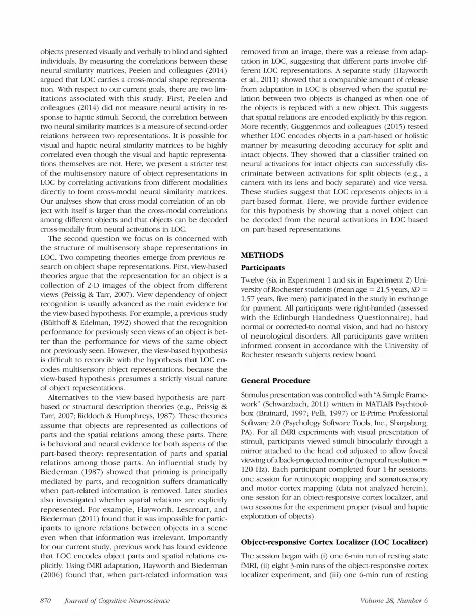
The stimuli used in Experiment 1 were taken from the setof objects known as Fribbles (Tarr, 2003). We picked 12Fribbles (four objects from three categories) for Experi-ment 1. For the stimuli used in Experiment 2, we created
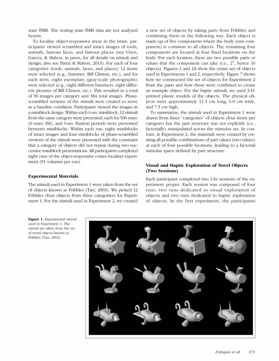
a new set of objects by taking parts from Fribbles andcombining them in the following way. Each object ismade up of five components where the body (one com-ponent) is common to all objects. The remaining fourcomponents are located at four fixed locations on thebody. For each location, there are two possible parts orvalues that the component can take (i.e., 24, hence 16objects). Figures 1 and 2A show the entire set of objectsused in Experiments 1 and 2, respectively. Figure 7 showshow we constructed the set of objects for Experiment 2from the parts and how these were combined to createan example object. For the haptic stimuli, we used 3-D-printed plastic models of the objects. The physical ob-jects were approximately 11.5 cm long, 6.0 cm wide,and 7.5 cm high.
To summarize, the stimuli used in Experiment 1 weredrawn from three “categories” of objects (four items percategory) but the part structure was not explicitly (i.e.,factorially) manipulated across the stimulus set. In con-trast, in Experiment 2, the materials were created by cre-ating all possible combinations of part values (two values)at each of four possible locations, leading to a factorialstimulus space defined by part structure.
Visual and Haptic Exploration of Novel Objects(Two Sessions)
Each participant completed two 1-hr sessions of the ex-periment proper. Each session was composed of fourruns, two runs dedicated to visual exploration ofobjects and two runs dedicated to haptic explorationof objects. In the first experiment, the participants
Figure 1. Experimental stimuliused in Experiment 1. Thestimuli are taken from the setof novel objects known asFribbles (Tarr, 2003).
Erdogan et al. 871

observed each novel object stimulus in the visual andhaptic conditions; that is, all 12 objects were presentedin each run. In the second experiment, the novel objectstimuli were divided (arbitrarily) into two sets, A and B.Within a given scanning session, a participant was pre-sented (for instance) Set A for haptic exploration andSet B for visual exploration; that is, in each run, partic-ipants saw eight objects. In their second session for theexperiment proper, that same participant was presentedSet B for haptic exploration and Set A for visual explora-
tion. This ensured that participants only viewed or onlyhaptically explored a given object in a given scanning ses-sion. The order of a given item set (Set A first, Set B first)by modality (visual, haptic) was also counterbalancedacross participants. For both Experiments 1 and 2, visualand haptic exploration was blocked by run, organized in anABBA/BAAB fashion, and counterbalanced evenly acrossparticipants.While laying supine in the scanner, participants were
visually presented with the objects or were required to
Figure 2. (A) Experimental stimuli used in Experiment 2. The stimuli are based on Fribbles (Tarr, 2003). Each object is made up of four components atfour fixed locations. For each location, there are two possible values or parts (i.e., 24; hence 16 objects). (B) Results of agglomerative clustering appliedto behavioral similarity data from the visual condition. In the behavioral experiment (Erdogan, Yildirim, & Jacobs 2015), participants either viewedor haptically explored a pair of objects and provided similarity ratings on a scale of 1–7. Similarity judgments are averaged across participants to geta condition level similarity matrix. (C) Results of agglomerative clustering applied to haptic behavioral similarity data. (D) Scatter plot of cross-modalbehavioral similarity judgments versus similarities calculated from part structure. In the cross-modal condition, participants viewed one of the objects andhaptically explored the other object. Similarities based on part structure are calculated by counting the number of shared parts between pairs of objects.
872 Journal of Cognitive Neuroscience Volume 28, Number 6

keep their eyes closed while haptically exploring theobjects. In the haptic condition, the objects were handedto the participant by the experimenter. For runs in whichitems were visually presented, participants were in-structed to deploy their attention to the features of theobject.In the visual condition in Experiment 1, the objects
were presented in the center of the screen for the par-ticipants to fixate upon. Miniblocks were 4 sec long andwere interspersed by 8-sec fixation periods. Each objectwas presented in four miniblocks per run, with the con-straint that the same object did not repeat on two suc-cessive miniblocks. This meant that there were a totalof 48 (12 × 4) object presentations in each run. InExperiment 2, the objects were presented centrally androtated 40 degrees per second along the vertical axis(i.e., the objects revolved in the depth plane). Miniblocksin the visual condition were 9 sec long and were inter-spersed by 9-sec fixation periods. Each object was pre-sented in four miniblocks per run, in a similar mannerto Experiment 1. Therefore, there were in total 32 (8 ×4) object presentations in each run. In the haptic condi-tion, participants were instructed to form a mental imageof the plastic object while haptically exploring the objectwith their hands. In Experiment 1, miniblocks were 12 seclong and were interspersed by 9-sec periods in which theirhands were unoccupied. Each plastic object was presentedin four miniblocks per run, with the constraint that thesame item did not repeat across two successive miniblockpresentations. Miniblocks in Experiment 2 were 16 seclong and were interspersed by 16-sec periods in whichtheir hands were unoccupied. Each plastic object was pre-sented in four miniblocks per run, in a similar manner toExperiment 1.In our experiments, participants performed no explicit
task other than visually or haptically exploring the pre-sented objects. We believe such a design enables us toinvestigate visual-haptic processing without any potentialtask-related effects. Previous research shows that, evenin the absence of any explicit task, visual and hapticprocessing converges in LOC (Naumer et al., 2010). Al-though our participants did not perform an explicit task,we asked them to mentally picture the object they wereexploring in the haptic condition. This might raise suspi-cions about whether the activation in LOC was due tomental imagery rather than haptic processing. However,previous research suggests that LOC is minimally acti-vated by mental imagery ( James et al., 2002; Amediet al., 2001).Before the experiment began, participants were intro-
duced to comparable plastic objects outside the scanner.These objects were not used in the experiment properand were dissimilar to the experimental stimuli. Visualanalogs of the objects were also presented to the partic-ipants to inform them of the format of the visual exper-iment and to practice the implicit task that they wererequired to carry out while in the scanner.
MR Acquisition and Analysis
MRI Parameters
Whole-brain BOLD imaging was conducted on a 3-TSiemens (Amsterdam, TheNertherlands)MAGNETOMTrioscanner with a 32-channel head coil located at the Roch-ester Center for Brain Imaging. High-resolution structuralT1 contrast images were acquired using a magnetizationprepared rapid gradient-echo pulse sequence at the startof each participant’s first scanning session (repetitiontime = 2530, echo time = 3.44 msec, flip angle = 7°, fieldof view = 256 mm, matrix = 256 × 256, 1 × 1 × 1 mmsagittal left-to-right slices). An EPI pulse sequence wasused for T2* contrast (repetition time = 2000 msec, echotime = 30 msec, flip angle = 90°, field of view = 256 ×256 mm, matrix = 64 × 64, 30 sagittal left-to-right slices,voxel size = 4 × 4 × 4 mm). The first six volumes of eachrun were discarded to allow for signal equilibration (fourat acquisition and two at analysis).
fMRI Data Analysis
fMRI data were analyzed with the BrainVoyager softwarepackage (Version 2.8) and in-house scripts drawing on theBVQX toolbox written in MATLAB (wiki2.brainvoyager.com/bvqxtools). Preprocessing of the functional data in-cluded, in the following order, slice scan time correction(sinc interpolation), motion correction with respect tothe first volume of the first functional run, and linear trendremoval in the temporal domain (cutoff: two cycles withinthe run). Functional data were registered (after contrastinversion of the first volume) to high-resolution deskulledanatomy on a participant-by-participant basis in nativespace. For each participant, echo-planar and anatomi-cal volumes were transformed into standardized space(Talairach & Tournoux, 1988). Functional data for thelocalizer experiment (object-responsive cortex localizer)were smoothed at 6 mm FWHM (1.5 mm voxels) and inter-polated to 3mm3 voxels; functional data for the experimentproper (visual and haptic exploration of objects) wereinterpolated to 3 mm3 but were not spatially smoothed.
For all experiments, the general linear model was usedto fit beta estimates to the experimental events of inter-est. Experimental events were convolved with a standard2-gamma hemodynamic response function. The first de-rivatives of 3-D motion correction from each run wereadded to all models as regressors of no interest to attractvariance attributable to head movement. Thus, all multi-voxel pattern analyses were performed over beta estimates.
In all multivoxel analyses, we normalized individual voxelactivations within a run to remove baseline differencesacross runs. In other words, for each voxel, we subtractedthe mean activation for that voxel over all objects in the runand divided it by the standard deviation of that voxel’s ac-tivation across objects. Additionally, for linear correlationmultivoxel analyses, activations for all eight repeats of asingle item (in a given modality, i.e., visual/haptic) were
Erdogan et al. 873

averaged to obtain a single activation vector for each item. Inour correlation analyses, we transformed correlation valuesusing Fisher’s z transformation and ran all statistical tests onthose transformed values. When calculating correlations be-tween correlationmatrices, weusedonly the upper trianglesof matrices. All statistical tests were two-tailed. For trainingthe support vector machine (SVM) for decoding, we usedthe library libsvm (www.csie.ntu.edu.tw/∼cjlin/libsvm/). Weused linear kernels with cost parameter set to 1.
Whole-brain pattern analyses were performed using asearchlight approach (Kriegeskorte, Goebel, & Bandettini,2006). Whole-brain searchlight maps were computed witha mask fit to the deskulled Talairach anatomy of individ-ual participants. The “searchlight” passes over each voxel(in each participant) and extracts the beta estimates (for16 items) for the cube of voxels (n = 125) that surroundthe voxel. The analysis was carried out based on the patternof responses across the 125 voxels, and the results wereassigned to the center voxel of that cube. All whole-brainanalyses were thresholded at p < .005 (corrected), clusterthreshold for nine contiguous voxels. If no regions wereobserved at that threshold, a more lenient threshold wasused ( p < .05, uncorrected, nine voxels).
Definition of ROIs (LOC)
Left and right LOC were identified at the group levelusing the object-responsive localizer experiment withthe contrast of [intact images] > [scrambled images].The result used cluster size corrected alpha levels bythresholding individual voxels at p < .05 (uncorrected)and applying a subsequent cluster size threshold gener-ated with a Monte Carlo style permutation test (1000 iter-ations) on cluster size to determine the appropriate alphalevel that maintains Type I error at 1% (using AlphaSimas implemented in Brain Voyager). The Talairach coor-
dinates were as follows: left LOC: x = −40, y = −71, z =−9; right LOC: x= 38, y=−65, z=−12. We note as wellthat none of the results in this study change qualitativelyif LOC is defined individually for each participant, ratherthan at the group level.
RESULTS
Our study consisted of two experiments. In both experi-ments, participants either viewed or haptically explored aset of objects during fMRI. The stimuli for Experiment 1consisted of 12 objects (four objects from three catego-ries; see Figure 1) picked from the set of objects knownas Fribbles (Tarr, 2003). For Experiment 2, we created anovel set of objects based on Fribbles. Each object in thisset was composed of one component that was commonto all objects and four components that varied acrossobjects. The variable components were located at fourfixed locations (Figure 2A), and there were two possibleparts (or values) that each component could take (i.e.,24 = 16 objects in total).
Cross-modal Decoding of Novel Objects in LOC
If object representations in LOC are multisensory acrosshaptic and visual modalities, it should be possible to de-code object identity using cross-modal representationalsimilarity analyses. To that end, we correlated the voxelpatterns in LOC elicited when a participant was viewingobjects with the voxel patterns elicited when the sameparticipant haptically explored the objects. The resultingrepresentational similarity analysis quantifies the similar-ity of voxel patterns across modalities, comparing everyobject to every other object as well as to itself. Previousstudies have calculated neural similarity matrices sepa-rately for each modality and then subsequently correlated
Figure 3. Comparison between diagonals and nondiagonals of cross-modal similarity matrices for both experiments. Participants 1–6 are inExperiment 1, and participants 7–12 are in Experiment 2. Avg = average of all 12 participants. (A) Results for left LOC. (B) Results for right LOC.
874 Journal of Cognitive Neuroscience Volume 28, Number 6

those matrices (e.g., Peelen et al., 2014). Such an approachamounts to showing that neural correlations among ob-jects in one modality correlate with the neural correlationsamong objects in another modality. The goal of the currentanalysis is to run a stricter test of the hypothesis that LOCencodes objects in a multisensory manner by correlatingvoxel patterns from different modalities directly to form across-modal neural similarity matrix.Two predictions are made by the hypothesis that object
representations in LOC are multisensory. First, cross-modal correlations between the visual and haptic voxelpatterns for the same object will be higher than cross-modal correlations among the voxel patterns for differ-ent objects (i.e., the diagonal values will be greater than
the nondiagonal values in the cross-modal representa-tional similarity matrix). The results of this analysis foreach participant in Experiments 1 and 2 can be seen inFigure 3. For every participant in right LOC and for 10of 12 participants in left LOC, cross-modal correlationswere in fact higher for identical objects than they werefor different objects (see Figure 4 for average cross-modal correlation matrices). Because an initial ANOVAanalysis found no effect of Experiment (L-LOC, F = 0.17,p = .69; R-LOC, F = 0.48, p = .50), we combined theresults from both experiments. Diagonal versus non-diagonal differences reached statistical significance in bothL-LOC and R-LOC (L-LOC, difference = 0.06; t= 3.86, p <.004; R-LOC, difference = 0.05; t = 5.08, p < .001),
Figure 4. Cross-modal similarity matrices for both experiments. (A, B) Cross-modal similarity matrices calculated from left (A) and right (B) LOCactivations from Experiment 1. (C, D) Cross-modal similarity matrices calculated from left (C) and right (D) LOC activations from Experiment 2.
Erdogan et al. 875

indicating that LOC contains multisensory representationsof objects. A second and stricter prediction is that it shouldbe possible to decode object identity using the repre-sentational similarity matrix by testing whether each ob-ject is more correlated with itself (across modalities)than it is with each of the other objects in the set (alsoacross modalities). We calculated the decoding accuraciesfor each participant and compared these to the chancedecoding accuracy (1/12 for Experiment 1 and 1/16 forExperiment 2). Again, because an initial ANOVA analysisfound no effect of Experiment (L-LOC, F = 0.67, p =.43; R-LOC, F = 0.82, p = .39), we combined the resultsfrom both experiments. Our results showed that it is pos-
sible to decode object identity cross-modally in both L-LOCand R-LOC (L-LOC, difference from chance accuracy =0.09, t = 2.48, p < .04; R-LOC, difference = 0.10, t =3.48, p < .006). These data indicate that LOC containsmultisensory representations of objects.We then tested whether multisensory coding of novel
objects was specific to LOC or was a property observedthroughout the brain. To that end, a whole brain searchlightanalysis was conducted in which each voxel was codedaccording to whether it (and its immediate neighbors)showed higher pattern similarity for an object correlatedwith itself (across modalities) than with other objects (alsocross modality). Converging with the ROI analyses, the
Figure 5.Whole searchlight analysis of brain regions in which the diagonal of the cross-modal neural similarity matrix is greater than the off-diagonalvalues. The cross-modal similarity matrix was created by correlating the voxel patterns elicited when visually exploring objects with the voxelpatterns elicited when haptically exploring objects. If the diagonal of the matrix is greater than the off-diagonal values, that means that the patternof voxel activations elicited by an object (across modalities) is more similar than the patterns elicited by two different objects.
876 Journal of Cognitive Neuroscience Volume 28, Number 6

results (Figure 5) identified the right LOC in both exper-iments (see Table 1 for coordinates). The left posteriortemporal-occipital cortex was also identified in the search-light analyses from both experiments.
A Common Similarity Space of Novel Objects asDerived from Neural and Behavioral Metrics
The stimuli used in Experiment 2 were designed to havea clear part-based structure for the purpose of testing thepart-based hypothesis through representational similarity
and neural decoding analyses. In a prior study (Erdogan,Yildirim, & Jacobs, 2015), we collected behavioral simi-larity judgments for these stimuli while participantsviewed or haptically explored the objects. Similarityratings consisted of Likert similarity ratings (range 1:7)for each pair of objects. We evaluated how well partici-pants’ judgments of the similarity among the objectswere explained by the part-based structure of the objects.As shown in Figure 2D, the agreement was extremelygood (R2 = .96). This indicates that participants perceivethe similarity among these object stimuli in terms oftheir part structure. Therefore, a significant agreement
Table 1. Talairach Coordinates, Cluster Sizes, Significance Levels, and Anatomical Regions for the Searchlight Results
Region
Talairach CoordinatesCluster Size
(mm2) t px y z
Exp1: Diagonal of the Cross-modal Neural Similarity Matrix > Off-diagonal Values (p < .05, Cluster > 9 Voxels)
Precentral gyrus LH −51 −13 34 6790 8.55 <.001
Middle occipital gyrus LH −24 −88 19 25007 12.28 <.001
Lateral occipital cortex LH −39 −67 −14 8.40 <.001
Precentral gyrus RH 57 −1 19 1469 7.06 <.001
Postcentral gyrus RH 63 −25 38 3175 7.51 <.001
Lateral occipital cortex RH 39 −55 −5 23568 13.83 <.001
Exp2: Diagonal of the Cross-modal Neural Similarity Matrix > Off-diagonal Values (p < .05, Cluster > 9 Voxels)
Inferior frontal gyrus LH −39 20 10 1100 5.47 <.01
Precentral gyrus LH −30 −16 52 1514 7.30 <.001
Superior parietal lobule LH −21 −58 58 4417 12.27 <.001
Inferior frontal gyrus RH 39 17 16 1450 6.19 <.002
Superior temporal gyrus RH 51 −25 7 877 10.04 <.001
Lateral occipital cortex RH 50 −62 −18 257 3.88 <.01
Exp2: Correlation between Neural and Behavioral Similarity for Visual Exploration of Objects (p < .05, Cluster > 9 Voxels)
Parietal lobe LH −18 −58 46 539 8.67 <.001
Lateral occipital cortex RH 42 −70 1 742 9.84 <.001
Lingual gyrus RH 0 −73 −11 2607 6.07 <.002
Exp2: Correlation between Neural and Behavioral Similarity for Haptic Exploration of Objects (p < .005, Cluster > 9 Voxels)
Lateral occipital cortex LH −39 −67 −14 1230 9.99 <.001
Precentral gyrus RH 42 −13 34 1577 10.78 <.001
Postcentral gyrus RH 51 20 34 2323 19.19 <.001
Parietal lobe RH 9 −37 61 3143 12.86 <.001
Superior temporal gyrus RH 42 −49 19 2110 13.79 <.001
Lateral occipital cortex RH 33 −73 −8 2190 11.84 <.001
LH = left hemisphere; RH = right hemisphere.
Erdogan et al. 877

between the neural similarity matrices and behavioralsimilarity judgments will lend support to both the hy-pothesis that LOC representations are multisensory andto the hypothesis that they are part based. We tested thisprediction by calculating correlations between behavioralsimilarity judgments and measures of object similarityderived from neural data. A visual similarity matrix wasformed by correlating voxel patterns when participantsviewed the objects during fMRI, and a haptic similaritymatrix was formed when participants haptically exploredthe objects during fMRI. As predicted by the hypothesisthat LOC encodes multisensory, part-based representa-tions of objects, the neural similarity matrices obtainedfrom R-LOC for both modalities were correlated withthe behavioral similarity matrices (neural similarity mea-sures based on visual exploration: L-LOC: r= .02, t= 1.10,p = .33, R-LOC: r = .08, t = 4.21, p < .009; Haptic con-dition, L-LOC: r = .08, t = 1.52, p = .187, R-LOC: r =.14, t = 3.28, p < .03).
To evaluate the degree to which the observed relation-ship between behavioral and neural similarity measureswas specific to LOC, we again carried out a whole-brainsearchlight analysis that maps how similar the neural sim-ilarity matrices were to the behavioral similarity matrices.The most stringent test of whether LOC encodes multi-sensory representations of novel objects is to test whetherLOC is identified by two independent searchlight ana-lyses: The first analysis relates neural and behavioral sim-ilarity data for visual exploration of objects, and thesecond analysis relates neural and behavioral similaritydata for haptic exploration of objects. Thus, the key testis whether these two independent searchlight analysesoverlap in LOC. The results indicate overlap in rightLOC (see Table 1 for Talairach coordinates). As can beseen in Figure 6, there is good overlap (35 voxels,958 mm3, across the maps in Figure 6A, B, and C) betweenthe independent functional definition of right LOC (objects> scrambled images) and right LOC as identified by thetwo independent multivoxel pattern searchlight analyses.Interestingly, the whole-brain searchlight analysis overhaptic data also identified several other regions in thetemporal and frontal lobes involved in sensory processing(see Table 1 for coordinates).
Object Category Representations in LOC
Stimuli in Experiment 1 formed three families or categoriesof objects (Figure 1). This raises the possibility of evalu-ating whether LOC object representations encode categorystructure. Using analyses of the LOC cross-modal similaritymatrix, we found that neural activations were more simi-lar when considering two objects belonging to the samecategory than when considering two objects belonging todifferent categories. Using decoding analyses, we foundthat we can decode the category to which an object belongsat above-chance levels. However, because we are uncertainabout the proper interpretation of these results, we do
not study LOC object category representations here.One possibility is that LOC encodes the category structureof objects. Another possibility is that LOC encodes objectshape and that the results regarding category structure aredue to the fact that objects belonging to the same cate-gory have similar shapes in our experiment and objectsbelonging to different categories have dissimilar shapes.Because we cannot distinguish these two possibilities
Figure 6. Overlap in right LOC for the (A) functional localizer (i.e.,objects > scrambled objects), (B) a whole brain searchlight analysisof the correlation between neural similarity matrices and behavioralsimilarity for visual exploration of objects, and (C) a whole brainsearchlight analysis of the correlation between neural similarity matricesand behavioral similarity for haptic exploration of objects.
878 Journal of Cognitive Neuroscience Volume 28, Number 6

based on the stimuli used here and because there is sub-stantial evidence indicating that LOC represents objectshape, a stronger test of the nature of LOC object repre-sentations is provided by fine-grained analysis of the partstructure within the materials from Experiment 2.
Part-based Object Representations in LOC
Finally, we sought to directly test the hypothesis thatLOC encodes objects in a part-based manner. If theshape representations in LOC are encoding object parts,we should be able to decode the parts that make up anobject from neural activations. We focused these analysesonly on our second experiment because the stimuli inour first experiment are not suited to testing the part-based hypothesis. Although all objects used in Experi-ment 1 have a clear part-based structure, each part is atmost shared by two objects, which drastically limits theamount of data available for decoding part identities.However, the stimuli in our second experiment were de-signed specifically to test the part-based hypothesis, witheach part being shared by 8 of 16 objects in the stimulus set.The objects in our second experiment can be represented
as four binary digits with each digit coding which one ofthe two possible parts for each of the part locations ispresent (see Figure 7 for a schematic of this analysisapproach). In our decoding analyses, we thus soughtto predict the four-digit binary representation of eachobject using neural activity patterns. We trained fourseparate linear SVMs, one for each location. Each SVMmodel was trained to predict which of the two possiblepart values for that location was present in an object.Each of the four classifiers was trained on 15 of the16 objects, and the classifiers were tested by havingthem jointly predict the four-digit binary representationfor the 16th object. If all four of the predictions (one foreach location) were correct, we counted that as a suc-cessful decoding of the object (see Figure 7B). Thus,chance for this classification test was 0.54 = 0.0625. Thisanalysis approach was performed using 16-fold leave-one-out cross-validation, each time leaving one object out (fortest) and training the classifiers on the remaining 15 ob-jects. We then averaged the classification accuraciesover folds to obtain an estimate of the classification ac-curacy across all objects for each participant. Statisticalanalysis was then performed over subject means. The
Figure 7. (A) Design of stimuli.Each object is composed offour components at four fixedlocations. (Parts are coloredfor illustration purposes. Allimages were grayscale in theexperiment.) (B) Schematic ofthe decoding model. Neuralactivations for 15 of the objectsare used as the training set totrain four linear SVMs to predictparts at each location. Then,the trained classifiers are usedto predict the parts of theleft-out test object, and thesepredictions are comparedwith the true parts of theobject.
Erdogan et al. 879

results of this analysis indicated that it was possible todecode novel objects in LOC, both for fMRI data ob-tained during visual and during haptic exploration of theobjects (visual condition, L-LOC: classification accuracy =0.198, t = 3.61, p < .016, R-LOC: classification accuracy =0.250, t = 5.81, p < .003; haptic condition, L-LOC: classifi-cation accuracy = 0.167, t= 2.50, p= .055, R-LOC: classifi-cation accuracy = 0.302, t = 5.86, p < .003).
DISCUSSION
We have shown that it is possible to decode object iden-tity from a cross-modal similarity matrix created by cor-relating LOC voxel patterns during visual and hapticexploration of the same set of objects. This suggests thatthere is a unique neural code generated during percep-tual exploration of each of the novel objects that is similarregardless of whether the sensory modality is vision ortouch. We also found that linear classifiers successfullypredict a novel object based on its part structure. Thus,the fundamental units of object representation in LOCare expressed in terms of an object’s composite parts.These findings provide further evidence for part-basedvisual representations of objects in LOC and multisensoryrepresentations of whole objects, at least across the hap-tic and visual modalities (Peelen et al., 2014; Naumeret al., 2010; Amedi et al., 2001, 2002; James et al., 2002).Crucially, our cross-modal decoding analyses relied on adirect comparison between activations from differentmodalities, representing a more direct test of the multi-sensory nature of object representations in LOC thanwas present in prior studies. Additionally, we believeour part-based decoding of novel objects presents a sig-nificant step towards understanding the nature of objectrepresentations in LOC. The only previous study thatused a similar decoding analysis (Guggenmos et al., 2015)employed simpler stimuli (two-part objects) and presentedobjects only visually. Our study used a richer set of stimuliand showed that decoding of a novel object is possiblefrom both visual and haptic activation in LOC. We believethat the findings we have reported strongly suggest thatobject representations in LOC are multisensory and partbased.
Our results show an interesting hemispheric asym-metry; in most of our analyses, the findings are strongerin R-LOC. We do not have a clear understanding of whythis is the case. A recent study suggests that haptic pro-cessing is stronger in LOC for the nondominant hand(Yalachkov, Kaiser, Doehrmann, & Naumer, 2015). How-ever, it is important to note that participants in our ex-periment used both of their hands to explore objects.Additionally, these hemispheric differences are seen inthe visual condition as well, making an explanation basedon haptic processing unlikely. Future research shouldinvestigate whether this hemispheric asymmetry is aconsistent characteristic of object shape processing ormerely an artifact of our particular sample.
Although we have referred to the object representa-tions in LOC as multisensory, it is worth pointing out thatour study focused on visual and haptic processing, simplybecause shape information is conveyed mainly throughthese two modalities. For example, as previous research(Naumer et al., 2010; Amedi et al., 2002) shows, LOCdoes not respond to auditory stimulation. Similarly, ourstudy says little about the representation of objects thatlack a clear part-based structure, for example, bell pep-pers, or that are processed holistically, for example, faces.The question of how an object without a clear part-basedstructure is represented lies at a finer level than that onwhich our study focused; we did not investigate how anindividual part might be neurally represented but whetherparts are explicitly represented in the first place. Futureresearch should focus on this more difficult question ofhow individual parts are represented.In this study, we focused mainly on LOC and the nature
of object representations in this region. However, lookingat Table 1, we see that our searchlight results identifiedother regions, for instance, the precentral gyrus and theleft posterior temporal-occipital cortex. Although noneof those regions show the consistent activity that LOCshows across various analyses, it is possible that multi-sensory object representations reside in a larger networkof brain regions and likely that multisensory object repre-sentations in LOC are embedded in a broader network ofregions that support multisensory processing. This is anempirical question that needs to be addressed by futureresearch.A key claim of the part-based hypothesis is that objects
are represented as a combination of shape primitivesfrom a finite set. Although our data cannot speak to theinventory of shape-based primitives that the brain mayencode, further research using the methods we havedeveloped may be able to describe that inventory. A sec-ond key aspect of part-based theories of object represen-tation is that spatial relations among parts are directlyrepresented. The findings we have reported motivate anew approach to test whether the spatial arrangementamong an object’s parts are encoded in the same region(LOC) that encodes the part information. Alternatively,information about the spatial arrangement of parts maybe stored elsewhere in the brain.Our findings also bear on the principal alternative
theoretical model to part-based object representations:image- or view-based models. View-based theories arguethat the representation of an object is a concatenation of2-D images of the object from different views (for discus-sion, see Peissig & Tarr, 2007). View dependency in ob-ject recognition is advanced as the main evidence for theview-based hypothesis. However, view-based modelshave difficulty accounting for our finding that there is ahigh degree of similarity in the voxel patterns elicited byhaptic and visual exploration of objects and that the sharedvariance in voxel patternmaps onto thepart structure of thestimuli.
880 Journal of Cognitive Neuroscience Volume 28, Number 6

In this study, we have presented evidence that LOCcarries multisensory and part-based representations ofobjects. In addition to the empirical evidence presentedhere and in earlier studies, we believe this hypothesis isalso appealing from a theoretical perspective as it ele-gantly captures how information can be transferredacross modalities, how inputs from multiple modalitiescan be combined, and more generally, how we cope witha world that is in its essence multisensory.
Acknowledgments
We thank Elon Gaffin-Cahn for assistance with fMRI data collec-tion. Preparation of this paper was supported by NIH R01NS089609 to B. Z. M. and by AFOSR FA9550-12-1-0303 andNSF BCS-1400784 to R. A. J. F. E. G. was supported by a Univer-sity of Rochester Center for Visual Science predoctoral trainingfellowship (NIH Training Grant 5T32EY007125-24).
Reprint requests should be sent to Robert A. Jacobs, Department ofBrain and Cognitive Sciences, University of Rochester, Rochester,NY 14627-0268, or via e-mail: [email protected].
REFERENCES
Amedi, A., Jacobson, G., Hendler, T., Malach, R., & Zohary, E.(2002). Convergence of visual and tactile shape processing inthe human lateral occipital complex. Cerebral Cortex,12, 1202–1212.
Amedi, A., Malach, R., Hendler, T., Peled, S., & Zohary, E.(2001). Visuo-haptic object-related activation in the ventralvisual pathway. Nature Neuroscience, 4, 324–330.
Biederman, I. (1987). Recognition-by-components: A theory ofhuman image understanding. Psychological Review, 94,115–147.
Brainard, D. H. (1997). The psychophysics toolbox. SpatialVision, 10, 433–436.
Bülthoff, H. H., & Edelman, S. (1992). Psychophysical supportfor a two-dimensional view interpolation theory of objectrecognition. Proceedings of the National Academy ofSciences, U.S.A., 89, 60–64.
Chen, Q., Garcea, F. E., & Mahon, B. Z. (in press). Therepresentation of object-directed action and functionknowledge in the human brain. Cerebral Cortex.doi: 10.1093/cercor/bhu328.
Cooke, T., Jäkel, F., Wallraven, C., & Bülthoff, H. H. (2007).Multimodal similarity and categorization of novel,three-dimensional objects. Neuropsychologia, 45, 484–495.
Cooke, T., Kannengiesser, S., Wallraven, C., & Bülthoff, H. H.(2006). Object feature validation using visual and hapticsimilarity ratings. ACM Transactions on Applied Perception,3, 239–261.
Erdogan, G., Yildirim, I., & Jacobs, R. A. (2015). From sensorysignals to modality-independent conceptual representations:A probabilistic language of thought approach. PLoSComputational Biology, 11, e1004610.
Fintzi, A. R., & Mahon, B. Z. (2013). A bimodal tuning curvefor spatial frequency across left and right human orbitalfrontal cortex during object recognition. Cerebral Cortex,24, 1311–1318.
Gaissert, N., Bülthoff, H. H., & Wallraven, C. (2011). Similarityand categorization: From vision to touch. Acta Psychologica,138, 219–230.
Gaissert, N., & Wallraven, C. (2012). Categorizing naturalobjects: A comparison of the visual and the haptic modalities.Experimental Brain Research, 216, 123–134.
Gaissert, N., Wallraven, C., & Bülthoff, H. H. (2010). Visual andhaptic perceptual spaces show high similarity in humans.Journal of Vision, 10, 1–20.
Grill-Spector, K., Kourtzi, Z., & Kanwisher, N. (2001). The lateraloccipital complex and its role in object recognition. VisionResearch, 41, 1409–1422.
Guggenmos, M., Thoma, V., Cichy, R. M., Haynes, J. D., Sterzer,P., & Richardson-Klavehn, A. (2015). Non-holistic codingof objects in lateral occipital complex with and withoutattention. Neuroimage, 107, 356–363.
Hayworth, K. J., & Biederman, I. (2006). Neural evidence forintermediate representations in object recognition. VisionResearch, 46, 4024–4031.
Hayworth, K. J., Lescroart, M. D., & Biederman, I. (2011). Neuralencoding of relative position. Journal of ExperimentalPsychology: Human Perception and Performance, 37,1032–1050.
James, T. W., Humphrey, G. K., Gati, J. S., Servos, P., Menon, R. S.,& Goodale, M. A. (2002). Haptic study of three-dimensionalobjects activates extrastriate visual areas. Neuropsychologia,40, 1706–1714.
Kourtzi, Z., & Kanwisher, N. (2001). Representation of perceivedobject shape by the human lateral occipital cortex. Science,293, 1506–1509.
Kriegeskorte, N., Goebel, R., & Bandettini, P. (2006).Information-based functional brain mapping. Proceedingsof the National Academy of Sciences, U.S.A., 103,3863–3868.
Lacey, S., Peters, A., & Sathian, K. (2007). Cross-modal objectrecognition is viewpoint-independent. PLoS One, 2, e890.
Lawson, R. (2009). A comparison of the effects of depth rotationon visual and haptic three-dimensional object recognition.Journal of Experimental Psychology: Human Perceptionand Performance, 35, 911–930.
Naumer, M. J., Ratz, L., Yalachkov, Y., Polony, A., Doehrmann,O., Van De Ven, V., et al. (2010). Visuohaptic convergencein a corticocerebellar network. European Journal ofNeuroscience, 31, 1730–1736.
Norman, J. F., Norman, H. F., Clayton, A. M., Lianekhammy, J.,& Zielke, G. (2004). The visual and haptic perception ofnatural object shape. Perception & Psychophysics, 66,342–351.
Peelen, M., He, C., Han, Z., Caramazza, A., & Bi, Y. (2014).Nonvisual and visual object shape representations inoccipitotemporal cortex: Evidence from congenitallyblind and sighted adults. Journal of Neuroscience, 34,163–170.
Peissig, J. J., & Tarr, M. J. (2007). Visual object recognition:Do we know more now than we did 20 years ago? AnnualReview of Psychology, 58, 75–96.
Pelli, D. G. (1997). The VideoToolbox software for visualpsychophysics: Transforming numbers into movies. SpatialVision, 10, 437–442.
Riddoch, M. J., & Humphreys, G. W. (1987). A case ofintegrative visual agnosia. Brain, 110, 1431–1462.
Schwarzbach, J. (2011). A simple framework (ASF) forbehavioral and neuroimaging experiments based on thepsychophysics toolbox for MATLAB. Behavior ResearchMethods, 43, 1194–1201.
Talairach, J., & Tournoux, P. (1988). Co-planar stereotaxicatlas of the human brain. New York: Thieme.
Tarr, M. J. (2003). Visual object recognition: Can asingle mechanism suffice? In M. A. Peterson & G.Rhodes (Eds.), Perception of faces, objects, and scenes(pp. 177–207). New York: Oxford University Press.
Yalachkov, Y., Kaiser, J., Doehrmann, O., & Naumer, M. J. (2015).Enhanced visuo-haptic integration for the non-dominanthand. Brain Research, 1614, 75–85.
Erdogan et al. 881


