Embed Size (px)
Citation preview

The multisensory function of primary visual cortex in humans
Micah M. Murray a,b,c,n, Antonia Thelen c, Gregor Thut d, Vincenzo Romei e,Roberto Martuzzi f, Pawel J. Matusz a,g,nQ1
a The Laboratory for Investigative Neurophysiology (The LINE), Neuropsychology and Neurorehabilitation Service and Department of Radiology, UniversityHospital Center and University of Lausanne, Lausanne, Switzerlandb EEG Brain Mapping Core, Center for Biomedical Imaging (CIBM) of Lausanne and Geneva, Lausanne, Switzerlandc Department of Hearing and Speech Sciences, Vanderbilt University Medical Center, Nashville, TN, USAd Centre for Cognitive Neuroimaging, Department of Psychology, University of Glasgow, Glasgow G12 8QB, United Kingdome Centre for Brain Science, Department of Psychology, University of Essex, Colchester, United Kingdomf Laboratory of Cognitive Neuroscience, Brain-Mind Institute, Ecole Polytechnique Fédérale de Lausanne, Switzerlandg Attention, Brain, and Cognitive Development Group, Department of Experimental Psychology, University of Oxford, United Kingdom
a r t i c l e i n f o
Article history:Received 25 May 2015Received in revised form8 August 2015Accepted 10 August 2015
Keywords:MultisensoryCross-modalPrimary cortexHumansBrain imagingBrain mapping
a b s t r a c t
It has been nearly 10 years since Ghazanfar and Schroeder (2006) proposed that the neocortex is es-sentially multisensory in nature. However, it is only recently that sufficient and hard evidence thatsupports this proposal has accrued. We review evidence that activity within the human primary visualcortex plays an active role in multisensory processes and directly impacts behavioural outcome. Thisevidence emerges from a full pallet of human brain imaging and brain mapping methods with whichmultisensory processes are quantitatively assessed by taking advantage of particular strengths of eachtechnique as well as advances in signal analyses. Several general conclusions about multisensory pro-cesses in primary visual cortex of humans are supported relatively solidly. First, haemodynamic methods(fMRI/PET) show that there is both convergence and integration occurring within primary visual cortex.Second, primary visual cortex is involved in multisensory processes during early post-stimulus stages (asrevealed by EEG/ERP/ERFs as well as TMS). Third, multisensory effects in primary visual cortex directlyimpact behaviour and perception, as revealed by correlational (EEG/ERPs/ERFs) as well as more causalmeasures (TMS/tACS). While the provocative claim of Ghazanfar and Schroeder (2006) that the whole ofneocortex is multisensory in function has yet to be demonstrated, this can now be considered establishedin the case of the human primary visual cortex.
& 2015 Published by Elsevier Ltd.
1. Introduction
Until recently, the archetypical view of sensory organisationwas that multisensory integration is a process that is restricted tohigher-order brain regions and occurs only after substantial in-formation processing within lower-level and sensory-specificcortices. As such, regions like primary visual cortex had tradi-tionally been considered as exclusively visual in their function.Multisensory research has ushered a new view of brain organisa-tion and perception, wherein the convergence and integration ofinformation from different senses within low-level cortices is arule rather than an exception (e.g. Ghazanfar and Schroeder, 2006;van Atteveldt et al., 2014a; de Meo et al., 2015; ten Oever et al.,2015) (Fig. 1). This new view is supported by multiple sources of
evidence coming from studies involving various experimentalparadigms, populations/species, as well as brain mapping meth-ods. One consequence of this new view is that it is no longer ac-curate to functionally characterise primary visual cortex as ex-clusively visual, but rather as inherently multisensory.
This new characterisation, however, is considered by some tobe controversial. The principal issue is to what degree multi-sensory effects in primary cortices are the first cortical loci ofmultisensory processes or instead simply a downstream by-pro-duct of multisensory processes elsewhere. A corollary controversyis therefore to what extent multisensory processes in primarycortices are directly affecting behaviour. In what follows, we ad-dress these controversies and review the current evidence for themultisensory nature of the primary visual cortex in humans (seeKayser et al. (2009) for auditory cortex). To facilitate a response tothe above controversies, we focus here in large part on studiesusing simple, rudimentary stimuli (e.g. flashes/checkerboards andtones/noises). Some advantage of using such stimuli include:(1) catalysering inter-species generalisations, (2) they are a
123456789
101112131415161718192021222324252627282930313233343536373839404142434445464748495051525354555657585960616263646566
676869707172737475767778798081828384858687888990
Contents lists available at ScienceDirect
journal homepage: www.elsevier.com/locate/neuropsychologia
Neuropsychologia
http://dx.doi.org/10.1016/j.neuropsychologia.2015.08.0110028-3932/& 2015 Published by Elsevier Ltd.
n Correspondence to: Centre Hospitalier Universitaire Vaudois (CHUV), BH7.081,rue du Bugnon 46 1011 Lausanne, Switzerland.
E-mail addresses: [email protected] (M.M. Murray),[email protected] (P.J. Matusz).
Please cite this article as: Murray, M.M., et al., The multisensory function of primary visual cortex in humans. Neuropsychologia (2015),http://dx.doi.org/10.1016/j.neuropsychologia.2015.08.011i
Neuropsychologia ∎ (∎∎∎∎) ∎∎∎–∎∎∎

reasonable starting point for addressing controversies in how toidentify and qualitatively describe multisensory phenomena, and(3) their properties can be parametrically varied to render themphysically (and therefore perceptually and/or behaviourally) morecomplex and ethologically more valid (e.g. Ghazanfar et al., 2005;Cappe et al., 2009b; see also Fort et al. (2002a,b) for effects ofparametric variation of task demands).
2. The anatomic scaffolding for multisensory processes in theprimary visual cortex
One line of support for the view of the primary visual cortex asthe locus of multisensory processes is based on anatomical evi-dence for monosynaptic afferents from primary and/or low-levelauditory association cortices inQ3 monkeys (Falchier et al., 2002,2009; Rockland and Ojima, 2003; Cappe and Barone, 2005; seealso Clarke and Innocenti (1990), Clemo et al. (2008) for evidencein cats, Vaudano et al. (1991) for evidence in rats, Laramée et al.(2011) for evidence in mice; and Henschke et al. (2014) for evi-dence in Mongolian gerbils). These direct pathways complementthe poly-synaptic pathways via higher-order association corticesas well as cortico-thalamo-cortical pathways (e.g. Cappe et al.(2009a); see also Smiley and Falchier (2009) and Meredith et al.(2009) for reviews). The current evidence further indicates thatthese connections follow a feedback-like laminar profile, origi-nating and terminating in layers 6 and layers 1/6, respectively(Rockland and Ojima, 2003; Clavagnier et al., 2004). Finally, somehave claimed that these projections are heterogeneously dis-tributed across the retinotopic representations within the primaryvisual cortex, with more peripheral visual field representationsreceiving denser projections (Falchier et al., 2002; Rockland andOjima, 2003). However, projections to neurons representing the
central visual field should not be discounted (detailed below). Inhumans, comparable anatomic tracing data are unavailable. Forone, the tracer substances used in human tissue themselves mi-grate over distances of only a few centimetres and thus permit anevaluation of intrinsic but not long-range connectivity (Tardif andClarke, 2001; Marguiles et al., 2009). Second, while focal lesionsprovide an opportunity to study long-range connections, the le-sions must be limited to the grey matter to offer firm evidenceregarding the sources of fibre tracts; a situation that is extremelyrare (e.g. Di Virgilio and Clarke (1997) for a demonstration ofheterotopic interhemispheric connectivity between the right in-ferior temporal cortex and both Wernicke's and Broca's areas). Analternative method to detail the anatomic connectivity in humansis offered by non-invasive diffusion-based imaging. While it hasone major benefit in that it can be conducted in vivo (and there-fore correlated with functional measures), the majority of diffu-sion-based parameters is qualitative in nature and provides nodirect quantification of the axonal or other morphological prop-erties of the underlying anatomy (Lemkaddem et al., 2014; Dau-guet et al., 2007).
Such limitations notwithstanding, there is a growing number ofstudies reporting the presence of connectivity between the pri-mary visual cortex and primary auditory cortex (as well as otherhigher-level visual and auditory cortices). For example, in a pair ofstudies, Beer et al. (2011, 2013) have reported the existence of fibretracts between a seed region within the Heschl's gyrus and theoccipital pole as well as the anterior portions of the calcarinesulcus. Additional tracts were found between the planum tem-porale and both the occipital pole as well as anterior portions ofthe calcarine sulcus. Notably, the size of connected regions withinthe occipital pole and anterior calcarine sulcus were comparable(Beer et al., 2011). This pattern suggests that both central andperipheral visual field representations receive projections from
123456789
101112131415161718192021222324252627282930313233343536373839404142434445464748495051525354555657585960616263646566
676869707172737475767778798081828384858687888990919293949596979899
100101102103104105106107108109110111112113114115116117118119120121122123124125126127128129130131132
Fig. 1. Schemas of cortical loci of multisensory processes. The schemas are depicted on a right hemisphere, with the occipital lobe on the left side of the image and thefrontal lobe on the right side. Low-level visual, auditory, and somatosensory (tactile) cortices are indicated by the blue, red, and green shaded regions, respectively. The solidlines depict a schema where interactions are restricted to higher-order association cortices, such as the prefrontal and parietal cortices (indicated by superimposed coloureddiscs). The dotted lines depict a schema where interactions occur directly between low-level cortices. There is now evidence in support of both schemas. Therefore,multisensory processes undoubtedly involve a dynamic combination of these schemas that probably emerge as a consequence of experience-dependant processes. (Forinterpretation of the references to color in this figure legend, the reader is referredQ2 to the web version of this article.)
M.M. Murray et al. / Neuropsychologia ∎ (∎∎∎∎) ∎∎∎–∎∎∎2
Please cite this article as: Murray, M.M., et al., The multisensory function of primary visual cortex in humans. Neuropsychologia (2015),http://dx.doi.org/10.1016/j.neuropsychologia.2015.08.011i

primary and low-level auditory cortices. Resolving the discrepancybetween the abovementioned studies in humans and those carriedout in animal models is undoubtedly one major challenge for thenearest future. Nonetheless, what is important to note is that bothlines of research converge in demonstrating the presence of au-ditory inputs to the primary visual cortex.
Data from blind and visually-impaired individuals as well asanimal models of visual deprivation provide another line of evi-dence in favour of multisensory congruence and interactionswithin the primary visual cortex (e.g. reviewed in Bavelier andNeville( 2002), Sadato (2006), Renier et al. (2014), and Ricciardiet al. (2014)). These effects include both auditory as well as hapticresponses within visual cortices. For example, primary visualcortex appears to be causally linked to the accuracy of readingBraille by early-blind individuals (Cohen et al., 1997, 1999; Sadatoet al., 2002) and to also correlate with performance on taskscompleted either via touch or sound (e.g. Amedi et al., 2003; Razet al., 2005; reviewed in Ricciardi et al. (2014)).
The studies reviewed above provide strong evidence that thereis anatomic scaffolding that would permit multisensory processeswithin primary visual cortex. In what follows, we review func-tional evidence that multisensory processes are indeed occurringwithin these loci. First, however, we address a major, historicalobstacle; namely the appropriate quantification of multisensoryprocesses.
3. The challenge of quantifying multisensory and cross-modalresponses in human brain imaging and brain mapping studies
A particular challenge in human multisensory research thatinvolves brain imaging methods, such as functional magnetic re-sonance imaging (fMRI) or event-related potentials/fields (ERPs/ERFs), is determining the appropriate statistical criterion foridentifying multisensory interactions within primary cortices aswell as throughout the brain (originally reviewed in e.g. Calvert(2001), Beauchamp (2005) and Laurienti et al. (2005)). One majorproblem is the difficulty in transposing established principles ofmultisensory processing, defined on the basis of single-unit re-cordings in animals (Stein and Meredith, 1993), to population-levelresponses (and behaviour) in humans. Another difficulty is relatedto the ability to differentiate between and understand the natureof super-additive and sub-additive nonlinear responses, i.e. mul-tisensory responses that are greater than or less than the summedunisensory responses, respectively (see Laurienti et al. (2005) formodels based on the extrapolation of single-unit findings; also seebelow). Yet another persisting major controversy concerns theapplicability of a linear model (i.e. the response to a multisensorypair is contrasted with the summed responses to the constituentunisensory signals presented in isolation) to data from humanbrain imaging. Several recent reviews have treated these issues indetail (e.g. Murray et al., 2012; Stevenson et al., 2014; Besle et al.,2004).
In the case of haemodynamic imaging, there is still no generalconsensus or an objective metric by which one could assert that agiven statistical criterion is more robust at identifying multi-sensory brain activity than others. Some favour using a non-line-arity criterion (e.g. Calvert, 2001). Others suggest comparingmultisensory responses to the mean or the maxima of the uni-sensory responses (e.g. Beauchamp, 2005). Still, others proposeusing inverse effectiveness (i.e. presence of stronger multisensoryresponses for multisensory stimuli involving weak unisensorysignals) as a hallmark of multisensory integration (James et al.,2012). The fundamental problem in identifying the presence ofmultisensory processes using haemodynamic imaging methodslies in the fact that these methods cannot differentiate within a
given voxel between populations composed of truly multisensoryneurons versus a mixture of unisensory neurons (cf. Fig. 1 inLaurienti et al. (2005)). One solution is to analyse the temporalrather than the strength aspects of the neural response providedby the haemodynamic brain imaging methods (see below).
Analyses of electromagnetic signals at the scalp provide parti-cularly strong evidence regarding the presence of multisensoryprocessing because of their sub-millisecond temporal resolution,especially when combined with knowledge regarding signal pro-pagation in different sensory systems (e.g. reviewed in Murray andSpierer (2009), and Musacchia and Schroeder (2009)). An ad-vantage of temporal information (i.e. when interactions occur) isthat it helps to constrain where these interactions take place basedon knowledge regarding how far throughout spatio-temporalhierarchies signals from each sensory modality have propagated.While it is undeniably true that the anatomic circuitry and theirshaping by experience are prerequisites for multisensory pro-cesses, only functional studies can provide direct evidence for thepresence of multisensory interactions within the primary visualcortex. In spite of this, ERP/ERF studies of multisensory processeshave traditionally been criticised on two levels.
The first criticism, shared with haemodynamic methods, con-cerns the appropriateness of the additive model. While biophysicalprinciples clearly describe how electric fields summate at the scalplevel (e.g. Besle et al., 2004), there are valid concerns regarding thepotential confound of common activity within the additive model(Teder-Sälejärvi et al., 2002). The second line of critique focusesmore on signal processing and its consequences for statisticaloutcomes. Some researchers have demonstrated the impact ofdigital filters and baseline correction procedures (Teder-Sälejärviet al., 2002), whereas others have scrutinised the fact that thelatency, direction (super-additive vs. sub-additive), and scalp dis-tribution of statistical effects involving voltage waveforms areentirely reference-dependant and therefore have very limitedneurophysiologic interpretability (Cappe et al., 2010). The latterhas been overcome by advances in signal processing techniques,which improve not only the capability to localise multisensoryinteractions, but also to provide a more mechanistic character-isation of their neurophysiologic bases (reviewed in Murray et al.(2008), Brunet et al. (2011), Michel and Murray (2012) and Koeniget al. (2014)). From a more mechanistic standpoint, the analysesfalling under the umbrella of electrical neuroimaging (e.g. Murrayet al., 2008) can reveal whether and, if so, when multisensorystimuli engage distinct configurations of brain regions and/ormodulate the gain of regions already active under unisensoryconditions. Moreover, the nature (i.e. directionality) of such effectscan be determined; one can statistically distinguish between su-per-additive and sub-additive interactions, as a function of time.
Throughout this review we will discuss in more detail the ap-proaches that enable one to circumvent, if not overcome alto-gether, the challenges in the quantification of multisensory pro-cesses in the human brain. In so doing, we will present how evi-dence from various sources supports multisensory convergenceand integration within primary visual cortices in humans.
4. Haemodynamic imaging
Numerous haemodynamic imaging studies have documentedauditory-visual convergence within primary visual cortices. Forexample, using a blocked design, Laurienti et al. (2002) reporteddeactivation within the primary visual cortex in response tosounds, but not if these sounds were accompanied by visual sti-muli. However, in this and other similar studies (e.g. Haxby et al.,1994; Kawashima et al., 1995) attention was explicitly directedtowards the visual stimuli, making it possible that top-down, goal-
123456789
101112131415161718192021222324252627282930313233343536373839404142434445464748495051525354555657585960616263646566
676869707172737475767778798081828384858687888990919293949596979899
100101102103104105106107108109110111112113114115116117118119120121122123124125126127128129130131132
M.M. Murray et al. / Neuropsychologia ∎ (∎∎∎∎) ∎∎∎–∎∎∎ 3
Please cite this article as: Murray, M.M., et al., The multisensory function of primary visual cortex in humans. Neuropsychologia (2015),http://dx.doi.org/10.1016/j.neuropsychologia.2015.08.011i
based attention to vision had obstructed positive-going activationsand the corresponding multisensory effects (e.g. Mozolic et al.,2008).
Other studies have focused on brain activity occurring in theabsence of external stimulation or task and have revealed theexistence of intrinsic functional coupling between primary visualand primary auditory cortices (Eckert et al., 2008). Still others havelikewise reported increased coupling between auditory and pri-mary visual cortices, particularly under conditions of synchronousstimulation across the senses (Lewis and Noppeney, 2010; Tyllet al., 2013), that may perhaps be mediated by thalamic circuits(Noesselt et al., 2010; Bonath et al., 2013). In the same vein, soundshave been shown to activate visual cortices as a function of priormultisensory experiences (Zangenehpour and Zatorre, 2010;Meylan and Murray, 2007; see also Murray et al. (2004, 2005),Thelen et al. (2012, 2014); Matusz et al. (2015a) for effects of priormultisensory contexts on sensory processing). Such effects ofmultisensory exposure on visual cortex have been recently linkedto the expression of immediate early genes (zif268) (Hirst et al.,2012). The same sound-induced visual cortex activations can beobserved in humans also across a very short time scale using theso-called flash-beep illusion, wherein participants often reportseeing two flashes when a single flash is presented close in timewith two beeps (Shams et al., 2000). Watkins et al. (2006, 2007)have shown that activity within the primary visual cortex, morespecifically the retinotopic representation of the veridical flash, isenhanced when a second flash is perceived and suppressed whentwo veridical flashes are perceived as one (see also Mishra et al.(2007) for comparable ERP results). de Haas et al. (2012) foundthat grey matter volume in early human visual cortex predicts
proneness to the flash-beep illusion. Notably, very recently, Vetteret al. (2014) demonstrated that it is possible to decode the cate-gory of natural sounds heard by participants based on the patternsof activity within the primary visual cortex (see also de Haas et al.(2013) for results of impaired decoding of visual stimuli within V2in the presence of semantically incongruent sounds). Collectively,these findings provide strong evidence for a direct link betweencross-modal responses in primary visual cortex and perception(see also van Atteveldt et al. (2014b) for variations in multisensoryprocesses as a function of task as well as across retinotopicrepresentations).
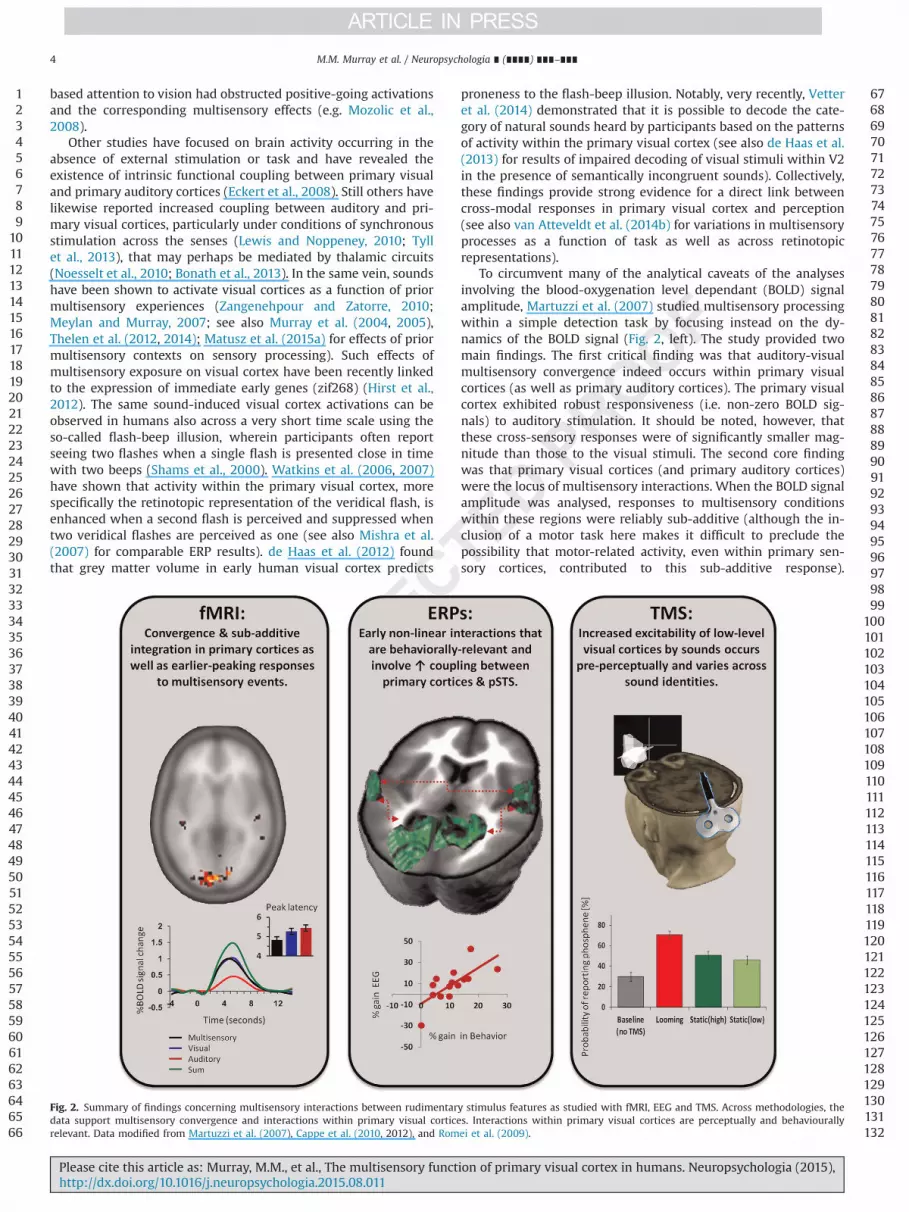
To circumvent many of the analytical caveats of the analysesinvolving the blood-oxygenation level dependant (BOLD) signalamplitude, Martuzzi et al. (2007) studied multisensory processingwithin a simple detection task by focusing instead on the dy-namics of the BOLD signal (Fig. 2, left). The study provided twomain findings. The first critical finding was that auditory-visualmultisensory convergence indeed occurs within primary visualcortices (as well as primary auditory cortices). The primary visualcortex exhibited robust responsiveness (i.e. non-zero BOLD sig-nals) to auditory stimulation. It should be noted, however, thatthese cross-sensory responses were of significantly smaller mag-nitude than those to the visual stimuli. The second core findingwas that primary visual cortices (and primary auditory cortices)were the locus of multisensory interactions. When the BOLD signalamplitude was analysed, responses to multisensory conditionswithin these regions were reliably sub-additive (although the in-clusion of a motor task here makes it difficult to preclude thepossibility that motor-related activity, even within primary sen-sory cortices, contributed to this sub-additive response).
123456789
101112131415161718192021222324252627282930313233343536373839404142434445464748495051525354555657585960616263646566
676869707172737475767778798081828384858687888990919293949596979899
100101102103104105106107108109110111112113114115116117118119120121122123124125126127128129130131132
Fig. 2. Summary of findings concerning multisensory interactions between rudimentary stimulus features as studied with fMRI, EEG and TMS. Across methodologies, thedata support multisensory convergence and interactions within primary visual cortices. Interactions within primary visual cortices are perceptually and behaviourallyrelevant. Data modified from Martuzzi et al. (2007), Cappe et al. (2010, 2012), and Romei et al. (2009).
M.M. Murray et al. / Neuropsychologia ∎ (∎∎∎∎) ∎∎∎–∎∎∎4
Please cite this article as: Murray, M.M., et al., The multisensory function of primary visual cortex in humans. Neuropsychologia (2015),http://dx.doi.org/10.1016/j.neuropsychologia.2015.08.011i

Importantly, also the dynamics of the BOLD response within pri-mary cortices revealed that responses to multisensory and uni-sensory stimuli differed significantly from each other. Specifically,for the multisensory as compared to either unisensory conditionwithin primary visual cortices (and primary auditory cortices) theBOLD signal peaked earlier (following also a steeper slope). Im-portantly, these latency effects were not a result of a simple am-plitude/latency trade-off (see Table 1 in Martuzzi et al. (2007), fordetailed statistics; see also Narsude et al. (2015) for a recent re-plication at 7T). A pressing issue that will require additional re-search is resolving the neurophysiologic mechanisms underlyingmodulations of the BOLD signal latency. One proposition is that anearlier peak reflects facilitated neural processing time (Hensonet al., 2002;Q4 see Wang et al. (2008) for initial findings of facilitatedresponses latencies to multisensory stimuli in the monkey primaryvisual cortex). Notwithstanding, analyses of BOLD dynamics thusprovide an important extension of the available repertoire of ap-proaches for identifying brain regions involving multisensoryprocessing using fMRI as well as putative neural mechanisms.
5. Electromagnetic signals (EEG/MEG)
The advances in the analysis of electromagnetic signals, fallingjointly under the umbrella-term ‘electrical neuroimaging frame-work’ surmount many of the traditional caveats and interpreta-tional limitations (see Michel et al. (2009), Murray et al. (2008,2009), and Michel and Murray (2012) for recent, more detaileddiscussions).
To understand better the dynamics of multisensory processesgauged in perceptual tasks, we applied these analytical methods toERPs in response to task-irrelevant AV stimuli that required top-down attention but no motor responses (Cappe et al., 2010; Fig. 2,centre). The core findings of this ERP study were the following:(1) nonlinear multisensory neural response interactions occurredas early as �60 ms post-stimulus onset, in line with other findings(early multisensory integration, eMSI; De Meo et al., 2015), (2) in-teractions followed from changes in the ERP topography ratherthan, simply, changes in the strength (gain) of the response,meaning that multisensory stimuli engage distinct configurationsof intracranial sources, (3) a network comprised of the primaryvisual cortex, primary auditory cortex, and the posterior superiortemporal sulcus mediated these early interactions, (4) activitythroughout this network was correlated under multisensory, butnot unisensory stimulations, and (5) these nonlinear interactionswere sub-additive both at the level of surface ERP topography andsource estimations within specific brain regions.
The observed eMSI shared timing and scalp topography withthe results of prior studies that involved task-relevant or passivelypresented stimuli (reviewed in De Meo et al. (2015)). These studieshave typically reported multisensory interactions in the ERPsmeasured at individual electrodes onsetting �40–55 ms andvisible as a parieto-occipital positivity in the difference map at thescalp (Fort et al., 2002a; Giard and Peronnet, 1999; Molholm et al.,2002; Vidal et al., 2008; Senkowski et al., 2011; Stekelenburg andVroomen, 2012; Cappe et al., 2012; Altieri et al., 2013; see alsoStevenson et al. (2012) as well as Barth et al. (1995) for corre-sponding data in the rat brain). The analysis of ERPs evoked byattended but task-irrelevant stimuli that required no motor re-sponse circumvented the caveat concerning motor-related activity(Gondan and Röder, 2006; Teder-Sälejärvi et al., 2002). Moreover,the varied inter-stimulus interval ensured that post-stimulus ef-fects were not due to pre-stimulus anticipatory or state-dependantmodulations (Teder-Sälejärvi et al., 2002). In another study, Cappeet al. (2012) demonstrated that these early non-linear interactionscould themselves be further enhanced by looming signals and that
the extent of enhancement correlated with behavioural gains,thereby demonstrating the behavioural relevance of early multi-sensory processes. The robustness of the results of Cappe et al.(2010, 2012) further validates the suitability of an additive modelto test for multisensory interactions (Besle et al., 2004). Becausethese results were obtained using reference-independent mea-sures, their neurophysiologic underpinnings can be interpretedwith greater certainty than those achieved with traditional voltagewaveform analyses. Moreover, the reference-independence of thisanalytical framework facilitates direct comparison of results acrosslaboratories; something that will require concerted coordinationin the near-future.
The application of distributed source estimations (and statis-tical analyses thereof) further allowed Cappe et al. (2010) to lo-calise early nonlinear effects to primary visual cortices, primaryauditory cortices, as well as the posterior superior temporal sulcus(see also Raij et al. (2010) for MEG findings using a distributedinverse solution). These results resolve an ongoing debate re-garding whether or not the early effects emanate from nominallyvisual (Fort et al., 2002a; Molholm et al., 2002) or nominally au-ditory cortices (Vidal et al., 2008) or both (Raij et al., 2010; Sen-kowski et al., 2007; Teder-Sälejärvi et al., 2002). Specifically, thefindings of Cappe et al. and Raij et al. show there to be a widelydistributed network of functionally coupled brain regions operat-ing in concert during the time period of these early multisensoryinteractions. It has previously been suggested that early multi-sensory processes will focus in cortices associated with the less-efficient sensory modality. For example, an individual who isbetter at processing visual stimuli would show multisensory ef-fects within auditory cortices and vice versa (Giard and Perronet,1999). In contrast, the distributed network has been observed tobe active during early stages of multisensory processing in afashion independent of whether or not a given individual wasmore efficient in their behaviour with visual or auditory stimuli(Cappe et al., 2010, 2012).
What remains to be fully characterised is the finer temporaldynamics of early multisensory integrative effects. Because sourceestimations were performed after first averaging across time,Cappe et al. (2010) could not reveal if effects within their dis-tributed network actually had distinct time courses such that ac-tivation within one region led that of the others. By contrast, Raijet al. (2010) used a more refined approach and estimated dis-tributed sources on a millisecond-by-millisecond scale. Auditoryresponses within primary visual cortices onset at �53 ms, andvisual responses within primary auditory cortices onset at �75–82 ms post-stimulus. Non-linear multisensory interactions beganat approximately the same latency as the responses to the cross-modal inputs (cf. Table 4 in Raij et al. (2010)), suggesting thatpathways for convergence may be the same as those generatinginteraction effects (although this remains to be more thoroughlyexamined; reviewed in Meredith et al. (2009) and van Atteveldtet al. (2014a)). In agreement with this notion are data from ani-mals demonstrating auditory as well as somatosensory cross-modal convergence within visual cortex of monkeys (Schroederand Foxe, 2002; Wang et al., 2008), cats (Murata et al., 1965;Spinelli et al., 1968; Morrell, 1972; Majkowski and Sobieszek, 1972;Fishman and Michael, 1973), and mice (Iurilli et al., 2012). Forexample, Iurilli et al. (2012) showed that auditory inputs sup-pressed visual responses within infragranular layers of primaryvisual cortex via GABAergic synapses and moreover that thissuppression was directly linked to performance (as measured by aconditioned motor response). These authors suggested that audi-tory inputs degrade the perception of the visual stimulus; some-thing that will need to be reconciled with the many studiesshowing multisensory and cross-modal enhancement of percep-tion and behaviour (a sampling of which we review here). More
123456789
101112131415161718192021222324252627282930313233343536373839404142434445464748495051525354555657585960616263646566
676869707172737475767778798081828384858687888990919293949596979899
100101102103104105106107108109110111112113114115116117118119120121122123124125126127128129130131132
M.M. Murray et al. / Neuropsychologia ∎ (∎∎∎∎) ∎∎∎–∎∎∎ 5
Please cite this article as: Murray, M.M., et al., The multisensory function of primary visual cortex in humans. Neuropsychologia (2015),http://dx.doi.org/10.1016/j.neuropsychologia.2015.08.011i

generally, the work of Iurilli et al. (2012) constitutes an importantstep in not only characterizing the neural bases of multisensoryprocesses across circuit and synaptic levels, but also in linkingphysiology with behaviour. That said, however, few studies havecharacterised non-linear neural response interactions in visualcortex of animals; something that will undoubtedly change in thecoming years alongside improvements in recording techniques inawake, behaving animals as well as techniques for simultaneousrecordings from multiple brain sites (e.g. Pigarev et al., 2009; Lanzet al., 2013; Gindrat et al., 2015).
The overall pattern observed in EEG/MEG studies in humans issuggestive of a network that may achieve its dynamic couplingthrough oscillatory activity. There is growing evidence that oscil-lations along with cross-frequency coupling may be particularlyimportant in understanding some of the mechanistic bases ofmultisensory interactions as well as their link to behaviouraloutcome (e.g. van Atteveldt et al., 2014a; Gleiss and Kayser, 2014a,b; Mercier et al., 2013; Schepers et al., 2013, 2014; see also Lakatoset al., 2007, 2008). By way of two recent examples, Romei et al.,(2012) found that a single beep can phase-align alpha oscillationsto the sound within the occipital pole, and Cecere et al. (2015)used EEG together with tACS to show there is a tight link betweenan individual's alpha frequency and the temporal window of theflash-beep illusion (Shams et al., 2000) as well as between alphapower and the proneness to the illusion.
6. Brain stimulation (TMS/tACS)
Brain stimulation methods are a particularly effective meansfor drawing causal inference between brain activity and behaviour.Because the timing of stimulation can be finely controlled, one canalso track the dynamics of these processes with a resolution on parwith EEG/MEG. Likewise, they can allow the experimenter to assaythe excitability of primary visual cortex and other early sensoryareas and its modulation by information from other sensory sys-tems. As will be detailed below, this is most readily achieved withphosphene induction. A full review of TMS as a methodology canbe found in works by Pascual-Leone et al. (Pascual-Leone et al.,2000, 2002). In what follows, we summarise evidence from brainstimulation studies that demonstrate a causal role of multisensoryprocesses within the primary visual cortex.
Phosphenes are the perceived sensation of flashes of light inthe absence of visual stimulation following occipital TMS, and arebelieved to be generated by an activation current that is inducedby the magnetic field of the TMS pulse (e.g., Allen et al., 2007;Moliadze et al., 2003). Phosphenes generated by stimulation oflow-level visual areas (V1/V2) are typically perceived as brief,static sensations along the horizontal meridian or in the lowerquadrant of the hemifield contralateral to the stimulated hemi-sphere. There is considerable inter-individual and inter-trialvariability in the presence as well as the nature and strength ofphosphenes, which may stem from anatomical/morphologicaldifferences as well as state-dependant effects (Silvanto and Pasc-ual-Leone, 2008; Romei et al., 2008a). Nonetheless, there are nowstandard procedures for establishing the minimum intensity ofoccipital TMS required to elicit phosphenes (i.e., phosphenethreshold or PT) (see, e.g., Pascual-Leone and Walsh, 2001; Ramos-Estebanez et al., 2007; Romei et al., 2007, 2008a,b, 2009).
Several independent laboratories have now demonstrated thatnon-visual stimuli enhance the excitability of low-level visualcortices within the occipital pole (Bolognini et al., 2010; Ramos-Estebanez et al., 2007; Romei et al., 2007, 2009; 2012; 2013;Spierer et al., 2013; see also Leo et al. (2011); Cecere et al. (2014)).In these studies, the experimenters first identified the PT for eachparticipant and then set the TMS intensity at a level below this PT
value so that, under baseline conditions, phosphenes were re-ported on roughly 30–40% of trials. The core finding is that thelikelihood of perceiving a phosphene dramatically increased whenthis same TMS pulse was paired with a sound (Fig. 2, right). Nu-merous control experiments have ruled out an explanation interms of general attention or alerting (e.g. Romei et al., 2007,2009; Spierer et al., 2013). Examination of the temporal dynamicsof this excitability enhancement show on the one hand that itoccurs during early stages after sound onset (Romei et al., 2007),even at pre-perceptual stages (Romei et al., 2009), and that theeffects depend on the pre-stimulus alpha phase of visual cortices(Romei et al., 2012). Moreover, the effects persist in time, againfollowing an alpha oscillation, and the persistence extends beyondsound offset (Romei et al., 2013). It is only during these extendedtime periods, however, that inter-individual differences in atten-tional preference on an independent task for the auditory or visualmodality seem to play a role (Romei et al., 2013). Most critically,this brain stimulation evidence also supports a direct role of visualcortices in behavioural responses to sounds. Reaction times tosounds are facilitated by single-pulse TMS to the occipital pole,and the magnitude of this facilitation is comparable to and in factcorrelates with that observed with veridical multisensory stimuli(Romei et al., 2007). In a similar manner, the latency at which theexcitability of visual cortices is differentially enhanced by loomingversus stationary sounds preceded and positively correlated withthe duration required for an individual to reliability discriminatethese sounds (Romei et al., 2009). In other words, the visual cortexseems to “know” the nature of the sound prior to an individualbeing aware of it. Finally, tuning of visual cortex activity to anexternal alpha oscillator via tACS stimulation causally modulatesthe temporal window of audio-visual integration shown to beresponsible for the flash-beep illusion phenomenon (Cecere et al.,2015).
7. Discussion
The above studies provide convergent evidence that humanprimary visual cortex is a locus of multisensory processing. Thiswas demonstrated at both anatomical (or at least fibre tracking)and functional levels. Moreover, there is reliable evidence thateffects within primary visual cortex directly impact behaviouraloutcome (at least for certain tasks). In parallel, the works reviewedhere demonstrate how multisensory phenomena can be studiednon-invasively and, more importantly, quantitatively assessed bytaking advantage of particular strengths of each technique as wellas advances in signal analyses.
Several general conclusions about multisensory processes inprimary visual cortex of humans are supported relatively solidly.First, there is both convergence and integration occurring withinprimary visual cortex. This localisation is supported by all of thebrain mapping methods reviewed above: diffusion-based as wellas functional MRI measures, electrical neuroimaging of ERPs, EEGoscillations, single-pulse TMS and tACS over the occipital pole.While this review focused on primary visual cortex, we wouldhasten to note that a similar conclusion regarding convergenceand integration would apply as well to low-level (near primary)auditory cortex of humans, which most likely work in concert withregions such as the superior temporal sulcus to orchestrate manymultisensory processes. Second, primary visual cortex is involvedin multisensory processes during early post-stimulus stages, asrevealed by ERP/ERFs as well as TMS. As reviewed by De Meo et al.(2015), the eMSI have been observed across a wide variety of po-pulations, experimental paradigms and task demands. Third,multisensory effects in primary visual cortex directly relate tobehaviour and perception, as revealed by correlational (EEG/ERPs/
123456789
101112131415161718192021222324252627282930313233343536373839404142434445464748495051525354555657585960616263646566
676869707172737475767778798081828384858687888990919293949596979899
100101102103104105106107108109110111112113114115116117118119120121122123124125126127128129130131132
M.M. Murray et al. / Neuropsychologia ∎ (∎∎∎∎) ∎∎∎–∎∎∎6
Please cite this article as: Murray, M.M., et al., The multisensory function of primary visual cortex in humans. Neuropsychologia (2015),http://dx.doi.org/10.1016/j.neuropsychologia.2015.08.011i
ERFs) as well as more causal measures (TMS/tACS).Collectively, these results are consistent with the emerging
view that multisensory processes in primary visual cortex, ex-emplified by the eMSI, constitute a hallmark of bottom-up multi-sensory processes that occur and affect behaviour in a fashion thatis (largely) independent of the observer’s goals (Matusz and Eimer,2011) as well as the context in which stimuli are presented (tenOever et al., 2015). One possibility is that the eMSI constitute amechanism by which the brain can differentiate potentially im-portant external events at a sufficiently early stage of stimulusprocessing, which may be advantageous at later stages betweensensation and behaviour. The studies reviewed here converge toprovide an important source of support for this perspective. Onthe one hand, the eMSI are observed in response to detection ofmultisensory stimuli that are devoid of established links betweenthem, while also being modulated by such factors as the perceivedlooming/receding nature of the stimuli. On the other hand, higher-level factors, such as semantic congruence, seem ineffective inmodulating multisensory processes in primary visual cortex (Fortet al., 2002b; Molholm et al., 2004; Yuval-Greenberg and Deouell,2007).
While we focused here on the correspondences of findingsacross diverse methods, the precise neurophysiologic mechanismsthat underlie multisensory processes remain largely unknown.Some candidate mechanisms whereby cross-modal inputs couldact include: (1) driving of visual responses (2) sub-thresholdmodulation, and (3) resetting of the phase of spontaneous ongoingactivity (reviewed in van Atteveldt et al. (2014a); see also Cecereet al., 2015). Shedding light onto the relative contributions of thesemechanisms is a domain of current research, which is complicatedby the fact that the neural bases of non-invasive brain measures(and by extension their correspondence across methods) remainlargely unresolved. For example, further studies will be required torelate changes in BOLD dynamics to nonlinear interactions evidentin ERPs on the one hand and to changes in phosphene thresholdswithin visual cortex on the other. Some significant efforts in thisdirection are revealing that (i) the changes in visual cortex excit-ability (as indexed by phosphene induction) are directly related topre-stimulus alpha phase over the occipital scalp (Romei et al.,2012); (ii) that proneness to the flash-beep illusion are related tointer-individual differences in baseline visual cortex excitability asindexed by occipital alpha power (Cecere et al., 2015; c.f. Romeiet al. (2008b) for a direct link between phosphene perception andalpha power) as well as inter-individual differences in primaryvisual cortex grey matter volume (de Haas et al., 2012); and (iii)individual alpha frequency over occipital areas is likely to set thetemporal pace of multisensory binding (Cecere et al., 2015). Moregenerally, though, drawing direct links between single-unit ac-tivity and BOLD/ERP etc. responses is not forcibly straightforward,and phenomena at the synaptic level might not be a directtranslation of those at the single-unit level (we would defer theinterested reader to Panzeri et al. (2015) for a recent treatment ofthis issue). This issue of linking single-unit and brain imagingmeasures is of tremendous importance not only from an analyticalstandpoint, but also with regard to extrapolating classical rules ofmultisensory interactions based on single-unit recordings (e.g.Stein and Meredith, 1993) to measurements in humans (and in-creasingly other species) based on brain imaging data (Stevensonet al., 2014).
Such is not to suggest that stimuli in realistic settings wouldnot also be subjected to multisensory processes that are top-downin nature, such as those related to matching current goals and/orlong-termmemory (Matusz and Eimer, 2013; Matusz et al., 2015b).Some evidence in this direction is provided by studies of cross-modal selective attention that have focused on cross-frequencycoupling as a potential neural mechanism (e.g. Zion-Golumbic
et al., 2013 for evidence concerning auditory cortex; Lakatos et al.,2009 for evidence from non-human primates concerning visualcortex).
In summary, evidence from a full pallet of human brain imagingand brain mapping methods clearly demonstrates that multi-sensory processes occur within the human primary visual cortex.What is more, these processes can directly impact behaviouraloutcome. While the provocative claim of Ghazanfar and Schroeder(2006) that the whole of neocortex is multisensory in function hasyet to be demonstrated, this can now be considered established inthe case of the human primary visual cortex.
Uncited Q5reference
Laramée et al. (2013).
Acknowledgements
The authors thank Céline Cappe Q6for helpful comments on aprior version of the manuscript. MMM is supported by the SwissNational Science Foundation (Grant 320030-149982 as well as theNational Centre of Competence in Research project “SYNAPSY: TheSynaptic Bases of Mental Disease” [project 51AU40-125759]) and
Q7the Swiss Brain League (2014 Research Prize). AT is supported bythe Swiss National Science Foundation (Grant P2LAP3_151771). GTis supported by a Wellcome Trust Investigator Award (098434).
References
Altieri, N., Stevenson, R.A., Wallace, M.T., Wenger, M.J., 2013. Learning to associateauditory and visual stimuli: behavioral and neural mechanisms. Brain Topogr.28, 1–15.
Allen, E.A., Pasley, B.N., Duong, T., Freeman, R.D., 2007. Transcranial magnetic sti-mulation elicits coupled neural and hemodynamic consequences. Science 317,1918–1921.
Amedi, A., Raz, N., Pianka, P., Malach, R., Zohary, E., 2003. Early ‘visual’ cortex ac-tivation correlates with superior verbal memory performance in the blind. Nat.Neurosc. 6 (7), 758–766.
Barth, D.S., Goldberg, N., Brett, B., Di, S., 1995. The spatiotemporal organization ofauditory, visual and auditory-visual evoked potentials in rate cortex. Brain Res.678, 177–190.
Bavelier, D., Neville, H.J., 2002. Cross-modal plasticity: where and how? Nat. Rev.Neurosci. 3, 443–452.
Beauchamp, M.S., 2005. Statistical criteria in fMRI studies of multisensory in-tegration. Neuroinformatics 3, 93–113.
Beer, A.L., Plank, T., Greenlee, M.W., 2011. Diffusion tensor imaging shows whitematter tracts between human auditory and visual cortex. Exp. Brain Res. 213(2–3), 299–308.
Beer, A.L., Plank, T., Meyer, G., Greenlee, M.W., 2013. Combined Q8diffusion-weightedand functional magnetic resonance imaging reveals a temporal-occipital net-work involved in auditory-visual object processing. Front. Integr. Neurosci. 7, 5.
Besle, J., Fort, A., Giard, M.H., 2004. Interest and validity of the additive model inelectrophysiological studies of multisensory interactions. Cogn. Process. 5,189–192.
Bonath, B., Tyll, S., Budinger, E., Krauel, K., Hopf, J.M., Noesselt, T., 2013. Task-de-mands and audio-visual stimulus configurations modulate neural activity in thehuman thalamus. Neuroimage 66, 110–118.
Brunet, D., Murray, M.M., Michel, C.M., 2011. Spatiotemporal analysis of multi-channel EEG: CARTOOL. Comput. Intell. Neurosci. 2011, 2.
Calvert, G.A., 2001. Crossmodal processing in the human brain: insights fromfunctional neuroimaging studies. Cereb. Cortex 11, 1110–1123.
Cappe, C., Barone, P., 2005. Heteromodal connections supporting multisensory in-tegration at low levels of cortical processing in the monkey. Eur. J. Neurosci. 22,2886–2902.
Cappe, C., Rouiller, E.M., Barone, P., 2009a. Multisensory anatomical pathways. Hear.Res. 258, 28–36.
Cappe, C., Thut, G., Romei, V., Murray, M.M., 2009b. Selective integration of audi-tory-visual looming cues by humans. Neuropsychologia 47, 1045–1052.
Cappe, C., Thut, G., Romei, V., Murray, M.M., 2010. Auditory-visual multisensoryinteractions in humans: timing, topography, directionality, and sources. J.Neurosci. 30, 12572–12580.
Cecere, R., Rees, G., Romei, V., 2015. Individual differences in alpha frequency drivecrossmodal illusory perception. Curr. Biol. 25, 231–235.
123456789
101112131415161718192021222324252627282930313233343536373839404142434445464748495051525354555657585960616263646566
676869707172737475767778798081828384858687888990919293949596979899
100101102103104105106107108109110111112113114115116117118119120121122123124125126127128129130131132
M.M. Murray et al. / Neuropsychologia ∎ (∎∎∎∎) ∎∎∎–∎∎∎ 7
Please cite this article as: Murray, M.M., et al., The multisensory function of primary visual cortex in humans. Neuropsychologia (2015),http://dx.doi.org/10.1016/j.neuropsychologia.2015.08.011i

Cecere, R., Romei, V., Bertini., C., Làdavas, E., 2014. Crossmodal enhancement ofvisual orientation discrimination by looming sounds requires functional acti-vation of primary visual areas: a case study. Neuropsychologia 56, 350–358.
Clarke, S., Innocenti, G.M., 1990. Auditory neurons with transitory axons to visualareas form short permanent projections. Eur. J. Neurosci. 2 (3), 227–242.
Clavagnier, S., Falchier, A., Kennedy, H., 2004. Long-distance feedback projections toarea V1: Implications for multisensory integration, spatial awareness, and vi-sual consciousness. Cogn. Affect. Behav. Neurosci. 4 (2), 117–126.
Clemo, H.R., Sharma, G.K., Allman, B.L., Meredith, M.A., 2008. Auditory projectionsto extrastriate visual cortex: connectional basis for multisensory processing in‘unimodal’ visual neurons. Exp. Brain Res. 191 (1), 37–47.
Cohen, L.G., Celnik, P., Pascual-Leone, A., Corwell, B., Falz, L., Dambrosia, J., Honda,M., Sadato, N., Gerloff, C., Catalá, M.D., Hallett, M., 1997. Functional relevance ofcross-modal plasticity in blind humans. Nature 389 (6647), 180–183.
Cohen, L.G., Weeks, R.A., Sadato, N., Celnik, P., Ishii, K., Hallett, M., 1999. Period ofsusceptibility for cross-modal plasticity in the blind. Ann. Neurol. 45 (4),451–460.
Dauguet, J., Peled, S., Berezovskii, V., Delzescaux, T., Warfield, S.K., Born, R., et al.,2007. Comparison of fiber tracts derived from in-vivo DTI tractography with 3Dhistological neural tract tracer reconstruction on a macaque brain. NeuroImage37, 530–538.
de Haas, B., Schwarzkopf, D.S., Urner, M., Rees, G., 2013. Auditory modulation ofvisual stimulus encoding in human retinotopic cortex. NeuroImage 70,258–267.
de Haas, B., Kanai, R., Jalkanen, L., Rees, G., 2012. Grey matter volume in earlyhuman visual cortex predicts proneness to the sound-induced flash illusion.Proc. Biol Sci. 279, 4955–4961.
De Meo, R., Murray, M.M., Clarke, S., Matusz, P.J., 2015. Top-down control and earlymultisensory processes: chicken vs. egg. Front. Integr. Neurosci. 9, 17.
Di Virgilio, G., Clarke, S., 1997. Direct interhemispheric visual input to humanspeech areas. Hum. Brain Mapp. 5, 347–354.
Eckert, M.A., Kamdar, N.V., Chang, C.E., Beckmann, C.F., Greicius, M.D., Menon, V.,2008. A cross-modal system linking primary auditory and visual cortices: evi-dence from intrinsic fMRI connectivity analysis. Hum. Brain Mapp. 29, 848–857.
Falchier, A., Clavagnier, S., Barone, P., Kennedy, H., 2002. Anatomical evidence ofmultimodal integration in primate striate cortex. J. Neurosci. 22, 5749–5759.
Falchier, A., Schroeder, C.E., Hackett, T.A., Lakatos, P., Nascimento-Silva, S., Ulbert, I.,et al., 2009. Projection from visual areas V2 and prostriata to caudal auditorycortex in the monkey. Cereb. Cortex 20, 1529–1538.
Fishman, M.C., Michael, P., 1973. Integration of auditory information in the cat’svisual cortex. Vis. Res. 13, 1415–1419.
Fort, A., Delpuech, C., Pernier, J., Giard, M.H., 2002a. Dynamics of cortico-subcorticalcross-modal operations involved in audio-visual object detection in humans.Cereb. Cortex 12, 1031–1039.
Fort, A., Delpuech, C., Pernier, J., Giard, M.H., 2002b. Early auditory-visual interac-tions in human cortex during nonredundant target identification. Brain Res.Cogn. Brain Res. 14, 20–30.
Ghazanfar, A.A., Maier, J.X., Hoffman, K.L., Logothetis, N.K., 2005. Multisensory in-tegration of dynamic faces and voices in rhesus monkey auditory cortex. J.Neurosci. 25, 5004–5012.
Ghazanfar, A.A., Schroeder, C.E., 2006. Is neocortex essentially multisensory?Trends Cogn. Sci. 10, 278–285.
Giard, M.H., Peronnet, F., 1999. Auditory–visual integration during multimodalobject recognition in humans: a behavioral and electrophysiological study. J.Cogn. Neurosci. 11, 473–490.
Gindrat, A.D., Quairiaux, C., Britz, J., Brunet, D., Lanz, F., Michel, C.M., Rouiller, E.M.,2015. Whole-scalp EEG mapping of somatosensory evoked potentials in ma-caque monkeys. Brain Struct. Funct. 220 (4), 2121–2142.
Gleiss, S., Kayser, C., 2014. Oscillatory mechanisms underlying the enhancement ofvisual motion perception by multisensory congruency. Neuropsychologia 53,84–93.
Gleiss, S., Kayser, C., 2014. Acoustic noise improves visual perception and modulatesoccipital oscillatory states. J. Cogn. Neurosci. 26, 699–711.
Gondan, M., Röder, B., 2006. A new method for detecting interactions between thesenses in event-related potentials. Brain Res. 1073–1074, 389–397.
Haxby, J.V., Horwitz, B., Ungerleider, L.G., Maisog, J.M., Pietrini, P., Grady, C.L., 1994.The functional organization of human extrastriate cortex: a PET-rCBF study ofselective attention to faces and locations. J. Neurosci. 14, 6336–6353.
Henschke, J.U., Noesselt, T., Scheich, H., Budinger, E., 2014. PossibleQ9 anatomicalpathways for short-latency multisensory integration processes in primarysensory cortices. Brain Struct. Funct., 1–23.
Hirst, P., Javadi Khomami, P., Gharat, A., Zangenehpour, S., 2012. Cross-modal re-cruitment of Primary visual cortex by auditory stimuli in the nonhuman pri-mate brain: a molecular mapping study. Neural Plast. 2012, 11.
Iurilli, G., Ghezzi, D., Olcese, U., Lassi, G., Nazzaro, C., Tonini, R., Tucci, V., Benfenati,F., Medini, P., 2012. Sound-driven synaptic inhibition in primary visual cortex.Neuron 73 (4), 814–828.
James, T.W., Stevenson, R.A., Kim, S., 2012. Inverse effectiveness in multisensoryprocessing. The new handbook of multisensory processes. In: Stein, B.E. (Ed.),2012. MIT Press, Cambridge.
Kawashima, R., O’Sullivan, B.T., Roland, P.E., 1995. Positron-emission tomographystudies of cross-modality inhibition in selective attentional tasks: closing themind’s eye. Proc. Natl. Acad. Sci. USA 92, 5969–5972.
Kayser, C., Petkov, C.I., Logothetis, N.K., 2009. Multisensory interactions in primateauditory cortex: fMRI and electrophysiology. Hear. Res. 258, 80–88.
Koenig, T., Stein, M., Grieder, M., Kottlow, M., 2014. A tutorial on data-driven
methods for statistically assessing ERP topographies. Brain Topogr. 27 (1),72–83.
Lakatos, P., Chen, C.M., O’Connell, M.N., Mills, A., Schroeder, C.E., 2007. Neuronaloscillations and multisensory interaction in primary auditory cortex. Neuron53, 279–292.
Lakatos, P., Karmos, G., Mehta, A.D., Ulbert, I., Schroeder, C.E., 2008. Entrainment ofneuronal oscillations as a mechanism of attentional selection. Science 320,110–113.
Lanz, F., Lanz, X., Scherly, A., Moret, V., Gaillard, A., Gruner, P., Hoogewoud, H.M.,Belhaj-Saif, A., Loquet, G., Rouiller, E.M., 2013. Refined methodology for im-plantation of a head fixation device and chronic recording chambers in non-human primates. J. Neurosci. Methods 219 (2), 262–270.
Laramée, M.E., Kurotani, T., Rockland, K.S., Bronchti, G., Boire, D., 2011. Indirectpathway between the primary auditory and visual cortices through layer Vpyramidal neurons in V2L in mouse and the effects of bilateral enucleation. Eur.J. Neurosci. 34 (1), 65–78.
Laramée, M.E., Rockland, K.S., Prince, S., Bronchti, G., Boire, D., 2013. Principalcomponent and cluster analysis of layer V pyramidal cells in visual and non-visual cortical areas projecting to the primary visual cortex of the mouse. Cereb.Cortex 23 (3), 714–728.
Laurienti, P.J., Burdette, J.H., Wallace, M.T., Yen, Y.F., Field, A.S., Stein, B.E., 2002.Deactivation of sensory-specific cortex by cross-modal stimuli. J. Cogn. Neu-rosci. 14, 420–429.
Laurienti, P.J., Perrault, T.J., Stanford, T.R., Wallace, M.T., Stein, B.E., 2005. On the useof superadditivity as a metric for characterizing multisensory integration infunctional neuroimaging studies. Exp. Brain Res. 166, 289–297.
Lemkaddem, A., Skiöldebrand, D., Dal Palú, A., Thiran, J.P., Daducci, A., 2014. Globaltractography with embedded anatomical priors for quantitative connectivityanalysis. Front. Neurol. 5, 232.
Leo, F., Romei, V., Freeman, E., Ladavas, E., Driver, J., 2011. Looming sounds enhanceorientation sensitivity for visual stimuli on the same side as such sounds. Exp.Brain Res. 213, 193–201.
Lewis, R., Noppeney, U., 2010. Audiovisual synchrony improves motion dis-crimination via enhanced connectivity between early visual and auditory areas.J. Neurosci. 30 (37), 12329–12339.
Majkowski, J., Sobieszek, A., 1972. Cross-modality comparisons of averaged evokedpotentials, their relation to vigilance and stimulus parameters in cats. Elec-troencephalogr. Clin. Neurophysiol. 33 (1), 61–70.
Margulies, D.S., Vincent, J.L., Kelly, C., Lohmann, G., Uddin, L.Q., Biswal, B.B., Pet-rides, M., 2009. Precuneus shares intrinsic functional architecture in humansand monkeys. Proc. Natl. Acad. Sci. USA 106 (47), 20069–20074.
Martuzzi, R., Murray, M.M., Michel, C.M., Maeder, P.P., Thiran, J.P., Clarke, S., et al.,2007. Multisensory interactions within human primary cortices revealed byBOLD dynamics. Cereb. Cortex 17, 1672–1679.
Matusz, P.J., Eimer, M., 2011. Multisensory enhancement of attentional capture invisual search. Psychon. Bull. Rev. 18 (5), 904–909.
Matusz, P.J., Eimer, M., 2013. Top‐down control of audiovisual search by bimodalsearch templates. Psychophysiology 50 (10), 996–1009.
Matusz, P.J., Broadbent, H., Ferrari, J., Forrest, B., Merkley, R., Scerif, G., 2015a. Multi-modal distraction: Insights from children’s limited attention. Cognition 136,156–165.
Matusz, P.J., Thelen, A., Amrein, S., Geiser, E., Anken, J., Murray, M.M., 2015b. Therole of auditory cortices in the retrieval of single‐trial auditory–visual objectmemories. Eur. J. Neurosci. 41 (5), 699–708.
Mercier, M.R., Foxe, J.J., Fiebelkorn, I.C., Butler, J.S., Schwartz, T.H., Molholm, S., 2013.Auditory-driven phase reset in visual cortex: human electrocorticography re-veals mechanisms of early multisensory integration. Neuroimage 79, 19–29.
Meredith, M.A., Allman, B.L., Keniston, L.P., Clomo, H.R., 2009. Auditory influenceson non-auditory cortices. Hear. Res. 258 (1–2), 64–71.
Meylan, R., Murray, M.M., 2007. Auditory-visual multisensory interactions attenu-ate subsequent visual responses in humans. NeuroImage 35, 244–254.
Michel, C.M., Koenig, T., Brandeis, D., Gianotti, L.R.R., Wackermann, J., 2009. Elec-trical Neuroimaging. Cambridge University Press, Cambridge.
Michel, C.M., Murray, M.M., 2012. Towards the utilization of EEG as a brain imagingtool. Neuroimage 61 (2), 371–385.
Mishra, J., Martinez, A., Sejnowski, T.J., Hillyard, S.A., 2007. Early cross-modal in-teractions in auditory and visual cortex underlie a sound-induced visual illu-sion. J. Neurosci. 27 (15), 4120–4131.
Molholm, S., Ritter, W., Murray, M.M., Javitt, D.C., Schroeder, C.E., Foxe, J.J., 2002.Multisensory auditory-visual interactions during early sensory processing inhumans: a high-density electrical mapping study. Brain Res. Cogn. Brain Res.14, 115–128.
Moliadze, V., Zhao, Y., Eysel, U., Funke, K., 2003. Effect of transcranial magneticstimulation on single-unit activity in the cat primary visual cortex. J. Physiol.553, 665–679.
Morrell, F., 1972. Visual system’s view of acoustic space. Nature 238, 44–46.Mozolic, J.L., Hugenschmidt, C.E., Peiffer, A.M., Laurienti, P.J., 2008. Modality-specific
selective attention attenuates multisensory integration. Exp. Brain Res. 184,39–52.
Murata, K., Cramer, H., Bach-y-Rita, P., 1965. Neuronal convergence of noxious,acoustic, and visual stimuli in the visual cortex of the cat. J. Neurophysiol. 28(6), 1223–1239.
Murray, M.M., Brunet, D., Michel, C.M., 2008. Topographic ERP analyses: a step-by-step tutorial review. Brain Topogr. 20, 249–264.
Murray, M.M., Cappe, C., Romei, V., Martuzzi, R., Thut, G., 2012. Auditory-visualmultisensory interactions in humans: a synthesis of findings from Q10behavior,
123456789
101112131415161718192021222324252627282930313233343536373839404142434445464748495051525354555657585960616263646566
676869707172737475767778798081828384858687888990919293949596979899
100101102103104105106107108109110111112113114115116117118119120121122123124125126127128129130131132
M.M. Murray et al. / Neuropsychologia ∎ (∎∎∎∎) ∎∎∎–∎∎∎8
Please cite this article as: Murray, M.M., et al., The multisensory function of primary visual cortex in humans. Neuropsychologia (2015),http://dx.doi.org/10.1016/j.neuropsychologia.2015.08.011i

ERPs, fMRI, and TMS. In: Stein, B.E. (Ed.), The New Handbook of MultisensoryProcesses. MIT Press, Cambridge, pp. 223–238.
Murray, M.M., De Lucia, M., Brunet, D., Michel, C.M., 2009. Principles of topographicanalyses for electrical neuroimaging. In: Handy, T.C. (Ed.), Brain Signal Analysis:Advances in Neuroelectric and Neuromagnetic Methods. MA: MIT Press, Cam-bridge, pp. 21–54.
Murray, M.M., Foxe, J.J., Wylie, G.R., 2005. The brain uses single-trial multisensorymemories to discriminate without awareness. NeuroImage 27, 473–478.
Murray, M.M., Michel, C.M., Grave de Peralta, R., Ortigue, S., Brunet, D., Andino, S.G.,et al., 2004. Rapid discrimination of visual and multisensory memories revealedby electrical neuroimaging. NeuroImage 21, 125–135.
Murray, M.M., Spierer, L., 2009. Auditory spatio-temporal brain dynamics and theirconsequences for multisensory interactions in humans. Hear. Res. 258, 118–130.
Musacchia, G., Schroeder, C.E., 2009. Neuronal mechanisms, response dynamics andperceptual functions of multisensory interactions in auditory cortex. Hear. Res.258, 72–79.
Narsude, M., Gallichan, D., van der Zwaag, W., Gruetter, R., Marques, J.P., 2015.Three-dimensional echo planar imaging with controlled aliasing: a sequencefor high temporal resolutionQ11 functional MRI. Magn. Reson. Med. . http://dx.doi.org/10.1002/mrm.25835
Noesselt, T., Tyll, S., Boehler, C.N., Budinger, E., Heinze, H.J., Driver, J., 2010. Sound-induced enhancement of low-intensity vision: multisensory influences on hu-man sensory-specific cortices and thalamic bodies relate to perceptual en-hancement of visual detection sensitivity. J. Neurosci. 30 (41), 13609–13623.
Panzeri, S., Macke, J.H., Gross, J., Kayser, C., 2015. Neural population coding: com-bining insights from microscopic and mass signals. Trends Cogn. Sci. 19 (3),162–172.
Pascual-Leone, A., Walsh, V., 2001. Fast backprojections from the motion to theprimary visual area necessary for visual awareness. Science 292, 510–512.
Pigarev, I.N., Saalmann, Y.B., Vidyasagar, T.R., 2009. A minimally invasive and re-versible system for chronic recordings from multiple brain sites in macaquemonkeys. J. Neurosci. Methods 181 (2), 151–158.
Raij, T., Ahveninen, J., Lin, F.H., Witzel, T., Jääskeläinen, L.P., Letham, B., et al., 2010.Onset timing of cross-sensory activations and multisensory interactions inauditory and visual sensory cortices. Eur. J. Neurosci. 31, 1772–1782.
Ramos-Estebanez, C., Merabet, L.B., Machii, K., Fregni, F., Thut, G., Wagner, T.A.,et al., 2007. Visual phosphene perception modulated by subthreshold cross-modal sensory stimulation. J. Neurosci. 27, 4178–4181.
Raz, N., Amedi, A., Zohary, E., 2005. V1 activation in congenitally blind humans isassociated with episodic retrieval. Cereb. Cortex 15 (9), 1459–1468.
Renier, L., De Volder, A.G., Rauschecker, J.P., 2014. Cortical plasticity and preservedfunction in early blindness. Neurosci. Biobehav. Rev. 41, 53–63.
Ricciardi, E., Bonino, D., Pellegrini, S., Pietrini, P., 2014. Mind the blind brain tounderstand the sighted one! Is there a supramodal cortical functional archi-tecture? Neurosci. Biobehav. Rev. 41, 64–77.
Rockland, K.S., Ojima, H., 2003. Multisensory convergence in calcarine visual areasin macaque monkey. Int. J. Psychophys. 50, 19–26.
Romei, V., Murray, M.M., Merabet, L., Thut, G., 2007. Occipital TMS has opposingeffects on visual and auditory stimulus detection: implications for multisensoryinteractions. J. Neurosci. 27, 11465–11472.
Romei, V., Brodbeck, V., Michel, C., Amedi, A., Pascual-Leone, A., Thut, G., 2008a.Spontaneous fluctuations in posterior alpha-band EEG activity reflect varia-bility in excitability of human visual areas. Cereb. Cortex 18, 2010–2018.
Romei, V., Rihs, T., Brodbeck, V., Thut, G., 2008b. Resting electroencephalogramalpha-power over posterior sites indexes baseline visual cortex excitability.Neuroreport 19, 203–208.
Romei, V., Murray, M.M., Cappe, C., Thut, G., 2009. Preperceptual and stimulus-selective enhancement of low-level human visual cortex excitability by sounds.Curr. Biol. 19, 1799–1805.
Romei, V., Gross, J., Thut, G., 2012. Sounds reset rhythms of visual cortex and cor-responding human visual perception. Curr. Biol. 22 (9), 807–813.
Romei, V., Murray, M.M., Cappe, C., Thut, G., 2013. The contributions of sensorydominance and attentional bias to cross-modal enhancement of visual cortexexcitability. J. Cogn. Neurosci. 25 (7), 1122–1135.
Sadato, N., Okada, T., Honda, M., Yonekura, Y., 2002. Critical period for cross-modalplasticity in blind humans: a functional MRI study. Neuroimage 16, 389–400.
Sadato, N., 2006. Cross-modal plasticity in the blind revealed by functional neu-roimaging. Suppl. Clin. Neurophys. 59, 75–79.
Schepers, I.M., Schneider, T.R., Hipp, J.F., Engel, A.K., Senkowski, D., 2013. Noisealters beta-band activity in superior temporal cortex during audiovisual speechprocessing. Neuroimage 70, 101–112.
Schepers, I.M., Yoshor, D., Beauchamp, M.S., 2014. Electrocorticography revealsenhanced visual cortex responses to visual speech. Cerebral Cortex, published
online June 5.Schroeder, C.E., Foxe, J.J., 2002. The timing and laminar profile of converging inputs
to multisensory areas of the macaque neocortex. Cogn. Brain Res. 14 (1),187–198.
Senkowski, D., Saint-Amour, D., Höfle, M., Foxe, J.J., 2011. Multisensory interactionsin early evoked brain activity follow the principle of inverse effectiveness.Neuroimage 56 (4), 2200–2208.
Senkowski, D., Saint-Amour, D., Kelly, S.P., Foxe, J.J., 2007. Multisensory processingof naturalistic objects in motion: A high-density electrical mapping and sourceestimation study. Neuroimage 36 (3), 877–888.
Shams, L., Kamitani, Y., Shimojo, S., 2000. Illusions: what you see is what you hear.Nature 408 (6814), 788.
Silvanto, J., Pascual-Leone, A., 2008. State-dependency of transcranial magneticstimulation. Brain Topogr. 21, 1–10.
Smiley, J.F., Falchier, A., 2009. Multisensory connections of monkey auditory cere-bral cortex. Hear. Res. 258, 37–46.
Spierer, L., Manuel, A.L., Bueti, D., Murray, M.M., 2013. Contributions of pitch andbandwidth to sound-induced enhancement of visual cortex excitability in hu-mans. Cortex 49 (10), 2728–2734.
Spinelli, D.N., Starr, A., Barrett, T.W., 1968. Auditory specificity in unit recordingsfrom cat’s visual cortex. Exp. Neurol. 22, 75–84.
Stein, B.E., Meredith, M.A., 1993. The Merging of the Senses. MIT Press, Cambridge.Stekelenburg, J.J., Vroomen, J., 2012. Electrophysiological correlates of predictive
coding of auditory location in the perception of natural audiovisual events.Front. Integr. Neurosci. 6, 26.
Stevenson, R.A., Bushmakin, M., Kim, S., Wallace, M.T., Puce, A., James, T.W., 2012.Inverse effectiveness and multisensory interactions in visual event-relatedpotentials with audiovisual speech. Brain Topogr. 25 (3), 308–326.
Stevenson, R.A., Ghose, D., Fister, J.K., Sarko, D.K., Altieri, N.A., Nidiffer, A.R., Wallace,M.T., 2014. Identifying and quantifying multisensory integration: a tutorial re-view. Brain Topogr. 27 (6), 707–730.
Tardif, E., Clarke, S., 2001. Intrinsic connectivity of human auditory areas: a tracingstudy with DiI. Eur. J. Neurosci. 13, 1045–1050.
Teder-Sälejärvi, W.A., McDonald, J.J., Di Russo, F., Hillyard, S.A., 2002. An analysis ofaudio–visual crossmodal integration by means of event-related potential (ERP)recordings. Brain Res. Cogn. Brain Res. 14, 106–114.
Thelen, A., Cappe, C., Murray, M.M., 2012. Electrical neuroimaging of memory dis-crimination based on single-trial multisensory learning. NeuroImage 62 (3),1478–1488.
Thelen, A., Matusz, P.J., Murray, M.M., 2014. Multisensory context portends objectmemory. Curr. Biol. 24 (16), R734–R735.
ten Oever, S., Romei, V., van Atteveldt, N., Soto-Faraco, S., Murray, M.M., Matusz, P.J.,2015. Control mechanisms put COGs (Context-Object-Goals) into multisensoryprocessing.
Tyll, S., Bonath, B., Schoenfeld, M.A., Heinze, H.J., Ohl, F.W., Noesselt, T., 2013. Neuralbasis of multisensory looming signals. Neuroimage 65, 13–22.
van Atteveldt, N., Murray, M.M., Thut, G., Schroeder, C.E., 2014a. Multisensory in-tegration: flexible use of general operations. Neuron 81 (6), 1240–1253.
van Atteveldt, N., Peterson, B.S., Schroeder, C.E., 2014b. Contextual control ofaudiovisual integration in low-level sensory cortices. Hum. Brain Mapp. 35 (5),2394–2411.
Vaudano, E., Legg, C.R., Glickstein, M., 1991. Afferent and efferent connections oftemporal association cortex in the rat: a horseradish peroxidase study. Eur. J.Neurosci. 3, 317–330.
Vetter, P., Smith, F.W., Muckli, L., 2014. Decoding sound and imagery content inearly visual cortex. Curr. Biol. 24 (11), 1256–1262.
Vidal, J., Giard, M.H., Roux, S., Barthélémy, C., Bruneau, N., 2008. Cross-modalprocessing of auditory-visual stimuli in a no-task paradigm: a topographicevent-related potential study. Clin. Neurophys. 119, 763–771.
Wang, Y., Celebrini, S., Trotter, Y., Barone, P., 2008. Visuo-auditory interactions inthe primary visual cortex of the behaving monkey: electrophysiological evi-dence. BMC Neurosci. 9, 79.
Watkins, S., Shams, L., Tanaka, S., Haynes, J.D., Rees, G., 2006. Sound alters activity inhuman V1 in association with illusory visual perception. Neuroimage 31 (3),1247–1256.
Watkins, S., Shams, L., Josephs, O., Rees, G., 2007. Activity in human V1 followsmultisensory perception. Neuroimage 37 (2), 572–578.
Yuval-Greenberg, S., Deouell, L.Y., 2007. What you see is not (always) what youhear: Induced gamma band responses reflect cross-modal interactions in fa-miliar object recognition. J. Neurosci. 27, 1090–1096.
Zangenehpour, S., Zatorre, R.J., 2010. Crossmodal recruitment of primary visualcortex following brief exposure to bimodal audiovisual stimuli. Neuropsycho-logia 48, 591–600.
123456789
101112131415161718192021222324252627282930313233343536373839404142434445464748495051525354555657585960616263646566
676869707172737475767778798081828384858687888990919293949596979899
100101102103104105106107108109110111112113114115116117118119120121122123124125126127128129130131132
M.M. Murray et al. / Neuropsychologia ∎ (∎∎∎∎) ∎∎∎–∎∎∎ 9
Please cite this article as: Murray, M.M., et al., The multisensory function of primary visual cortex in humans. Neuropsychologia (2015),http://dx.doi.org/10.1016/j.neuropsychologia.2015.08.011i


