Embed Size (px)
Citation preview

Murraya koenigii leaf extract inhibits proteasomeactivity and induces cell death in breastcancer cellsNoolu et al.
Noolu et al. BMC Complementary and Alternative Medicine 2013, 13:7http://www.biomedcentral.com/1472-6882/13/7

Noolu et al. BMC Complementary and Alternative Medicine 2013, 13:7http://www.biomedcentral.com/1472-6882/13/7
RESEARCH ARTICLE Open Access
Murraya koenigii leaf extract inhibits proteasomeactivity and induces cell death in breastcancer cellsBindu Noolu1, Rajanna Ajumeera2, Anitha Chauhan1, Balakrishna Nagalla3, Raghunath Manchala1
and Ayesha Ismail1*
Abstract
Background: Inhibition of the proteolytic activity of 26S proteasome, the protein-degrading machine, is nowconsidered a novel and promising approach for cancer therapy. Interestingly, proteasome inhibitors have beendemonstrated to selectively kill cancer cells and also enhance the sensitivity of tumor cells to chemotherapeuticagents. Recently, polyphenols/flavonoids have been reported to inhibit proteasome activity. Murraya koenigiiSpreng, a medicinally important herb of Indian origin, has been used for centuries in the Ayurvedic system ofmedicine. Here we show that Murraya koenigii leaves (curry leaves), a rich source of polyphenols, inhibit theproteolytic activity of the cancer cell proteasome, and cause cell death.
Methods: Hydro-methanolic extract of curry leaves (CLE) was prepared and its total phenolic content [TPC]determined by, the Folin-Ciocalteau’s method. Two human breast carcinoma cell lines: MCF-7 and MDA-MB-231and a normal human lung fibroblast cell line, WI-38 were used for the studies. Cytotoxicity of the CLE was assessedby the MTT assay. We studied the effect of CLE on growth kinetics using colony formation assay. Growth arrest wasassessed by cell cycle analysis and apoptosis by Annexin-V binding using flow cytometry. Inhibition of theendogenous 26S proteasome was studied in intact cells and cell extracts using substrates specific to 20Sproteasomal enzymes.
Results: CLE decreased cell viability and altered the growth kinetics in both the breast cancer cell lines in adose-dependent manner. It showed a significant arrest of cells in the S phase albeit in cancer cells only. Annexin Vbinding data suggests that cell death was via the apoptotic pathway in both the cancer cell lines. CLE treatmentsignificantly decreased the activity of the 26S proteasome in the cancer but not normal cells.
Conclusions: Our study suggests M. koenigii leaves to be a potent source of proteasome inhibitors that lead tocancer cell death. Therefore, identification of active component(s) from the leaf extract could lead to thedevelopment of anti-cancer agents which could be useful in the treatment of different types of cancers.
Keywords: Murraya koenigii, 26S proteasome, Breast cancer, Polyphenols, Methanolic extract, Proteasome inhibitor
* Correspondence: [email protected] of Endocrinology & Metabolism, National Institute of Nutrition,Hyderabad, IndiaFull list of author information is available at the end of the article
© 2013 Noolu et al.; licensee BioMed Central Ltd. This is an Open Access article distributed under the terms of the CreativeCommons Attribution License (http://creativecommons.org/licenses/by/2.0), which permits unrestricted use, distribution, andreproduction in any medium, provided the original work is properly cited.

Noolu et al. BMC Complementary and Alternative Medicine 2013, 13:7 Page 3 of 17http://www.biomedcentral.com/1472-6882/13/7
BackgroundBreast cancer is the second most prevalent cancer in theworld next only to lung cancer [1] and is a major publichealth problem in developing countries like India. Everyyear 75,000 new cases of breast cancer are reported inIndia. The increase in the number of cases has beenattributed to factors such as genetics, environmental pol-lution, urbanization and changing food habits.Murraya koenigii Spreng (curry-leaf tree), is a small aro-
matic tree belonging to the family Rutaceae. It is a tropicalto sub-tropical tree native to India. Of the 14 globalspecies belonging to the genus Murraya, only two areavailable in India, namely, M. koenigii and M. paniculata.Of the two M. koenigii is more popular due to its largespectrum of medicinal properties. M. koenigii leaves havea slightly pungent, bitter and feebly acidulous taste andthese characteristics are retained even after drying. Freshand dried curry leaves are extensively used in South Indianculinary practices for seasoning and flavouring dishes [2].Different parts of the plant such as leaves, root, bark and
A
C
E
020406080
100120
% C
ell V
iab
ility
Conc of CLE (µg of GAE)
0
20
40
60
80
100
120
% C
ell V
iab
ility
Conc of MG-132 (µM )
0
20
40
60
80
100
120
% C
ell V
iab
ility
Conc of CLE (µg of GAE)
Figure 1 Curry leaf extract decreases cell viability of breast cancer cetreatment with CLE, whereas Panel C shows results of MTT assay after 24 hMDA-MB-231 cell line. Panels D & E shows results of MTT assay after 12 hassay after 24 h treatment with MG-132, in MCF-7 cell line. The data repre
fruit are known to possess various biological activities.Traditionally, this plant is used in Indian systems of medi-cine for a variety of ailments and also used as a tonic,stomachic, and carminative [3-5].The major chemical constituents of the plant reported
are carbazole alkaloids, coumarins and flavonoids [6]. M.koenigii leaf extracts exhibit hypoglycemic and hypolipi-demic effects in experimental animals [7-9]. Carbazolealkaloids and methanolic extracts of M. koenigii are alsoreported to possess anti-oxidative [10-12], anti-diarrhealand anti-trichomonal activities [13,14]. M. koenigii leafextracts reduced blood cholesterol and glucose levels inob/ob mice [15]. Methanolic extract of M. koenigii leavespossess anti-inflammatory [16] and immunomodulatoryactivity [17]. Mahanine, a carbazole alkaloid purifiedfrom M. koenigii leaves has apoptotic effects in humanleukemia cells [18-20].We and others have shown that a hydro-methanolic
extract of M. koenigii leaves is rich in phenolic content[10,21]. Polyphenols have a wide spectrum of biological
B
D
F
020406080
100120
% C
ell V
iab
ility
Conc of CLE (µg of GAE)
0
20
40
60
80
100
120
% C
ell V
iab
ilty
Conc of MG-132 (µM)
020406080
100120
% C
ell V
iab
ility
Conc of CLE (µg of GAE)
lls: Panels A & B shows results of MTT assay after 12 h and 24 htreatment with MG-132, a specific proteasome inhibitor respectively inand 24 h treatment with CLE, whereas Panel F shows results of MTTsents mean+/− SEM of three independent experiments.

A
B
Vehicle g
Vehicle g
Figure 2 Curry leaf extract alters growth kinetics of breast cancer cells: Panel A depicts results of colony formation assay in MDA-MB-231cell line. Panel B depicts results of colony formation assay in MCF-7 cell line. Cells were grown in 6-well plates and treated with variousconcentrations of the CLE (0- 50 μg GAE). After a week cells were stained with crystal violet and photographed.
Noolu et al. BMC Complementary and Alternative Medicine 2013, 13:7 Page 4 of 17http://www.biomedcentral.com/1472-6882/13/7
activities, including anti-oxidant, anti-inflammatory andmetal-chelating properties [22,23]. Recent studies haveshown that naturally occurring polyphenols/flavanoidsmodulate the functionality of the 26S proteasome, a multi-enzymatic, multi-catalytic complex localized both in thecytoplasm and nucleus of eukaryotic cells [24,25]. The 26Sproteasome is a huge 2.4 MDa complex comprising of twosub-complexes – the 19S regulatory subunit and the 20Scatalytic subunit [26]. The 20S sub-unit possesses at leastthree distinct activities, which are associated with the threedifferent β subunits respectively: chymotrypsin-like activity(β5), trypsin-like activity (β2) and the caspase-like activity(β1) [27]. The 26S proteasome is the major non-lysosomalpathway of protein degradation in eukaryotic cells. Thisproteolytic machine is involved in the degradation of oxi-dized, unfolded and misfolded proteins and antigen presen-tation [28-31]. It regulates several cellular processes such asapoptosis, signal transduction, cell-cycle regulation and celldifferentiation [32]. Two important functions of the prote-asome system are to promote tumor cell proliferation andprotect tumor cells against apoptosis [27,33,34].In the present work, we demonstrate for the first time
that the hydro-methanolic extract of M. koenigii leaves richin phenolic content, potently inhibits the activity of the pro-teasome both in vitro and in vivo. The CLE induced celldeath in two breast cancer cell lines in a time and dose-dependent manner. The leaf extract altered the growthkinetics of the cancer cells in a dose-dependent manner asdemonstrated by the colony formation assay. Cancer cellsbut not normal cells were arrested in the S phase of the cellcycle. Annexin V binding experiments demonstrate that
apoptosis was induced by CLE in both the breast carcinomacell lines.
MethodsChemicals & reagentsDulbecco’s Modified Eagle’s Medium (DMEM)- cell culturemedia, antibiotic-antimycotic mix, sodium pyruvate, non-essential amino acid mix and stable glutamine werepurchased from Himedia (Mumbai, India); fetal bovineserum (FBS) was purchased from (GIBCO, Invitro-gen USA), 3-[4, 5-dimethyltiazol-2-yl]-2.5-diphenyl-tetra-zolium bromide (MTT), Dimethylsulfoxide (DMSO),Propidium Iodide, Ribonuclease A, Dithiothreitol (DTT),3-[(3-Cholamidopropyl)dimethylammonio]-1-propanesul-fonate (CHAPS), Ethylene diamine tetra acetic acid(EDTA), Phenyl methyl sulfoxide (PMSF), Crystal-violet,Sodium dodecyl sulphate (SDS) and 4-(2-Hydroxyethyl)piperazine-1-ethanesulfonic acid N-(2-Hydroxyethyl)pi-perazine-N′-(2-ethanesulfonic acid) (HEPES) were pur-chased from Sigma-Aldrich (St Louis, MO, USA). Thefluorogenic proteasomal peptide substrates Suc-LLVY-AMC (chymotrypsin-like substrate), BOC-Leu-Arg-Arg-AMC (trypsin -like substrate) and Z-Leu-Leu-Glu-AMC(caspase- like substrate) and MG-132 (carbobenzoxy-Leu-Leu-leucinal - a specific inhibitor of the 26S proteasome)were procured from ENZO Life sciences, USA. 20S rabbitproteasome was purchased from Boston Biochem, USA. Allother reagents were procured from Qualigens fine chemi-cals (Mumbai, India). Annexin staining was done using a kit(Annexin V-FITC Apoptosis detection kit; BD Pharmingen,San Jose, CA, USA).

Vehicle 3µg CLE
6µg CLE 12.5µg CLE
25µg CLE
Figure 3 Cell cycle arrest by CLE in the MDA-MB-231 breast carcinoma cell line. Cell cycle analysis of MDA-MB-231 cells treated withvarying concentrations of CLE for 24 h was done by flow cytometry.
Noolu et al. BMC Complementary and Alternative Medicine 2013, 13:7 Page 5 of 17http://www.biomedcentral.com/1472-6882/13/7

Vehicle 12.5ug CLE
25ug CLE 37.5ug CLE
50ug CLE
Figure 4 Cell cycle arrest by CLE in the MCF-7 breast carcinoma cell line. Cell cycle analysis of MCF-7 cells treated with varyingconcentrations of CLE for 24 h was done by flow cytometry.
Noolu et al. BMC Complementary and Alternative Medicine 2013, 13:7 Page 6 of 17http://www.biomedcentral.com/1472-6882/13/7
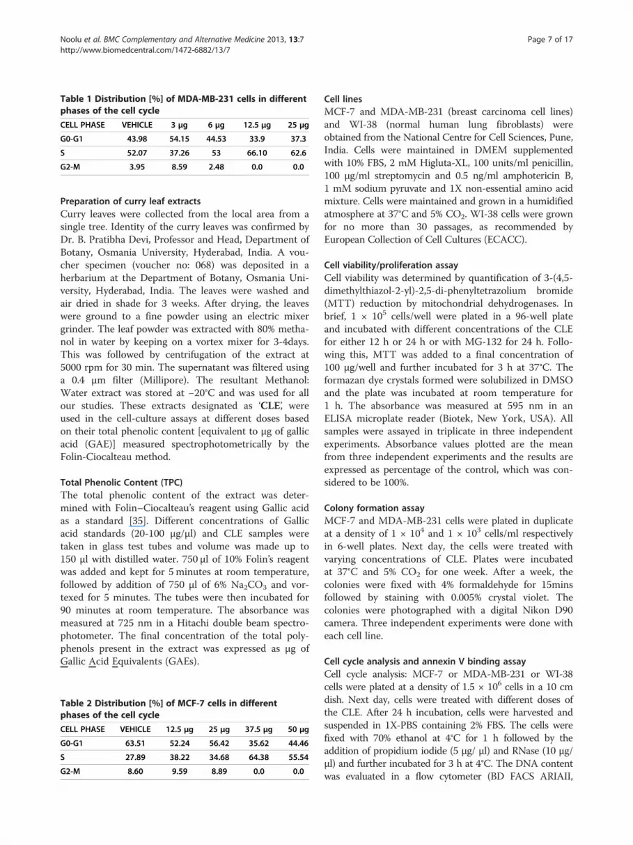
Table 1 Distribution [%] of MDA-MB-231 cells in differentphases of the cell cycle
CELL PHASE VEHICLE 3 μg 6 μg 12.5 μg 25 μg
G0-G1 43.98 54.15 44.53 33.9 37.3
S 52.07 37.26 53 66.10 62.6
G2-M 3.95 8.59 2.48 0.0 0.0
Noolu et al. BMC Complementary and Alternative Medicine 2013, 13:7 Page 7 of 17http://www.biomedcentral.com/1472-6882/13/7
Preparation of curry leaf extractsCurry leaves were collected from the local area from asingle tree. Identity of the curry leaves was confirmed byDr. B. Pratibha Devi, Professor and Head, Department ofBotany, Osmania University, Hyderabad, India. A vou-cher specimen (voucher no: 068) was deposited in aherbarium at the Department of Botany, Osmania Uni-versity, Hyderabad, India. The leaves were washed andair dried in shade for 3 weeks. After drying, the leaveswere ground to a fine powder using an electric mixergrinder. The leaf powder was extracted with 80% metha-nol in water by keeping on a vortex mixer for 3-4days.This was followed by centrifugation of the extract at5000 rpm for 30 min. The supernatant was filtered usinga 0.4 μm filter (Millipore). The resultant Methanol:Water extract was stored at −20°C and was used for allour studies. These extracts designated as ‘CLE’, wereused in the cell-culture assays at different doses basedon their total phenolic content [equivalent to μg of gallicacid (GAE)] measured spectrophotometrically by theFolin-Ciocalteau method.
Total Phenolic Content (TPC)The total phenolic content of the extract was deter-mined with Folin–Ciocalteau’s reagent using Gallic acidas a standard [35]. Different concentrations of Gallicacid standards (20-100 μg/μl) and CLE samples weretaken in glass test tubes and volume was made up to150 μl with distilled water. 750 μl of 10% Folin’s reagentwas added and kept for 5 minutes at room temperature,followed by addition of 750 μl of 6% Na2CO3 and vor-texed for 5 minutes. The tubes were then incubated for90 minutes at room temperature. The absorbance wasmeasured at 725 nm in a Hitachi double beam spectro-photometer. The final concentration of the total poly-phenols present in the extract was expressed as μg ofGallic Acid Equivalents (GAEs).
Table 2 Distribution [%] of MCF-7 cells in differentphases of the cell cycle
CELL PHASE VEHICLE 12.5 μg 25 μg 37.5 μg 50 μg
G0-G1 63.51 52.24 56.42 35.62 44.46
S 27.89 38.22 34.68 64.38 55.54
G2-M 8.60 9.59 8.89 0.0 0.0
Cell linesMCF-7 and MDA-MB-231 (breast carcinoma cell lines)and WI-38 (normal human lung fibroblasts) wereobtained from the National Centre for Cell Sciences, Pune,India. Cells were maintained in DMEM supplementedwith 10% FBS, 2 mM Higluta-XL, 100 units/ml penicillin,100 μg/ml streptomycin and 0.5 ng/ml amphotericin B,1 mM sodium pyruvate and 1X non-essential amino acidmixture. Cells were maintained and grown in a humidifiedatmosphere at 37°C and 5% CO2. WI-38 cells were grownfor no more than 30 passages, as recommended byEuropean Collection of Cell Cultures (ECACC).
Cell viability/proliferation assayCell viability was determined by quantification of 3-(4,5-dimethylthiazol-2-yl)-2,5-di-phenyltetrazolium bromide(MTT) reduction by mitochondrial dehydrogenases. Inbrief, 1 × 10⁵ cells/well were plated in a 96-well plateand incubated with different concentrations of the CLEfor either 12 h or 24 h or with MG-132 for 24 h. Follo-wing this, MTT was added to a final concentration of100 μg/well and further incubated for 3 h at 37°C. Theformazan dye crystals formed were solubilized in DMSOand the plate was incubated at room temperature for1 h. The absorbance was measured at 595 nm in anELISA microplate reader (Biotek, New York, USA). Allsamples were assayed in triplicate in three independentexperiments. Absorbance values plotted are the meanfrom three independent experiments and the results areexpressed as percentage of the control, which was con-sidered to be 100%.
Colony formation assayMCF-7 and MDA-MB-231 cells were plated in duplicateat a density of 1 × 10⁴ and 1 × 10³ cells/ml respectivelyin 6-well plates. Next day, the cells were treated withvarying concentrations of CLE. Plates were incubatedat 37°C and 5% CO2 for one week. After a week, thecolonies were fixed with 4% formaldehyde for 15minsfollowed by staining with 0.005% crystal violet. Thecolonies were photographed with a digital Nikon D90camera. Three independent experiments were done witheach cell line.
Cell cycle analysis and annexin V binding assayCell cycle analysis: MCF-7 or MDA-MB-231 or WI-38cells were plated at a density of 1.5 × 10⁶ cells in a 10 cmdish. Next day, cells were treated with different doses ofthe CLE. After 24 h incubation, cells were harvested andsuspended in 1X-PBS containing 2% FBS. The cells werefixed with 70% ethanol at 4°C for 1 h followed by theaddition of propidium iodide (5 μg/ μl) and RNase (10 μg/μl) and further incubated for 3 h at 4°C. The DNA contentwas evaluated in a flow cytometer (BD FACS ARIAII,

Vehicle 12.5µg CLE
25µg CLE 50µg CLE
Figure 5 CLE treatment does not arrest cell cycle in the WI-38 normal lung fibroblast cell line. Cell cycle analysis of WI-38 cells treatedwith varying concentrations of CLE for 24 h was done by flow cytometry.
Noolu et al. BMC Complementary and Alternative Medicine 2013, 13:7 Page 8 of 17http://www.biomedcentral.com/1472-6882/13/7

Table 3 Distribution [%] of WI-38 cells in different phasesof the cell cycle
CELL PHASE VEHICLE 12.5 μg 25 μg 50 μg
G0-G1 48.7 55.83 48.57 52.8
S 50.35 44.16 51.43 47.20
G2-M 0.87 0.00 0.00 0.00
Noolu et al. BMC Complementary and Alternative Medicine 2013, 13:7 Page 9 of 17http://www.biomedcentral.com/1472-6882/13/7
USA). The data was analyzed using Modfit software (BDBiosciences, USA).Annexin-V staining: MCF-7 or MDA-MB-231 cells were
plated at a density of 1.5 × 10⁶ cells in a 10 cm dish. Nextday, cells were treated with different doses of the CLE.After 24 h incubation, cells were washed with 1X-PBS andre-suspended in 100μl binding buffer (supplied by thevendor). Cells were stained with Annexin V-FITC andpropidium iodide according to the manufacturer’s proto-col before analysis by flow cytometry.
Inhibition of purified 20S proteasome activityChymotrypsin-like activity of the purified 20S proteasomewas measured as follows: In brief, 200 ng of purified 20Sproteasome was incubated in 200μl of assay buffer(50 mmol/LTris–HCl, pH 8.0 containing 0.035% SDS) withor without different concentrations of CLE and 40 μMsubstrate Suc-Leu-Leu-Val-Tyr-AMC (for chymotrypsin-like activity) and incubated for 2h at 37°C. The free 7-amino-4-methylcoumarin (AMC) liberated was measuredfluorimetrically using a multi-mode reader [Spectra MaxM5] using an excitation filter (380 nm) and emission filter(460 nm). The data is plotted as mean (+/− standard error)and is expressed as a percentage of the control, which wasconsidered to be 100%. The assay was repeated thrice.
Inhibition of 26S proteasome activity in intact cellsTo measure inhibition of the proteasome activity inliving tumor cells, MCF-7 or MDA-MB-231 or WI-38cells were plated at a density of 1 × 10⁴ in a 24 wellplate. Next day, cells were treated with or without theCLE at the indicated concentrations. After 24 h of treat-ment, the media was aspirated out and 500 μl of 1X-PBSwas added followed by addition of fluorogenic substrates(20 μM final concentration) specific for the chymotrypsin-like (Ch-L), trypsin-like (T-L) and caspase-like (Cp-L)activities of the 20S proteasome. The plate was then incu-bated for 2 h at 37°C. 200 μl of the 1X- PBS was thentransferred into a black plate and the free 7-amino-4-methylcoumarin (AMC) liberated was measured fluori-metrically in a multi-mode reader [Spectra Max M5] atexcitation (380 nm) and emission (460 nm). The results aredisplayed as mean (+/− standard error) and are expressedas a percentage of the control, which was considered to be100%. All samples were assayed in triplicate in three inde-pendent experiments.
Inhibition of 26S proteasome activity in cell extractsMCF-7 or MDA-MB-231 cells (1 × 10⁷) were harvested,washed twice in 1X-PBS and re-suspended in 1 ml ATP/DTT lysis buffer [10 mmol/L Tris–HCl (pH 7.8), 5 mmol/L ATP, 0.5 mmol/l DTT, 5 mmol/L MgCl2]. Cells wereincubated on ice for 10 minutes, followed by sonicationfor 15 seconds. The lysate was centrifuged at 2000 rpm for10 minutes at 4°C. The supernatant enriched in 26S pro-teasomes is the cell extract which was used for the assay.This was mixed with glycerol (20% final concentration),aliquoted and stored at −80°C, and was stable for at least 1month. The total protein content of the cell extract wasestimated by the Bicinchoninic Acid (BCA) method usinga kit (Bangalore Genei, Bangalore, India). The assay wascarried out in a total of 200 μl reaction volume containingproteasome extract (50 μg protein), 50 mM EDTA, vary-ing concentrations of the CLE/MG-132 and 50 μM of theproteasomal fluorogenic substrates and incubated for 2 hat 37°C. The amount of free 7-amino-4-methylcoumarin(AMC) liberated was measured fluorimetrically. Theresults are expressed as mean (+/− standard error) as apercentage of the control, which was considered to be100%. All samples were assayed in triplicate in three in-dependent experiments.
Statistical analysisAll experiments were performed in triplicates and repeatedat least three times and the data are presented as mean+/−SEM. Mean values were compared across concentrations ofCLE using non-parametric test of Kruskal-Wallis one-wayANOVA for each cell line using the SPSS statistical soft-ware. Differences between groups were considered signifi-cant at α (probability) level of </= 0.05.
ResultsM. koenigii leaf extract alters viability and growth kineticsof breast cancer cellsThe TPC of the methanolic extract of curry leaves was 3 μgof GAEs/μl of the CLE. MTT assays were performed withdifferent concentrations of CLE (GAE) in both the cell linesat the 12 h and 24 h time points to assess the effect of theextract on cell viability. There was a significant (p<0.05)time and dose-dependent decrease in cell viability in boththe cell lines. As expected, the decrease in cell viabilityobserved after 24 h of treatment was higher compared to12 h. In addition, MDA-MB-231 cells (Figure 1A and 1B)appeared to be more sensitive to CLE induced cell deaththan MCF-7 cells (Figure 1D and 1E). 24 h post treatment,a 50% reduction in cell viability was observed in MDA-MB-231 and MCF-7 cells at 15 μg and 37.5 μg GAE of CLErespectively. As a positive control, the effect of MG-132- aspecific inhibitor of the proteolytic activity of the 26S prote-asome was also assessed on cell viability in both the breastcancer cell lines. 24 h post treatment with MG-132, a 50%

Vehicle 6µg CLE
12.5µg CLE 18µg CLE
25µg CLE 37.5µg CLE
Figure 6 Exposure to CLE induces apoptosis in the MDA-MB-231 breast carcinoma cell line. Phosphatidylserine levels were detected byannexin V-FITC binding. MDA-MB-231 cells were treated with varying concentrations of the CLE for 24 h and stained with FITC-conjugatedannexin V and propidium iodide (PI).
Noolu et al. BMC Complementary and Alternative Medicine 2013, 13:7 Page 10 of 17http://www.biomedcentral.com/1472-6882/13/7
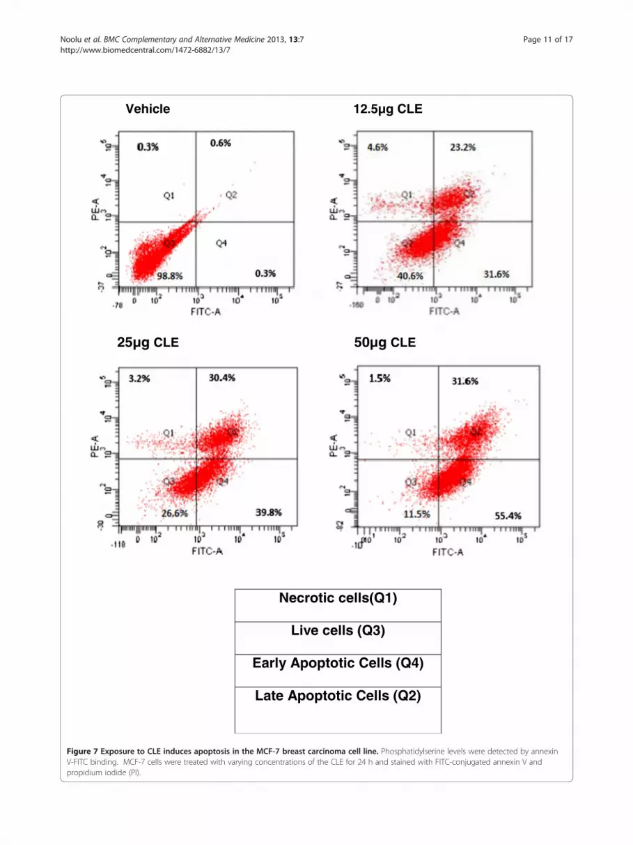
Vehicle 12.5µg CLE
25µg CLE 50µg CLE
Necrotic cells(Q1)
Live cells (Q3)
Early Apoptotic Cells (Q4)
Late Apoptotic Cells (Q2)
Figure 7 Exposure to CLE induces apoptosis in the MCF-7 breast carcinoma cell line. Phosphatidylserine levels were detected by annexinV-FITC binding. MCF-7 cells were treated with varying concentrations of the CLE for 24 h and stained with FITC-conjugated annexin V andpropidium iodide (PI).
Noolu et al. BMC Complementary and Alternative Medicine 2013, 13:7 Page 11 of 17http://www.biomedcentral.com/1472-6882/13/7

B
C
0
20
40
60
80
100
120
% C
h-L
act
ivit
y
Conc of CLE (µg of GAE)
0
20
40
60
80
100
120
%T
-L a
ctiv
ity
Conc of CLE (µg of GAE)
0
20
40
60
80
100
120
% C
p-L
aci
vity
Conc of CLE(µg of GAE)
A
Figure 9 Inhibition of the enzymatic activities of the 26Sproteasome by the CLE in intact MDA-MB-231 breast cancercell line. Intact MDA-MB-231 were treated for 24 h with or withoutthe CLE followed by 2 h incubation at 37°C with the fluorogenicsubstrate for Ch-L or T-L or Cp-L activities respectively. Thefluorescent intensity of the free AMCs was determined in amultimode reader with excitation (380 nm) and emission (460 nm).Each activity was expressed as the percentage of the control(defined as 100%). Panel A, B & C represents Ch-L, T-L and Cp-Lactivities respectively.
0
20
40
60
80
100
120
% C
h-L
act
ivit
y
Conc of CLE (µg of GAE)Figure 8 Curry leaf extract inhibits activity of purified 20Sproteasome. Purified 20S proteasome from rabbit was incubatedwith 40 μM of fluorogenic peptide substrate for Ch-L activity in thepresence of various concentrations of the CLE for 2 h at 37°C. Thefluorescence intensity of the free AMCs was determined usingfluorescence mode in a multimode reader with excitation (380 nm)and emission (460 nm).
Noolu et al. BMC Complementary and Alternative Medicine 2013, 13:7 Page 12 of 17http://www.biomedcentral.com/1472-6882/13/7
reduction in cell viability was observed at 20 μM and>40 μM of MG-132 in MDA-MB-231and MCF-7 cells re-spectively (Figure 1C and 1F).To test the effect of CLE on growth kinetics, MCF-7 and
MDA-MB-231 cells were seeded at a lower density andtreated with different concentrations of the CLE. After in-cubation for a week, it was observed that at a dose of 25 μgGAE of CLE, no colonies were found in either MDA-MB-231 (Figure 2A) or MCF-7 cells (Figure 2B). In line withour observations on cell viability (MTT assay), MDA-MB-231 cells appeared to be more sensitive than MCF-7 cells.This is supported by our findings where a lesser concentra-tion of CLE was needed to inhibit the formation of coloniesin MDA-MB-231 cells in comparison to MCF-7 cells.
M. koenigii leaf extract induces growth arrest andapoptosis in breast cancer cellsCell cycle experiments were done to determine whetherCLE treatment arrested growth in MDA-MB-231 andMCF-7 cells. In both the breast carcinoma cell lines CLEtreatment showed a dose-dependent arrest in the Sphase of the cell cycle resulting in complete inhibition ofcell proliferation (Figures 3 and 4; Tables 1 and 2). Thatinhibition of cell proliferation denoted by G2-M phasewas observed at 12.5 μg GAE in MDA-MB-231 cells,whereas, in MCF-7 cells it was seen at 37.5 μg GAE,corroborates the greater sensitivity of MDA-MB-231

Noolu et al. BMC Complementary and Alternative Medicine 2013, 13:7 Page 13 of 17http://www.biomedcentral.com/1472-6882/13/7
than MCF-7 cells to the CLE induced effects. Interest-ingly, CLE had no effect on cell cycle in the normal WI-38cell line at any of the concentrations tested (Figure 5 andTable 3), indicating that CLE could arrest growth only incancer but not normal cells.Further, Annexin-V binding experiments were conducted
in both cell lines to determine the probable mechanism ofcell death. That 6μg CLE resulted in 45% of live MDA-MB-231 cells whereas, a dose of 12.5 μg CLE was required for asimilar effect in MCF-7 cells not only confirms the greatersensitivity of MDA-MB-231 than MCF-7 cells but also sug-gests apoptosis to be the probable mechanism of cell death.This is confirmed by our finding that CLE demonstrated adose dependent increase in the % of apoptotic cells in bothMDA-MB-231 and MCF-7 cells. 50% of cells were apop-totic with 6 μg CLE in MDA-MB-231 cell line and itincreased to 66% at a dose of 37.5 μg CLE (Figure 6). Asimilar dose dependent increase in the % apoptotic cellswas seen in MCF-7 cells (Figure 7).
M. koenigii leaf extract inhibits 20S purified proteasomeactivityWe then tested whether or not CLE inhibited the activityof the purified 20S rabbit proteasome in a cell-free system.Indeed CLE decreased the chymotrypsin-like activity ofthe 20S proteasome in a dose-dependent manner and a
C D
0
20
40
60
80
100
120
% C
h-L
act
ivit
y
Conc of CLE (µg of GAE)
0
20
40
60
80
100
120
%C
p-L
act
ivit
y
Conc of CLE (µg of GAE)
A B
Figure 10 Inhibition of the enzymatic activities of the 26S proteasomnormal cell line. Intact MCF-7 or WI-38 cells were treated for 24 h with ofluorogenic substrate for either Ch-L or T-L or Cp-L activities respectively. Tmultimode reader with excitation (380 nm) and emission (460 nm). Each a100%). Panel D depicts results from WI-38 cells. Panels A & D represents
50% decrease (IC50) in activity was seen at a concentrationof CLE equivalent to 3 ug of Gallic Acid (GAE)/μl of theextract (Figure 8).
M. koenigii leaf extract inhibits cellular 26S proteasomeactivity in intact cellsWhether the CLE also inhibited the activity of the 26Sproteasome in living cancer cells was assessed next inboth MCF-7 and MDA-MB-231 cells. Similar to itseffects on the purified 20S rabbit proteasome, CLEshowed a significant (p<0.05), dose-dependent decreasein the chymotrypsin-like, trypsin-like and caspase-likeactivities of the 26S proteasome in intact cancer cells(Figures 9 and 10).On the other hand, CLE did not inhibit the
chymotrypsin-like activity of the 26S proteasome atany of the concentrations tested in the normal WI-38 cells (Figure 10D) indicating the specificity of theeffect to cancer cells.
M. koenigii leaf extract inhibits cellular 26S proteasomeactivity in cell extractsFurther to confirm that the CLE inhibits the 26S prote-asome, cell extracts enriched in 26S proteasomes wereprepared from both MCF-7 and MDA-MB-231 cells.The cell extracts were then treated with the CLE and
0
20
40
60
80
100
120
% C
h-L
act
ivit
y
Conc of CLE (µg of GAE)
0
20
40
60
80
100
120
%T
-L a
ctiv
ity
Conc of CLE (µg of GAE)
e by the CLE in intact MCF-7 breast cancer cell line and WI-38r without the CLE followed by 2 h incubation at 37°C with thehe fluorescent intensity of the free AMCs was determined in activity was expressed as the percentage of the control (defined asCh-L activity; panels B & C depicts T-L and Cp-L activities respectively.

B
C D
0
20
40
60
80
100
120
%C
h-L
act
ivit
y
Conc of CLE (µg of GAE)
0
20
40
60
80
100
120
%T
-L a
ctiv
ity
Conc of CLE((µg of GAE)
0
20
40
60
80
100
120
%C
p-L
act
ivit
y
Conc of CLE(µg of GAE)
0
20
40
60
80
100
120
% C
h-L
act
ivit
y
Conc of MG-132 (nM)
A
Figure 11 Concentration-dependent inhibition of the enzymatic activities of the 26S proteasome by the CLE or MG-132 in MDA-MB-231 cell extracts. Cell extracts (=50 μg protein) were incubated with 50 μM of the fluorogenic substrates specific for Ch-L, T-L and Cp-L activitiesin the presence of various concentrations of the CLE for 2 h at 37°C. Each activity was expressed as the percentage of the control (defined as100%). Panel A, B & C depicts Ch-L, T-L and Cp-L activities respectively, whereas panel D depict Ch-L activity with the synthetic proteasomeinhibitor MG-132.
Noolu et al. BMC Complementary and Alternative Medicine 2013, 13:7 Page 14 of 17http://www.biomedcentral.com/1472-6882/13/7
inhibition of the three proteasomal activities was assessed.It was interesting that CLE inhibited the chymotrypsin-like, trypsin-like and caspase-like activities of the 26S pro-teasome in cell extracts in a dose-dependent manner inboth MDA-MB-231 [IC50 of 22.5 μg CLE] and MCF-7cells [IC50 of 30 μg CLE] (Figures 11 and 12). As a positivecontrol MG-132, a proteasome inhibitor was also tested. Itwas observed that MG-132 decreased the chymotrypsin-like activity of the 26S proteasome in a dose-dependentmanner in cell extracts prepared from both MDA-MB-231 and MCF-7 cells with an IC50 of >50 nM and 25 nMin the two cell lines respectively (Figures 11D & 12C).
DiscussionSearch for new anti-cancer drugs from natural sources isone of the most important approaches for cancer pre-vention and treatment. In recent years, more emphasisis laid on Complementary and Alternative [CAM] formsof medicine for the treatment of various cancers, amongwhich herbal medicine is now being explored for cancertherapy [36]. Dietary constituents have chemopreventiveand chemotherapeutic potential, in addition to amelior-ating the side effects associated with conventionalchemotherapy. In this context, a recent approach in can-cer therapy advocates the inhibition of the proteolyticactivity of 26S proteasome, the multi-enzymatic proteasecomplex in cells. Unlike normal cells, cancer cells have
increased proteasomal activity which is essential for theirsurvival and uninhibited proliferation [37-39]. Inhibitionof the proteasome results in apoptosis and cancer celldeath [40]. Importantly, inhibitors of the 20S proteolyticunit of the proteasome have been shown to induceapoptosis and cell cycle arrest only in neoplastic cellsbut not in normal cells [39-41]. Therefore, the prote-asome has emerged as an attractive molecular target forcancer therapy [42]. A number of synthetic proteasomeinhibitors have been described and most of them inter-fere with the proteolytic activity of the β subunits of the20S proteasome. These inhibitors which bind the activesite either reversibly or irreversibly include peptide alde-hydes such as MG-132, non-peptide inhibitors such aslactacystin and epoxomycin and peptide boronates suchas bortezomib [43].Bortezomib/Velcade/PS-341 is the first-in-line and
the only dipeptide boronate proteasome inhibitor tobe approved by the FDA in 2003 for the treatment ofpatients with refractory multiple myeloma. Bortezomibis now being tested in a variety of hematological andsolid tumors including non-Hodgkin’s lymphoma, pros-tate, breast and non-small-cell-lung cancer [44,45]. Inrecent years, synthetic polyphenols such as apigenin,epigallocatechin gallate [EGCG], quercitin and myrcetinhave been reported to act as proteasome inhibitors andinduce cell death in cancer cells [24].

A
B
C
0
20
40
60
80
100
120
%C
h-L
act
ivit
y
Conc of CLE (µg of GAE)
0
20
40
60
80
100
120
% C
h-L
act
ivit
y
Conc of MG-132 (nM)
0
20
40
60
80
100
120
%T
-L a
ctiv
ity
Conc of CLE (µg of GAE)
Figure 12 Concentration-dependent inhibition of the enzymaticactivities of the 26S proteasome by the CLE or MG-132 inMCF-7 cell extracts. Cell extracts (=50 μg protein) were incubatedwith 50 μM of the fluorogenic substrates specific for Ch-L, T-L and Cp-Lactivities in the presence of various concentrations of the CLE for 2 h at37°C. Each activity was expressed as the percentage of the control(defined as 100%). Panel A & B depicts Ch-L and T-L activitiesrespectively, whereas panel C depicts Ch-L activity with MG-132.
Noolu et al. BMC Complementary and Alternative Medicine 2013, 13:7 Page 15 of 17http://www.biomedcentral.com/1472-6882/13/7
Drug resistance limits the effectiveness of existing treat-ment options and is a major challenge faced in currentcancer research. Interestingly, it has been shown that lac-tacystin and bortezomib enhance sensitivity of cancer cellsthat are resistant to routine chemotherapy [46,47]. Never-theless, synthetic proteasome inhibitors are associatedwith some toxicity. Therefore, proteasome inhibitors fromnatural food sources with minimal or no toxicity can bepotential anticancer agents.In the present study, we report the anticancer potential
of M. koenigii leaf extracts in two human breast carcin-oma cell lines. In recent years, dietary polyphenols haveattracted lot of attention owing to their anti-tumor activ-ities [48,49]. One such activity is the inhibition of theproteasome in cancer cells leading to cell death. Recentwork from our laboratory [21] has demonstrated that M.koenigii leaf extract is a rich source of polyphenols. Inthis study, we found that a hydro-methanolic extract ofcurry leaves is rich in polyphenol content. Extracts of M.koenigii leaves have been reported to possess various bio-logical activities such as anti-diabetic, anti-oxidative andanti-inflammatory [8-10]. Recently, carbazole alkaloidsfrom M. koenigii have shown anti-cancer activity inleukemia cells [18-20]. However, the underlying mechan-ism(s) are not reported yet. In the present work, we dem-onstrate for the first time that the hydro-methanolicextract of curry leaf has proteasome-inhibitory potentialand induces cell death in human breast cancer cells.We found that the methanolic extract of curry leaves
significantly decreased cell viability and proliferation ofboth MCF-7 and MDA-MB-231 breast cancer cells in adose-dependent manner. This was further supported bythe significant reduction in the number of colonies inCLE treated cells compared to vehicle treated cells. Ourcell viability and colony formation data shows that CLEaltered the growth kinetics of both MCF-7 and MDA-MB-231 cells. Therefore, curry leaves appear to be apromising drug candidate for restricting the growth ofbreast cancer cells.In order to assess the stage at which the cell growth
was arrested by CLE, we performed cell cycle experi-ments and observed that there was a clear arrest of cellsin the synthetic or S phase. In contrast to its effect onthe breast carcinoma cell lines, CLE interestingly, hadno effect on the different phases of the cell cycle in thenormal fibroblast cell line. Anti-cancer drugs can resulteither in programmed cell death/apoptosis or necrosis.In order to identify the probable cell death pathwayinvolved, we used Annexin V binding to test if the celldeath occurred through apoptosis or necrosis. Indeed itwas found that in both the breast cancer cell lines CLEinduced apoptotic cell death.We tested next, whether the anti-cancerous effect of the
CLE was due its potential of inhibiting the proteolytic

Noolu et al. BMC Complementary and Alternative Medicine 2013, 13:7 Page 16 of 17http://www.biomedcentral.com/1472-6882/13/7
activity of the protein degrading machine present ineukaryotic cells – the 26S proteasome, which is now con-sidered to be a novel approach for cancer therapy. Weobserved that CLE inhibited the purified 20S proteasomeenzyme. Furthermore, it significantly inhibited all thethree enzymatic activities associated with the 26S prote-asome in living cells in a dose-dependent manner. To fur-ther confirm the findings in live cells, cell extracts wereprepared from both breast cancer cells and tested for thepotential of the CLE in inhibiting the cellular proteasome.Similar to our findings in intact cells we found that CLEinhibited the 26S proteasome in cell extracts also in adose-dependent manner. To test whether or not the in-hibitory effect of the CLE on the proteasome activity wasspecific to cancer cells, we tested its effects in WI-38, anormal lung fibroblast cell line. Interestingly, CLE had noeffect on the chymotrypsin–like activity of the 26Sproteasome in live WI-38 cells. Hence, data from ourproteasome-inhibition experiments suggests that theCLE could inhibit the cellular proteasome leading tocell death in cancer cells but not normal cells. Our datais in accordance with earlier reports that proteasomeinhibitors selectively inhibited proteasome activity onlyin neoplastic cells [39,50,51].
ConclusionOur results indicate that the hydro-methanolic extractof curry leaves is a good source of active compound(s)that can potentially inhibit the 26S proteasome specific-ally in cancer cells. The inhibition of the proteasome incancer cells appears to be one of the important biologicalactivities in M. koenigii leaves that can be exploited forcancer treatment. Hence, isolation and characterization ofactive component(s) from methanolic extracts of curryleaves could lead to the discovery of novel anticanceragents.
Abbreviations(MTT): 3-(4,5-dimethylthiazol-2-yl)-2,5-di-phenyltetrazolium bromide;(GAE): Gallic Acid Equivalents; (CLE): Curry leaf extract; (AMC): 7-amino-4-methylcoumarin; (DMSO): Dimethyl sulphoxide; (EGCG): Epigallocatechingallate; (DTT): Dithiothretol; (FITC): Fluorescein isothiocyanate;(PBS): Phosphate buffered saline.
Competing interestsThe authors declare that they have no competing interests.
Authors’ contributionsAYI designed the study and wrote the manuscript. BN carried out most ofthe experiments. RA assisted with the cell cycle and Annexin-V experimentsand data analysis. AC helped with cytotoxicity assays. BKN has done thestatistical analysis of the data. RM has critically read and revised themanuscript for intellectual content. All authors read and approved the finalmanuscript.
AcknowledgementsThis work was funded by a grant from the Department of Biotechnology,India (Project No: BR/PR/11128/FNS/20/403/2008) to AYI. We thank theUniversity Grants Commission for providing fellowship to BN. We thank ourDirector Dr. B. Sesikeran for the valuable support and encouragement during
the course of the studies. We thank Ms. GV Asha for the assistance providedin estimation of the phenolic content of the CLEs.
Author details1Department of Endocrinology & Metabolism, National Institute of Nutrition,Hyderabad, India. 2Department of Stem Cell Research, National Institute ofNutrition, Hyderabad, India. 3Department of Statistics, National Institute ofNutrition, Hyderabad, India.
Received: 26 July 2012 Accepted: 4 January 2013Published: 9 January 2013
References1. Parkin DM, Bray F, Ferlay J, Pisani P: Estimating the world cancer burden:
Globocan. Int J Cancer 2000, 94:153–156.2. Pruthi JS: Spices and condiments. New Delhi: National Book Trust India; 1976.3. Chevallier A: The encyclopedia of medicinal plants. London: Dorlon Kindersley
Publisher; 1996.4. Sivarajan VV, Balachandran I: Ayurvedic drugs and their plant sources. New
Delhi: Oxford & IBH Publishing; 1994.5. Muthumani P, Venkatraman S, Ramseshu KV, Meera R, Devi P, Kameswari B:
Pharmacological studies of anticancer, anti inflammatory activities ofMurraya koenigii (Linn) Spreng in experimental animals. J Pharm Sci Res2009, 1:137–141.
6. Nayak A, Banerji J, Banerji A, Mandal S: Review on chemistry andpharmacology of Murraya koenigii Spreng (Rutaceae). J Chem Pharm Res2010, 2:286–299.
7. Kesari AN, Kesari S, Singh SK, Gupta RK, Watal G: Studies on the glycemicand lipidemic effect of Murraya koenigii in experimental animals.J Ethnopharmacol 2007, 112(2):305–311.
8. Arulselvan P, Senthilkumar GP, Sathish Kumar D, Subramanian S: Anti-diabetic effect of Murraya koenigii leaves on streptozotocin induceddiabetic rats. Pharmazie 2006, 61(10):874–877.
9. Lawal HA, Atiku MK, Khelpai DG: Hypoglycaemic and hypolipidemic effectof aqueous leaf extract of Murraya koenigii in normal and alloxan-diabetic rats. Niger J Physiol Sci 2008, 23(1–2):37–40.
10. Gupta S, Prakash J: Studies on Indian green leafy vegetables for theirantioxidant activity. Plant Foods Hum Nutr 2009, 64(1):39–45.
11. Tachibana Y, Kikuzaki H, Lajis NH, Nakatani N: Antioxidative activity ofcarbazoles from Murraya koenigii leaves. J Agric Food Chem 2001,49(11):5589–5594.
12. Khan BA, Abraham A, Leelamma S: Anti-oxidant effects of curry leaf,Murraya koenigii and mustard seeds, Brassica juncea in rats fed withhigh fat diet. Indian J Exp Biol 1997, 35(2):148–150.
13. Adebajo AC, Ayoola OF, Iwalewa EO, Akindahunsi AA, Omisore NO,Adewunmi CO, Adenowo TK: Anti-trichomonal, biochemical andtoxicological activities of methanolic extract and some carbazolealkaloids isolated from the leaves of Murraya koenigii growing inNigeria. Phytomedicine 2006, 13(4):246–254.
14. Mandal S, Nayak A, Kar M, Banerjee SK, Das A, Upadhyay SN, Singh RK,Banerji A, Banerji J: Antidiarrheal activity of carbazole alkaloids fromMurraya koenigii Spreng (Rutaceae) seeds. Fitoterapia 2010, 81(1):72–74.
15. Xie JT, Chang WT, Wang CZ, Mehendale SR, Li J, Ambihaipahar R,Ambihaipahar U, Fong HH, Yuan CS: Curry leaf (Murraya koenigii Spreng.)reduces blood cholesterol and glucose levels in ob/ob mice. Am J ChinMed 2006, 34(2):279–284.
16. Gupta S, George M, Singhal M, Sharma GN, Garg V: Leaves extract ofmurraya koenigii linn for anti-inflammatory and analgesic activity inanimal models. J Adv Pharm Technol Res 2010, 1(1):68–77.
17. Shah AS, Wakade AS, Juvekar AR: Immunomodulatory activity ofmethanolic extract of Murraya koenigii (L) Spreng. Leaves. Indian J ExpBiol 2008, 46(7):505–509.
18. Ito C, Itoigawa M, Nakao K, Murata T, Tsuboi M, Kaneda N, Furukawa H:Induction of apoptosis by carbazole alkaloids isolated from Murrayakoenigii. Phytomedicine 2006, 13(5):359–365.
19. Roy MK, Thalang VN, Trakoontivakorn G, Nakahara K: Mechanism ofmahanine-induced apoptosis in human leukemia cells (HL-60). BiochemPharmacol 2004, 67(1):41–51.
20. Bhattacharya K, Samanta SK, Tripathi R, Mallick A, Chandra S, Pal BC, ShahaC, Mandal C: Apoptotic effects of mahanine on human leukemic cells aremediated through crosstalk between Apo-1/Fas signaling and the Bid

Noolu et al. BMC Complementary and Alternative Medicine 2013, 13:7 Page 17 of 17http://www.biomedcentral.com/1472-6882/13/7
protein and via mitochondrial pathways. Biochem Pharmacol 2010,79(3):361–372.
21. Ayesha I, Bindu N, Shulagna S, Chandana M, Mehrajuddin B, Raghunath M:Proteasome inhibitory potential of commonly consumed dietaryingredients. Int J Food Nutr Sci 2012, 1(4):27–31.
22. Scalbert A, Manach C, Morand C, Rémésy C, Jiménez L: Dietarypolyphenols and the prevention of diseases. Crit Rev Food Sci Nutr 2005,45(4):287–306. Review.
23. Scalbert A, Johnson IT, Saltmarsh M: Polyphenols: antioxidants andbeyond. Am J Clin Nutr 2005, 81(Suppl 1):215S–217S. Review.
24. Chen D, Daniel KG, Chen MS, Kuhn DJ, Landis-Piwowar KR, Dou QP: Dietaryflavonoids as proteasome inhibitors and apoptosis inducers in humanleukemia cells. Biochem Pharmacol 2005, 69(10):1421–1432.
25. Pettinari A, Amici M, Cuccioloni M, Angeletti M, Fioretti E, Eleuteri AM: Effectof polyphenolic compounds on the proteolytic activities of constitutiveand immuno-proteasomes. Antioxid Redox Signal 2006, 8(1–2):121–129.
26. Hershko A, Ciechanover A: The ubiquitin system. Annu Rev Biochem 1998,67:425–479. Review.
27. Groll M, Ditzel L, Lowe J, Stock D, Bochtler M, Bartunik HD, Huber R:Structure of 20S proteasome from yeast at 2.4 A resolution. Nature 1997,386:463–471.
28. Rivett AJ: The multicatalytic proteinase. Multiple proteolytic activities.J Biol Chem 1989, 264(21):12215–12219.
29. Jung T, Bader N, Grune T: Oxidized proteins: intracellular distribution andrecognition by the proteasome. Arch Biochem Biophys 2007,462(2):231–237. Review.
30. Tambyrajah WS, Bowler LD, Medina-Palazon C, Sinclair AJ: Cell cycle-dependent caspase-like activity that cleaves p27(KIP1) is the beta(1)subunit of the 20S proteasome. Arch Biochem Biophys 2007,466(2):186–193.
31. Chen WJ, Lin JK: Induction of G1 arrest and apoptosis in human jurkat Tcells by pentagalloylglucose through inhibiting proteasome activity andelevating p27Kip1, p21Cip1/WAF1, and Bax proteins. J Biol Chem 2004,279(14):13496–13505.
32. Hilt W, Wolf DH: Proteasomes: The World of Regulatory Proteolysis.Georgetown, Texas: Landes Bioscience; 2000.
33. Goldberg AL: Functions of the proteasome: the lysis at the end of thetunnel. Science 1995, 268(5210):522–523. Review.
34. Hochstrasser M: Ubiquitin, proteasomes, and the regulation ofintracellular protein degradation. Curr Opin Cell Biol 1995,7(2):215–223. Review.
35. Singleton VL, Orthofer R, Lamuela-Raventos RM: Analysis of total phenolsand other oxidation substrates and antioxidants by means of Folin-Ciocalteu Reagent. Methods Enzymol 1999, 299:152–178.
36. Adams M, Jewell AP: The use of complementary and alternative medicineby cancer patients. Int Semin Surg Oncol 2007, 4:10.
37. Wada M, Kosaka M, Saito S, Sano T, Tanaka K, Ichihura A: Serumconcentrations and localization in tumor cells of proteasomes inpatients with hematologic malignancy and their pathophysiologicsignificance. J Lab Clin Med 1993, 121:215–223.
38. Li B, Dou QP: Bax degradation by the ubiquitin/proteasome-dependentpathway: involvement in tumor survival and progression. Proc Natl AcadSci USA 2000, 97(8):3850–3855.
39. An B, Goldfarb RH, Siman R, Dou QP: Novel dipeptidyl proteasomeinhibitors overcome Bcl-2 protective function and selectively accumulatethe cyclin-dependent kinase inhibitor p27 and induce apoptosis intransformed, but not normal, human fibroblasts. Cell Death Differ 1998,5(12):1062–1075.
40. Dou QP, Li B: Proteasome inhibitors as potential novel anticancer agents.Drug Resist Update 1999, 2(4):215–223.
41. Kazi A, Urbizu DA, Kuhn DJ, Acebo AL, Jackson ER, Greenfelder GP, KumarNB, Dou QP: A natural musaceas plant extract inhibits proteasomeactivity and induces apoptosis selectively in human tumor andtransformed, but not normal and non-transformed, cells. Int J Mol Med2003, 12(6):879–887.
42. Landis-Piwowar KR, Milacic V, Chen D, Yang H, Zhao Y, Chan TH, Yan B, DouQP: The proteasome as a potential target for novel anticancer drugs andchemosensitizers. Drug Resist Update 2006, 9(6):263–273. Review.
43. Adams J: The proteasome: structure, function, and role in the cell. CancerTreat Rev 2003, 29(Suppl 1):3–9. Review.
44. Richardson PG, Hideshima T, Anderson KC: Bortezomib (PS-341): a novel,first-in-class proteasome inhibitor for the treatment of multiple myelomaand other cancers. Cancer Control 2003, 10(5):361–369. Review.
45. Richardson PG, Anderson KC: Bortezomib: a novel therapy approved formultiple myeloma. Clin Adv Hematol Oncol 2003, 1(10):596–600. Review.
46. Delic J, Masdehors P, Omura S, Cosset JM, Dumont J, Binet JL, MagdelénatH: The proteasome inhibitor lactacystin induces apoptosis and sensitizeschemo- and radio resistant human chronic lymphocytic leukemialymphocytes to TNF-alpha-initiated apoptosis. Br J Cancer 1998,77(7):1103–1107.
47. Ma MH, Yang HH, Parker K, Manyak S, Friedman JM, Altamirano C, Wu ZQ,Borad MJ, Frantzen M, Roussos E, Neeser J, Mikail A, Adams J, Sjak-Shie N,Vescio RA, Berenson JR: The proteasome inhibitor PS-341 markedlyenhances sensitivity of multiple myeloma tumor cells tochemotherapeutic agents. Clin Cancer Res 2003, 9(3):1136–1144.
48. Kandaswami C, Lee LT, Lee PP, Hwang JJ, Ke FC, Huang YT, Lee MT: Theantitumor activities of flavonoids. In Vivo 2005, 19:895–909.
49. Thomasset SC, Berry DP, Garcea G, Marczylo T, Steward WP, Gescher J:Dietary polyphenolic phytochemicals - promising cancerchemopreventive agents in humans? A review of their clinicalproperties. Int J Cancer 2007, 120:451–458.
50. Nam S, Smith DM, Dou QP: Ester bond-containing tea polyphenolspotently inhibit proteasome activity in vitro and in vivo. J Biol Chem2001, 276(16):13322–13330.
51. Kuhn DJ, Lam WH, Kazi A, Daniel KG, Sung S, Chow LMC, Chan TH, Dou QP:Synthetic peracetate tea polyphenols as potent proteasome inhibitorsand apoptosis inducers in human cancer cells. Front Bio 2005,10:1010–1023.
doi:10.1186/1472-6882-13-7Cite this article as: Noolu et al.: Murraya koenigii leaf extract inhibitsproteasome activity and induces cell death in breast cancer cells. BMCComplementary and Alternative Medicine 2013 13:7.
Submit your next manuscript to BioMed Centraland take full advantage of:
• Convenient online submission
• Thorough peer review
• No space constraints or color figure charges
• Immediate publication on acceptance
• Inclusion in PubMed, CAS, Scopus and Google Scholar
• Research which is freely available for redistribution
Submit your manuscript at www.biomedcentral.com/submit


