Embed Size (px)
Citation preview
FEBS 29936 FEBS Letters 579 (2005) 5193–5198
Mutation of the extracellular domain of tumour necrosis factor receptor1 causes reduced NF-jB activation due to decreased surface expression
Stefan Sieberta, Ceri A. Fieldinga, Bryan D. Williamsb, Paul Brennana,*
a Department of Medical Biochemistry and Immunology, Wales College of Medicine, Cardiff University, Heath Park,Cardiff CF14 4XX, United Kingdom
b Department of Rheumatology, Wales College of Medicine, Cardiff University, Heath Park, Cardiff CF14 4XX, United Kingdom
Received 15 June 2005; revised 29 July 2005; accepted 17 August 2005
Available online 31 August 2005
Edited by Lukas Huber
Abstract Tumour necrosis factor receptor-associated periodicsyndrome (TRAPS) results from point mutations in the extracel-lular domain of TNF receptor 1 (TNFRSF1A), but the effects ofthe mutations are controversial. This study shows that reducedNF-jB signalling is a feature of four TRAPS mutations. Re-duced signalling correlates with reduced surface expression, mea-sured by flow cytometry and microscopy. This suggests thatcorrect formation of the extracellular domain of TNFRSF1Ais important for localisation and receptor function. Importantly,our data provides a mechanism for the reduced TNFRSF1 sig-nalling observed in a patient cell line.� 2005 Federation of European Biochemical Societies. Publishedby Elsevier B.V. All rights reserved.
Keywords: Tumour necrosis factor; TRAPS; NF-jB
1. Introduction
Tumour necrosis factor receptor-associated periodic syn-
drome (TRAPS; MIM 142680) is an autoinflammatory syn-
drome, characterised by recurrent fevers with cutaneous,
muscle and joint inflammation, associated with autosomal
dominantly-inherited mutations in the gene that encodes
tumour necrosis factor (TNF) receptor superfamily 1A
(TNFRSF1A) [1]. TNFa exerts its many pro-inflammatory
effects through two distinct receptors: TNFRSF1A (TNFR1,
p55/p60-TNFR) and TNFRSF1B (TNFR2, p75/p80-TNFR).
TNFRSF1A is widely expressed and appears to be the major
receptor for soluble TNFa-induced signaling [2]. At least 60
TNFRSF1A mutations associated with TRAPS have been re-
ported on the INFEVERS (Internet periodic fevers) website
(http://fmf.igh.cnrs.fr/infevers/). The majority are located in
the first or second cysteine-rich extracellular domains (CRD1
and CRD2). The binding site for TNFa is formed by CRD2
and CRD3 of TNFRSF1A [3], while CRD1, also known as
the pre-ligand assembly binding domain, is thought to mediate
TNFRSF1A self-assembly [4].
The initial study of TRAPS suggested that the TNFRSF1A
mutations impair activation-induced shedding of the receptor
Abbreviations: TRAPS, tumour necrosis factor receptor-associatedperiodic syndrome; TNFa, tumour necrosis factor alpha; TNFRSF,TNF receptor superfamily; CRD, cysteine-rich domain
*Corresponding author. Fax: +44 292074 3868.E-mail address: [email protected] (P. Brennan).
0014-5793/$30.00 � 2005 Federation of European Biochemical Societies. Pu
doi:10.1016/j.febslet.2005.08.037
[1]. However, this is not the case for all mutations [5,6] and var-
ies according to cell type [7]. Another study has shown that
overexpression of TRAPS-associated TNFRSF1A mutants
spontaneously induce apoptosis and interleukin-8 production
although TNFa binding to the mutated receptors appears to
be defective [8].We have recently shown that cells from a patient
with TRAPS, bearing the C43S mutation, have decreased
NF-jB induction and TNFa-induced apoptosis, although
IL-6 and IL-8 production were normal [6] suggesting that
reduced TNFRSF1A signalling may be a feature of TRAPS.
This study was initiated to investigate whether reduced
TNFRSF1A signalling was a general feature of TRAPS muta-
tions. We transfected cells with either wild-type (WT) or the
following mutant recombinant forms of TNFRSF1A: C30R,
C43S, T50M and C52F. We investigated the effects of these
clinically relevant mutations on receptor function, in terms
of NF-jB activation, on cell death and on receptor expression.
Our work shows that all of these TRAPS mutations result in
decreased TNFRSF1A signalling and that this loss of signal-
ling correlates with reduced expression of the receptor on the
surface of cells.
2. Materials and methods
2.1. Production of recombinant TNFRSF1A DNA clones and plasmidsThe TNFRSF1A coding region was ligated into the pcDNA3.1 myc/
His B vector (Clontech). The TRAPS mutations were generated usingthe Quikchange site-directed mutagenesis kit (Stratagene). Primersused: C30R: 5 0-CGATTTGCCGTACCAAGTGCCAC-3 0, C43S:5 0-CTTGTACAATGACTCTCCAGGCCCGGGGC-30, T50M 5 0-CC-GGGGCAGGATATGGACTGCAGGGAG-30, C52F: 5 0-GGCAG-GATACGGACTTCAGGGAGTGTG AGAG-30 and their reversecomplements. Mutations were confirmed by sequencing. The 3Enh.jB-ConALuc reporter (3EnhLuc) contains three NF-jB binding [9].
2.2. Cells, transfection, luciferase reporter assay and cell viability assayEli-BL and DG75 B-cell lymphoma culture has been described [10].
107 cells in 0.5 ml culture medium with 100 mMHEPES (pH 7.2), weretransfected using a Biorad Genepulser II (270 V/950 lF). Transfectionefficiency was typically 5–20% for the Eli-BL and 40–50% for theDG75. Luciferase assays were performed in the transfected Eli-BLcells. Cells were stimulated after 16 h. Luciferase activity was measured8 h later [11]. Transfection efficiency was assessed using the PromegaDual-Luciferase reporter system.Cell viability was assessed by alamar blue assay (Biosource). Briefly,
2 · 104 transfected cells were resuspended in 200 ll of 10% alamar bluereagent in growth medium on a 96 well plate. After 4 h incubation at37 �C, readings were made on a FLUOstar Optima (BMG Laborato-ries) using the fluorescence configuration.
blished by Elsevier B.V. All rights reserved.
5194 S. Siebert et al. / FEBS Letters 579 (2005) 5193–5198
2.3. Detection of TNFRSF1A expression by flow cytometry or
immunofluorescenceFor surface expression, cells were harvested 24 h after transfection,
washed with phosphate buffered saline (PBS) and fixed in 2% parafor-maldehyde. Cells were washed and resuspended in 10% normal rabbitserum (NRS) in PBS for 20 min. Cells were stained with anti-TNFRSF1A monoclonal antibody (Ab-1, Calbiochem) followed byan anti-mouse R-phycoerytherin (RPE)-conjugated F(ab 0)2 fragment(Dako), or with mouse IgG1-conjugated PE negative control (Serotec).Washed cells were resuspended in 2% paraformaldehyde and analysedusing a Becton Dickinson FACScalibur. Total (surface and intracellu-lar) expression of TNFRSF1A was assayed following permeabilisationwith 0.1% Triton X-100/PBS after fixation and stained as before.For confocal microscopy, cells were washed, fixed with 2% parafor-
maldehyde, resuspended in PBS and air-dried. Cells were permeabi-lised with 0.1% Triton X-100/PBS, incubated in 10% NRS/PBS andstained with the same anti-TNFRSF1A antibody followed by an AlexaFluor 488 anti-mouse IgG antibody (excited 488 nm – detected520 nm). For nuclear staining, cells were incubated with DRAQ510 lM (Biostatus) for 10 min (excited 647 nm – detected 680/30 nm).Images were acquired using a confocal laser scanning microscope (Bio-rad), with a krypton/argon ion laser and a Zeiss Axiovert 135. Resultsare representative of five experiments.
3. Results
3.1. The TNFRSF1A mutants result in reduced NF-jB activity
NF-jB is a critical transcription factor for the induction of
inflammation by TNFa [12]. NF-jB activation by wild-type
and mutant recombinant forms of TNFRSF1A was assessed
0100020003000400050006000700080009000
10000
0 5 10 15 20DNA dose (µg)
WT
C43S
T50M
C52F
C30R*
*
*
*
0
1000
2000
3000
4000
5000
6000
7000
8000
0 2.5 5 7.5 10
TNFα (ng/ml)
pcDNA3.1WT C43S C30R
** * * *
A B
C D
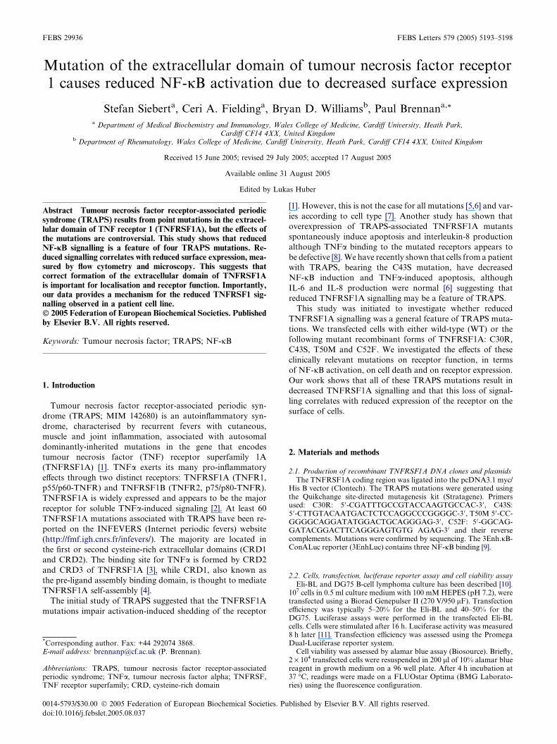
Fig. 1. NF-jB activation by WT and mutant recombinant forms of TNFRreporter (3 lg) and TNFRSF1A constructs. Total DNA was constant at 23 lgone half was stimulated with TNFa (10 ng/ml). After 8 h, luciferase activity wstimulated cells. (C) TNFa dose response for cells transfected with TNFexperiments. Asterisks (*) indicate P < 0.05 between WT and the TRAPS conjB firefly luciferase reporter (3 lg), a Renilla luciferase reporter (1 lg) andLuciferase activities were measured after 8 h of TNFa (10 ng/ml) stimulation
by luciferase reporter assay in transiently transfected Eli-BL
cells. Eli-BL cells express undetectable levels of membrane
TNFRSF1A by flow cytometry and have low endogenous re-
sponses to TNFa [10], allowing measurement of alterations
in NF-jB activity as a result of transfection with recombinant
TNFRSF1A. Eli-BL cells were co-transfected with the NF-jBluciferase reporter plasmid and either empty mammalian vec-
tor, mammalian expression vectors for wild-type TNFRSF1A
or a TNFRSF1A mutant. The mutants used were C30R, C43S,
T50M and C52F. C43S was the TRAPS mutation identified
and described by us previously [6]. C30R is the cysteine on
the opposite side of the disulfide bond to C43S. C52F was
one of the first mutants described, and as there may be differ-
ences between cysteine and non-cysteine mutations, we chose a
close by non-cysteine mutation, T50M.
Overexpression of TNFRSF1A resulted in increased NF-jBactivity, which was dependent on the amount of DNA trans-
fected (Fig. 1A). Interestingly, the level of NF-jB activation
was consistently, and significantly, higher for WT TNFRSF1A
than for any of the four TRAPS mutants. In addition, there
appear to be differences in NF-jB activity between these
TRAPS mutants themselves, with the C30R mutation display-
ing minimal NF-jB activation. The differences in NF-jB activ-
ity between wild-type and the TRAPS mutants were more
pronounced in cells stimulated with TNFa (Fig. 1B). To inves-
tigate whether the TRAPS mutations had a different dose re-
sponse curve, wild-type receptor and two of the TRAPS
0
2000
4000
6000
8000
10000
12000
14000
16000
0 5 10 15 20DNA dose (µg)
WT
C43S
T50M
C52F
C30R
**
*
*
0
10
20
30
40
50
pcDNA3.1 WT C43S C30R
Construct
Mock stimulated
TNF stimulated
SF1A. Eli-BL cells were transiently transfected with NF-jB luciferaseby addition of pcDNA3.1. Transfections were split into two. 16 h lateras assayed. Results are shown for (A) mock stimulated and (B) TNFaRSF1A constructs (10 lg). All results are the mean of at least fivestructs, by Student�s t-test. (D) Eli-BL cells were transfected with NF-TNFRSF1A constructs (10 lg) or empty vector. Firefly and Renilla, according to the manufacturer�s instructions (Promega).
2030405060708090
100110120130
WT
C43S
T50MC52F
C30R
** * *
Cel
l via
bili
ty(%
of
emp
ty v
ecto
r)
A
S. Siebert et al. / FEBS Letters 579 (2005) 5193–5198 5195
mutants were stimulated with a range of TNFa concentra-
tions. Reduced NF-jB was seen with the TRAPS mutants over
the full range of TNFa doses (Fig. 1C). Transfection efficiency
was assessed by dual luciferase assays. Cells were transfected
with the NF-jB reporter, expressing firefly luciferase, and an
SV40 promoter driving Renilla luciferase. Fig. 1D shows data
with normalised NF-jB activity for WT TNFRSF1A and two
TRAPS mutants. Both mutants show reduced NF-jB activity
even when normalised for transfection efficiency.
0102030405060708090
100110
0 2.5 5 7.5 10TNFα (ng/ml)
WT
C43S
T50M
C30R
* * * * *
010
0 5 10 15 20
Cel
l via
bili
ty(%
of
emp
ty v
ecto
r)
200
250 WT
C43S
T50M
B
C
DNA (µg)
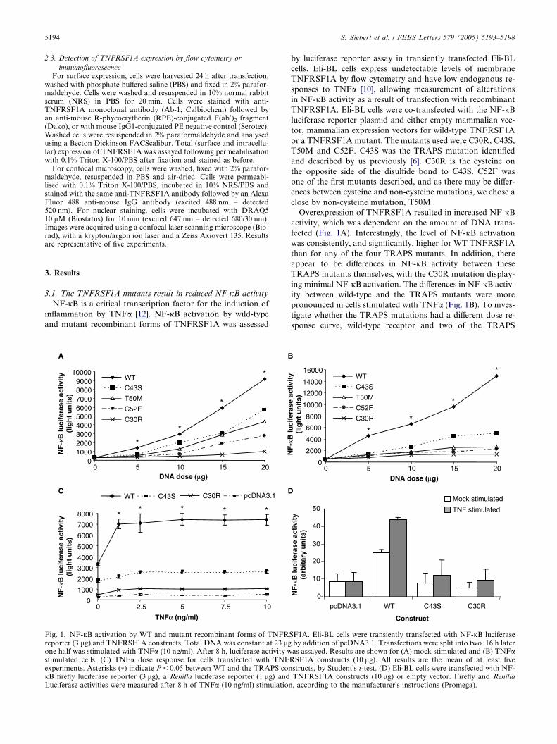
3.2. Overexpression of the TRAPS mutants result in less celldeath than WT TNFRSF1A
As overexpression of TNFRSF1A itself can result in sponta-
neous cell death [8,13], cell viability assays were performed on
the transfected cells used for the luciferase assays. Alamar blue,
a non-toxic dye that is chemically reduced by the innate meta-
bolic activity of cells, was used to quantify cell viability [14]. Cell
viability was consistently lower in the Eli-BL cells transfected
with WT TNFRSF1A than those transfected with the mutant
forms of TNFRSF1A (Fig. 2A). The degree of cell death was
dependent on the amount of TNFRSF1A DNA transfected
but was independent of the TNFa dose (Fig. 2B). The decreased
cell death seen with the mutant recombinant forms of
TNFRSF1Ademonstrates that the reducedNF-jBactivity seen
with thesemutants is not as a result of differences in cell death. In
fact,when the percentage of viable cells is taken into account, the
difference in NF-jB activity between WT TNFRSF1A and the
TRAPS mutants increases further (Fig. 2C).
0
50
100
150
0 5 10 15 20DNA (µg)
C52F
C30R
Fig. 2. Cell viability of the TNFRSF1A constructs. Cell viability wastested, 16 h post-transfection with TNFRSF1A constructs (10 lg), inuntreated (A) and TNFa treated (B) in Eli-BL cells. The results areshown relative to cells transfected with empty pcDNA3.1, expressed asa percentage. Results are the means ± S.E.M. of three experiments.
* = P < 0.05 versus TRAPS constructs, by Student�s t-test. (C) NF-jBactivation by TNFRSF1A constructs adjusted for cell viability. NF-jBactivity, measured by luciferase assay, was divided by the percentage ofviable cells. All results shown are the means ± S.E.M. of threeexperiments.
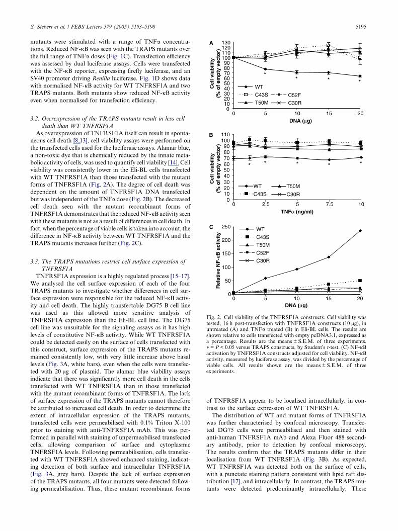
3.3. The TRAPS mutations restrict cell surface expression of
TNFRSF1A
TNFRSF1A expression is a highly regulated process [15–17].
We analysed the cell surface expression of each of the four
TRAPS mutants to investigate whether differences in cell sur-
face expression were responsible for the reduced NF-jB activ-
ity and cell death. The highly transfectable DG75 B-cell line
was used as this allowed more sensitive analysis of
TNFRSF1A expression than the Eli-BL cell line. The DG75
cell line was unsuitable for the signaling assays as it has high
levels of constitutive NF-jB activity. While WT TNFRSF1A
could be detected easily on the surface of cells transfected with
this construct, surface expression of the TRAPS mutants re-
mained consistently low, with very little increase above basal
levels (Fig. 3A, white bars), even when the cells were transfec-
ted with 20 lg of plasmid. The alamar blue viability assays
indicate that there was significantly more cell death in the cells
transfected with WT TNFRSF1A than in those transfected
with the mutant recombinant forms of TNFRSF1A. The lack
of surface expression of the TRAPS mutants cannot therefore
be attributed to increased cell death. In order to determine the
extent of intracellular expression of the TRAPS mutants,
transfected cells were permeabilised with 0.1% Triton X-100
prior to staining with anti-TNFRSF1A mAb. This was per-
formed in parallel with staining of unpermeabilised transfected
cells, allowing comparison of surface and cytoplasmic
TNFRSF1A levels. Following permeabilisation, cells transfec-
ted with WT TNFRSF1A showed enhanced staining, indicat-
ing detection of both surface and intracellular TNFRSF1A
(Fig. 3A, grey bars). Despite the lack of surface expression
of the TRAPS mutants, all four mutants were detected follow-
ing permeabilisation. Thus, these mutant recombinant forms
of TNFRSF1A appear to be localised intracellularly, in con-
trast to the surface expression of WT TNFRSF1A.
The distribution of WT and mutant forms of TNFRSF1A
was further characterised by confocal microscopy. Transfec-
ted DG75 cells were permeabilised and then stained with
anti-human TNFRSF1A mAb and Alexa Fluor 488 second-
ary antibody, prior to detection by confocal microscopy.
The results confirm that the TRAPS mutants differ in their
localisation from WT TNFRSF1A (Fig. 3B). As expected,
WT TNFRSF1A was detected both on the surface of cells,
with a punctate staining pattern consistent with lipid raft dis-
tribution [17], and intracellularly. In contrast, the TRAPS mu-
tants were detected predominantly intracellularly. These
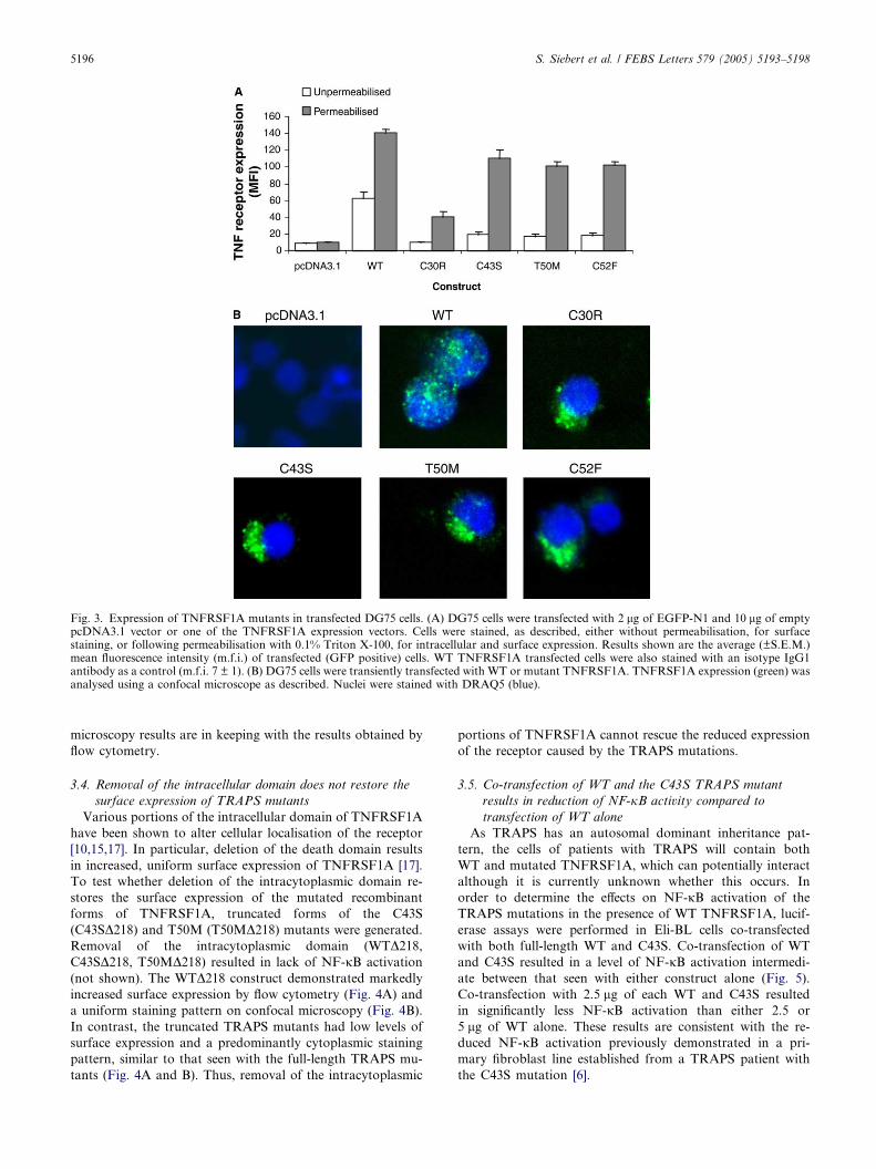
Fig. 3. Expression of TNFRSF1A mutants in transfected DG75 cells. (A) DG75 cells were transfected with 2 lg of EGFP-N1 and 10 lg of emptypcDNA3.1 vector or one of the TNFRSF1A expression vectors. Cells were stained, as described, either without permeabilisation, for surfacestaining, or following permeabilisation with 0.1% Triton X-100, for intracellular and surface expression. Results shown are the average (±S.E.M.)mean fluorescence intensity (m.f.i.) of transfected (GFP positive) cells. WT TNFRSF1A transfected cells were also stained with an isotype IgG1antibody as a control (m.f.i. 7 ± 1). (B) DG75 cells were transiently transfected with WT or mutant TNFRSF1A. TNFRSF1A expression (green) wasanalysed using a confocal microscope as described. Nuclei were stained with DRAQ5 (blue).
5196 S. Siebert et al. / FEBS Letters 579 (2005) 5193–5198
microscopy results are in keeping with the results obtained by
flow cytometry.
3.4. Removal of the intracellular domain does not restore the
surface expression of TRAPS mutants
Various portions of the intracellular domain of TNFRSF1A
have been shown to alter cellular localisation of the receptor
[10,15,17]. In particular, deletion of the death domain results
in increased, uniform surface expression of TNFRSF1A [17].
To test whether deletion of the intracytoplasmic domain re-
stores the surface expression of the mutated recombinant
forms of TNFRSF1A, truncated forms of the C43S
(C43SD218) and T50M (T50MD218) mutants were generated.
Removal of the intracytoplasmic domain (WTD218,C43SD218, T50MD218) resulted in lack of NF-jB activation
(not shown). The WTD218 construct demonstrated markedly
increased surface expression by flow cytometry (Fig. 4A) and
a uniform staining pattern on confocal microscopy (Fig. 4B).
In contrast, the truncated TRAPS mutants had low levels of
surface expression and a predominantly cytoplasmic staining
pattern, similar to that seen with the full-length TRAPS mu-
tants (Fig. 4A and B). Thus, removal of the intracytoplasmic
portions of TNFRSF1A cannot rescue the reduced expression
of the receptor caused by the TRAPS mutations.
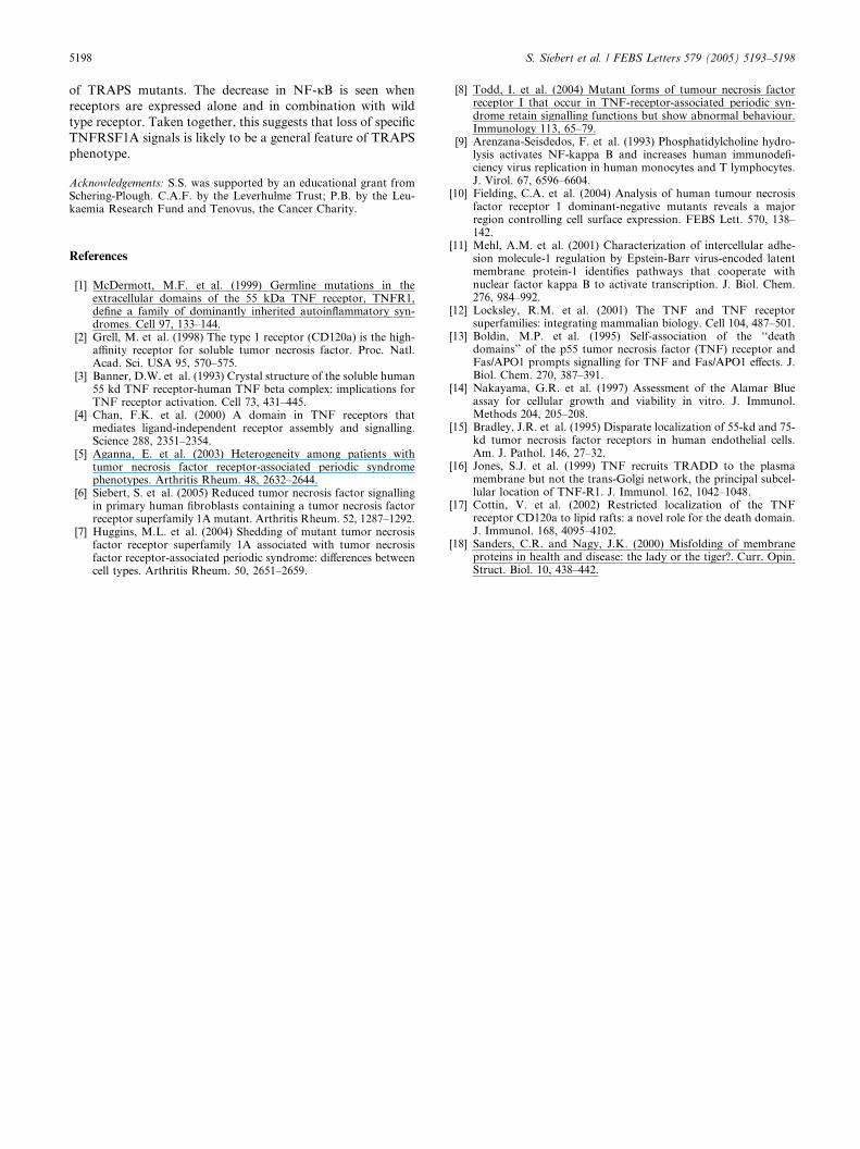
3.5. Co-transfection of WT and the C43S TRAPS mutant
results in reduction of NF-jB activity compared to
transfection of WT alone
As TRAPS has an autosomal dominant inheritance pat-
tern, the cells of patients with TRAPS will contain both
WT and mutated TNFRSF1A, which can potentially interact
although it is currently unknown whether this occurs. In
order to determine the effects on NF-jB activation of the
TRAPS mutations in the presence of WT TNFRSF1A, lucif-
erase assays were performed in Eli-BL cells co-transfected
with both full-length WT and C43S. Co-transfection of WT
and C43S resulted in a level of NF-jB activation intermedi-
ate between that seen with either construct alone (Fig. 5).
Co-transfection with 2.5 lg of each WT and C43S resulted
in significantly less NF-jB activation than either 2.5 or
5 lg of WT alone. These results are consistent with the re-
duced NF-jB activation previously demonstrated in a pri-
mary fibroblast line established from a TRAPS patient with
the C43S mutation [6].
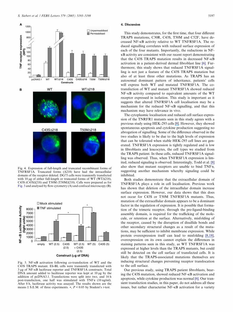
Fig. 4. Expression of full-length and truncated recombinant forms ofTNFRSF1A. Truncated forms (D218) have had the intracellulardomain of the receptor deleted. DG75 cells were transiently transfectedwith 10 lg of either full-length or truncated forms of WT (WTD218),C43S (C43SD218) and T50M (T50MD218). Cells were prepared as forFig. 3 and analysed by flow cytometry (A) and confocal microscopy (B).
010002000300040005000600070008000
empty WT (2.5) C43S(2.5)
WT (2.5)+ C43S
(2.5)
WT (5) C43S (5)
Construct (µg of DNA)
Mock stimulated
TNF stimulated*
*
Fig. 5. NF-jB activation following co-transfection of WT and theC43S TRAPS mutant. Eli-BL cells were transiently transfected with3 lg of NF-jB luciferase reporter and TNFRSF1A constructs. TotalDNA amount added to luciferase reporter was kept at 10 lg by theaddition of pcDNA3.1. Transfections were split into two, and 16 hpost-transfection, one half was stimulated with TNFa (10 ng/ml).After 8 h, luciferase activity was assayed. The results shown are themeans ± S.E.M. of three experiments. *, P < 0.05 by Student�s t-test.
S. Siebert et al. / FEBS Letters 579 (2005) 5193–5198 5197
4. Discussion
This study demonstrates, for the first time, that four different
TRAPS mutations, C30R, C43S, T50M and C52F, have de-
creased NF-jB activity relative to WT TNFRSF1A. The re-
duced signalling correlates with reduced surface expression of
each of the four mutants. Importantly, the reductions in NF-
jB activity are consistent with our recent report demonstrating
that the C43S TRAPS mutation results in decreased NF-jBactivation in a patient-derived dermal fibroblast line [6]. Fur-
thermore, this study shows that reduced TNFRSF1A signal-
ling is not just a feature of the C43S TRAPS mutation but
also of at least three other mutations. As TRAPS has an
autosomal dominant pattern of inheritance, patients� cells
will express both WT and mutated TNFRSF1A. The co-
transfection of WT and mutant TNFRSF1A showed reduced
NF-jB activity compared to equivalent amounts of the WT
receptor expressed in isolation. This study is important as it
suggests that altered TNFRSF1A cell localisation may be a
mechanism for the reduced NF-jB signalling, and that this
mechanism may have relevance in vivo.
The cytoplasmic localisation and reduced cell surface expres-
sion of the TNRFR1 mutants seen in this study agrees with a
previous study using HEK-293 cells [8]. However, they showed
spontaneous apoptosis and cytokine production suggesting no
abrogation of signalling. Some of the difference observed in the
two studies is likely to be due to the high levels of expression
that can be tolerated when stable HEK-293 cell lines are gen-
erated. TNFRSF1A expression is tightly regulated and is low
in fibroblasts and leucocytes, the cell types we studied from
the TRAPS patient. In these cells, reduced TNFRSF1A signal-
ling was observed. Thus, when TNFRSF1A expression is lim-
ited, reduced signaling is observed. Interestingly, Todd et al. [8]
also show that mutant receptors are unable to bind TNFa,suggesting another mechanism whereby signaling could be
inhibited.
Both studies demonstrate that the extracellular domain of
TNFRSF1A plays a role in cell localisation. Previous work
has shown that deletion of the intracellular domain increase
surface expression. However, our data shows that this does
not occur for C43S or T50M TNFRSF1A mutants. Thus,
mutation of the extracellular domain appears to be a dominant
factor in the regulation of expression. It is possible that forma-
tion of the trimeric receptor, through the pre-ligand-binding
assembly domain, is required for the trafficking of the mole-
cule, or retention at the surface. Alternatively, misfolding of
the receptor, caused by the disruption of disulfide bonds and
other secondary structural changes as a result of the muta-
tions, may be sufficient to inhibit membrane expression. While
protein overexpression itself can lead to misfolding [8,18],
overexpression on its own cannot explain the differences in
staining patterns seen in this study, as WT TNFRSF1A was
expressed at higher levels than the TRAPS mutants, but could
still be detected on the cell surface of transfected cells. It is
likely that the TRAPS-associated mutations themselves are
inducing structural changes preventing receptor translocation
to the cell surface.
Our previous study, using TRAPS patient fibroblasts, bear-
ing the C43S mutation, showed reduced NF-jB activation and
apoptosis, while cytokine production was normal [6]. Our tran-
sient transfection studies, in this paper, do not address all these
issues, but rather characterise NF-jB activation for a variety
5198 S. Siebert et al. / FEBS Letters 579 (2005) 5193–5198
of TRAPS mutants. The decrease in NF-jB is seen when
receptors are expressed alone and in combination with wild
type receptor. Taken together, this suggests that loss of specific
TNFRSF1A signals is likely to be a general feature of TRAPS
phenotype.
Acknowledgements: S.S. was supported by an educational grant fromSchering-Plough. C.A.F. by the Leverhulme Trust; P.B. by the Leu-kaemia Research Fund and Tenovus, the Cancer Charity.
References
[1] McDermott, M.F. et al. (1999) Germline mutations in theextracellular domains of the 55 kDa TNF receptor, TNFR1,define a family of dominantly inherited autoinflammatory syn-dromes. Cell 97, 133–144.
[2] Grell, M. et al. (1998) The type 1 receptor (CD120a) is the high-affinity receptor for soluble tumor necrosis factor. Proc. Natl.Acad. Sci. USA 95, 570–575.
[3] Banner, D.W. et al. (1993) Crystal structure of the soluble human55 kd TNF receptor-human TNF beta complex: implications forTNF receptor activation. Cell 73, 431–445.
[4] Chan, F.K. et al. (2000) A domain in TNF receptors thatmediates ligand-independent receptor assembly and signalling.Science 288, 2351–2354.
[5] Aganna, E. et al. (2003) Heterogeneity among patients withtumor necrosis factor receptor-associated periodic syndromephenotypes. Arthritis Rheum. 48, 2632–2644.
[6] Siebert, S. et al. (2005) Reduced tumor necrosis factor signallingin primary human fibroblasts containing a tumor necrosis factorreceptor superfamily 1A mutant. Arthritis Rheum. 52, 1287–1292.
[7] Huggins, M.L. et al. (2004) Shedding of mutant tumor necrosisfactor receptor superfamily 1A associated with tumor necrosisfactor receptor-associated periodic syndrome: differences betweencell types. Arthritis Rheum. 50, 2651–2659.
[8] Todd, I. et al. (2004) Mutant forms of tumour necrosis factorreceptor I that occur in TNF-receptor-associated periodic syn-drome retain signalling functions but show abnormal behaviour.Immunology 113, 65–79.
[9] Arenzana-Seisdedos, F. et al. (1993) Phosphatidylcholine hydro-lysis activates NF-kappa B and increases human immunodefi-ciency virus replication in human monocytes and T lymphocytes.J. Virol. 67, 6596–6604.
[10] Fielding, C.A. et al. (2004) Analysis of human tumour necrosisfactor receptor 1 dominant-negative mutants reveals a majorregion controlling cell surface expression. FEBS Lett. 570, 138–142.
[11] Mehl, A.M. et al. (2001) Characterization of intercellular adhe-sion molecule-1 regulation by Epstein-Barr virus-encoded latentmembrane protein-1 identifies pathways that cooperate withnuclear factor kappa B to activate transcription. J. Biol. Chem.276, 984–992.
[12] Locksley, R.M. et al. (2001) The TNF and TNF receptorsuperfamilies: integrating mammalian biology. Cell 104, 487–501.
[13] Boldin, M.P. et al. (1995) Self-association of the ‘‘deathdomains’’ of the p55 tumor necrosis factor (TNF) receptor andFas/APO1 prompts signalling for TNF and Fas/APO1 effects. J.Biol. Chem. 270, 387–391.
[14] Nakayama, G.R. et al. (1997) Assessment of the Alamar Blueassay for cellular growth and viability in vitro. J. Immunol.Methods 204, 205–208.
[15] Bradley, J.R. et al. (1995) Disparate localization of 55-kd and 75-kd tumor necrosis factor receptors in human endothelial cells.Am. J. Pathol. 146, 27–32.
[16] Jones, S.J. et al. (1999) TNF recruits TRADD to the plasmamembrane but not the trans-Golgi network, the principal subcel-lular location of TNF-R1. J. Immunol. 162, 1042–1048.
[17] Cottin, V. et al. (2002) Restricted localization of the TNFreceptor CD120a to lipid rafts: a novel role for the death domain.J. Immunol. 168, 4095–4102.
[18] Sanders, C.R. and Nagy, J.K. (2000) Misfolding of membraneproteins in health and disease: the lady or the tiger?. Curr. Opin.Struct. Biol. 10, 438–442.


