Embed Size (px)
Citation preview
© The Author (2009) New Phytologist (2009) 182: 555–557 555Journal compilation © New Phytologist (2009) www.newphytologist.org 555
Forum
Blackwell Publishing LtdOxford, UKNPHNew Phytologist0028-646X1469-8137© The Authors (2009). Journal compilation © New Phytologist (2009)281710.1111/j.1469-8137.2009.02817.xMarch 200900555???557???CommentaryCommentary Commentary
Commentary
Plants circling in outer space
Oscillatory movements are ubiquitous in plants
Plants do not grow in a strict linear manner, rather theycircumnutate. That is, they exhibit an oscillatory or helicalgrowth pattern around an axis. Circumnutation is readilyapparent in vines such as morning glory or grape (Fig. 1), butin fact it is nearly ubiquitous in plants. Circumnutation occursin almost all plant organs throughout all stages of development(Johnsson, 1997; Larson, 2000). In the late 19th century,plant scientists noted that plant organs, including roots, shoots,stems, hypocotyls, branches, leaves and flower stalks, did notgrow exactly in a linear direction. The mean growth directionmay be maintained for long periods of time, but the organ’sinstantaneous growth direction usually rotates or oscillatesslowly around a mean. Circumnutation is best visualized usingtime-lapse photography (Fig. 2), and numerous examples,including sunflower seedlings, Arabidopsis stems and morningglory stems, are illustrated in movies found at the Plants-in-Motion website (http://plantsinmotion.bio.indiana.edu/). Thepaper by Johnsson et al. (pp. 621–629) in this issue of NewPhytologist uses microgravity as a tool to study this interestingphenomenon in plants.
‘... this paper suggests not only that endogenous nutations
occur in stems as Darwin predicted, but also that grav-
itational accelerations amplify these circumnutations.’
Plant movement was the subject of two of Charles Darwin’sbooks, The movements and habits of climbing plants (Darwin,1875) and The power of movement in plants (Darwin & Darwin,1880). Darwin hypothesized that circumnutation is the majordevelopmental phenomenon in plants and that understandingthis oscillatory movement was the key to understanding allaspects of plant movements, including gravitropism, photot-ropism, thigmotropism and nastic movements. He viewed allof these phenomena as an outgrowth of circumnutation.
In order to verify his observations, Charles Darwin per-formed his studies on many types of plants. In his book, helists the 320 plant species he studied and concludes ‘everygrowing part of every plant is continually circumnutating,
though often on a small scale’ (Darwin & Darwin, 1880).Darwin hypothesized that circumnutation was an endogenousmechanism found in all plants that they employ in order toexplore their immediate environment.
Is circumnutation dependent upon gravity?
Supporting Darwin’s endogenous hypothesis, Shabala (2006)summarized other possible functions of circumnutation,including synchronizing events between cells at differentsites, functioning as a filter that helps separate signal fromenvironmental noise and decreasing the response time whenreacting to external stimuli. However, an alternate hypothesis isthat circumnutation is dependent upon gravity and thereforeis not a strictly endogenous feature of plants (Brown, 1993).
Several modern experiments support Darwin’s ‘endogenoushypothesis’. For example, in a spaceflight experiment, 93% ofsunflower (Helianthus annuus) seedlings exhibited circumnuta-tion in microgravity compared with 100% of control seedlingson the ground (Brown et al., 1990). The circumnutation of theseedlings in microgravity had a reduced period and amplitude
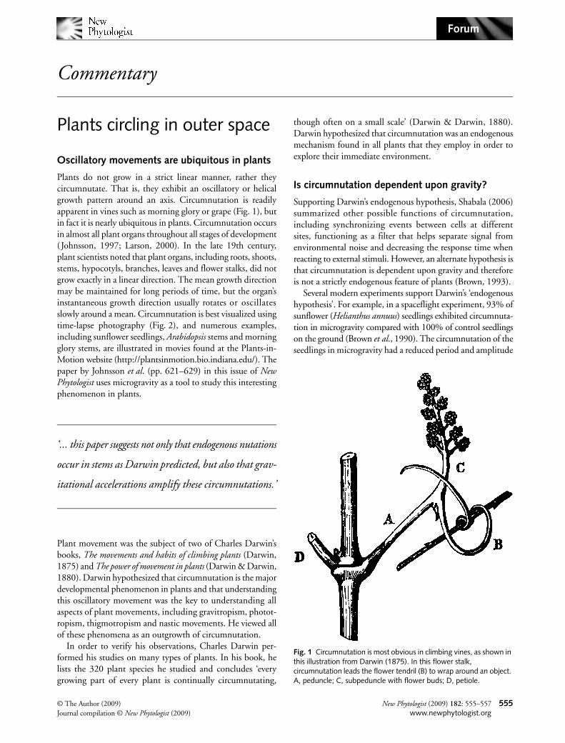
Fig. 1 Circumnutation is most obvious in climbing vines, as shown in this illustration from Darwin (1875). In this flower stalk, circumnutation leads the flower tendril (B) to wrap around an object. A, peduncle; C, subpeduncle with flower buds; D, petiole.
CommentaryForum556
relative to the ground control plants. By contrast, a series ofpapers by the Takahashi group in Japan suggested that gravitywas required for circumnutation to occur. In their first report,they suggested a link between circumnutation and gravitybased on their finding that an agravitropic mutant of morningglory (Pharbitis nil ) also was defective in circumnutation(Hatakeda et al., 2003). In a follow-up study, this group alsoshowed that mutants of P. nil and Arabidopsis thaliana lackingthe endodermal layer, which is involved in gravity sensing inshoots (Kiss, 2000), exhibited severely reduced circumnuta-tions – thus linking this phenomenon directly to mechanismsof gravity perception (Kitazawa et al., 2005).
Space experiments provide a research opportunity for fundamental biology
How can the results between the Brown group and the Takahashigroup be reconciled? Kitazawa et al. (2005) suggested that thesunflower seedlings in the experiments by Brown et al. (1990)sensed gravity before the space experiment started becausesome seedlings germinated prior to the launch of the spacecraft.Thus, an experiment in which all seeds were germinated inspace (and seedlings developed completely in microgravity)would help to resolve these controversies. The paper byJohnsson et al. uses this very approach with Arabidopsis plants.
In these elegant spaceflight studies (Johnsson et al.), alaboratory incubator facility with a centrifuge, termed theEuropean Modular Cultivation System (EMCS), was used onthe International Space Station (Kiss et al., 2007). Centrifugesprovide important controls for spaceflight studies but havenot been available for most biological experiments performedin space to date (Perbal & Driss-Ecole, 2002). Arabidopsisplants developed from seeds in microgravity, and onceinflorescence stems were formed, the centrifuge provided 0.8 gof acceleration, which is similar to the earth nominal control.After acceleration, the centrifuge was turned off so that theplants would again experience microgravity.
Johnsson et al. detected small nutational movements (withminute amplitude) of the side stems in microgravity beforecentrifugation. However, when the gravitational accelerationwas provided to the level of 0.8 g, the amplitude of thecircumnutations increased five to ten times. Light also had aneffect on circumnutations in that the period was decreasedfrom 85 min (dark) to 60 min (light). Thus, the resultspresented in this paper suggest not only that endogenousnutations occur in stems, as Darwin predicted, but also thatgravitational accelerations amplify these circumnutations.
What is the overall significance of these results? Johnsson et al.seem to favor a model that incorporates both hypotheses – theendogenous model and the idea that circumnutation is related to,and dependent upon, gravity. This space study was able to showthat small, endogenous circumnutations do occur in microgravity,but that the gravitational accelerations provided by the centrifugeclearly increased their magnitude. Johnsson et al. and others inthe field have referred to this idea as the ‘combined model’, whichhas been outlined in Brown (1991) and in Johnsson (1997).
In summary, the unique microgravity environment was usedto test the hypothesis that circumnutations are an internal, endog-enous feature of plant organs. This is important because, in pre-vious studies, researchers could not study circumnutations in plantswithout the ‘complicating’ effects of gravity. In a similar manner,the microgravity environment obtained in orbiting spacecrafthas been used effectively to study phototropism without theinterference of gravity or gravitropism (Heathcote et al., 1995;Kiss et al., 2007). Thus, the experiments of Johnsson et al.provide a fine example of using the microgravity environmentaboard orbiting spacecraft as a unique research tool to studyimportant problems in fundamental biology (Perbal & Driss-Ecole, 2002). We look forward to further contributions fromthe science programs of the European Space Agency and theNational Aeronautics and Space Administration from thelaboratories aboard the International Space Station.
John Z. Kiss
Department of Botany, Miami University,Oxford, OH 45056, USA
(tel + 1 513 529 5428;email [email protected])
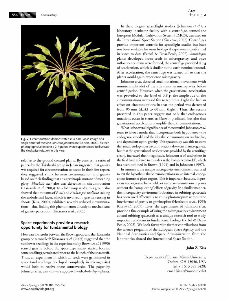
Fig. 2 Circumnutation demonstrated in a time-lapse image of a single shoot of the vine Lonicera japonicaare (Larson, 2000). Sixteen photographs taken over a 2-h period were superimposed to illustrate the clockwise rotation in this vine.
New Phytologist (2009) 182: 555–557 © The Author (2009)www.newphytologist.org Journal compilation © New Phytologist (2009)
Commentary Forum 557
References
Brown AH. 1991. Gravity perception and circumnutation in plants. Advances in Space Biology and Medicine 1: 129–153.
Brown AH. 1993. Circumnutations: from Darwin to space flights. Plant Physiology 101: 345–348.
Brown AH, Chapman DK, Lewis RF, Venditti AL. 1990. Circumnutations of sunflower hypocotyls in satellite orbit. Plant Physiology 94: 233–238.
Darwin C. 1875. The movements and habits of climbing plants. London, UK: John Murray.
Darwin C, Darwin F. 1880. The power of movement in plants. London, UK: John Murray.
Hatakeda Y, Kamada M, Goto N, Fukaki H, Tasaka M, Suge H, Takahashi H. 2003. Gravitropic response plays an important role in the nutational movements of the shoots of Pharbitis nil and Arabidopsis thaliana. Physiologia Plantarum 118: 464–473.
Heathcote DG, Brown AH, Chapman DK. 1995. The phototropic response of Triticum aestivum coleoptiles under conditions of low gravity. Plant, Cell & Environment 18: 53–60.
Johnsson A. 1997. Circumnutations: results from recent experiments on earth and in space. Planta 203: S147–S158.
Johnsson A, Solheim BGB, Iversen T-H. 2009. Gravity amplifies and microgravity decreases circumnutations in Arabidopsis stems: results from a space experiment. New Phytologist 182: 621–629.
Kiss JZ. 2000. Mechanisms of the early phases of plant gravitropism. Critical Reviews in Plant Sciences 19: 551–573.
Kiss JZ, Kumar P, Bowman RN, Steele MK, Eodice MT, Correll MJ, Edelmann RE. 2007. Biocompatibility studies in preparation for a spaceflight experiment on plant tropisms (TROPI). Advances in Space Research 39: 1154–1160.
Kitazawa D, Hatakeda Y, Kamada M, Fujii N, Miyazawa Y, Hoshino A, Iida S, Fukaki H, Morita MT, Tasaka M et al. 2005. Shoot circumnutation and winding movements require gravisensing cells. Proceedings of the National Academy of Sciences, USA 102: 18742–18747.
Larson KC. 2000. Circumnutation behavior of an exotic honeysuckle vine and its native congener: influence on clonal mobility. American Journal of Botany 87: 533–538.
Perbal G, Driss-Ecole D. 2002. Contributions of space experiments to the study of gravitropism. Journal of Plant Growth Regulation 21: 156–165.
Shabala S. 2006. Oscillations in plants. In: Baluška F, Manusco S, Volkmann D eds. Communications in plants. Berlin, Germany: Springer-Verlag, 261–275.
Key words: circumnutation, gravitropism, nutation, space biology.284710.1111/j.0021-8782.2007.02847.xMarch 200900555???557???CommentaryCommentary
CommentaryCommentary
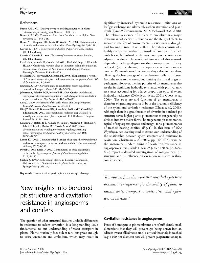
New insights into bordered pit structure and cavitation resistance in angiosperms and conifersThe question of what structural features underlie differencesin resistance to xylem cavitation is a long-standing issuefundamental to our understanding of water transport inplants. Plants routinely face xylem tensions great enoughto cause cavitation and embolism, which may result in
significantly increased hydraulic resistance, limitations onleaf gas exchange and ultimately carbon starvation and plantdeath (Tyree & Zimmermann, 2002; McDowell et al., 2008).The relative resistance of a plant to embolism is a majordeterminant of species distribution and the ability of plants tosurvive in the face of environmental stresses such as droughtand freezing (Stuart et al., 2007). The xylem consists of ahighly compartmentalized network of conduits in whichemboli can be isolated while water transport continues inadjacent conduits. The continued function of this networkdepends to a large degree on the nano-porous primarycell walls (pit membranes) that separate conduits from oneanother. Pit membranes function as safety valves in the xylem,allowing the free passage of water between cells as it movesfrom the roots to the leaves, but limiting the spread of gas orpathogens. However, the fine porosity of pit membranes alsoresults in significant hydraulic resistance, with pit hydraulicresistance accounting for a large proportion of total xylemhydraulic resistance (Zwieniecki et al., 2001; Choat et al.,2006). The structure and function of pit membranes istherefore of great importance in both the hydraulic efficiencyof the xylem and cavitation resistance (Choat et al., 2008).Although there is a great breadth of diversity in bordered pitstructure across higher plants, pit membranes can generally bedivided into two major forms: homogeneous pit membranes,typical of angiosperm species; and margo–torus pit membranesof tracheid-bearing conifers (Fig. 1). In this issue of NewPhytologist, two exciting studies extend our understanding ofthe relationship between xylem structure and resistance tocavitation: Christman et al. (2009, pp. 664–674) examinethe anatomical underpinning of cavitation resistance inangiosperm species, while Hacke & Jansen (2009, pp. 675–686) report a detailed investigation of margo–torus pitstructure and its influence on cavitation resistance in threeconifer species.
‘It is obvious from this work that rare, leaky pits have
dramatic consequences for the ability of plants to
sustain water transport as water stress and xylem
tension increases.’
Cavitation resistance in angiosperms
Pores of homogenous pit membranes are of sufficiently smalldimensions that they will prevent gas being drawn into anadjacent water-filled vessel until a critical threshold is reached(e.g. a 100-nm-diameter pore will prevent gas penetration up to
© The Authors (2009) New Phytologist (2009) 182: 557–560Journal compilation © New Phytologist (2009) www.newphytologist.org
CommentaryForum558
a pressure difference of 2.88 MPa across the pit membrane).The potential for the spread of embolism between vessels andthroughout the xylem is therefore dictated by the porosityof pit membranes and the minimum value of xylem waterpotential (negative hydrostatic pressure in the xylem fluid).Species with smaller pit-membrane pores are predicted to havegreater cavitation resistance, and thus to tolerate greater degreesof water stress, than those with larger pit-membrane pores.However, although the relationship between pit-membranepore size and cavitation resistance has sound theoreticalunderpinnings, it has been difficult to confirm this empiricallyby matching observed pore sizes to measured cavitationresistance across a range of species, with many studies failingto find pores large enough to be responsible for air seeding atrealistic pressures (Wheeler, 1983; Shane et al., 2000; Choatet al., 2003). One explanation for this discrepancy is that thepores responsible for air seeding are actually extremely rare.Because air seeding will always occur first at the largest pore,it is only required that there be one large pore present in all ofthe many thousands of pit connections between two vessels.A rare, large pore may therefore escape detection by electronmicroscopy or particle-exclusion experiments. Support forthis idea is provided by the work of Wheeler et al. (2005),which shows a strong correlation between cavitation resistanceand the average area of pit overlap between vessels. This suggeststhat cavitation resistance might be determined stochastically,with the probability of having a rare, large pore increasingwith the area of contact between vessels.
Christman et al. provide further support for this hypothesis,using an elegant pairing of theory and empirical data.Probability theory was used to model the cavitation threshold
of pit membranes in three Acer species that have differingresistances to cavitation. The model incorporates the theorythat if there is a normal distribution of pore diameters in anyconnection between vessels, only the extreme tail of thedistribution will be responsible for air seeding. In fact, themodel suggests that only one in 10 000 pits would be ‘leaky’enough to cause air seeding at measured air-seeding thresholds.To test this model, Christman et al. measured air-seedingthresholds on different stem lengths of the three Acer species.This is analogous to a membrane-filter bubble test, where thepore diameter of a filter can be predicted from the pressurerequired for gas penetration through the filter. The modelpredicts that short stem segments with fewer vessel end wallsshould air seed at lower pressures than longer stem sections inwhich air must penetrate an increasing number of intervesselend walls to move through the entire segment. The empiricaldata matched the modeled predictions of air-seeding pressuresclosely. As the stem length increased, air-seeding pressures alsoincreased, indicating that the effects of rare, large pit-membranepores was masked by the majority of end walls, which lackvery leaky pits. In the shortest stem segments, air-seedingpressures were consistently lower than the average cavitationpressures of each species. This evidence confirms that there iswide variation in the porosity of pit connections within eachstem, and strongly suggests that a very small variation in thefrequency of the rare, large pores can have a significant effecton cavitation resistance, which is independent of the numberof pits or the total pit area.
It is obvious from this work that rare, leaky pits havedramatic consequences for the ability of plants to sustain watertransport as water stress and xylem tension increases. The
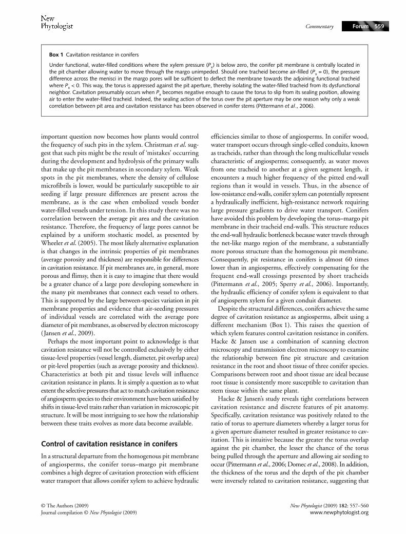
Fig. 1 Variation in pit structure. (a) A homogeneous pit membrane of an angiosperm species, Acer negundo, and (b) a margo–torus type pit membrane of a conifer, Calocedrus decurrens. Homogeneous pit membranes, typical of angiosperm species, have a relatively uniform array of microfibrils, whereas in margo–torus pit membranes of tracheid-bearing conifers, the conductive and protective functions of the membrane are spatially distinct as a porous outer region (margo) that allows for movement of water between conduits and a central thickened plug (torus).
New Phytologist (2009) 182: 557–560 © The Authors (2009)www.newphytologist.org Journal compilation © New Phytologist (2009)
Commentary Forum 559
important question now becomes how plants would controlthe frequency of such pits in the xylem. Christman et al. sug-gest that such pits might be the result of ‘mistakes’ occurringduring the development and hydrolysis of the primary wallsthat make up the pit membranes in secondary xylem. Weakspots in the pit membranes, where the density of cellulosemicrofibrils is lower, would be particularly susceptible to airseeding if large pressure differences are present across themembrane, as is the case when embolized vessels borderwater-filled vessels under tension. In this study there was nocorrelation between the average pit area and the cavitationresistance. Therefore, the frequency of large pores cannot beexplained by a uniform stochastic model, as presented byWheeler et al. (2005). The most likely alternative explanationis that changes in the intrinsic properties of pit membranes(average porosity and thickness) are responsible for differencesin cavitation resistance. If pit membranes are, in general, moreporous and flimsy, then it is easy to imagine that there wouldbe a greater chance of a large pore developing somewhere inthe many pit membranes that connect each vessel to others.This is supported by the large between-species variation in pitmembrane properties and evidence that air-seeding pressuresof individual vessels are correlated with the average porediameter of pit membranes, as observed by electron microscopy(Jansen et al., 2009).
Perhaps the most important point to acknowledge is thatcavitation resistance will not be controlled exclusively by eithertissue-level properties (vessel length, diameter, pit overlap area)or pit-level properties (such as average porosity and thickness).Characteristics at both pit and tissue levels will influencecavitation resistance in plants. It is simply a question as to whatextent the selective pressures that act to match cavitation resistanceof angiosperm species to their environment have been satisfied byshifts in tissue-level traits rather than variation in microscopic pitstructure. It will be most intriguing to see how the relationshipbetween these traits evolves as more data become available.
Control of cavitation resistance in conifers
In a structural departure from the homogenous pit membraneof angiosperms, the conifer torus–margo pit membranecombines a high degree of cavitation protection with efficientwater transport that allows conifer xylem to achieve hydraulic
efficiencies similar to those of angiosperms. In conifer wood,water transport occurs through single-celled conduits, knownas tracheids, rather than through the long multicellular vesselscharacteristic of angiosperms; consequently, as water movesfrom one tracheid to another at a given segment length, itencounters a much higher frequency of the pitted end-wallregions than it would in vessels. Thus, in the absence oflow-resistance end-walls, conifer xylem can potentially representa hydraulically inefficient, high-resistance network requiringlarge pressure gradients to drive water transport. Conifershave avoided this problem by developing the torus–margo pitmembrane in their tracheid end-walls. This structure reducesthe end-wall hydraulic bottleneck because water travels throughthe net-like margo region of the membrane, a substantiallymore porous structure than the homogenous pit membrane.Consequently, pit resistance in conifers is almost 60 timeslower than in angiosperms, effectively compensating for thefrequent end-wall crossings presented by short tracheids(Pittermann et al., 2005; Sperry et al., 2006). Importantly,the hydraulic efficiency of conifer xylem is equivalent to thatof angiosperm xylem for a given conduit diameter.
Despite the structural differences, conifers achieve the samedegree of cavitation resistance as angiosperms, albeit using adifferent mechanism (Box 1). This raises the question ofwhich xylem features control cavitation resistance in conifers.Hacke & Jansen use a combination of scanning electronmicroscopy and transmission electron microscopy to examinethe relationship between fine pit structure and cavitationresistance in the root and shoot tissue of three conifer species.Comparisons between root and shoot tissue are ideal becauseroot tissue is consistently more susceptible to cavitation thanstem tissue within the same plant.
Hacke & Jansen’s study reveals tight correlations betweencavitation resistance and discrete features of pit anatomy.Specifically, cavitation resistance was positively related to theratio of torus to aperture diameters whereby a larger torus fora given aperture diameter resulted in greater resistance to cav-itation. This is intuitive because the greater the torus overlapagainst the pit chamber, the lesser the chance of the torusbeing pulled through the aperture and allowing air seeding tooccur (Pittermann et al., 2006; Domec et al., 2008). In addition,the thickness of the torus and the depth of the pit chamberwere inversely related to cavitation resistance, suggesting that
Box 1 Cavitation resistance in conifers
Under functional, water-filled conditions where the xylem pressure (Px) is below zero, the conifer pit membrane is centrally located inthe pit chamber allowing water to move through the margo unimpeded. Should one tracheid become air-filled (Px = 0), the pressuredifference across the menisci in the margo pores will be sufficient to deflect the membrane towards the adjoining functional tracheidwhere Px < 0. This way, the torus is appressed against the pit aperture, thereby isolating the water-filled tracheid from its dysfunctionalneighbor. Cavitation presumably occurs when Px becomes negative enough to cause the torus to slip from its sealing position, allowingair to enter the water-filled tracheid. Indeed, the sealing action of the torus over the pit aperture may be one reason why only a weakcorrelation between pit area and cavitation resistance has been observed in conifer stems (Pittermann et al., 2006).
© The Authors (2009) New Phytologist (2009) 182: 557–560Journal compilation © New Phytologist (2009) www.newphytologist.org
CommentaryForum560
a combination of a thinner torus and a shallow pit chambermay form a tighter seal over the pit aperture. The authorssuggest that deeper pit chambers may require the margo tostretch further to seal the aperture, thereby predisposing thefibrils to irreparable damage by tearing.
Hacke & Jansen’s study arrives on the heels of recent workthat underscores the functional significance of conifer pitmembranes on tree height. The pit aperture may represent asignificant proportion of transport resistance in their xylem,so if apertures shrink to improve cavitation resistance, theresulting decrease in aperture conductance thus represents aclear trade-off in hydraulic efficiency at the pit level. Indeed,a linear relationship between the torus : aperture ratio andcavitation resistance has been observed with increasing height invery tall Douglas-fir trees: at greater branch heights, cavitationresistance increases to compensate for increasing xylemtensions but at the cost of reduced transport efficiencythrough the pit aperture (Domec et al., 2008). Given thelinear relationship between the torus : aperture diameter andheight, this compromise in pit structure places an importantconstraint on the maximum height that these trees can reach.Whether any clear relationship exists between pit architectureand tree height in tall angiosperms remains to be seen.
Future directions for research
While great strides have recently been made in our understandingof structure–function relationships in the xylem, importantgaps still remain. For example, the spatially complex structure ofthe angiosperm vessel network has not often been incorporatedinto measurements of xylem function. The three-dimensionalarrangement and connectivity of vessels has enormous potentialto influence the efficiency and the propagation of embolismthrough the xylem. As imaging technology, such as X-raycomputed tomography and magnetic resonance imaging, isrefined, our ability to resolve flow and propagation of embolism,in three dimensions and in real time, will be greatly improved.We can now measure hydraulic function directly at the pit level,so given the great variation in the structure of interconduitpits and their importance to hydraulic function, further directmeasurements are warranted. In conifers, additional evaluationof the margo structure, and its implications for hydraulictrade-offs, should be considered. Because of its delicate naturethe margo is often difficult to visualize using scanningelectron microscopy. A combination of careful observation ofmargo structure and improved capability to simulate flowthrough complex structures should allow an improvedresolution of the role that variation in margo structure playsin trade-offs at the tissue and whole-plant levels.
Brendan Choat1* and Jarmila Pittermann2
1Functional Ecology Group, Research School of BiologicalSciences, The Australian National University, Canberra,
ACT, 2601, Australia; 2Department of Ecology andEvolutionary Biology, University of California, Santa Cruz,
California, 95064, USA (*Author for correspondence:tel +61 2 6125 4558; email [email protected])
References
Choat B, Ball M, Luly J, Holtum J. 2003. Pit membrane porosity and water stress-induced cavitation in four co-existing dry rainforest tree species. Plant Physiology 131: 41–48.
Choat B, Brodie TW, Cobb AR, Zwieniecki MA, Holbrook NM. 2006. Direct measurements of intervessel pit membrane hydraulic resistance in two angiosperm tree species. American Journal of Botany 93: 993–1000.
Choat B, Cobb AR, Jansen S. 2008. Structure and function of bordered pits: new discoveries and impacts on whole-plant hydraulic function. New Phytologist 177: 608–625.
Christman MA, Sperry JS, Alder FR. 2009. Testing the ‘rare pit’ hypothesis for xylem cavitation resistance in three species of acer. New Phytologist 182: 664–674.
Domec JC, Lachenbruch B, Meinzer FC, Woodruff DR, Warren JM, McCulloh KA. 2008. Maximum height in a conifer is associated with conflicting requirements for xylem design. Proceedings of the National Academy of Sciences, USA 105: 12069–12074.
Hacke UG, Jansen S. 2009. Embolism resistance of three boreal conifer species varies with pit structure. New Phytologist 182: 675–686.
Jansen S, Choat B, Pletsers A. 2009. Morphological variation of intervessel pit membranes and implications to xylem function in angiosperms. American Journal of Botany 96: 409–419.
McDowell N, Pockman WT, Allen CD, Breshears DD, Cobb N, Kolb T, Plaut J, Sperry J, West A, Williams DG et al. 2008. Mechanisms of plant survival and mortality during drought: why do some plants survive while others succumb to drought? New Phytologist 178: 719–739.
Pittermann J, Sperry JS, Hacke UG, Wheeler JK, Sikkema EH. 2005. Torus–margo pits help conifers compete with angiosperms. Science 310: 1924–1924.
Pittermann J, Sperry JS, Hacke UG, Wheeler JK, Sikkema EH. 2006. Inter-tracheid pitting and the hydraulic efficiency of conifer wood: the role of tracheid allometry and cavitation protection. American Journal of Botany 93: 1265–1273.
Shane MW, McCully ME, Canny MJ. 2000. Architecture of branch-root junctions in maize: Structure of the connecting xylem and the porosity of pit membranes. Annals of Botany 85: 613–624.
Sperry JS, Hacke UG, Pittermann J. 2006. Size and function in conifer tracheids and angiosperm vessels. American Journal of Botany 93(10): 1490–1500.
Stuart SA, Choat B, Martin KC, Holbrook NM, Ball MC. 2007. The role of freezing in setting the latitudinal limits of mangrove forests. New Phytologist 173: 576–583.
Tyree MT, Zimmermann MH. 2002. Xylem structure and the ascent of sap. New York, USA: Springer-Verlag.
Wheeler EA. 1983. Intervascular pit membranes in Ulmus and Celtis native to the United States. International Association of Wood Anatomists Bulletin 4: 79–88.
Wheeler JK, Sperry JS, Hacke UG, Hoang N. 2005. Inter-vessel pitting and cavitation in woody rosaceae and other vesselled plants: a basis for a safety versus efficiency trade-off in xylem transport. Plant, Cell and Environment 28: 800–812.
Zwieniecki MA, Melcher PJ, Holbrook NM. 2001. Hydrogel control of xylem hydraulic resistance in plants. Science 291: 1059–1062.
Key words: air seeding, bordered pit, cavitation, embolism, torus–margo pit membranes, xylem.280510.1111/j.1469-8137.2009.02805.xFebruary 200900555???557???LettersLetters
New Phytologist (2009) 182: 557–560 © The Authors (2009)www.newphytologist.org Journal compilation © New Phytologist (2009)
Letters Forum 561
Letters
Letters
Mycorrhizas in Upper Carboniferous Radiculites-type cordaitalean rootletsMycorrhizas are mutualistic associations between plants andfungi; > 90% of embryophytes are capable of formingsymbioses of this type. The fungus uses the host as a source ofcarbon, while the host is supplied with mineral elements bythe fungus. Endomycorrhizal fungi associated with prostateaxes of Aglaophyton major (paramycorrhizas sensu Strullu-Derrien & Strullu, 2007) from the Lower Devonian Rhyniechert represent the oldest occurrence of mycorrhizas (Remyet al., 1994; Taylor et al., 2005). The fungi involved in thisand other mycorrhizal associations from the Rhynie chertbelong to the Glomeromycota, a fungal phylum establishedby Schüßler et al. (2001) using molecular data. Evidence fromthese plant-bearing deposits indicates that all main spore typesin the Glomeromycota were in existence before the evolutionof true roots (Dotzler et al., 2008).
Extensive collections of thin sections of petrified plantmaterial were manufactured during the early twentieth century.These collections are an invaluable source of informationabout associations between plants and microorganisms(Krings et al., 2007). Our study focuses on fungal associationsin permineralized Radiculites rootlets of the Radiculites-type(assigned to cordaitalean reticulatus) from the flora fromGrand’Croix (France) based on the original slides containedin the Lignier, Florin and Carpentier collections. The thinsections were prepared in the early twentieth century frommaterial collected from the Late Pennsylvanian (UpperCarboniferous) ‘Poudingue Mosaïque’ of Grand’Croix, whichbelongs to the Saint Etienne coal basin (Massif Central, centralFrance). This basin is situated c. 50 km southwest of thetown of Lyon. Information about the geological setting ofGrand’Croix can be found in Doubinger et al. (1995). Thinsections were prepared according to standard techniques. Apiece of silicified rock was cemented to a glass slide and thenground to a thickness sufficiently thin to allow for examinationin transmitted light. Slides were studied using dry or oilimmersion objectives. The Lignier slide collection is todayhoused in the Herbarium (C.N.) of the University of Caen(France), the Carpentier slides are kept in the collections ofLille Catholic University (France) and the Florin collec-tion is housed in the Natural History Museum of Stockholm(Sweden).
Characterization of the rootlets
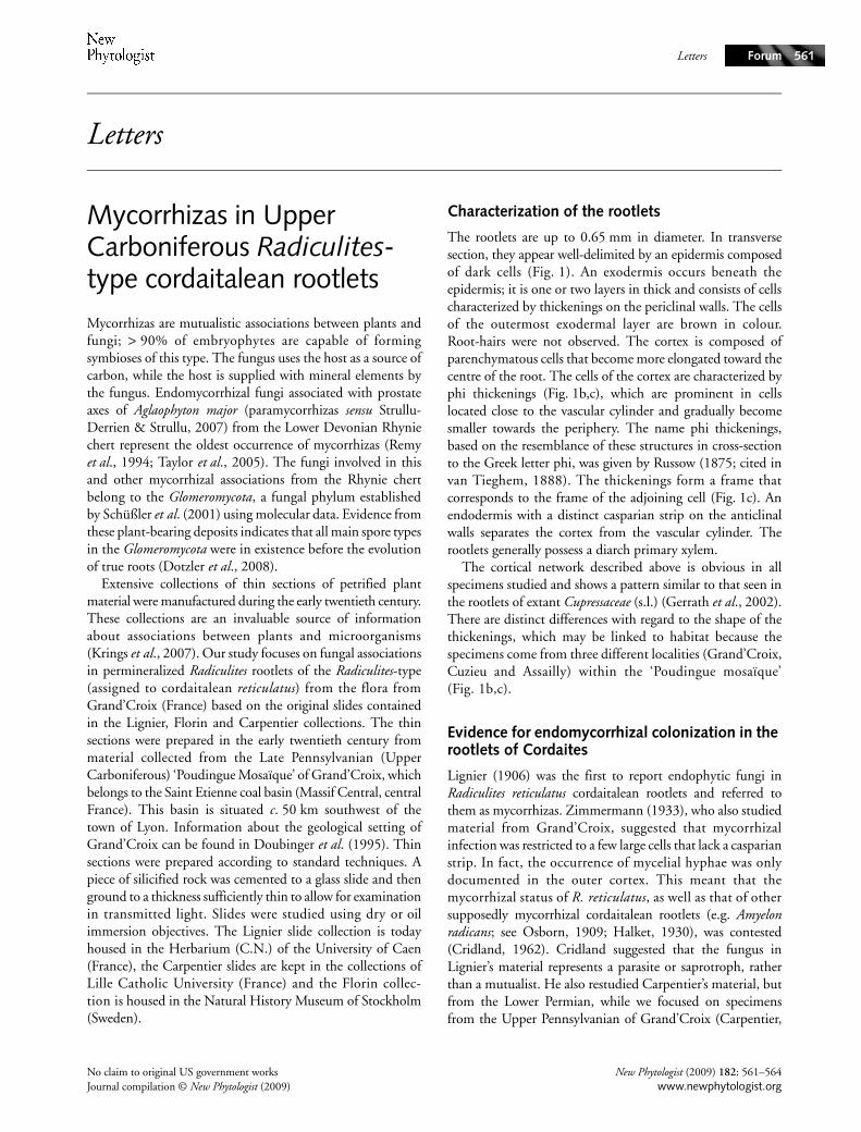
The rootlets are up to 0.65 mm in diameter. In transversesection, they appear well-delimited by an epidermis composedof dark cells (Fig. 1). An exodermis occurs beneath theepidermis; it is one or two layers in thick and consists of cellscharacterized by thickenings on the periclinal walls. The cellsof the outermost exodermal layer are brown in colour.Root-hairs were not observed. The cortex is composed ofparenchymatous cells that become more elongated toward thecentre of the root. The cells of the cortex are characterized byphi thickenings (Fig. 1b,c), which are prominent in cellslocated close to the vascular cylinder and gradually becomesmaller towards the periphery. The name phi thickenings,based on the resemblance of these structures in cross-sectionto the Greek letter phi, was given by Russow (1875; cited invan Tieghem, 1888). The thickenings form a frame thatcorresponds to the frame of the adjoining cell (Fig. 1c). Anendodermis with a distinct casparian strip on the anticlinalwalls separates the cortex from the vascular cylinder. Therootlets generally possess a diarch primary xylem.
The cortical network described above is obvious in allspecimens studied and shows a pattern similar to that seen inthe rootlets of extant Cupressaceae (s.l.) (Gerrath et al., 2002).There are distinct differences with regard to the shape of thethickenings, which may be linked to habitat because thespecimens come from three different localities (Grand’Croix,Cuzieu and Assailly) within the ‘Poudingue mosaïque’(Fig. 1b,c).
Evidence for endomycorrhizal colonization in the rootlets of Cordaites
Lignier (1906) was the first to report endophytic fungi inRadiculites reticulatus cordaitalean rootlets and referred tothem as mycorrhizas. Zimmermann (1933), who also studiedmaterial from Grand’Croix, suggested that mycorrhizalinfection was restricted to a few large cells that lack a casparianstrip. In fact, the occurrence of mycelial hyphae was onlydocumented in the outer cortex. This meant that themycorrhizal status of R. reticulatus, as well as that of othersupposedly mycorrhizal cordaitalean rootlets (e.g. Amyelonradicans; see Osborn, 1909; Halket, 1930), was contested(Cridland, 1962). Cridland suggested that the fungus inLignier’s material represents a parasite or saprotroph, ratherthan a mutualist. He also restudied Carpentier’s material, butfrom the Lower Permian, while we focused on specimensfrom the Upper Pennsylvanian of Grand’Croix (Carpentier,
No claim to original US government works New Phytologist (2009) 182: 561–564Journal compilation © New Phytologist (2009) www.newphytologist.org
LettersForum562
1932). However, the cortical network that characterizes theRadiculites-type rootlets has not been reported for Amyelon-type rootlets (Cridland, 1964). As a result, these two types ofrootlets appear to belong to different genera of Cordaites. Thecolonization of Amyelon radicans by endophytic fungi has notyet been reinvestigated.
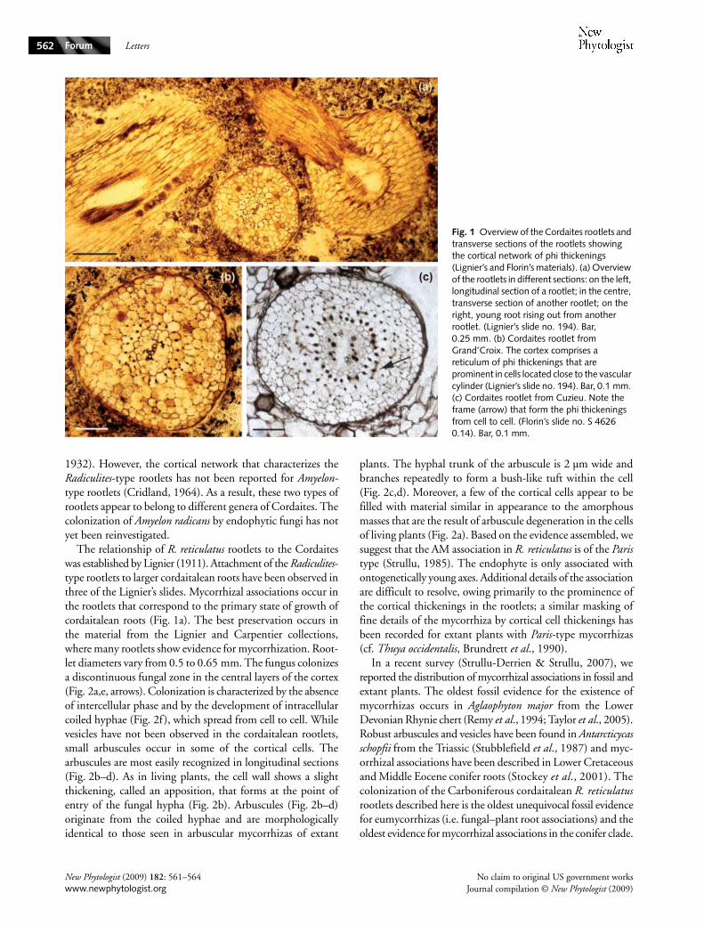
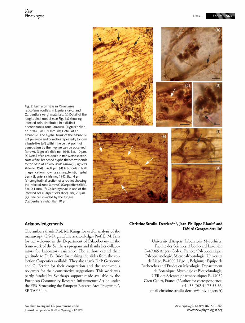
The relationship of R. reticulatus rootlets to the Cordaiteswas established by Lignier (1911). Attachment of the Radiculites-type rootlets to larger cordaitalean roots have been observed inthree of the Lignier’s slides. Mycorrhizal associations occur inthe rootlets that correspond to the primary state of growth ofcordaitalean roots (Fig. 1a). The best preservation occurs inthe material from the Lignier and Carpentier collections,where many rootlets show evidence for mycorrhization. Root-let diameters vary from 0.5 to 0.65 mm. The fungus colonizesa discontinuous fungal zone in the central layers of the cortex(Fig. 2a,e, arrows). Colonization is characterized by the absenceof intercellular phase and by the development of intracellularcoiled hyphae (Fig. 2f), which spread from cell to cell. Whilevesicles have not been observed in the cordaitalean rootlets,small arbuscules occur in some of the cortical cells. Thearbuscules are most easily recognized in longitudinal sections(Fig. 2b–d). As in living plants, the cell wall shows a slightthickening, called an apposition, that forms at the point ofentry of the fungal hypha (Fig. 2b). Arbuscules (Fig. 2b–d)originate from the coiled hyphae and are morphologicallyidentical to those seen in arbuscular mycorrhizas of extant
plants. The hyphal trunk of the arbuscule is 2 µm wide andbranches repeatedly to form a bush-like tuft within the cell(Fig. 2c,d). Moreover, a few of the cortical cells appear to befilled with material similar in appearance to the amorphousmasses that are the result of arbuscule degeneration in the cellsof living plants (Fig. 2a). Based on the evidence assembled, wesuggest that the AM association in R. reticulatus is of the Paristype (Strullu, 1985). The endophyte is only associated withontogenetically young axes. Additional details of the associationare difficult to resolve, owing primarily to the prominence ofthe cortical thickenings in the rootlets; a similar masking offine details of the mycorrhiza by cortical cell thickenings hasbeen recorded for extant plants with Paris-type mycorrhizas(cf. Thuya occidentalis, Brundrett et al., 1990).
In a recent survey (Strullu-Derrien & Strullu, 2007), wereported the distribution of mycorrhizal associations in fossil andextant plants. The oldest fossil evidence for the existence ofmycorrhizas occurs in Aglaophyton major from the LowerDevonian Rhynie chert (Remy et al., 1994; Taylor et al., 2005).Robust arbuscules and vesicles have been found in Antarcticycasschopfii from the Triassic (Stubblefield et al., 1987) and myc-orrhizal associations have been described in Lower Cretaceousand Middle Eocene conifer roots (Stockey et al., 2001). Thecolonization of the Carboniferous cordaitalean R. reticulatusrootlets described here is the oldest unequivocal fossil evidencefor eumycorrhizas (i.e. fungal–plant root associations) and theoldest evidence for mycorrhizal associations in the conifer clade.
Fig. 1 Overview of the Cordaites rootlets and transverse sections of the rootlets showing the cortical network of phi thickenings (Lignier’s and Florin’s materials). (a) Overview of the rootlets in different sections: on the left, longitudinal section of a rootlet; in the centre, transverse section of another rootlet; on the right, young root rising out from another rootlet. (Lignier’s slide no. 194). Bar, 0.25 mm. (b) Cordaites rootlet from Grand’Croix. The cortex comprises a reticulum of phi thickenings that are prominent in cells located close to the vascular cylinder (Lignier’s slide no. 194). Bar, 0.1 mm. (c) Cordaites rootlet from Cuzieu. Note the frame (arrow) that form the phi thickenings from cell to cell. (Florin’s slide no. S 4626 0.14). Bar, 0.1 mm.
New Phytologist (2009) 182: 561–564 No claim to original US government workswww.newphytologist.org Journal compilation © New Phytologist (2009)
Letters Forum 563
Acknowledgements
The authors thank Prof. M. Krings for useful analysis of themanuscript. C.S-D. gratefully acknowledges Prof. E. M. Friisfor her welcome in the Department of Palaeobotany in theframework of the Synthesys program and thanks her collabo-rators for Laboratory assistance. The authors extend theirgratitude to Dr D. Brice for making the slides from the col-lection Carpentier available. They also thank Dr P. Gerrienneand C. Ferrier for their cooperation and the anonymousreviewers for their constructive suggestions. This work waspartly funded by Synthesys support made available by theEuropean Community-Research Infrastructure Action underthe FP6 ‘Structuring the European Research Area Programme’,SE-TAF 3444.
Christine Strullu-Derrien1,2*, Jean-Philippe Rioult3 andDésiré-Georges Strullu1
1Université d’Angers, Laboratoire Mycorhizes,Faculté des Sciences, 2 boulevard Lavoisier,
F–49045 Angers Cedex, France; 2Paléobotanique,Paléopalynologie, Micropaléontologie, Université
de Liège, B–4000 Liège 1, Belgium; 3Equipe deRecherches et d’Etudes en Mycologie, Département
de Botanique, Mycologie et Biotechnologie,UFR des Sciences pharmaceutiques F–14032
Caen Cedex, France (*Author for correspondence:tel +33 (0)2 41 73 53 56;
email [email protected])
Fig. 2 Eumycorrhizas in Radiculites reticulatus rootlets in Lignier’s (a–d) and Carpentier’s (e–g) materials. (a) Detail of the longitudinal rootlet (see Fig. 1a) showing infected cells distributed in a distinct discontinuous zone (arrows). (Lignier’s slide no. 194). Bar, 0.1 mm. (b) Detail of an arbuscule. The hyphal trunk of the arbuscule is 2 µm wide and branches repeatedly to form a bush-like tuft within the cell. A point of penetration by the hyphae can be observed (arrow). (Lignier’s slide no. 194). Bar, 10 µm. (c) Detail of an arbuscule in transverse section. Note a fine-branched hypha that corresponds to the base of an arbuscule (arrow) (Lignier’s slide no. 194). Bar, 8 µm. (d) Arbuscule in high magnification showing a characteristic hyphal trunk (Lignier’s slide no. 194). Bar, 4 µm. (e) Longitudinal section of a rootlet showing the infected zone (arrows) (Carpentier’s slide). Bar, 0.1 mm. (f) Coiled hyphae in one of the infected cell (Carpentier’s slide). Bar, 20 µm. (g) One cell invaded by the fungus (Carpentier’s slide). Bar, 10 µm.
No claim to original US government works New Phytologist (2009) 182: 561–564Journal compilation © New Phytologist (2009) www.newphytologist.org
LettersForum564
References
Brundrett M, Murase G, Kendrick B. 1990. Comparative anatomy of roots and mycorrhizae of common Ontario trees. Canadian Journal of Botany 68: 551–578.
Carpentier A. 1932. Etude de végétaux à structure conservée. Silex Stéphanien de Grand’Croix (Loire). Mémoires et travaux des Facultés catholiques de Lille 40: 1–30.
Cridland AA. 1962. The fungi in cordaitean rootlets. Mycologia 54: 230–234.
Cridland AA. 1964. Amyelon in American coal-balls. Palaeontology 7: 186–209.
Dotzler N, Walker C, Krings M, Hass H, Kerp H, Taylor TN, Agerer R. 2008. Acaulosporoid glomeromycotan spores with a germination shield from the 400-million-year-old Rhynie chert. Mycological Progress 112: 1107–1114.
Doubinger J, Vetter P, Langiaux J, Galtier J, Broutin J. 1995. La flore fossile du bassin houiller de Saint Etienne. Mémoires du Muséum d’Histoire Naturelle, Paris 164: 1–351.
Gerrath JM, Covington L, Doubt J, Larson DW. 2002. Occurrence of phi thickenings is correlated with gymnosperm systematics. Canadian Jounal of Botany 80: 852–860.
Halket AC. 1930. The rootlets of Amyelon radicans, Will.; their anatomy, their apices and their endophytic fungus. Annals of Botany 44: 865–905.
Krings M, Dotzler N, Taylor TN, Galtier J. 2007. A microfungal assemblage in Lepidodendron from the Upper Visean (Carboniferous) of central France. Comptes Rendus Palevol 6–7: 431–437.
Lignier O. 1906. Radiculites reticulatus radicelle fossile de Sequoïnée. Bulletin de la Société Botanique de France 53: 193–201.
Lignier O. 1911. Les Radiculites reticulatus Lignier sont probablement des
radicelles de Cordaitales. Association Française pour l’ Avancement des Sciences 40: 509–513.
Osborn TGB. 1909. The lateral roots of Amyelon radicans Will. and their mycorrhiza. Annals of Botany 23: 603–611.
Remy W, Taylor TN, Hass H, Kerp H. 1994. 400 million year old vesicular arbuscular mycorrhizae (VAM). Proceedings of the National Academy of Sciences, USA 91: 11841–11843.
Schüßler A, Schwarzott D, Walker C. 2001. A new fungal phylum, the Glomeromycota: phylogeny and evolution. Mycological Research 105: 1413–1421.
Stockey RA, Rothwell GW, Addy HD, Currah RS. 2001. Mycorrhizal association of the extinct conifer Metasequoia milleri. Mycological Research 105: 202–205.
Strullu DG. 1985. Les Mycorhizes, handbuch der pflanzenanatomie. Berlin–Stuttgart, Germany: Gebrüder Borntraeger.
Strullu-Derrien C, Strullu DG. 2007. Mycorrhization of fossil and living plants. Comptes Rendus Palevol 6–7: 483–494.
Stubblefield SP, Taylor TN, Trappe JM 1987. Vesicular–arbuscular mycorrhizae from the Triassic of Antarctica. American Journal of Botany 74: 1904–1911.
Taylor TN, Hass H, Kerp H. 2005. Life history biology of early land plants: deciphering the gametophyte phase. Proceedings of the National Academy of Sciences, USA 102: 5892–5897.
van Tieghem P. 1888. Le réseau de soutien de l’écorce de la racine. Annales des Sciences Naturelles Botanique 7: 375–378.
Zimmermann W. 1933. Paläobotanische und phylogenetische beiträge1-4. Palaeobiologica 5: 321–348.
Key words: arbuscules, Cordaites, cortical network, mycorrhizas, phi thickenings, Radiculites rootlets.
New Phytologist (2009) 182: 561–564 No claim to original US government workswww.newphytologist.org Journal compilation © New Phytologist (2009)
About New Phytologist
• New Phytologist is owned by a non-profit-making charitable trust dedicated to the promotion of plant science, facilitating projectsfrom symposia to open access for our Tansley reviews. Complete information is available at www.newphytologist.org.
• Regular papers, Letters, Research reviews, Rapid reports and both Modelling/Theory and Methods papers are encouraged.We are committed to rapid processing, from online submission through to publication ‘as-ready’ via Early View – our averagesubmission to decision time is just 29 days. Online-only colour is free, and essential print colour costs will be met if necessary.We also provide 25 offprints as well as a PDF for each article.
• For online summaries and ToC alerts, go to the website and click on ‘Journal online’. You can take out a personal subscription tothe journal for a fraction of the institutional price. Rates start at £139 in Europe/$259 in the USA & Canada for the online edition(click on ‘Subscribe’ at the website).
• If you have any questions, do get in touch with Central Office ([email protected]; tel +44 1524 594691) or, for a localcontact in North America, the US Office ([email protected]; tel +1 865 576 5261).


