Embed Size (px)
Citation preview

Chapter 23
New Salivary Biomarkers of Human Exposure to MalariaVector Bites
Papa M. Drame, Anne Poinsignon, Alexandra Marie,Herbert Noukpo, Souleymane Doucoure,Sylvie Cornelie and Franck Remoue
Additional information is available at the end of the chapter
http://dx.doi.org/10.5772/55613
1. Introduction
Mosquitoes are the most menacing worldwide arthropod disease vectors. They transmit abroad range of viral, protozoan and metazoan pathogens responsible of the most devastatinghuman and animal diseases [1]. Among the main frequent mosquito-borne diseases, malariarepresents the most widespread and serious infection in terms of heavy burden on health andeconomic development throughout the world. Despite substantial efforts and increasinginternational funding to eliminate it, malaria is still a major public health problem with nearlya million of deaths per year, especially in children younger than 5 years old (86%) [2]. Ap‐proximately two thirds of the world's population live in areas at risk for malaria [3, 4].Understanding mechanisms that govern its transmission remains therefore a major scientificchallenge, but also an essential step in the design and the evaluation of effective controlprograms [5, 6].
Entomological, parasitological and clinical assessments are routinely used to evaluate theexposure of human populations to Anopheles vector bites and the risk of malaria transmission.However, these methods are labor intensive and difficult to sustain on large scales, especiallywhen transmission and exposure levels are low (dry season, high altitude, urban settings orafter vector control) [7, 8]. In particular, the entomological inoculation rate (EIR), the goldstandard measure for mosquito–human transmission intensity of Plasmodium, is highlydependent on the density of human-biting Anopheles [9]. This latter is estimated by usingtrapping methods such as human-landing catches (HLC) of adult mosquitoes, the commonlyused for sampling host-seeking mosquitoes and then for assessing the human exposure level.
© 2013 Drame et al.; licensee InTech. This is an open access article distributed under the terms of the CreativeCommons Attribution License (http://creativecommons.org/licenses/by/3.0), which permits unrestricted use,distribution, and reproduction in any medium, provided the original work is properly cited.

HLC may be limited because of ethical and logistical constraints to relevantly apply it tochildren [10]. Transmission estimates based on the prevalence or density of human infectionare susceptible to micro-heterogeneity caused by climatic factors and the socioeconomicdeterminants of the host-seeking behavior [8]. Incidence of disease may be the closest logicalcorrelate of the burden of disease on health systems. However, it can be subject to variabilitybetween sites and may not be appropriate for the evaluation of early phase studies of vectorcontrol or reliable for epidemic prediction [10]. More recently, serological correlates oftransmission intensity have been described, yet they represent long-term rather than short-term exposure data [8]. They are not then suitable in evaluating the short-term impact of vectorcontrol programs. Therefore, it is currently emphasized the need to develop new toolsassessing reliably human malaria risk and control interventions, and monitoring changes overtime at both population and individual levels [5, 6].
Malaria is a parasitic disease caused by protozoan agents of the genus Plasmodium (Aplicom‐plexa; Haemosporida). Five Plasmodium species are pathogen for humans: P. falciparum, P. vivax,P. ovale, P. malariae and P. knowlesi. During their complex life cycle in the female Anophelesmosquito (Insecta; Diptera), Plasmodium parasites go through several developmental transi‐tions, traverse the midgut and reach the salivary gland (SG) epithelium. They acquire theirmaturity within SGs of the vector and can be then transmitted by the bite of the femalemosquito. This latter needs, during the first days after emergence, to feed on sugar to meet theenergy demands of basic metabolism and flight, but also to feed on vertebrate blood for itseggs’ development and maturation [11], and therefore to keep perennial its life cycle andindirectly malaria transmission cycle.
Anopheles mouthparts comprise six pieces that form a long stylus allowing to perforate humantissues and to suck the internal liquid. However, it is clear that Anopheles mosquito acts notonly as syringe injecting parasites during the bite. When taking a blood meal, it also injectsinto human skin avascular tissue [12] a cocktail of bioactive molecules including enzymes thatare injected in human skin by saliva [13, 14]. Some of these salivary compounds are essentialto the Plasmodium life cycle [15]. They have substantial anti-hemostatic, anti-inflammatory, andimmunomodulatory activities that assist the mosquito in the blood-feeding process byinhibiting several defense mechanisms of the human host [16]. Furthermore, many of them areimmunogenic and elicit strong immune responses, evidenced by the swelling and itching thataccompany a mosquito bite [17]. Specific acquired cellular [18, 19] or/and humoral responsesare developed by human individuals when exposed to bites of Anopheles mosquitoes [20-23].These immune responses may play several roles in the pathogen transmission ability and thedisease outcomes [24]. In addition, recent studies have demonstrated that the intensity of theantibody response specific to salivary proteins could be a biomarker of the exposure level ofhuman to Anopheles bites [22, 25]. Therefore, studying Anopheles-human immunologicalrelationships can provide new promising tools for monitoring the real human-Anophelescontact and identifying individuals at risk of malaria transmission. It can also allow thedevelopment of novel methods for monitoring control and mosquito-release programmes’effectiveness.
Anopheles mosquitoes - New insights into malaria vectors756

However, whole saliva could be inadequate as a biomarker tool, because it is a cocktail ofvarious molecular components with different nature and biological functions. Some of theseelements are ubiquitous and may potentially cause cross-reactivities with common salivaryepitopes of other haematophagous arthropods [26]. In addition, a lack of reproducibilitybetween collected whole Anopheles saliva batches has been observed and difficulties to obtainsufficient quantities needed for large-scale studies were highlighted [26]. Therefore, specificand antigenic proteins have been identified in the secretome of Anopheles mosquitoes and aspecific biomarker of Anopheles bites was developed by coupling bioinformatic and immuno-epidemiological approaches. This promising candidate, namely, the gSG6-P1 (An. gambiaeSalivary Gland Protein-6 peptide 1), has been described to be highly antigenic [26]. It has beenthen validated as a pertinent biomarker assessing specifically and reliably the exposure levelto Anopheles bites [27-29] and/or the effectiveness of malaria vector control [30] in all age-classesof human populations (newborns, infants, children and adults) from several malaria epide‐miological settings (rural, semi-urban and urban areas…) throughout sub-Saharan Africacountries (Senegal, Angola and Benin).
The present chapter contributes therefore to a better understanding of the human-mosquitoimmunological relationship. It resumes most of the studies highlighting the roles of mosquitosaliva on the human physiology and immunology, approaches, techniques, and methods usedto develop and validate specific candidate-biomarkers of exposure to Anopheles bites and theirapplications on malaria control in several different epidemiological settings. Effects of variousexplanatory variables (age, sex, seasonality, differential use of vector control…) on humanantibody responses to Anopheles salivary antigens are also discussed in the aim to optimizetheir use in epidemiological and vector-borne disease (VBD) control studies. Finally, differentways of application of such salivary biomarker of exposure of Anopheles vector bites in the fieldof operational research by National Malaria Control Programmes (NMCP) are highlighted.
2. Human host-mosquito relationship: Roles of mosquito saliva
Arthropods represent the vast majority of described metazoan life forms throughout the world,with species’ richness estimated between 5 to 10 million [31]. The blood feeding habit has arisenand evolved independently in more than 14,000 species from 400 genera in the arthropodtaxonomy [32]. In mosquitoes, only the adult female is hematophagous, whereas both maleand female take sugar meals [33]. During the probing and the feeding stages, like all blood-sucking arthropods, female Anopheles must circumvent the highly sophisticated barriersrepresented by human defense systems (Fig. 1): haemostatic and inflammatory reactions,innate and adaptive immune system defenses. Therefore, they express in their saliva potentpharmacological and immunogenic components.
2.1. Pharmacological properties of mosquito saliva
The first-line of the human host non-specific defense to the insect bite is the haemostaticreaction. It provides an immediate response to the vascular injury caused by the intrusion of
New Salivary Biomarkers of Human Exposure to Malaria Vector Biteshttp://dx.doi.org/10.5772/55613
757

the mosquito mouthparts in host vessels, thus preventing the extensive loss of host blood [32,34]. The haemostatic reaction consists of three not physiologically distinct mechanisms: i) theblood coagulation that leads to the production of fibrin clots, ii) the thrombus formation andwound healing mediated by platelet aggregation, and iii) the vasoconstriction that leads torestricted influx of blood to the injured site. Each mechanism is activated by several pathways,in response to different exogenous and endogenous stimuli. Platelet aggregation is the firststep in the haemostatic cascade and follows the interaction between blood platelets and theexposed extracellular matrix. This latter contains a large number of adhesive macromoleculessuch as collagen which is abundant underneath endothelial cells (not found in blood). Thisinteraction results to the activation of platelets by mainly collagen and adenosine diphosphate(ADP, released by damaged cells and by activated platelets), the primary agonists of plateletaggregation. Platelets can be also activated by other agonists such as thrombin (produced bythe coagulation cascade) and thromboxane A2 (TXA2, produced by activated platelets) [35].Activated platelets release endogeneous secretions such as serotonin and TXA2, two potentvasoconstrictors. In parallel, the blood coagulation mechanism is getting underway. The maintask of the coagulation cascade is to produce fibrin that supports aggregated platelets in athrombus formation. The coagulation process consists of an enzymatic cascade with two waysof activation, the exogenous and the endogenous, where several amplification points andregulatory mechanisms are known.
Figure 1. Effects of Anopheles saliva on hemostatic, inflammatory and immune reactions of the human to the vectorbites.
Anopheles mosquitoes - New insights into malaria vectors758

However, mosquitoes can successfully engorge on their hosts within a half-minute becauseantihemostatic components of their saliva facilitate location of blood vessels and the bloodsampling [36]. These salivary secretions, named sialogenins (from the Greek sialo, saliva; gen,origin, source; and ins for proteins), are mainly an array of potent anticoagulants, anti-platelets,vasodilators and anti-inflammatory substances [16, 32, 37, 38].
2.1.1. Inhibition of platelet aggregation
Compared to other blood-sucking arthropods like ticks and sand flies, only a limited numberof Anopheles mosquito sialogenins involved in the inhibition of platelet aggregation have beencharacterized. Apyrase (Adenosine triphosphate (ATP)-diphosphohydrolase EC 3.6.1.5) isubiquitous for hematophagous arthropods (mosquitoes, bugs, sand flies, fleas, triatomines,and ticks) and hydrolyses ATP and ADP into adenosine monophosphate (AMP) and inorganicphosphate (Pi), thus inhibiting platelet aggregation [16]. Three classes of apyrase have beencharacterized at the molecular level in different blood-sucking arthropods (reviewed by [39]).One named 5′-nucleotidase family is highly expressed in the salivary gland of Anophelesgambiae [40]. The D7 protein family is one of the most abundantly expressed sialogenins ofmosquitoes. Two classes have been described in the saliva of mosquitoes: long (28–30 kDa)and short (15–20 kDa) forms [41-43]. The D7-related proteins may inhibit activation of hostplasma. It has been described in Anopheles mosquitoes in a short form and may block theplatelet activation by scavenging serotonin (agonist-positive feedback loop to increase plateletaggregation), while it principal function is reported to modulate tonus of vessels (vasocon‐striction) [44]. Anophelin from An. stephensi saliva is a 30-kDa protein that directly binds toimmobilized collagen and specifically inhibits collagen-induced platelet aggregation and theintracellular Ca2+ increase [45]. It can also act by inhibiting the activity of thrombin which playsa role in concentration of platelet aggregation [46].
2.1.2. Inhibition of blood coagulation cascade
Arthropod anticoagulants mostly target factor X-active (fXa), which plays a central role at thenexus of the intrinsic and extrinsic pathways, as well as an ultimate role of thrombin in drivingproduction of fibrin from fibrinogen. However, Anopheles mosquitoes produce an anti-thrombin [38]. In An. albimanus for example, Anophelin protein has been shown to be a potentanticoagulant that acts as a specific and tight-binding thrombin inhibitor [46], blocking ordelaying then the clot formation process until blood meal completion [34]. In addition, a D7-related protein of An. stephensi saliva has been characterized as an inhibitor of fXII [47].
2.1.3. Vasodilator effect on host blood vessels
In human, various types of endogenous vasoconstrictors (serotonin, TXA2, noradrenalin…) arereleased few seconds after tissue injury in order to stop the blood flow locally at the bite site.Diverse types of vasodilators have been characterized in the saliva of hematophagousarthropods. Aedes mosquitoes use sialokinins that mimic the endogenous tachykinin substanceP which stimulate the production of nitric oxide (NO), a potent dilator of blood vessels [48,49]. In contrast, the saliva of the adult female Anopheles mosquito has been shown to contain
New Salivary Biomarkers of Human Exposure to Malaria Vector Biteshttp://dx.doi.org/10.5772/55613
759

a myeloperoxidase with a vasodilator activity associated with a catechol oxidase/peroxidaseactivity [50]. This latter drives the H2O2-dependent destruction of noradrenalin and serotonin,two important endogenous vasoconstrictors [50]. In addition, some D7 proteins of Anopheleshave been described to bind to biogenic amines such as serotonin, histamine, and norepi‐nephrine [44]. These strategies remove the human host’s ability to maintain vascular tone atthe bite site, resulting to a weak but persistent local vasodilatation [14].
2.2. Immunological effects of mosquito saliva
The tissue injury causes an immediate onset of acute inflammation and innate immunity,which promote tissue repair, prevent colonization of the damaged tissues by opportunisticpathogens and initiates adaptive immunity, which is more specific [51]. These responsesmobilize multiple elements such as phagocytes and antigen-presenting cells, cytokine-producing cells, T and B lymphocytes (TL and BL) and complement (classical and alternativepathways). It may result to the development of strong cell and humoral immune reactions,thereby altering physiologically the environment at the bite site and leading to the rejection ofthe blood-sucker [52]. The saliva of Anopheles mosquitoes (like blood-feeding arthropods ingeneral) has selected, during evolution, compounds that can counter these host responses bymodulating immune cells and cytokines’ production [52, 53]. This certainly allows mosquitoesto complete successfully a blood meal in only few seconds. Immunomodulatory effects ofAnopheles mosquito saliva can therefore affect the transmission of pathogens and the devel‐opment of associated pathologies [54]. Understanding the mechanisms which govern thisimmunomodulation could then allow the development of new prevention tools or strategiesagainst malaria transmission [54-56].
2.2.1. Inhibition of host inflammatory reaction
The host inflammatory reaction following tissue injury consists of the triple response ofLewis: redness, heat and pain, triggering the awareness of the host to the blood sucker action[16]. If redness and heat are ones of the direct consequences of the dilatation of blood vessels,pain is induced by an increased vascular permeability under the effect of ADP, serotoninand histamine released by platelets and mast cells, following activation of the fXII by tissue-exposed collagen [16]. The fXIIa converts prekallikrein to kallikrein, which hydrolyzes bloodkininogen to produce the vasodilator peptide, bradykinin. This latter induces TNF-α (TumorNecrosis Factor alpha) release by neutrophils [57], which in turn stimulates the release of IL(interleukin)-1β and IL-6 from various cell types. These cytokines contribute to the phenom‐enon of hyperalgesia (increased sensitivity to pain) that accompanies inflammation. Hostinflammatory reaction to bites has been described as mast cells-dependent in individualsbitten by Anopheles mosquitoes [58]. In contrast to ticks which need to be attached to theirhost for several hours (tick Argasidæ) or weeks (tick Ixodidæ), mosquitoes take just few secondsfor a successful blood meal. This certainly explains the poverty of anti-inflammatorycomponents in their saliva in contrast to the ticks’ one. Nevertheless, some salivarycomponents of Anopheles mosquitoes can inhibit the human inflammatory reaction. Inparticular, a 16kDa D7 family proteins of An. stephensi (Hamadarin) inhibits the contact
Anopheles mosquitoes - New insights into malaria vectors760

system by preventing the mutual activation between the fXIIa and the kallikrein in thepresence of Zn2+ [47].
2.2.2. Modulation of host immune response
A role for arthropod saliva in modifying the outcome of transmission and infection is not anovel idea introduced in the context of mosquitoes and malaria parasites. The increasedpathogen infectivity in association with ticks, sand flies, and mosquitoes saliva has beendescribed previously [54]. If ticks that take a long time to engorge must additionallynecessitate in their saliva anti-inflammatory and immunosuppressive factors, rapidly feedingdipterans, in particular mosquitoes and sand flies, clearly have evolved salivary factors thatdirectly modulate host immune defenses [52]. One possible explanation is that these moleculeshave evolved because they have long-term beneficial effects for the populations rather thanto the individual at the time of feeding [24]. Although the molecular mechanisms by whichmosquito saliva induces alteration of the host immune response are unclear [59, 60], dataevidently demonstrate that effects depend on the global regulation of the Th1/Th2 cyto‐kines’ balance, as it has been described in sand flies/Leishmania model, the most studiedstriking host-parasite vector system [61]. The Th1 response has been described to lead to aprotective immunity and the resistance of the host to intracellular pathogens, while the Th2response might favor the survivor of pathogens (parasites, virus…) and then the diseasetransmission and evolution [24]. For mosquitoes, studies have globally shown an enhance‐ment of transmission and disease when pathogens are introduced in the presence of vectorsaliva. Mosquito saliva is commonly associated with a downregulation of the expression ofTh1 and an upregulation of the Th2-type cytokines. In mouse models, mosquito saliva canpotentiate the infection of arboviruses [24, 62, 63]. The co-inoculation of Sindbis virus withAedes aegypti salivary gland extract resulted on a reduced interferon- gamma (IFN-γ)expression, when compared to injection of virus alone [64]. It has been also shown that Ae.aegypti saliva contains multiple factors that can affect various components of the host immuneresponse [65]. For example, factor Xa inhibitor may inhibit complement activation andleukocyte migration to the bite site [24] and other factors inhibit TNF-α release from activatedmast cells [66]. Chickens subcutaneously infected with P. gallinaceum sporozoites in thepresence of Aedes fluviatillis salivary gland homogenates showed a higher level of parasitae‐mia when compared to those that received only sporozoites [67]. For Anopheles, mice exposedto mosquito feeding in tandem with the inoculation of sporozoites had higher parasitemiaand an elevated progression to cerebral malaria. This was associated with, in particular,elevated levels of IL-4 and IL-10, suppression of overall transcription in response to infection,and decreased mobility of dendritic cells and monocytes [19]. It was also described thatAnopheles stephensi saliva downregulates specific antibody (Ab) immune responses by amechanism that is mast cell and IL-10 -dependent [60]. IL-10, by inhibiting pro-inflammato‐ry and Th1 cytokines, stimulates certain T, mast and B cells and has pleiotropic effects inimmunoregulation and inflammation, while IL-4 is the prototypical Th2 cytokine (itdifferentiates CD4+ T-cells and up-regulates MHC class II production). The enhancement ofIL-10 expression could account for reduction in secretion of other cytokines because it inhibitsantigen presentation, IFN–γ expression, and macrophage activation [68]. However, some
New Salivary Biomarkers of Human Exposure to Malaria Vector Biteshttp://dx.doi.org/10.5772/55613
761

data have suggested a paradoxical protective role of mosquito saliva against pathogentransmission and disease infection. Ae. aegypti saliva can inhibit infection of dendritic cellsby dengue virus, and the pre-sensitization of dendritic cells with saliva prior to infectionenhanced this inhibition. Moreover, the proportion of dead cells was also reduced in virus-infected dendritic cell cultures exposed to mosquito saliva, and an enhanced production ofIL-12 and TNF-α was detected in these cultures [69]. In addition to these effects on cellularimmunity, Anopheles saliva can also acts on humoral host immune response. Indeed, specificantibodies (immunoglobulins [Ig] G, M and E) to salivary antigens have been described inseveral studies [20, 22, 23, 25, 56, 70]. However, the implication of these Ab responses indisease pathogenesis or protection is not yet elucidated.
Therefore, future studies are needed for an overall understanding of mosquito saliva effect,especially Anopheles mosquito saliva, in pathogen transmission, disease development andpathogenesis.
2.2.3. Human host-Anopheles vector immune relationship and applications
The study of immunological properties of salivary proteins of Anopheles mosquitoes representsa new research thematic which can significantly improve the understanding of Plasmodiumtransmission mechanisms and therefore help for the effective prevention and control ofmalaria. It can notably lead to major applications in three areas: i) development of vaccines,diagnosis, treatment, ii) prevention of allergies, and iii) development of biomarkers ofexposure to bites and malaria disease risk.
The development of parasite transmission-blocking vaccines, by stimulating the immuneresponse against the vector is an attractive alternative way for malaria control. Several studiestargeted the effect of Abs specific to the mosquito midgut antigens have shown promisingresults [71-73]. The study of the immune response induced by vector saliva at the biting siteand its potential effect on the transmission and the development of pathogens suggests thepossibility to control parasite transmission by vaccinating the host with immunogenic salivarycompounds [54, 74]. In a mouse model, it has been shown that two salivary proteins (29 and100 kDa) of the female An. gambiae can induce production of Ab which can block about 75%of the invasion of An. stephensi salivary glands by P. yoelii sporozoites [75]. In addition, theprior exposition to non infective An. stephensi bites induces a Th1 immune response withincreased production of IL-12 and IFN-γ. Its effect can subsequently limit future P. yoeliiinfection (reduced rate of liver and blood parasites) and the development of cerebral malariain mouse [18]. In this context, saliva can be thought as a non-specific “adjuvant” which couldbe effective at inducing a Th1-biased environment that is known to be protective againstmalaria infection. However, the development of such vaccines is complex. For example, Abproduced by immunization (with salivary proteins) must be ingested by the mosquito duringa bite, cross it midgut and digestive enzymes, migrate to the salivary glands, before they canblock the invasion by sporozoites. Nevertheless, the possibility to develop a pan-arthropodvaccine has been recently demonstrated by another mechanism. Indeed, an immune responsedirected to salivary proteins that adsorb to pathogens can turn the microorganism into aninnocent bystander of anti-salivary immunity as it has been recently reported in a salivary
Anopheles mosquitoes - New insights into malaria vectors762

protein (Salp15) from the hard tick Ixodes scapularis [76] and vaccine candidate for the controlof Lyme disease [77]. Unfortunately, any hematophagous arthropod saliva-based vaccine hasnot yet been tested on humans.
In the field of allergic reactions to salivary proteins of mosquitoes, the first studies were mainlyconducted in Canada and Finland. They concerned Aedes and Culex mosquitoes which expressa panel of allergens in their saliva during the blood feeding time [17, 56, 78]. These proteinscan thus be used in recombinant form, as diagnostic tool of the level of human exposure toallergens or in immunotherapy injections for desensitization of human [56, 70, 79]. It exists yetno study highlighting the presence and effect of allergens in the Anopheles mosquitoes’ saliva.
The study of immunological relationship between human-vector by quantifying specific Abresponses to salivary proteins may also allow the identification and characterization ofbiological markers for epidemiological assessment of the exposure of individuals and popu‐lations to the Anopheles bites and thus to the risk of malaria transmission [22]. The developmentof such biomarkers or indicators (see next chapter) can be a complementary alternative tocurrent referent entomological and parasitological methods which present several limitationsespecially in low exposure/transmission contexts.
3. Development of biomarkers of human exposure to Anopheles bites andindicators of malaria vector control effectiveness
3.1. Validation of concept with whole Anopheles saliva
To improve the fight against malaria and regarding numerous limitations described withcurrent entomological and parasitological tools, the World Health Organization (WHO) hasemphasized the need of new indicators and methods to evaluate, at individual and populationlevels, the exposure level to Anopheles vectors and the effectiveness of vector control strategies.One promising concept is based on the fact that mosquito saliva injected to the human hostduring the vector bite is antigenic and can induce an adaptive humoral host response (seeFigure 1). Therefore, a logical positive correlation between the human exposure level toAnopheles bites and human anti-mosquito saliva Ab level can be expected. In this way, anti-mosquito saliva Ab response can be a pertinent epidemiological biomarker of human exposureto vector bites.
The epidemiological importance of human exposure to the saliva of vectors has been firstlydescribed in Lyme disease [80, 81], leishmaniasis [82] and Chagas disease [83]. During the lastdecade, studies have provided data on human exposure to anopheline saliva and its interactionwith malaria transmission. In particular, Remoue et al. [22] have shown that children living ina seasonal malaria transmission region of Senegal developed IgG responses to An. gambiaewhole saliva (WS). Interestingly, these specific IgG levels were positively associated with anincreased rainfall and the Anopheles mosquito density, measured by referent entomologicalmethods. Indeed, an increase in the level of IgG was observed according to the Anophelesaggressiveness and density in September (Figure 2), the peak of malaria transmission.
New Salivary Biomarkers of Human Exposure to Malaria Vector Biteshttp://dx.doi.org/10.5772/55613
763

Importantly, IgG response to An. gambiae WS can predict clinical malaria cases. Indeed,children who developed a malaria attack in December had higher levels of anti-WS IgG inSeptember of the same year, i.e. three months before they develop the disease (Figure 3) [22].
Malaria morbiditySept. to Dec.
P= 0.046
n= 132 n= 89
0.0
0.1
0.2
0.3
No Yes
ΔO.D
.
Figure 3. Anti-salivary IgG according to malaria morbidity. The results of individual absorbance (OD) values in Septem‐ber are shown according to subsequent detection of clinical malaria for the age ≥1 year. Bars indicate the median val‐ue for each group. Statistical significance between groups is indicated by a non-parametric Mann–Whitney U-test).
Anti-mosquito saliva Ab appeared transitional. Soldier travelers transiently exposed to An.gambiae bites in endemic areas of Africa (especially Ivory Coast and Gabon) developed specific
Exposure level(entomology)
0.0
0.2
0.4
0.6
0.8
low highmoderate
Anti-saliva I gG
P< 0.01 P< 0.05
Figure 2. Anti-saliva IgG according to the intensity of exposure [22]. Individual absorbance (OD) values in Septemberare shown for the three groups with different levels of exposure. Bars indicate the median value for each group. Statis‐tical significances between each group by non-parametric Mann–Whitney U-test are indicated.
Anopheles mosquitoes - New insights into malaria vectors764

IgG responses to anti-An. gambiae WS which strongly decreased several weeks after the end oftheir trip [21]. In addition, anti-An. gambiae saliva IgG levels waned rapidly after 6 weeks ofInsecticide-Treated Nets (ITNs) well-use in a semi-urban population in Angola, before a newsignificant increase two months later following the stop of ITN use [84]. Data on humanexposure to anopheline saliva and its interaction with malaria were also provided by studiesfrom other none African areas. In South-eastern Asia, it has been described that anti-An.dirus salivary protein Ab occur predominantly in patients with acute P. falciparum or P. vivaxmalaria; people from non-endemic areas do not carry such Abs [23]. In the Americas, thepresence of anti-Anopheles saliva Ab has been also described. In adult volunteers from Brazil,anti-An. darlingi WS Ab levels increased with P. vivax infections [20]. The presence of anti-An.albimanus WS Ab with exposure to mosquito bite has been recently described in Haiti [25].Specific IgG response to An. gambiae WS has also been described as an immunological indicatorevaluating the efficacy of malaria vector control strategies. Indeed, Drame et al. have recentlyshown in a semi-urban area (Lobito, Provence Benguela) in Angola that specific IgG levelsdrastically decreased after the introduction of ITNs and this was associated with a drop inparasite load (Figure 4) [84].
Mar
ch-A
pril
May ju
ly
August
Sep
tem
ber
Oct
ober
Dec
ember
Januar
y
Febru
ary
Apri
l
June-
July
August
Oct
ober
0.0
0.1
0.2
0.3
0.4
0.5
1.0
1.5
2.0
2.5
3.0
O
.D.
(med
ian
valu
es) P
ara
site
s/
L o
f blo
od
(geo
metric
mean
s)
2005 2006
Peak of Ano
ITNs
Peak of Ano
IgG response
Parasitemia
Figure 4. Evolution of anti-Anopheles gambiae saliva IgG and Plasmodium falciparum infections before and after ITNimplementation, (Ano=Anopheles).
Anti-Anopheles saliva IgG response has also been recently used to evaluate and compare theeffectiveness of three malaria vector control strategies in another area (Balombo) of Angola [85].Indeed, Brosseau et al. [85] have investigated over a period of two years (2008-2009) Ab responseto An. gambiae WS in children between 2 to 9 years old, before and after the introduction of threedifferent malaria vector control methods: deltamethrin treated long lasting impregnated nets(LLIN) and insecticide treated plastic sheeting (ITPS) - Zero Fly®) (ITPS-ZF), deltamethrinimpregnated Durable (Wall) Lining (ITPS-DL - Zerovector®) alone, and indoor residual spraying
New Salivary Biomarkers of Human Exposure to Malaria Vector Biteshttp://dx.doi.org/10.5772/55613
765

(IRS) with lambdacyhalothrin alone. They observed considerable decreases in entomological(82.4%), parasitological (54.8%) and immunological criteria analyzed. In particular, theimmunological data based on the level of anti-saliva IgG Ab in children of all villages significant‐ly dropped from 2008 to 2009, especially with LLIN+ZF and with IRS (Figure 5).
2008 2009 2008 2009 2008 20090.0
0.5
1.0
1.5
2.0
2.5
3.0
Ant
i-sal
iva
IgG
resp
onse
s(
O.D
.)
LLIN + ZF DL IRS
P<0.0001 P=0.0001 P<0.0001
Figure 5. Comparison of median values of the IgG antibody response to Anopheles saliva obtained before and afterimplementation of each vector control method [85].
Taken together, these studies indicated that the estimation of human IgG Ab responses specificto Anopheles WS could provide a reliable biomarker for evaluating the Anopheles exposure level,the risk of malaria transmission, the disease outcomes and the effectiveness of vector controlstrategies. However, the pertinence and the practical large-scale application of serological testsfor epidemiological purposes have been hampered by several limitations. First, WS is a cocktailof various molecular components with different nature and biological functions. Somecomponents are Anopheles-specific and other widely distributed within genus, families, ordersor classes of bloodsucking Diptera or Arthropods [16]. Therefore, the evaluation of Anophelesexposure or vector control effectiveness based on the immunogenicity of WS could be skewedand over or underestimated by possible cross-reactivities between common epitopes betweenmosquito species or other organisms [26]. Second, the collection of saliva or salivary glandextracts is tedious and time-consuming; therefore it will be difficult or impossible to have anadequate production of mosquito saliva needed for large-scale epidemiological studies [26].
Anopheles mosquitoes - New insights into malaria vectors766

Third, saliva composition can be affected by several ecological parameters such as age, feedingstatus or infectivity of Anopheles [86],which in turn may influence the anti-saliva immuneresponse measured and may cause a lack of reproducibility between saliva batches. Analternative for optimizing the specificity of this immunological test would thus be to identifyAnopheles genus-specific proteins [87].
3.2. Methods for the identification of specific Anopheles salivary proteins
The isolation of salivary components has been a challenge for many years. Many functionalactive salivary proteins have been isolated following classical biochemical and molecularbiology approaches [88]. Protocols mainly consisted of the isolation of salivary componentsfrom hundreds of salivary gland pairs, obtaining amino-terminal or internal peptide sequenceof the purified component, screening of a salivary gland library with the information obtained,and isolation of the cDNA or gene of interest (Fig. 6).
Activity detected on saliva or salivary gland homogenate (SGH)
Purificat ion of active component
Edman degradation of N-terminal part or from internal peptide
Design degenerate primers based on obtained protein sequence
Produce PCR probe with designed primers
Screen salivary gland cDNA library
Secondary screen is usually performed to isolate cDNA of interest
Sequence cDNA of interest
Express recombinant protein either in bacteria, mammalian or insect cell-expression system
Test for biologic activity
Figure 6. Classical biochemical and molecular biology protocol used for isolation and characterisation of salivary pro‐teins and cDNA from vectors of disease [90].
New Salivary Biomarkers of Human Exposure to Malaria Vector Biteshttp://dx.doi.org/10.5772/55613
767

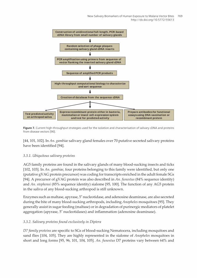
During the last decade, technical advances in molecular biology have allowed the sequencingof the genome, including transcripts of salivary glands [89], of most disease vectors, comprisingAnopheles mosquitoes [90]. However, protocols do not allow to obtain entire sequences [89].Nowadays, researchers have switched from testing one salivary molecule at a time to studyingthe whole complex of genes and secreted proteins in blood-feeding arthropods using tran‐scriptomic and/or proteomic approaches. The transcriptomic is the complete set of transcriptsin an organism for a specific developmental stage or physiological condition. Transcriptomictechniques help to interpret the functional elements of the genome, and to understand thetransmission and development of diseases [91]. They aim to catalogue transcript of majorAnopheles species, including mRNAs, non-coding RNAs and small RNAs; to determine thetranscriptional structure of genes and to quantify the changing expression levels of eachtranscript during development and under different conditions [91]. Proteomic is a large-scalestudy of the gene expression at the protein level, which ultimately provides direct measure‐ment of protein expression levels [92]. The proteomic revolution is hitting the vector biologyfield as well as many other fields. The isolation and sequencing of all the proteins from SGs ofdisease vectors and, more specifically, secreted salivary proteins, is clarifying the complexityof proteins present in the saliva of various blood-feeding arthropods [93]. During the last years,a comprehensive high-throughput approach has been developed (Figure 7) [88]. It combinesmassive sequencing protocol of high quality full-length salivary gland cDNA libraries, aproteomic approach to isolate a large set of salivary proteins, and high-throughput computa‐tional biology and functional assays to analyze and test the biologic activities of these novelmolecules. It is a powerful tool which can help easily and rapidly to identify and characterizegenes or transcripts encoding for various proteins of SGs (the sialome) of blood-suckingarthropods. This high-throughput approach has then allowed an unprecedented insight intothe complexity of salivary gland compounds of mosquito vectors of disease agents, indicatingthat the diversity of their targets is still larger than previously thought [16].
3.3. Salivary proteins (sialome) of Anopheles mosquitoes
The increasing power of large-scale genomic, transcriptomic and proteomic analyses allowedthe accumulation of a considerable amount of information on the salivary secretions of blood-sucking arthropods [86]. As far as mosquitoes are concerned, the analysis of salivary tran‐scriptomes of a number of Anopheles have allowed the discovery of a variety of genes thatmatched the sequence of various protein families, providing some clues on the evolution ofblood feeding [15, 41-43, 92, 94-100]. Many of the salivary protein sequences are coded by genesrelated to intrinsic functions of the cell (housekeeping genes). However, the large number ofsalivary proteins is secreted during plant or blood feeding. Finally, a little number has nosimilarities to sequences deposited in databases, representing unknown and novel sequences[41, 94, 101]. This emphasizes how much still need to be learned concerning the biologicalfunctions of salivary proteins in blood feeding, pathogen transmission and manipulation ofhost responses.
The analysis of the adult Anopheles sialome has shown that secreted proteins and/or peptides(secretome) can be ubiquitous or specific to arthropod classes, orders, families, genus or species
Anopheles mosquitoes - New insights into malaria vectors768
[44, 101, 102]. In An. gambiae salivary gland females over 70 putative secreted salivary proteinshave been identified [94].
3.3.1. Ubiquitous salivary proteins
AG5 family proteins are found in the salivary glands of many blood-sucking insects and ticks[102, 103]. In An. gambiae, four proteins belonging to this family were identified, but only one(putative gVAG protein precursor) was coding for transcripts enriched in the adult female SGs[94]. A precursor of gVAG protein was also described in An. funestus (84% sequence identity)and An. stephensi (85% sequence identity) sialome [95, 100]. The function of any AG5 proteinin the saliva of any blood-sucking arthropod is still unknown.
Enzymes such as maltase, apyrase, 5′ nucleotidase, and adenosine deaminase, are also secretedduring the bite of many blood-sucking arthropods, including Anopheles mosquitoes [95]. Theygenerally assist in sugar feeding (maltase) or in degradation of purinergic mediators of plateletaggregation (apyrase, 5′ nucleotidases) and inflammation (adenosine deaminase).
3.3.2. Salivary proteins found exclusively in Diptera
D7 family proteins are specific to SGs of blood-sucking Nematocera, including mosquitoes andsand flies [104, 105]. They are highly represented in the sialome of Anopheles mosquitoes inshort and long forms [95, 96, 101, 104, 105]. An. funestus D7 proteins vary between 64% and
Construction of unidirectional full- length, PCR-basedcDNA library from small number of salivary glands
Random selection of phage plaques containing salivary gland cDNA inserts
PCR amplificat ion using primers from sequence of λvector flanking the inserted salivary gland cDNA
Sequence of amplified PCR products
High-throughput computational biology to characterizeand sort sequence
Creation of database from the sequence cDNA
Test predicted activityon arthropod saliva
Prepare antibodies for functionalassaysusing DNA vaccination or
recombinant protein
Express recombinant protein either in bacteria, mammalian or insect cell-expression system
and test for predicted activity
Figure 7. Current high-throughput strategies used for the isolation and characterisation of salivary cDNA and proteinsfrom disease vectors [90].
New Salivary Biomarkers of Human Exposure to Malaria Vector Biteshttp://dx.doi.org/10.5772/55613
769

75% identity with their An. gambiae closest match [105]. D7 proteins could act as anti-hemostaticfactors by trapping agonists of hemostasis [44, 47]. However, further investigations are neededto clearly describe their function.
Other Diptera-specific protein families or peptides have also been described in the sialome ofblood-feeding mosquitoes [95]. However their function is still unknown, even if some wereknown to play a role in antimicrobial property of mosquito saliva.
3.3.3. Protein families found exclusively in mosquitoes
The 30-kDa antigen family found exclusively in the SGs of adult female mosquitoes has beenfound in both culicine and anopheline mosquitoes [95, 100, 101, 106-108]. Only one geneenriched in SGs of adult females is known in An. gambiae. The An. funestus homologue is alsoabundantly expressed and shares 63% identity with the An. gambiae orthologue. The functionof this protein family is still unknown [95].
The gSG (An. gambiae Salivary Gland)-5 family was first discovered in the SGs of An. gambiaeand shown to be exclusively expressed in the adult female [94, 109]. This protein shows a highsimilarity to Aedes and Culex proteins [101]. Transcripts coding for this family were found inthe sialotranscriptome of An. darlingi with 46% identical to the An. gambiae orthologue andonly 26% and 23% identical to the culicine proteins [101]. The function of this mosquito-specificprotein remains unknown, but its tissue- and sex-specific expression profile suggests it ispossibly related to blood feeding.
The gSG8 family is highly divergent with members only found in An. gambiae and Ae. aegypti.In An. gambiae, this protein is specifically expressed in female SGs [109], suggesting a likelyrole in blood feeding.
Various types of mucins have been described in the saliva of adult mosquitoes and mayfunction/act as a lubricant of their mouthparts [15, 41, 94, 102]. Three mucins encodingtranscripts have been identified in the An. gambiae larval SG [110], suggesting the importanceof mucins at multiple developmental stages. Mucins may also play a crucial role in Anophe‐les salivary gland invasion by P. berghei sporozoites [111]. Several protein families are alsorepresented in this group, including gSG-3, gSG-10, and 13.5-kDa families [101]. These familieswere also found abundantly expressed in the sialotranscriptome of An. gambiae adult male[112], indicating their function is not related specifically to blood feeding.
3.3.4. Protein families found exclusively in Anophelines
Anophelin was described as a short acidic peptide with strong thrombin inhibitory activity inAn. albimanus [46]. An. funestus anophelin is 59% identical to the An. gambiae orthologue [95],and An. darlingi anophelin is 86% identical to An. albimanus [101].
The 8.2-kDa family is represented in several Anopheles species. In An. funestus the peptide have42% identity with the 8.2-kDa salivary peptide of An. stephensi and similar proteins from An.gambiae and An. darlingi [95]. In An. gambiae, this peptide was found enriched in adult femaleSGs, suggesting a role in blood feeding.
Anopheles mosquitoes - New insights into malaria vectors770

The 6.2-kDa family was first described in a sialotranscriptome of An. gambiae [94], where it wasfound enriched in adult female SGs compared to other tissues. The An. funestus member of thisfamily is 61% identical to the An. gambiae [95], and 53% to an An. darlingi [101] homologues.
The SG-1 family proteins appear to be exclusively expressed in the female SGs of Anophelesmosquitoes and not observed in other tissues [94, 101]. However, their function remains to bedetermined.
The SG-2 family proteins were identified from An. gambiae saliva and shown to be expressedin female SGs and adult males but not in other tissues [113]. Related, but very divergent,sequences were obtained from salivary transcriptomes of other anopheline species [95, 101].Because this protein family is expressed in both male and female An. gambiae, and due to itsrelatively small size, it may display antimicrobial function [101].
The hyp 8.2 and hyp 6.2 proteins are similarly enriched in An. gambiae adult female SGs [94]. An.stephensi and An. funestus also have members of these protein families.
The SG-7/Anophensin family is also unique to anophelines. In An. gambiae, it is highly enrichedin female SGs [94]. More recently, the An. stephensi homologue was determined to inhibitkallikrein and production of bradykinin, a pain-producing substance [114]. Four putativealleles representing the homologue(s) of gSG7 in An. darlingi were identified. These An.darlingi transcripts have no more than 45% identity to the An. gambiae gSG7 and An. stephensianophensin [101].
The SG6 protein is a small protein first described in An. gambiae [109] and a unique sequencecodes for a mature peptide/protein of ~10 kDa (116 amino-acids) with ten cysteine residuesmaking probably five disulphide bonds. A homologue was later found in the sialotranscrip‐tome of An. stephensi [100] and An. funestus [95]. An. funestus SG6/fSG6 (f for funestus) has 81%and 76% identities with An. stephensi and An. gambiae polypeptides, respectively. It is not foundin the transcriptomes of the Culicinae subfamily members analyzed so far, i.e. C. pipiensquinquefasciatus, Ae. aegypti and Ae. albopictus [108, 115, 116]. In An. gambiae, the transcriptcoding for gSG6 (g for gambiae) was found to be 16 times more expressed in SGs of adult femalesthan in males [94]. The gSG6 protein plays some essential blood feeding role and was recruitedin the anopheline subfamily most probably after the separation of the lineage which gave originto Cellia and Anopheles subgenera [99]. The gSG6 protein, because immunogenic, can betherefore a reliable indicator of human exposure specific to Anopheles mosquito bites [99],vectors of malaria.
3.4. Specific salivary biomarker of exposure to Anopheles bites: The gSG6-P1 peptidecandidate
The SG6 salivary protein has been reported to be immunogenic in travelers exposed for shortperiods to Anopheles bites [21], and in Senegalese children living in a malaria endemic area byan immuno-proteomic, coupling 2D immunoblot and mass spectrometry [117], and by an ELISA[26] approaches. Recently, its immunogenicity has been confirmed in individuals from a malariahyperendemic area of Burkina Faso [118, 119], by using a recombinant form expressed as purifiedN-terminal His-tagged recombinant protein in the E. coli vector pET28b(+) (Novagen) [99, 119].
New Salivary Biomarkers of Human Exposure to Malaria Vector Biteshttp://dx.doi.org/10.5772/55613
771

In particular, increased anti-gSG6 IgG levels were observed in exposed individuals during themalaria transmission/rainy season [119]. In addition, anti-gSG6 IgG response appeared to be areliable serological indicator of exposure to bites of the main African malaria vectors (An. gambiae,An. arabiensis and An. funestus) in the same area [119]. However, gSG6 recombinant protein hasbeen described to relatively generate a high background in control sera from individuals notexposed to Anopheles bites, and considerable variations in specific Ab response between childrensupposed to be similarly exposed to Anopheles bites [26]. Therefore, with the objective ofoptimizing Anopheles specificity and reproducibility of the immunological assay, a peptidedesign approach was undertaken using bioinformatic tools [26].
3.4.1. Identification and sequence of gSG6-P1 peptide
Several algorithms were employed for prediction of potential immunogenic sites of the gSG6protein by using bioinformatics. The prediction of immunogenicity was based on the deter‐mination of physico-chemical properties of the amino-acid (AA) sequences with BcePred andFIMM databases and on the identification of MHC class 2 binding regions using the ProPred-2online service. This led to define five gSG6 peptides (gSG6-P1 to gSG6-P5) of 20 to 27 AA
Figure 8. Sequences of the anopheline gSG6 proteins [99]. (A) Clustal alignment of anopheline gSG6 proteins. Signalpeptides and conserved Cysteines are boxed. Conserved sites are shaded. (B) Phylogenetic tree (NJ algorithm, boot‐strapped 10,000 times) constructed from the alignment of the nucleotide sequence encoding the mature gSG6 poly‐peptides.
Anopheles mosquitoes - New insights into malaria vectors772

residues in length (Fig. 9), overlapping by at least 3 residues and spanning the entire sequenceof the mature gSG6 protein. Both predictive methods for putative linear B-cell epitopes (FIMMand BcePred) assigned the highest immunogenicity to gSG6-P1, gSG6-P2, gSG6-P3, and thengSG6-P4.
MAIRVELLLAMVLLPLLLLESVVPHAAAEKVWVDRDNVY
CGHLDCTRVATFKGERFCTLCDTRHFCECKETREPLPYMY
ACPGTEPCQSSDRLGSCSKSMHDVLCDRIDQAFLEQ
gSG6-P1Signal Peptide
gSG6-P2 gSG6-P3
gSG6-P4 gSG6-P5
Figure 9. Amino-acid sequence of gSG6 Peptides. Amino-acid sequence of the SG6 protein of Anopheles gambiae (gi:13537666) is presented and sequences of the selected peptides, gSG6-P1 to gSG6-P5, are underlined. Signal peptide(SP) sequence is indicating by dotted underline [26].
Similarities were also searched using the Blast family programs, including both thegenome/EST libraries of other vector arthropods available in Vectorbase and of pathogens/organisms in non-redundant GenBank CDS databases. No relevant identity was found withproteins of other blood-sucking arthropods. Indeed, the longest perfect match was 6 AAsbetween a putative protein from Pediculus humanus and gSG6-P2 and gSG6-P3 peptides. In thecase of gSG6-P1, the best match was 4 AAs in length with Culex pipiens quinquefasciatus salivaryadenosine deaminase. Moreover, no relevant similarity was found with sequences frompathogens or other organisms. The highest hits of gSG6-P1 were with the cyanobacteriumMicrocystis aeruginosa (3 AAs) and with Ostreococcus OsV5 virus (4 AAs). Altogether, thisanalysis confirmed the bona fide high specificity of the five selected gSG6 peptides for theAnopheles species. Peptides were then synthesized.
3.4.2. Antigenicity of gSG6 peptides
IgG Ab responses to the five gSG6 peptides were evaluated by ELISA in a randomly selectedsubsample of children (n<30) living in a rural area of Senegal. All peptides were immunogenic,but the intensity of the IgG level was clearly peptide-dependent; weak immunogenicity wasobserved for gSG6-P3, gSG6-P4 and gSG6-P5, whereas gSG6-P1 and gSG6-P2 appeared highlyimmunogenic (Fig. 10).
New Salivary Biomarkers of Human Exposure to Malaria Vector Biteshttp://dx.doi.org/10.5772/55613
773

IgG response to gSG6 peptides
Figure 10. IgG antibody response according to gSG6 peptides [26]. For each peptide, the IgG Ab level was evaluated in asubsample of exposed children. Results at the peak of the season of Anopheles exposure are reported according to gSG6peptides. Results are presented by box plot graph where lines of the boxes represent the 75th percentile, median and 25thpercentile of individual average ΔOD values; whiskers represent the lower and upper adjacent values.
3.4.3. Validation as a biomarker of exposure in several epidemiological settings
The specific IgG level to the two most antigenic gSG6 peptides (gSG6-P1 et gSG6-P2) was thenevaluated according to the level of exposure (estimated by entomological data) in a largersample (n=241) of children living in a malaria seasonal area [26]. A positive trend was foundfor both peptides, but only significant for gSG6-P1 (Figure 11). Altogether, these resultsindicated that only the IgG response to gSG6-P1 is suitable to be a pertinent biomarker ofexposure to Anopheles bites and thus to risk of malaria.
P<0.0001
Low1.75
High62.2
Medium33.5
gSG6-P1
0.0
0.5
1.0
1.5
DO
gSG6-P2
0.0
0.5
1.0
1.5
D
O
P<0.05
Low
1.75
High62.2
Medium33.5
An. gambiaemean/trap/village
(In September)
Figure 11. IgG response to gSG6-P1 and gSG6-P2 according to intensity of exposure to Anopheles gambiae bites [26].Individual ΔOD (Optical Density) values in September (peak of the season of Anopheles exposure) are shown for thethree different exposure groups. Results are presented for the same children (n=241) for gSG6-P1 (A) and gSG6-P2 (B).Exposure groups were defined by entomological data. Bars indicate median value for each exposure group. Statisticalsignificance between the 3 groups is indicated (non-parametric Mann-Whitney U-test).
Anopheles mosquitoes - New insights into malaria vectors774

Therefore, the gSG6-P1 was selected as the most pertinent candidate as marker of exposure.Indeed, this peptide appeared to satisfy several requirements that an exposure biomarkershould fulfill. First, it thus far appears to be specific to Anopheles genus and therefore, no rel‐evant cross-reactivity phenomena with epitopes from other proteins of arthropods or patho‐gens would be expected. Second, because it is of a synthetic nature, it guarantees highreproducibility of the immunological assay. Third, it elicits a specific Ab response whichcorrelates well with the level of exposure to An. gambiae bites.
3.4.3.1. Biomarker of Anopheles vector bites
As previously suggested, anti-gSG6-P1 IgG response was described as a biomarker of An.gambiae bites in children living in Senegalese villages where malaria transmission seasonallyand moderately occurred [26]. In the same area, a specific IgG response to the peptide has beendetected in 36% of children living in villages where very few An. gambiae, or none, werecollected by classical entomological methods [28]. This deals with a high sensitivity andspecificity of the gSG6-P1 epitope(s) after a low immunological boost induced by weak bitesexposure. This result points to the potential use of such serological tool as an epidemiologicalbiomarker of An. gambiae bites in very low exposure areas, where the sensitivity of currententomological methods of malaria risk assessment is weak.
One study aimed to evaluate the risk of malaria transmission in children and adults living inurban area of Senegal (Dakar region) by using the gSG6-P1 peptide biomarker. Results showedconsiderable individual variations in anti-gSG6-P1 IgG levels between and within districts, inspite of a context of a global low Anopheles exposure level and malaria transmission [27].Despite this individual heterogeneity, the median level of specific IgG and the percentage ofimmune responders differed significantly between districts. In addition, a positive associationwas observed between the exposure levels to An. gambiae bites, estimated by classical ento‐mological methods, and the median IgG levels or the percentage of immune respondersreflecting the real contact between human populations and Anopheles mosquitoes [27].Differences in exposure levels to An. gambiae bites could then partly explain district and/orgroup-variations in anti-gSG6-P1 IgG Ab response as previously described in a low-exposurerural area of Senegal [28]. Interestingly, in urban Dakar area, immunological parametersseemed to better discriminate the Anopheles exposure level between different groups comparedto referent entomological data. Moreover, in this study, some discrepancies were observed inthe correlation between immunological parameters and the exposure level to An. gambiae bitesassessed by entomological data in districts. This suggests the main role of the human behaviorinfluencing the contact with vectors. A differential use of Vector Control Measures (ITNs,sprays, curtains) can for example drastically reduce human-vector contact. Many householdcharacteristics (height, type, use of air conditioning, well-closed windows), which can differbetween districts, could also be crucial factors. Importantly, the effect of these factors may benot taken into account by assessing the mosquito exposure level and malaria risk with classicalentomological tools. This strengthens the usefulness of such biomarker as an alternative toolin the evaluation of exposure levels to Anopheles bites, especially in low/very low exposure,where current entomological methods can give inaccurate estimations of the human-mosquitocontact [27].
New Salivary Biomarkers of Human Exposure to Malaria Vector Biteshttp://dx.doi.org/10.5772/55613
775

In a population from a malaria hyperendemic area of Burkina Faso, the use of gSG6 recombi‐nant protein as reliable indicator of exposure to the 3 main African malaria vectors (An. gambiaes.s., An. arabiensis and An. funestus) has been suggested [119]. This probably could be relied toa wide cross-reactivity between SG6 sequences of principal Anopheles vectors, which highlyshare identical epitopes between species. Moreover, the gSG6-P1 peptide has been used toaccurately evaluate the exposure level to An. funestus bites in a rural area in Senegal [29].Indeed, two-thirds of 2-9 years old children from this area developed an IgG response to gSG6-P1, in an area where An. funestus only was reported. In addition, IgG response increased duringthe An. funestus exposure season, and a positive association was observed with the level ofexposure to An. funestus bites [29]. This result deals with the cross-reactivity between An.gambiae gSG6-P1 and An. funestus fSG6-P1 sequences which share a high level of identity.Indeed, these sequences differ only by the substitution of two AAs: asparagine by glutamine(position 9) and leucine by isoleucine (position 15) (Fig. 12).
Figure 12. Sequences of the SG6-P1 salivary peptide [29]. Sequences are shown for An. funestus (fSG6-P1), for An.gambiae (gSG6-P1). Identities are marked with ‘*’ and strong AA conservations with ‘:’.
AAs from fSG6-P1 are close in terms of polarity and charge to those from An. gambiae gSG6-P1. The main consequence is that individuals exposed to An. funestus bites can sufficientlydevelop a specific Ab response against gSG6-P1 An. gambiae antigen. This observation, inconjunction with present results, suggests that these substitutions do not alter the synthesisand the recognition of specific Ab because epitope appears to be conserved.
All mentioned studies were conducted on subjects older than 1 year. However, to be morerelevant in epidemiological surveys and studies on malaria, such biomarker tool mustpertinently be applicable to all human age-classes, including newborns and young infants (<1year old) who can be also bitten by Anopheles and at high risk of malaria transmission [120]. Inthis way, a recent study has indicated that human Ab responses to gSG6-P1 biomarker helpto assess Anopheles exposure level and the risk of malaria in younger than 1 year old infantsliving in moderate to high transmission area of Benin (Drame et al., submitted).
Indeed, the presence of anti-gSG6-P1 IgG and IgM in the blood of respectively 93.28 and 41.79%of 3-months old infants (the majority of infants) and their gradual increasing levels until 12months (Fig. 13), whatever the Anopheles exposure level or the season. These observations areconsistent with the development and maturation patterns of the newborn immune systemduring the first months of life. Indeed, the immature human immune system completes itsmaturation during infancy following exposition to antigens. Therefore, newborns are naiveand increasingly susceptible to infectious agents; their immune system is not or insufficiently
Anopheles mosquitoes - New insights into malaria vectors776

stimulated by antigens. In endemic malaria transmission area, they are progressively exposedto salivary antigens of Anopheles [121], probably explaining the progressive increase of anti-gSG6-P1 IgG and IgM from 3 to 12 months-old. Individual or population factors and behaviorsenhancing the level of the human-Anopheles contact with age can play a crucial role onaccelerating this gradual acquisition [122, 123].
3.4.3.2. Factors of variation of antibody response to gSG6-P1 and their consequences
Specific gSG6-P1 Ab responses can be influenced by several determinant factors in theirvariations between individuals, districts, villages, regions... Therefore, identifying effects ofhuman intrinsic (gender, age…) and extrinsic (period of sampling, use of vector controlmeasure…) factors will be useful to the application of the gSG6-P1 biomarker in epidemio‐logical studies or monitoring, evaluation and surveillance of risk of malaria programmes.
Effect of age
Studies have globally reported an increasing anti-gSG6-P1 Ab level according to individualage. In a moderate transmission semi-urban area in Angola, the lowest and highest specific
Figure 13. IgG and IgM responses to Anopheles gSG6-P1 salivary peptide in the first year-life. Individual IgG (A) andIgM (B) responses to the Anopheles gSG6-P1 are represented for infants in months 3 (white), 6 (light-gray), 9 (dark-gray) and 12 (black box) after their birth. Horizontal lines in the boxes indicate medians of the individual data. Hori‐zontal black dotted lines represent the cut-off of IgG (0.204) and IgM (0.288) responder. Statistical significantdifferences between all age groups (multivariate linear mixed model analysis) are indicated.
New Salivary Biomarkers of Human Exposure to Malaria Vector Biteshttp://dx.doi.org/10.5772/55613
777

IgG levels have been described in young children (0-7 years old) and in teenagers/ adults (>14years old) respectively [30]. In a low malaria transmission urban area (Dakar region) in Senegal,specific IgG levels were significantly higher in adults (>18 years old) compared to 6-10 yearsold children and in this latter group compared to those aged from 2 to 5 years [27] [124]. InTori Bossito, moderate-high rural transmission area of Benin, both anti-gSG6-P1 IgG and IgMlevels were low at 3 months of age and gradually increased until 12 months after birth (Drameet al., submitted). The increase of specific IgG response with age is consistent with the gradualacquired immunity against Anopheles mosquito saliva [30] following the development ofindividual factors and behaviors enhancing the probability of human-vector contact [122,123]. However, few data have reported a decrease of IgG levels to gSG6-P1 peptide [28] or toSG6 protein [118] with age. In particular, in Senegalese children (0 to 60 months old), thehighest specific IgG levels were reported in the youngest children in spite of a probable veryweak exposure to An. gambiae [30]. It can be explained by a passive IgG transfer from motherto child during pregnancy or breastfeeding as recently reported in young infants from Benin(Drame et al., submitted). This represents a way of overestimation of the assessment of human-Anopheles contact level and the risk of malaria in young infants by using anti-gSG6-P1 IgG Ab.Therefore, the evaluation of specific IgM Ab levels could be a relevant solution to bias in IgGmeasurements. Indeed, IgM Ab, in a form of polymers (usually pentamers) in the humanorganism, could not cross the maternal-foetal barrier [125] and are the first Ab to appear inresponse to initial or primary exposure to antigen [126]. Interestingly, in Tori Bossito, specificIgM levels seemed to be a serological marker only during the first 6-months of exposure. Ininfants older to 6 months, the assessment of gSG6-P1-specific IgG showed a more pertinentevaluation of exposure level.
Effect of sex
Some studies have reported higher levels of anti-gSG6-P1 in female individuals (children andwomen) compared to males (children and men) [27, 30] ([124]; Drame et al., submitted).However, this difference was not significant, suggesting that it might be only physiological.
The season of Anopheles exposure
The season of individual sampling may be also a factor of confusion in the use gSG6-P1 biomarkerin epidemiological studies on malaria risk assessment or control. Indeed, significant seasonal‐ly variations in anti-gSG6-P1 IgG or/and IgM levels have been reported in studies conducted innewborns, children or/and adults from endemic malaria areas in Senegal [27-29, 124], Angola[30] and Benin (Drame et al., submitted). In Senegal, in particular, specific gSG6-P1 in urbanchildren and adults steadily waned from the beginning (October) to the end (December) of thestudy, due to an important drop in human exposure level to An. gambiae s. l. bites from the endof rainfalls (October) to the beginning of the dry season (December) [127, 128].
One direct application of a salivary biomarker of exposure could serve in the elaboration ofmaps representing the risk of exposure to Anopheles bites. Such immuno-epidemiologicalmarker might represent a quantitative tool applied to field conditions and a complementarytool to those currently available, such as entomological, ecological and environmental data [59,129]. It could represent a geographic indicator of the risks of malaria transmission and thus a
Anopheles mosquitoes - New insights into malaria vectors778

useful tool for predicting malaria morbidity risk as previously described [22]. Furthermore, itmay represent a powerful tool for evaluation of vector control strategies (impregnated bed-net, intradomiciliary aspersion, etc.) and could here constitute a direct criterion for effective‐ness and appropriate use (malaria control program) [84].
3.4.3.3. Indicator of malaria vector control effectiveness
Long and short-term evaluation of ITN efficacy
A longitudinal study associating parasitological, entomological and immunological assess‐ments of the efficacy of ITN-based strategies using the gSG6-P1 biomarker has been conductedin a malaria-endemic area in Angola. Human IgG responses to gSG6-P1 peptide were evalu‐ated in 105 individuals (adults and children) before and after the introduction of ITNs andcompared to entomo-parasitological data. A significant decrease of anti-gSG6-P1 IgG responsewas observed just after the effective use of ITNs (Fig. 14). The drop in gSG6-P1 IgG levels wasassociated with a considerable decrease of P. falciparum parasitaemia, the current WHOcriterion for vector control efficacy [130]. It was particularly marked in April-August 2006,corresponding to the season peak of An. gambiae exposure. Interestingly, the entomologicaldata indicated that this season-dependent peak was of similar intensity before (2005) and after(2006) ITN use, suggesting ITN installation had no impact on An. gambiae density, probablybecause of the low percentage of the overall human population covered in the studied area[131]. This study indicated also that the drop of anti-gSG6-P1 IgG response was associatedwith correct ITN use and not due to low Anopheles density. In addition, this was observed inall age groups studied (<7 years, 7–14 years, and >14 years), suggesting that this biomarker isrelevant for ITN evaluation in all age groups. This rapid decrease after correct ITN usageappears to be a special property of anti-gSG6-P1 IgG which is short-lived (4-6 weeks) in theabsence of ongoing antigenic stimulation, at/for all age classes.
The response does not seem to build up but wanes rapidly, when exposure failed. This propertyrepresents a major strength when using such salivary biomarker of exposure for evaluatingthe efficacy of vector control. In addition, using a response threshold (ΔOD=0.204) combinedwith ΔODITNs - the difference between April (after ITNs) and January 2006 (before) - makespossible the use of this operational biomarker at individual level (Fig. 15). The thresholdresponse (TR) represents the non-specific background IgG response (the cut-off of immuneresponse) and was calculated in non-Anopheles exposed individuals (n= 14- neg; North ofFrance) by using this formula: TR= mean (∆DOneg) + 3SD = 0.204. An exposed individual wasthen classified as an immune responder if its ΔOD> 0.204. If the ΔODITNs value is comprisedbetween -0.204 and +0.204, no clear difference in exposure level to Anopheles bites can bedefined.In contrast, if the individual ΔODITNs value <−0.204, it could be concluded with a high levelof confidence that this individual is benefiting from ITN installation. The ΔODITNs parametercould therefore provide a measure of ITN efficacy at the individual level. An individual bio‐marker would also be relevant at the large-scale operational studies or surveillance in thefield, e.g. in National Malaria Control Programs (NMCP). In addition, the high sensitivityand specificity of the gSG6-P1 Ab response make it ideal for the evaluation of low-level ex‐
New Salivary Biomarkers of Human Exposure to Malaria Vector Biteshttp://dx.doi.org/10.5772/55613
779

posure to Anopheles bites [27, 28], even when exposure or transmission is curtailed by NMCPefforts. Taken together, the estimation of human IgG responses to Anopheles gSG6-P1 couldprovide a reliable indicator for evaluating the efficacy of ITN-based strategies against malar‐ia vectors, at individual and population levels, even after vector control generating particu‐lar low exposure/transmission contexts. This salivary biomarker is a relevant tool for theevaluation of short-term efficacy as well as longer-term monitoring of malaria VCMs.
Evaluation of effectiveness of diverse vector control measures
A recent cross-sectional study conducted from October to December 2008 on 2,774 residents(children and adults) of 45 districts of urban Dakar (Senegal) has validated IgG responses togSG6-P1 as an epidemiological indicator evaluating the effectiveness of a range of VCMs.Indeed, in this area, IgG levels to gSG6-P1 as well as the use of diverse malaria VCMs (ITNs,mosquito coils, spray bombs, ventilation and/or incense) highly varied between districts [124].This difference of use suggests some socio-economical and cultural discrepancies betweenhouseholders as described in large cities of Ivory Coast [132] and Tanzania [123]. At the districtlevel, specific IgG levels significantly decreased with VCM use in children as well as in adults.
Figure 14. IgG Ab responses to gSG6-P1 before and after ITN use [30]. The percentage (%) of anti-gSG6-P1 IgG im‐mune responders (thick-dotted line) in the “immunological” sub-population (n=105), before (2005) and after (2006and January 2007) the installation of ITNs (A). These results are presented together with the intensity of P. falciparuminfection (mean parasitaemia – fine-dotted line) measured in the same population and the mean of number of An.gambiae (solid line) in the studied area (A). Entomological data were not available in December 2006 and January2007 (the last two months of the study). Arrows indicate the installation of Insecticide Treated Nets (ITNs) in February2006. Individual anti-gSG6-P1 IgG levels (ΔOD) are presented before (2005) and after (2006) the installation of ITNs(B). Bars indicate the median value for each studied month. Statistically significant differences between months areindicated.
Anopheles mosquitoes - New insights into malaria vectors780

Among used VCM, ITNs, the 1st chosen preventive method (43.35% rate of use), by reducingdrastically the human-Anopheles contact level and specific IgG levels in children as well as inadults, were by far the most efficient whatever age, period of sampling or the exposure levelto mosquito bites. Spray bombs were secondarily associated to a decrease of specific IgG level,due certainly to their power and fast knock-down action. But, their effects can be limited bythe non-persistence of used products and some socio-economic considerations [133]. Inaddition, they only have been recently adopted and are more expensive in the majority of sub-Saharan Africa cities [133], explaining their less frequent use (9.57% rate of use) in the Dakararea. The non-effect of mosquito coil use is surprising, regardless to their well-adoption byresidents (36.68% of rate of use), but it can be explained by their power deterrent effect whichtends to push Anopheles vectors outside where they can remain active [133]. However, theprotection ensured by ITN use seemed to be insufficient because anti-gSG6-P1 IgG levels inITN users were specifically high in some periods of fairly high exposure to Anopheles bites.Changes in An. arabiensis behaviour, the major malaria vector in the area, can also explain thislack of protection. It can bite outside the rooms/ habitations with a maximal activity around10.00 pm, when people are not in bed and ITNs not hanged [123]. Therefore, ITNs must beassociated to a complementary VCM for an effective protection against Anopheles bites.
Taken together, these results suggest that the assessment of human IgG responses to Anophe‐les gSG6-P1 salivary peptide can provide a reliable evaluation of the effectiveness of malariavector control in urban settings of Dakar whatever the age, sex, level of exposure to bites orperiod of malaria transmission. Therefore, this salivary biomarker can be used to compare theeffectiveness of different anti-malaria vector strategies in order to identify the most suitablefor a given area.
Janvier 2006 Avril 20060.0
0.2
0.4
0.6
0.8
1.0
1.2
D
.O.
MIIs
-1.5
-1.0
-0.5
0.0
0.5
1.0
-0.204
0.204
D
.O. A
vril
06
-
D.O
. Jan.
06
A B
ITNs
Figure 15. IgG response to gSG6-P1 as biomarker for short-term ITN efficacy. Changes in individual IgG levels (ΔOD)are presented between “just before” (January 2006) and “just after” (April 2006) ITN introduction (n=105; childrenand adults) (A). The arrow indicates the installation of Insecticide Treated Nets (ITNs) in February 2006. Individual IgGlevel changes from January (before) to April are presented (B) by individual ΔODITNs, value (ΔODITNs=ΔODApril06, -ΔODJanuary06). The threshold of specific IgG responders (TR=0.204) is indicated (dotted line). Significant positive(ΔOD>0.204) or negative (ΔOD<−0.204) changes are therefore individually presented.
New Salivary Biomarkers of Human Exposure to Malaria Vector Biteshttp://dx.doi.org/10.5772/55613
781

Comparing effectiveness of combined or not vector control measures
In parallel to an entomological and parasitological evaluation, IgG responses to gSG6-P1 werealso used to assess, in a randomized controlled trial in 28 villages in southern Benin, fourmalaria vector control interventions: Long-Lasting Insecticide-treated Net (LLIN) targetedcoverage to pregnant women and children younger than 6 years (TLLIN, reference group),LLIN universal coverage of all sleeping units (ULLIN), TLLIN plus full coverage of carbamate-indoor residual spraying (IRS) applied every 8 months (TLLIN+IRS), and ULLIN plus fullcoverage of carbamate-treated plastic sheeting (CTPS) lined up to the upper part of thehousehold walls (ULLIN+CTPS). Results from this study have shown that specific IgG levelswere similar in the 4 groups before intervention and only significantly lower in the ULLINgroup compared to the others after intervention. In contrast to immunological data, clinicalincidence density of malaria, the prevalence and parasite density of asymptomatic infections,and the density and aggressiveness of Anopheles mosquitoes, were not significantly differentbetween the four groups before as well as after interventions [134]. These findings mean thatLLIN used along by all the population of a given area may be more suitable in reducing thecontact between human populations and the Anopheles vectors, even if any effect on malariamorbidity, infection, and transmission was not observed. Therefore, combining anti-vectortools do not undeniably reduce individual exposure to malaria vectors, even if significant effecton reducing more rapidly malaria transmission and burden has been reported [135]. Thesefindings confirm that anti-vector saliva Ab response as a biomarker of exposure is alsoimportant for NMCPs and should help the design of more cost-effective strategies for malariacontrol and elimination.
3.4.4. Importance to develop a specific biomarker of infecting Anopheles bites
Recent data have shown that the use of the gSG6-P1 biomarker for the assessment of thedifferential risk of the disease transmission may have some limitations in high exposure areas(Drame et al., submitted). Indeed, the gSG6-P1 assesses the exposure level to both infective andnot infective Anopheles bites. In malaria hyperendemic areas, resident people are highlyexposed to mainly not infective bites and present almost all Ab specific to gSG6-P1 levelsrelatively high. Therefore it should be relevant to develop a biomarker of exposure specific toinfective bites in order to assess the human risk of malaria transmission in such contexts. Suchepidemiological parameter would be important to define in the context of malaria control. Thetransmission depends on the density of competent Anopheles, of their Plasmodium infective rateand of the intensity of human-vector contact. In addition, current methods to measure theintensity of malaria transmission show several limitations, especially in low transmissionareas. The EIR (entomological inoculation rate) is a commonly used metric rate that estimatesthe number of bites by infectious mosquitoes per person per unit time. It is the product of the"human biting rate" – the number of bites per person per day by vector mosquitoes – and thefraction of vector mosquitoes that are infectious (the "sporozoite rate"). The classical methodto estimate the density of sporozoites in mosquitoes is the dissection of salivary glands andthe sporozoites counting under microscope. But in area of low exposure and because fewmosquitoes are infected, many mosquitoes must be caught and dissected. The salivary glands
Anopheles mosquitoes - New insights into malaria vectors782

dissection is a tedious technique which required well trained and studious personnel. More‐over this technique cannot differentiate Plasmodium species. Another technique named CSP-ELISA detects the CSP (Circumsporozoite protein) parasite surface protein and is generallydone on head/thorax of mosquitoes. However the CSP protein is expressed at the oocyst stage,consequently the CSP can be detected in the mosquito before the sporozoites have reached thesalivary glands (until 2-3 days) [136, 137]. Therefore, this method induced a bias with anoverestimation of sporozoites index [138, 139]. Other traditional epidemiological estimatesmainly based on parasitological tests are very sensitive and specific allowing the determinationof parasite species, but the examination of finger prick and thick blood smear is also labourintensive and time-consuming requiring well trained staff for a reliable examination [140]. Toimprove the measure of transmission, antibody responses against parasite proteins (CSP,AMA1, MSP1, MSP3, etc…) could be used but several studies have highlighted limits of thisapproach. Actually, people exposed to malaria can be seropositive during several months [141,142], even after transmission has stopped [141] or in the context of low transmission [143]. Soby using this method we are not able to distinguish old and new infection which is particularlyimportant in the context of evaluation of the effectiveness of vector control program. Consid‐ering these limits, these serological parameters seem inappropriate to assess the malariaexposure at the individual level. Some proteomic and transcriptomic studies highlighted thatthe composition of Anopheles salivary glands could be modified with the presence of Plasmo‐dium parasite [15, 144, 145]. Therefore, the development of a biomarker specific of infectivebites based on the analysis of antibody response against salivary proteins should represent analternative method to assess the parasite transmission to the human.
The principle of biomarker of infective bites is based on the use of immunogenic salivaryprotein like marker of transmission. The expression of some salivary proteins could be inducedor regulated when the salivary glands are infected. Therefore, if one of such protein presentsalso immunogenic properties, we can probably use the specific immune response to thisprotein like a marker of transmission in human. Such a biomarker will be also particularlyrelevant in the context of re-emergence after malaria transmission reduction or in area of lowexposure. This tool will allow focusing the intervention (vector control strategies and drugsdistribution) on the most exposed and the most susceptible population.
4. Conclusions
In the present chapter, we have described the development of a biomarker (the An. gambiae gSG6-P1 peptide) of Anopheles mosquito bites by using an original approach coupling bioinformatictools and immuno-epidemiological assays. Then, measurements of IgG level specific to gSG6-P1 at individual as well as population level, represent a tool/biomarker for accurately evaluatethe level of human exposure to Anopheles bites and the risk of malaria in all age-classes ofpopulations (newborns, infants, children, adults) living in various settings (very-low, low,moderate, and high malaria transmission areas) of rural, semi-urban and urban regions ofSenegal, Angola and Benin. In the majority of these areas, this biomarker appeared to bepromising and complementary to classical entomological methods, because it can give a reliable
New Salivary Biomarkers of Human Exposure to Malaria Vector Biteshttp://dx.doi.org/10.5772/55613
783
evaluation of the individual contact with anthropophilic Anopheles even if exposure to bites islow/very low (urban area). Therefore, such biomarker would be particularly relevant in placeswhere malaria transmission is low, e.g. in foci of urban, high-altitude or seasonal malaria, andin travelers in endemic areas. This chapter has also shown that the availability of such a biomarkercould allow the evaluation of the exposure to the main P. falciparum vectors (An. gambiae s.s., An.arabiensis, An. funestus, An. melas) in Africa where different species of malaria vector co-inhab‐it. One direct application of such a gSG6 peptide marker of exposure could be in the elabora‐tion of maps representing the risk of exposure to Anopheles bites. It could represent a geographicindicator of the risks of malaria transmission and thus a useful tool for predicting malariamorbidity risk as previously described. Furthermore, it represents a powerful and reliable toolfor the evaluation of the effectiveness of vector control strategies. Such an indicator could alsorepresent an alternative to classical entomological-parasitological monitoring methods formeasuring and following the effectiveness of vector control strategies used by the NationalMalaria Control Programmes in various settings across Africa. Finally, this biomarker ap‐proach could be similarly applied to vector-control strategies for other mosquito-borne diseasessuch as emergent or re-emergent arbovirus diseases and trypanosomiasis.
Author details
Papa M. Drame1*, Anne Poinsignon1, Alexandra Marie1, Herbert Noukpo2,Souleymane Doucoure1, Sylvie Cornelie1,2 and Franck Remoue1,2
*Address all correspondence to: [email protected]
1 Universités Montpellier 1 et 2, Institut de Recherche pour le Développement (IRD), Mont‐pellier, France
2 Universités Montpellier 1 et 2, Centre de Recherche Entomologique de Cotonou (CREC),Cotonou, Bénin
References
[1] Gubler DJ. Resurgent vector-borne diseases as a global health problem. Emerg InfectDis 1998;4:442-50.
[2] WHO. 2011. In: Organization WH, ed. Geneva: World Health Organization, WorldMalaria Report 2011.
[3] Guerra CA, Gikandi PW, Tatem AJ, et al. The limits and intensity of Plasmodium falci‐parum transmission: implications for malaria control and elimination worldwide.PLoS Med 2008;5:e38.
Anopheles mosquitoes - New insights into malaria vectors784

[4] Hay SI, Guerra CA, Gething PW, et al. A world malaria map: Plasmodium falciparumendemicity in 2007. PLoS Med 2009;6:e1000048.
[5] WHO. Global Malaria Action Plan World Health Organization. Geneva, Switzerland:World Health Organization, 2009.
[6] The MalERA Consultative group on Monitoring EaS. A research agenda for malariaeradication: monitoring, evaluation, and surveillance. PLoS Med. 2011;8:e1000400.
[7] Beier JC, Killeen GF and Githure JI. Short report: entomologic inoculation rates andPlasmodium falciparum malaria prevalence in Africa. Am J Trop Med Hyg1999;61:109-13.
[8] Drakeley CJ, Corran PH, Coleman PG, et al. Estimating medium- and long-termtrends in malaria transmission by using serological markers of malaria exposure.Proc Natl Acad Sci U S A 2005;102:5108-13.
[9] Hay SI, Rogers DJ, Toomer JF and Snow RW. Annual Plasmodium falciparum entomo‐logical inoculation rates (EIR) across Africa: literature survey, Internet access and re‐view. Trans R Soc Trop Med Hyg 2000;94:113-27.
[10] Smith DL, Dushoff J, Snow RW and Hay SI. The entomological inoculation rate andPlasmodium falciparum infection in African children. Nature 2005;438:492-5.
[11] Clements AP, Ferry JG. Cloning, nucleotide sequence, and transcriptional analyses ofthe gene encoding a ferredoxin from Methanosarcina thermophila. J Bacteriol1992;174:5244-50.
[12] Vanderberg JP, Frevert U. Intravital microscopy demonstrating antibody-mediatedimmobilisation of Plasmodium berghei sporozoites injected into skin by mosquitoes.Int J Parasitol 2004;34:991-6.
[13] Champagne DE. Antihemostatic strategies of blood-feeding arthropods. Curr DrugTargets Cardiovasc Haematol Disord 2004;4:375-96.
[14] Champagne DE. Antihemostatic molecules from saliva of blood-feeding arthropods.Pathophysiol Haemost Thromb 2005;34:221-7.
[15] Choumet V, Carmi-Leroy A, Laurent C, et al. The salivary glands and saliva ofAnopheles gambiae as an essential step in the Plasmodium life cycle: a global proteomicstudy. Proteomics 2007;7:3384-94.
[16] Ribeiro JM, Francischetti IM. Role of arthropod saliva in blood feeding: sialome andpost-sialome perspectives. Annu Rev Entomol 2003;48:73-88.
[17] Peng Z, Simons FE. Mosquito allergy: immune mechanisms and recombinant saliva‐ry allergens. Int Arch Allergy Immunol 2004;133:198-209.
New Salivary Biomarkers of Human Exposure to Malaria Vector Biteshttp://dx.doi.org/10.5772/55613
785

[18] Donovan MJ, Messmore AS, Scrafford DA, Sacks DL, Kamhawi S and McDowellMA. Uninfected mosquito bites confer protection against infection with malaria para‐sites. Infect Immun 2007;75:2523-30.
[19] Schneider BS, Mathieu C, Peronet R and Mecheri S. Anopheles stephensi saliva enhan‐ces progression of cerebral malaria in a murine model. Vector Borne Zoonotic Dis2011;11:423-32
[20] Andrade BB, Rocha BC, Reis-Filho A, et al. Anti-Anopheles darlingi saliva antibodiesas marker of Plasmodium vivax infection and clinical immunity in the Brazilian Ama‐zon. Malar J 2009;8:121.
[21] Orlandi-Pradines E, Almeras L, Denis de Senneville L, et al. Antibody responseagainst saliva antigens of Anopheles gambiae and Aedes aegypti in travellers in tropicalAfrica. Microbes Infect 2007;9:1454-62.
[22] Remoue F, Cisse B, Ba F, et al. Evaluation of the antibody response to Anopheles sali‐vary antigens as a potential marker of risk of malaria. Trans R Soc Trop Med Hyg2006;100:363-70
[23] Waitayakul A, Somsri S, Sattabongkot J, Looareesuwan S, Cui L and UdomsangpetchR. Natural human humoral response to salivary gland proteins of Anopheles mosqui‐toes in Thailand. Acta Trop 2006;98:66-73.
[24] Schneider BS, Higgs S. The enhancement of arbovirus transmission and disease bymosquito saliva is associated with modulation of the host immune response. Trans RSoc Trop Med Hyg 2008;102:400-8.
[25] Londono-Renteria BL, Eisele TP, Keating J, James MA and Wesson DM. Antibody re‐sponse against Anopheles albimanus (Diptera: Culicidae) salivary protein as a measureof mosquito bite exposure in Haiti. J Med Entomol 2010;47:1156-63.
[26] Poinsignon A, Cornelie S, Mestres-Simon M, et al. Novel peptide marker correspond‐ing to salivary protein gSG6 potentially identifies exposure to Anopheles bites. PLoSOne 2008;3:e2472
[27] Drame PM, Machault V, Diallo A, et al. IgG responses to the gSG6-P1 salivary pep‐tide for evaluating human exposure to Anopheles bites in urban areas of Dakar region,Senegal. Malar J 2012;11:72.
[28] Poinsignon A, Cornelie S, Ba F, et al. Human IgG response to a salivary peptide,gSG6-P1, as a new immuno-epidemiological tool for evaluating low-level exposureto Anopheles bites. Malar J 2009;8:198.
[29] Poinsignon A, Samb B, Doucoure S, et al. First attempt to validate the gSG6-P1 sali‐vary peptide as an immuno-epidemiological tool for evaluating human exposure toAnopheles funestus bites. Trop Med Int Health 2010;15:1198-203.
Anopheles mosquitoes - New insights into malaria vectors786

[30] Drame PM, Poinsignon A, Besnard P, et al. Human antibody responses to the Anoph‐eles salivary gSG6-P1 peptide: a novel tool for evaluating the efficacy of ITNs in ma‐laria vector control. PLoS One 2010;5:e15596.
[31] Odegaard F. How many species of arthropods? Erwin's estimate revised. BiologicalJournal of the Linnean Society 2000;71:583-597.
[32] Ribeiro JM. Blood-feeding arthropods: live syringes or invertebrate pharmacologists?Infect Agents Dis 1995;4:143-52.
[33] Foster WA. Mosquito sugar feeding and reproductive energetics. Annu Rev Entomol1995;40:443-74.
[34] Ribeiro JM. Role of saliva in blood-feeding by arthropods. Annu Rev Entomol1987;32:463-78.
[35] Francischetti IM, Sa-Nunes A, Mans BJ, Santos IM and Ribeiro JM. The role of salivain tick feeding. Front Biosci 2009;14:2051-88.
[36] Ribeiro JM, Rossignol PA and Spielman A. Role of mosquito saliva in blood vessellocation. J Exp Biol 1984;108:1-7.
[37] Champagne DE. The role of salivary vasodilators in bloodfeeding and parasite trans‐mission. Parasitol Today 1994;10:430-3.
[38] Stark KR, James AA. Salivary gland anticoagulants in culicine and anopheline mos‐quitoes (Diptera:Culicidae). J Med Entomol 1996;33:645-50.
[39] Francischetti IM. Platelet aggregation inhibitors from hematophagous animals. Toxi‐con 2010;56:1130-44.
[40] Lombardo F, Di Cristina M, Spanos L, Louis C, Coluzzi M and Arca B. Promoter se‐quences of the putative Anopheles gambiae apyrase confer salivary gland expression inDrosophila melanogaster. J Biol Chem 2000;275:23861-8.
[41] Arca B, Lombardo F, Capurro M, et al. Salivary gland-specific gene expression in themalaria vector Anopheles gambiae. Parassitologia 1999;41:483-7.
[42] Arca B, Lombardo F, de Lara Capurro M, et al. Trapping cDNAs encoding secretedproteins from the salivary glands of the malaria vector Anopheles gambiae. Proc NatlAcad Sci U S A 1999;96:1516-21.
[43] Calvo E, Andersen J, Francischetti IM, et al. The transcriptome of adult female Anoph‐eles darlingi salivary glands. Insect Mol Biol 2004;13:73-88.
[44] Calvo E, Mans BJ, Andersen JF and Ribeiro JM. Function and evolution of a mosquitosalivary protein family. J Biol Chem 2006;281:1935-42.
[45] Yoshida S, Sudo T, Niimi M, et al. Inhibition of collagen-induced platelet aggregationby anopheline antiplatelet protein, a saliva protein from a malaria vector mosquito.Blood 2008;111:2007-14.
New Salivary Biomarkers of Human Exposure to Malaria Vector Biteshttp://dx.doi.org/10.5772/55613
787

[46] Francischetti IM, Valenzuela JG and Ribeiro JM. Anophelin: kinetics and mechanismof thrombin inhibition. Biochemistry 1999;38:16678-85.
[47] Isawa H, Yuda M, Orito Y and Chinzei Y. A mosquito salivary protein inhibits acti‐vation of the plasma contact system by binding to factor XII and high molecularweight kininogen. J Biol Chem 2002;277:27651-8.
[48] Champagne DE, Ribeiro JM. Sialokinin I and II: vasodilatory tachykinins from theyellow fever mosquito Aedes aegypti. Proc Natl Acad Sci U S A 1994;91:138-42.
[49] Ribeiro JM. Characterization of a vasodilator from the salivary glands of the yellowfever mosquito Aedes aegypti. J Exp Biol 1992;165:61-71.
[50] Ribeiro JM, Valenzuela JG. Purification and cloning of the salivary peroxidase/cate‐chol oxidase of the mosquito Anopheles albimanus. J Exp Biol 1999;202:809-16.
[51] Fontaine A, Pascual A, Diouf I, et al. Mosquito salivary gland protein preservation inthe field for immunological and biochemical analysis. Parasit Vectors 2011;4:33.
[52] Wikel SK. Immune responses to arthropods and their products. Annu Rev Entomol1982;27:21-48.
[53] Konik P, Slavikova V, Salat J, Reznickova J, Dvoroznakova E and Kopecky J. Anti-tumour necrosis factor-alpha activity in Ixodes ricinus saliva. Parasite Immunol2006;28:649-56.
[54] Titus RG, Bishop JV and Mejia JS. The immunomodulatory factors of arthropod sali‐va and the potential for these factors to serve as vaccine targets to prevent pathogentransmission. Parasite Immunol 2006;28:131-41.
[55] Andrade BB, Barral-Netto M. Biomarkers for susceptibility to infection and diseaseseverity in human malaria. Mem Inst Oswaldo Cruz 2011;106 Suppl 1:70-8.
[56] Peng Z, Estelle F and Simons R. Mosquito allergy and mosquito salivary allergens.Protein Pept Lett 2007;14:975-81.
[57] Poole S, Lorenzetti BB, Cunha JM, Cunha FQ and Ferreira SH. Bradykinin B1 and B2receptors, tumour necrosis factor alpha and inflammatory hyperalgesia. Br J Pharma‐col 1999;126:649-56.
[58] Demeure CE, Brahimi K, Hacini F, et al. Anopheles mosquito bites activate cutaneousmast cells leading to a local inflammatory response and lymph node hyperplasia. JImmunol 2005;174:3932-40
[59] Billingsley PF, Baird J, Mitchell JA and Drakeley C. Immune interactions betweenmosquitoes and their hosts. Parasite Immunol 2006;28:143-53.
[60] Depinay N, Hacini F, Beghdadi W, Peronet R and Mecheri S. Mast cell-dependentdown-regulation of antigen-specific immune responses by mosquito bites. J Immunol2006;176:4141-6.
Anopheles mosquitoes - New insights into malaria vectors788

[61] Gillespie RD, Mbow ML and Titus RG. The immunomodulatory factors of blood‐feeding arthropod saliva. Parasite Immunol 2000;22:319-31.
[62] Edwards JF, Higgs S and Beaty BJ. Mosquito feeding-induced enhancement of CacheValley Virus (Bunyaviridae) infection in mice. J Med Entomol 1998;35:261-5.
[63] Schneider BS, Soong L, Girard YA, Campbell G, Mason P and Higgs S. Potentiationof West Nile encephalitis by mosquito feeding. Viral Immunol 2006;19:74-82.
[64] Schneider BS, Soong L, Zeidner NS and Higgs S. Aedes aegypti salivary gland extractsmodulate anti-viral and TH1/TH2 cytokine responses to sindbis virus infection. ViralImmunol 2004;17:565-73.
[65] Esmon CT. Interactions between the innate immune and blood coagulation systems.Trends Immunol 2004;25:536-42.
[66] Bissonnette EY, Rossignol PA and Befus AD. Extracts of mosquito salivary gland in‐hibit tumour necrosis factor alpha release from mast cells. Parasite Immunol1993;15:27-33.
[67] Rocha AC, Braga EM, Araujo MS, Franklin BS and Pimenta PF. Effect of the Aedes flu‐viatilis saliva on the development of Plasmodium gallinaceum infection in Gallus (gal‐lus) domesticus. Mem Inst Oswaldo Cruz 2004;99:709-15.
[68] Thomson SA, Sherritt MA, Medveczky J, et al. Delivery of multiple CD8 cytotoxic Tcell epitopes by DNA vaccination. J Immunol 1998;160:1717-23.
[69] Ader DB, Celluzzi C, Bisbing J, et al. Modulation of dengue virus infection of den‐dritic cells by Aedes aegypti saliva. Viral Immunol 2004;17:252-65.
[70] Remoue F, Alix E, Cornelie S, et al. IgE and IgG4 antibody responses to Aedes salivain African children. Acta Trop 2007;104:108-15.
[71] Ramasamy MS, Srikrishnaraj KA, Wijekoone S, Jesuthasan LS and Ramasamy R.Host immunity to mosquitoes: effect of antimosquito antibodies on Anopheles tessella‐tus and Culex quinquefasciatus (Diptera: Culicidae). J Med Entomol 1992;29:934-8.
[72] Siden-Kiamos I, Louis C. Interactions between malaria parasites and their mosquitohosts in the midgut. Insect Biochem Mol Biol 2004;34:679-85.
[73] Suneja A, Gulia M and Gakhar SK. Blocking of malaria parasite development in mos‐quito and fecundity reduction by midgut antibodies in Anopheles stephensi (Diptera:Culicidae). Arch Insect Biochem Physiol 2003;52:63-70.
[74] Gomes R, Teixeira C, Teixeira MJ, et al. Immunity to a salivary protein of a sand flyvector protects against the fatal outcome of visceral leishmaniasis in a hamster mod‐el. Proc Natl Acad Sci U S A 2008;105:7845-50.
New Salivary Biomarkers of Human Exposure to Malaria Vector Biteshttp://dx.doi.org/10.5772/55613
789

[75] Brennan JD, Kent M, Dhar R, Fujioka H and Kumar N. Anopheles gambiae salivarygland proteins as putative targets for blocking transmission of malaria parasites. ProcNatl Acad Sci U S A 2000;97:13859-64.
[76] Ramamoorthi N, Narasimhan S, Pal U, et al. The Lyme disease agent exploits a tickprotein to infect the mammalian host. Nature 2005;436:573-7.
[77] Dai J, Wang P, Adusumilli S, et al. Antibodies against a tick protein, Salp15, protectmice from the Lyme disease agent. Cell Host Microbe 2009;6:482-92.
[78] Peng Z, Beckett AN, Engler RJ, Hoffman DR, Ott NL and Simons FE. Immune re‐sponses to mosquito saliva in 14 individuals with acute systemic allergic reactions tomosquito bites. J Allergy Clin Immunol 2004;114:1189-94.
[79] Peng Z, Ho MK, Li C and Simons FE. Evidence for natural desensitization to mosqui‐to salivary allergens: mosquito saliva specific IgE and IgG levels in children. Ann Al‐lergy Asthma Immunol 2004;93:553-6.
[80] Schwartz BS, Ford DP, Childs JE, Rothman N and Thomas RJ. Anti-tick saliva anti‐body: a biologic marker of tick exposure that is a risk factor for Lyme disease seropo‐sitivity. Am J Epidemiol 1991;134:86-95.
[81] Schwartz BS, Ribeiro JM and Goldstein MD. Anti-tick antibodies: an epidemiologictool in Lyme disease research. Am J Epidemiol 1990;132:58-66.
[82] Barral A, Honda E, Caldas A, et al. Human immune response to sand fly salivarygland antigens: a useful epidemiological marker? Am J Trop Med Hyg 2000;62:740-5.
[83] Nascimento RJ, Santana JM, Lozzi SP, Araujo CN and Teixeira AR. Human IgG1 andIgG4: the main antibodies against Triatoma infestans (Hemiptera: Reduviidae) salivarygland proteins. Am J Trop Med Hyg 2001;65:219-26.
[84] Drame PM, Poinsignon A, Besnard P, et al. Human antibody response to Anophelesgambiae saliva: an immuno-epidemiological biomarker to evaluate the efficacy of in‐secticide-treated nets in malaria vector control. Am J Trop Med Hyg 2010;83:115-21.
[85] Brosseau L, Drame PM, Besnard P, et al. Human antibody response to Anopheles sali‐va for comparing the efficacy of three malaria vector control methods in Balombo,Angola. PLoS One 2012;7:e44189.
[86] Ribeiro JM. A catalogue of Anopheles gambiae transcripts significantly more or less ex‐pressed following a blood meal. Insect Biochem Mol Biol 2003;33:865-82.
[87] Lombardo F, Lanfrancotti A, Mestres-Simon M, Rizzo C, Coluzzi M and Arca B. Atthe interface between parasite and host: the salivary glands of the African malariavector Anopheles gambiae. Parassitologia 2006;48:573-80.
[88] Valenzuela JG. High-throughput approaches to study salivary proteins and genesfrom vectors of disease. Insect Biochem Mol Biol 2002;32:1199-209
Anopheles mosquitoes - New insights into malaria vectors790

[89] Charlab R, Valenzuela JG, Rowton ED and Ribeiro JM. Toward an understanding ofthe biochemical and pharmacological complexity of the saliva of a hematophagoussand fly Lutzomyia longipalpis. Proc Natl Acad Sci U S A 1999;96:15155-60
[90] Holt RA, Subramanian GM, Halpern A, et al. The genome sequence of the malariamosquito Anopheles gambiae. Science 2002;298:129-49
[91] Wang Z, Gerstein M and Snyder M. RNA-Seq: a revolutionary tool for transcriptom‐ics. Nat Rev Genet 2009;10:57-63
[92] Jariyapan N, Roytrakul S, Paemanee A, et al. Proteomic analysis of salivary glands offemale Anopheles barbirostris species A2 (Diptera: Culicidae) by two-dimensional gelelectrophoresis and mass spectrometry. Parasitol Res 2012. 111(3), 1239-49.
[93] Valenzuela JG, Belkaid Y, Garfield MK, et al. Toward a defined anti-Leishmania vac‐cine targeting vector antigens: characterization of a protective salivary protein. J ExpMed 2001;194:331-42
[94] Arca B, Lombardo F, Valenzuela JG, et al. An updated catalogue of salivary glandtranscripts in the adult female mosquito, Anopheles gambiae. J Exp Biol2005;208:3971-86
[95] Calvo E, Dao A, Pham VM and Ribeiro JM. An insight into the sialome of Anophelesfunestus reveals an emerging pattern in anopheline salivary protein families. InsectBiochem Mol Biol 2007;37:164-75
[96] Das S, Radtke A, Choi YJ, Mendes AM, Valenzuela JG and Dimopoulos G. Transcrip‐tomic and functional analysis of the Anopheles gambiae salivary gland in relation toblood feeding. BMC Genomics 2010;11:566.
[97] Jariyapan N, Baimai V, Poovorawan Y, et al. Analysis of female salivary gland pro‐teins of the Anopheles barbirostris complex (Diptera: Culicidae) in Thailand. ParasitolRes 2010;107:509-16.
[98] Jariyapan N, Choochote W, Jitpakdi A, et al. Salivary gland proteins of the humanmalaria vector, Anopheles dirus B (Diptera: Culicidae). Rev Inst Med Trop Sao Paulo2007;49:5-10.
[99] Lombardo F, Ronca R, Rizzo C, et al. The Anopheles gambiae salivary protein gSG6: ananopheline-specific protein with a blood-feeding role. Insect Biochem Mol Biol2009;39:457-66.
[100] Valenzuela JG, Francischetti IM, Pham VM, Garfield MK and Ribeiro JM. Exploringthe salivary gland transcriptome and proteome of the Anopheles stephensi mosquito.Insect Biochem Mol Biol 2003;33:717-32.
[101] Calvo E, Pham VM, Marinotti O, Andersen JF and Ribeiro JM. The salivary glandtranscriptome of the neotropical malaria vector Anopheles darlingi reveals acceleratedevolution of genes relevant to hematophagy. BMC Genomics 2009;10:57.
New Salivary Biomarkers of Human Exposure to Malaria Vector Biteshttp://dx.doi.org/10.5772/55613
791
[102] Francischetti IM, Valenzuela JG, Pham VM, Garfield MK and Ribeiro JM. Toward acatalog for the transcripts and proteins (sialome) from the salivary gland of the ma‐laria vector Anopheles gambiae. J Exp Biol 2002;205:2429-51.
[103] Li S, Kwon J and Aksoy S. Characterization of genes expressed in the salivary glandsof the tsetse fly, Glossina morsitans morsitans. Insect Mol Biol 2001;10:69-76.
[104] Arca B, Lombardo F, Lanfrancotti A, et al. A cluster of four D7-related genes is ex‐pressed in the salivary glands of the African malaria vector Anopheles gambiae. InsectMol Biol 2002;11:47-55.
[105] Valenzuela JG, Charlab R, Gonzalez EC, et al. The D7 family of salivary proteins inblood sucking Diptera. Insect Mol Biol 2002;11:149-55.
[106] Simons FE, Peng Z. Mosquito allergy: recombinant mosquito salivary antigens fornew diagnostic tests. Int Arch Allergy Immunol 2001;124:403-5.
[107] Jariyapan N, Choochote W, Jitpakdi A, et al. A glycine- and glutamate-rich protein isfemale salivary gland-specific and abundant in the malaria vector Anopheles dirus B(Diptera: Culicidae). J Med Entomol 2006;43:867-74.
[108] Ribeiro JM, Arca B, Lombardo F, et al. An annotated catalogue of salivary gland tran‐scripts in the adult female mosquito, Aedes aegypti. BMC Genomics 2007;8:6.
[109] Lanfrancotti A, Lombardo F, Santolamazza F, et al. Novel cDNAs encoding salivaryproteins from the malaria vector Anopheles gambiae. FEBS Lett 2002;517:67-71.
[110] Neira Oviedo M, Ribeiro JM, Heyland A, VanEkeris L, Moroz T and Linser PJ. Thesalivary transcriptome of Anopheles gambiae (Diptera: Culicidae) larvae: A microar‐ray-based analysis. Insect Biochem Mol Biol 2009;39:382-94.
[111] Okulate MA, Kalume DE, Reddy R, et al. Identification and molecular characteriza‐tion of a novel protein Saglin as a target of monoclonal antibodies affecting salivarygland infectivity of Plasmodium sporozoites. Insect Mol Biol 2007;16:711-22.
[112] Shen Z, Jacobs-Lorena M. A type I peritrophic matrix protein from the malaria vectorAnopheles gambiae binds to chitin. Cloning, expression, and characterization. J BiolChem 1998;273:17665-70.
[113] Calvo E, Pham VM, Lombardo F, Arca B and Ribeiro JM. The sialotranscriptome ofadult male Anopheles gambiae mosquitoes. Insect Biochem Mol Biol 2006;36:570-5.
[114] Isawa H, Orito Y, Iwanaga S, et al. Identification and characterization of a new kallik‐rein-kinin system inhibitor from the salivary glands of the malaria vector mosquitoAnopheles stephensi. Insect Biochem Mol Biol 2007;37:466-77.
[115] Arca B, Lombardo F, Francischetti IM, et al. An insight into the sialome of the adultfemale mosquito Aedes albopictus. Insect Biochem Mol Biol 2007;37:107-27.
Anopheles mosquitoes - New insights into malaria vectors792

[116] Ribeiro JM, Charlab R, Pham VM, Garfield M and Valenzuela JG. An insight into thesalivary transcriptome and proteome of the adult female mosquito Culex pipiens quin‐quefasciatus. Insect Biochem Mol Biol 2004;34:543-63.
[117] Cornelie S, Remoue F, Doucoure S, et al. An insight into immunogenic salivary pro‐teins of Anopheles gambiae in African children. Malar J 2007;6:75.
[118] Rizzo C, Ronca R, Fiorentino G, et al. Humoral response to the Anopheles gambiae sali‐vary protein gSG6: a serological indicator of exposure to Afrotropical malaria vec‐tors. PLoS One 2011;6:e17980.
[119] Rizzo C, Ronca R, Fiorentino G, et al. Wide cross-reactivity between Anopheles gam‐biae and Anopheles funestus SG6 salivary proteins supports exploitation of gSG6 as amarker of human exposure to major malaria vectors in tropical Africa. Malar J2011;10:206.
[120] Larru B, Molyneux E, Ter Kuile FO, Taylor T, Molyneux M and Terlouw DJ. Malariain infants below six months of age: retrospective surveillance of hospital admissionrecords in Blantyre, Malawi. Malar J 2009;8:310.
[121] Rogier C. [Childhood malaria in endemic areas: epidemiology, acquired immunityand control strategies]. Med Trop (Mars) 2003;63:449-64.
[122] Carnevale P, Frezil JL, Bosseno MF, Le Pont F and Lancien J. [The aggressiveness ofAnopheles gambiae A in relation to the age and sex of the human subjects]. Bull WorldHealth Organ 1978;56:147-54.
[123] Geissbuhler Y, Chaki P, Emidi B, et al. Interdependence of domestic malaria preven‐tion measures and mosquito-human interactions in urban Dar es Salaam, Tanzania.Malar J 2007;6:126.
[124] Drame PM, Diallo A, Poinsignon A, et al. Evaluation of the effectiveness of malariavector control measures in urban settings of Dakar by a specific Anopheles salivary bi‐omarker. PLoS One (2013). In press.
[125] Palmeira P, Quinello C, Silveira-Lessa AL, Zago CA and Carneiro-Sampaio M. IgGplacental transfer in healthy and pathological pregnancies. Clin Dev Immunol2012;2012:985646.
[126] Kinyanjui SM, Bull P, Newbold CI and Marsh K. Kinetics of antibody responses toPlasmodium falciparum-infected erythrocyte variant surface antigens. J Infect Dis2003;187:667-74.
[127] Gadiaga L, Machault V, Pages F, et al. Conditions of malaria transmission in Dakarfrom 2007 to 2010. Malar J 2011;10:312.
[128] Machault V, Gadiaga L, Vignolles C, et al. Highly focused anopheline breeding sitesand malaria transmission in Dakar. Malar J 2009;8:138.
New Salivary Biomarkers of Human Exposure to Malaria Vector Biteshttp://dx.doi.org/10.5772/55613
793

[129] Kalluri S, Gilruth P, Rogers D and Szczur M. Surveillance of arthropod vector-borneinfectious diseases using remote sensing techniques: a review. PLoS Pathog2007;3:1361-71.
[130] Smith T, Killeen G, Lengeler C and Tanner M. Relationships between the outcome ofPlasmodium falciparum infection and the intensity of transmission in Africa. Am JTrop Med Hyg 2004;71:80-6.
[131] Corran P, Coleman P, Riley E and Drakeley C. Serology: a robust indicator of malariatransmission intensity? Trends Parasitol 2007;23:575-82
[132] Doannio JM, Doudou DT, Konan LY, et al. [Influence of social perceptions and prac‐tices on the use of bednets in the malaria control programme in Ivory Coast (WestAfrica)]. Med Trop (Mars) 2006;66:45-52.
[133] Pages F, Orlandi-Pradines E and Corbel V. [Vectors of malaria: biology, diversity,prevention, and individual protection]. Med Mal Infect 2007;37:153-61.
[134] Corbel V, Akogbeto M, Damien GB, et al. Combination of malaria vector control in‐terventions in pyrethroid resistance area in Benin: a cluster randomised controlledtrial. Lancet Infect Dis 2012;12:617-26.
[135] Okumu FO, Moore SJ. Combining indoor residual spraying and insecticide-treatednets for malaria control in Africa: a review of possible outcomes and an outline ofsuggestions for the future. Malar J 2011;10:208.
[136] Beier JC. Malaria parasite development in mosquitoes. Annu Rev Entomol1998;43:519-43.
[137] Lombardi S, Esposito F, Zavala F, et al. Detection and anatomical localization of Plas‐modium falciparum circumsporozoite protein and sporozoites in the Afrotropical ma‐laria vector Anopheles gambiae s.l. Am J Trop Med Hyg 1987;37:491-4.
[138] Burkot TR, Williams JL and Schneider I. Identification of Plasmodium falciparum-in‐fected mosquitoes by a double antibody enzyme-linked immunosorbent assay. Am JTrop Med Hyg 1984;33:783-8.
[139] Fontenille D, Meunier JY, Nkondjio CA and Tchuinkam T. Use of circumsporozoiteprotein enzyme-linked immunosorbent assay compared with microscopic examina‐tion of salivary glands for calculation of malaria infectivity rates in mosquitoes (Dip‐tera: Culicidae) from Cameroon. J Med Entomol 2001;38:451-4.
[140] Siala E, Ben Abdallah R, Bouratbine A, Aoun K. Current biological diagnosis of ma‐laria. Revue Tunisienne d'Infectiologie 2010;4:5-9.
[141] Druilhe P, Pradier O, Marc JP, Miltgen F, Mazier D and Parent G. Levels of antibod‐ies to Plasmodium falciparum sporozoite surface antigens reflect malaria transmissionrates and are persistent in the absence of reinfection. Infect Immun 1986;53:393-7.
Anopheles mosquitoes - New insights into malaria vectors794

[142] Greenhouse B, Ho B, Hubbard A, et al. Antibodies to Plasmodium falciparum antigenspredict a higher risk of malaria but protection from symptoms once parasitemic. J In‐fect Dis 2011;204:19-26.
[143] Clark EH, Silva CJ, Weiss GE, et al. Plasmodium falciparum malaria in the PeruvianAmazon, a region of low transmission, is associated with immunologic memory. In‐fect Immun 2012;80:1583-92.
[144] Dixit R, Sharma A, Mourya DT, Kamaraju R, Patole MS and Shouche YS. Salivarygland transcriptome analysis during Plasmodium infection in malaria vector Anophelesstephensi. Int J Infect Dis 2009;13:636-46.
[145] Rosinski-Chupin I, Briolay J, Brouilly P, et al. SAGE analysis of mosquito salivarygland transcriptomes during Plasmodium invasion. Cell Microbiol 2007;9:708-24.
New Salivary Biomarkers of Human Exposure to Malaria Vector Biteshttp://dx.doi.org/10.5772/55613
795


![[Biomarkers of alcohol abuse. Part I. Traditional biomarkers and their interpretation]](https://img.pdfslide.net/doc/110x75/6338c558e5d84507a20c3ac6/biomarkers-of-alcohol-abuse-part-i-traditional-biomarkers-and-their-interpretation.jpg)


