Embed Size (px)
Citation preview

NKT Cell Responses to B Cell Lymphoma
Junxin Li1, Wenji Sun1, Priyanka B. Subrahmanyam1, Carly Page1, Kenisha M. Younger1,Irina V. Tiper1, Matthew Frieman1, Amy S. Kimball2, and Tonya J. Webb1,*
Junxin Li: [email protected]; Wenji Sun: [email protected]; Priyanka B. Subrahmanyam:[email protected]; Carly Page: [email protected]; Kenisha M. Younger: [email protected];Irina V. Tiper: [email protected]; Matthew Frieman: [email protected]; Amy S. Kimball:[email protected] of Microbiology and Immunology, University of Maryland School of Medicine,Baltimore, MD 21201, USA
2Department of Medicine and the Marlene and Stewart Greenebaum Cancer Center, University ofMaryland School of Medicine, Baltimore, MD 21201, USA
Abstract
Natural killer T (NKT) cells are a unique subset of CD1d-restricted T lymphocytes that express
characteristics of both T cells and natural killer cells. NKT cells mediate tumor immune-
surveillance; however, NKT cells are numerically reduced and functionally impaired in lymphoma
patients. Many hematologic malignancies express CD1d molecules and co-stimulatory proteins
needed to induce anti-tumor immunity by NKT cells, yet most tumors are poorly immunogenic. In
this study, we sought to investigate NKT cell responses to B cell lymphoma. In the presence of
exogenous antigen, both mouse and human NKT cell lines produce cytokines following
stimulation by B cell lymphoma lines. NKT cell populations were examined ex vivo in mouse
models of spontaneous B cell lymphoma, and it was found that during early stages, NKT cell
responses were enhanced in lymphoma-bearing animals compared to disease-free animals. In
contrast, in lymphoma-bearing animals with splenomegaly and lymphadenopathy, NKT cells were
functionally impaired. In a mouse model of blastoid variant mantle cell lymphoma, treatment of
tumor-bearing mice with a potent NKT cell agonist, -galactosylceramide (-GalCer), resulted in
a significant decrease in disease pathology. Ex vivo studies demonstrated that NKT cells from -
GalCer treated mice produced IFN- following -GalCer restimulation, unlike NKT cells from
vehicle-control treated mice. These data demonstrate an important role for NKT cells in the
immune response to an aggressive hematologic malignancy like mantle cell lymphoma.
© 2014 by the authors; licensee MDPI, Basel, Switzerland.
This article is an open access article distributed under the terms and conditions of the Creative Commons Attribution license (http://creativecommons.org/licenses/by/3.0/).*Author to whom correspondence should be addressed; [email protected]; Tel.: +1-410-706-4109; Fax: +1-410-706-6970.
Author ContributionsJ.L., W.S., P.B.S., C.P., K.M.Y. and I.V.T. performed research, analyzed data, and helped write the manuscript. A.S.K. and M.F.contributed samples and reagents and helped write the manuscript. T.J.W. designed and performed research, analyzed and interpreteddata, and wrote the manuscript.
Conflicts of InterestThe authors declare no conflict of interest.
NIH Public AccessAuthor ManuscriptMed Sci (Basel). Author manuscript; available in PMC 2014 June 19.
Published in final edited form as:Med Sci (Basel). 2014 June 1; 2(2): 82–97. doi:10.3390/medsci2020082.
NIH
-PA
Author M
anuscriptN
IH-P
A A
uthor Manuscript
NIH
-PA
Author M
anuscript

Keywords
NKT cells; CD1d; mouse models of lymphoma; mantle cell lymphoma; -galactosylceramide
1. Introduction
Non-Hodgkin’ s lymphomas (NHL) are one of the few cancers of which incidence has
increased over the past thirty years. NHL are a large heterogeneous group lymphomas, of
which >80% arise from B cells. Diffuse large B-cell lymphoma (DLBCL) is the most
common subtype; it is an aggressive lymphoma with heterogeneous clinical behaviors.
DLBCL accounts for 25%–30% of NHL among adults in the United States and although
60% of patients respond well to current therapy and have prolonged survival, the remainder
succumb to the disease [1]. Another subtype of NHL, mantle cell lymphoma (MCL) is an
aggressive disease that is characterized by the abnormal accumulation of CD20+CD5+ B
cells in the lymph nodes, spleen, bone marrow, and blood. Although treatment with
combination chemotherapy can be effective, most patients relapse, and the outcome for
MCL remains poor with a median survival of only five years [2–4]. Thus, novel approaches
for the treatment of both DLBCL and MCL are essential.
Clinical studies have demonstrated that while immunotherapy can effectively treat
lymphoma, many patients relapse [2–4]. New strategies that focus on restoring the host’ s
anti-tumor immune responses are needed to improve therapeutic outcomes (as reviewed in
[5–9]). Many studies have demonstrated that the host’ s immune system has critical
functions, such as cancer immune surveillance, and can recognize neoplastic transformation
and destroy malignant cells (as reviewed in [10,11]). This is a multifaceted process where
the first recognition events are contributed by early, innate immunity. These events lead to
the activation of the adaptive immune system, which results in the effective clearance of
tumor cells.
Natural killer T (NKT) cells are a unique subset of lymphocytes that recognize lipid antigens
in the context of CD1d, a non-classical MHC class I molecule and serve as a link between
the innate and adaptive immune system through their expeditious release of a number of
different cytokines (as reviewed in [12–14]). There are two defined subsets of NKT cells.
Type I NKT cells (also known as invariant NKT cells, or iNKT cells) express a semi-
invariant V 14J 18 TCR in mice and V24J 18 TCR in humans [15–18]. Type II NKT
cells are CD1d restricted T cells that express a more diverse set of chains in their TCR.
The two types of NKT cells often exert opposing effects, especially in tumor immunity,
where type II cells generally suppress tumor immunity, while type I NKT cells enhance anti-
tumor immune responses [19]. In addition to playing an important regulatory role in tumor
surveillance [20,21], iNKT cells have been demonstrated to play a role in autoimmune
disease [22], infectious disease, and inflammatory conditions, such as ischemia reperfusion
injury [23,24].
-Galactosylceramide (-GalCer) is the prototypical NKT cell agonist [25]. It was
discovered during a screen for anti-tumor agents derived from the marine sponge Agelas
mauritianus [26], and is now widely used as a synthetic ligand because it activates both
Li et al. Page 2
Med Sci (Basel). Author manuscript; available in PMC 2014 June 19.
NIH
-PA
Author M
anuscriptN
IH-P
A A
uthor Manuscript
NIH
-PA
Author M
anuscript

human and murine NKT cells. Following with the recognition of -GalCer, NKT cells
produce cytokines, undergo expansion, and subsequently activate NK cells, dendritic cells,
B cells, and T cells [27–30]. Moreover, activated NKT cells induce cell death in tumor cells,
like other cytotoxic cells, such as NK cells and cytotoxic T lymphocytes (CTL).
Several studies have sought to ascertain the role of NKT cells in modulating anti-tumor
immune responses to B cell lymphomas [24,31–36]. While many of these studies have
utilized established tumor models to examine the efficacy of autologous B cell lymphoma
vaccines in combination with -GalCer, the goal of this study was to evaluate NKT cell
responses to B cell lymphomas, assess NKT cell function during lymphomagenesis, and
determine the efficacy of -GalCer in a spontaneous mouse model of B cell lymphoma in
immunocompetent mice. We found that in the presence of an NKT cell agonist, both mouse
and human NKT cells produce high levels of IFN- following recognition of malignant B
cells; however, autologous NKT cell function diminishes during lymphomagenesis.
Importantly, we found that treatment with a single dose of -GalCer elicited effective anti-
tumor immunity in a spontaneous mouse model of blastoid variant MCL.
2. Experimental Section
2.1. Peripheral Blood Mononuclear Cells (PBMC)
All donors gave written informed consent before enrolling in the study. The Institutional
Review Board at the University of Maryland School of Medicine (UMSOM) approved this
investigation. Peripheral blood was collected from patients undergoing treatment at the
Marlene and Stewart Greenebaum Cancer Center at the UMSOM. The clinical diagnosis
was confirmed in our patient population using cytogenetics. Data shown are from newly
diagnosed patients prior to treatment. Peripheral blood mononuclear cells (PMBC) were also
obtained from commercial vendors. Specifically, buffy coats were purchased from
Biological Specialty Corporation and peripheral blood from two different, newly diagnosed
MCL patients was purchased from AllCells, LLC (Alameda, CA, USA). PBMCs were
isolated by Ficoll-Hypaque (Amersham Pharmacia Biotek, Uppsala, Sweden) density
gradient centrifugation. Human primary B cells were isolated using the Pan B cell isolation
kit from StemCell Technologies (Vancouver, BC, Canada) according to the manufacturer’ s
instructions. NKT cells were isolated and expanded as previously reported [37].
2.2. Mice
Wild-type C57BL/6 mice were purchased from The Jackson Laboratory (Bar Harbor, ME,
USA). IL-14 transgenic mice and c-myc transgenic mice were generously provided by Dr.
Julian L. Ambrus Jr. (State University of New York (SUNY) at Buffalo School of Medicine
and Biomedical Sciences), and bred in specific pathogen-free facilities at the University of
Maryland School of Medicine. All experiments were performed in accordance with
procedures approved by the University of Maryland School of Medicine animal use and care
committee. In order to generate the BV-MCL mouse model, we crossed c-myc transgenic
(TG) mice with IL-14 TG mice to obtain double transgenic mice (DTG), as previously
described [38]. Every DTG mouse is characterized by an initial leukemic phase and
develops widespread lymphadenopathy and splenomegaly within three to four months of
Li et al. Page 3
Med Sci (Basel). Author manuscript; available in PMC 2014 June 19.
NIH
-PA
Author M
anuscriptN
IH-P
A A
uthor Manuscript
NIH
-PA
Author M
anuscript

age. Isolation of liver MNC was performed as described previously [39]. Spleens and lymph
nodes were harvested from tumor free and tumor-bearing mice, and processed into single-
cell suspensions. Erythrocytes were lysed by hypotonic shock using ACK cell lysing buffer
(Quality Biological, Inc., Gaithersburg, MD, USA). The remaining cells were washed twice
with IMDM supplemented with 5% FBS (complete medium), then resuspended in the same
medium.
2.3. Cell Lines
The V 14+ NKT cell hybridoma cell lines DN32.D3 and N38-3C3 have been described
[40–42] and were cultured in IMDM medium supplemented with 5% FBS, Pen/Strep and 2
mM L-glutamine. L-CD1dwt cells are CD1d1-transfected L cells, kindly provided by Dr.
Randy Brutkiewicz (Indiana University School of Medicine, Indianapolis, IN, USA) and
were cultured in DMEM supplemented with 10% FBS, 2 mM L-glutamine, Pen/Strep, and
500 μg/mL G418. Farage, a human B cell lymphoma line, was generously provided by Dr.
Ronald Gartenhaus (University of Maryland School of Medicine, Baltimore, MD, USA);
Mantle cell lymphoma lines, SP53 and JeKo-1 were kindly provided by Dr. Raymond Lai
(University of Alberta, Edmonton, AB, Canada). Murine B cell lymphoma cell lines,
WEHI-231, CH31, and CH33 were graciously provided by Dr. Gregory Carey (University
of Maryland School of Medicine, Baltimore, MD, USA). The human B lymphoblastoid cell
line transfected with human CD1d, C1R-CD1d, kindly provided by Dr. Mark Exley
(Harvard Medical School, Boston, MA, USA), was used as a control. All B cell lymphoma
lines were cultured in RPMI 1640 medium supplemented with non-essential amino acids
(Sigma-Aldrich, St. Louis, MO, USA), sodium pyruvate (Gibco, Life Technologies,
Carlsbad, CA, USA), vitamin solution (Gibco), 2-mercaptoethanol (Gibco), 10% fetal
bovine serum (Gibco), and Pen/Strep (Gibco).
2.4. NKT Cell Assays
To measure NKT cell responses to B cell lymphomas, murine B cell lymphoma lines were
incubated in the presence or absence of exogenous antigen (200 ng/mL), washed and
cocultured (5 × 10⁵ cells/well) with the NKT cell hybridomas (5 × 10⁴ cells/well) in
triplicate wells in 96-well microtiter plates. After a 20- to 24 h coculture, supernatants were
harvested, and IL-2 was measured by ELISA, as it is the prototypical cytokine used to assess
their activation [40,41]. To assess primary human NKT cell responses to B cell lymphomas,
human B cell lines or primary B cells were incubated in the presence or absence of -
GalCer (100 ng/mL) and cocultured (105 cells/well) with primary human NKT cells (2 × 10⁴
cells/well) in triplicate wells in 96-well microtiter plates. To assess endogenous primary
murine NKT cell responses to B cell lymphomas in DTG mice following treatment with -
GalCer, splenocytes (5 × 10⁵ cells/well) were incubated in the presence or absence of -
GalCer (100 ng/mL) in triplicate wells in 96-well microtiter plates. After a 48 h coculture,
supernatants were harvested, and IFN- and IL-4 were measured by ELISA. The limit of
detection for each ELISA was <4 pg/mL.
2.5. FACS Analysis
Mouse B cell lymphoma lines were stained with PE-labeled mouse CD1d-specific antibody
(clone 1B1, Biolegend, San Diego, CA, USA). Human B cell lines were analyzed for surface
Li et al. Page 4
Med Sci (Basel). Author manuscript; available in PMC 2014 June 19.
NIH
-PA
Author M
anuscriptN
IH-P
A A
uthor Manuscript
NIH
-PA
Author M
anuscript

expression of CD1d by flow cytometry. Cells were stained with PE-labeled human CD1d
specific clone CD45.2 (BD Biosciences, San Jose, CA, USA) or 51.1 (Biolegend, San
Diego, CA, USA). Isotype control staining was performed to demonstrate specificity. Liver
mononuclear cells were stained with anti-mouse TCR mAb (Biolegend, San Diego, CA,
USA) and PBS57 loaded- (-GalCer) CD1d tetramer (generously provided by the NIH
tetramer facility, Emory University, Atlanta, GA, USA) to determine the percentage of NKT
cells. Data were acquired using an LSR II (BD Biosciences, San Jose, CA, USA) and
analyzed with FCS Express V3 (De Novo Software, Los Angeles, CA, USA).
2.6. Histological Analysis
Spleen sections were fixed in 4% paraformaldehyde (PFA) in phosphate-buffered saline
(PBS) and sent to the Histology Core at the University of Maryland, Baltimore, for paraffin
embedding and sectioning. Five-micrometer sections were prepared and used for
hematoxylin and eosin (H&E) staining by the Histology Core Services (University of
Maryland).
3. Results and Discussion
3.1. B Cell Lymphoma Lines Do Not Present an Endogenous Activating Antigen to NKTCells
It has been previously reported that murine splenocytes do not present an activating
endogenous antigen to NKT cells [42]. However, it was unclear whether cellular alterations
during malignant transformation could alter the lipid repertoire such that NKT cells would
recognize B cell lymphomas. To determine if B cell lymphoma cells could directly induce
NKT cell activation, we utilized a panel of murine B lymphoma cell lines. Mouse fibroblast
cells stably transfected with CD1d1, LCD1d, were used as a positive control because these
cells present an endogenous, activating antigen [41,43]. As shown in Figure 1A, it was
found that, in the absence of exogenous antigen, there was minimal NKT cell activation by
B cell lymphoma lines. However, the addition of the potent NKT cell agonist, -
Galactosylceramide (-GalCer) resulted in differential activation of NKT cell hybridomas
(Figure 1B). Interestingly, we found that culture with B cell lymphomas resulted in higher
levels of activation by DN32.D3, compared to N38-3C3, suggesting some degree of
variability within the NKT cell hybridomas. In addition, CD1d cell surface expression was
highly variable in B cell lymphoma lines (Figure 1C). In CH31 and CH33 B cell lymphoma
lines, CD1d expression was undetectable by flow cytometry, however, WEHI-231 cells
expressed high levels of CD1d. Antigen-pulsed mouse B lymphoma cells stimulated
cytokine production by NKT cell hybridomas. Taken together, these data demonstrate that
the CD1d molecules expressed on the cell surface of the murine B cell lymphomas cells are
indeed functional, however, B cell lymphomas do not present an endogenous, activating
ligand.
3.2. Human NKT Cells Respond to B Cell Lymphoma in the Presence of α-GalCer
The level of CD1d cell surface expression on tumors is thought be directly correlated with
NKT cell mediated cytotoxicity. High levels of CD1d expression result in higher tumor cell
death, whereas low levels of CD1d expression result in minimal tumor cell lysis [44,45]. In
Li et al. Page 5
Med Sci (Basel). Author manuscript; available in PMC 2014 June 19.
NIH
-PA
Author M
anuscriptN
IH-P
A A
uthor Manuscript
NIH
-PA
Author M
anuscript

addition, specific subsets of human B cells have been reported to express CD1d [46–48].
Therefore, we next examined CD1d expression on human B cell lymphoma cell lines
(Figure 2A). It was found that CD1d is differentially expressed in B cell lymphomas.
Specifically, there was high expression CD1d on Farage and intermediate expression on
JeKo-1 and SP53 cells. C1R-CD1d cells have been stably transfected with hCD1d and
served as positive controls for flow cytometry and for NKT cell activation. We have found
that primary NKT cells can be activated by C1R-CD1d in the absence of exogenous antigen.
We next assessed whether CD1d expression on the B cell lymphomas correlated with CD1d-
induced cytokine production by NKT cells. We found that in the absence of exogenous
antigen ( -GalCer), human B cell lymphomas did not activate NKT cells (Figure 2B). The
addition of -GalCer, resulted in similar levels of IFN- induction by all of the B cell
lymphoma lines examined, which did not directly correlate with CD1d expression (Figure
2A). This may be due to the potent activity of -GalCer. When we compared CD1d
expression on primary B cells in healthy donors and lymphoma patients, CD1d expression
was highly variable. Our data suggest that presentation of -GalCer by low levels of CD1d
is sufficient to induce cytokine production by NKT cells. Importantly, it was found that
while NKT cells are reduced in MCL patients, malignant B cells from an MCL patient
stimulated high levels of IFN- production when incubated with primary NKT cells
expanded from a healthy donor (Figure 2C–E). These data demonstrate that healthy NKT
cells respond to malignant B cells and further suggest that appropriate modulation of NKT
cells may enhance anti-tumor responses.
3.3. NKT Cell Responses Are Reduced during Lymphomagenesis
Our in vitro studies show that human NKT cells recognize B cell lymphomas. These data
strongly suggest a role for CD1d-restricted NKT cells in cancer immune surveillance. There
is a critical need for valid animal models in order to understand the role of NKT cells in
cancer immunotherapy and develop novel, effective therapeutics. Ford et al. developed a
spontaneous MCL mouse model for the aggressive blastoid variant of this disease (MCL-
BV). It is generated by crossing IL-14 transgenic mice with c-myc transgenic mice [38],
referred to here as double transgenic (DTG). C-myc is increased in most cases of MCL and
c-myc transgenic mice (129 background) crossed with C57BL/6 wildtype have been shown
to develop NKT cell responsive B cell lymphoma. The type of B cell lymphoma has been
shown to be time dependent and is characterized as follicular to DLBCL (reviewed in [49]).
Thus, we sought to use these spontaneous models of lymphoma to examine ex vivo NKT cell
responses during lymphomagenesis.
To examine the role of NKT cells during lymphomagenesis, splenocytes were harvested
from wildtype C57BL/6, tumor-free and tumor-bearing DTG and c-myc transgenic mice
(Figure 3A–D). To assess the endogenous NKT response to the tumor, splenic leukocytes
were cultured in medium in the presence or absence of -GalCer. Supernatants were
harvested 48 h later, and cytokine production was measured by ELISA. As shown in Figure
3C, mouse NKT cells respond to B cell lymphomas early during malignancy; however,
following the establishment of large tumor masses as indicated by splenomegaly and
lymphadenopathy, NKT cell responses are significantly reduced. In addition, we examined
Li et al. Page 6
Med Sci (Basel). Author manuscript; available in PMC 2014 June 19.
NIH
-PA
Author M
anuscriptN
IH-P
A A
uthor Manuscript
NIH
-PA
Author M
anuscript

total T cell responses via stimulation of splenocytes with anti-CD3/CD28, and found that
activation of conventional T cells was also lost during lymphomagenesis (Figure 3D). In
these studies, PMA/ionomycin was used as a positive control.
3.4. Treatment of Tumor-Bearing Mice with α-GalCer Results in Reduced MCL Pathology
The DTG mouse model is characterized by an initial leukemic phase and all of the mice
have been reported to develop a B-cell malignancy within 4 months, which is preceded by
high peripheral white blood cell counts that are 1.5 to 3 times higher than controls at two
months [38]. Due to the high penetrance and high expression of Cyclin D1 (data not shown),
which makes this model phenotypically similar to human MCL, we chose the DTG MCL-
BV model to assess the therapeutic efficacy of NKT cell-based immunotherapy. In this
study, eight-week-old DTG mice were injected with 2 μg -GalCer (i.v.) or 0.02% DMSO
in PBS (vehicle) and the mice were examined for disease six weeks post treatment.
Littermates served as controls for each experiment. It was found that treatment with a single
intravenous dose of -GalCer was sufficient to delay disease progression in vivo (Figure
4A). Gross pathology clearly shows the effectiveness of activating NKT cells early during
disease onset in order to delay tumor outgrowth (Figure 4B,C).
3.5. α-GalCer Stalls Progression of MCL-BV
Several studies have shown that the use of immunomodulators is effective in controlling
hematological malignancies by initiating or restoring the host anti-tumor immune responses
to poorly immunogenic or immunosuppressive tumors. In a previous study, it was reported
that a single therapeutic vaccination of irradiated, -GalCer loaded autologous tumor cells
was sufficient to inhibit the growth of established tumors [31]. In that study, the authors
used an Eμ-myc transgenic tumor transplant model and demonstrated that anti-lymphoma
immunity required NKT cells, NK cells, and CD8+ T cells. In the current study, we observed
inhibition of disease development following a single injection of -GalCer alone in a
spontaneous mouse model of lymphoma. Survival curves were plotted based on severe
disease, characterized by overtly visible lymphadenopathy, difficulty breathing, and lack of
normal movement, indicating that the mice were terminal and had to be euthanized. Mice
that did not show detectable lymphadenopathy upon outward examination were classified as
non-terminal. We found that, at the indicated timepoints, vehicle treated mice showed severe
terminal disease while their -GalCer treated counterparts did not show any outward
pathological manifestation of the disease (Figure 5A). These observations prompted us to
examine the lymphocyte populations ex vivo to assess whether the treatment had an effect on
the NKT cell population and to determine if the autologous NKT cells remained responsive
to -GalCer. Therefore, six weeks post-treatment, organs were harvested from 14-week-old
DTG mice and NKT cell populations were assessed by flow cytometry and ELISA. No
difference was observed in the percentage of NKT cells in the -GalCer treated mice,
compared to the controls (Figure 5B). To assess the immune response to NKT cell
modulation, splenic leukocytes were cultured in medium in the presence or absence of -
GalCer. Supernatants were harvested 48 hours later, and cytokine production was measured
by ELISA. We found that production of IFN- and IL-4 could be detected in splenocyte
cultures following -GalCer restimulation (Figure 5C,D). Higher levels of IFN- were
produced by splenocytes from -GalCer-treated mice, but there was no change in the
Li et al. Page 7
Med Sci (Basel). Author manuscript; available in PMC 2014 June 19.
NIH
-PA
Author M
anuscriptN
IH-P
A A
uthor Manuscript
NIH
-PA
Author M
anuscript

production of IL-4. Overall, these data suggest that the activation of NKT cells can influence
the host’ s anti-tumor- response by producing and inducing other immune cells to produce
high levels of IFN-.
4. Conclusions
Although MCL responds well to initial therapy, almost all patients relapse within 1–4 years
even after intensive therapy. A more aggressive form, the blastoid variant presents at an
earlier age and patients have a mean survival time of less than 20 months [2–4]. Therefore,
the development of better treatment options is needed for this patient population. The goal of
this study was to examine the efficacy of -GalCer in inducing NKT cell responses to B cell
lymphoma. We first performed in vitro studies to investigate NKT cell responses to both
murine and human B cell lymphoma lines. It was found that the addition of -GalCer to
NKT cell/lymphoma co-cultures resulted in the induction of cytokine production by NKT
cells, even when lymphoma CD1d expression was undetectable by flow cytometry. In
primary cultures, human NKT cells produced higher levels of IFN- when cultured with
MCL patient derived B cells, compared to B cells from a healthy donor indicating that there
are mechanisms by which NKT cells recognize and respond to malignancy.
Given the anti-tumor role of NKT cells, we examined the percentage and ex vivo function of
splenic NKT cells in two spontaneous, myc oncogene-driven mouse models of B cell
lymphoma. It was found that prior to the development of splenomegaly, the percentage of
splenic NKT cells was high and they were responsive to stimulation by -GalCer. Treatment
of DTG mice at early stages of disease with -GalCer resulted in a significant reduction in
disease pathology and the maintenance of IFN- producing NKT cells. Our data suggest that
in treated mice, type I iNKT cells produce IFN- and can play an immunoregulatory role on
the NK and CD8+ T cell populations by inducing cytokine production. Perhaps, in the
absence of appropriate NKT cell activation; this regulation may not occur, resulting in lower
IFN- production and poor anti-tumor immunity.
Similar to work by Mattarollo [31], in which they transplanted primary B-cell lymphomas
derived from Eμ-myc transgenic mice into immunocompetent recipients and treated by
vaccination with -GalCer–loaded autologous tumor cells, we found that the activation of
NKT cells resulted in significant inhibition of tumor growth and was associated with high
levels of IFN- . In contrast to our study, this group did not see an effect when they treated
with soluble antigen alone. This discrepancy may be due to the fact that in this model the
tumors were injected, and control of this burden of lymphomas may require a more rapid
induction of the immune response. In our spontaneous model tumors develop slowly over
time, which may render them more sensitive to NKT cell responses.
Bjordahl et al. found that mice lacking iNKT cells were able to reject lymphoma [32], and
proposed that this effect was due to tumor-specific CD8+ T cells being suppressed by iNKT
cells. We believe that the role of iNKT cells is more nuanced. The tumor suppression in our
mice may have arisen from the interplay between iNKT cells and CD8+ T cells, with the
early activation of NKT cells facilitating the expansion of tumor specific CD8+ T cells;
whereas, in their model, type I NKT cells may be playing an immunoregulatory role and that
Li et al. Page 8
Med Sci (Basel). Author manuscript; available in PMC 2014 June 19.
NIH
-PA
Author M
anuscriptN
IH-P
A A
uthor Manuscript
NIH
-PA
Author M
anuscript

this model may induce a potent CD8+ T cell responses that iNKT cells are attempting to
down regulate. In good agreement with our work, studies by Dong and colleagues showed
that a single vaccination with -GalCer loaded A20 lymphoma cells induced significant
tumor regression in tumor bearing mice and elicited effective anti-tumor immunity against
tumor challenge [33]. Their depletion and adoptive transfer studies implicated a role for
CD4+ T cells in mediating anti-tumor immunity. It will be important in our future studies to
clearly delineate the relative contributions of NK cells, NKT cells, CD4+, and CD8+ T cells.
We are currently backcrossing each of the transgenic strains with CD1−/− mice, so that we
can further ascertain the role of NKT cells in MCL. Consistent with data reported by
Renukaradhya et al. [34], we found that pro- and anti-inflammatory cytokines secreted by
splenocytes from tumor-bearing mice correlated with tumor progression.
How do NKT cells regulate anti-tumor immunity and how does their activation result in a
sustained increase in their number and function? Our data suggests that healthy primary
human NKT cells specifically recognize and respond to B cell lymphomas, when
appropriately stimulated. This results in higher levels of IFN- production, which is
important for anti-tumor responses. -GalCer-induced cytokine production, ex vivo, is also
substantially greater in the transgenic mice early during malignancy, again demonstrating a
level of control by NKT cells. We show that early NKT cell activation results in restored
immune surveillance, and the host is able to delay tumor progression. Thus, in untreated
mice, NKT cell function is impaired. Our work importantly demonstrates that during
lymphomagenesis, NKT cells not only generate strong and effective responses, but also play
a role in controlling the magnitude of the adaptive anti-tumor immune response and have a
significant impact on disease outcome.
Acknowledgments
The authors would like to thank Pat Lesho and Nancy Tait, our research coordinators, for managing samplecollection, and the patients and healthy donors who allowed their samples to be studied. This work was supportedby grants from the American Cancer Society, NIH/NCI K01 CA131487, R21 CA162273, R21 CA162277, and P30Tumor Immunology and Immunotherapy Program to T.J. Webb.
References
1. Michallet AS, Lebras L, Coiffier B. Maintenance therapy in diffuse large B-cell lymphoma. CurrOpin Oncol. 2012; 24:461–465. [PubMed: 22759738]
2. Williams ME, Densmore JJ. Biology and therapy of mantle cell lymphoma. Curr Opin Oncol. 2005;17:425–431. [PubMed: 16093790]
3. Marcus, R.; Sweetenham, JW.; Williams, ME. Lymphoma: Pathology, Diagnosis, and Treatment.Cambridge University Press; Cambridge, UK; New York, NY, USA: 2007. p. xivp. 277
4. Perez-Galan P, Dreyling M, Wiestner A. Mantle cell lymphoma: Biology, pathogenesis, and themolecular basis of treatment in the genomic era. Blood. 2011; 117:26–38. [PubMed: 20940415]
5. Vanneman M, Dranoff G. Combining immunotherapy and targeted therapies in cancer treatment.Nat Rev Cancer. 2012; 12:237–251. [PubMed: 22437869]
6. Vivier E, Ugolini S, Blaise D, Chabannon C, Brossay L. Targeting natural killer cells and naturalkiller T cells in cancer. Nat Rev Immunol. 2012; 12:239–252. [PubMed: 22437937]
7. Pardoll DM. The blockade of immune checkpoints in cancer immunotherapy. Nat Rev Cancer.2012; 12:252–264. [PubMed: 22437870]
8. Restifo NP, Dudley ME, Rosenberg SA. Adoptive immunotherapy for cancer: Harnessing the T cellresponse. Nat Rev Immunol. 2012; 12:269–281. [PubMed: 22437939]
Li et al. Page 9
Med Sci (Basel). Author manuscript; available in PMC 2014 June 19.
NIH
-PA
Author M
anuscriptN
IH-P
A A
uthor Manuscript
NIH
-PA
Author M
anuscript

9. Scott AM, Wolchok JD, Old LJ. Antibody therapy of cancer. Nat Rev Cancer. 2012; 12:278–287.[PubMed: 22437872]
10. Finn OJ. Cancer immunology. N Engl J Med. 2008; 358:2704–2715. [PubMed: 18565863]
11. Bhardwaj N. Harnessing the immune system to treat cancer. J Clin Investig. 2007; 117:1130–1136.[PubMed: 17476342]
12. Cerundolo V, Kronenberg M. The role of invariant NKT cells at the interface of innate andadaptive immunity. Semin Immunol. 2010; 22:59–60. [PubMed: 20172739]
13. Singh AK, Gaur P, Das SN. Natural killer T cell anergy, co-stimulatory molecules andimmunotherapeutic interventions. Hum Immunol. 2014; 75:250–260. [PubMed: 24373798]
14. Monteiro M, Graca L. iNKT cells: Innate lymphocytes with a diverse response. Crit Rev Immunol.2014; 34:81–90. [PubMed: 24579702]
15. Brennan PJ, Brigl M, Brenner MB. Invariant natural killer T cells: An innate activation schemelinked to diverse effector functions. Nat Rev Immunol. 2013; 13:101–117. [PubMed: 23334244]
16. Berzins SP, Smyth MJ, Baxter AG. Presumed guilty: Natural killer T cell defects and humandisease. Nat Rev Immunol. 2011; 11:131–142. [PubMed: 21267014]
17. Dellabona P, Padovan E, Casorati G, Brockhaus M, Lanzavecchia A. An invariant V alpha 24-Jalpha Q/V beta 11 T cell receptor is expressed in all individuals by clonally expanded CD4-8- Tcells. J Exp Med. 1994; 180:1171–1176. [PubMed: 8064234]
18. Lantz O, Bendelac A. An invariant T cell receptor alpha chain is used by a unique subset of majorhistocompatibility complex class I-specific CD4+ and CD4−8− T cells in mice and humans. J ExpMed. 1994; 180:1097–1106. [PubMed: 7520467]
19. Terabe M, Berzofsky JA. The immunoregulatory role of type I and type II NKT cells in cancer andother diseases. Cancer Immunol Immunother. 2014; 63:199–213. [PubMed: 24384834]
20. Terabe M, Berzofsky JA. The role of NKT cells in tumor immunity. Adv Cancer Res. 2008;101:277–348. [PubMed: 19055947]
21. Swann JB, Uldrich AP, van Dommelen S, Sharkey J, Murray WK, Godfrey DI, Smyth MJ. Type Inatural killer T cells suppress tumors caused by p53 loss in mice. Blood. 2009; 113:6382–6385.[PubMed: 19234138]
22. Illes Z, Kondo T, Newcombe J, Oka N, Tabira T, Yamamura T. Differential expression of NK Tcell V alpha 24J alpha Q invariant TCR chain in the lesions of multiple sclerosis and chronicinflammatory demyelinating polyneuropathy. J Immunol. 2000; 164:4375–4381. [PubMed:10754338]
23. Kinjo Y, Wu D, Kim G, Xing GW, Poles MA, Ho DD, Tsuji M, Kawahara K, Wong CH,Kronenberg M. Recognition of bacterial glycosphingolipids by natural killer T cells. Nature. 2005;434:520–525. [PubMed: 15791257]
24. Ghalamfarsa G, Hadinia A, Yousefi M, Jadidi-Niaragh F. The role of natural killer T cells in B cellmalignancies. Tumour Biol. 2013; 34:1349–1360. [PubMed: 23504588]
25. Burdin N, Brossay L, Koezuka Y, Smiley ST, Grusby MJ, Gui M, Taniguchi M, Hayakawa K,Kronenberg M. Selective ability of mouse CD1 to present glycolipids: -galactosylceramidespecifically stimulates V14+ NK T lymphocytes. J Immunol. 1998; 161:3271–3281. [PubMed:9759842]
26. Kawano T, Cui J, Koezuka Y, Toura I, Kaneko Y, Sato H, Kondo E, Harada M, Koseki H,Nakayama T, et al. Natural killer-like nonspecific tumor cell lysis mediated by specific ligand-activated V 14 NKT cells. Proc Natl Acad Sci USA. 1998; 95:5690–5693. [PubMed: 9576945]
27. Seino K, Fujii S, Harada M, Motohashi S, Nakayama T, Fujisawa T, Taniguchi M. Valpha14 NKTcell-mediated anti-tumor responses and their clinical application. Springer Semin Immunopathol.2005; 27:65–74. [PubMed: 15650847]
28. Carnaud C, Lee D, Donnars O, Park SH, Beavis A, Koezuka Y, Bendelac A. Cutting edge: Cross-talk between cells of the innate immune system: NKT cells rapidly activate NK cells. J Immunol.1999; 163:4647–4650. [PubMed: 10528160]
29. Hermans IF, Silk JD, Gileadi U, Salio M, Mathew B, Ritter G, Schmidt R, Harris AL, Old L,Cerundolo V. NKT cells enhance CD4+ and CD8+ T cell responses to soluble antigen in vivothrough direct interaction with dendritic cells. J Immunol. 2003; 171:5140–5147. [PubMed:14607913]
Li et al. Page 10
Med Sci (Basel). Author manuscript; available in PMC 2014 June 19.
NIH
-PA
Author M
anuscriptN
IH-P
A A
uthor Manuscript
NIH
-PA
Author M
anuscript

30. Fujii S, Shimizu K, Smith C, Bonifaz L, Steinman RM. Activation of natural killer T cells byalpha-galactosylceramide rapidly induces the full maturation of dendritic cells in vivo and therebyacts as an adjuvant for combined CD4 and CD8 T cell immunity to a coadministered protein. JExp Med. 2003; 198:267–279. [PubMed: 12874260]
31. Mattarollo SR, West AC, Steegh K, Duret H, Paget C, Martin B, Matthews GM, Shortt J, Chesi M,Bergsagel PL, et al. NKT cell adjuvant-based tumor vaccine for treatment of myc oncogene-drivenmouse B-cell lymphoma. Blood. 2012; 120:3019–3029. [PubMed: 22932803]
32. Bjordahl RL, Gapin L, Marrack P, Refaeli Y. iNKT cells suppress the CD8+ T cell response to amurine Burkitt’ s-like B cell lymphoma. PLoS One. 2012; 7:e42635. [PubMed: 22880059]
33. Chung Y, Qin H, Kang CY, Kim S, Kwak LW, Dong C. An NKT-mediated autologous vaccinegenerates CD4 T-cell dependent potent antilymphoma immunity. Blood. 2007; 110:2013–2019.[PubMed: 17581919]
34. Renukaradhya GJ, Khan MA, Vieira M, Du W, Gervay-Hague J, Brutkiewicz RR. Type I NKTcells protect (and type II NKT cells suppress) the host’ s innate antitumor immune response to a B-cell lymphoma. Blood. 2008; 111:5637–5645. [PubMed: 18417738]
35. Renukaradhya GJ, Sriram V, Du W, Gervay-Hague J, van Kaer L, Brutkiewicz RR. Inhibition ofantitumor immunity by invariant natural killer T cells in a T-cell lymphoma model in vivo. Int JCancer. 2006; 118:3045–3053. [PubMed: 16395717]
36. Hong S, Lee H, Jung K, Lee SM, Lee SJ, Jun HJ, Kim Y, Song H, Bogen B, Choi I. Tumor cellsloaded with alpha-galactosylceramide promote therapeutic NKT-dependent anti-tumor immunityin multiple myeloma. Immunol Lett. 2013; 156:132–139. [PubMed: 24148970]
37. Webb TJ, Li X, Giuntoli RL 2nd, Lopez PH, Heuser C, Schnaar RL, Tsuji M, Kurts C, Oelke M,Schneck JP. Molecular identification of GD3 as a suppressor of the innate immune response inovarian cancer. Cancer Res. 2012; 72:3744–3752. [PubMed: 22649190]
38. Ford RJ, Shen L, Lin-Lee YC, Pham LV, Multani A, Zhou HJ, Tamayo AT, Zhang C, Hawthorn L,Cowell JK, et al. Development of a murine model for blastoid variant mantle-cell lymphoma.Blood. 2007; 109:4899–4906. [PubMed: 17311992]
39. Tupin E, Kronenberg M. Activation of natural killer T cells by glycolipids. Methods Enzymol.2006; 417:185–201. [PubMed: 17132506]
40. Brutkiewicz RR, Bennink JR, Yewdell JW, Bendelac A. TAP-independent, 2-microglobulin-dependent surface expression of functional mouse CD1.1. J Exp Med. 1995; 182:1913–1919.[PubMed: 7500037]
41. Roberts TJ, Sriram V, Spence PM, Gui M, Hayakawa K, Bacik I, Bennink JR, Yewdell JW,Brutkiewicz RR. Recycling CD1d1 molecules present endogenous antigens processed in anendocytic compartment to NKT cells. J Immunol. 2002; 168:5409–5414. [PubMed: 12023333]
42. Park SH, Roark JH, Bendelac A. Tissue-specific recognition of mouse CD1 molecules. J Immunol.1998; 160:3128–3134. [PubMed: 9531267]
43. Renukaradhya GJ, Webb TJ, Khan MA, Lin YL, Du W, Gervay-Hague J, Brutkiewicz RR. Virus-induced inhibition of CD1d1-mediated antigen presentation: Reciprocal regulation by p38 andERK. J Immunol. 2005; 175:4301–4308. [PubMed: 16177070]
44. Metelitsa LS, Naidenko OV, Kant A, Wu HW, Loza MJ, Perussia B, Kronenberg M, Seeger RC.Human NKT cells mediate anti-tumor cytotoxicity directly by recognizing target cell CD1d withbound ligand or indirectly by producing IL-2 to activate NK cells. J Immunol. 2001; 167:3114–3122. [PubMed: 11544296]
45. Nicol A, Nieda M, Koezuka Y, Porcelli S, Suzuki K, Tadokoro K, Durrant S, Juji T. Humaninvariant V 24+ natural killer T cells activated by -galactosylceramide (KRN7000) havecytotoxic anti-tumour activity through mechanisms distinct from T cells and natural killer cells.Immunology. 2000; 99:229–234. [PubMed: 10692041]
46. Brigl M, Brenner MB. CD1: Antigen presentation and T cell function. Annu Rev Immunol. 2004;22:817–890. [PubMed: 15032598]
47. Blair PA, Norena LY, Flores-Borja F, Rawlings DJ, Isenberg DA, Ehrenstein MR, Mauri C.CD19(+)CD24(hi)CD38(hi) B cells exhibit regulatory capacity in healthy individuals but arefunctionally impaired in systemic Lupus Erythematosus patients. Immunity. 2010; 32:129–140.[PubMed: 20079667]
Li et al. Page 11
Med Sci (Basel). Author manuscript; available in PMC 2014 June 19.
NIH
-PA
Author M
anuscriptN
IH-P
A A
uthor Manuscript
NIH
-PA
Author M
anuscript

48. Bosma A, Abdel-Gadir A, Isenberg DA, Jury EC, Mauri C. Lipid-antigen presentation by CD1d(+)B cells is essential for the maintenance of invariant natural killer T cells. Immunity. 2012; 36:477–490. [PubMed: 22406267]
49. Donnou S, Galand C, Touitou V, Sautes-Fridman C, Fabry Z, Fisson S. Murine models of B-celllymphomas: Promising tools for designing cancer therapies. Adv Hematol. 2012; 2012:701704.[PubMed: 22400032]
Li et al. Page 12
Med Sci (Basel). Author manuscript; available in PMC 2014 June 19.
NIH
-PA
Author M
anuscriptN
IH-P
A A
uthor Manuscript
NIH
-PA
Author M
anuscript
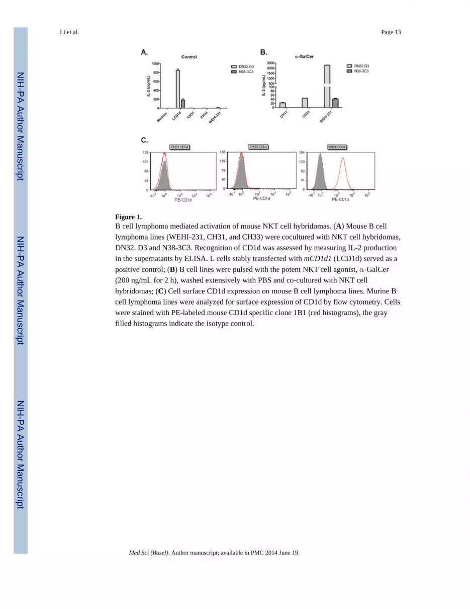
Figure 1.B cell lymphoma mediated activation of mouse NKT cell hybridomas. (A) Mouse B cell
lymphoma lines (WEHI-231, CH31, and CH33) were cocultured with NKT cell hybridomas,
DN32. D3 and N38-3C3. Recognition of CD1d was assessed by measuring IL-2 production
in the supernatants by ELISA. L cells stably transfected with mCD1d1 (LCD1d) served as a
positive control; (B) B cell lines were pulsed with the potent NKT cell agonist, -GalCer
(200 ng/mL for 2 h), washed extensively with PBS and co-cultured with NKT cell
hybridomas; (C) Cell surface CD1d expression on mouse B cell lymphoma lines. Murine B
cell lymphoma lines were analyzed for surface expression of CD1d by flow cytometry. Cells
were stained with PE-labeled mouse CD1d specific clone 1B1 (red histograms), the gray
filled histograms indicate the isotype control.
Li et al. Page 13
Med Sci (Basel). Author manuscript; available in PMC 2014 June 19.
NIH
-PA
Author M
anuscriptN
IH-P
A A
uthor Manuscript
NIH
-PA
Author M
anuscript

Figure 2.Healthy human NKT cells are stimulated by human B cell lymphomas, but circulating NKT
cells are reduced in lymphoma patients. (A) Differential CD1d expression in human B cell
lymphoma cell lines. Human B cell lymphoma cell lines were analyzed for surface
expression of CD1d by flow cytometry. Cells were stained with PE-labeled human CD1d
specific clone CD51.1 (red histograms). Isotype staining was performed to demonstrate
specificity (black histograms). C1R-CD1d served as the positive control; (B) Primary human
NKT cells were cocultured with human B cell lymphomas in the absence or presence of
antigen- -GalCer. Cytokine production following stimulation with C1R-CD1d served as
the positive control; (C) Circulating NKT cells are reduced in MCL patients. PBMC were
isolated from healthy donors (HD) and mantle cell lymphoma (MCL) patients and stained
for flow cytometry; (D) Peripheral blood mononuclear cells (PBMC) were isolated from
healthy donors and cancer patients. Cells were stained for V24+V 11+ TCR and analyzed
by FACS. Scatterplots demonstrate the variation in the percentages of NKT cells. MZL-
marginal zone lymphoma; DLBCL-diffuse large B cell lymphoma; MCL- mantle cell
lymphoma; FL-follicular lymphoma. % NKT cells of healthy donors vs. lymphoma patients;
Statistical analysis was performed using one way ANOVA *** p < 0.001; (E) Primary NKT
cells expanded from a healthy donor were co-cultured with B cells isolated from a healthy
donor or a mantle cell lymphoma (MCL) patient in the presence or absence of -GalCer.
Culture supernatants were harvested and ELISA was used to measure IFN- production.
Data are representative of three independent experiments.
Li et al. Page 14
Med Sci (Basel). Author manuscript; available in PMC 2014 June 19.
NIH
-PA
Author M
anuscriptN
IH-P
A A
uthor Manuscript
NIH
-PA
Author M
anuscript

Figure 3.NKT cell responses are impaired during lymphoma progression. (A) Splenic NKT cell
profiles in WT, DTG and c-myc Tg mice; (B) Splenic NKT cell % in WT, DTG, and c-myc
Tg mice at six to seven weeks and 10–15 weeks of age. The term “ disease” means that the
mice were sick, as indicated by splenomegaly; (C) Primary mouse splenocytes cultured in
medium alone or -GalCer. After 48 h, IFN- levels were measured in the supernatant by
ELISA; (D) To assess T cell function, splenocytes were cultured with anti- CD3/CD28
microbeads or PMA/ionomycin for 48 h. IFN- production in the culture supernatant was
measured by ELISA. Data shown from one experiment and are representative of seven
similar experiments.
Li et al. Page 15
Med Sci (Basel). Author manuscript; available in PMC 2014 June 19.
NIH
-PA
Author M
anuscriptN
IH-P
A A
uthor Manuscript
NIH
-PA
Author M
anuscript

Figure 4.Activation of NKT cells reduces tumor burden in vivo. Eight-week old IL-14/c-myc double
transgenic mice were treated with vehicle alone (DMSO) or -GalCer (2 μg/mouse) in PBS
i.v. and after six weeks, lymph nodes and spleens were harvested and examined for disease.
(A) H&E staining of spleen sections shows improved splenic architecture and lower
frequency of blastoid variant MCL cells as compared to vehicle treated controls; (B) spleens
and (C) lymph nodes of -GalCer treated mice show reduced splenomegaly and
lymphadenopathy, respectively, as compared to vehicle-treated, littermate controls. These
data are representative of three independent experiments of two to five mice per group; a
total of 11 mice were treated with vehicle, and 10 were treated with -GalCer.
Li et al. Page 16
Med Sci (Basel). Author manuscript; available in PMC 2014 June 19.
NIH
-PA
Author M
anuscriptN
IH-P
A A
uthor Manuscript
NIH
-PA
Author M
anuscript

Figure 5.Activation of NKT cells with a single dose of -GalCer (i.v.) increases survival in a mouse
model of MCL-BV. (A) DTG mice were treated with -GalCer or vehicle alone as described
above. Survival curves showing mice treated with vehicle alone (n = 11) or with -GalCer
(n = 10). Mice were euthanized upon detection of severe lymphadenopathy along with their
non-terminal counterparts; (B) Percentage of TCR+, PBS57 loaded (-GalCer)-CD1d
tetramer+ NKT cells in vehicle treated or -GalCer treated mice; (C,D) Splenocytes were
cultured for 48 h ex vivo in medium alone, or with -GalCer for restimulation. Both baseline
and restimulated levels were higher for (C) IFN- but not (D) IL-4 in mice treated with -
GalCer.
Li et al. Page 17
Med Sci (Basel). Author manuscript; available in PMC 2014 June 19.
NIH
-PA
Author M
anuscriptN
IH-P
A A
uthor Manuscript
NIH
-PA
Author M
anuscript


