Embed Size (px)
Citation preview

Phosphatidylinositol-3 kinase is distinctively required for l-,but not j-opioid receptor-induced activation of c-Jun N-terminal
kinase
Angel Y. F. Kam, Anthony S. L. Chan and Yung H. Wong
Department of Biochemistry, the Molecular Neuroscience Center, and the Biotechnology Research Institute, Hong Kong
University of Science and Technology, Clear Water Bay, Kowloon, Hong Kong
Abstract
Opioid receptors are the therapeutic targets of narcotic anal-
gesics. All three types of opioid receptors (l, d and j) are
prototypical Gi-coupled receptors with common signaling
characteristics in their regulation of intracellular events. Nev-
ertheless, numerous signaling processes are differentially
regulated by the three receptors. We have recently demon-
strated that stimulation of d-opioid receptor can up-regulate
the activity of the c-Jun N-terminal kinase (JNK) in a pertussis
toxin-sensitive manner (Kam et al. 2003; J. Neurochem. 84,
503–513). The present study revealed that the l-opioid
receptor could stimulate JNK in both SH-SY5Y cells and
transfected COS-7 cells. The mechanism by which the
l-opioid receptor stimulated JNK was delineated with the use
of specific inhibitors and dominant-negative mutants of signa-
ling intermediates. Activation of JNK by the l-opioid receptor
was mediated through Gbc, Src kinase, son-of-sevenless
(Sos), Rac and Cdc42. Interestingly, unlike the d-opioid recep-
tors, the l-opioid receptor required phosphatidylinositol-3
kinase (PI3K) to activate JNK. The l-opioid receptor-induced
JNK activation was effectively inhibited by wortmannin or the
coexpression of a dominant negative mutant of PI3Kc. Like
the d-opioid receptor, activation of JNK by the j-opioid
receptor occurred in a PI3K-independent manner. These
studies revealed that the l-opioid receptor utilize a distinct
mechanism to regulate JNK.
Keywords: JNK, l-opioid receptor, PI3K, small GTPases,
Son-of-sevenless (Sos), Src.
J. Neurochem. (2004) 89, 391–402.
Opioids are multifunctional peptides that regulate numerous
physiological effects including inhibition of neuronal firing
and neurotransmitter release, induction of tolerance and
dependence, reduction of gastrointestinal motility, as well as
modulation of the immune and endocrine responses (Mattes
et al. 1996). The actions of these peptides are manifested by
specific opioid receptors, which are mainly distributed
throughout the nervous system, but are also expressed in
immune cells such as monocytes and lymphocytes. The
opioid receptors are classified into three major types, termed
l, d and j according to their ligand selectivities and
pharmacological profiles. All three opioid receptors inhibit
adenylyl cyclase, reduce Ca2+ currents, stimulate inward
rectifying K+ currents and activate mitogen-activated protein
kinases (MAPK) via pertussis toxin (PTX)-sensitive Gi/Go
proteins (reviewed in Law et al. 2000). There is increasing
evidence, however, that molecular processes mediated by l-,d- and j-opioid receptors are not identical. Activation of thed-opioid receptor triggers translocation of G protein-coupled
receptor kinase (GRK)2 and 3 to the plasma membrane,
which is followed by cointernalization of the receptor and
GRKs. In contrast, stimulation of l-opioid receptor does notresult in the accumulation of GRKs at the membrane nor its
cointernalization with GRK2 or 3 (Schulz et al. 2002).
Received August 2, 2003; revised manuscript received December 1,
2003; accepted December 12, 2003.
Address correspondence and reprint requests to Yung H. Wong,
Department of Biochemistry, Hong Kong University of Science and
Technology, Clear Water Bay, Kowloon, Hong Kong.
E-mail: [email protected]
Abbreviations used: DAMGO, [D-Ala2,N-Me-Phe4,Gly5-ol]enkepha-
lin; DMEM, Dulbecco’s modified Eagle’s medium; EGF, epidermal
growth factor; ERK, extracellular signal-regulated protein kinases; G
protein, guanine nucleotide-binding regulatory protein; GEF, guanine
nucleotide exchange factor; HA, hemagglutinin; JNK, c-Jun N-terminal
kinase; MAPK, mitogen-activated protein kinase; PI3K, phosphatidy-
linositol-3 kinase; PTX, pertussis toxin; SDS–PAGE, sodium dodecyl
sulfate-polyacrylamide gel electrophoresis; Sos, son-of-sevenless;
U-50,488, (±)-trans-U-50,488 methanesulfonate.
Journal of Neurochemistry, 2004, 89, 391–402 doi:10.1111/j.1471-4159.2004.02338.x
� 2004 International Society for Neurochemistry, J. Neurochem. (2004) 89, 391–402 391
Likewise, l- and j-opioid receptors exhibit different abilitiesto utilize Gaz, a PTX-insensitive member of the Gi family,
for the regulation of extracellular signal-regulated kinase
(ERK) in PTX-treated COS-7 cells (Belcheva et al. 2000).
These differences in the molecular pharmacology of opioid
receptors may in part account for their functional specificity
in various physiological responses, such as the addiction
potential of different opioid ligands.
We have recently observed that activated d-opioid recep-
tors can stimulate c-Jun N-terminal kinase (JNK) activity
(Kam et al. 2003). Along with ERK and p38, JNK
constitutes the family of MAPKs that are widely involved
in diverse biological functions ranging from proliferation,
survival, differentiation, to apoptosis. JNK is broadly
expressed in the nervous system and it appears to exhibit
multiple functions. In rat pheochromocytoma PC-12 cells,
withdrawal of nerve growth factor (NGF) activates JNK and
causes apoptosis (Xia et al. 1995). However, opposite
perspectives suggest that JNK also contributes to neuronal
protection by inhibition of caspases (Kuan et al. 1999).
Moreover, apoptosis was increased in the hindbrain and
forebrain of jnk1–/–jnk2–/– mice at E10.5, indicative of the
requirement of JNK in neuronal survival during development
(Sabapathy et al. 1999). Indeed, stimulation of opioid
receptors has been shown to activate ERK1/2 (Belcheva
et al. 1998), p38 MAPK (Singhal et al. 2002) and JNK
(Kam et al. 2003) in various cell types. Hayashi et al. (2002)
have demonstrated that a low concentration of d-opioidpeptide DADLE requires ERK activation to promote cell
survival in serum-deprived PC-12 cells. Moreover, inhibition
of p38 MAPK phosphorylation by using specific inhibitors
can protect morphine-treated macrophages from apoptosis
(Singhal et al. 2002), suggesting the involvement of p38
MAPK in morphine-induced macrophage apoptosis. How-
ever, the role of opioid receptor-regulated JNK activity in the
nervous system still remains unclear and so mapping the
signaling pathway from opioid receptor to JNK is important.
In a previous study, we have shown that the d-opioidreceptor-mediated JNK pathway involves Gbc, Src kinase
and the small GTPases Rac and Cdc42, but does not require
phosphatidylinositol 3-kinase (PI3K) or transactivation of
EGF receptor (Kam et al. 2003). Here we demonstrate that
JNK activity is stimulated by the l-opioid receptor in humanneuroblastoma SH-SY5Y cells. By heterologous expression
assays in COS-7 cells, we have further characterized the
l-opioid receptor-regulated JNK activity and the pathway
has been shown to depend on Gbc, Src kinase, Rac and
Cdc42, but not EGF receptor. These intermediate steps in the
pathway are identical with the d-opioid receptor-stimulated
signaling cascade. Moreover, a guanine nucleotide exchange
factor (GEF) Son-of-sevenless (Sos) is identified for
transmitting signals from the l-opioid receptor to JNK.
Surprisingly, PI3K is uniquely necessary for l-opioidreceptor-stimulated JNK as shown by using a PI3K inhibitor,
wortmannin, and a dominant negative mutant of PI3Kc. Thisis a distinct difference between l- and d-opioid receptor
signaling. Like the d-opioid receptor, j-opioid receptor doesnot require PI3K to mediate JNK activity.
Materials and methods
Materials
The cDNAs of the rat l-opioid receptor and the mouse j-opioidreceptor (both in the pCMV6 vector) were kindly provided by Dr L.
Yu (Indiana University School of Medicine) and Dr G. Bell
(University of Chicago, Chicago, IL, USA), respectively. JNK-HA
cDNAwas supplied by Dr T. A. Voyno-Yasenetskaya (University of
Illinois, Chicago, IL, USA). The plasmids containing wild-type
(WT) and dominant-negative mutant of PI3Kc (PI3KcK832R), SrcWT, dominant negative Src (SrcK295R/527F) as well as dominant
negative Ras (RasS17N), Rac (RacT17N), Cdc42 (Cdc42T17 N)
and RhoA (RhoAT19N) were obtained as previously described
(Kam et al. 2003). The cDNAs encoding p21-binding domain
(PBD) from human PAK1 cloned into the bacterial expression
vector pGEX-2TK and Sos-Pro were kind donations from Dr Udo
Schmitz (Medizinische Universitats-Poliklinik, Germany) and Dr R.
J. Lefkowitz (Duke University Medical Center, Durham, NC, USA),
respectively. The cDNA of kinase-deficient mutant of Akt was a
generous gift from Dr Z.G. Wu (Hong Kong University of Science
and Technology, Hong Kong, China). [c-32P]ATP was purchased
from DuPont NEN (Boston, MA, USA). Anti-phospho-JNK, anti-
JNK, anti-phospho-Akt, anti-Akt antibodies and antiphospho-Src-
Y416 antibodies were obtained from Cell Signaling Technology,
Inc. (Beverly, MA, USA). Antiserum against Gat was from
Transduction Laboratories. Mouse monoclonal antibody against
human Rac was purchased from Upstate (Lake Placid, NY, USA).
Rabbit polyclonal antibody against human Cdc42 was from Santa
Cruz Biotechnology (Santa Cruz, CA, USA). PTX and 12CA5 (anti-
HA) antibody were purchased from List Biological Laboratories
(Campbell, CA, USA) and Roche Molecular Biochemicals (Indi-
anapolis, IN, USA), respectively. Wortmannin, tyrphostin AG1478
and pyrazolopyrimidine PP2 were purchased from Calbiochem-
Novabiochem Co. (La Jolla, CA, USA). Cell culture
reagents, including LipofectAMINE PLUS were obtained from
Invitrogen, Carlsbad (CA, USA). [D-Ala2,N-Me-Phe4,Gly5-ol]en-
kephalin (DAMGO), (±)-trans-U-50,488 methanesulfonate
(U-50,488H), [D-Phe-Cys-Tyr-D-Trp-Orn-Pen-Thr-NH2] (CTOP) and
all other chemicals were purchased from Sigma (St. Louis, MO, USA).
Cell culture and transfection
COS-7 cells were grown in Dulbecco’s modified Eagle’s medium
(DMEM) supplemented with 10% (v/v) fetal bovine serum (FBS),
50 units/mL penicillin and 50 lg/mL streptomycin in a humidified
atmosphere containing 5% CO2 at 37�C. One day before the
transfection, cells were seeded onto six-well plates at a density of
3 · 105 cells/well. Transfection was performed by means of
LipofectAMINE PLUS reagents according to the supplier’s instruc-
tions, and the transfected cells were kept in the growth medium for
36 h. Under the same physical condition, human neuroblastoma SH-
SY5Y cells were maintained in a mixture (1 : 1) of Eagle’s
minimum essential medium (MEM) and Ham’s F12 medium
392 A. Y. F. Kam et al.
� 2004 International Society for Neurochemistry, J. Neurochem. (2004) 89, 391–402

supplemented with 10% (v/v) FBS, 50 units/mL penicillin and
50 lg/mL streptomycin. Human monocytic THP-1 cells were
cultured in RPMI 1640 medium supplemented with 0.05 mM
2-mercaptoethanol, 10% (v/v) FBS, 50 units/mL penicillin and
50 lg/mL streptomycin at 37�C in 5% CO2.
In vitro JNK assay
Transfected COS-7 cells were serum starved for 18 h in the presence
or absence of PTX (100 ng/mL) before drug treatment. Where
appropriate, additional treatments of wortmannin (100 nM, 15 min),
AG1478 (500 nM, 30 min) and PP2 (10 lM, 30 min) were applied
to the starved cells. Cells were then treated with the assay medium
(DMEM with 20 mM HEPES) in the absence or presence of 100 nM
DAMGO or U-50,488H for 15 min at 37�C and lysed in 500 lL of
lysis buffer (50 mM Tris-HCl, pH 7.5, 100 mM NaCl, 5 mM EDTA,
40 mM NaP2O7, 1% Triton X-100, 1 mM dithiothreitol, 200 lMNa3VO4, 100 lM phenylmethylsulfonyl fluoride, 2 lg/mL leupep-
tin, 4 lg/mL aprotinin, and 0.7 lg/mL pepstatin) and shaken on ice
for 30 min. The lysates were centrifuged at 14,000 · g for 5 min at
4�C. Fifty microliters of each sample was used for the detection of
JNK-HA expression or 100 lL was used for phosphorylated and
total Akt if necessary, 400 lL was incubated for 1 h at 4�C with
anti-HA antibody (2 lg/sample), followed by incubation with 30 lLof protein A-agarose (50% slurry) at 4�C for 1 h. The resulting
immunoprecipitates were washed twice with lysis buffer and twice
with kinase assay buffer [40 mM HEPES, pH 8.0, 5 mM
Mg(C2H3O2)2, 1 mM EGTA, 1 mM dithiothreitol, 200 lM Na3VO4].
Washed immunoprecipitates were resuspended in 40 lL of kinase
assay buffer containing 5 lg of GST-c-Jun per reaction, and the
kinase reactions were initiated by the addition of 10 lL of ATP
buffer (50 lM ATP with 2 lCi of [c-32P]ATP per sample). After
30 min incubation at 30�C with occasional shakings, the reactions
were terminated by adding 10 lL of 6X sample buffer and boiled
for 5 min, and the samples were subjected to 12% sodium dodecyl
sulfate–polyacrylamide gel electrophoresis (SDS–PAGE). The
radioactivity incorporated into GST-c-Jun was detected by autora-
diogram, and the signal intensity was quantified by PhosphorImager
(Molecular Dynamics 445 SI).
Western blot
SH-SY5Y cells were seeded onto six-well plates at a density of
2.5 · 105 cells/well and allowed to reach confluency. The cells were
then serum starved for 4 h in the absence or presence of PTX
(100 ng/mL), followed by pretreatments with inhibitors if necessary.
The cells were stimulated with DAMGO at 37�C for the indicated
time and then lysed in 200 lL of lysis buffer. THP-1 cells were
plated at 1 · 106 cells/mL in serum-free media and maintained for
18–24 h with or without PTX (100 ng/mL). If necessary, THP-1
cells were preincubated with different inhibitors before addition of
U-50,488H. After stimulation with the agonist, cells were lysed as
with SH-SY5Y cells. Supernatants of both cell lysates were
collected by centrifugation at 14 000 · g for 5 min, mixed with
40 lL of 6X sample buffer and boiled for 5 min. 150 lg proteins ofeach sample were resolved by 12% SDS–PAGE, and then
transferred to nitrocellulose membranes. Phosphorylated or total
kinases (JNK, Akt and Src) were detected by specific antibodies as
mentioned under Materials, followed with horseradish peroxidase-
conjugated secondary antibody. Immunoblots were developed in the
presence of enhanced chemiluminescence reagents, and the images
detected in X-ray films were quantified by densitometric scanning
using the Eagle Eye II still video system (Stratagene, La Jolla, CA,
USA).
GTPase pull-down assay
GTPase pull-down assay was performed as described previously
(Kam et al. 2003). The cDNAs of PAK-PBD in pGEX-2TK was
expressed in Escherichia coli as a fusion protein with glutathione-S-
transferase. The fusion proteins were purified from glutathione-
Sepharose beads. Transfected COS-7 cells expressing l-opioidreceptor were serum starved and then lysed with 500 lL of Mg2+-
containing lysis buffer (MLB; 25 mM HEPES, pH 7.5, 150 mM
NaCl, 1% Triton X-100, 0.25% sodium deoxycholate, 10% glycerol,
25 mM NaF, 10 mM MgCl2, 1 mM EDTA, 1 mM sodium orthovana-
date, 10 lg/mL leupeptin, 10 lg/mL aprotinin). Cell lysates were
centrifuged at 4�C for 10 min at 14 000 · g. Fifty microliters of the
supernatant was used for detecting total Rac and Cdc42. 450 lL cell
lysates were incubated with 10 lg GST-PAK-PBD and 15 lL of
50% slurry of glutathione-Sepharose beads at 4�C for 60 min with
constant rotations. Bound proteins were collected by centrifugation
and pellets were washed three times in MLB and finally suspended
in 2 · Laemmli sample buffer (40 lL). Proteins were resolved by
12% SDS–PAGE and the bound Rac or Cdc42 were analyzed by
immunoblotting using antiserum against Rac or Cdc42, respectively.
Results
Stimulation of l-opioid receptor induce a time- and
concentration-dependent JNK activation
We have recently demonstrated that d-opioid receptor can
induce JNK stimulation in both COS-7 and NG108-15 cells
(Kam et al. 2003). Here, we asked if the l-opioid receptor
can similarly regulate JNK activity. Heterologous expression
of the l-opioid receptor and HA-tagged JNK (HA-JNK) in
COS-7 cells were performed as described previously (Kam
et al. 2003). The JNK activity was examined by immuno-
complex kinase assay. Treatment of COS-7 cells with the
l-selective agonist, DAMGO, resulted in a time-dependent
JNK activation, as reflected by increases in the phosphory-
lation of GST-c-Jun proteins. The DAMGO-induced JNK
activation peaked at 15 min after drug addition, thereafter it
gradually recovered to near basal level (Fig. 1a, left panel).
Anti-HAwestern blotting of total cell lysates verified that the
total amount of HA-JNK was unaffected by the agonist
treatment, suggesting that the elevated JNK activities did not
result from increased JNK expression but from phosphory-
lation. In order to examine the mechanism of JNK activation
by the l-opioid receptor under more native conditions, we
also examined human neuroblastoma SH-SY5Y cells en-
dogenously expressing the receptor (Kazmi and Mishra
1987). Activation of JNK was determined by western
blotting using an antiserum against the p46/p54 isoforms of
JNK dually phosphorylated at threonine 183 and tyrosine
PI3K-dependent JNK activation by l-opioid receptor 393
� 2004 International Society for Neurochemistry, J. Neurochem. (2004) 89, 391–402

185 residues. Figure 1(a) (right panel) showed time course
studies for JNK activation in DAMGO-treated SH-SY5Y
cells. Interestingly, in comparison with studies in COS-7
cells, JNK activity was rapidly stimulated upon DAMGO
application to SH-SY5Y cells and reached the maximum at
5 min. The elevated JNK activity was sustained for up to
15 min. Therefore, the time course of agonist-induced JNK
activity was shifted to earlier time points in SH-SY5Y cells
as compared to COS-7 cells. Furthermore, administration of
DAMGO in SH-SY5Y cells preferentially activated p46
JNK, as indicated in the immunoblots of phosphorylated
JNK. Transfected COS-7 and SH-SY5Y cells were then
challenged with different agonist concentrations (0.1 nM to
1 lM) for 15 min and 5 min, respectively. In both cells,
maximal JNK activity-induced by DAMGO was obtained at
around 100 nM, but the half-maximum response was about
10 nM in COS-7 and 1 nM in SH-SY5Y cells, respectively
(Fig. 1b).
l-Opioid receptors activate JNK in a PTX-sensitive
and Gbc-dependent manner
As prototypical Gi/Go-coupled receptors, opioid receptors
regulate an array of effectors via pertussis toxin (PTX)-
sensitive Gi/Go proteins (reviewed in Law et al. 2000).
Nevertheless, couplings between opioid receptors and a
variety of PTX-insensitive G proteins are also plausible.
Chan and Wong (2000) have demonstrated that the activated
ORL1 receptor can functionally interact with PTX-insensitive
Gz, G12, G14 and G16 to induce JNK activation in COS-7
cells. Therefore, it is pertinent to investigate the participation
of Gi/Go proteins in the opioid receptor-regulated JNK
activation. Transfected COS-7 and SH-SY5Y cells were
pretreated with 100 ng/mL PTX for 18 h before exposure to
DAMGO. The toxin ADP-ribosylates Gi/Go proteins, as a
result their couplings with receptors can be abrogated. As
illustrated in Fig. 2(a), JNK was significantly activated upon
stimulation with DAMGO in both cells. However, pretreat-
ment of cells with PTX completely abolished the JNK
activation, demonstrating that l-opioid receptor preferen-
tially interacts with Gi/Go proteins to regulate JNK activity in
COS-7 and SH-SY5Y cells. Because SH-SY5Y cells not
only express l-opioid receptor, but also d- and j-opioidreceptors (Cheng et al. 1995), we next performed experi-
ments to confirm whether the JNK activation is mediated via
the activation of l-opioid receptor in SH-SY5Y cells by
using a selective antagonist CTOP. The JNK stimulation by
DAMGO was suppressed in the presence of CTOP (Fig. 2a,
right panel), indicating the requirement of l-opioid receptor
in SH-SY5Y cells.
Both Gai-GTP and Gbc dimers are responsible for
transducing signals from receptors to downstream effectors.
However, in the case of regulation of JNK, Gbc are seeminglymore effective. Coso et al. (1996) have reported that only
Gbc subunits, but not the activated mutants of Gai signifi-cantly increase JNK activity. Moreover, JNK activation by
Gi-coupled m2 muscarinic acetylcholine receptor is inhibited
by overexpressing the b-adrenergic receptor kinase (b-ARK)carboxyl-terminal domain, which acts as a Gbc scavenger
protein. To confirm the role of Gbc in signaling from the
ligand-activated l-opioid receptors to JNK, the a subunit of
transducin (Gat) was cotransfected into COS-7 cells. Gat canalso sequester free Gbc subunits that are concomitantly
released upon stimulation of receptors, thereby suppressing
Gbc-dependent biological responses. In cells coexpressing
Gat and l-opioid receptor, the JNK activation in response to
DAMGO was markedly reduced (Fig. 2b). Therefore, Gbc
0
1
2
3
0 10 20 30 40 50 60
*
(a)
Time (min.)0 5 15 30 45 60
JNK
acti
vity
(fol
dst
imul
atio
n)
JNK-HA
GSTc-Jun
P0 1 5 15 30 45
0
1
2
3
0 10 20 30 40
**
TotalJNK
p54P
p46P
B -10 -9 -8 -7 -6Log[DAMGO] (M)
0
1
2
3
-10 -9 -8 -7 -6
* *
(b)
JNK
acti
vity
(fol
dst
imul
atio
n)
JNK-HA
GSTc-Jun
PB -10 -9 -8 -7 -6
0
1
2
3
-10 -9 -8 -7 -6
* *
TotalJNK
p54P
p46P
COS-7 SH-SY5Y
COS-7 SH-SY5Y
Fig. 1 l-Opioid receptor-induced JNK activation in COS-7 cells and
SH-SY5Y cells. COS-7 cells were transiently cotransfected with the
cDNAs of JNK-HA and l-opioid receptor (0.5 lg for each). The
transfectants were serum-starved for 18 h, before application of
DAMGO. The lysate of COS-7 cells were subjected to in vitro kinase
assay as described under ‘Materials and methods’. SH-SY5Y cells
were serum-starved for 4 h prior to stimulation with DAMGO, and
activated JNK in the cell lysates was detected by anti-phospho-JNK
antiserum. (a) Time course of JNK activation by 100 nM DAMGO. (b)
Dose–response of JNK activation following DAMGO treatment of
COS-7 cells for 15 min or SH-SY5Y cells for 5 min. Representative
autoradiograms illustrate phosphorylated GST-c-Jun, total JNK-HA,
phosphorylated and total JNK. Band intensities were quantified by
densitometry. Data are presented as fold increase in JNK activity over
non-stimulated cells (Con), and represents the mean ± SEM from four
or five separate experiments. *DAMGO significantly induced JNK
activation (Bonferroni t-test, p < 0.05).
394 A. Y. F. Kam et al.
� 2004 International Society for Neurochemistry, J. Neurochem. (2004) 89, 391–402

appeared to play an essential role in regulating l-opioidstimulated-JNK activity. As determined by immunodetection
with anti-Gat antiserum, coexpression of Gat did not alter theamount of JNK (shown in the anti-HA immunoblot).
Small GTPases Rac and Cdc42 contribute to JNK
activation by l-opioids
Small GTPases are believed to be a link between G protein-
coupled receptors (GPCRs) or receptor tyrosine kinases and
MAPKs. They may operate upstream of Raf in ERK
activation or MEKK in JNK activation. Belcheva et al.
(1998) have demonstrated that the three classical opioid
receptors require Ras GTPases to activate ERK via Raf and
MEK1/2. Like the ERK pathway, several small GTPases act
as upstream activators of JNK and they are members of the
Rho family, Rac and Cdc42 (Coso et al. 1995). In agreement
with this observation, we have previously found that the
pathway from the d-opioid receptor to JNK primarily
requires Rac or Cdc42 (Kam et al. 2003). To determine
which GTPases participate in signaling from the l-opioidreceptor toward JNK, dominant-negative mutants, including
RasS17N, RacT17N, Cdc42T17N or RhoT19N, were co-
transfected together with l-opioid receptor and HA-JNK into
the COS-7 cells. These inhibitory mutants are stable in a
GDP conformation, and so competitively prevent endog-
enous GTPases from accessing their specific guanine
nucleotide exchange factors. Like the d-opioid receptor, the
ability of the l-opioid receptor to stimulate JNK was
essentially abolished by RacT17N and Cdc42T17N, whereas
RasS17N was ineffective in inhibiting the JNK activation
(Fig. 3a). RhoT19N produced a slight but statistically
insignificant inhibition on DAMGO-induced JNK activity
(Fig. 3a). These results indicate that Rac and Cdc42 GTPases
link the l-opioid receptor to the regulation of JNK. In orderto further validate the important roles of Rac and Cdc42, we
(a)
(b)
Fig. 2 JNK stimulation by l-opioid receptor is attenuated in the
presence of PTX or Gbc-scavenging proteins. (a) The l-opioid
receptor and JNK-HA were coexpressed in COS-7 cells as in legend to
Fig. 1. Transfectants, with or without PTX pretreatment (100 ng/mL,
18 h), were incubated in the absence (basal) or presence of DAMGO
(100 nM) for 15 min. Similarly, serum-starved SHSY-5Y cells were
pretreated with or without PTX (100 ng/mL, 18 h) prior to application of
DAMGO (100 nM) in the absence or presence of CTOP (10 lM) for
5 min. (b) COS-7 cells were cotransfected with cDNAs encoding JNK-
HA and l-opioid receptor, together with vector or Gat (0.5 lg for each).
Serum-starved cells were exposed to 100 nM DAMGO for 15 min.
Values shown represent the mean ± SEM from five or six separate
experiments. *DAMGO significantly stimulated JNK activity (Bonfer-
roni t-test, p < 0.05).
(a)
(b)
Fig. 3 Role of small GTPases Ras, Rac, Cdc42 and Rho in l-opioid-
dependent JNK activation. (a) COS-7 cells were cotransfected with the
cDNAs of l-opioid receptor and JNK-HA, together with dominant
negative RasS17N, RacT17N, N17Cdc42 or N17Rho (0.5 lg each).
Transfectants were exposed to 100 nM DAMGO for 15 min. Results
are the mean ± SEM from eight independent experiments. *DAMGO
significantly stimulated JNK activity (Bonferroni t-test, p < 0.05). (b)
COS-7 cells expressing l-opioid receptors were stimulated by 100 nM
DAMGO for the indicated time, activation of Cdc42 and Rac was
determined by GST-PAK-PBD pull down assay and western blotting
as described in ‘Materials and Methods’. Total cell lysates were also
immunoblotted with Rac and Cdc42 antibodies to determine protein
expressions.
PI3K-dependent JNK activation by l-opioid receptor 395
� 2004 International Society for Neurochemistry, J. Neurochem. (2004) 89, 391–402

next investigated the activation state of Rac and Cdc42
following stimulation of the l-opioid receptor. This was
achieved by using GST-PAK-PBD pull-down assay. Because
the p21-binding domain (PBD) of the PAK protein binds
only to the activated GTP-bound Rac and Cdc42, the amount
of such activated GTPases in the assay was measured by anti-
Rac/Cdc42 immunoblotting. As shown in Fig. 3(b), stimu-
lation of the l-opioid receptor led to a time-dependent
activation of Rac and Cdc42, with a peak at around
15–30 min incubation of DAMGO. After 60 min, activations
of both GTPases decreased to basal levels.
The guanine nucleotide exchange factor (GEF)
Son-of-sevenless (Sos) mediates l-opioid-stimulated
JNK activity
Activations of small GTPases occur through stimulation of
specific GEF by promoting their exchange of GDP for GTP.
It was therefore required to identify which Rac-GEFs and/or
Cdc42-GEFs provide the link between l-opioid receptor andJNK activation. Sos proteins belong to Dbl family GEF and
are widely expressed. They specifically activate Rac but not
Cdc42 (Han et al. 1998; Nimnual et al. 1998). Hawes et al.
(1998) have demonstrated that l-opioid receptor requires Sosto regulate the activation of ERK, implying that the
stimulated receptor may transmit signal to Sos. Additionally,
expression of the Dbl homology domain of Sos (DH-Sos) has
been shown to induce a robust JNK stimulation in COS-1
cells (Nimnual et al. 1998). Sos therefore was considered as
the candidate to mediate the signaling from l-opioid receptortoward JNK. The inhibition on the activity of Sos is achieved
by expressing their proline-rich domains (Sos-Pro) at the
C-terminus, as Sos-Pro compete with the endogenous Sos for
binding to the SH3 domain of Grb2 (van Biesen et al. 1995).
As shown in Fig. 4, expression of Sos-Pro blocked the JNK
activation in response to DAMGO, indicating that Sos is able
to provide a relay from the l-opioid receptor to Rac and
subsequently JNK.
Like Sos protein, Vav2 is also a ubiquitously expressed
member of Dbl family, but displays overlapping GEF activity
for Rac1, Cdc42 and RhoA (Abe et al. 2000). To determine
the participation of Vav2 in the l-opioid-dependent JNKactivation, a dominant negative mutant of Vav2 was coex-
pressed in COS-7 cells together with the receptor and JNK-
HA. However, as the dominant negative mutant of Vav2
robustly increased the basal activity of JNK (data not
shown), it is difficult to observe the effect of DAMGO on the
JNK activity in the presence of the mutant. Owing to this
technical limitation, involvement of Vav2 in the l-opioid-signaling could not be determined.
l-Opioid receptor, but not j-opioid receptor requires
PI3Kc to stimulate JNK activity
As illustrated in Fig. 4, Sos proteins provide the link between
the l-opioid receptor and JNK activation. Numerous studies
revealed that Sos catalyze exchange for Rac in response to
lipid products of phosphatidylinositol 3-kinase (PI3K) such
as PI-3,4,5-P3 via binding to their Pleckstrin Homology (PH)
domains (PH-Sos; Han et al. 1998; Nimnual et al. 1998). In
addition, overexpression of PI3Kc significantly stimulates
JNK in COS-7 cells (Lopez-Ilasaca et al. 1998). We then
asked whether the l-opioid receptor can activate PI3K,
which in turn evoked the Rac and Cdc42-dependent JNK
activations via specific GEFs such as Sos. Agonist stimula-
tion of the l-opioid receptor in COS-7 cells was demonstra-ted to increase the phosphorylation of Akt at Ser-473, a
downstream target for the activated PI3K (Fig. 5a, immuno-
blot of antiphospho-Akt in left panel). Thus, it implied the
stimulatory effect of l-opioids on PI3K. To assess the
requirement of PI3K for l-opioid receptor signaling toward
Rac, Cdc42 and JNK, a specific inhibitor of PI3K wortman-
nin was utilized. The inhibitor significantly abrogated JNK
phosphorylation by the l-opioid receptor in both transfectedCOS-7 cells and SH-SY5Y cells (Fig. 5a). Wortmannin
(100 nM, 15 min) also attenuated the l-opioid receptor-
induced Rac and Cdc42 stimulation in COS-7 cells as
determined with the GST-PAK-PBD pull down assay (data
not shown). Surprisingly, these results were inconsistent with
the previous observation that no inhibitory effect by wort-
mannin was observed when JNK was stimulated by d-opioidreceptor (Kam et al. 2003). Hence, the effect of wortmannin
on the j-opioid signaling was also examined. COS-7 cells
were transiently expressed j-opioid receptor and HA-JNK.
As shown in Fig. 5(b) (left panel), JNK activity was
significantly increased upon stimulation with j-opioidU-50,488H, irrespective of whether COS-7 cells were
preincubated with wortmannin or not. In order to confirm
the insensitivity of wortmannin in j-opioid-stimulated JNK
Fig. 4 Suppression of l-opioid-stimulated JNK activation by SOS-
Pro. The l-opioid receptor and JNK-HA were coexpressed in COS-7
cells, together with vector or SOS-Pro (0.5 lg each). JNK activity was
determined at 15 min in the absence (basal) or presence of DAMGO
(100 nM). Results are the mean ± SEM from five independent
experiments. *DAMGO significantly induced JNK activation (Bonfer-
roni t-test, p < 0.05).
396 A. Y. F. Kam et al.
� 2004 International Society for Neurochemistry, J. Neurochem. (2004) 89, 391–402

pathway, human monocytic THP-1 cells endogenously
expressing the j-opioid receptor were utilized. Administra-
tion of U-50,488H in quiescent THP-1 cells increased the
phosphorylation of JNK (Fig. 5b; right panel). In agreement
with studies in COS-7 cells, wortmannin still had no
inhibitory effect on the JNK activation in the j-opioid-treated THP-1 cells. Hence, the crucial role for PI3K was
only observed in the Rac and Cdc42-dependent JNK
activation by l-opioid receptor, but not j- and d-opioidreceptors. Unlike COS-7 cells, elevations of basal JNK
activity were observed in wortmannin-treated SH-SY5Y and
THP-1 cells. The reason may be the presence of a PI3K
inhibitory pathway in the naive cells. The immunoblots
against Akt and phospho-Akt in Fig. 5 revealed that
pretreating COS-7 cells with wortmannin substantially
reduced the Akt activity, while total amount of Akt was
not altered, confirming the inhibitory effect of wortmannin
on PI3K.
Wortmannin inhibits all isoforms of PI3K and the
c-isoform has been well documented to be dependent on
Gbc subunits (Stephens et al. 1994). Although PI3Kc is
mainly expressed in hematopoietic cells, its limited expres-
sion is still detected in COS-7 cells (Murga et al. 1998).
Moreover, COS-7 cells expressing p101 (a regulatory subunit
specific for c isoform) alone can elicit a level of Akt
activation similar to that induced by PI3Kc overexpression,
implicating that p101 functionally interacts with endogenous
PI3Kc isoform in COS-7 cells to stimulate Akt (Murga et al.
1998). Therefore, the function of PI3Kc in the JNK
activation by l-opioid was determined by using a kinase-
deficient mutant (PI3KcK832R). Coexpression of
PI3KcK832R suppressed JNK activation in response to
DAMGO, but not U-50,488H (Fig. 6a). Collectively, these
findings reveal a marked difference in signaling between the
three opioid receptors, whereby only l-opioid receptor
regulates JNK activity through PI3Kc-dependent pathway.Expression of PI3KcK832R clearly reduced the activity of
Akt in comparison with the wild type of PI3Kc (data not
shown), confirming the negative effect of the kinase-deficient
mutant on endogenous PI3K signaling.
Akt phosphorylation by l-opioid receptor is not essential
for JNK activation
Although activation of l- and j-opioid receptors increased
the Akt phosphorylation in the absence of wortmannin
(Fig. 5), only the l-opioid receptor required PI3K to
stimulate JNK. To test whether the observed phosphorylation
of Akt correlated with l-opioid receptor-mediated JNK
activation, a kinase deficient Akt containing a Lys179 to Met
mutation was utilized (Xu and Wu 2000). Overexpression of
this kinase deficient Akt mutant in mouse C2C12 has been
shown to suppress insulin-like growth factor-1-mediated
myogenin expression (Xu and Wu 2000). Furthermore, Akt-
dependent phosphorylations of glycogen synthase kinase 3
and tuberin induced by platelet-derived growth factor (30 ng/
mL, 5 min) were attenuated in COS-7 cells transiently
overexpressing this Akt mutant (data not shown). However,
the Akt mutant failed to inhibit the PI3K-dependent JNK
activation induced by l-opioid receptor (Fig. 6b, left panel),therefore excluding the role for Akt in the pathway. Similar
to the insensitivity to wortmannin and PI3KcK832R,expression of the kinase deficient Akt still had no demon-
strable effect on the j-opioid receptor-stimulated JNK
(Fig. 6b, right panel). Taken together, these studies indicate
that signal transductions from l- and j-opioid receptors to
JNK and Akt activation might occur independently.
Fig. 5 Wortmannin inhibits JNK activation by l-opioid receptor, but
not j-opioid receptor. (a) COS-7 cells were cotransfected with the
cDNAs of l-opioid receptor and JNK-HA (0.5 lg each). Transfectants
were serum-starved for 18 h and pretreated with or without wort-
mannin (100 nM, 15 min). The JNK activity was stimulated with 100 nM
DAMGO (15 min). Fifty microliters of lysate of COS-7 cells were
probed for phosphorylated and total Akt. SH-SY5Y cells were similarly
pretreated with wortmannin and then stimulated with DAMGO
(100 nM, 5 min). (b) COS-7 cells coexpressing j-opioid receptor and
JNK-HA were preincubated with or without wortmannin (100 nM,
15 min) prior to application of 100 nM U-50,488H (U-50; 15 min). THP-
1 cells were similarly preincubated with wortmannin after 18 h star-
vation, and then stimulated with 100 nM U-50 for 5 min. Values shown
represent the mean ± SEM from five to six separate experiments.
*Opioid agonists significantly induced JNK activation (Bonferroni t-test,
p < 0.05). **Wortmanin significantly increased the basal JNK activity
as compared to untreated cells (Bonferroni t-test, p < 0.05).
PI3K-dependent JNK activation by l-opioid receptor 397
� 2004 International Society for Neurochemistry, J. Neurochem. (2004) 89, 391–402

l-Opioid receptor stimulates JNK through Src family
tyrosine kinases but not transactivation of EGF receptor
Li and Smithgall (1998) have demonstrated that v-src
transformed Rat-2 cells induce JNK activation. Furthermore,
Src kinase activity can be stimulated by transient over-
expression of Gbc (Luttrell et al. 1996) or a2-adrenergic andmuscarinic m1 receptors (Chen et al. 1994). Therefore, these
studies suggest that Src family tyrosine kinases seem to
participate in the mitogenic signaling by G protein-coupled
receptors, in addition to PI3Kc. A pyrazolopyrimidine PP2 is
a potent and selective inhibitor of Src family tyrosine kinase.
COS-7 and SH-SY5Y cells were preincubated with PP2
(10 lM) for 30 min before stimulation by DAMGO. The
JNK stimulation in response to DAMGO was completely
suppressed following pretreatment with PP2 (Fig. 7a,b, left
panels). We next examined the sole contribution of the
ubiquitous Src kinase in the pathway. Upon coexpression of
dominant negative Src kinase with the l-opioid receptor in
COS-7 cells, a complete inhibition of JNK was observed
JNK
act
ivit
y (f
old
stim
ulat
ion)
JNK-HA
GSTc-Jun
PWT DN
0
1
2
3*
WT DN0
1
2
3
* *
Basal DAMGO U50(a)
Vector AktDN
JNK
act
ivit
y (f
old
stim
ulat
ion)
0
1
2
3*
*
0
1
2
3
**
Vector AktDN
JNK-HA
GSTc-Jun
P
Basal DAMGO U50(b)
PI3Kγ
Fig. 6 Effect of dominant negative PI3Kc and Akt on l- and j-opioid
receptor-mediated stimulation of JNK. (a) cDNAs encoding l- (left
panel) or j- (right panel) opioid receptor and JNK-HA were cotrans-
fected into COS-7 cells, together with wild-type (WT) or dominant
negative (DN) PI3Kc (0.5 lg each). (b) COS-7 cells were cotrans-
fected with the cDNAs encoding l- (left panel) or j- (right) opioid
receptor and JNK-HA in the presence of vector or dominant negative
Akt (Akt DN). Transfected cells were stimulated for 15 min with 100 nM
DAMGO or U-50,488H. Results are the mean ± SEM from four sep-
arate experiments. *Opioid agonists significantly induced JNK activa-
tion (Bonferroni t-test, p < 0.05).
Fig. 7 Differential involvement of Src family tyrosine kinase and EGF
receptor in the activation of JNK by opioid receptors. (a) The l-opioid
receptor and JNK-HA were coexpressed in COS-7 cells in the
absence or presence of vector or dominant negative Src (Src DN;
0.5 lg each). In case of inhibitor pretreatments, transfectants were
preincubated with either PP2 (10 lM, 3 h) or AG1478 (500 nM, 30 min)
as indicated. JNK activity was determined at 15 min in the absence
(basal) or presence of DAMGO (100 nM). (b) SH-SY5Y cells were
pretreated with either PP2 or AG1478 (as in a) before application of
100 nM DAMGO for 5 min. (c) Serum starved SH-SY5Y cells were
stimulated with 100 nM DAMGO for the indicated durations. Activated
and total Src were determined by western blotting using antiphospho-
Src-Y416 and antic-Src antisera, respectively. *DAMGO significantly
stimulated JNK activity (Bonferroni t-test, p < 0.05).
398 A. Y. F. Kam et al.
� 2004 International Society for Neurochemistry, J. Neurochem. (2004) 89, 391–402

following incubation of DAMGO (Fig. 7a, right panel).
Furthermore, DAMGO treatment of SH-SY5Y cells led to a
time-dependent increase in phosphorylation of c-Src at Tyr-
416, which represented the activated c-Src (Fig. 7c). The
maximal stimulation was obtained at 5–15 min. These results
suggest that Src kinases are essential mediators for l-opioidreceptor to activate JNK.
Recently, Belcheva et al. (2001) have demonstrated
increased EGF receptor phosphorylation upon DAMGO
treatment in HEK293 cells stably expressing l-opioidreceptors. Moreover, l-opioid receptor-mediated ERK acti-
vation has also been shown to require calmodulin-dependent
transactivation of EGF receptor, where a metalloprotease is
involved in promoting the shedding of membrane-bound
EGF. The contribution of EGF receptor transactivation to
activation of JNK by l-opioid receptor remains unclear. As
shown in Fig. 7a,b, inhibition of endogenous EGF receptor
tyrosine kinase in transfected COS-7 and SH-SY5Y cells by
500 nM of the specific tyrphostine AG1478 (30 min) did not
abolish l-opioid-elicited JNK activation. Therefore, transac-
tivation of EGF receptor is not required in l-opioid receptorssignaling toward JNK. The Src-dependent, but EGF receptor-
independent JNK activation by l-opioid receptor displays thesame signaling properties as that of the d-opioid receptor
(Kam et al. 2003).
Discussion
The present study together with our previous findings (Kam
et al. 2003) suggested that molecular mechanisms behind
opioid-stimulated JNK were similar among three opioid-
receptors, except for the differential participation of PI3K.
The l-opioid receptor-activated JNK was transduced by Gbcreleased upon Gi/o stimulation, and required Src, Rac and
Cdc42, as shown previously for the d-opioid receptor (Kam
et al. 2003). Furthermore, l- and d-opioid receptors did not
entail EGF transactivation to stimulate JNK. One of Rac-
GEFs, Sos, appeared to transmit signals from l-opioidreceptor to JNK. Remarkably, a significant difference
between l-, d- and j-opioid receptors signaling was
observed, whereby PI3K was involved in l-, but not d-and j-opioid receptors-induced JNK activation. Presumably,
an alternative pathway from l-opioid receptor to JNK might
exist.
The l-opioid receptor-activated JNK was completely
inhibited by PTX (Fig. 2a), suggesting Gi/o as the primary
mediator for functional coupling of the receptor to JNK.
Notably, opioid receptors also utilize PTX-insensitive G
proteins, such as Gz to regulate downstream effectors under
certain circumstances. When SH-SY5Y cells are differenti-
ated by retinoic acid, the expression of Gz increases (Ammer
and Schulz 1994) and ORL1 receptor-inhibited cAMP
accumulation becomes PTX-resistant (Chan et al. 1998). It
may be interpreted as functional coupling between ORL1
receptor and Gz upon differentiation of the cells. It remains to
be determined if the l-opioid receptor utilizes Gz to stimulate
JNK in differentiated SH-SY5Y cells. Activation of the
l-opioid receptor releases free Gbc and Gai-GTP, which thenmediates downstream signals. In endothelial cells, JNK is
regulated by mechanisms involving Gbc, Ras and tyrosine
kinases, but not by Gai2 (Jo et al. 1997). Predictably,
sequestration of Gbc by transducin blocked l-opioid recep-
tor-induced JNK activation, implicating the involvement of
Gbc (Fig. 2b). It is further supported by a study where Gbcoverexpression in COS-7 cells potently stimulates JNK,
whereas activated Gai2 fails to do that (Coso et al. 1996).
Yet, a different involvement of Gai in JNK activation is
illustrated by Yamauchi et al. (2000), wherein activated Gai2can stimulate JNK through MKK4- and MKK7-independent
pathways in HEK 293 cells. This disparate JNK regulation
by Gai might be attributed to cell-type specificity.
JNK is potently stimulated by constitutively activated Rac
and Cdc42, but is inhibited by their dominant-negative
mutants, so they appear as upstream mediators of the JNK
cascade (Coso et al. 1995). Similarly, dominant-negative Rac
and Cdc42, but not Ras and Rho, significantly abrogated the
l-opioid receptor-activated JNK (Fig. 3a). In the PAK pull-
down assay, participation of Rac and Cdc42 were further
highlighted by the induction of GDP/GTP exchange after
l-opioid incubation (Fig. 3b). This finding is compatible withour previous study revealing Rac- and Cdc42-dependent JNK
regulation by d-opioid receptor (Kam et al. 2003). Then,
specific GEF in the pathway was identified here. Dominant-
interfering Sos attenuated the l-opioid receptor-stimulated
JNK (Fig. 4), implying that Sos may link the receptor to Rac
and subsequently JNK. DH-Sos can activate JNK via Rac, but
does not affect ERK activity (Nimnual et al. 1998). PH-Sos,
on the other hand, negatively regulates the activity of its DH
domain by structural contact (Nimnual et al. 1998). Although
it is unclear how Sos couples the l-opioid receptor to Rac-
dependent JNK activation, several reports provide a glimpse
of the pathway. An interaction between the PH-Sos and Gb1c2(Sawai et al. 1999) provides a possible means for Gbc to
regulate Sos activity. Moreover, the inhibition of PH domain
on Sos activity toward Rac is disrupted by interaction with
PI3K-generated PI-3,4,5-P3 and PI-3,4-P3 (Nimnual et al.
1998). Because Akt activity was increased after incubation of
DAMGO (Fig. 5) and a report documented Akt activation
requires PI-3,4,5-P3 and PI-3,4-P3 (Andjelkovic et al. 1997),
we speculate that activated l-opioid receptor may trigger
PI3K stimulation and so produce such lipid products.
Collectively, a putative pathway from l-opioid receptors to
JNK is proposed. PI3K stimulations by l-opioid receptors
increase the amount of intracellular PI-3,4,5-P3, which
subsequently bind to the PH-Sos. Consequently, exposed
DH-Sos catalyzes the GDP/GTP exchange specifically on
Rac and initiates the JNK cascade. However, at this point, the
identitiy of Cdc42-GEF for the pathway remains ambiguous.
PI3K-dependent JNK activation by l-opioid receptor 399
� 2004 International Society for Neurochemistry, J. Neurochem. (2004) 89, 391–402
The crucial role for PI3K is only observed in JNK
activation by l-opioid receptor, but not j- and d-opioidreceptors (Figs 5 and 6a; Kam et al. 2003). PI3K thus
differentially mediates their JNK regulations, but the reason
for this discrepancy is unclear. Coexpression of kinase-
deficient Akt had no effect on l- and j-opioid receptor-
induced JNK activations (Fig. 6b). This exclusion of Akt
narrows down the potential targets of PI3K that might
contribute to the disparate opioid signaling. Indeed, signifi-
cant differences in many signalings regulated by l-, d- andj-opioid receptor have been observed. The l- but not
j-opioid receptor utilizes overexpressed Gaz to trigger acuteERK activation in a PTX-insensitive manner (Belcheva et al.
2000). When COS-7 cells express the l- or j-opioid receptortogether with Gaz or Ga12, EGF-stimulated ERK is sup-
pressed by chronic l-agonist, but not j-agonist, after PTXtreatment (Belcheva et al. 2000). Additionally, DAMGO has
a synergistic effect with NGF on the survival of chick dorsal
root ganglion (DRG) neurons, but DPLPE (d-agonist) andU-50 488 failed to do that (Sakaguchi et al. 1999).
An interesting role of PI3K is implicated in neuronal
protection from apoptosis. Cannabinoids require PI3K to
protect oligodendrocyte progenitors from apoptosis in the
absence of trophic support (Molina-Holgado et al. 2002).
Similarly, recent studies have reported the association of
PI3K signaling with l-opioid-promoted neuronal survival.
Morphine dose-dependently decreases peroxynitrite-induced
cell death of rat neonatal astrocytes via Gi and PI3K (Kim
et al. 2001). Furthermore, PI3K participates in DAMGO-
induced anti-apoptotic effect on differentiated SH-SY5Y
cells and cortical neurons following serum withdrawal
(Iglesias et al. 2003). Based on these reports and our results
demonstrating the involvement of PI3K in l-opioid receptor-stimulated JNK in SH-SY5Y cells (Figs 5 and 6A), we
postulate that the ability of l-opioid receptor-activated JNK
through PI3K will be ultimately involved in the neuronal
survival, especially since JNK has been shown to possess
neuronal protection capacity. In hindbrain and forebrain of
jnk1–/–jnk2–/– mice at E10.5, apoptosis is augmented (Sab-
apathy et al. 1999). Hence, future efforts should be directed
to confirm the physiological significance of this signaling
cascade. Moreover, the differential involvement of PI3K in
JNK pathway regulated by three opioid receptors possibly
indicates their functional specificity in certain biological
responses or in specific cell types. For example, l-opioidreceptor exclusively has a survival-promoting effect on chick
DRG neurons in the presence of NGF, whereas d- and j-opioid receptors have no such effect (Sakaguchi et al. 1999).
Our experiments support Src participations in l-opioidreceptors-activated JNK, with no role for EGF receptor
(Fig. 7a,b). The importance of Src is further confirmed by
Src activations after DAMGO administration (Fig. 7c).
Present understanding of the molecular mechanism by which
GPCRs stimulate Src is limited. A recent study by Ma et al.
(2000) has successfully demonstrated a direct linkage
between Gai1 and Src, whereas Gbc failed to directly
stimulate the kinase activity. Nevertheless, this observation in
a reconstituted system may not totally reflect the situation
in a cellular environment. Luttrell et al. (1996) have
µ-Opioid Receptor
JNKJNK
RacCdc42
PI3KPI3K
SosGEF?GEF?
Src
. GTP
MKK4/7
MEKK
AC
Gα
γ
β
i
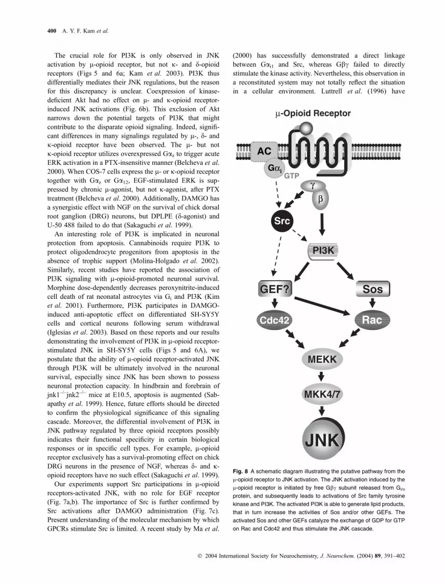
Fig. 8 A schematic diagram illustrating the putative pathway from the
l-opioid receptor to JNK activation. The JNK activation induced by the
l-opioid receptor is initiated by free Gbc subunit released from Gi/o
protein, and subsequently leads to activations of Src family tyrosine
kinase and PI3K. The activated PI3K is able to generate lipid products,
that in turn increase the activities of Sos and/or other GEFs. The
activated Sos and other GEFs catalyze the exchange of GDP for GTP
on Rac and Cdc42 and thus stimulate the JNK cascade.
400 A. Y. F. Kam et al.
� 2004 International Society for Neurochemistry, J. Neurochem. (2004) 89, 391–402

demonstrated that overexpressed Gbc in COS-7 cells elicits
c-Src activation. Intracellular signal-adapters might be required
for Gbc-stimulated Src activity. JNK activation by l-opioidreceptor is primarily mediated by Gbc due to a complete
suppression of the response after transducin coexpression
(Fig. 2b). Either direct activation of Src by Gai is insufficientto activate JNK or a Gbc-dependent signal is concomitantlyrequired for l-opioid receptor-induced JNK activation.
It is generally believed that PI3K and Src lie upstream of
Rho family GTPases (Han et al. 1998; Nagao et al. 1999).
The next issue is the relative position of PI3K and Src within
the l-opioid-activated JNK pathway. We employed an
experimental model in which JNK activation by SrcWT
was studied in contransfection system with PI3KcK832R, asSrcWT can functionally act as the active mutant to stimulate
MAPK (Della Rocca et al. 1997). A potent JNK stimulation
by SrcWT was partially suppressed by the coexpression of
PI3KcK832R (approximate 40% reduction; data not shown).
Thus, Src is presumably located upstream of PI3K in the
pathway. The incomplete reduction on the JNK activation
may be due to the presence of PI3K-independent pathway
from Src toward JNK. In agreement with a study, PI3K
stimulation by factors-VIIa was sensitive to Src inhibitor PP1
(Versteeg et al. 2000), indicating that Src is perhaps located
upstream of PI3K.
The present study provides a putative mechanism by
which l-opioid receptor activated JNK (Fig. 8). l-Opioidreceptor stimulated JNK in a PTX-sensitive and Gbc-dependent manner. The signal transduction undergoes
through Src family tyrosine kinase, Sos, Rac and Cdc42,
but not EGF receptor. However, one intermediate step
exclusively occurs in the l-opioid signaling, whereby l-,but not j- and d-opioid receptor requires PI3K to mediate
JNK activity. Hence, this is a distinct difference in the
signaling pathway between three opioid receptors.
Acknowledgements
We are extremely grateful to the following individuals for kindly
providing the cDNAs: Dr L. Yu for rat l-opioid receptor,
T. VoynoYasenetskaya for the JNK-HA, Dr Matthias P. Wymann
for wild-type and dominant negative mutant of PI3Kc, Dr S. Lin forwild-type and dominant negative mutant of Src, Dr E. J. Stanbridge
for RasS17N and RacT17N, Dr M. Symons for Cdc42T17 N
and RhoT17N, Dr Udo Schmitz for PAK1-PBD as well as
Dr R. J. Lefkowitz for Sos-Pro. This work was supported in part
by grants from the University Grants Committee of Hong Kong
(AoE/B-15/01), the Research Grants Council of Hong Kong
(HKUST 6115/00 M and 2/99C) and the Hong Kong Jockey Club.
References
Abe K., Rossman K. L., Liu B., Ritola K. D., Chiang D., Campbell S. L.,
Burridge K. and Der C. J. (2000) Vav2 is an activator of Cdc42,
Rac1, and RhoA. J. Biol. Chem. 275, 10141–10149.
Ammer H. and Schulz R. (1994) Retinoic acid-induced differentiation of
human neuroblastoma SH-SY5Y cells is associated with changes
in the abundance of G proteins. Neurochem. 62, 1310–1318.
Andjelkovic M., Alessi D. R., Meier R., Fernandez A., Lamb N., Frech
M., Cron P., Cohen P., Lucocq J. M. and Hemmings B. A. (1997)
Role of translocation in the activation and function of protein
kinase B. J. Biol. Chem. 272, 31515–31524.
Belcheva M. M., Wong Y. H. and Coscia C. J. (2000) Evidence for
transduction of mu but not kappa opioid modulation of extracel-
lular signal-regulated kinase activity by GZ and G12 proteins.
Cellular Signaling 12, 481–489.
Belcheva M. M., Vogel Z., Ignatova E., Avidor-Reiss T., Zippel R.,
Levy R., Young E. C., Barg J. and Coscia C. J. (1998) Opioid
modulation of extracellular signal-regulated protein kinase activity
is ras-dependent and involves Gbc subunits. J. Neurochem. 70,
635–645.
Belcheva M. M., Szucs M., Wang D., Sadee W. and Coscia C. J. (2001)
l-Opioid receptor-mediated ERK activation involves calmodulin-
dependent epidermal factor receptor transactivation. J. Biol. Chem.
276, 33847–33853.
van Biesen T., Hawes B. E., Luttrell D. K., Krueger K. M., Touhara K.,
Porfiri E., Sakaue M., Luttrell L. M. and Lefkowitz R. J. (1995)
Receptor-tyrosine-kinase- and Gbc-mediated MAP kinase activa-
tion by a common signaling pathway. Nature 376, 781–784.
Chan A. S. L. and Wong Y. H. (2000) Regulation of c-Jun N-terminal
kinase by the ORL1 receptor through multiple G proteins.
J. Pharmacol. Exp. Ther. 295, 1094–1100.
Chan J. S. C., Yung L. Y., Lee J. W. M., Wu Y. L., Pei G. and Wong Y.
H. (1998) Pertussis toxin-insensitive signaling of the ORL1receptor: coupling to Gz and G16 proteins. J. Neurochem. 71,
2203–2210.
Chen Y. H., Pouyssegur J., Courtneidge S. A. and Obberghen-Schilling
E. V. (1994) Activation of Src family kinase activity by the G
protein-coupled thrombin receptor in growth-responsive fibro-
blasts. J. Biol. Chem. 269, 27372–27377.
Cheng J., Standifer K. M., Tublin P. R., Su W. and Pasternak G. W.
(1995) Demonstration of kappa 3-opioid receptors in the SH-SY5Y
human neuroblastoma cell line. J. Neurochem. 65, 170–175.
Coso O. A., Chlariello M., Yu J. C., Teramoto H., Crespo P., Xu N., Miki
T. and Gutkind J. S. (1995) The small GTP-binding proteins Rac1
and Cdc42 regulate the activity of the JNK/SAPK signaling
pathway. Cell 81, 1137–1146.
Coso O. A., Teramoto H., Simonds W. F. and Gutkind J. S. (1996)
Signaling from G protein-coupled receptors to c-Jun kinase
involves bc subunits of heterotrimeric G proteins acting on a Ras
and Rac1-dependent pathway. J. Biol. Chem. 271, 3963–3966.
Della Rocca G. J., van Biesen T., Daaka Y., Luttrell D. K., Luttrell L. M.
and Lefkowitz R. J. (1997) Ras-dependent mitogen-activated
protein kinase activation by G protein-coupled receptors: conver-
gence of Gi- and Gq-mediated pathways on calcium/calmodulin,
Pyk2, and Src kinase. J. Biol. Chem. 272, 19125–19132.
Han J., Luby-Phelps K., Das B., Shu X., Xia Y., Mosteller R. D., Krishna
U. M., Falck J. R., White M. A. and Broek D. (1998) Role of
substrates and products of PI3-kinase in regulating activation of
Rac-related guanosine triphosphatases by Vav. Science 279, 558–
560.
Hawes B. E., Fried S., Yao X., Weig B. and Graziano M. P. (1998)
Nociceptin (ORL-1) and l-opioid receptors mediate mitogen-
activated protein kinase activation in CHO cells through a
Gi-coupled signaling pathway: evidence for distinct mechanisms of
agonist-mediated desensitization. J. Neurochem. 71, 1024–1033.
Hayashi T., Tsao L. I. and Su T. P. (2002) Antiapoptotic and cytotoxic
properties of delta opioid peptide [D-Ala(2),D-Leu(5)]enkephalin in
PC12 cells. Synapse 43, 86–94.
PI3K-dependent JNK activation by l-opioid receptor 401
� 2004 International Society for Neurochemistry, J. Neurochem. (2004) 89, 391–402

Iglesias M., Segura M. F., Comella J. X. and Olmos G. (2003) l-Opioidreceptor activation prevents apoptosis follwong serum withdrawal
in differentiated SH-SY5Y cells and cortical neurons via phos-
phatidylinositol 3-kinase. Neuropharmacology 44, 482–492.
Jo H., Sipos K., Go Y. M., Law R., Rong J. and McDonald J. M. (1997)
Differential effect of shear stress on extracellular signal-regulated
kinase and N-terminal Jun kinase in endothelial cells. J. Biol.
Chem. 272, 1395–1401.
Kam A. Y. F., Chan A. S. L. and Wong Y. H. (2003) Rac and Cdc42-
dependent regulation of c-JUN N-terminal kinases by the d-opioidreceptor. J. Neurochem. 84, 503–513.
Kazmi S. M. I. and Mishra R. K. (1987) Comparative pharmacological
properties and functional coupling of l and d opioid receptor sites
in human neuroblastoma SH-SY5Y cells. Mol. Pharmacol. 32,
109–118.
Kim M. S., Cheong Y. P., So H. S., Lee K. M., Kim T. Y., Oh J., Chung
Y. T., Son Y., Kim B. R. and Park R. (2001) Protective effects of
morphine in peroxynitrite-induced apoptosis of primary rat neo-
natal astrocytes: potential involvement of G protein and phospha-
tidylinositol 3-kinase (PI3 kinase). Biochem. Pharmacol. 61, 779–
786.
Kuan C. Y., Yang D. D., Roy D. R. S., Davis R. J., Rakic P. and Flavell
R. A. (1999) The JNK1 and JNK2 protein kinase regulate regional-
specific apoptosis during early brain development. Neuron 22,
667–676.
Law P. Y., Wong Y. H. and Loh H. H. (2000) Molecular mechanisms and
regulation of opioid receptor signaling. Annu. Rev. Pharmacol.
Toxicol. 40, 389–430.
Li J. and Smithgall T. E. (1998) Fibroblast transformation by Fps/Fes
tyrosine kinases requires Ras, Rac and Cdc42 and induces extra-
cellular signal-regulated and c-Jun N-terminal kinase activation.
J. Biol. Chem. 273, 13828–13834.
Lopez-Ilasaca M., Gutkind J. S. and Wetzker R. (1998) Phosphoinositide
3-kinase c is a mediator of Gbc-dependent Jun kinase activation.
J. Biol. Chem. 273, 2505–2508.
Luttrell L.M., Hawes B. E., van Biesen T., Luttrell D. K., Lansing T. J. and
Lefkowitz R. J. (1996) Role of c-Src tyrosine kinase in G protein-
coupled receptor- and Gbc subunit-mediated activation of mitogen-activated protein kinases. J. Biol. Chem. 271, 19443–19450.
Ma Y. C., Huang J., Ali S., Lowery W. and Huang X. Y. (2000) Src
tyrosine kinase is a novel direct effector of G proteins. Cell 102,
635–646.
Mattes H. W., Maldonado R., Simonin F. et al. (1996) Loss of morphine-
induced analgesia, reward effect and withdrawal symptoms in mice
lacking the l-opioid-receptor gene. Nature 383, 819–823.
Molina-Holgado E., Vela J. M., Arevalo-Martın A., Almazan G., Mo-
lina-Holgado F., Borrell J. and Guaza C. (2002) Cannabinoids
promote oligodendrocyte progenitor survival: involvement of
cannabinoid receptors and phosphatidylinositol-3 kinase/Akt
signaling. J. Neurosci. 22, 9742–9753.
Murga C., Laguinge L., Wetzker E., Cuadrado A. and Gutkind J. S.
(1998) Activation of Akt/protein kinase B by G protein-coupled
receptors: a role for a and bc subunits of heterotrimeric G proteins
acting through phosphatidylinositol-3-OH kinasec. J. Biol. Chem.273, 19080–19085.
Nagao M., Kaziro Y. and Itoh H. (1999) The Src family tyrosine kinase
is involved in Rho-dependent activation of c-Jun N-terminal kinase
by Ga12. Oncogene 18, 4425–4434.Nimnual A. S., Yatsula B. A. and Bar-Sagi D. (1998) Coupling of Ras
and Rac guanine-triphosphatases through the Ras exchanger Sos.
Science 279, 560–563.
Sabapathy K., Jochum W., Hochedlinger K., Chang L., Karin M. and
Wagner E. F. (1999) Defective neural tube morphogenesis and
altered apoptosis in the absence of both JNK1 and JNK2. Mech.
Dev. 89, 115–124.
Sakaguchi M., Fujimori T., Satoh T., Satoh M., Takeuchi M. and Mat-
sumura E. (1999) Effects of opioids on neuronal survival in culture
of embryonic chick dorsal root ganglion neurons. Neurosci. Lett.
262, 17–20.
Sawai T., Hirakawa T., Yamada K. and Nishizawa Y. (1999) Interaction
between pleckstrin homology domains and G protein bc-subunits:analyses of kinetic parameters by a biosensor-based method. Biol.
Pharm. Bull. 22, 229–233.
Schulz R., Wehmeyer A. and Schulz K. (2002) Opioid receptor types
selectively cointernalize with G protein-coupled receptor kinases 2
and 3. J. Pharmacol. Exp. Ther. 300, 376–384.
Singhal P. C., Bhaskaran M., Patel J., Patel K., Kasinath B. S., Du-
raisamy S., Franki N., Reddy K. and Kapasi A. A. (2002) Role of
p38 mitogen-activated protein kinase phosphorylation and Fas–Fas
ligand interaction in morphine-induced macrophage apoptosis.
J. Immunol. 168, 4025–4033.
Stephens L., Smrcka A., Cooke F. T., Jackson T. R., Sternweis P. C. and
Hawkins P. T. (1994) A novel phosphotidylinositol 3-kinase
activity in myeloid-derived cells is activated by G protein bcsubunits. Cell 77, 83–93.
Versteeg H. H., Hoedemaeker I., Diks S. H., Stam J. C., Spaargaren M.,
van Bergen en Henegouwen P. M. P., van Deventer S. J. H. and
Peppelenbosch M. P. (2000) Factor VIIa/tissue factor-induced
signaling via activation of Src-like kinases, phosphatidylinositol
3-kinase, and Rac. J. Biol. Chem. 275, 28750–28756.
Xia Z., Dickens M., Raingeaud J., Davis R. J. and Greenberg M. E.
(1995) Opposing effects of ERK and JNK-p38 MAP kinases on
apoptosis. Science 270, 1326–1331.
Xu Q. and Wu Z. (2000) The insulin-like growth factor-phosphatidy-
linositol 3-kinase-Akt signaling pathway regulates myogenin
expression in normal myogenic cells but not in rhabdomyosarco-
ma-derived RD cells. J. Biol. Chem. 275, 36750–36757.
Yamauchi J., Kawano T., Nagao M., Kaziro Y. and Itoh H. (2000)
Gi-dependent activation of c-Jun N-terminal kinase in human
embryonal kidney 293 cells. J. Biol. Chem. 275, 7633–7640.
402 A. Y. F. Kam et al.
� 2004 International Society for Neurochemistry, J. Neurochem. (2004) 89, 391–402


