Embed Size (px)
Citation preview

Wawrzynska et al. 2008 Plant Physiology
1
Running title: Suppression of edr1-mediated resistance to powdery mildew
Research Area: Plants Interacting with Other Organisms
*For correspondence: Roger Innes, Department of Biology, Indiana University, Bloomington, IN
47401
Phone: 1-812-855-2219; fax: 1-812-855-6082; email: [email protected]
Plant Physiology Preview. Published on September 24, 2008, as DOI:10.1104/pp.108.127605
Copyright 2008 by the American Society of Plant Biologists

Wawrzynska et al. 2008 Plant Physiology
2
Powdery Mildew Resistance Conferred by Loss of the EDR1 Protein
Kinase is Suppressed by a Missense Mutation in KEG, a Regulator
of ABA Signaling1
Anna Wawrzynska2, Katy M. Christiansen, Yinan Lan, Natalie L. Rodibaugh and Roger
W. Innes*
Department of Biology, Indiana University, Bloomington, IN 47405, USA

Wawrzynska et al. 2008 Plant Physiology
3
1This work was supported by the National Institutes of Health (grant number R01 GM063761 to
R.W.I.). Y.L. was supported by a Howard Hughes Medical Institute undergraduate research
award.
2Present address: Institute of Biochemistry and Biophysics, Polish Academy of Sciences,
Pawińskiego 5A Str, 02-106 Warsaw, Poland
*Corresponding author; email [email protected]; fax 1-812-855-6082
The author responsible for distribution of materials integral to the findings presented in
this article in accordance with the policy described in the Instructions for Authors
(www.plantphysiol.org) is: Roger Innes ([email protected])

Wawrzynska et al. 2008 Plant Physiology
4
ABSTRACT
Loss-of-function mutations in the Arabidopsis ENHANCED DISEASE RESISTANCE 1
(EDR1) gene confer enhanced resistance to infection by powdery mildew (Golovinomyces
cichoracearum). EDR1 encodes a protein kinase, but its substrates, and the pathways regulated
by EDR1 are unknown. To identify components of the EDR1 signal transduction pathway(s) we
conducted a forward genetic screen for mutations that suppressed edr1-mediated disease
resistance. Genetic mapping and cloning of one of these suppressor mutations revealed a
recessive missense mutation in the KEEP ON GOING gene (KEG; At5g13530), which we
designated keg-4. KEG encodes a multi-domain protein that includes a RING E3 ligase domain,
a kinase domain, ankyrin repeats and HERC2-like repeats. The KEG protein has previously
been shown to have ubiquitin ligase activity and to negatively regulate protein levels of the
transcription factor ABCISIC ACID INSENSITIVE 5 (ABI5). KEG mRNA levels were found
to be three fold higher in edr1 mutant plants compared to wild type. Loss-of-function mutations
in KEG are seedling lethal and are hypersensitive to glucose and abscisic acid (ABA). The keg-4
mutation, in contrast, conferred resistance to 6% glucose, and suppressed edr1-mediated
hypersensitivity to ABA, suggesting that the keg-4 mutation suppresses ABA signaling by
altering KEG function. Several ABA-responsive genes were found to be further upregulated in
the edr1 mutant following ABA treatment, and this upregulation was suppressed by the keg-4
mutation. We conclude that edr1-mediated resistance to powdery mildew is mediated, in part, by
enhanced ABA signaling.

Wawrzynska et al. 2008 Plant Physiology
5
Powdery mildew fungi are obligate biotrophic pathogens that can grow only on living plant
tissues. These pathogens must evade or suppress host defenses until their life cycle is complete.
A number of Arabidopsis mutants displaying enhanced disease resistance to powdery mildew
(Golovinomyces cichoracearum) have been characterized (Frye and Innes, 1998; Vogel and
Somerville, 2000; Vogel et al., 2002; Tang et al., 2005, 2006). These mutants can be grouped
into two broad classes based on the presence or absence of mildew-induced lesions. The edr1,
edr2 and edr3 mutants typify the former class (Frye and Innes, 1998; Tang et al., 2005, 2006). In
these mutants, fungal growth is inhibited at a very late stage of the infection process and
resistance correlates with a more rapid activation of host defenses relative to wild-type plants,
including programmed cell death (PCD). The most striking phenotypes caused by the edr1
mutation besides powdery mildew-induced lesions, are enhanced drought-induced growth
inhibition, and enhanced ethylene-induced senescence (Frye et al., 2001; Tang et al., 2005). The
former two phenotypes require an intact salicylic acid (SA) signaling pathway, while the latter
does not (Tang et al., 2005). The general processes of PCD, drought responses and senescence
have all been linked to enhanced sensitivity to abscisic acid (ABA) (Beaudoin et al., 2000;
Ghassemian et al., 2000; Anderson et al., 2004; Mohr and Cahill, 2007; Xie et al., 2007),
suggesting that EDR1 may be also be involved in ABA signaling (Frye et al., 2001).
ABA regulates many important events during both vegetative and reproductive growth of
plants. These range from relatively slow effects, such as promotion of seed storage reserve
synthesis, acquisition of desiccation tolerance and dormancy, and tolerance to drought, salt, and
cold stresses (Leung and Giraudat, 1998), to rapid effects, such as stomatal closure (Leung and
Giraudat, 1998; Finkelstein et al., 2002). Cumulative evidence suggests that the crosstalk
between ABA and SA is important for adaptation of plants to combinations of abiotic and biotic
stresses (Kunkel and Brooks, 2002; Mauch-Mani and Mauch, 2005). SA inhibits ABA-induced
stomatal closure (Rai et al., 1986), leaf abscission (Apte and Laloraya, 1982), and inhibition of
seedling growth (Ray, 1986), while ABA increases susceptibility to biotrophic pathogens by
counteracting SA-dependent defenses (Mohr and Cahill, 2003; de Torres-Zabala et al., 2007;
Mohr and Cahill, 2007). Conversely, ABA-dependent priming of callose biosynthesis promotes
enhanced resistance to some necrotrophic pathogens (Ton and Mauch-Mani, 2004).
The complex connections between SA signaling and ABA signaling are also observed
during leaf senescence, which shares many physiological events with pathogen-induced defence

Wawrzynska et al. 2008 Plant Physiology
6
responses, such as increases in ethylene and SA levels (Ryals et al., 1996; Morris et al., 2000),
accumulation of H2O2 (Levine et al., 1994; Pastori and Del Rio, 1997) and accumulation of
transcripts from pathogenesis-related (PR) genes (Hanfrey et al., 1996; Butt et al., 1998; Pontier
et al., 1999; Quirino et al., 1999; Quirino et al., 2000; Yoshida et al., 2001). ABA is considered a
senescence promoter, although evidence for an in vivo role is rather poor compared with ethylene
(Nooden and Leopold, 1988; Madhu et al., 1999; Panavas et al., 1999). Several mutations that
inhibit defense responses in Arabidopsis also inhibit senescence (Morris et al., 2000). For
example, the pad4 mutation, which enhances disease susceptibility and reduces SA accumulation
(Jirage et al., 1999), displays a dramatic delay in PCD during senescence (Morris et al., 2000).
Consistent with these observations, SA levels increase approximately 4-fold in senescing
Arabidopsis leaves (Morris et al., 2000). Determining cause and effect in these processes is
difficult, however, as SA signaling pathways include positive feedback loops. For example, cell
death promotes SA production, but SA production also promotes cell death (Glazebrook, 2005).
Accordingly, it has been proposed that high concentrations of SA, such as those generated at the
sites of pathogen entry, are required for cell death induction, whereas SA at low levels, detected
beyond the margins of the initial infection sites, might lead to cell survival and lesion
containment (Alvarez, 2000).
Because loss of EDR1 function leads to enhanced PCD and senescence, it is considered
to be a negative regulator of these processes. The EDR1 protein belongs to a small family of
protein kinases in Arabidopsis that includes the CTR1 protein (Frye et al., 2001), a negative
regulator of ethylene responses (Kieber et al., 1993; Cao et al., 1997). Unlike loss of CTR1
function, however, loss of EDR1 does not activate ethylene signaling pathways (Frye et al.,
2001). The specific function of EDR1 thus remains unknown. To uncover additional genes in
the EDR1 kinase pathway, or identify other pathways that interact with the EDR1 pathway, we
performed a suppressor screen to identify mutations that suppress the edr1 mutant phenotype.
Here we describe one such suppressor mutation, which was found to be a missense mutation in
the KEG gene. KEG encodes a putative ubiqutin ligase thought to be involved in ABA signaling
(Stone et al., 2006). Interestingly, we found that transgenic overexpression of KEG induces
massive cell death in Arabidopsis.
RESULTS

Wawrzynska et al. 2008 Plant Physiology
7
The supp69 Mutation Blocks EDR1-Dependent Resistance to G. cichoracearum
Because edr1 mutant plants show enhanced drought-induced growth inhibition (Tang et
al., 2005), we suspected that the edr1 mutation might be enhancing sensitivity to ABA. To test
this hypothesis, we performed a seed germination assay on varying levels of ABA, which is
known to inhibit germination (Finkelstein, 1994). Figure 1 shows that the edr1 mutant is indeed
hypersensitive to exogenous ABA as the percent germination at three days of incubation on 0.6
µM ABA was only ~15% for edr1 seeds compared to greater than 50% for wild type. This
enhanced ABA sensitivity suggested that we could enrich for suppressor mutants by germinating
mutagenized seed on ABA-containing plates.
To enrich for edr1 suppressor mutants, we screened an ethyl-methane sulfonate
mutagenized edr1 population on agar plates containing 0.7 µM ABA (60,000 M2 seeds derived
from 3500 M1 edr1 plants). Approximately 1000 seedlings were identified that germinated
within the first three days of incubation, a time period during which very few edr1 mutant seed
had germinated. Seedlings were transplanted to pots containing Metromix 360. Four to five
weeks later these plants were inoculated with G. cichoracearum and scored for disease responses
8 days post inoculation (dpi). Seventy-four mutants displaying visible powder were selected and
their respsonse to G. cichoracearum retested in the next generation. Among these, 11 mutants
were found to be fully susceptible to G. cichoracearum, lacking edr1-dependent necrotic lesions
and allowing abundant development of G. cichoracearum conidiophores. Here we describe one
mutant, which was designated supp69. Characterization of the other mutants is ongoing and will
be described elsewhere.
The supp69 mutant displayed a wild-type Col-0-like phenotype 8 days after infection
with G. cichoracearum (Fig. 2). No other obvious developmental or morphological phenotypes
of supp69 plants were observed when grown under normal conditions. Complementation tests
revealed that supp69 is not allelic to pad4 or npr1 (not shown), which have been previously
shown to suppress the edr1 phenotype (Tang et al., 2005). Segregation analysis of a back-cross
to the edr1 mutant revealed that susceptibility to G. cichoracearum was caused by a single
recessive mutation. We therefore proceeded with genetic mapping of the supp69 mutation and a
detailed characterization of the supp69 mutant phenotype.

Wawrzynska et al. 2008 Plant Physiology
8
The supp69 Mutation Maps to Chromosome 5
Genetic mapping of the supp69 mutation was complicated by a lack of edr1 mutant
alleles in Arabidopsis accessions other than Col-0. We therefore crossed the supp69 mutant
(edr1-supp69) to the Landsberg erecta accession (Ler) and identified F3 families that were
homozygous for the edr1 mutation and segregating for the supp69 mutation (see Methods).
Twenty-eight F3 families were selected and pooled for mapping purposes. F3 plants were scored
for susceptibility to G. cichoracearum. Susceptibility segregated in an approximately 1:3 ratio,
confirming that supp69 was caused by a single recessive mutation. DNA was isolated from 629
susceptible F3 plants and scored for microsatellite markers distributed across the Arabidopsis
genome. Initially, the supp69 mutation was mapped to a region between microsatellite markers
MYH9 and nga151 on chromosome 5 (Fig. 3). To further localize the mutated gene, we created
PCR-based markers at intervals between MYH9 and nga151 using small insertions/deletions that
are polymorphic between Ler and Col-0 (Jander et al., 2002). Fine mapping localized the
mutation to a 126 kb interval covering the 3’-end of bacterial artificial chromosome (BAC) clone
T6I14 (GenBank accession AL391710) and the 5’-end of BAC MSH12 (GenBank accession
AB006704) defined by one recombinant at each border (Fig. 3). This region harbors 33 genes
(from At5g13470 to At5g13750). Twenty-one of these were amplified by PCR and sequenced.
A single G to A transition mutation was identified in At5g13540, which was recently found to be
misannotated, with the full ORF encompassing both the At5g13530 and At5g13540 loci (Stone
et al., 2006). The combined gene has been named KEG for “keep on going” (Stone et al., 2006).
The supp69 mutation was located in the 15th exon of KEG, and causes a glycine to serine
substitution (G1144S) in the HERC2-like domain of the KEG protein (Fig. 3) (Stone et al.,
2006).
Loss-of-function keg mutants display a strong post-germinative growth arrest shortly
after the emergence of the first true leaves (Stone et al., 2006), indicating that KEG is essential
for plant development. Because supp69 plants show normal growth and development, we
conclude that the mutation in KEG does not cause a complete a loss-of-function. As three keg
mutants have been described previously (Stone et al., 2006), the keg mutation in supp69 was
designated keg-4.

Wawrzynska et al. 2008 Plant Physiology
9
Complementation of the supp69 Mutation
To confirm that the mutation in At5g13530 was responsible for the suppression of the
edr1 phenotype, we transformed supp69 plants with a genomic copy of the KEG gene under
control of its native promoter and tested for restoration of the edr1 mutant phenotype. Thirteen
independent T1 transgenic plants were inoculated with G. cichoracearum. All thirteen were
resistant to G. cichoracearum and showed necrotic lesions and almost no conidiation 8 days after
infection, demonstrating that the KEG genomic construct complemented the keg-4 mutation (data
not shown). The transgene did not cause any growth phenotypes, as all transgenic lines were
indistinguishable from WT Col-0 plants prior to inoculation.
The keg-4 Mutation Suppresses the Enhanced Ethylene-Induced Senescence Phenotype of
edr1
Besides showing enhanced resistance to powdery mildew, edr1 mutants display an
enhanced ethylene-induced senescence phenotype (Frye et al., 2001). To test whether the keg-4
mutation also suppressed this trait, we exposed plants to ethylene (100 ppm) for 3 days. The
supp69 plants showed the same rate of senescence as Col-0 plants in contrast to the enhanced
senescence phenotype of edr1 (Fig. 4A). To determine whether the keg-4 mutation by itself
affected senescence rates, we crossed out the edr1 mutation by backcrossing to wild-type Col-0
plants and selecting homozygous EDR1/EDR1 keg-4/keg-4 plants in the F2. Ethylene induced
visible chlorosis (yellowing) on the oldest 2-3 leaves of WT Col-0, supp69 and keg-4 plants after
3 days exposure to ethylene. However, in edr1 mutant plants, chlorosis were observed on much
younger leaves and appeared earlier (Fig. 4A). Quantification of chlorophyll levels revealed
significant differences between ethylene-treated edr1 plants and the other three genotypes (Fig.
4B). Consistent with this finding, edr1 plants also showed a faster rate of senescence under
standard short-day growth conditions, which became visibly obvious by 11 weeks of growth
(Fig. 4C). We thus conclude that the keg-4 mutation by itself does not delay senescence in a
wild-type background, but fully suppresses the enhanced senescence of the edr1 mutant.
The keg-4 Mutation Suppresses edr1-Mediated Drought Induced Growth Inhibition

Wawrzynska et al. 2008 Plant Physiology
10
We previously reported that edr1 plants appeared more sensitive than WT Col-0 plants to
under-watering, often growing slower than WT Col-0 plants (Tang et al., 2005). To further
characterize supp69, we grew plants under standard growth conditions for 3 weeks and then
stopped watering them for 2 weeks. The edr1 plants were significantly smaller than WT Col-0,
supp69 or keg-4 plants at the end of the 2-week-drought period, although they were the same size
at the start (Fig. 5A). Control edr1 plants grown with the standard watering regime did not
significantly differ in size from Col-0, supp69 or keg-4 at 5 weeks (Fig. 5A). To quantify the
edr1-mediated drought-induced growth phenotype, we weighed the individual plants (fresh
weight) grown under standard or drought conditions. Figure 5B shows that edr1 mutant plants
weighed almost the same as the other tested plants when grown under standard conditions, but
weighed significantly less when grown under drought conditions. These data indicate that keg-4
also suppresses the edr1-mediated drought sensitivity.
The keg-4 Mutation Suppresses the ABA Hypersensitivity of edr1
The suppression of edr1-mediated drought sensitivity suggested that the keg-4 mutation
should also suppress the ABA hypersensitivity of edr1 mutants. We therefore tested the supp69
mutant for sensitivity to ABA using the seed germination assay described above. We plated
seeds on MS agar containing 0.7 µM ABA. As described above, this level of ABA inhibited
germination of edr1 seeds more than wild-type seeds, which resulted in noticeably smaller
seedlings at 5 days of incubation (Fig. 6). The supp69 and keg-4 mutant seedlings were
indistinguishable from wild-type plants in this assay, confirming that keg-4 also suppresses edr1-
mediated ABA hypersensitivity.
The supp69 and keg-4 Mutants Show Lowered Sensitivity to Glucose
Null alleles of KEG4 have been shown previously to confer hypersensitivity to
exogenous glucose (Stone et al., 2006), which causes growth arrest of wild-type Arabidopsis
seedlings when added to agar at 6% (Zhou et al., 1998; To et al., 2002). Glucose influences
post-germinative growth of Arabidopsis via its ability to activate ABA biosynthesis genes and

Wawrzynska et al. 2008 Plant Physiology
11
consequent activation of ABA-inducible genes (Cheng et al., 2002; Finkelstein and Gibson,
2002), and many glucose insensitive mutants are also ABA insensitive. We therefore tested
whether the keg-4 mutation affected glucose sensitivity. Significantly, the keg-4 mutation
conferred glucose insensitivity (Fig. 7). After 15 days on glucose-containing media, the supp69
and keg-4 seedlings showed only a slight growth inhibition relative to plants grown in the
absence of glucose, while WT Col-0 and edr1 seeds showed almost no growth (Fig. 7). All
mutants and wild-type plants grew similarly on plates containing 6% mannitol, a non-
metabolizable sugar, demonstrating that the response was not simply the result of osmotic stress
(Fig. 7). There were also no significant differences between plants grown on MS with 6%
sucrose (data not shown). These data indicate that the keg-4 allele confers phenotypes opposite to
that of a keg loss-of-function mutation (Stone et al., 2006), and further support our conclusion
that the keg-4 mutation inhibits ABA signaling.
The keg-4 Mutation Suppresses edr1-Mediated Changes In Gene Expression
ABA induces the expression of many genes that are important for adaptation to stress.
Based on the edr1 mutant phenotypes observed above and the ability of the keg-4 mutation to
suppress them, we hypothesized that EDR1 might play a role in ABA-induced changes in gene
expression. To test this hypothesis, we examined the expression of RD29A (RESPONSIVE TO
DESSICATION 29A, At5g52310), a well-characterized ABA-inducible gene (Yamaguchi-
Shinozaki and Shinozaki, 2006). In WT Col-0, treatment with 100 µM ABA for 3h induced
RD29A (Fig. 8A). Induction of this gene was greater in the edr1 mutant, consistent with edr1
being hypersensitive to ABA. The keg-4 mutation fully suppressed the enhanced expression in
edr1, indicating that keg-4 suppresses edr1-mediated hypersensitivity at the level of gene
induction (Fig. 8A).
We also analyzed expression of ABI5 (abscisic acid insensitive 5, At2g36270) and MPK3
(MAP kinase 3, At3g45640). ABI5 is a key transcription factor required for the induction of
many ABA responsive genes, and is itself inducible by ABA (Lopez-Molina et al., 2001;
Brocard et al., 2002). It is most highly expressed in germinating seeds (Lopez-Molina et al.,
2001; Brocard et al., 2002). MPK3 has been linked to ABA signaling in seedlings because
overexpression of MPK3 increases ABA sensitivity in ABA-induced postgermination growth

Wawrzynska et al. 2008 Plant Physiology
12
arrest (Lu et al., 2002). In addition, Arabidopsis plants with guard cell specific silencing of
MPK3 display a partial insensitivity to ABA-mediated inhibition of stomatal opening (Gudesblat
et al., 2007). Although ABA treatment does not activate MPK3 in leaf mesophyll protoplasts
(Kovtun et al., 2000), ABA does appear to activate MPK3 in Arabidopsis seedlings (Lu et al.,
2002). ABI5 was highly induced by ABA treatment in both wild-type and edr1 seedlings (Fig.
8B), indicating that there is a positive feedback loop regulating this gene. Induction appeared to
be slightly enhanced in the edr1 mutant, but this difference was not statistically significant. In
contrast, MPK3 transcript levels were suppressed approximately two-fold in wild-type plants in
response to ABA treatment, whilst in edr1 seedlings MPK3 was induced two-fold, resulting in a
four-fold difference in MPK3 transcript levels between wild-type and edr1 mutant seedlings.
This difference was fully suppressed by the keg-4 mutation.
We also examined the expression of a two powdery-mildew inducible genes, ERF1
(ethylene responsive binding factor 1, At4g17500) and XET (xyloglucan endotransglycosylase,
At5g57560). At 30 hours post inoculation (hpi), both genes were induced about three fold in
wild-type Col-0 plants (Fig. 8C). The basal and induced levels of ERF1 were higher in the edr1
mutant, while only the induced levels of XET1 were higher. The keg-4 mutation restored the
expression of both genes to wild-type levels. Thus the keg-4 mutation is able to suppress the
effect of edr1-induced changes in gene expression for both ABA- and pathogen-responsive
genes.
Overexpression of KEG Leads to Massive Cell Death
To further analyze the impact of KEG expression in plant development, we tested the
effect of overexpressing the KEG gene in transgenic Arabidopsis plants. We constructed
transgenic plants expressing the full-length KEG cDNA under the control of the constitutive
CaMV 35S promoter (35S::KEG). We were unable to obtain any transformants containing the
35S::KEG construct, which suggested that constitutive overexpression of KEG in Arabidopsis
may be lethal. We therefore constructed transgenic Col-0 plants expressing the KEG cDNA
under the control of a steroid-inducible promoter (Aoyama and Chua, 1997). We generated 24
transgenic lines containing this construct. All developed large necrotic lesions within 40 hrs of
treatment with 50 µM dexamethasone (Fig. 9). In contrast, none of the DEX-treated leaves of

Wawrzynska et al. 2008 Plant Physiology
13
control plants or ethanol-treated plants containing the DEX::KEG transgene showed any visible
cell death (data not shown). These data suggest that ectopic overexpression of KEG results in cell
death and that the level of KEG is tightly controlled.
Analysis of KEG Expression
To gain insight into the spatial and temporal pattern of KEG expression, we searched the
Arabidopsis microarray data available through the Genevestigator Web interface
(https://www.genevestigator.ethz.ch/). Microarray analyses showed that KEG is expressed in
various tissues and organs at all developmental stages and is not specifically induced by any
factor. To investigate KEG expression more directly, we constructed transgenic plants expressing
a KEG promoter::GUS fusion. A 1044 base-pair fragment 5’ to the KEG start codon was fused to
the β-glucuronidase (GUS) reporter gene, and the construct was transformed into WT Col-0
plants. We obtained a number of pKEG::GUS transformants and analyzed a total of 24 transgenic
lines. Figure S1 shows representative GUS staining patterns. GUS staining was observed in all
tissues of 8 days old seedlings (most prominent in the meristem parts), consistent with the
microarray data. However, in 7 week-old flowering plants, GUS staining was only observed in
the youngest parts of the stem, anthers, and the receptacle of immature siliques. No staining was
observed in mature leaves, older parts of the stem, flower parts other than anthers, or mature
siliques. These results suggest that the expression of KEG may be under developmental
regulation and that KEG is expressed mainly in the actively growing and dividing cells.
To further investigate the expression KEG and EDR1, we performed quantitative RT-
PCR analyses. In one week-old seedlings, treatment with 100 µM ABA for 3h induced the
expression of both EDR1 and KEG (Fig. S2A). The edr1 mutation did not affect KEG transcript
levels. We also examined expression of these genes in 5 week-old plants 30h after powdery
mildew treatment (Fig. S2B). Both EDR1 and KEG were slightly induced by powdery mildew.
The transcript level of EDR1 was reduced about two-fold in the edr1 mutant compared to the
WT Col-0. Because the edr1 mutation creates an early stop codon (Frye, et al., 2001), this
reduction in edr1 transcript level is likely due to nonsense mediated decay (NMD), a cellular
mechanism of mRNA surveillance that detects nonsense mutations and prevents the expression
of truncated or erroneous proteins (Chang et al., 2007). Interestingly, the level of KEG transcript
Wawrzynska et al. 2008 Plant Physiology
14
in 5 week-old plants was about 3-fold greater in edr1 than WT Col-0 plants (Fig. S2B),
suggesting that that EDR1 negatively regulates KEG transcription in rosette leaves.
DISCUSSION
The edr1 mutant of Arabidopsis displays enhanced resistance to powdery mildew and
undergoes more rapid senescence than wild-type plants when exposed to ethylene (Frye and
Innes, 1998). In addition, edr1 mutants display enhanced growth inhibition and spontaneous cell
death in response to drought (Tang et al., 2005), and enhanced cell death mediated by the RPW8
powdery mildew resistance gene (Xiao et al., 2005). All of these phenotypes, except for
ethylene-induced senescence, can be suppressed by mutations in the salicylic acid (SA) signaling
pathway (sid2, npr1, pad4 and eds1) (Frye et al., 2001; Tang et al., 2005; Xiao et al., 2005).
However, edr1 mutant plants do not show constitutive expression of SA-inducible genes when
grown under optimal conditions (Frye and Innes, 1998), thus it has been unclear why loss of
EDR1 function leads to these various phenotypes. The data presented above suggest that there
may be a mechanistic link between EDR1 function, ABA signaling, and SA enhancement of
programmed cell death.
Several lines of evidence point to ABA as a central player in edr1-mediated phenotypes.
The most direct is the hypersensitivity of edr1 mutant seeds to ABA-mediated inhibition of
germination (Fig. 1). In addition, the enhanced drought-induced growth inhibition of edr1 plants
is consistent with enhanced ABA sensitivity (Fig. 5), as is the enhanced induction of RD29A by
exogenous ABA (Fig. 8A). Most compelling, however, is the identification of the keg-4 missense
mutation in KEG, which suppresses all known edr1-mediated phenotypes. Because loss of
function mutations in KEG cause accumulation of the ABI5 transcription factor and ABA
hypersensitivity, and because KEG physically associates with ABI5, KEG is believed to be a
central regulator of ABA signaling (Stone et al., 2006). Furthermore, loss-of-function mutations
in ABI5 substantially suppress the phenotypes caused by loss-of-function mutations in KEG,
indicating that a primary role of KEG is regulating ABI5 levels (Stone et al., 2006). The
observation that keg-4 suppresses all known edr1-mediated phenotypes, including ethylene-
induced senescence, appears to place KEG function upstream of SA signaling, because mutations
in SID2, NPR1, PAD4 and EDS1 do not suppress the ethylene induced senescence phenotype of
edr1 (Tang et al., 2005). Understanding the function of KEG and the nature of the keg-4

Wawrzynska et al. 2008 Plant Physiology
15
mutation thus appears key to understanding how the edr1 mutation confers its various
phenotypes.
The KEG protein is quite large (178 kD) and contains multiple functional domains (Fig.
3). Starting at the N-terminal end, these are the RING (for Really Interesting New Gene) E3
ligase domain, a kinase domain, nine tandem ankyrin repeats, and twelve HERC2-like (for
HECT and RCC1-like) repeats. The RING domain of KEG has been shown to have E3
ubiquitin-ligase activity in vitro, and the kinase domain autophosphorylates in vitro (Stone et al.,
2006). The ankyrin-repeats are required for interaction between KEG and the ABI5 transcription
factor, at least in in vitro pull down assays (Stone et al., 2006). The function of the HERC2-like
repeats is unknown. The HERC2-like repeats is where the keg-4 mutation is located, however,
suggesting that this domain plays a critical role in KEG function.
The keg-4 mutation causes a glycine to serine substitution in the fifth HERC2-like repeat
(Fig. 3). This glycine residue is highly conserved among the 12 HERC2-like repeats of KEG
(Stone et al., 2006). The HERC2-like repeat were first defined in the Arabidopsis KEG protein
as a 61 amino acid motif with similarity to the mammalian HERC2 protein, which is a HECT-
type ubiquitin E3 ligase (Garcia-Gonzalo and Rosa, 2005; Stone et al., 2006). However, HERC2
contains only a single copy of this motif. The combination of multiple HERC2-like repeats and
a RING E3-ligase domain appears to be unique to plants. Single KEG homologs have been
identified in Oryza sativa (rice), Medicago truncatula, and Populus tricocarpa (Stone et al.,
2006), thus KEG appears to be highly conserved among angiosperms.
The phenotypes conferred by the keg-4 mutation are generally opposite to the phenotypes
conferred by loss-of-function mutations in KEG. In particular, the keg-4 mutant is resistant to
high levels of exogenous glucose, while keg loss-of-function mutants are hypersensitive to
glucose (Fig. 7) (Stone et al., 2006), suggesting that keg-4 has reduced sensitivity to ABA rather
than hypersensitivity. Consistent with this, keg-4 suppressed the ABA hypersensitivity of edr1
(Fig. 6). Also, keg loss-of-function mutants undergo a growth arrest shortly after germination,
consistent with hypersensitivity to endogenous ABA, while keg-4 mutants germinate and grow
similar to wild-type plants on half-strength MS agar. One plausible explanation for these
observations is that the HERC2-like repeat domain functions to regulate the E3 ligase activity of
KEG in response to ABA. In the absence of ABA, KEG presumably keeps ABI5 protein levels
low via ubiquitylating ABI5 and thus targeting it for degradation. In the presence of ABA,

Wawrzynska et al. 2008 Plant Physiology
16
protein levels of ABI5 increase dramatically, thus ABA must somehow prevent the
ubiquitylation of ABI5, presumably by modifying KEG activity (Stone et al., 2006). We
speculate that the keg-4 mutation renders KEG insensitive to ABA by modifying the structure of
the HERC2-like repeats, locking KEG in an ‘on position’ relative to ABI5 ubiquitylation.
One observation that seems inconsistent with the above model is the recessive nature of
the keg-4 mutation. If the keg-4 mutation causes KEG to constitutively ubiquitylate ABI5 even
in the presence of ABA, then one would expect reduced ABI5 levels, and thus reduced
responsiveness to ABA, even in a heterozygous state. We attempted to assess ABI5 protein
levels directly in edr1 and keg-4 mutant seedlings, both in the presence and absence of
exogenous ABA, using a previously described antibody (Lopez-Molina et al., 2001), but were
unable to detect any protein, even in wild-type ABA-treated seedlings. It was thus not possible
to compare ABI5 protein levels between the various mutant and wild-type plants. Given the
recessive nature of keg-4, we speculate that the level of mutant KEG protein present in
heterozygous plants is insufficient to ubiquitylate all of the ABI5 protein, and that there may be a
minimum threshold level of ABI5 that the plant must go below to suppress edr1-mediated
signaling.
The edr1 mutant displays approximately three-fold elevated levels of KEG mRNA in
rosette leaves, both before and after powdery mildew inoculation (Fig. S2). One would expect
this to lead to increases in KEG protein levels and thus decreases in ABA sensitivity. It is
possible, however, that this increase simply reflects an elevated steady-state level of ABA
signaling in the edr1 mutant as exogenous application of ABA induces KEG transcript levels
about three fold in wild-type seedlings (Fig. S2). If EDR1 normally functions as a negative
regulator of ABA signaling, then plants may compensate for loss of EDR1 function by increasing
KEG levels. This compensation may allow for normal growth under non-stressed conditions, but
under times of abiotic or biotic stress, is insufficient, leading to overactivation of ABA pathways.
KEG overexpression in mature rosettes leads to rapid cell death (Fig. 9). This phenotype
cannot be explained by reduction in ABI5 protein levels, alone, as ABI5 null mutants are viable
(Finkelstein and Lynch, 2000; Lopez-Molina and Chua, 2000). This observation suggests that
KEG may have other substrates in addition to ABI5, and/or that KEG overexpression leads to
ubiquitylation and degradation of inappropriate substrates. The former hypothesis is supported
by the finding that abi5 mutations do not fully suppress KEG loss-of-function mutant phenotypes

Wawrzynska et al. 2008 Plant Physiology
17
(Stone et al., 2006). Furthermore, KEG loss-of-function mutations partially suppress the ABA
insensitivity of abi5-1 mutants (Stone et al., 2006), which suggests that loss of KEG causes
accumulation of an ABA-responsive transcription factor that can compensate for loss of ABI5
function. If KEG regulates the levels of multiple transcription factors, then keg-4 suppression of
edr1 phenotypes may be a consequence of reducing the levels of these specific transcription
factors in addition to ABI5.
Our data indicate that edr1 plants have enhanced ABA signaling and that this is causally
related to enhanced resistance to powdery mildew. This conclusion would seem to be at odds
with recent work showing that the hemibiotrophic bacterial pathogen Pseudomonas syringae
strain DC3000 specifically induces ABA biosynthesis in Arabidopsis to promote virulence, and
that ABA insensitive Arabidopsis mutants have enhanced resistance to strain DC3000 (de
Torres-Zabala et al., 2007). If our model is correct, one would predict that the edr1 mutant
should have enhanced susceptibility to P. syringae, but in fact, edr1 was originally isolated in a
screen for mutants with enhanced resistance to P. syringae (Frye and Innes, 1998). Further
analysis of the edr1 mutant phenotype, however, revealed that the enhanced resistance to P.
syringae was variable, and in non-stressed plants, was not reproducible (Frye and Innes, 1998).
We speculate that the enhanced responses of edr1 to abiotic stresses such as drought may
indirectly promote resistance to P. syringae, and that the edr1 mutant was isolated in the original
screen because it had undergone a stress response prior to inoculation.
The role of ABA in regulating defense responses is still poorly understood and,
depending on the pathogen studied, ABA may enhance resistance or enhance susceptibility
(Mauch-Mani and Mauch, 2005). For example, both salt stress and exogenous ABA enhance the
resistance of barley to the biotrophic powdery mildew fungus Blumeria graminis (Wiese et al.,
2004). Likewise, ABA application enhances resistance of Arabidopsis to the oomycete Pythium
irregulare, while ABA deficient and ABA insensitive mutants are more susceptible (Adie et al.,
2007). Similarly, ABA application protects Arabidopsis against the necrotrophic fungi Alternaria
brassicicola and Plectosphaerella cucumerina, while the ABA deficient and ABA insensitive
mutants display enhanced susceptibility to these pathogens (Ton and Mauch-Mani, 2004; Adie et
al., 2007). Finally, ABA deficient and ABA insensitive Arabidopsis mutants also display
enhanced susceptibility to soil-borne bacterium Ralstonia solanacearum (Hernandez-Blanco et
al., 2007). We are currently testing whether the abi-5 mutation, and other mutations that confer

Wawrzynska et al. 2008 Plant Physiology
18
ABA insensitivity or ABA deficiency, affect edr1-mediated resistance to powdery mildew. We
predict that abi-5 should at least partially suppress edr1.
Although the above examples show that ABA can positively regulate resistance against
some pathogens, including powdery mildews, the majority of studies have shown that ABA
promotes susceptibility to pathogens. Early studies showed that exogenous application of ABA
enhanced susceptibility of potato tubers to Phytophthora infestans and Cladosporium
cucumerinum (Henfling et al., 1980), the susceptibility of soybean to Phytophthora megasperma
f.sp. glycinea (Ward et al., 1989), the susceptibility of rice to rice blast (Magnaportha grisea)
(Matsumoto et al., 1980), and the susceptibility of tobacco to blue mold (Peronospora tabacina)
(Salt et al., 1986). More recently, ABA treatment has been shown to increase the susceptibility
of Arabidopsis to Peronospora parasitica and the avirulent bacterium P. syringae pathovar
tomato strain 1065 (Mohr and Cahill, 2003), but not to the virulent strain DC3000. As described
above, though, strain DC3000 upregulates endogenous ABA levels in Arabidopsis (de Torres-
Zabala et al., 2007), thus exogenous ABA may have little effect on this strain. ABA application
also enhances the susceptibility of Arabidopsis to Fusarium oxysporum (Anderson et al., 2004)
and the susceptibility of tomato to Botrytis cineria (Audenaert et al., 2002). Consistent with this,
ABA deficient Arabidopsis mutants are more resistant to F. oxysporum (Anderson et al., 2004)
and B. cinerea (Adie et al., 2007), and the ABA deficient sitiens mutant of tomato is more
resistant to B. cineria (Audenaert et al., 2002), and to P. syringae (Thaler and Bostock, 2004).
The molecular mechanisms underlying ABA regulation of defense responses are just
beginning to be defined. In Arabidopsis, exogenous application of ABA suppresses transcription
of defense genes induced by jasmonic acid and ethylene (Anderson et al., 2004), as well as
defenses induced by salicylic acid (Yasuda et al., 2008), thus the enhanced resistance of ABA
deficient mutants could be explained by upregulation of JA/ethylene- and/or SA-mediated
defense pathways. The enhanced resistance of the sitiens mutant of tomato also correlated with
enhanced SA-dependent defense signaling (Thaler and Bostock, 2004), suggesting that ABA
negatively regulates SA-dependent defenses in tomato as it does in Arabidopsis. In light of these
data, it is difficult to reconcile how the edr1 mutant could have both enhanced ABA sensitivity
(this study) and enhanced expression of PR-1 (Frye and Innes, 1998), and why resistance of edr1
plants to powdery mildew is dependent on SA signaling (Frye et al., 2001).

Wawrzynska et al. 2008 Plant Physiology
19
The regulatory interactions between ABA, JA and ethylene are clearly complex, as SA
signaling is usually considered antagonistic to JA/ethylene signaling (Kunkel and Brooks, 2002;
Thaler et al., 2002; Li et al., 2004), yet both signaling pathways are upregulated in ABA
deficient mutants. To add to this complexity, transcriptional profiling analyses have revealed a
large number of genes that respond similarly to exogenous methyl jasmonate and SA application
(Schenk et al., 2000), despite their generally antagonistic effects on well-characterized defense
genes such as PR-1 and PDF1.2. It may thus be an oversimplification to conclude that ABA
antagonizes all JA and SA-induced defenses, when most studies to date have focused on just a
few well-characterized genes (Anderson et al., 2004; Yasuda et al., 2008). It is plausible that
ABA acts synergistically with some SA-inducible genes and antagonistically with others, with
the former set being particularly important for resistance to powdery mildew.
The challenge in front of us is to determine which ABA-regulated responses contribute to
resistance to some pathogens, and which responses contribute to susceptibility to others. This
will require careful analyses, including transcriptional profiling, of mutants blocked in more
defined defense signaling steps than analyzed to date, and these mutants need to be tested against
a diverse collection of pathogens.
MATERIALS AND METHODS
ABA Germination Assay
For testing ABA sensitivity (Fig. 1), seeds were sterilized then plated on one-half-
strength Murashige and Skoog (MS) salts (Sigma-Aldrich), supplemented with varying
concentrations of ABA and 0.8% agar. Plates were placed at 4ºC for 72 hrs then transferred to a
growth room set to 23ºC and a 9 h light (150 microEinsteins m-2 s-1) /15 h dark cycle for 3 days
at which time seeds were scored for germination (a root emerging from the seed coat).
Plant Growth Conditions and Mutant Screening
Ethyl methanesulfonate-mutagenized edr1 plants (M2 generation) were planted on one-
half-strength MS plates supplemented with 0.7 µM ABA, 1% sucrose and 0.8% agar and grown

Wawrzynska et al. 2008 Plant Physiology
20
in growth rooms as decribed in the previous paragraph. Seedlings germinating by day 3 were
transplanted to MetroMix 360 and allowed to grow for 5 weeks in the same growth rooms, at
which time they were inoculated with G. cichoracearum. Disease phenotypes were scored 8 days
after inoculation. Plants displaying powder and no necrotic lesions were selected and allowed to
set seeds. Approximately 60,000 M2 plants derived from 3,500 M1 plants were screened. For
liquid cultures, seeds were put into half strength MS without agar and shaken continuously at
200 rpm under continuous light.
Powdery Mildew Infections
Golovinomyces cichoracearum strain UCSC1 was maintained on hyper-susceptible
Arabidopsis thaliana pad4-2 mutant plants. Plants were inoculated between 4 and 6 weeks of age
by gently brushing the leaves of diseased plants and healthy plants together to pass the conidia
(asexual spores). The disease phenotype was scored 8 days after inoculation.
Ethylene-Induced Senescence Assay
Five-week-old plants were placed in a sealed chamber containing 100 ppm of ethylene
for 3 days. Leaves five to eight (leaf one being the oldest true leaf) were removed and
chlorophyll was extracted and measured as previously described (Frye et al., 2001).
Drought Stress Assay
Plants were grown in growth rooms as described above for 3 weeks, and then watering
was stopped for 2 weeks. The aerial portions of plants were then weighed and photographed. The
mean weight from ten plants of each line was used to represent the growth phenotype.
Genetic and Physical Mapping of Supp69

Wawrzynska et al. 2008 Plant Physiology
21
Genetic mapping was accomplished using an F2 population derived from a cross between
the supp69 mutant (carrying the edr1 mutation in the Columbia genotype, Col-0) and Landsberg
erecta (Ler). F2 seeds were planted and inoculated with G. cichoracearum as described above
and plants displaying an edr1 phenotype were selected for collection of F3 seeds. Fifty-seven F3
families were planted (12 plants per family) and scored for disease susceptibility to G.
cichoracearum. Twenty-eight of these families segregated susceptible plants, indicating that they
contained the supp69 mutation. Genomic DNA was isolated from 84 susceptible F3 plants
chosen from these 28 F3 families and scored with published microsatellite markers. This initial
mapping localized the supp69 mutation between molecular markers MYH9 and nga151 on
chromosome 5. New molecular markers at intervals between these two markers were next
developed using the Monsanto Col-0 and Ler polymorphism database
(http://www.arabidopsis.org/Cereon/index.jsp; primer sequences available upon request). We
then selected 629 susceptible F3 plants representing 1258 meioses and scored them for
recombination between markers MYH9 and nga151. Ultimately, the supp69 mutation was
localized to BAC clone T6I14. This analysis defined a 126 kb region that co-segregated with the
supp69 mutation.
Sequencing of Candidate Genes
The genetic interval to which the supp69 mutation was mapped contained 33 genes
within a 126 kb region. We amplified 21 of these genes from the supp69 mutant using the
polymerase chain reaction (PCR) and directly sequenced the PCR products. Once a mutation was
identified, sequencing was stopped and the identity of supp69 confirmed by complementation.
All sequencing reactions were performed using BigDye Terminator Kits (Applied Biosystems,
Foster City, CA, USA) and separated on an ABI 3730 automated DNA sequencer (Applied
Biosystems).
Complementation of supp69 by KEG and Overexpression of KEG
The genomic sequence of KEG together with its promoter (1044 bp upstream of ATG)
was PCR amplified from BAC T6I14 using primers to create attB-end products and inserted into

Wawrzynska et al. 2008 Plant Physiology
22
the pDONR207 vector using an Invitrogen BP ClonaseTM kit (Invitrogen, USA). The insert was
next recloned into the pGWB19 vector (Nakagawa et al., 2007). All cloning products were
checked for proper sequences.
The full-length KEG cDNA was amplified by PCR from a plasmid containing a KEG
cDNA (a kind gift of Judy Callis, University of California, Davis) using primers with attB sites
for recombination. The PCR product was introduced into the pDONR207 vector. The resulting
clone was sequence-verified and inserts recombined into the C-terminal HA-tagged,
dexamethasone-inducible Gateway® destination vector pBAV154 (Vinatzer et al., 2006) using
the Invitrogen LR ClonaseTM kit.
Plant Transformation
Plasmids were transformed into Agrobacterium tumefaciens strain GV3101 by
electroporation with selection on LB plates containing 50 µg ml-1 kanamycin sulfate (Sigma).
Arabidopsis plants were transformed using the floral dip method (Clough and Bent, 1998).
Transgenic plants were selected either by growing on one-half-strength MS salts plus 0.8% agar
and 50 µg ml-1 kanamycin or by spraying 1-week old seedlings grown in soil with 300 µM
BASTA (Finale; Fornam Companies, Inc, Phoenix) five times in 2-days intervals. Transformants
were transplanted to soil and allowed to set seeds.
Construction of The KEG Promoter::GUS Reporter and GUS Activity Assays
A 1044 bp promoter fragment of KEG was amplified by PCR from genomic DNA of WT
Col-0 using primers to create attB-end products. The resulting PCR products were then gel-
purified using the QIAquick® Gel Extraction Kit (Qiagen), and the Invitrogen BP ClonaseTM kit
was then used to recombine the products into the Gateway® donor vector pDONR207
(Invitrogen). The resulting clones were sequence-verified and inserts recombined into the C-
terminal GUS-tagged pGWB3 vector (constructed by Tsuyoshi Nakagawa (Shimane University,
Izumo, Japan)). The clone was also verified by sequencing and transformed into Agrobacterium
strain GV3101 by electroporation. Plant transformation was conducted as described above. GUS

Wawrzynska et al. 2008 Plant Physiology
23
activity analysis was performed as described (Jefferson et al., 1987) using 8 day-old seedlings
and 7 week old flowering plants.
Quantitative RT-PCR Analysis
For G. cichoracearum treatment, plants were grown and inoculated as described above.
Leaves were removed from plants at 30 hpi. For ABA-treatment seedlings were grown in liquid
MS media for one week in a room at 25°C under constant light. The seedlings were exposed to
100 µM ABA for 3h before RNA was extracted. For checking ABI5 and MPK3 expression, seeds
were germinated for 72 h on half-strength MS plates with or without addition of 0.7 µM ABA.
Total RNA was isolated using the Qiagen RNeasy kit and treated with DNase
(Invitrogen) to remove DNA contamination. The High Capacity Reverse Transcriptase Kit
(Applied Biosystems) was utilized to obtain cDNA and the samples purified with Qiagen
QIAquick PCR Purification Kit. qRT-PCR was performed using primers listed in Supplemental
Table S1. A tubulin gene (At5g19770) was used as a control for normalizing the amount of
cDNA. The Takara SYBR Premix Extaq was used for all qRT-PCR runs and the Mx3000P
(Stratagene) protocol was followed.
Statistical analysis
Statistical significance of observed differences in datasets was determined using 1-way
ANOVA as implemented in the Analyse-it add-in to Microsoft Excel (Analyse-It Software, Ltd.,
Leeds, UK). The Tukey post hoc test was used to identify differences between single treatments
when the ANOVA was significant.
Materials
Upon request, all novel materials described in this publication will be made available in a
timely manner for non-commercial research purposes, subject to the requisite permission from
any third-party owners of all or parts of the material. Obtaining any permission will be the
responsibility of the requestor.

Wawrzynska et al. 2008 Plant Physiology
24
ACKNOWLEDGEMENTS
We would like to thank Jean Greenburg for the kind gift of the pBAV154 plasmid and Judy
Callis for a KEG cDNA clone. We also thank the Arabidopsis Biological Resource Center at
Ohio State University for providing the T6I14 BAC clone.
LITERATURE CITED
Adie BA, Perez-Perez J, Perez-Perez MM, Godoy M, Sanchez-Serrano JJ, Schmelz EA, Solano R (2007) ABA is an essential signal for plant resistance to pathogens affecting JA biosynthesis and the activation of defenses in Arabidopsis. Plant Cell 19: 1665-1681
Alvarez ME (2000) Salicylic acid in the machinery of hypersensitive cell death and disease resistance. Plant Mol Biol 44: 429-442
Anderson JP, Badruzsaufari E, Schenk PM, Manners JM, Desmond OJ, Ehlert C, Maclean DJ, Ebert PR, Kazan K (2004) Antagonistic interaction between abscisic acid and jasmonate-ethylene signaling pathways modulates defense gene expression and disease resistance in Arabidopsis. Plant Cell 16: 3460-3479
Aoyama T, Chua NH (1997) A glucocorticoid-mediated transcriptional induction system in transgenic plants. Plant J 11: 605-612
Apte P, Laloraya M (1982) Inhibitory action of phenolic compounds on abscisic acid-induced abscission. J Exp Bot 33: 826–830
Audenaert K, De Meyer GB, Hofte MM (2002) Abscisic acid determines basal susceptibility of tomato to Botrytis cinerea and suppresses salicylic acid-dependent signaling mechanisms. Plant Physiol 128: 491-501
Beaudoin N, Serizet C, Gosti F, Giraudat J (2000) Interactions between abscisic acid and ethylene signaling cascades. Plant Cell 12: 1103-1115
Brocard IM, Lynch TJ, Finkelstein RR (2002) Regulation and role of the Arabidopsis abscisic acid-insensitive 5 gene in abscisic acid, sugar, and stress response. Plant Physiol 129: 1533-1543
Butt A, Mousley C, Morris K, Beynon J, Can C, Holub E, Greenberg JT, Buchanan-Wollaston V (1998) Differential expression of a senescence-enhanced metallothionein

Wawrzynska et al. 2008 Plant Physiology
25
gene in Arabidopsis in response to isolates of Peronospora parasitica and Pseudomonas syringae. Plant J 16: 209-221
Cao H, Glazebrook J, Clarke JD, Volko S, Dong X (1997) The Arabidopsis NPR1 gene that controls systemic acquired resistance encodes a novel protein containing ankyrin repeats. Cell 88: 57-63
Chang YF, Imam JS, Wilkinson MF (2007) The nonsense-mediated decay RNA surveillance pathway. Annu Rev Biochem 76: 51-74
Cheng WH, Endo A, Zhou L, Penney J, Chen HC, Arroyo A, Leon P, Nambara E, Asami T, Seo M, Koshiba T, Sheen J (2002) A unique short-chain dehydrogenase/reductase in Arabidopsis glucose signaling and abscisic acid biosynthesis and functions. Plant Cell 14: 2723-2743
Clough SJ, Bent AF (1998) Floral dip: a simplified method for Agrobacterium-mediated transformation of Arabidopsis thaliana. Plant J 16: 735-743
de Torres-Zabala M, Truman W, Bennett MH, Lafforgue G, Mansfield JW, Rodriguez Egea P, Bogre L, Grant M (2007) Pseudomonas syringae pv. tomato hijacks the Arabidopsis abscisic acid signalling pathway to cause disease. Embo J 26: 1434-1443
Finkelstein RR (1994) Maternal effects govern variable dominance of two abscisic acid response mutations in Arabidopsis thaliana. Plant Physiol 105: 1203-1208
Finkelstein RR, Gampala SS, Rock CD (2002) Abscisic acid signaling in seeds and seedlings. Plant Cell 14 Suppl: S15-45
Finkelstein RR, Gibson SI (2002) ABA and sugar interactions regulating development: cross-talk or voices in a crowd? Curr Opin Plant Biol 5: 26-32
Finkelstein RR, Lynch TJ (2000) The Arabidopsis abscisic acid response gene ABI5 encodes a basic leucine zipper transcription factor. Plant Cell 12: 599-609
Frye CA, Innes RW (1998) An Arabidopsis mutant with enhanced resistance to powdery mildew. Plant Cell 10: 947-956
Frye CA, Tang D, Innes RW (2001) Negative regulation of defense responses in plants by a conserved MAPKK kinase. Proc Natl Acad Sci U S A 98: 373-378
Garcia-Gonzalo FR, Rosa JL (2005) The HERC proteins: functional and evolutionary insights. Cell Mol Life Sci 62: 1826-1838
Ghassemian M, Nambara E, Cutler S, Kawaide H, Kamiya Y, McCourt P (2000) Regulation of abscisic acid signaling by the ethylene response pathway in Arabidopsis. Plant Cell 12: 1117-1126
Wawrzynska et al. 2008 Plant Physiology
26
Glazebrook J (2005) Contrasting mechanisms of defense against biotrophic and necrotrophic pathogens. Annu Rev Phytopathol 43: 205-227
Gudesblat GE, Iusem ND, Morris PC (2007) Guard cell-specific inhibition of Arabidopsis MPK3 expression causes abnormal stomatal responses to abscisic acid and hydrogen peroxide. New Phytol 173: 713-721
Hanfrey C, Fife M, Buchanan-Wollaston V (1996) Leaf senescence in Brassica napus: expression of genes encoding pathogenesis-related proteins. Plant Mol Biol 30: 597-609
Henfling JWDM, Bostock R, Kuc J (1980) Effect of abscisic acid on rishitin and lubimin accumulation and resistance to Phytophthora infestans and Cladosporium cucumerinum in potato tuber tissue slices. Phytopathology 70: 1074–1078
Hernandez-Blanco C, Feng DX, Hu J, Sanchez-Vallet A, Deslandes L, Llorente F, Berrocal-Lobo M, Keller H, Barlet X, Sanchez-Rodriguez C, Anderson LK, Somerville S, Marco Y, Molina A (2007) Impairment of cellulose synthases required for Arabidopsis secondary cell wall formation enhances disease resistance. Plant Cell 19: 890-903
Jander G, Norris SR, Rounsley SD, Bush DF, Levin IM, Last RL (2002) Arabidopsis map-based cloning in the post-genome era. Plant Physiol 129: 440-450
Jefferson RA, Bevan M, Kavanagh T (1987) The use of the Escherichia coli beta-glucuronidase as a gene fusion marker for studies of gene expression in higher plants. Biochem Soc Trans 15: 17-18
Jirage D, Tootle TL, Reuber TL, Frost LN, Feys BJ, Parker JE, Ausubel FM, Glazebrook J (1999) Arabidopsis thaliana PAD4 encodes a lipase-like gene that is important for salicylic acid signaling. Proc Natl Acad Sci U S A 96: 13583-13588
Kieber JJ, Rothenberg M, Roman G, Feldmann KA, Ecker JR (1993) CTR1, a negative regulator of the ethylene response pathway in Arabidopsis, encodes a member of the raf family of protein kinases. Cell 72: 427-441
Kovtun Y, Chiu WL, Tena G, Sheen J (2000) Functional analysis of oxidative stress-activated mitogen-activated protein kinase cascade in plants. Proc. Natl. Acad. Sci. U.S.A. 1997: 2940-2945
Kunkel BN, Brooks DM (2002) Cross talk between signaling pathways in pathogen defense. Curr Opin Plant Biol 5: 325-331
Leung J, Giraudat J (1998) Abscisic acid signal transduction. Annu Rev Plant Physiol Plant Mol Biol 49: 199-222
Levine A, Tenhaken R, Dixon R, Lamb C (1994) H2O2 from the oxidative burst orchestrates the plant hypersensitive disease resistance response. Cell 79: 583-593

Wawrzynska et al. 2008 Plant Physiology
27
Li J, Brader G, Palva ET (2004) The WRKY70 transcription factor: a node of convergence for jasmonate-mediated and salicylate-mediated signals in plant defense. Plant Cell 16: 319-331
Lopez-Molina L, Chua NH (2000) A null mutation in a bZIP factor confers ABA-insensitivity in Arabidopsis thaliana. Plant Cell Physiol 41: 541-547
Lopez-Molina L, Mongrand S, Chua NH (2001) A postgermination developmental arrest checkpoint is mediated by abscisic acid and requires the ABI5 transcription factor in Arabidopsis. Proc Natl Acad Sci U S A 98: 4782-4787
Lu C, Han MH, Guevara-Garcia A, Fedoroff NV (2002) Mitogen-activated protein kinase signaling in postgermination arrest of development by abscisic acid. Proc Natl Acad Sci U S A 99: 15812-15817
Madhu A, Thomas G, Edward N (1999) The roles of abscisic acid and ethylene in the abscission and senescence of cocoa flowers. Plant Growth Regul 27: 149–155
Matsumoto K, Suzuki Y, Mase S, Watanabe T, Sekizawa Y (1980) On the relationship between plant hormones and rice blast resistance. Ann. Phytopathol. Soc. Japan 46 307–314
Mauch-Mani B, Mauch F (2005) The role of abscisic acid in plant-pathogen interactions. Curr Opin Plant Biol 8: 409-414
Mohr PG, Cahill DM (2003) Abscisic acid influences the susceptibility of Arabidopsis thaliana to Pseudomonas syringae pv. tomato and Peronospora parasitica. Func Plant Biol 30: 461-469
Mohr PG, Cahill DM (2007) Suppression by ABA of salicylic acid and lignin accumulation and the expression of multiple genes, in Arabidopsis infected with Pseudomonas syringae pv. tomato. Funct Integr Genomics 7: 181-191
Morris K, MacKerness SA, Page T, John CF, Murphy AM, Carr JP, Buchanan-Wollaston V (2000) Salicylic acid has a role in regulating gene expression during leaf senescence. Plant J 23: 677-685
Nakagawa T, Kurose T, Hino T, Tanaka K, Kawamukai M, Niwa Y, Toyooka K, Matsuoka K, Jinbo T, Kimura T (2007) Development of series of gateway binary vectors, pGWBs, for realizing efficient construction of fusion genes for plant transformation. J Biosci Bioeng 104: 34-41
Nooden LD, Leopold AC (1988) Senescence and aging in plants. Academic Press, San Diego
Panavas T, Pikula A, Reid PD, Rubinstein B, Walker EL (1999) Identification of senescence-associated genes from daylily petals. Plant Mol Biol 40: 237-248
Pastori GM, Del Rio LA (1997) Natural senescence of pea leaves (an activated oxygen-mediated function for peroxisomes). Plant Physiol 113: 411-418

Wawrzynska et al. 2008 Plant Physiology
28
Pontier D, Gan S, Amasino RM, Roby D, Lam E (1999) Markers for hypersensitive response and senescence show distinct patterns of expression. Plant Mol Biol 39: 1243-1255
Quirino BF, Noh YS, Himelblau E, Amasino RM (2000) Molecular aspects of leaf senescence. Trends Plant Sci 5: 278-282
Quirino BF, Normanly J, Amasino RM (1999) Diverse range of gene activity during Arabidopsis thaliana leaf senescence includes pathogen-independent induction of defense-related genes. Plant Mol Biol 40: 267-278
Rai V, Sharma S, Sharma S (1986) Reversal of ABA-induced stomatal closure by phenolic compounds. J Exp Bot 37: 129-134
Ray S (1986) GA, ABA, phenol interaction and control of growth:phenolic compounds as effective modulators of GA-ABA interaction in radish seedlings. Biol Plant 28: 361-369
Ryals JA, Neuenschwander UH, Willits MG, Molina A, Steiner HY, Hunt MD (1996) Systemic Acquired Resistance. Plant Cell 8: 1809-1819
Salt SD, Tuzun S, Kuc J (1986) Effect of β-inonone and abscisic acid on the growth of tobacco and resistance to blue mold: Mimicry of effects of stem infection by Peronospora tabacina Asam. Physiol. Mol. Plant Pathol. 28: 287–297
Schenk PM, Kazan K, Wilson I, Anderson JP, Richmond T, Somerville SC, Manners JM (2000) Coordinated plant defense responses in Arabidopsis revealed by microarray analysis. Proc Natl Acad Sci U S A 97: 11655-11660
Stone SL, Williams LA, Farmer LM, Vierstra RD, Callis J (2006) KEEP ON GOING, a RING E3 ligase essential for Arabidopsis growth and development, is involved in abscisic acid signaling. Plant Cell 18: 3415-3428
Tang D, Ade J, Frye CA, Innes RW (2005) Regulation of plant defense responses in Arabidopsis by EDR2, a PH and START domain-containing protein. Plant J 44: 245-257
Tang D, Ade J, Frye CA, Innes RW (2006) A mutation in the GTP hydrolysis site of Arabidopsis dynamin-related protein 1E confers enhanced cell death in response to powdery mildew infection. Plant J 47: 75-84
Tang D, Christiansen KM, Innes RW (2005) Regulation of plant disease resistance, stress responses, cell death, and ethylene signaling in Arabidopsis by the EDR1 protein kinase. Plant Physiol 138: 1018-1026
Thaler J, Bostock R (2004) Interactions between abscisic-acid-mediated responses and plant resistance to pathogens and insects. Ecology 85: 48-58
Thaler JS, Fidantsef AL, Bostock RM (2002) Antagonism between jasmonate- and salicylate-mediated induced plant resistance: effects of concentration and timing of elicitation on

Wawrzynska et al. 2008 Plant Physiology
29
defense-related proteins, herbivore, and pathogen performance in tomato. J Chem Ecol 28: 1131-1159
To JP, Reiter WD, Gibson SI (2002) Mobilization of seed storage lipid by Arabidopsis seedlings is retarded in the presence of exogenous sugars. BMC Plant Biol. 2: 4
Ton J, Mauch-Mani B (2004) Beta-amino-butyric acid-induced resistance against necrotrophic pathogens is based on ABA-dependent priming for callose. Plant J 38: 119-130
Vinatzer BA, Teitzel GM, Lee MW, Jelenska J, Hotton S, Fairfax K, Jenrette J, Greenberg JT (2006) The type III effector repertoire of Pseudomonas syringae pv. syringae B728a and its role in survival and disease on host and non-host plants. Mol Microbiol 62: 26-44
Vogel J, Somerville S (2000) Isolation and characterization of powdery mildew-resistant Arabidopsis mutants. Proc Natl Acad Sci U S A 97: 1897-1902
Vogel JP, Raab TK, Schiff C, Somerville SC (2002) PMR6, a pectate lyase-like gene required for powdery mildew susceptibility in Arabidopsis. Plant Cell 14: 2095-2106
Ward EW, Cahill DM, Bhattacharyya MK (1989) Abscisic acid suppression of phenylalanine ammonia-lyase activity and mRNA, and resistance of soybeans to Phytophthora megasperma f.sp. glycinea. Plant Physiol 91: 23-27
Wiese J, Kranz T, Schubert S (2004) Induction of pathogen resistance in barley by abiotic stress. Plant Biol (Stuttg) 6: 529-536
Xiao S, Calis O, Patrick E, Zhang G, Charoenwattana P, Muskett P, Parker JE, Turner JG (2005) The atypical resistance gene, RPW8, recruits components of basal defence for powdery mildew resistance in Arabidopsis. Plant J 42: 95-110
Xie Z, Zhang ZL, Hanzlik S, Cook E, Shen QJ (2007) Salicylic acid inhibits gibberellin-induced alpha-amylase expression and seed germination via a pathway involving an abscisic-acid-inducible WRKY gene. Plant Mol Biol 64: 293-303
Yamaguchi-Shinozaki K, Shinozaki K (2006) Transcriptional regulatory networks in cellular responses and tolerance to dehydration and cold stresses. Annu. Rev. Plant Biol. 57: 781–803
Yasuda M, Ishikawa A, Jikumaru Y, Seki M, Umezawa T, Asami T, Maruyama-Nakashita A, Kudo T, Shinozaki K, Yoshida S, Nakashita H (2008) Antagonistic interaction between systemic acquired resistance and the abscisic acid-mediated abiotic stress response in Arabidopsis. Plant Cell 20: 1678-1692
Yoshida S, Ito M, Nishida I, Watanabe A (2001) Isolation and RNA gel blot analysis of genes that could serve as potential molecular markers for leaf senescence in Arabidopsis thaliana. Plant Cell Physiol 42: 170-178

Wawrzynska et al. 2008 Plant Physiology
30
Zhou L, Jang JC, Jones TL, Sheen J (1998) Glucose and ethylene signal transduction crosstalk revealed by an Arabidopsis glucose-insensitive mutant. Proc Natl Acad Sci U S A 95: 10294-10299

Wawrzynska et al. 2008 Plant Physiology
31
Figure Legends
Figure 1. ABA hypersensitivity of the edr1 mutant. Seeds of either WT Col-0 or the edr1 mutant
were planted on one-half strength MS agar supplemented with the indicated concentrations of
ABA. The percentage of seeds germinating after 3 days is shown.
Figure 2. Suppression of the edr1 powdery mildew resistant phenotype. Col-0, edr1 and supp69
plants 8 days after powdery mildew infection. Abundant white powder visible on Col-0 and
supp69 indicates asexual sporulation, thus a susceptible response; the lower leaves of the edr1
mutant display regions of chlorosis and necrosis and are free of visible powder.
Figure 3. Positional cloning of the supp69 mutation. The number of recombination events
between the indicated markers and the supp69 mutation over the total number of chromosomes
scored is shown. Horizontal lines indicate BAC clones spanning the region to which keg-4 was
mapped. Sequencing of candidate genes in this interval revealed a G to A transition in the KEG
gene. The genomic structure of the KEG gene with exons indicated by black boxes and the
position of the keg-4 mutation are shown. The domain structure of the KEG protein and the
location of the glycine to serine substitution caused by keg-4 are also shown.
Figure 4. The keg-4 mutation suppresses enhanced ethylene-induced senescence in the edr1
mutant. A, Plants were photographed after 3 days of exposure to 100 ppm ethylene. B, The
chlorophyll content in leaves five to eight (leaf one being the first true leaf) of the plants shown
in panel A. Bars represent the relative mean and standard deviation of values obtained from four
plants in comparison to untreated controls (* - significantly different (P<0.001) by 1-way
ANOVA with Tukey's multiple-comparison post hoc test). C, The edr1 mutant shows signs of
accelerated senescence at 11 weeks. All plants were grown under standard short day growth
conditions. Similar results were obtained in two additional independent experiments.
Figure 5. The keg-4 mutation suppresses edr1-mediated drought-induced growth inhibition. A,
Plants were grown under standard growth conditions for 3 weeks, and then watering was
withheld for 2 weeks. B, Quantification of drought-induced growth inhibition. Aerial portions

Wawrzynska et al. 2008 Plant Physiology
32
were weighed immediately after removal from soil. Bars represent the mean and SD of values
from 10 plants. (* - significantly different (P<0.001)from others by 1-way ANOVA with Tukey's
multiple-comparison post hoc test). The experiment was repeated two additional times with
similar results.
Figure 6. The keg-4 mutation suppresses edr1-mediated ABA hypersensitivity.
Plants were germinated on plates containing 0.7 µM ABA and the photo was taken at day 5. The
experiment was repeated three additional times with similar results.
Figure 7. The keg-4 mutation confers glucose insensitivity. Col-0, edr1, supp69 and keg-4 seeds
were germinated on solid MS media with the addition of either 6% glucose or 6% mannitol. The
photograph was taken 15 days after seeds germinated on the mannitol plates. The experiment
was repeated four additional times with similar results.
Figure 8. The keg-4 mutation suppresses edr1-mediated changes in gene expression. A, qRT-
PCR analysis of RD29A expression in 8 day-old seedlings in response to 100 µM ABA
treatment. Total RNA was isolated from whole seedlings 30 hours after treatment. B, ABI5 and
MPK3 expression in 3 day old seedlings germinated on MS agar containing 0.7µM ABA. C,
ERF and XET expression in 5 week old plants inoculated with powdery mildew. Total RNA was
isolated from leaves 30 hpi. All data represent the average and SD of three biological replicates,
from independent experiments.
Figure 9. Overexpression of the KEG gene is toxic to plants. Wild type Col-0 and transgenic T1
supp69 plants carrying a dexamethasone-inducible wild-type copy of KEG were sprayed with 50
µM dexamethasone. The photos show the same plant at time 0 (before spraying) and 7 days after
induction for three independent transgenic lines. Note that large necroses develop on all leaves
irrespective of their ages.

Wawrzynska et al. 2008 Plant Physiology
33
Supplementary Information
Figure S1. KEG is expressed in all tissues of seedlings, but is not in mature leaves.
KEG promoter–GUS expression in transgenic Col-0 T1 plants. A, 8 day old seedlings. B,
flower. C, rosette leaf. D, stem and E, silique from 7 week-old plant.
Figure S2. EDR1 and KEG are induced by ABA and powdery mildew. A, RNA was isolated
from 8 day old seedlings treated with 100µM ABA for 3 h. B, RNA was isolated from 5 week
old plants 30 hrs after inoculation with powdery mildew. Shown is the average and SD of three
biological replicates from independent experiments.
Table S1. Oligonucleotides used for qRT-PCR experiments.

Figure 1. ABA hypersensitivity of the edr1 mutant. Seeds of either WT Col-0 or theedr1 mutant were planted on one-half strength MS agar supplemented with theindicated concentrations of ABA. The percentage of seeds germinating after 3 daysis shown.
0102030405060708090
100
0 0.1 0.3 0.6 1ABA concentration, µM
Per
cen
t se
eds
ger
min
ated
Col-0edr1

Col-0 edr1 supp69
Figure 2. Suppression of the edr1 powdery mildew resistant phenotype. Col-0, edr1 andsupp69 plants 8 days after powdery mildew infection. Abundant white powder visible onCol-0 and supp69 indicates asexual sporulation, thus a susceptible response; the lowerleaves of the edr1 mutant display regions of chlorosis and necrosis and are free of visiblepowder.

Figure 3. Positional cloning of the supp69 mutation. The number of recombination eventsbetween the indicated markers and the supp69 mutation over the total number ofchromosomes scored is shown. Horizontal lines indicate BAC clones spanning the regionto which keg-4 was mapped. Sequencing of candidate genes in this interval revealed a G toA transition in the KEG gene. The genomic structure of the KEG gene with exonsindicated by black boxes and the position of the keg-4 mutation are shown. The domainstructure of the KEG protein and the location of the glycine to serine substitution causedby keg-4 are also shown.
keg-4 (GGC AGC)
CHR5nga151
KEG *1kb
31629
8629
T6I14MSH12
1629
1629
chromosomesrecombinant
RING kinase ankyrin repeats HERC2-like repeats
*G S
MYH9

0%
15%
30%
45%
60%
75%
90%
% o
f chl
orop
hyll
left
afte
r tre
atm
ent
Col-0 edr1 supp69 keg4
Col-0 edr1 supp69 keg-4
Col-0 edr1 supp69 keg-4
A
B
C
Figure 4. The keg-4 mutation suppresses enhanced ethylene-inducedsenescence in the edr1 mutant. A, Plants were photographed after 3 days ofexposure to 100 ppm ethylene. B, The chlorophyll content in leaves five to eight(leaf one being the first true leaf) of the plants shown in panel A. Bars representthe relative mean and standard deviation of values obtained from four plants incomparison to untreated controls (* - significantly different by 1-way ANOVAwith Tukey's multiple-comparison post hoc test). C, The edr1 mutant showssigns of accelerated senescence at 11 weeks. All plants were grown understandard short day growth conditions. Similar results were obtained in twoadditional independent experiments.
*

Figure 5. The keg-4 mutation suppresses edr1-mediated drought-inducedgrowth inhibition. A, Plants were grown under standard growth conditions for3 weeks, and then watering was withheld for 2 weeks. B, Quantification ofdrought-induced growth inhibition. Aerial portions were weighed immediatelyafter removal from soil. Bars represent the mean and SD of values from 10plants. (* - significantly different from others by 1-way ANOVA with Tukey'smultiple-comparison post hoc test). The experiment was repeated twoadditional times with similar results.
g FW
0.00.10.20.30.40.50.60.70.80.91.0
Col-0 edr1 supp69 keg-4
CO
NTR
OL
UN
WAT
ERED
Col-0 edr1 supp69 keg-4
B
A
*
WateredUnwatered

Col-0 edr1 supp69 keg-4
Figure 6. The keg-4 mutation suppresses edr1-mediated ABA hypersensitivity.Plants were germinated on plates containing 0.7 オM ABA and the photo wastaken at day 5. The experiment was repeated three additional times with similarresults.

Col-0 edr1 supp69 keg-4
6% G
LUC
OS
E6%
MA
NN
ITO
L
Figure 7. The keg-4 mutation confers glucose insensitivity. Col-0, edr1, supp69 and keg-4seeds were germinated on solid MS media with the addition of either 6% glucose or 6%mannitol. The photograph was taken 15 days after seeds germinated on the mannitolplates. The experiment was repeated four additional times with similar results.
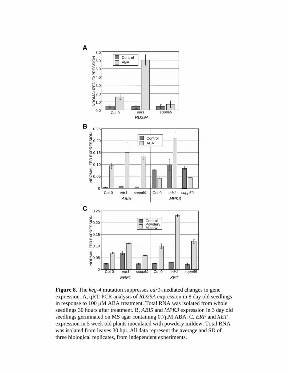
Figure 8. The keg-4 mutation suppresses edr1-mediated changes in geneexpression. A, qRT-PCR analysis of RD29A expression in 8 day old seedlingsin response to 100 M ABA treatment. Total RNA was isolated from wholeseedlings 30 hours after treatment. B, ABI5 and MPK3 expression in 3 day oldseedlings germinated on MS agar containing 0.7 M ABA. C, ERF and XETexpression in 5 week old plants inoculated with powdery mildew. Total RNAwas isolated from leaves 30 hpi. All data represent the average and SD ofthree biological replicates, from independent experiments.
0.0
1.0
2.0
3.0
4.0
5.0
6.0
7.0
Col-0 edr1 supp69
A
C
B
0
0.05
0.10
0.15
0.20
0.25
ABI5 MPK3
NR
OM
ALI
ZED
EX
PR
ES
SIO
NN
OR
MA
LIZE
DE
XP
RE
SS
ION
0
0.05
0.10
0.15
0.20
0.25
Col-0 edr1 supp69 Col-0 edr1 supp69ERF1 XET
ControlABA
ControlPowderyMildew
ControlABA
RD29A
Col-0 edr1 supp69 Col-0 edr1 supp69
NR
OM
ALI
ZED
EX
PR
ES
SIO
N

Figure 9. Overexpression of the KEG gene is toxic to plants. Wildtype Col-0 and transgenic T1 supp69 plants carrying adexamethasone-inducible wild-type copy of KEG were sprayed with50 M dexamethasone. The photos show the same plant at time 0(before spraying) and 7 days after induction for three independenttransgenic lines. Note that large necroses develop on all leavesirrespective of their ages.
0 DAYS 7 DAYS
LIN
E 2
3LI
NE
7LI
NE
9C
ol-0


