Embed Size (px)
Citation preview

Protein-Based Virtual Screening of Chemical Databases.II. Are Homology Models of G-Protein Coupled ReceptorsSuitable Targets?Caterina Bissantz,1,2 Philippe Bernard,1 Marcel Hibert,1 and Didier Rognan,1*1Laboratoire de Pharmacochimie de la Communication Cellulaire, UMR CNRS 7081, 74 route du Rhin, B.P. 24 ,F-67401 Illkirch, France.2Department of Applied Biosciences, ETH Zurich, Winterthurerstrasse 190, CH-8057 Zurich, Switzerland
ABSTRACT The aim of the current study is toinvestigate whether homology models of G-Protein-Coupled Receptors (GPCRs) that are based on bo-vine rhodopsin are reliable enough to be used forvirtual screening of chemical databases. Startingfrom the recently described 2.8 Å-resolution X-raystructure of bovine rhodopsin, homology models ofan “antagonist-bound” form of three human GPCRs(dopamine D3 receptor, muscarinic M1 receptor,vasopressin V1a receptor) were constructed. Thehomology models were used to screen three-dimen-sional databases using three different docking pro-grams (Dock, FlexX, Gold) in combination with sevenscoring functions (ChemScore, Dock, FlexX, Fresno,Gold, Pmf, Score). Rhodopsin-based homology mod-els turned out to be suitable, indeed, for virtualscreening since known antagonists seeded in thetest databases could be distinguished from ran-domly chosen molecules. However, such models arenot accurate enough for retrieving known agonists.To generate receptor models better suited for ago-nist screening, we developed a new knowledge- andpharmacophore-based modeling procedure thatmight partly simulate the conformational changesoccurring in the active site during receptor activa-tion. Receptor coordinates generated by this newprocedure are now suitable for agonist screening.We thus propose two alternative strategies for thevirtual screening of GPCR ligands, relying on adifferent set of receptor coordinates (antagonist-bound and agonist-bound states). Proteins 2003;50:5–25. © 2002 Wiley-Liss, Inc.
Key words: docking; scoring; structure-based li-gand design; homology modeling;GPCRs
INTRODUCTION
High-throughput screening of chemical libraries is awell-established method for finding new lead compoundsin drug discovery.1 However, as the available databasesget larger and larger, the costs of such screenings risewhereas the hit rates decrease. A possibility to avoid theseproblems is not to screen the whole database experimen-tally but only a small subset, which should be enriched incompounds that are likely to bind to the target. This
preselection can be done by virtual screening (VS), acomputational method to select the most promising com-pounds from an electronic database for experimentalscreening.2 Virtual screening can be carried out by search-ing databases for molecules fitting either a known pharma-cophore3 or a three-dimensional (3-D) structure of themacromolecular target.4 Pharmacophore-based screeninghas been extensively used over the last decade and hasproven to be successful in many cases.5 It has the advan-tage of being applicable to ligands for which the target 3-Dstructure is unknown, but it requires several 3- or 4-pointpharmacophoric descriptions.6 Protein-based virtualscreening should be more efficient than the pharmacophore-based method since the protein environment of the ligandis taken into account. However, it still suffers from docking/scoring inaccuracies,7,8 and it requires knowledge of the3-D structure of the target. Therefore, with few excep-tions,9,10 it has only been applied to targets for which ahigh-resolution X-ray structure is known.11 However, withthe sequencing of the human genome, computationalchemists will have to face an overwhelming number ofpotential targets for which no or very few experimental3-D information is available. Therefore, it will be veryimportant in the near future to be able to use not onlyX-ray or NMR structures, but also protein models forprotein-based virtual screening of chemical libraries.
This is notably true for G-Protein coupled receptors(GPCRs) that represent one of the most important familiesof pharmaceutical targets.12 Thus, there is high interest indeveloping new lead compounds for most human GPCRs.However, difficulties in obtaining significant amounts ofpure and active recombinant GPCRs have rendered thedetermination of high-resolution GPCR X-ray structuresvery challenging. For long, GPCR models13,14 have been
Grant sponsor: MaproTech (Freiburg, Germany); Grant sponsor:Fondation pour la Recherche Medicale (Paris, France).
Philippe Bernard’s present address is GreenPharma, Faculte desSciences, Universite d’Orleans, B.P.6759, F-45067 Orleans Cedex,France.
*Correspondence to: Didier Rognan, Laboratoire de Pharmaco-chimie de la Communication Cellulaire, UMR CNRS 7081, 74 route duRhin, B.P. 24 , F-67401 Illkirch, France.E-mail: [email protected]
Received 3 December 2001; Accepted 8 June 2002
Published online 00 Month 2002 in Wiley InterScience(www.interscience.wiley.com). DOI: 10.1002/prot.10237
PROTEINS: Structure, Function, and Genetics 50:5–25 (2003)
© 2002 WILEY-LISS, INC.

based on low-resolution maps of either bacteriorhodop-sin15 or bovine rhodopsin.16,17 These models were usefulfor studying the functional architecture of GPCRs, butwere not reliable enough for structure-based ligand de-sign.13,18 This situation hopefully changed with the recentdetermination of a high-resolution X-ray structure ofbovine rhodopsin.19 Based on this X-ray structure, it ispossible to build refined models of other GPCRs, whichmight be close enough to their true 3-D structures forprotein-based ligand design.
In the current study, we present two consecutive studiesin which we investigated whether rhodopsin-based GPCRhomology models are reliable enough for carrying outvirtual screening of chemical libraries focused on eitherantagonists or agonist ligands of test GPCRs. We firstconstructed “antagonist-bound” molecular models of threehuman GPCRs (dopamine D3 receptor, acetylcholine mus-carinic M1 receptor, vasopressin V1a receptor) for which alarge amount of information is available.20–22 The threemodels were evaluated in terms of their ability to identifyknown antagonists seeded into a database of randomlychosen “drug-like” compounds. A previously describedvirtual screening procedure23 combining three differentdocking algorithms (Dock,24 Gold,25 FlexX26) in associa-tion with seven scoring functions (Dock,24 Gold,25 FlexX,26
PMF,27 ChemScore,28 Fresno,29 Score30) was used. Consen-sus scoring31 was then applied to generate small subsets(hit lists) comprising only the top scorers common to two orthree scoring lists. In the second part of this study,different models of an “agonist-bound: form of three hu-man GPCRs (dopamine D3, �2-adrenergic, and �-opioidreceptors) were constructed and used for identifying trueagonists embedded in test databases.
COMPUTATIONAL METHODSPreparation of 3-D Databases
The Advanced Chemical Directory (ACD v.2000-1)32 wasfiltered in order to eliminate unwanted compounds such aschemically reactive molecules, inorganic compounds, andmolecules with unsuitable molecular weights (lower than250, higher than 600) using an in-house SLN (Sybyl LineNotation)33 script. These boundaries were applied to re-trieve most of the “drug-like” compounds of the startingdatabase and to avoid molecular weight biases whencomparing known ligands with randomly chosen com-pounds. We have previously shown that larger moleculestend to be overestimated by free energy scoring functionsas they can statistically provide more interactions to thetarget.23 The randomly chosen compounds should, there-fore, have a molecular weight not below that of the knownligands seeded into the test databases.
Out of the 75,000 remaining molecules, 990 were ran-domly chosen. The most likely protonation state of ioniz-able moieties (amines, amidines, carboxylic acids, etc.)present in this test database was then assigned usinganother SLN script. For each of the 990 compounds,three-dimensional coordinates and Gasteiger atomiccharges34 were then generated using Concord 4.0.35 Finalcoordinates were stored in a multi mol2 file (a TRIPOS file
format33 for storing coordinates of several molecules in asingle file). For each of the three receptors used forantagonist screening (D3, M1, and V1a receptors), a set of10 known antagonists of the respective target was pre-pared (Fig. 1). For each of the receptors used for agonistscreening (D3, �2, � receptors), a set of 10 known fullagonists was prepared (Fig. 2). The 10 reference ligandswere chosen to be as different as possible in order to spanthe broadest chemical diversity for a given target. In orderto keep our docking results as unbiased as possible, we didnot include any of the ligands used for receptor refinement(see the following) in these test sets. Starting from Isis/Draw36 2D structures, a 3-D structure of each knownligand was generated with Gasteiger charges using theConcord conversion program. These new structures werethen added to the random database to create six finallibraries of 1,000 molecules.
Alignment of Amino Acid Sequences
The amino acid sequences of the five target receptorswere retrieved from the Swiss-Prot database (accessionnumbers: human dopamine D3 receptor, P35462; humanmuscarinic acetylcholine M1 receptor, P11229; humanvasopressin V1a receptor, P37288; human �2-adrenergicreceptor, P07550; human �-opioid receptor, P41143) andaligned to the sequence of bovine rhodopsin (accessionnumber: P02699) using the ClustalW multiple alignmentprogram.37 A slow pairwise alignment using BLOSUMmatrix series38 (the matrix of the series used for thealignment is decided by ClustalW itself based on thesimilarity of the amino acid sequences to align and is thusnot specified by the user) and a gap opening penalty of 15.0were chosen for aligning the amino acid sequences to thesequence of bovine rhodopsin in two steps: (1) from theN-terminus to the first 5 residues of the third intracellularloop I3, (2) from the last 5 residues of the I3 loop to theC-terminus. Because the disulfide bridge occurring be-tween the third transmembrane segment (TM III) and thesecond extracellular loop (E2) in the structure of bovinerhodopsin is conserved in all five test receptors, we manu-ally adjusted the alignment of the extracellular loop E2 toalign the respective cysteines.
Preparation of Starting Protein Coordinates
The 3-D models of the five test receptors were con-structed by mutating the side chains of the amino acids inrhodopsin to the respective side chains in M1, D3, V1a, �2,and �-opioid receptors. Standard geometries for the mu-tated side chains were given by the BIOPOLYMER moduleof SYBYL.33 Whenever possible, the side chain torsionalangles were kept to the values occurring in bovine rhodop-sin. Otherwise, a short scanning of side chain angles wasperformed to remove steric clashes between the mutatedside chain and the other amino acids. The third intracellu-lar loop between helices 5 and 6, which shows a highdegree of variability, was not included in any of the threemodels. This loop is responsible for G protein coupling butis not involved in direct interactions with the ligand.39 We,therefore, assume that omitting this loop should not
6 C. BISSANTZ ET AL.
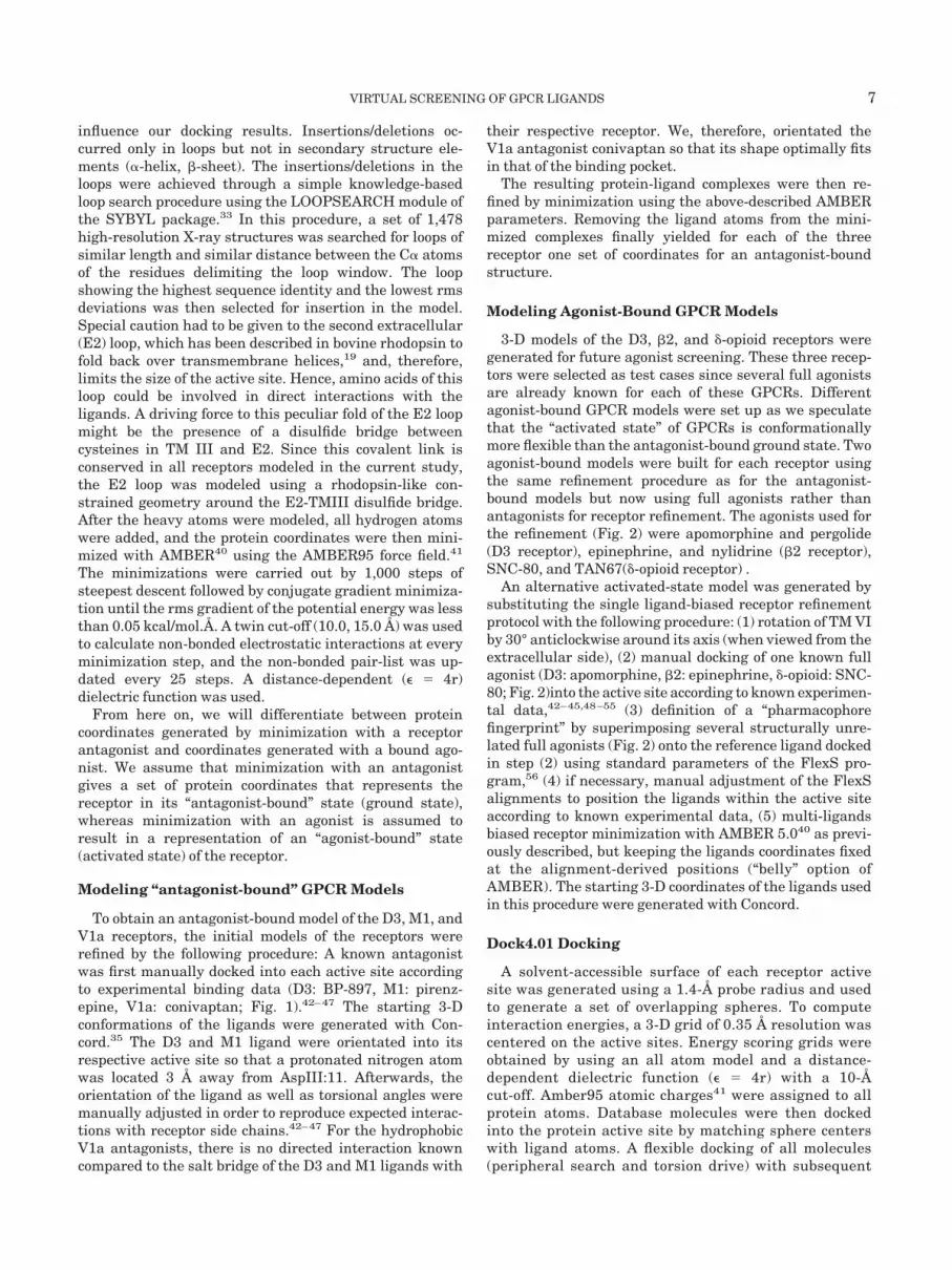
influence our docking results. Insertions/deletions oc-curred only in loops but not in secondary structure ele-ments (�-helix, �-sheet). The insertions/deletions in theloops were achieved through a simple knowledge-basedloop search procedure using the LOOPSEARCH module ofthe SYBYL package.33 In this procedure, a set of 1,478high-resolution X-ray structures was searched for loops ofsimilar length and similar distance between the C� atomsof the residues delimiting the loop window. The loopshowing the highest sequence identity and the lowest rmsdeviations was then selected for insertion in the model.Special caution had to be given to the second extracellular(E2) loop, which has been described in bovine rhodopsin tofold back over transmembrane helices,19 and, therefore,limits the size of the active site. Hence, amino acids of thisloop could be involved in direct interactions with theligands. A driving force to this peculiar fold of the E2 loopmight be the presence of a disulfide bridge betweencysteines in TM III and E2. Since this covalent link isconserved in all receptors modeled in the current study,the E2 loop was modeled using a rhodopsin-like con-strained geometry around the E2-TMIII disulfide bridge.After the heavy atoms were modeled, all hydrogen atomswere added, and the protein coordinates were then mini-mized with AMBER40 using the AMBER95 force field.41
The minimizations were carried out by 1,000 steps ofsteepest descent followed by conjugate gradient minimiza-tion until the rms gradient of the potential energy was lessthan 0.05 kcal/mol.Å. A twin cut-off (10.0, 15.0 Å) was usedto calculate non-bonded electrostatic interactions at everyminimization step, and the non-bonded pair-list was up-dated every 25 steps. A distance-dependent (� � 4r)dielectric function was used.
From here on, we will differentiate between proteincoordinates generated by minimization with a receptorantagonist and coordinates generated with a bound ago-nist. We assume that minimization with an antagonistgives a set of protein coordinates that represents thereceptor in its “antagonist-bound” state (ground state),whereas minimization with an agonist is assumed toresult in a representation of an “agonist-bound” state(activated state) of the receptor.
Modeling “antagonist-bound” GPCR Models
To obtain an antagonist-bound model of the D3, M1, andV1a receptors, the initial models of the receptors wererefined by the following procedure: A known antagonistwas first manually docked into each active site accordingto experimental binding data (D3: BP-897, M1: pirenz-epine, V1a: conivaptan; Fig. 1).42–47 The starting 3-Dconformations of the ligands were generated with Con-cord.35 The D3 and M1 ligand were orientated into itsrespective active site so that a protonated nitrogen atomwas located 3 Å away from AspIII:11. Afterwards, theorientation of the ligand as well as torsional angles weremanually adjusted in order to reproduce expected interac-tions with receptor side chains.42–47 For the hydrophobicV1a antagonists, there is no directed interaction knowncompared to the salt bridge of the D3 and M1 ligands with
their respective receptor. We, therefore, orientated theV1a antagonist conivaptan so that its shape optimally fitsin that of the binding pocket.
The resulting protein-ligand complexes were then re-fined by minimization using the above-described AMBERparameters. Removing the ligand atoms from the mini-mized complexes finally yielded for each of the threereceptor one set of coordinates for an antagonist-boundstructure.
Modeling Agonist-Bound GPCR Models
3-D models of the D3, �2, and �-opioid receptors weregenerated for future agonist screening. These three recep-tors were selected as test cases since several full agonistsare already known for each of these GPCRs. Differentagonist-bound GPCR models were set up as we speculatethat the “activated state” of GPCRs is conformationallymore flexible than the antagonist-bound ground state. Twoagonist-bound models were built for each receptor usingthe same refinement procedure as for the antagonist-bound models but now using full agonists rather thanantagonists for receptor refinement. The agonists used forthe refinement (Fig. 2) were apomorphine and pergolide(D3 receptor), epinephrine, and nylidrine (�2 receptor),SNC-80, and TAN67(�-opioid receptor) .
An alternative activated-state model was generated bysubstituting the single ligand-biased receptor refinementprotocol with the following procedure: (1) rotation of TM VIby 30° anticlockwise around its axis (when viewed from theextracellular side), (2) manual docking of one known fullagonist (D3: apomorphine, �2: epinephrine, �-opioid: SNC-80; Fig. 2)into the active site according to known experimen-tal data,42–45,48–55 (3) definition of a “pharmacophorefingerprint” by superimposing several structurally unre-lated full agonists (Fig. 2) onto the reference ligand dockedin step (2) using standard parameters of the FlexS pro-gram,56 (4) if necessary, manual adjustment of the FlexSalignments to position the ligands within the active siteaccording to known experimental data, (5) multi-ligandsbiased receptor minimization with AMBER 5.040 as previ-ously described, but keeping the ligands coordinates fixedat the alignment-derived positions (“belly” option ofAMBER). The starting 3-D coordinates of the ligands usedin this procedure were generated with Concord.
Dock4.01 Docking
A solvent-accessible surface of each receptor activesite was generated using a 1.4-Å probe radius and usedto generate a set of overlapping spheres. To computeinteraction energies, a 3-D grid of 0.35 Å resolution wascentered on the active sites. Energy scoring grids wereobtained by using an all atom model and a distance-dependent dielectric function (� � 4r) with a 10-Åcut-off. Amber95 atomic charges41 were assigned to allprotein atoms. Database molecules were then dockedinto the protein active site by matching sphere centerswith ligand atoms. A flexible docking of all molecules(peripheral search and torsion drive) with subsequent
VIRTUAL SCREENING OF GPCR LIGANDS 7

minimization was performed as follows: (1) automaticselection and matching of an anchor fragment within amaximum of 100 orientations, (2) iterative growing ofthe ligand using at least 30 conformations (peripheralseeds) for seeding the next growing stage with assign-ment of energy-favored torsion angles, (3) simultaneousrelaxation of the base fragments as well as of allperipheral segments and final relaxation of the entiremolecule. Orientations/conformations were relaxed in100 cycles of 100 simplex minimization steps to aconvergence of 0.1 kcal/mol. The top solution correspond-ing to the best Dock energy or chemical score of eachligand was then stored in a single multi mol2 file.
FlexX1.8 Docking
Standard parameters of the FlexX1.9 program as imple-mented in the 6.62 release of the SYBYL package wereused for iterative growing and subsequent scoring of FlexXposes. Only the top solution was retained and furtherstored in a single mol2 file.
Gold1.1 Docking
For each of the 10 independent genetic algorithm (GA)runs, a maximum number of 1,000 GA operations wasperformed on a single population of 50 individuals.Operator weights for crossover, mutation, and migra-tion were set to 100, 100, and 0, respectively. To allowpoor non-bonded contacts at the start of each GA run,the maximum distance between hydrogen donors andfitting points was set to 5 Å, and non-bonded van derWaals energies were cut off at a value equal to kij (welldepth of the van der Waals energy for the atom pair i,j).To further speed up the calculation, the GA docking wasstopped when the top three solutions were within 1.5 Årmsd. If this criterion is met, we can assume that thesetop solutions represent a reproducible pose for theligand.
It should be mentioned that all docking tests wereapplied using fast virtual screening parameters (pace ofapproximately 60–90 sec/ligand on a standard UNIXworkstation) to closely match database screening condi-
Fig. 1. Structures of receptor antagonists used in the test databases (top boxes) and in the minimization protocol (bottom boxes). A: Dopamine D3antagonists. B: Muscarinic M1 antagonists. C: Vasopressin V1a antagonists. Pirenzepine and conivaptan were used for refining the M1 and V1aantagonist-bound receptor models. For the dopamine D3 receptor, three independent minimizations were carried out using BP-897, sulpiride, andGR-218231, thus leading to three sets of coordinates for the antagonist-bound form of the D3 receptor. Underlined numbers represent true antagonistspredicted as virtual hits by the best virtual screening strategy.
8 C. BISSANTZ ET AL.

tions. This strategy explains why the top-ranked pose onlywas selected for further rescoring.
Consensus Scoring: Definition of a Hit List
Docked poses were rescored using the CScore™ moduleof SYBYL6.62 that comprises the following scoring func-tions: ChemScore, Dock, FlexX, Gold, and Pmf. It shouldbe noted that the original Dock4.0 and Gold scores asimplemented in the respective docking program signifi-cantly differ from the respective scores calculated byCScore™. The scoring function of Cscore corresponding tothe native scoring function of the applied docking algo-rithm (Dock, FlexX, Gold) was always discarded. Twoadditional scoring functions, Fresno29 and Score,30 wereused as part of in-house SPL (Sybyl Programming Lan-guage)33 scripts. For rescoring, no relaxation of the boundligands was performed. Rescoring the whole database withall scoring functions took about 30 min on a standardUNIX workstation.
Having rescored the docked poses with these sevenscoring functions, we then carried out the “consensusscoring.” For the D3, V1a, and �2 receptor screenings, wedefined the top 15% of the individual ranking lists as topscorers. For the �-opioid receptors, the top 20% scorers
were selected. Last, the top 25% scorers were retrievedwhen screening the M1 receptor A pairwise comparison ofthe lists of top scorers (21 combinations) yielded theconsensus lists (from here on also called “hit lists”) thatconsequently contain only those compounds that wereranked among the top scorers from the two comparedscoring functions. We, furthermore, generated the consen-sus lists comparing the top scorers of three single rankinglists (giving 35 additional consensus lists).
The reason why we had to adjust the definition of “topscorers” from one GPCR to another resides in the expectedvariation of accuracy of our homology models. The numberof top scorers used for consensus scoring represents foreach receptor the best possible compromise between thetotal size of the virtual hit lists and the number of trueligands it contains (see Quantitative Descriptors of the HitLists).
Quantitative Descriptors of the Hit Lists
Two properties of every generated hit list were com-puted: the hit rate and the yield. The hit rate (or purity)describes the accuracy of the virtual screening and isdefined as the percentage of known true ligands in the hitlist (Eqn. 1). A random screening of the full databasewould thus have given a hit rate of 1% (10 true hits out of1,000 molecules). The yield is defined as the percentage oftrue hits retrieved by our virtual screening protocol (Eqn.2). It describes the sensitivity of the screening.
Hit Rate � (t/l) � 100 (1)
Yield � (t/T) � 100 (2)
t � number of true hits in the hit listT � number of true hits in the full databasel � number of compounds in the hit list
RESULTSBuilding Starting GPCR Models
The amino acid sequences of the target receptors werealigned to the sequence of the bovine rhodopsin template(Fig. 3), excluding the third intracellular loop, whichshows too high variability in amino acid composition andlength. The sequence identity between the transmem-brane (TM) domains of bovine rhodopsin and the humandopamine D3, muscarinic M1,Vasopressin V1a, �2-adren-ergic, and �-opioid receptor is 29, 21, 21, 22, and 23%,respectively. The alignment is in agreement with knownstructural features of GPCRs.57 Amino acids known to behighly conserved in the family of the (rhodopsin-like)GPCRs (AsnI:21, AspII:13, ArgIII:29, TrpIV:11, ProV:16,ProVI:21, NPxxY in TM VII; A:n, position n of the trans-membrane helix A) are aligned against each other. Thedisulfide bridge in bovine rhodopsin is conserved in all testreceptors, and the respective cysteines are also aligned toeach other. It should be emphasized that we did not modelthe loops exactly with the deletions/insertions as indicatedin the alignment since there are obviously several ways ofaligning these very variable sequences. We rather carriedout a loop search to make sure that our loops have
Figure 1. (Continued)
VIRTUAL SCREENING OF GPCR LIGANDS 9

conformations similar to loops in X-ray structures thathave similar length and similar distance between the C�atoms of the residues delimiting the loop.
Based on this alignment, we mutated the side chains ofthe amino acids in the X-ray structure of bovine rhodopsinto the respective side chains in the new receptor (seeComputational Methods). Deletions occurring in the E2loop of the three GPCRs (see Fig. 3) could be accommo-dated easily without modifying the respective orientationsof the seven �-helices. Minimizing the resulting structuresgave for each receptor one initial model. Any amino acidside chain directed towards the inner part of the transmem-brane ligand binding domain was selected as a potentialanchor and thus selected as part of a putative binding site(Fig. 3).
Building Antagonist-Bound Models of D3, M1, andV1a Receptors
The X-ray structure of bovine rhodopsin corresponds toits ground state19 in which retinal is covalently bound
(antagonist-bound conformation). Homology models con-structed from this structure are not directly suitable fordocking purpose for two main reasons: (1) depending onthe type of bound ligand (agonist, antagonist), GPCRs areknown to adopt different conformational states,58–60 (2)retinal is a rather flat molecule, and thus the modeledbinding sites are too narrow—especially since the E2 loopin bovine rhodopsin folds deeply into the center of thereceptor to completely enclose retinal—for most of theknown GPCR ligands to fit in. We, therefore, have to“expand” the active site in our models somewhat bypushing the E2 loop out of the TM cavity center. Hence,preliminary attempts to automatically dock known li-gands into the starting models directly obtained fromrhodopsin (minimized in absence of any ligand) usuallyfailed whatever the docking tool used (data not shown).
Thus, we feel it is necessary to refine the receptor modelby minimization in complex with a known ligand toachieve expansion of the TM cavity. Minimization with anantagonist is supposed to give a model close to an antago-
Fig. 2. Structures of receptor agonists used in the test databases (top boxes) and in the minimization protocol (bottom boxes). A: Dopamine D3agonists. B: �2-adrenergic agonists. C: �-opioid agonists. Underlined numbers represent true agonists predicted as virtual hits by the best virtualscreening. Single-ligand minimized receptor models were obtained with apomorphine and pergolide (D3 receptor), epinephrine and nylidrine (�2receptor), and SNC-80 and TAN67 (�-opioid receptor). Pharmacophore-based receptor models were obtained by first docking one reference agonist(apomorphine, epinephrine, and SNC-80) into the TM cavity and then superimposing other ligands (bottom boxes) with FlexS onto the receptor-boundreference agonist.
10 C. BISSANTZ ET AL.

nist-bound state (ground state) of the receptor, whereasminimization with a full agonist should lead to a modelcloser to the agonist-bound state (activated state). For thispurpose, we have chosen ligands for which a large amountof experimental data (location of the binding site) wasavailable.
To get 3-D coordinates of the antagonist-bound state ofthe D3, M1, and V1a receptors, we chose BP-897 (D3receptor), pirenzepine (M1 receptor), and conivaptan (V1areceptor), docked them according to known experimentaldata into their respective receptors,42–47 and energy mini-mized the complexes. These reference ligands were chosenin the light of their conformational freedom (as rigid aspossible) and of known experimental data to guide us inthe initial docking. The D3 antagonist BP-897 has beenorientated so that its protonated nitrogen forms a saltbridge with AspIII:11. Additionally, the amide group ofBP-897 interacts with ThrVII:7. The basic nitrogen of themuscarinic antagonist pirenzepine also forms a salt bridgewith AspIII:11. The lactam group interacts with AsnVI:23,and its aromatic nitrogen with ThrV:8. The V1a antago-nist conivaptan is orientated so that the benzazepinemoiety lies in the pocket between TM III, V, and VI,whereas the diphenyl group occupies the pocket betweenTM I, II, and VII. It should be noted that the three dockingmodes reported herein are compatible with all knownexperimental data.42–47 Removing the bound ligand fromthe energy minimized complex (refined as previously de-scribed for the starting models) gave a set of proteincoordinates (inactive state) for each receptor.
Building Agonist-Bound Models of the D3, �2, and�-Opioid Receptors
Two agonist-bound models were built for each GPCRusing the same strategy as that used for the antagonist-bound models. The two dopamine agonists apomorphine(S1 model) and pergolide (S2 model) were manually dockedinto the active site with their protonated nitrogens withinsalt bridge distance (3 Å) from AspIII:11. Additionally, thetwo phenolic groups of apomorphine and the indolic nitro-gen atom of pergolide H-bond to three serines on TM V(SerV:8, SerV:9, SerV:12). For generating the �2-models,epinephrine (S1 model) and nylidrine (S2 model) wereused as reference agonists. They were also docked with thebasic nitrogens forming a salt bridge with AspIII:11, andthe phenolic groups interacting with the three serines onTM V. �-opioid receptor models were generated by dockingSNC-80 (S1 model) and TAN67 (S2 model) into the activesite, with the protonated amine establishing a salt bridgewith AspIII:11, the phenol rings lying in the pocketbetween TMs III,V,VI, whereas the diethylamide (SNC-80) and the quinoline ring (TAN67) were embedded in thepocket between TMs VI and VII. All complexes were thenrefined using the above-described AMBER refinementprotocol.
For each of the three receptors, we furthermore gener-ated one activated-state model using a multi-ligand basedmodeling procedure (M models). This protocol requiresfirst defining a pharmacophoric fit of few reference ago-
Figure 2. (Continued)
VIRTUAL SCREENING OF GPCR LIGANDS 11

nists. For defining a dopamine D3 agonist pharmacophore,5 full agonists [Fig. 2(A)] were selected: two catecholamines(dopamine, apomorphine), one ergoline (pergolide), onepyrazoloquinoline (quinpirole), and one 2-aminotetralinederivative (7-OH-DPAT). The protonated amine commonto all D3 ligands is supposed to form a salt bridge with thehighly conserved AspIII:11, and the catechol moiety of thecatecholamines or its bioisostere in other ligand classesare expected to H-bond to three serine side chains on helix5 (SerV:8, SerV:9, SerV:12).43 The phenylalanine residuesPheVI:22 and PheVI:23 also seem to be important foragonist binding, probably via �-� interactions with thearomatic catechol ring or its bioisostere.45 We thus manu-ally docked a rigid molecule first (apomorphine) as previ-ously described and used this structural template forsuperimposing the other full dopamine D3 receptor ago-nists by fitting three chemical features (protonated amine,aromatic ring, H-bond donor moiety) with the FlexS pro-gram.56 The FlexS-aligned conformations were slightlymodified in order to correctly orientate the N-alkyl substitu-ents and the thioether chain of pergolide within the activesite. In the proposed alignment, protonated amines andH-bond forming groups of the aromatic rings are superim-
posed [Fig. 4(A)]. The agonists are orientated in the activesite so that the protonated amines form salt bridges withAspIII:11; the para-hydroxyl group of apomorphine anddopamine H-bonds to SerV:12, the meta-hydroxyl substitu-ent of apomorphine, dopamine, and 7-OH-DPAT to SerV:8and SerV:9. The indolic nitrogen atom in the ergolinederivatives binds to SerV:12. Since TM VI was previouslyrotated by 30°, the aromatic ring of PheVI:22 lies now in aplane parallel to that of the aromatic rings of the agonistsand can thus form �-� interactions with the ligands. This�-� interaction cannot be formed when docking the li-gands directly in the rhodopsin-derived model as it wasdone for generating the models S1 and S2.
Like the D3 receptor ligands, all �2 agonists have aprotonated nitrogen [Fig. 2(B)] expected to form a saltbridge with AspIII:11,49 and the catechol moiety (or itsbioisostere) is probably H-bonded to three serines on helix5 (SerV:8, SerV:9, SerV:12).48,52 Additionally, the �2 ago-nists possess a �-hydroxyl group expected to H-bond toAsnVI:26.51 The phenylalanine residues PheVI:22 andPheVI:23 have also been proposed to be involved in ligandbinding.50 After manually docking epinephrine accordingto the expected interactions, other �2 full agonists were
Fig. 3. Amino acid sequence alignment of 5 GPCRs (ACM1_HUMAN: human acetylcholine muscarinic M1, D3DR_HUMAN: dopamine D3,V1AR_HUMAN: human vasopressin V1a, OPRD_HUMAN: human �-opioid receptor, B2AR_HUMAN: human �-opioid receptor) with bovine rhodopsin(OPSD_BOVIN). Transmembrane domains (TM I to TM VII) are boxed. E1-3 and I1-3 indicate the positions of extracellular and intracellular loops,respectively. Residues colored in red delimit a putative binding site for GPCR ligands. Amino acids indicated in blue are conserved in the GPCR family.For facilitating the comparison of different receptors, transmembrane helical sequences (TM I to TM VII), assigned as in the X-ray structure of bovinerhodopsin,29 are numbered from the N- to the C-terminal end. Numbers inserted in the I3 loop describe the number of residues omitted in the currentstudy. The residues referenced in the text are underlined. The numbering of residues referenced in the text is as follows: A:n indicates position n of thetransmembrane helix A.
12 C. BISSANTZ ET AL.

superimposed onto the reference agonist using FlexS [Fig.2(B)]. This alignment is straightforward due to the com-mon phenyl-2-aminoethanol core of the �2 agonists [Figs.2(B), 4(B)]. We only had to adjust the conformations of the
large N-aralkyl substituents in fenoterol and nylidrine sothat they do not bump into protein atoms. The ligands areorientated in the active site so that the protonated aminesform salt bridges with AspIII:11 [Fig. 4(B)]. The para-hydroxyl group of epinephrine and nylidrine H-bonds tothe side chain of SerV:12. The meta-hydroxyl of epineph-rine and one of the two phenolic moieties of fenoterolH-bond to the side chains of SerV:8 and SerV:9, the otherhydroxyl group of fenoterol to either the side chain ofSerV:12 or to the backbone carbonyl of ValIII:15. Theamino group of clenbuterol is proposed to interact withSerV:12. All aromatic catechol moieties can again form a�-� interaction with PheVI:22. Last, the large hydropho-bic residues on the protonated amines of fenoterol andnylidrine occupy the pocket between TMs I, II, and VII[Fig. 4(B)]. As for the D3 receptor, the interactions be-tween the ligands and the important residues on TM VI(PheVI:22, AsnVI :26) cannot be formed in the singleligand-biased models (S1 and S2 models).
Last, a pharmacophoric alignment was defined for �-opi-oid receptor agonists from 5 compounds [Fig. 2(C)]: twocompounds of the SNC series (SNC-80 and 5661), oneopiate (SIOM) and one simplified opiate (TAN67), as wellas one true non-peptide62 compound (SL-3111). The �-ago-nists are regarded as consisting of two parts: the “address”and the “message” part.63 The address part is generally alarge hydrophobic residue that is supposed to bind in thepocket between TMs VI and VII; important residueshereby are TrpVI:29 and LeuVII:3.53,55 The message partis expected to bind in the central cavity between TMs III,V, and VI; important residues are AspIII:11, TyrIII:12,TrpVI:19, and TyrVII:11.54 FlexS was again used to super-impose the agonists onto the manually docked referenceligand (SNC-80). The alignment of the SNC derivative 56onto SNC-80 is straightforward. The two compounds of theSNC series are orientated in the active site so that theprotonated amine can form a salt bridge with AspIII:11;the N,N-diethylamide (address-part), which is orientatedout-of-plane with respect to the benzene ring, lies in thepocket between TMs VI and VII, with the carbonyl point-ing to TrpVI:29 and one of the two ethyl groups interactingwith LeuVII:3 [Fig. 4(C)]. The methoxyphenyl and quino-line moiety, respectively, fit in the central pocket betweenTMs III, V, and VI, with the methoxy group of SNC-80pointing away from HisVI:23. Though structurally verysimilar, the true non-peptide compounds are expected tobind in a different mode than the SNC derivatives.62
Whereas O-methylation is tolerated in the SNC-series, itresults in a loss of activity of the true non-peptides. We,therefore, adjusted the FlexS fitted solution of SL-3111 sothat its phenolic OH group can form a H-bond to HisVI:23.Besides this small modification of the automated align-ment, SL-3111 is docked similarly to the SNC compounds,with the protonated amine superimposed onto the amineof SNC-80 and the t-butyl group onto the N,N-diethylam-ide. The N-benzyl substituent of SL-3111 occupies thepocket between TMs II and VII. The FlexS solution forSIOM was not accepted because the address part wassuperimposed onto the phenoxy ring of SNC-80, thus lying
Fig. 4. Receptor-based alignment of the ligands used for receptorrefinement of model M of the dopamine D3 receptor (A), the �2-adrenergic receptor (B), and the �-opioid receptor (C). Ligands are shownas capped sticks, whereas amino acid side chains of the receptor areindicated in ball-and-stick representation. The color coding is the follow-ing: carbon (agonists, green; receptor, white); oxygen, red; nitrogen, blue;sulfur, yellow.
VIRTUAL SCREENING OF GPCR LIGANDS 13

in the central cavity rather than in the pocket betweenTMs VI and VII. We, therefore, manually superimposedSIOM onto the other agonists. The address part of theopiate SIOM overlaps now with the N,N-diethylamide ofSNC-80, its phenolic OH with the one of SL-3111, thusinteracting with HisVI:23 [Fig. 4(C)]. The FlexS alignmentof the simplified opiate TAN67 to the reference agonistSNC-80 was modified in order to maximize overlap withthe opiate derivative (SIOM) so that its perhydroisoquino-line overlaps the corresponding part of SIOM, thus position-ing the protonated nitrogens, the phenolic hydroxyl moities,and the address parts in the same regions of space.
Again, energy minimization (previously described AM-BER protocol) and removal of the bound ligands affordedfor each receptor three independent sets of receptorcoordinates (S1, S2, M models) in its supposed activatedstate.
Virtual Screening of Receptor Antagonists
Screening the antagonist D3 receptor model with FlexXand Gold, hit rates up to 5, 14, and 30% were obtained aftersingle, double, and triple scoring, respectively [Fig. 5(A)].Only the consensus hit lists out of Dock poses did notcontain any true antagonist. The best compromise be-tween hit rate and yield is achieved when combining FlexXas docking tool with PMF, Gold, and ChemScore forconsensus scoring (hit rate of 18.92%, yield of 70%). Thisconsensus list of 37 molecules contains 7 true ligands(compounds 2, 4–5, 7–10) [Fig. 1(A)]. Regarding the dockedconformations, we can observe a clear difference betweenthe conformations proposed by Dock and those generatedby Gold and FlexX. Whereas Dock was unable to place anyof the 10 chosen D3 antagonists into the transmembranecavity, the conformations proposed by FlexX and Gold aremostly in agreement with the expected interactions: thebasic nitrogens form salt bridges with AspIII:11, and theamide oxygen found in many D3 antagonists H-bonds toThrVII:7 [Fig. 6(A)].
When screening the M1 receptor, poorer hit rates (2–2.5% for Dock and Gold poses, almost 10% for FlexX poses)were achieved [Fig. 5(B)]. The best consensus hit list(FlexX as docking program in combination with FlexX,Gold, and PMF as scoring functions) comprised 53 mol-ecules. It contains 5 of the 10 known ligands (compounds14, 17, 18, 22, 23) [Fig. 1(B)]. Interestingly, in contrast toboth the retinal binding mode19 and the previous dopa-mine D3 antagonist binding mode, the M1 antagonistbinding site seems to be restricted to the pocket betweenTMs III, V, and VI. Whereas the D3 receptor cavity is wideenough to allow complete occupation of the binding sitefrom one extreme (TM I) to the other (TM V in ourorientation) [Fig. 6(A)], the M1 binding site is almostseparated in two distinct cavities [Fig. 6(B)] by two ty-rosine side chains (TyrIII:12, TyrE2:17), one of which(TyrIII:12) is part of TM3, whereas the other one (TyrE2:17) belongs to the second extracellular loop (E2) whoserhodopsin-like fold still remains putative and has to beexperimentally verified.
Fig. 5. Virtual screening of receptor antagonists. Hit rates among thetop scorers after single (dark bars) or consensus scoring (double scoring:hashed bars, triple scoring: white bars). The top 15% scorers of the singleranking lists were used for the D3 and V1a receptor whereas the top 25%scorers were retrieved for the M1 receptor screening. Only the bestdocking/scoring combinations are shown here. A: D3 receptor. B: M1receptor. C: V1a receptor.
14 C. BISSANTZ ET AL.

Screening the V1a receptor, FlexX, and Gold led to hitrates of up to 40% after triple scoring [Fig. 5(C)]. Theproposed docked orientations of V1a receptor antagonistsare compatible with site-directing mutagenesis data,47
identifying aromatic residues at TM VI (TrpVI:19, PheVI:22, PheVI:23) and hydrophilic amino acids at TM II(GlnII:20) and TM III (LysIII:8) as part of the “antagonist-binding site” [see docked orientation of compound 34 inFig. 6(C)]. Five ligands (compounds 26, 29, 30, 33, 34) [Fig.1(C)] were retrieved in the best consensus hit list (FlexX asdocking program in combination with FlexX, Gold, and
Fresno as scoring functions), which comprises only a totalnumber of 13 molecules.
Virtual Screening of Receptor Agonists
It should be emphasized again that none of the fullagonists used to assist receptor refinement in the differentprotocols (S1, S2 or M models) was included in the set of 10true agonists seeded in the test 1,000 compounds data-base. For clarity, we present herein for each screening onlythe best consensus combination (Fig. 7), giving the optimalcompromise between hit rate and yield for the computedconsensus hit lists.
Screening the D3 receptor models, the pharmacophore-based model M proved to be superior to both singleligand-biased receptor models for enriching hit lists in trueD3 agonists [Fig. 7(A)] whatever the docking tool used.Only the knowledge- and pharmacophore-based model Mafforded hit lists with both good hit rates and good yields.It is important to notice that 60–70% of the known ligandswere included in the model M-derived hit list [Fig. 2(A)],thus spanning a wide chemical diversity. The betterscreening results of the pharmacophore-based model canbe explained looking at the docked conformations of theligands. Only this model allows an automated docking ofthe 10 true agonists as expected with the protonatednitrogen atom facing AspIII:11, the catechol (or its bioiso-steric counterpart) H-bonding to the three serines of theTMV (SerV:8, SerV:9, SerV:12), and the phenyl ringdeveloping p-p stacking interactions with PheVI:22 [com-pare Fig. 8(A–C)]. The conformations achieved with theother models do not form these important interactions.
A similar result was found when comparing the different�2 adrenergic receptor models. After rescoring and consen-sus scoring analysis, the best results were again obtainedwith the multi-ligand-based model M. Starting from eitherFlexX or Dock poses, resulting hit rate and yield in trueagonists were significantly higher when the pharmacoph-ore-based model of the receptor was used [Fig. 7(B)]. As forthe D3 receptor screening, all agonist classes compoundswere represented in the optimal consensus hit list [Fig.2(B)]. Only model M of the �2 receptor is compatible with aproper docking of all known �2 full agonists with theprotonated nitrogen atom of the agonists forming a saltbridge with AspIII:11, the catechol moiety or its bioiso-stere H-bonded to the serines on TM V (SerV:8, SerV:9 andSerV:12), the �-OH groups interacting with AsnVI:26, andthe aromatic moiety establishing �-� interactions withPheVI:22 [compare Fig. 9(A–C)].
In our third test case, the �-opioid receptor, the docking,scoring, and consensus scoring was carried out as alreadyexplained for the D3 and �2 receptors with the exceptionthat here the top 20% scorers were selected for consensusscoring. Comparing the consensus hit lists obtained withthe different �-opioid receptor models [Fig. 7(C)], we stillobserve a clear difference between the knowledge- andmulti-ligand-based model M and the single-ligand mini-mized models S1 and S2. The best result was againobtained with model M. Rotation of helix 6 in the pharma-cophore model results in a much better interaction of two
Fig. 6. Proposed binding mode for (A) dopamine D3 receptor antago-nists (exemplified by sulpiride, compound 2), (B) muscarinic M1 receptorantagonists (exemplified by atropine, compound 14), (C) V1a receptorantagonists (exemplified by compound 34). Dock, FlexX, and Gold posesare displayed by green, yellow, and cyan carbon atoms, respectively.Receptor side chains are displayed by white carbon atoms.
VIRTUAL SCREENING OF GPCR LIGANDS 15

crucial residues (TrpVI:19, HisVI:23) with the bi-phenylmoiety found in most �-opioid agonists [Fig. 10(A, B)]. Asalready observed for the D3 and �2 receptors, screeningagainst model M enables us to retrieve true agonistsrepresenting different structural classes [Fig. 2(C)]. Nota-bly, 90% of the known agonists were included in theconsensus hit list of FlexX.
Strikingly, although Gold was able to propose reliableposes for most GPCR agonists, none of the 10 true agonistswas present in any of the final consensus lists for the threereceptors used in our validation study.
DISCUSSIONRhodopsin-Based Homology Modeling of GPCRs
Although the sequence identity between the TMs of themodeled receptors and the template (bovine rhodopsin) isonly between 21–29%, we believe that the receptors mod-eled in the current study share the same fold as thetemplate since the modeled GPCRs include several con-served motifs (sequences of highly conserved amino acids)showing that, despite the low sequence identity whentaking the whole TM sequence in account, the structurallyand functionally important amino acids are highly con-served or substituted by amino acids of high similarity(e.g., DRY on TMIII, NPxxY on TMVI). GPCR modelsbased on a template with an identity of 20–30% can thusbe expected to be of higher accuracy than when modelingother type of proteins based on a template with such alow-sequence identity. However, we have to clearly distin-guish between the transmembrane domains and loopregions. The above-stated reasoning holds true only for theTMs. We can assume that our models are of relatively highaccuracy in the TMs, but at this moment we are not able toachieve a fine modeling of the loop regions as well as the N-and C-terminal parts. These extramembrane parts aremuch too flexible and divergent in amino acid sequencefrom the rhodopsin template. Since the active site of theinvestigated receptors is located in the transmembranecore, for our purpose the loops—generating models thatare suitable for virtual screening—are not very important.The only exception is the second extracellular E2 loop,which folds back into the transmembrane core and thuscompletely encloses retinal in the rhodopsin X-ray struc-ture.19 The question arises whether this particular fold isconserved in other GPCRs. At this point, it is very difficultto give a definitive answer to this question. We can onlyimagine that a driving force to this fold is the disulfidebridge between cysteines in TM III and E2. Since thiscovalent link is conserved in our test receptors, we decidedto keep this special fold unchanged in our models.
Another important point is that our GPCR models arestatic though proteins are in reality more or less flexible.This might lead to further inaccuracies especially for
Fig. 7. Virtual screening of receptor agonists. Consensus scoring listshave been defined out of the top 15% scorers (D3 and �2 receptor) or thetop 20% scorers (�-opioid screening) of single ranking lists. For eachscreening of the three receptor models (S1, S2, and M), the bestconsensus combination is shown. The boxed numbers in the bars indicatethe number of true agonists present in the respective consensus list. Goldresults are not shown as consensus scoring of Gold poses did not allowthe retrievement of a single true agonist for any of the three receptorsinvestigated herein. A: D3 receptor screening. FlexX poses have beenscored with FlexX, Gold, and ChemScore, Dock poses with Dock, FlexX,and Gold The consensus lists from the S1-model screenings did notinclude any of the known agonists, thus giving hit rates of 0. B: �2receptor screening. FlexX poses have been scored with FlexX, Gold, andFresno, Dock poses scored with Dock, FlexX, and Gold. The consensuslists from the S1-model screenings did not include any of the knownagonists, thus giving hit rates of 0. C: �-opioid receptor screening. FlexXposes have been scored with FlexX, Dock, and Fresno, Dock posesscored with Gold, PMF, and Fresno. The consensus lists from the FlexXscreening against S1 and the Dock screenings against S1 and S2-modeldid not include any of the known agonists, thus giving hit rates of 0.
16 C. BISSANTZ ET AL.

activated GPCR models since the active site of the acti-vated state of GPCRs probably has a higher conforma-tional flexibility than in the ground state. We, therefore,cannot expect that all ligands will be optimally docked toour static models.
Setting-Up Test Ligand Databases
The test databases screened against these receptorscontained 990 randomly selected compounds from theACD. We assumed that these compounds are not ligands ofour test receptors, although no experimental data verify
this assumption. Thus, random compounds that are in-cluded in final consensus lists should be regarded as “falsepositives.” Assuming that a protonated nitrogen and anaromatic moiety are necessary features for binding, 15 outof the 990 randomly chosen compounds that possess thesetwo features have a higher probability to be a potentialtrue hit than the other random compounds. Regarding thefinal consensus lists, however, we could not observe thatthese 15 compounds are more often included in the consen-sus lists than the other compounds.
Fig. 8. Illustration of FlexX poses of the D3 receptor agonist 36 boundto different dopamine D3 receptor models. A: Model M. B: Model S1. C:Model S2. The ligands are represented as ball-and-sticks, receptor sidechains as capped sticks. Carbon atoms are colored in light gray,heteroatoms in black.
Fig. 9. Illustration of FlexX poses of the �2 receptor agonist 51 boundto different �2 adrenergic receptor models. A: Model M. B: Model S1. C:Model S2. The ligands are represented as ball-and-sticks, receptor sidechains as capped sticks. Carbon atoms are colored in light gray,heteroatoms in black.
VIRTUAL SCREENING OF GPCR LIGANDS 17

GPCR Models Are Reliable Enough for an UnbiasedScreening of Receptor Antagonists
Inactive states of three human GPCRs were built byminimizing the rhodopsin-derived homology models with aknown antagonist and challenged for their ability toaccommodate 10 known antagonists seeded in a databaseof 990 randomly selected “drug-like” molecules.
Screening against the D3 receptor model, hit rates up to30% were achieved. Thus, the herein presented D3 recep-tor model leads to hit rates similar to that already ob-tained when screening is performed against high-resolu-tion X-ray structures.23,31 These high hit rates can beexplained by the fact that both FlexX and Gold dockingalgorithms were able to generate poses for most D3receptor antagonists that are in good agreement withknown experimental data. The rationale to test severaldocking programs is here justified by the poor performanceof the third docking tool (Dock), which was not able toreliably dock any of the 10 chosen antagonists into the TMcavity. This failure of Dock is rather unexpected. Since theorientation of base fragments in Dock is based on shapecomplementarity between the ligand and the receptor,24
we would have expected Dock to perform rather well forthe very hydrophobic dopamine D3 antagonists. In thecases where the screening was successful, it is importantto notice the chemical diversity of the true D3 antagonistsincluded in the consensus hit lists. Using FlexX as dockingprogram and rescoring with PMF, Gold, and ChemScore,the consensus hit list contains 7 out of the 10 starting trueantagonists. Ligands structurally unrelated to the antago-nist (the arylpiperazine BP-897) used for refining the D3receptor minimization are indeed retrieved as virtual hits,such as the 2-aminotetraline derivative 10, the tetrahy-droisoquinoline 8, the benzopyranopyrrolidine 9, or chemi-cally diverse compounds (2, 4, 5). Thus, the presentinactive state of the dopamine D3 model seems to besuitable for a virtual screening program aimed at discover-ing new D3 antagonist lead structures.
Much poorer hit rates (approximately 10%) have beenobtained by screening the muscarinic M1 antagonistmodel. Although the best computed hit rate is still10-fold higher than that given by random screening, it isclear that virtual screening is less efficient for detectingM1 antagonists than dopamine D3 antagonists [com-pare Fig. 5(A,B)]. The most likely reason is that thecurrent M1 receptor model is of lower quality than thepreviously described D3 receptor model. One explana-tion for this observation might be the different predictedbinding mode of M1 and D3 receptor antagonists. D3antagonists, like retinal bound to rhodospin,19 are pro-posed to occupy the whole binding site from TMs I to VII.In contrast, the M1 antagonist binding site seemsrestricted to the narrow cavity between TMs III, V, andVI. Therefore, the X-ray structure of bovine rhodopsinmight be a better template for the D3 receptor than forthe M1 receptor. Since the M1 model seems of limitedaccuracy, screening results are somewhat influenced bythe ligand used in the minimization step. The bestconsensus list (FlexX as docking program in combina-
tion with FlexX, Gold, and PMF as scoring functions)contains 5 of the 10 known ligands (compounds 14, 17,18, 22, 23) [Fig. 1(B)], two of which (22,23) are structur-ally very close to the ligand used for the minimization(pirenzepine). The other three compounds belong, how-ever, to different structural classes than pirenzepine.
In the last screening test targeting the V1a vasopres-sin receptor, consensus scoring of FlexX and Gold posesled to remarkable hit rates of up to 40% after triplescoring [Fig. 5(C)]. The quality of the V1a receptormodel, from the virtual screening point of view, is thussimilar to that of high-resolution X-ray structures.23
This remarkable feature can be explained by the abilityof FlexX and Gold to place the true reference antago-nists in agreement with site-directed mutagenesisdata.18,47 Five ligands (compounds 26, 29, 30, 33, 34)[Fig. 1(C)] were retrieved in the best consensus hit list(FlexX poses scored with FlexX, Gold, and Fresno).Except for the SR-49059 compound (28), all knownvasopressin V1a antagonists are structurally similar.Within this limited diversity, it is, however, possible toretrieve antagonist with different chemical features likea pyrrolobenzodiazepine (compound 34), an oxime (com-pound 33), or the triazole 29.
The presented screening results against three differentGPCRs (two monoamine and a peptide receptor) suggestthat the herein described strategy for modeling GPCRinactive states is suitable for virtual screening. Impor-tantly, ligands structurally different from the antagonistused for the receptor refinement were present in the finalhit lists generated by consensus scoring. Thus, we feel thatthe ligand-based minimization protocol obviously does notstrongly bias the results of the virtual screening toward auser-defined antagonist. To ascertain this issue, the influ-ence of the ligand-based minimization procedure on thecomposition of final hit lists was studied by building twofurther inactive state models of one GPCR (dopamine D3receptor separately minimized with sulpiride and GR-218231, respectively) and challenging the two new modelsfor virtual screening of a 1,000-compound database com-prising 10 true D3 dopamine antagonists. As mentionedearlier, we took care that the ligands used for receptorrefinement were not included in the test database. Thus,sulpiride was replaced with BP-897 [Fig. 1(A)] in thedatabase docked to the sulpiride-biased receptor model(model 2) whereas GR-218231 was replaced by BP-897 inthe database docked to the GR-218231-biased receptormodel (model 3). For the sake of clarity, we used only thepreviously reported best screening strategy for dopamineD3 antagonists (FlexX docking, consensus scoring of the15% top scorers with Pmf, Gold, and ChemScore) [see Fig.5(A)].
The sulpiride-based receptor model 2 is very similar tothe previously used model 1 (the root-mean square devia-tion of heavy atoms of the active site is 0.3 Å only),differing mainly only in the conformation of TyrI:10 [Fig.11(A)]. The GR-218231-based model 3 differs more fromthe initial model (rmsd of 0.8 Å from model 1), mainly inthe positions of the side chains forming the pocket between
18 C. BISSANTZ ET AL.

TMs I, II, and VII [Fig. 11(A)]. This observation shows thatthe 3-D coordinates of the present models are not signifi-cantly biased by the ligand used for expanding the activesite, explaining that we are able to retrieve ligands ofdifferent structure than the one used for the receptorrefinement. Moreover, the final hit lists obtained by consen-sus scoring are consequently very similar in terms of hitrate, yield, and total size of the full hit list [Fig. 11(B)].Five known dopamine D3 antagonists were included in theconsensus list obtained when screening against “model 2”(compounds 4, 7, 9, 10, 11), 7 in the consensus listproduced by screening against “model 3” (compounds 1, 3,4, 6, 7, 8, 11). The ligands retrieved in the hit lists varyslightly in the different consensus lists, showing thatrelatively small differences in receptor side chain conforma-tions can nevertheless influence virtual screening outputs.However, none of the models shows biasing in the sensethat minimization with a specific ligand results in retriev-ing only ligands of the same structure and thus size andshape, for example, both the large arylpiperazine 7 as wellas the smaller-sized nafadotride (compound 4), were in-cluded in all three hit lists.
Pharmacophore-Biased Receptor RefinementGenerates Suitable Active State GPCR Models
In the second part of this work, we used the previousmodeling strategy—now minimizing with a known agonistdocked to the active site—to generate for each of three testreceptors (dopamine D3, �2-adrenergic, and �-opioid recep-tors) two independent models of an agonist-bound form ofthe receptor. However, we have to consider that thetemplate (bovine rhodopsin) has been crystallized in itsground state that conformationally differs from the acti-vated state.19 GPCRs models based on this X-ray struc-
Fig. 10. Illustration of FlexX poses of the �-opioid receptor agonist 67bound to different �-opioid receptor models. A: Model M. B: Model S1.FlexX did not find any docking solution for model S2. The ligands arerepresented as ball-and-sticks, receptor side chains as capped sticks.Carbon atoms are colored in light gray, heteroatoms in black. Fig. 11. Influence of the D3 receptor coordinates in the virtual
screening results. The rhodopsin-based receptor model was indepen-dently minimized with three ligands belonging to three different chemicalclasses (phenylpiperazine: BP-897; orthomethoxybenzamide: sulpiride;2-aminotetraline GR-218231) to lead to three independent receptormodels (Model1: BP-897-biased model; Model 2: sulpiride-biased model;Model3: GR-218231-biased model). For each screening, the ligand usedfor minimizing the receptor was discarded from the set of 10 knownantagonists seeded in the 990-compounds random library. A: Binding siteof three human D3 receptor models (model 1, white; model 2, cyan; model3, yellow). B: Hit rate and yield of the final hit lists, calculated from thepreviously-defined optimal protocol (consensus scoring of FlexX poses byPmf, Gold and ChemScore and selection of the top 15% scorers commonto the three lists) [recall Fig. 5(B)]. Numbers in brackets indicate thenumber of true hits and the total number of compounds in the final hit list,respectively.
VIRTUAL SCREENING OF GPCR LIGANDS 19

ture, are therefore expected to be closer to their inactiveform than to their activated, agonist-bound state. We,therefore, anticipated that a slight minimization with oneknown agonist docked to the active site might not beenough to generate correct models of an activated state of aGPCR.
Until now, there is only a crude picture of the conforma-tional changes that occur during receptor activation. Re-cent studies based on electron paramagnetic resonanceand fluorescence spectroscopy64 suggest an outward move-ment of the cytoplasmic end of TMs III and VI,65,66 as wellas an anticlockwise rotation of TM VI around its helicalaxis when viewed from the extracellular side. Other heli-ces probably adjust their positions upon activation as well.We developed a specific knowledge- and multi-ligand-based modeling procedure that better mirrors GPCR acti-vation than single-ligand minimization. This procedurenotably includes as the first step manually rotating TM VIby 30° anticlockwise around its helical axis. To simulateall the other changes that might occur in the active site, wethen performed as the second step of the new procedure amulti-ligand-based minimization. A receptor-restricted“pharmacophore” was first defined by superimposing sev-eral different full agonists onto a previously docked refer-ence ligand. The resulting pharmacophore is thus not onlydefined by the chemical structures of the agonists, but alsotakes into account the protein environment as well asknown experimental data. It represents all the importantstructural features of the different agonist classes of thetarget receptor. Following minimization of the receptor inthe presence of several different ligands avoids biasing thereceptor 3-D structure toward a specific agonist structure.We assume that the resulting receptor conformation ratherreflects a compromise between possible activated states.This is important to notice since minimization with severalligands at once results in a rather strong change of theactive site.
Comparing the virtual screening results obtained withthe different models generated by either single-ligand(models S1 and S2) or multi-ligand-based receptor refine-ment (models M) clearly shows that the pharmacophore-based refinement protocol gives receptor models that aremuch better suited for agonist screening (Fig. 7). Thisobservation can be explained by differences observed inthe conformations of the ligands docked to the differentmodels.
Most of the dopamine D3 receptor agonists were cor-rectly docked into the active site of model M. In this model,the side chains of the amino acids in the active site areorientated so that the D3 agonists can develop importantspecific interactions with the receptor, including a saltbridge with AspIII:11, H-bonds to three serines on TM V(SerV:8, SerV:9, SerV:12) and �-� interactions with PheVI:22. Therefore, the docked solutions get high scores fromthe scoring functions and are thus among the top scorers ofthe ranking lists used for consensus scoring. Using modelM, we not only achieved high hit rates and yields, but theretrieved agonists also show structural diversity, an impor-tant feature for virtual screening. At least one compound
of each chemical class represented in the test set has beenidentified as a potential hit in the consensus lists. Notably,we were able to retrieve agonists whose structures werenot represented in the pharmacophore definition. Thus,the knowledge- and pharmacophore-based modeling proce-dure seems to have properly simulated the conformationalrearrangements in the active site during receptor activa-tion. This is obviously not the case for the single-ligandminimization. Though the distances between AspIII:11and the serines on TM V in the models S1 and S2 aresimilar to the respective distances in model M, the orienta-tions of the side chains delimiting the active site are notreliable for docking D3 agonists (Fig. 8). In both model S1and S2, neither PheVI:22 nor PheVI:23 can form a �-�interaction with the aromatic moiety of the agonists,showing the importance of the rotation of TM VI in the newprocedure. It has, however, to be mentioned that we do notintend to suggest here that reorientation of PheVI:22 is themechanism of receptor activation. Such a conclusion can-not be drawn on the basis of the current model. Thedocking solutions found for the models S1 and S2 are,therefore, partially or totally in disagreement with theexpected interactions, resulting in low scores for theknown agonists and explaining the low hit rates and yieldsof the corresponding consensus lists.
In the case of the �2-adrenergic receptor, FlexX couldsuccessfully dock only one agonist to the active site of themodel S1, and Dock was unable to place any of the 10known �2 agonists into this active site. A reason for thisfailure might be that epinephrine, the ligand used forrefining model S1, has no large N-alkyl substituent. Thepocket between TMs II and VII is thus too small toaccommodate the larger hydrophobic residues of otherligands. Using nylidrine for receptor minimization (modelS2), the ligands could be placed into the binding sitebecause this pocket is now larger. But since nylidrine lacksthe meta hydroxyl group on the catechol ring, the positionsof the serines on TM V cannot be correctly adjusted. Sincethe position of TM VI and thus of the phenylalaninesPheVI:22 and PheVI:23 were not optimized either, thedocked conformations show the expected interactions onlypartially (Fig. 9). Nevertheless, we could achieve a rathergood hit rate and yield with the FlexX consensus list of theS2 screening. However, 60% of the retrieved known ago-nists (compounds 53–55) belong to the same chemical classas the compound used for the receptor refinement (nylid-rine) showing that in this case the model is obviouslybiased toward this agonist class. These results of the S1and S2 screenings suggest that we need a pharmacophoreto represent all the important pharmacophoric features ofthe �2-agonists. Only model M represents a correct modelof the activated state in which critical amino acids areproperly orientated for the majority of known agonists.Accordingly, in contrast to the conformations obtained forthe models S1 and S2, the docked conformations for modelM are in agreement with the expected interactions. There-fore, the docked solutions for model M get high scores fromthe scoring functions and are among the top scorers of theranking lists used for consensus scoring. As for the D3
20 C. BISSANTZ ET AL.
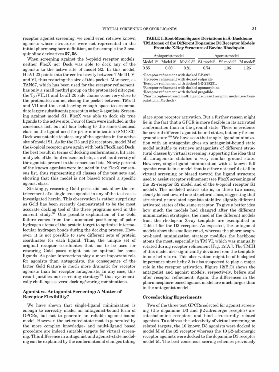
receptor agonist screening, we could even retrieve knownagonists whose structures were not represented in theinitial pharmacophore definition, as for example the 2-oxo-quinoline derivatives 57, 58.
When screening against the �-opioid receptor models,neither FlexX nor Dock was able to dock any of theagonists to the active site of model S2. In this model,HisVI:23 points into the central cavity between TMs III, V,and VI, thus reducing the size of this pocket. Moreover, asTAN67, which has been used for the receptor refinement,has only a small methyl group on the protonated nitrogen,the TyrVII:11 and LeuII:20 side chains come very close tothe protonated amine, closing the pocket between TMs IIand VII and thus not leaving enough space to accommo-date larger substituents present in other �-agonists. Screen-ing against model S1, FlexX was able to dock six trueligands to the active site. Four of them were included in theconsensus list, but all four belong to the same chemicalclass as the ligand used for prior minimization (SNC-80).Dock was not able to place any of the agonists in the activesite of model S1. As for the D3 and �2 receptors, model M ofthe �-opioid receptor gave again with both FlexX and Dock,the best result in terms of docking conformations, hit rate,and yield of the final consensus lists, as well as diversity ofthe agonists present in the consensus lists. Ninety percentof the known agonists were included in the FlexX consen-sus list, thus representing all classes of the test sets andshowing that this model is not biased toward a specificagonist class.
Strikingly, rescoring Gold poses did not allow the re-trievement of a single true agonist in any of the test casesinvestigated herein. This observation is rather surprisingas Gold has been recently demonstrated to be the mostaccurate docking tool of the three programs used in thecurrent study.67 One possible explanation of the Goldfailure comes from the automated positioning of polarhydrogen atoms of the protein target to optimize intermo-lecular hydrogen bonds during the docking process. How-ever, it is not possible to save different sets of proteincoordinates for each ligand. Thus, the unique set oforiginal receptor coordinates that has to be used forrescoring Gold poses might not be optimal for someligands. As polar interactions play a more important rolefor agonists than antagonists, the consequence of thelatter Gold feature is much more dramatic for receptoragonists than for receptor antagonists. In any case, thisresult justifies our screening strategy23 that systemati-cally challenges several docking/scoring combinations.
Agonist vs. Antagonist Screening: A Matter ofReceptor Flexibility?
We have shown that single-ligand minimization isenough to correctly model an antagonist-bound form ofGPCRs, but not to generate an reliable agonist-boundmodel. However, the activated-state models generated bythe more complex knowledge- and multi-ligand basedprocedure are indeed suitable targets for virtual screen-ing. This difference in antagonist and agonist-state model-ing can be explained by the conformational changes taking
place upon receptor activation. But a further reason mightlie in the fact that a GPCR is more flexible in its activatedconformation than in the ground state. There is evidencefor several different agonist-bound states, but only for oneground state.68 We have seen that single-ligand minimiza-tion with an antagonist gives an antagonist-bound statemodel suitable to retrieve antagonists of different struc-tural classes by virtual screening, supporting the idea thatall antagonists stabilize a very similar ground state.However, single-ligand minimization with a known fullagonist results in a model that is either not useful at all invirtual screening or biased toward the ligand structureused to assist receptor refinement (see FlexX screenings ofthe �2-receptor S2 model and of the �-opioid receptor S1model). The modeled active site is, in these two cases,clearly biased toward one structural class, suggesting thatstructurally unrelated agonists stabilize slightly differentactivated states of the same receptor. To give a better ideahow much the models had changed after the differentminimization strategies, the rmsd of the different modelsfrom the rhodopsin X-ray template are exemplified inTable I for the D3 receptor. As expected, the antagonistmodels show the smallest rmsd, whereas the pharmacoph-ore-based minimization strategy modifies the backboneatoms the most, especially in TM VI, which was manuallyrotated during receptor refinement [Fig. 12(A)]. The TMIIIof this model also significantly deviates from the templatein one helix turn. This observation might be of biologicalimportance since helix 3 is also suspected to play a majorrole in the receptor activation. Figure 12(B,C) shows theantagonist and agonist models, respectively, before andafter receptor refinement. Again, the differences in thepharmacophore-based agonist model are much larger thanin the antagonist model.
Crossdocking Experiments
Two of the three test GPCRs selected for agonist screen-ing (the dopamine D3 and �2-adrenergic receptor) arecatecholamine receptors and bind structurally relatedagonists. To address the selectivity of virtual screening onrelated targets, the 10 known D3 agonists were docked tomodel M of the �2 receptor whereas the 10 �2-adrenergicreceptor agonists were docked to the dopamine D3 receptormodel M. The best consensus scoring schemes previously
TABLE I. Root-Mean Square Deviations in Å (BackboneTM Atoms) of the Different Dopamine D3 Receptor Models
From the X-Ray Structure of Bovine Rhodopsin
Antagonist model Agonist model
Model 1a Model 2b Model 3c S1 modeld S2 modele M modelf
0.85 0.80 0.81 0.74 1.06 1.26aReceptor refinement with docked BP-897.bReceptor refinement with docked sulpiride.cReceptor refinement with docked GR-218231.dReceptor refinement with docked apomorphine.eReceptor refinement with docked pergolide.fPharmacophore-based multi ligands-biased receptor model (see Com-putational Methods).
VIRTUAL SCREENING OF GPCR LIGANDS 21

identified for both receptors [Fig. 7(A,B)] were then ap-plied to calculate the enrichment in true and “wrong”ligands. Though the positions of the AspIII:11 and theserines on helix 5 are very similar in both receptors,neither FlexX nor Dock was capable to find reliable posesupon docking the D3 agonists to the �2 receptor. Acomparison of the D3 and �2 models shows that D3agonists have to change their binding mode when bindingto the �2 receptor to avoid bumping into the secondextracellular loop E2. The E2 loop is 8 residues longer inthe �2 than in the D3 receptor (Fig. 3), thus decreasing theoverall size of the �2 binding pocket. Since the D3 agonistscannot form the expected interactions with the conservedaspartate (TM III) and the serines on TM V, they are allbadly scored whatever the scoring function used. Conse-quently, none of the true D3 agonists would have been
included in the consensus hit lists when screening for �2receptor agonists (Table II).
Docking the �2 agonists to the active site of the D3receptor gives better solutions, at least when using FlexXas docking algorithm. The small catecholamines 51–53 aswell as compound 57 can be docked as expected (salt bridgebetween the basic nitrogen and AspIII:11, H-bonds of thecatechol to the serines on TM V). Fitting the larger �2agonists (52, 54–56, 58–60) in the D3 active site is moredifficult because the pocket between TM II and VII is notas opened as in the �2 model. However, possible poses arenevertheless found in which the large N-aralkyl substitu-ents are directed towards TMs VI and VII. However, onlytwo �2-agonists (compounds 58, 59) [Fig. 2(B)] would havebeen selected as potential hits by screening the D3 recep-tor model. In conclusion, these “cross-docking” experi-
Fig. 12. Influence of the different refinement protocols on the overall GPCR structure exemplified by the dopamine D3 receptor. A: Ribbon view ofbackbone atoms of the seven transmembrane (TM) helices (bovine rhodopsin, white; D3 receptor antagonist model 1, red; D3 agonistpharmacophore-based model M, yellow; D3 agonist model S1, green; D3 agonist model S2, cyan). B: Comparison of the D3 antagonist model before(yellow) and after (cyan) receptor minimization. Ribbons indicate the backbone atoms of both structures. The docked ligand BP-897 is displayed bysticks. C: Comparison of the D3 pharmacophore-based agonist model before (yellow) and after (cyan) receptor minimization. Ribbons indicate thebackbone of both structures. For clarity, only one of the 5 superimposed ligands(apomorphine) is shown as sticks. D: Ribbon view of the backbone atomsof the D3 antagonist “model 1” (red) and the D3 agonist pharmacophore-based model M (yellow). Please note that helix 6 was manually rotated in thepharmacophore-based model to fit known pharmacophoric requirements. The usual locations of the antagonist and agonist binding sites in GPCRs areindicated by dashed lines. Generally, GPCR antagonists occupy the complete transmembrane cavity (from the pocket between TMs III, V, and VI to thepocket between TMs II and VII), whereas the agonist binding site is often restricted to the pocket between TMs III, V, VI. However, there are exceptions tothis rule, as for example �2-agonists that also partially occupy the pocket between TMs II and VII.
22 C. BISSANTZ ET AL.

ments show that virtual screening against the new agonist-bound states of related GPCRs is selective enough todistinguish not only true ligands from randomly chosendrug-like molecules but also true hits from chemicallyrelated inactive compounds.
CONCLUSIONS
Antagonist-bound state models of three human G-Protein coupled receptors have proven to be indeed suit-able for virtual screening of GPCR antagonists. For the D3and the V1a receptor, obtained hit rates are 20- to 40-foldhigher than what can be obtained by random screening.True antagonists structurally unrelated to the one used forreceptor refinement were predicted to be virtual hits,suggesting that the antagonist used in the minimizationstep does not significantly bias the virtual screening.Hence, the topology of the transmembrane cavity remainsrelatively similar after using different ligands for expand-ing the binding site, thus explaining that (1) hit listscontain ligands belonging to chemical series different fromthat used for receptor refinement and (2) the outcome ofthe virtual screening in terms of hit rate, yield, andstructures of the virtual hits is rather similar whateverligand used for expanding the binding site.
However, challenging single-ligand biased receptor mod-els for retrieving known full agonists was inefficient. Alogical explanation for this observation resides in the factthat all GPCR models have been derived from the inactivestate of bovine rhodospin, which is closer to an “antagonist-bound state” than to an “agonist-bound state” of the targetGPCR, as well as in the higher flexibility of the active sitein the activated state than in the ground state. Minimizingwith a known ligand in the active site is obviously suffi-cient to expand the active site but not for simulating theconformational changes occurring in the receptor activa-tion process. A pharmacophore-based receptor refinementmethod was proposed to generate agonist-bound statemodels of GPCRs still using the X-ray structure of bovine
rhodopsin as template. Applied to three human GPCRs,such receptor models are indeed precise enough for dis-criminating known agonists from randomly chosen “drug-like” molecules. Most importantly, we were able to retrievein the virtual hit lists true agonists whose chemicalstructures had not previously been used for generating thepharmacophore and refining the receptor model. Thisconfirms that the current GPCR activated-state modelsare not significantly biased toward the agonists used in theminimization step. This very important feature shouldenable us to retrieve new lead structures that could betotally unrelated to any known GPCR ligand.
We, thus, suggest two different strategies for virtualscreening of GPCRs, depending on the type of compoundsthat are searched for: (1) searching for antagonists re-quires only minimization of the rhodopsin-derived homol-ogy model with a single true ligand; (2) searching for fullagonists demands a much more complex procedure, stillstarting from rhodopsin, in which a proper activated statemodel has to be first derived by a pharmacophore-basedreceptor refinement.
ACKNOWLEDGMENTS
We sincerely thank TRIPOS SARL (Antony, France),the Cambridge Cristallographic Data Center (Cambridge,UK), and the Centre Informatique National del’Enseignement Superieur (CINES, Montpellier, France)for their software/hardware support. 3-D coordinates of allactivated (model M) and inactivated receptor models areavailable in PDB format at the following URL: http://med-chem.u-strasbg.fr/didier/gpcr_models/
REFERENCES
1. Mander T. Beyond uHTS: ridiculously HTS? Drug Discov Today2000;5:223–225.
2. Walters WP, Stahl MT, Murcko MA. Virtual screening: an over-view. Drug Discov Today 1998;3:160–178.
3. Pickett SD, McLay IM, Clark DE. Enhancing the hit-to-leadproperties of lead optimization libraries. J Chem Inf Comput Sci2000;2:263–272.
4. Gohlke H, Klebe G. Statistical potentials and scoring functionsapplied to protein-ligand binding. Curr Opin Struct Biol 2001;11:231–235.
5. Mason JS, Hermsmeier MA. Diversity assessment. Curr OpinChem Biol 1999;3:342–349.
6. Mason JS, Morize I, Menard PR, Cheney DL, Hulme C, Labaudi-niere RF. New 4-point pharmacophore method for molecularsimilarity and diversity applications: overview of the method andapplications, including a novel approach to the design of combina-torial libraries containing privileged substructures. J Med Chem1999;42:3251–3264.
7. Dixon JS. Evaluation of the CASP2 docking section. Proteins1997;(Suppl 1):198–204.
8. Tame JRH. Scoring functions: A view from the bench. J Comput-Aided Mol Design 1999;13:99–108.
9. Marhefka CA, Moore BM, Bishop C, Kirkovsky L, Mukherjee A,Dalton JT, Miller DD. Homology modeling using multiple molecu-lar dynamics simulations and docking studies of the humanandrogen receptor ligand binding domain bound to testosteroneand nonsteroidal ligands. J Med Chem 2001;44:1729–1740.
10. Schafferhans A, Klebe G. Docking ligands onto binding siterepresentations derived from proteins built by homology model-ling. J Mol Biol 2001;307:407–427.
11. Schneider, G., Bohm, H.J. Virtual screening and fast automateddocking methods. Drug Discov. Today 2002;7:64–70.
12. Marchese A, George SR, Kolakowski LF, Lynch KR, O’Dowd BF.Novel GPCRs and their endogenous ligands: expanding the bound-
TABLE II. Percentage of Known Ligands Identified asTrue Hits After Cross-Docking �2 Agonists to the D3
Receptor and D3 Agonists to the �2 Receptor†
LigandsReceptor(model M)
Dockingmethod Hit rate (%)
D3 agonists D3 Dock 24.1FlexX 31.6
�2 Dock 0.0FlexX 0.0
�2 agonists D3 Dock 0.0FlexX 7.7
�2 Dock 5.8FlexX 26.9
†Hit rates were calculated from the consensus list derived from the top15% scorers of single hit lists (D3 receptor: FlexX poses scored byFlexX, Gold, and ChemScore, Dock poses scored by Dock, FlexX, andGold; �2 receptor: FlexX poses scored by FlexX, Gold, and Fresno,Dock poses scored by Dock, FlexX, and Gold). In both dockings, theputative best model (model M) was used for each receptor. Gold wasnot used as docking tool since rescoring Gold poses for GPCR agonistswas previously proven to be inefficient.
VIRTUAL SCREENING OF GPCR LIGANDS 23

aries of physiology and pharmacology. Trends Pharmacol Sci1999;20:370–375.
13. Hibert MF, Trumpp-Kallmeyer S, Bruinvels A, Hoflack J. Three-dimensional models of neurotransmitter G-binding protein-coupled receptors. Mol Pharmacol 1991;40:8–15.
14. Bikker JA, Trumpp-Kallmeyer S, Humblet C. G-Protein coupledreceptors: models, mutagenesis, and drug design. J Med Chem1998;41:2911–2927.
15. Henderson R, Baldwin JM, Ceska TA, Zemlin F, Beckmann E,Downing KH. Model for the structure of bacteriorhodopsin basedon high-resolution electron cryo-microscopy. J Mol Biol 1990;213:899–929.
16. Schertler GF, Villa C, Henderson R. Projection structure ofrhodopsin. Nature 1993;362:770–772.
17. Baldwin JM, Schertler GF, Unger VM. An alpha-carbon templatefor the transmembrane helices in the rhodopsin family of G-protein-coupled receptors. J Mol Biol 1997;272:144–164.
18. Mouillac B, Chini B, Balestre MN, Elands J, Trumpp-Kallmeyer SHoflack J, Hibert M, Jard S, Barberis C. The binding site ofneuropeptide vasopressin V1a receptor. Evidence for a majorlocalization within transmembrane regions. J Biol Chem 1995;270:25771–25777.
19. Palczewski K, Kumasaka T, Hori T, Behnke CA, Motoshima H,Fox BA, Trong IL, Teller DC, Okada T, Stenkamp RE, YamamotoM, Miyano M. Crystal structure of Rhodopsin: a G protein-coupledreceptor. Science 2000;289:739–745.
20. Sokoloff P, Schwartz JC. Novel dopamine receptors half a decadelater. Trends Pharmacol Sci 1995;16:270–275.
21. Felder CC, Bymaster FP, Ward J, DeLapp N. Therapeutic opportu-nities for muscarinic receptors in the central nervous system.J Med Chem 2000;43:4333–4353.
22. Hibert M, Hoflack J, Trumpp-Kallmeyer S, Mouillac B, Chini B,Mahe E, Cotte N, Jard S, Manning M, Barberis C. Functionalarchitecture of vasopressin/oxytocin receptors. J Recept SignalTransduct Res 1999;19:589–596.
23. Bissantz C, Folkers G, Rognan D. Protein-based virtual screeningof chemical databases. 1. Evaluation of different docking/scoringcombinations. J Med Chem 2000;43:4759–4767.
24. Ewing TJA, Kuntz ID. Critical evaluation of search algorithms forautomated molecular docking and database screening. J ComputChem 1997;18:1175–1189.
25. Jones G, Wilett P, Glen RC, Leach AR, Taylor R. Development andvalidation of a genetic algorithm for flexible docking. J Mol Biol1997;267:727–748.
26. Rarey M, Kramer B, Lengauer T, Klebe G. A fast flexible dockingmethod using an incremental construction algorithm. J Mol Biol1996;261:470–489.
27. Muegge I, Martin YC. A general and fast scoring function forprotein-ligand interactions: a simplified potential approach. J MedChem 1999;42:791–804.
28. Eldridge M, Murray CW, Auton TA., Paolini GV, Lee RP. Empiri-cal scoring functions: I. The development of a fast empiricalscoring function to estimate the binding affinity of ligands inreceptor complexes. J Comput-Aided Mol Des 1997;11:425–445.
29. Rognan D, Laumoeller SL, Holm A, Buus S, Tschinke V. Predict-ing binding affinities of protein ligands from three-dimensionalcoordinates: application to peptide binding to class I major histo-compatibility proteins. J Med Chem 1999;42:4650–4658.
30. Wang R, Liu L, Lai L, Tang Y. SCORE: a new empirical method forestimating the binding affinity of a protein-ligand complex. J MolModel 1998;4:379–384.
31. Charifson PS, Corkery JJ, Murcko MA, Walters WP. Consensusscoring: A method for obtaining improved hit rates from dockingdatabases of three-dimensional structures into proteins. J MedChem 1999;42:5100–5109.
32. MDL Information Systems, Inc., San Leandro, CA 94577.33. SYBYL 6.62, Tripos Inc., St. Louis, MO 63144.34. Gasteiger J, Marsili M. Iterative partial equalization of orbital
electronegativity: a rapid access to atomic charges . Tetrahedron1980;36:3219–3228.
35. Concord 4.0 is part of the SYBYL software distribution (http://www.tripos.com).
36. Isis/Draw is distributed from MDL Information Systems (http://www.mdli.com).
37. Thompson JD, Higgins DG, Gibson TJ. CLUSTAL W: improvingthe sensitivity of progressive multiple sequence alignment through
sequence weighting, position-specific gap penalties and weightmatrix choice. Nucleic Acids Res 1994;22:4673–4680.
38. Henikoff S, Henikoff J. G. Amino acid substitution matrices fromprotein blocks Proc Natl Acad Sci USA 1992;89:10915–10919.
39. Strader CD, Dixon RA, Cheung AH., Candelore MR, Blake AD,Sigal IS. Mutations that uncouple the beta-adrenergic receptorfrom Gs and increase agonist affinity. J Biol Chem 1987;262:16439–16443.
40. Case DA, Pearlman DA, Caldwell JW, Cheatham III TE, Ross WS,Simmerling CL, Darden TA, Merz KM, Stanton RV, Cheng AL,Vincent JJ, Crowley M, Ferguson, DM, Radmer RJ, Seibel GL,Singh,UC, Weiner PK, Kollman PA. AMBER 5.0. University ofCalifornia, San Francisco; 1997.
41. Cornel WD, Cieplak P, Bayly CI, Gould IR, Merz JKM, FergusonDM, Spellmeyer DM, Fox T, Caldwell JW, Kollman PA. A secondgeneration force field for the simulation of proteins, nucleic acidsand organic molecules. J Am Chem Soc 1995;117:5179–5197.
42. Alberts GL, Pregenzer JF, Im WB. Identification of transmem-brane regions critical for ligand binding to the human D3 dopa-mine receptor using various D3/D1 transmembrane chimeras. MolPharmacol 1998;54:379–88.
43. Mansour A, Meng F, Meador-Woodruff JH, Taylor LP, Civelli O,Akil H. Site-directed mutagenesis of the human dopamine D2receptor. Eur J Pharmacol 1992;227:205–214.
44. Lundstrom K, Turpin P, Large C, Robertson G, Thomas P, LewellXQ. Mapping of dopamine D3 receptor binding site by pharmaco-logical characterization of mutants expressed in CHO cells withthe Semliki Forest virus system. J Recept Signal Transduct Res1998;18:133–150.
45. Cho W, Taylor LP, Mansour A, Akil H. Hydrophobic residues ofthe D2 dopamine receptor are important for binding and signaltransduction. J Neurochem 1995;65:2105–2115.
46. Bourdon H, Trumpp-Kallmeyer S, Schreuder H, Hoflack J, HibertM, Wermuth CG. Modelling of the binding site of the human m1muscarinic receptor: Experimental validation and refinement.J Comput-Aided Mol Design 1997;11:317–332.
47. Cotte N, Balestre MN, Aumelas A, Mahe E, Phalipou S, Morin D,Hibert M, Manning M, Durroux T, Barberis C, Mouillac B.Conserved aromatic residues in the transmembrane region VI ofthe V1a vasopressin receptor differentiate agonist vs. antagonistligand binding. Eur J Biochem 2000;267:4253–4263.
48. Strader CD, Candelore MR, Hill WS, Sigal IS, Dixon RAF.Identification of two serine residues involved in agonist activationof the �2-adrenergic receptor. J Biol Chem 1989;264:13572–13578.
49. Strader CD, Gaffney T, Sugg EE, Candelore MR, Keys R, PatchettAA, Dixon RAF. Allele-specific activation of genetically engi-neered receptors. J Biol Chem 1991;266:5–8.
50. Strader CD, Fong TM, Tota MR, Underwood D, Dixon RA.Structure and function of G protein-coupled receptors. Annu RevBiochem 1994;63:101–132.
51. Wieland K, Zuurmond HM, Krasel C, Ijzerman AP, Lohse MJ. In-volvement of Asn-293 in stereospecific agonist recognition and inactivation of the �2-adrenergic receptor. Pharmacology 1996;93:9276–9281.
52. Liapaski G, Ballesteros JA, Papachristou S, Chan WC, Chen X,Javitch JA. The Forgotten Serine. J Biol Chem 2000;275:37779–37788.
53. Valiquette M, Vu HK, Yue SY, Waehlestedt C, Walker P. Involve-ment of Trp-284, Val-296, and Val-297 of the human �-opioidreceptor in binding of �-selective ligands. J Biol Chem 1996;271:18789–18796.
54. Befort K, Tabbara L, Kling D, Maigret B, Kiefer BL. Role ofaromatic transmembrane residues of the �-opioid receptor inligand recognition. J Biol Chem 1996;271:10161–10168.
55. Pepin M, Yue SY, Roberts E, Wahlestedt C, Walker P. Novel“restoration of function” mutagenesis strategy to identify aminoacids of the �-opioid receptor involved in ligand binding. J BiolChem 1997;272:9260–9267.
56. Lemmen C, Lengauer T, Klebe G. FlexS: a method for fast flexibleligand superimposition. J Med Chem 1998;41:4502–4520.
57. Schwartz TW. Locating ligand-binding sites in 7 TM receptors byprotein engineering. Cur Opin Biotechnol 1994;5:434–444.
58. Gether U, Kobilka BK. G protein-coupled receptors. II. Mecha-nism of agonist activation. J Biol Chem 1998;273:17979–17982.
59. Hulme EC, Lu ZL, Ward SDC, Allman K, Curtis CA. The
24 C. BISSANTZ ET AL.
conformational switch in 7-transmembrane receptors: the musca-rinic receptor paradigm. Eur J Pharmacol 1999;375: 247–260.
60. Ghanouni P, Steenhuis JJ, Farrens DL, Kobilka BK. Agonist-induced conformational changes in the G-protein-coupling domainof the �2 adrenergic receptor. Proc Natl Acd Sci USA 2001;98:5997–6002.
61. Plobeck N, Delorme D, Wei Z, Yang H, Zhou F, Schwarz P, GawellL, Gagnon H, Pelcman B, Schmidt R, Yue SY, Walpole C, BrownW, Zhou E, Labarre M, Payza K, St-Onge S, Kamassah A, Morin P,Projean D, Ducharme J, Roberts E. New Diarylmethylpiperazinesas potent and selective nonpeptidic � opioid receptor agonists withincreased in vitro metabolic stability. J Med Chem 2000;43:3878–3894.
62. Liao S, Alfaro Lopez J, Shenderovich MD, Hosohata K, Lin J, Li X,Stropova D, Davis P, Jernigan KA, Porreca F, Yamamura HI,Hruby VJ. De novo design, synthesis, and biological activities ofhigh-affinity and selective non-peptide agonists of the �-opioidreceptor. J Med Chem 1998;41:4767–4776.
63. Portoghese PS. From models to molecules: opioid receptor dimers,
bivalent ligands, and selective opioid receptor probes. J Med Chem2001;44:2259–2269.
64. Farrens DL, Altenbach C, Yang K, Hubell WL, Khorana HG.Requirement of rigid-body motion of transmembrane helices forlight activation of rhodopsin. Science 1996;274:768–770.
65. Dunham TD, Farrens DL. Conformational changes in Rhodopsin.J Biol Chem 1999;274:1683–1690.
66. Jensen AD, Guarnieri F, Rasmussen SGF, Asmar F, BallesterosJA, Gether U. Agonist-induced conformational changes at thecytoplasmatic side of transmembrane segment 6 in the �2 adrener-gic receptor mapped by site-selective fluorescent labeling. J BiolChem 2001;276:9279–9290.
67. Paul N, Rognan D. ConsDock: A new program for the consensusanalysis of protein-ligand interactions. Proteins 2002;47:521–533.
68. Ghanouni P, Gryczynski Z, Steenhuis JJ, Lee TW, Farrens DL,Lakowicz JR, Kobilka BK. Functionally different agonistsinduce distinct conformations in the G protein coupling domainof the �2 adrenergic receptor. J Biol Chem 2001;276:24433–24436.
VIRTUAL SCREENING OF GPCR LIGANDS 25


