Embed Size (px)
Citation preview

Role of Vasodilator stimulated phosphoprotein in VEGF inducedblood-brain barrier permeability in endothelial cell monolayers
Brandon Davisa,#, Jun Tangc,#, Li Zhangc,#, Dezhi Mua,c, Xiangning Jianga, ValerieBirand, Zinaida Vexlera, and Donna M. Ferrieroa,b,*a Department of Neurology, University of California, San Francisco, San Francisco, CA 94143,USAb Department of Pediatrics, University of California, San Francisco, San Francisco, CA 94143,USAc Department of Pediatrics, West China Second University Hospital, Sichuan University, Chengdu610041, Chinad Department of Neonatal Intensive Care, Robert Debre Hospital, Paris, France
AbstractThe blood-brain barrier (BBB) plays an important role in the pathophysiology of central nervoussystem (CNS) disorders such as stroke and hypoxic-ischemic brain injury. Vascular endothelialgrowth factor (VEGF) is involved in angiogenesis and vasogenic edema during stroke andhypoxia. However, the role of VEGF in BBB permeability after hypoxia has not been fullyelucidated. We therefore investigated VEGF effects in an in vitro BBB model using rbcec4endothelial cell line with the stimulation of VEGF or hypoxia. In this study, BBB permeabilitywas studied using 14C-sucrose detection. The expression of BBB tight junction protein ZO-1, andthe expression and phosphorylation of vasodilator stimulated phosphoprotein (VASP), VEGF andVEGF receptor 2 (VEGFR2) were determined using fluorescent immunocytochemistry andwestern blot analyses. We found that hypoxia upregulated VEGF expression, and VEGF increasedBBB permeability. Hypoxia also increased VASP phosphorylation, which is mediated, in part,through VEGFR2. We also found that VASP at tight junctions was co-localized with ZO-1 in cell-cell contacts. Our findings show that VASP phosphorylation is affected by hypoxia and VEGFR2inhibition suggesting a role for VASP in BBB permeability.
KeywordsBlood-brain barrier; VEGF; Hypoxia; VEGFR2; Permeability; Vasodilator stimulatedphosphoprotein
The blood–brain barrier (BBB) plays an important role in the pathophysiology of centralnervous system (CNS) disorders such as hypoxia, hypoxia-ischemia, and stroke. IncreasedBBB permeability after CNS disorders can lead to cerebral edema resulting in an
*Corresponding author: Donna Ferriero, MD Departments of Neurology and Pediatrics, University of California San Francisco, CA94143, USA, [email protected].#These authors contributed equally to this work.Publisher's Disclaimer: This is a PDF file of an unedited manuscript that has been accepted for publication. As a service to ourcustomers we are providing this early version of the manuscript. The manuscript will undergo copyediting, typesetting, and review ofthe resulting proof before it is published in its final citable form. Please note that during the production process errors may bediscovered which could affect the content, and all legal disclaimers that apply to the journal pertain.
NIH Public AccessAuthor ManuscriptInt J Dev Neurosci. Author manuscript; available in PMC 2011 October 1.
Published in final edited form as:Int J Dev Neurosci. 2010 October ; 28(6): 423–428. doi:10.1016/j.ijdevneu.2010.06.010.
NIH
-PA Author Manuscript
NIH
-PA Author Manuscript
NIH
-PA Author Manuscript

exacerbation of injury (Stamatovic et al., 2008). BBB permeability can be changed inresponse to a variety of stimulators or stressors. These molecular stimulators, includingvascular endothelial growth factor (VEGF), can exert beneficial or deleterious effects on thebrain, depending on the context, timing, and functional cellular outcomes of signaling (Rouxand Couraud, 2005). Early after stroke, VEGF signaling increases paracellular permeabilityof endothelial cells, which causes vasogenic edema and contributes to cerebral swelling(Abumiya et al., 2005). At later points, VEGF signaling initiates the formation of newcerebral blood vessels (Wang et al., 2007). Studies that examine whether VEGF hasbeneficial or deleterious roles following cerebral stroke provide mixed results.
The controversial effects of exogenous VEGF on brain ischemia or stroke may be due to thetiming and route of VEGF administration. One study which looked specifically at the timingof VEGF administration by intravenous infusion following stroke illustrated that earlyVEGF administration at 1 hr after stroke increased BBB leakage. Conversely, delayedVEGF administration at 48 hr after stroke enhanced angiogenesis and improved neurologicaloutcome (Zhang et al., 2000). Another study suggested beneficial effects ofintracerebroventricular administration of VEGF following stroke was associated withactivation of the PI3K-Akt pathway without affecting BBB permeability and angiogenesis(Kaya et al., 2005). Early intravenous VEGF administration in the same study wasdeleterious and increased infarct volume (Kaya et al., 2005).
In vitro studies further reveal consequences of the route of VEGF intervention. Studiesutilizing in vitro BBB models containing endothelial monolayers which are in contact withmedia from separate apical and basolateral compartments demonstrate an ability of VEGF toincrease BBB monolayer permeability in a directional way, with the majority of the effectbeing exerted from the basolateral side of the monolayer (Nitz et al., 2003). Attempts toinhibit the permeability effects of VEGF have demonstrated limited efficacy in hypoxic-ischemic brain due to a variety of factors in vivo (Chi et al., 2005). Therefore, studying theBBB in vitro is scientifically and biologically important.
Using neutralizing antibody to VEGF, monolayers of endothelial cells have been shown torespond to hypoxia with increases in tight junction permeability in a VEGF-dependentmanner (Fischer et al., 2002). VEGF- induced permeability changes are dependent ondownstream signal transduction mediated via activation of the VEGF receptors (VEGFR).The precise mechanisms by which VEGF signaling increases vascular permeability are notwell understood and may depend on the context of the insult, but changes in ZO-1 at thetight junctions and actin reorganization appear to be major contributing factors.
Vasodilator stimulated phosphoprotein (VASP) can induce permeability changes anddynamic changes in localization of the cytoskeletal protein actin. VASP is phosphorylatedby kinases partially dependent on VEGFR2 signaling, including PKA, cGMP-dependentprotein kinase (PKG), and AMP-activated protein kinase (AMPK) at residues S157, S239,and T278, respectively. VASP has also been shown to interact with the tight junction proteinZO-1 in human microvascular endothelial cells (Comerford et al., 2002). It can also interactwith a structural protein alpha II-spectrin (SPCN) in HUVEC cells in a ZO-1 independentmanner (Benz et al., 2008). However, the roles of VASP in changes of BBB permeability inresponse to stressors are largely unknown. We therefore studied VASP phosphorylationfollowing VEGFR2 inhibition or hypoxia stimulation using the immortalized rbcec4 cellmonolayer.
Davis et al. Page 2
Int J Dev Neurosci. Author manuscript; available in PMC 2011 October 1.
NIH
-PA Author Manuscript
NIH
-PA Author Manuscript
NIH
-PA Author Manuscript

1. Materials and methods1.1. Immortalized Endothelial Cell cultures
The immortalized brain endothelial cell line, rbcec4 cells, retain an endothelial-likemorphology and express the endothelial cell markers such as von Willebrand factor, alkalinephosphatase, and γ-glutamyl transpeptidase (Blasig et al., 2001) and therefore can be utilizedas an in vitro BBB model. This cell line was derived from isolated rat brain endothelial cellstransformed with the polyoma virus large T antigen. Rbcec4 cells were kindly donated byDr. Ingolf E. Blasig (Forschungsinstitute für Molekulare Pharmakologie, Berlin, Germany).Rbcec4 cells were plated on collagen I (6 μg/ml)-coated dishes and maintained in DME/Ham’s F12 (2:1) medium supplemented with 10% heat inactivated fetal calf serum. Cultureswere kept in a humidified incubator with 5% CO2/95% air at 37°C for 5–7 days untilconfluence, with media changes every two days. Cells were then passaged once and grownon transwell inserts on day in vitro (DIV) 5 till monolayer formation. Monolayer integrityand tight junction formation were tested when the cells became confluent.
1.2 Hypoxia and VEGF treatmentsHypoxia was induced by placing endothelial cells in a modular incubator chamber(Anaerobic Chamber, Plas Labs, Lansing, MI) gassed with a mixture consisting of 5% CO2and 95% N2. Prior to the hypoxia experiments, the growth medium was replaced by themedium that had been equilibrated with a gas mixture of 5% CO2 and 95% N2 for 30 minbefore bathing the cells. Hypoxia was maintained for different periods of time, depending onprotocols.
The cells were incubated with recombinant VEGF (5–50 ng/ml, Sigma) for 20, 40 or 60 minfor acute measurements of tight junction permeability.
1.3 Fluorescence immunolabelingThe presence of tight junctions in rbcec4 monolayer was determined by protein expressionof ZO-1 using fluorescence immunolableling as previously described (Mu et al. 2005).Briefly, cultured rbcec4 cells on DIV 5 were fixed with 4% paraformaldhyde for 10 min.Endogenous peroxidase was inhibited with 0.3% hydrogen peroxide in methanol at roomtemperature (RT) for 20 min. Cells were incubated with Clean Vision ™ blocking solution(Immuno Vision Technologies Co, CA, USA) at RT for 1–2 h. After three washes with PBS,cells were incubated with monoclonal ZO-1 antibody (1:50, Zymed) at 4°C overnight. Afterwashing with PBS 3 times, cells were incubated with TRITC-conjugated secondary antibody(1:50, Sigma) at RT for 1 h. To delineate the cellular localization and expression ofVEGFR2 and phosphorylated VEGFR2 on rbcec4 cells after stimulation with VEGF,fluorescence immunolabeling was performed using Texas red-conjugated anti-phosphoY1054+Y1059 VEGFR2 antibody (1:100 Abcam) and FITC-conjugated anti-VEGFR2 c-terminus (1:100 Santa Cruz) as described above. After incubation with secondary antibodiesand washing with PBS, cells were coverslipped with ProLong Gold antifade reagent(Invitrogen) and images were captured with Openlab software (Improvision, England) inboth the FITC and TRITC channels. As negative controls, cells were processed in parallelwithout primary antibody.
1.4 VASP-eGFP transfection for rbcec4 cellsFor analysis of expression and localization of VASP and ZO-1 proteins, cells were seededovernight onto collagen-coated dishes and transfected with calcium phosphate precipitatesof the eukaryotic vectors coding for EGFP-VASP constructs (pEGFP-C2, Clontech, withcorresponding inserts) according to standard protocols. Forty-eight hrs after transfection,cells were washed 3 times with PBS containing 2 mM MgCl2 and 10% glycerol and fixed
Davis et al. Page 3
Int J Dev Neurosci. Author manuscript; available in PMC 2011 October 1.
NIH
-PA Author Manuscript
NIH
-PA Author Manuscript
NIH
-PA Author Manuscript

with the bifunctional membrane permeable crosslinker DSP (Pierce, Rockford, IL, USA) for30 min at 37°C. After additional washing, cells were permeabilized with 0.2% Triton X-100and excess crosslinker was quenched with 0.2 M glycine. Counterstaining for ZO-1 andfluorescent microscopy was performed to verify their co-localization.
1.5 Western Blot analysisWestern blot was performed as previously described (Jiang et al., 2008). Briefly, whole celllysates were used to quantify expression of phosphorylated VEGFR2 (p-VEGFR2) andphosphorylated VASP (pVASP) using the following antibodies and conditions: VEGFR2,pVEGFR2 antibodies (1:100, overnight, 4°C; Santa Cruz), VASP, pVASP Ser-239antibodies (1:100, overnight, 4°C; Upstate) or VEGF antibody (1:200, overnight, 4°C; SantaCruz). The phosphospecific antibody detects VEGFR2 molecules that areautophosphorylated at Y1054 and Y1059 in the catalytic domain of VEGFR2. The signalwas normalized to μ-actin expression (anti-β-actin antibody, 1:1500; Santa Cruz).
1.6 Inducible permeability of rbcec4 monolayersPermeability studies with 14C-sucrose (672 mCi/mmol, New England Nuclear Research,Boston, MA) were used to determine paracellular flux across confluent rbcec4 monolayers.Apical-to basolateral flux was determined by dividing the p moles of radioactive markerappearing in the receiver chamber by the time in minutes. The apparent permeabilitycoefficient (PC) was calculated using the equation: PC (cm/min)=Flux/(A×CDo), where fluxis the slope of the line, A is the area of the membrane and CDo is the initial donorconcentration of radioactive marker (Franke et al., 2000). Functional integrity of tightjunctions in the monolayers was confirmed via measurement of trans-endothelial electricalresistance (TEER). TEER measurement can be easily performed with the ENDOHM-24 ™chamber and the EVOHM ™ voltometer ( World Precision Instruments, Berlin, Germany) aspreviously described (Franke et al., 2000).
1.7 Statistical analysisData were presented as mean ± standard deviation (SD). One-way ANOVA withBonferroni/Dunnett post-hoc tests was performed for multiple comparisons. p<0.05 wasconsidered statistically significant.
2. Results2.1 Immortalized brain endothelial monolayers form tight junctions of BBB
In this study, immortal rat brain cell line rbcec4 cells were employed to set up the in vitroBBB model. Rbcec4 cells have been immortalized with the polyoma large T antigen andexpress the endothelial cell marker factor VIII and the mature BBB markers, gamma-glutamyl transpeptidase and alkaline phosphatase (Blasig et al., 2001). Rbcec4 cells grownto monolayers had an endothelial morphology and formed tight junctions on DIV5 asdetermined by expression of the tight junction protein, ZO-1, using fluorescentimmunocytochemistry. ZO-1 was observed at cell-cell contacts between rbcec4 cells (Fig1A), suggesting that these cultures could be used as an in vitro model for study of BBBpermeability and function.
Functional integrity of tight junctions in the monolayers was confirmed via measurement oftrans-endothelial electrical resistance (TEER) as previously described (Franke et al., 2000).Intact monolayers exhibiting TEER above 100 ohms were included in data analysis. Thesecharacteristics of rbcec4 cell are in agreement with previous reported BBB models (Deli etal., 2005).
Davis et al. Page 4
Int J Dev Neurosci. Author manuscript; available in PMC 2011 October 1.
NIH
-PA Author Manuscript
NIH
-PA Author Manuscript
NIH
-PA Author Manuscript

2.2 VEGFR2 activation after stimulation with VEGF and hypoxiaTo study the effect of VEGF on BBB permeability, we first determined whether VEGFcould activate its receptor VEGFR2, the predominant VEGF receptor in the brain. Fig. 1Bshows that both VEGF and hypoxia significantly increased VEGFR2 and phosphorylation ofVEGFR2 in whole cell lysates. Compared to untreated controls, VEGFR2 increasedapproximately 1.4 fold and 1.3 fold, whereas phosphorylation of VEGFR2 increasedapproximately 1.7 fold and 1.4 fold by hypoxia and VEGF respectively (Fig. 1B, C,*p<0.05, **p<0.01). We also found that hypoxia stimulates VEGF expression atapproximately 1.4 fold in this system (Fig. 1. D, E, *p<0.05).
We found that VEGFR2 expression was much stronger in VEGF-stimulated cells (Fig. 1G,in green) than that in DMEM control cells (Fig. 1H, in green). There is no positive stainingof cells in negative controls (Fig. 1F). Phosphorylation of VEGFR2, which is necessary forthe downstream kinase activity of activated VEGFR2 (Dougher and Terman, 1999), wasincreased in VEGF treated cells (Fig. 1E, red) compared with that seen in DMEM controlcells (Fig. 1F, red). These results suggest that both VEGF and hypoxia stimulate VEGFR2expression and phosphorylation in cultured rbcec4 cells.
2.3 BBB permeability is increased with VEGF stimulationTo determine whether VEGF increases permeability of a monolayer of rbcec4 cells tosucrose, we measured accumulation of 14C-sucrose in non-stimulated cells at 20 min, 40min and 60 min following VEGF exposure (Fig. 2). Compared to controls, there was atransient and significant increase in 14C-sucrose permeability at 20 min and 40 min afterexposure to 20 or 50 ng/ml VEGF (Fig. 2). At lower VEGF concentration (5ng/ml), therewas no difference in 14C-sucrose permeability at any time points studied (Fig. 2). Ourfindings suggest that VEGF could increase BBB permeability at doses higher than 20 ng/ml,and as early as 20 min following administration in this in vitro model.
2.4 VASP phosphorylation is increased by hypoxiaVEGF could increase BBB permeability but the underlying mechanism is not clear. VASPhas been shown to interact with the tight junction protein, ZO-1, in human vascularendothelial cells. Increased VASP phosphorylation has been shown to correlate withincreased barrier function (Comerford et al., 2002). To investigate whether VASP isinvolved in VEGF signaling following hypoxia, we performed Western blot analysis usingpVASP S239 phosphospecific antibody. We found that VASP phosphorylation wasincreased when measured immediately following either 24 h and 48 h duration of hypoxiaby an approximate 1.3 fold (p<0.05) and 1.6 fold (p<0.01), respectively (Fig. 3A). However,total VASP expression was not changed following hypoxia compared with the controls (Fig.3A).
To test whether VEGF/VEGFR2 signaling is involved in VASP phosphorylation, Westernblot analysis of rbcec4 monolayers treated with the VEGFR2 tyrosine kinase inhibitorSU5416 were performed. The induction of pVASP immediately following 24 h and 48 h ofhypoxia was abolished after SU5416 administration (Fig. 3B), suggesting that VEGFR2signaling pathway is involved in VASP phosphorlyation following hypoxia and that VASPis downstream of VEGF signaling following hypoxia. Total VASP levels did not changewith SU5416 treatment under hypoxia or control (Fig. 3B).
2.5 Colocalization of VASP with the tight junction protein ZO-1Since VASP and VEGF may be involved in BBB permeability, we next investigated therelationship between VASP and ZO-1 expression using fluorescent immunolabeling forZO-1 in VASP-eGFP transfected rbcec4 cells (Fig. 4; green-VASP and red-ZO1). VASP
Davis et al. Page 5
Int J Dev Neurosci. Author manuscript; available in PMC 2011 October 1.
NIH
-PA Author Manuscript
NIH
-PA Author Manuscript
NIH
-PA Author Manuscript

expression was observed as the characteristic “chicken-wire” staining pattern, while stainingfor ZO-1 resulted in a nearly identical staining pattern at the regions of cell-cell contacts(Fig. 4A, 4B). Colocalization of VASP with ZO-1 to the tight junction complex (Fig. 4C;yellow) may suggest its possible contribution to modulation of BBB permeability.
3. DiscussionHere we show for the first time that VASP phosphorylation initiated by hypoxia ismodulated by inhibition of VEGFR2 signaling in the in vitro rbcec4 cell BBB model. Wealso show that VASP and the tight junction protein ZO-1 co-localize at cellular contacts,likely contributing to the increased BBB permeability after exposure to VEGF.
In this rbcec4 BBB model, we found that VEGF can induce BBB permeability, which isconsistent with previous findings that VEGF is involved in vasodilation and endothelial cellpermeability (Gavard and Gutkind, 2006). Since VEGF functions through its receptorsespecially VEGFR2, we showed that both VEGF and hypoxia significantly increasedVEGFR2 expression and phosphorylation,. VEGFR2 dimerization and tyrosine kinaseactivity leads to the activation of several intracellular signaling cascades. Notably, therelease of intracellular calcium stores occurs in parallel with PI3K/Akt activation.Subsequently, an increase in NO and cGMP activates PKG. PKG has many substrates,including VASP (Comerford et al., 2002; Fischer S et al. 2004).
We decided to focus on the downstream signaling of VASP. VASP is composed of threemajor domains, an EVH1 domain, an EVH2 domain, and a central proline-rich region, andcontains both actin binding and actin monomer-nucleating capabilities. As a major actinbinding protein, VASP has been shown to colocalize with ZO-1 at tight junctions and play akey role in establishing and maintaining barrier function (Comerford et al., 2002). Loss ofVASP function results in loose associations between epithelia and failure to reseal barriersafter an insult (Vasioukhin et al., 2000). Inactivation of VASP prior to hypoxia in an in vitroepithelial barrier model by siRNA increased barrier permeability whereas VASPoverexpression protected from hypoxia-induced changes in permeability (Rosenberger et al.,2007). In endothelial cells, VASP can function in membrane ruffling, aggregation, andtethering of actin filaments during the formation of endothelial cell-substrate and cell-cellcontacts. Data that VASP depletion is associated with the more severe loss of ZO-1peripheral staining in response to LPS also suggest that VASP can regulate ZO-1 expressionand that PKA-dependent VASP phosphorylation contributes to the protective effect againstLPS-induced BBB disruption in vitro (Bogatcheva et al., 2009).
A recent study performed in HUVEC cells also revealed that patterns of subcellular VASPdistribution vary, depending on cell confluence and VASP phosphorylation (Benz et al.,2008). Expression of total VASP which has been shown to interact with a spectrin isoformvia the SH3 domain at sites of inter-endothelial adhesion was observed in cell–cell contacts,with almost complete spectrin isoform and ZO-1 localization (Benz et al., 2008). pS157-VASP, in turn, was mainly found at focal adhesions, with minor colocalization with spectrinisoform, suggesting multiple functions of this protein in endothelial cells (Benz et al.,2008).} Data in human placenta also suggest that VEGF mediated angiogenic andvasculogenic effects could be via stimulation of VASP expression (Kayisli et al., 2002).
Our results support the hypothesis that VASP phosphorylation after hypoxia is VEGFR2dependent since the VEGFR2 inhibitor, SU5416, showed a concurrent inhibition of VASPphosphorylation on s239 following hypoxia. Together these results suggest that VASP is adownstream effector of VEGF signaling following hypoxia, specifically via its substrateserine 239 in rbcec4 BBB model.
Davis et al. Page 6
Int J Dev Neurosci. Author manuscript; available in PMC 2011 October 1.
NIH
-PA Author Manuscript
NIH
-PA Author Manuscript
NIH
-PA Author Manuscript

VASP has also been shown to interact with ZO-1, a main BBB protein in human vascularendothelial cells. In this study, VASP was phosphorylated immediately after 24 h to 48 h ofhypoxia. This timeframe is in agreement with previous reports of tight junction breakdownand loss of ZO-1 tight junction localization following hypoxia in a different BBB model(Mark and Davis, 2002).
We also demonstrated that VASP participates in the BBB tight junction complex with ZO-1.VASP co-localized with the tight junction protein ZO-1 at regions of cell-cell contacts and,to a lesser extent, to focal adhesions. Since VASP interacts with the cytoskeletal proteinactin and participates in actin polymerization, VASP has the potential of being activelyinvolved in transmitting structural changes within cells (Collard et al., 2002).
It still needs to be determined whether VASP phosphorylation following hypoxia is directlyinvolved in modulation of BBB permeability in our model. However, from our results,VASP acts downstream of VEGF/VEGFR2 signaling and suggests its possible role inVEGF-mediated vascular permeability. Our previous in vivo data from neonatal strokemodel demonstrate that VEGFR2 inhibition with SU516 when given 48 hours after thestroke worsens injury and reduces endothelial cell proliferation in injured areas (Shimotakeet al., 2009). Again, whether VEGFR2 inhibition has beneficial or deleterious effects on thebrain depends on the stimulus, timing, cell type and pathways involved. Our data support therole of VEGF signaling in early disruption of the structural integrity of the BBB, and it ispossible that VASP phosphorylation is a critical step in disrupting the structural integrityneeded for cell to cell contact by increasing a migratory phenotype of the cells.
In conclusion, activation of VASP following hypoxia is mediated, at least in part, by theVEGF/VEGFR2 signaling pathway. VASP might play a role in VEGF-induced BBBpermeability. The ability to modulate this active regulation might be useful for efficientCNS drug delivery and the treatment of cerebral vasogenic edema and inflammationfollowing stroke.
AcknowledgmentsThis work was supported by grants from P50 NIH NINDS Program Project NS 35902, UCSF mentorship andresearch assistantship program, and also from the National Scientific Foundation of China (No.30825039, No.30973236 and No.30770748 to Dezhi Mu).
Abbreviations
BBB blood-brain barrier
CNS central nervous system
VEGF vascular endothelial growth factor
VEGFR2 VEGF receptor 2
VASP vasodilator stimulated phosphoprotein
ZO-1 zonula occludens 1
cGMP cyclic guanosine monophosphate
PKA protein kinase A
PKG cGMP-dependent protein kinase
AMPK AMP-activated protein kinase
PI3K phosphatidylinositol-3-kinase
Davis et al. Page 7
Int J Dev Neurosci. Author manuscript; available in PMC 2011 October 1.
NIH
-PA Author Manuscript
NIH
-PA Author Manuscript
NIH
-PA Author Manuscript

PBS phosphate buffered saline
TEER trans-endothelial electrical resistance
DIV day in vitro
DMEM dulbecco’s modified eagle’s medium
ReferencesAbumiya T, Yokota C, Kuge Y, Minematsu K. Aggravation of hemorrhagic transformation by early
intraarterial infusion of low-dose vascular endothelial growth factor after transient focal cerebralischemia in rats. Brain Res 2005;1049 (1):95–103. [PubMed: 15935998]
Benz PM, Blume C, Moebius J, Oschatz C, Schuh K, Sickmann A, Walter U, Feller SM, Renné T.Cytoskeleton assembly at endothelial cell–cell contacts is regulated by II-spectrin–VASPcomplexes. J Cell Biol 2008;180 (1):205–219. [PubMed: 18195108]
Blasig IE, Giese H, Schroeter ML, Sporbert A, Utepbergenov DI, Buchwalow IB, Neubert K,Schonfelder G, Freyer D, Schimke I, Siems WE, Paul M, Haseloff RF, Blasig R. *NO andoxyradical metabolism in new cell lines of rat brain capillary endothelial cells forming the blood-brain barrier. Microvasc Res 2001;62 (2):114–127. [PubMed: 11516240]
Bogatcheva NV, Zemskova MA, Kovalenkov Y, Poirier C, Verin AD. Molecular mechanismsmediating protective effect of cAMP on lipopolysaccharide (LPS)-induced human lungmicrovascular endothelial cells (HLMVEC) hyperpermeability. J Cell Physiol 2009;221 (3):750–759. [PubMed: 19725051]
Chi OZ, Hunter C, Liu X, Weiss HR. Effects of VEGF and nitric oxide synthase inhibition on blood-brain barrier disruption in the ischemic and non-ischemic cerebral cortex. Neurol Res 2005;27 (8):864–868. [PubMed: 16354548]
Collard CD, Park KA, Montalto MC, Alapati S, Buras JA, Stahl GL, Colgan SP. Neutrophil-derivedglutamate regulates vascular endothelial barrier function. J Biol Chem 2002;277 (17):14801–14811.[PubMed: 11847215]
Comerford KM, Lawrence DW, Synnestvedt K, Levi BP, Colgan SP. Role of vasodilator-stimulatedphosphoprotein in PKA-induced changes in endothelial junctional permeability. Faseb J 2002;16(6):583–585. [PubMed: 11919161]
Deli MA, Abraham CS, Kataoka Y, Niwa M. Permeability studies on in vitro blood-brain barriermodels: physiology, pathology, and pharmacology. Cell Mol Neurobiol 2005;25 (1):59–127.[PubMed: 15962509]
Dougher M, Terman BI. Autophosphorylation of KDR in the kinase domain is required for maximalVEGF-stimulated kinase activity and receptor internalization. Oncogene 1999;18 (8):1619–1627.[PubMed: 10102632]
Fischer S, Wobben M, Marti HH, Renz D, Schaper W. Hypoxia-induced hyperpermeability in brainmicrovessel endothelial cells involves VEGF-mediated changes in the expression of zonulaoccludens-1. Microvasc Res 2002;63 (1):70–80. [PubMed: 11749074]
Fischer S, Wiesnet M, Marti HH, Renz D, Schaper W. Simultaneous activation of several secondmessengers in hypoxia-induced hyperpermeability of brain derived endothelial cells. J Cell Physiol2004;198 (3):359–369. [PubMed: 14755541]
Franke H, Galla H, Beuckmann CT. Primary cultures of brain microvessel endothelial cells: a validand flexible model to study drug transport through the blood-brain barrier in vitro. Brain Res BrainRes Protoc 2000;5 (3):248–256. [PubMed: 10906490]
Gavard J, Gutkind JS. VEGF controls endothelial-cell permeability by promoting the beta-arrestin-dependent endocytosis of VE-cadherin. Nat Cell Biol 2006;8 (11):1223–1234. [PubMed:17060906]
Jiang X, Mu D, Biran V, Faustino J, Chang S, Rincon CM, Sheldon RA, Ferriero DM. Activated Srckinases interact with the N-methyl-D-aspartate receptor after neonatal brain ischemia. Ann Neurol2008;63 (5):632–641. [PubMed: 18384166]
Davis et al. Page 8
Int J Dev Neurosci. Author manuscript; available in PMC 2011 October 1.
NIH
-PA Author Manuscript
NIH
-PA Author Manuscript
NIH
-PA Author Manuscript

Kaya D, Gursoy-Ozdemir Y, Yemisci M, Tuncer N, Aktan S, Dalkara T. VEGF protects brain againstfocal ischemia without increasing blood-brain permeability when administeredintracerebroventricularly. J Cereb Blood Flow Metab 2005;25 (9):1111–1118. [PubMed:15829918]
Kayisli UA, Demir R, Erguler G, Arici A. Vasodilator-stimulated phosphoprotein expression and itscytokine-mediated regulation in vasculogenesis during human placental development. Mol HumReprod 2002;8 (11):1023–1030. [PubMed: 12397215]
Mark KS, Davis TP. Cerebral microvascular changes in permeability and tight junctions induced byhypoxia-reoxygenation. Am J Physiol Heart Circ Physiol 2002;282 (4):H1485–1494. [PubMed:11893586]
Mu D, Chang YS, Vexler ZS, Ferriero DM. Hypoxia-inducible factor 1alpha and erythropoietinupregulation with deferoxamine salvage after neonatal stroke. Exp Neurol 2005;195 (2):407–415.[PubMed: 16023639]
Nitz T, Eisenblatter T, Psathaki K, Galla HJ. Serum-derived factors weaken the barrier properties ofcultured porcine brain capillary endothelial cells in vitro. Brain Res 2003;981 (1–2):30–40.[PubMed: 12885423]
Rosenberger P, Khoury J, Kong T, Weissmüller T, Robinson AM, Colgan SP. Identification ofvasodilator-stimulated phosphoprotein (VASP) as an HIF-regulated tissue permeability factorduring hypoxia. FASEB J 2007;21 (10):2613–2621. [PubMed: 17412998]
Roux F, Couraud PO. Rat brain endothelial cell lines for the study of blood-brain barrier permeabilityand transport functions. Cell Mol Neurobiol 2005;25 (1):41–58. [PubMed: 15962508]
Shimotake J, Derugin N, Wendland M, Vexler ZS, Ferriero DM. Vascular endothelial growth factorreceptor-2 inhibition promotes cell death and limits endothelial cell proliferation in a neonatalrodent model of stroke. Stroke. 2009 in press.
Stamatovic SM, Keep RF, Andjelkovic AV. Brain endothelial cell-cell junctions: how to “open” theblood brain barrier. Curr Neuropharmacol 2008;6 (3):179–192. [PubMed: 19506719]
Vasioukhin V, Bauer C, Yin M, Fuchs E. Directed actin polymerization is the driving force forepithelial cell-cell adhesion. Cell 2000;100 (2):209–219. [PubMed: 10660044]
Wang L, Chopp M, Gregg SR, Zhang RL, Teng H, Jiang A, Feng Y, Zhang ZG. Neural progenitorcells treated with EPO induce angiogenesis through the production of VEGF. J Cereb Blood FlowMetab 2008;28 (7):1361–1368. [PubMed: 18414495]
Wang YQ, Guo X, Qiu MH, Feng XY, Sun FY. VEGF overexpression enhances striatal neurogenesisin brain of adult rat after a transient middle cerebral artery occlusion. J Neurosci Res 2007;85 (1):73–82. [PubMed: 17061257]
Zhang ZG, Zhang L, Jiang Q, Zhang R, Davies K, Powers C, Bruggen N, Chopp M. VEGF enhancesangiogenesis and promotes blood-brain barrier leakage in the ischemic brain. J Clin Invest2000;106 (7):829–838. [PubMed: 11018070]
Davis et al. Page 9
Int J Dev Neurosci. Author manuscript; available in PMC 2011 October 1.
NIH
-PA Author Manuscript
NIH
-PA Author Manuscript
NIH
-PA Author Manuscript

Fig. 1. VEGF and hypoxia stimulate VEGFR2 phosphorylation in rbcec4 cellsA. Rbcec4 cells form tight junctions, as demarcated by ZO-1 immunolabeling (red). Thenuclei are stained with DAPI (blue). B. VEGFR2 and VEGFR2 phosphorylation is increasedfollowing VEGF incubation or 24 h of hypoxia treatment, as demonstrated with Westernblot. C. Quantification of results from 1B (n=5, 5 independent experiments). D. VEGFexpression is increased following hypoxia, as demonstrated with Western blot. E.Quantification of results from 1D (n=4, 4 independent experiments). F–H. Both VEGFR2(G, green, arrows) and phosphorylated VEGFR2 (G, H, red, arrowheads) expression areincreased in the VEGF treated cells (G) than that seen in control cells (H). There is nopositive staining in the negative controls (F).*p<0.05, **p<0.01; Scale bar = 50 μM
Davis et al. Page 10
Int J Dev Neurosci. Author manuscript; available in PMC 2011 October 1.
NIH
-PA Author Manuscript
NIH
-PA Author Manuscript
NIH
-PA Author Manuscript

Fig. 2. VEGF increases permeability of rbcec4 cells in a dose-dependent mannerVehicle (0, medium), 5, 20 or 50 ng/ml of VEGF and C14-sucrose were administered torbcec4 cell monolayer. Media was collected from basolateral chamber at 20, 40, and 60 minafter administration. Permeability is expressed in proportion to controls. 20 or 50 ng/ml ofVEGF significantly and transiently increase C14-sucrose permeability at 20 min or 40 minafter VEGF administration. *p<0.05, **p<0.01, ***p<0.001 vs medium controls (n=5, 5independent experiments).
Davis et al. Page 11
Int J Dev Neurosci. Author manuscript; available in PMC 2011 October 1.
NIH
-PA Author Manuscript
NIH
-PA Author Manuscript
NIH
-PA Author Manuscript
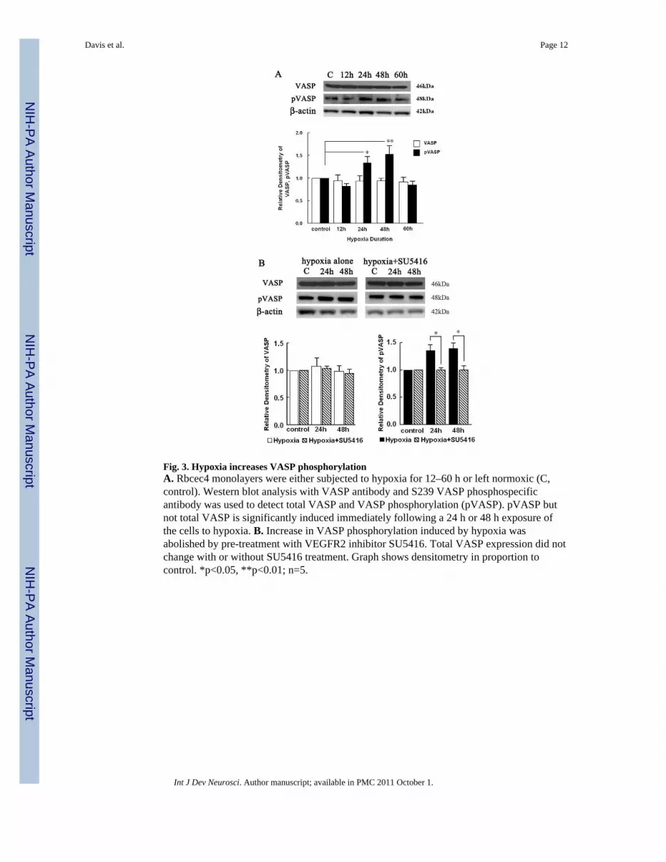
Fig. 3. Hypoxia increases VASP phosphorylationA. Rbcec4 monolayers were either subjected to hypoxia for 12–60 h or left normoxic (C,control). Western blot analysis with VASP antibody and S239 VASP phosphospecificantibody was used to detect total VASP and VASP phosphorylation (pVASP). pVASP butnot total VASP is significantly induced immediately following a 24 h or 48 h exposure ofthe cells to hypoxia. B. Increase in VASP phosphorylation induced by hypoxia wasabolished by pre-treatment with VEGFR2 inhibitor SU5416. Total VASP expression did notchange with or without SU5416 treatment. Graph shows densitometry in proportion tocontrol. *p<0.05, **p<0.01; n=5.
Davis et al. Page 12
Int J Dev Neurosci. Author manuscript; available in PMC 2011 October 1.
NIH
-PA Author Manuscript
NIH
-PA Author Manuscript
NIH
-PA Author Manuscript

Fig. 4. VASP and ZO-1 expression is co-localized in rbcec4 cellsA. Rbcec4 cells with VASP-eGFP expression show tight junctions in green (arrows). B.ZO-1 expression in rbcec4 cells transfected with VASP-eGFP is seen in cell-cell contacts inred (arrows). C. Overlay of immunofluorescent micrographs shown in A and B. Colocalizedexpression of ZO-1 and VASP is shown in yellow (arrows). Nuclei were stained blue withDAPI. Scale bar = 50 μM
Davis et al. Page 13
Int J Dev Neurosci. Author manuscript; available in PMC 2011 October 1.
NIH
-PA Author Manuscript
NIH
-PA Author Manuscript
NIH
-PA Author Manuscript


