Embed Size (px)
Citation preview

Savcheniuk et al. BMC Complementary and Alternative Medicine 2014, 14:247http://www.biomedcentral.com/1472-6882/14/247
RESEARCH ARTICLE Open Access
Short-term periodic consumption of multiprobioticfrom childhood improves insulin sensitivity,prevents development of non-alcoholic fattyliver disease and adiposity in adult rats withglutamate-induced obesityOleksandr Savcheniuk1, Nazarii Kobyliak2*, Maryana Kondro3, Oleksandr Virchenko1, Tetyana Falalyeyeva1
and Tetyana Beregova1
Abstract
Background: Today the impairment of metabolism and obesity are being extensively investigated due to thesignificant increase of the prevalence of these diseases. There is scientific evidence that probiotics are beneficial forhuman health. Thus, the aim of the study was to investigate the effect of multiprobiotic “Symbiter acidophilicconcentrated” on obesity parameters in the rats under experimental obesity.
Methods: The study was carried out on 60 newborn Wistar rats, divided into 3 groups, 20 animals in each(females – n = 10, males – n = 10): intact rats, monosodium glutamate (MSG-) and MSG + probiotic group. Rats ofintact group were administered with saline (8 μl/g, subcutaneously (s.c.)). Newborns rats of MSG-group and MSG +probiotic group were injected with a solution of MSG (4.0 mg/g) s.c. at 2nd – 10th postnatal days. The MSG + probioticgroup was treated with 140 mg/kg (1.4 × 10¹⁰ CFU/kg) of multiprobiotic “Symbiter”. MSG-group was treated with2.5 ml/kg of water (per os) respectively. Administration was started at the age of 4 weeks just after wean and continuedfor 3 month intermittently alternating two-week course of introduction with two-week course of break.
Results: Neonatal treatment with MSG caused a stunted growth in both MSG-groups, which manifested with significantlysmaller naso-anal length compared to adult intact rats. There was no significant difference in weight between intact andMSG-groups on 120th day. The adiponectin level in the serum of rats with MSG-induced obesity decreased by 2.43 times(p = 0.001) in males and 1.75 (p = 0.020) in females. Concentration of leptin in adipose tissue were significantly higherby 45.9% (p = 0.019) and 61.2% (p = 0.009) respectively in males and females compared to intact rats. Our study hasindicated that daily oral administration of multiprobiotic to neonatal MSG-treated rats by 2-week courses led tosignificant reduce of total body and VAT weight with subsequent improvement in insulin sensitivity andprevention of non-alcoholic fatty liver (NAFLD) development.
Conclusions: These results have shown that periodic treatment with multiprobiotic prevents the MSG-inducedobesity and NAFLD development.
Keywords: Probiotics, Obesity, Glutamate, Leptin, Adiponectin, Insulin, Non-alcoholic fatty liver disease
* Correspondence: [email protected] National Medical University, T. Shevchenko boulevard, 13, Kyiv01601, UkraineFull list of author information is available at the end of the article
© 2014 Savcheniuk et al.; licensee BioMed Central Ltd. This is an Open Access article distributed under the terms of theCreative Commons Attribution License (http://creativecommons.org/licenses/by/4.0), which permits unrestricted use,distribution, and reproduction in any medium, provided the original work is properly credited. The Creative Commons PublicDomain Dedication waiver (http://creativecommons.org/publicdomain/zero/1.0/) applies to the data made available in thisarticle, unless otherwise stated.

Savcheniuk et al. BMC Complementary and Alternative Medicine 2014, 14:247 Page 2 of 17http://www.biomedcentral.com/1472-6882/14/247
BackgroundObesity has dramatically increased during the past decadesand has now reached epidemic proportions in both devel-oped and developing countries [1,2]. It is a heterogeneousdisorder which has been associated with an increased riskof many serious illnesses such as cardiovascular diseases[3], hypertension [4], dyslipidemia [4], diabetes mellitus[5] and some types of cancer including colon cancer [6],lung cancer [7], breast cancer [8], uterine [9] and ovariancancer [10]. As of 2008, The World Health Organizationclaimed that 1.5 billion adults, in the age of 20 and older,were overweight. Among them over 200 million of menand nearly 300 million of women were obese [11]. Therate of obesity also increases with age, at least up to 50 or60 years old [12].Most of medications for treatment of obesity are taken
out the production because of their adverse effects. Orli-stat is the only drug that could be taken by patients forthe prolonged time. However, little attention is paid tothe search of means for obesity prophylaxis. In currentscientific literature there are a lot of studies that confirmbeneficial effects of probiotics on human organism. Thequestion about probiotics impact on fat metabolism andobesity is being actively debated in the scientific literature.The gut microbiota has been recently proposed to be anenvironmental factor involved in the control of bodyweight and energy homeostasis [13-17]. This “exteriorizedorgan” contributes to human homeostasis via multiplemetabolic functions and diverse control mechanisms.In several studies it was revealed the positive effects of
probiotics use under the conditions of experimentalobesity [18,19]. In our previous work we have shown thatprobiotics mixture of lyophilized strains Lactobacilluscasei IMVB-7280, Bifidobacterium animalis VKL andBifidobacterium animalis VKB at least partially preventthe MSG-induced obesity in rats [20]. But studies haveshown that multistrain probiotics are more effectivethan monostrain probiotics [21]. Also it is interesting tocompare the influence of the lyophilized and alive strainson the obesity and reveal the gender-specific differencesin the obesity development. Given the above the aim ofour work was to investigate the prophylactic influence ofshort periodical courses of the alive multiprobiotic admin-istration on the obesity and non-alcoholic fatty liver devel-opment (NAFLD) development induced by the neonatalMSG treatment in 4-month male and female rats.
MethodsEthics statementThis study was carried out in strict accordance with therecommendations in the Guide for the Care and Use ofLaboratory Animals of the National Institutes of Healthand the general ethical principles of animal experiments,approved by the First National Congress on Bioethics
Ukraine (September 2001). The protocol was approvedby the Committee on the Ethics of Animal Experimentsof the Taras Shevchenko National University of Kyiv(Protocol number: 18/2013). The rats were kept in col-lective cages in controlled conditions of temperature(22 ± 3°C), light (12 h light/dark cycle) and relativehumidity (60 ± 5%). The animals were fed laboratory chow(PurinaW) and tap water ad libitum.
Study designWe included 60 newborn Wistar rats, divided to 3 groupsof 20 animals each. All groups were equally representedby both sexes (females – n = 10, males – n = 10). Newbornsrats of intact group were administered with saline sub-cutaneously (s.c.) in the volume of 8 μl/g at 2nd, 4th, 6th,8th and 10th postnatal days. Newborns rats of MSG-groupand MSG + probiotic group received a solution of MSG(4.0 mg/g of body weight) s.c. at 2nd, 4th, 6th, 8th and 10th
postnatal days [22]. Within 4 months after birth rats wereon a normal diet. MSG+ probiotic group received multipro-biotic “Symbiter” in dose 140 mg/kg (1.4 × 10¹⁰ CFU/kg)at volume 2.5 ml/kg per os (p.o.). Multiprobiotic containsconcentrated biomass of 14 probiotic bacteria of generaBifidobacterium, Lactobacillus, Lactococcus, Propionibacter-ium. MSG-group respectively received 2.5 ml/kg of water(p.o.). Administration was started at the age of 4 weeks justafter wean and continued for 3 month intermittently alter-nating two-week course of introduction with two-weekcourse of break.
Anthropometric measurements and obesity parameterassessmentDuring 4 months in all groups the changes of bodyweight and food intake were analyzed. In adult age, ratsfrom three experimental groups (n = 60) were weighed andkilled by cervical dislocation under urethane anesthesia.We dissected and weighed visceral adipose tissue (VAT)(epididymal, perirenal and omental fat).For each animal at month 4 of life (120 days) we de-
termined existence of obesity using Lee index. It wascalculated as follow: the cube root of body weight (g)/nasoanal length (cm). Rats presenting values higher than0.300 were classified as obese, equal to or less than 0.300as normal [23].
Sample collection and blood biochemistry analysisRats of all groups were fasted for approximately 12 hoursprior sacrifice. Rats were sacrificed by cervical dislocationunder urethane anesthesia. Blood was drawn from theapex of the cardiac ventricle and few blood drops werecollected into a microcentrifuge tube containing a mixtureof NaF and EDTA in a 2:1 (w/w) ratio. For blood glucosedetermination we used Trinder glucose oxidase methodon this aliquot of blood. The remaining blood sample was

Savcheniuk et al. BMC Complementary and Alternative Medicine 2014, 14:247 Page 3 of 17http://www.biomedcentral.com/1472-6882/14/247
collected into a sterile tube and centrifuged at 3,500 rpm(2260 g) for 15 minutes. After centrifugation serumsupernatant for further analysis was aliquoted intomicrocentrifuge tubes and stored at -80°C. Serum insu-lin was determined using the Rat/Mouse Insulin ELISAKit (Linco Research, USA). The HOMA-IR was calcu-lated as the product of multiplying of fasting bloodglucose and serum insulin divided by 22.5 [24]. ELISAwas used for determination of serum adiponectin andleptin level and the VAT leptin by commercial kits«BioVendor» (Czech Republic). The VAT for measur-ing leptin was promptly harvested and immediatelyflash-frozen in liquid nitrogen and stored at -80°C.After defrosting the adipose tissue was homogenized withTES-buffer (pH = 7.4, 10 mM of tris(hydroxymethyl)ami-nomethane, 1 mM of EDTA, 250 mM of saccharose,inhibitors of protease (2,5 μg/ml of leupeptin, 2,5 μg/mlof aprotinin, 1 mM phenylmethanesulfonyl fluoride), 1%Triton X-100) (1:4). At next step the homogenate wascentrifuged at 14000 rpm for 15 min under +4°C. Thebottom layer of the supernatant was harvested for theleptin measuring.
Liver histology assessmentFor histological analysis liver tissue samples from boththe right and left hepatic lobes were taken (sample size0.5 × 0.5 cm). After fixed for 24 hours in a liquid Buena,fragments of liver were dehydrated in alcohols of in-creasing concentrations (from 70 ° to 96 °), embedded inparaffin and then cut with a thickness of 5-6 micronsand stained with hematoxylin-eosin. A pathologist blindedto group distributions performed the histological analysesof slides using light microscopy («Olympus», Japan). Toassess morphological changes in liver we used NAS(NAFLD activity score), which includes histologicalfeatures and has been defined as unweighted sum ofscores for steatosis (0-3), lobular inflammation (0-3)and ballooning (0-2). Acording to NAS scores ≥5 arediagnosed as non-alcoholic steatohepatitis (NASH), andcases with a NAS <3 are mentioned as not NASH [25].
Statistical analysisStatistical analysis performed by using SPSS-20 software.All data in this study were expressed as means ± standarddeviation (M± SD) or %. Data distribution was analyzedusing the Kolmogorov-Smirnov normality test. Continu-ous variables with parametric distribution were thenanalyzed using Analysis of Variance (ANOVA) and ifthe results were significant, a Fisher’s LSD Post Hoc testwas performed. For data with non-parametric distributionKruskall-Wallis test was used. Estimation of gender spe-cific changes performed using Student’s t test for unpairedvalues. For comparisons of categorical variables we con-ducted χ2 test. The difference between groups was defined
to be statistically significant when a p-value was less than0.05.
ResultsImpact of probiotic short-term courses on body weightand obesity parametersFigure 1 shows sex specific weight gain from 30 to120 days of age in all experimental rats groups. In femalerats on day 30, 60 and 90 we observed significant higherbody weight in MSG- group comparative to intact control.But on day 120 the weight did no differ between intactand MSG- groups (p = 0.914). Short-term periodic admin-istration of probiotic beginning from 30 days age lead toweight reduction by 17.3% (p = 0.001) at 120 days age inMSG-probiotic female rats comparative to MSG-group(Figure 1A). Also the body weight in this group were sig-nificantly lower by 14.3% (p = 0.001) comparative to intactrats (Figure 1A).Unlike female, in male rats on 30 days age we didn’t
find significant changes in body weight at all experimentalgroups. Beginning from day 60 we established significantweight gain in MSG-group comparative to other groupsand at day 120 the average rats body mass were 263.4 ±26.9 g. Specifically, that additional diet correction withmultiprobiotic “Symbiter” led to weight reduction by14.5% (p = 0.005) compared to the value seen in MSG-ratsafter 3 month of feeding (Figure 1B).Neonatal treatment with MSG caused a stunted growth
in both MSG-groups, which manifested with significantsmaller naso-anal length compared to intact rats. That’swhy in spite of lower weight in both sexes, after probioticadministration, we stated development of obesity in 50% offemale and 40% of male animals from MSG-probioticgroup compared to intact rats which was totally confirmedby Lee index higher than 0.300 (Figure 2A, B). On the otherhand, short-term periodic consumption of multiprobiotichad a preventive effect on glutamate-induced obesity. Es-pecially we observed significantly lower Lee index afterprobiotic correction in females (0.301 ± 0.01 vs 0.313 ±0.01, p = 0.024) and males (0.299 ± 0.007 vs 0.319 ± 0.01,p = 0.004) compared to MSG-group (Figure 2A,B). Theincidence of obesity was also higher in MSG-group – infemales (90% vs 50%, p = 0,051) and respectively inmales (90% vs 40%, p = 0,019).We observed 5-7 times increasing of total weight of
VAT in rats with MSG-group compared to intact con-trol (Figure 3A,B). Gender specific analysis showedthe development of more pronounced visceral obes-ity in males, because of significant higher depositionof VAT in MSG-rats (18.72 ± 5.46 g vs 14.1 ± 2.89 g;p = 0.036).Short-term concomitant administration of probiotic bac-
teria led to a decrease of VAT weight by 52.43% (p = 0.001)in females and respectively by 58.86% (p = 0.001) versus

Figure 1 Body weight changes in rats from birth to 4-month age (M ± SD, n = 10 in each group): A –females, B – males. * - p < 0.05compared to intact rats, # - p < 0.05 compared to MSG-group.
Savcheniuk et al. BMC Complementary and Alternative Medicine 2014, 14:247 Page 4 of 17http://www.biomedcentral.com/1472-6882/14/247
MSG-group (Figure 3A, B), although its level didn’t reachthe control values.Due to establish the alteration of eating behavior
under conditions of MSG-induced obesity we examinedfood intake in one-, two-, three- and four-month rats of allgroup. It was shown the slight age-dependent increase infood intake both in male and female rats. We have to noticethere was no any significant difference in food consumptionbetween experimental group that suggest MSG-obesity isnot a result of excessive caloric intake but associated withthe metabolic disorder (Figure 4А, B ).
Probiotic short-term courses improved insulin sensitivityin obese-induced ratsThe fasting blood glucose of MSG-rats were signifi-cant higher compared to intact animals in both sexes
(6.11 ± 1.15 vs 4.62 ± 0.42 mmol/L, p = 0.003 – in females;6.17 ± 0.64 vs 4.59 ± 0.67 mmol/L, p = 0.001 – respectivelyin males). Female rats from MSG-probiotic group pre-sented significant decrease of fasting glucose level by17.34% (p = 0.024) and males – by 16.04% (p = 0.010)compared to MSG-group (Figure 5 A,B).Analysis of the HOMA-IR and serum insulin concentra-
tions showed that under condition of MSG-induced obes-ity rats became insulin resistant. Both the HOMA-IRindex (2.3 ± 1.2 vs 0.46 ± 0.21, p = 0.001 – in females;2.77 ± 0.92 vs 0.66 ± 0.34, p = 0.001 – in males) andserum insulin concentration (8.19 ± 2.90 vs 2.33 ± 1.16,p = 0.001 – in females; 10.02 ± 2.62 vs 3.21 ± 1.53, p =0.001 – in males) were significant higher in rats withMSG-induced obesity compared to control (Figures 6A,B;7A,B). As we mentioned above the 2-week periodic

Figure 2 Index Lee in4-month rats (M ± SD, n = 10 in each group): A –females, B – males. * - p < 0.05 compared to intact rats, # - p < 0.05compared to MSG-group.
Savcheniuk et al. BMC Complementary and Alternative Medicine 2014, 14:247 Page 5 of 17http://www.biomedcentral.com/1472-6882/14/247
multiprobiotic courses led to improvement of insulinsensitivity which manifested with significant 2-fold de-crease of HOMA-IR and fasting insulinemia in relationto MSG-group. After treatment with probiotic theHOMA-IR index did not exceed the normal range, butwas not significantly higher compared to intact controlrats (Figure 7A, B).
Impact of short-term probiotic courses on adipocytokinelevels in obese-induced ratsAnalysis of the secretory function of adipose tissue showeda change in concentration adipose-derived hormonesin rats with experimental obesity. Thus, the level ofadiponectin in the serum of rats with MSG-induced
obesity decreased by 2.43 times (p = 0.001) in malesand 1.75 (p = 0.020) in females (Figure 8 A,B). Concen-tration of leptin in adipose tissue were higher by 45.9%(p = 0.019) and 61.2% (p = 0.009) respectively in malesand females in comparison with intact control group(Figure 9A,B). In serum leptin level was lesser than in theVAT. MSG increased the leptin level by 93.3% (p < 0.05)and 83.6% (p < 0.01) respectively both in males and females(Figure 10 A,B). Gender specific analysis did not confirmedchanges of leptin concentrations in adipose tissue but allexperimental groups represented significantly lower serumadiponectin concentrations in males compared to fe-males (intact – 4.29 ± 1.67 vs 6.5 ± 1.99 μg/ml, p = 0.001;MSG – 1.73 ± 0.56 vs 3.77 ± 1.64 μg/ml, p = 0.030).

Figure 3 Visceral adipose tissue weight in4-month rats (M ± SD, n = 10 in each group): A –females, B – males. * - p < 0.05 compared tointact rats, # - p < 0.05 compared to MSG-group.
Savcheniuk et al. BMC Complementary and Alternative Medicine 2014, 14:247 Page 6 of 17http://www.biomedcentral.com/1472-6882/14/247
It was found significant increase of serum adiponectinafter probiotic administration in males by 89% (p = 0.028)compared to MSG-group (Figure 8B) and females by38.2% (p = 0.039) compared to MSG-group (Figure 8A).The VAT leptin in males decreased by 14.3% (p = 0.047)compared to MSG-group (Figure 9B) and in females – by14.9% (p = 0.044) compared to MSG-group (Figure 9A).The serum leptin in MSG-probiotic group decreasedinsignificantly (Figure 10 A,B). Also showed that theconcentration of adiponectin and leptin in the case ofprobiotics ration did not differ from the level of intactrats (Figures 7A,B; 8A,B).Thus, intermittent administration of probiotics for
two weeks courses restored the hormonal activity ofadipose tissue.
Morphological changes in rat liver under the conditionsof obesity and probiotic administrationWe found significantly lower total score (0.95 ± 0.15 vs3.3 ± 0.28, p < 0.001), degree of steatosis (0.85 ± 0.13 vs2.15 ± 0.16, p < 0.001) and manifestation of lobular inflam-mation (0.1 ± 0.06 vs 0.95 ± 0.15, p < 0.001) due to NAFLDactivity score in MSG-probiotic group compared toMSG-obesity (Table 1).
DiscussionA lot of factors lead to development of obesity. The mostimportant factors contributing to fat accumulation arewidespread net of fast food restaurants, which offer thevariety of energy-dense foods and low physical activity andthe genetic inheritance. Also It was shown contributing of

Figure 4 Dynamics of food intake changes in rats from 1-month to 4-month age (M ± SD, n = 10 in each group): A –females, B – males.
Savcheniuk et al. BMC Complementary and Alternative Medicine 2014, 14:247 Page 7 of 17http://www.biomedcentral.com/1472-6882/14/247
microorganisms, increasing maternal age, greater fecundityamong people with higher adiposity, sleep debt, endocrinedisruptors, pharmaceutical iatrogenesis, reduction in vari-ability of ambient temperatures and intrauterine effects tothe obesity [26,27]. In general obesity develops when thebody receives more energy with food than it can spend.Not the least role in increased energy consumption playsmonosodium glutamate (C5H8NO4Na, MSG, E621) whichis food additive and is found especially in fast food.MSG can be formed naturally in various foods (cheese,
meat, etc.) [28]. Also it is largely used in the food indus-try because it improves the taste of food and even canmask the bad taste of stale products. That is why itsconsumption steadily increases worldwide. In 1968, a re-port appeared in the New England Journal of Medicine,describing a complex of symptoms in patients who dinedin Chinese restaurants. The symptoms of the so-called«Chinese restaurant syndrome» included numbness,radiating to the back, arms, and neck; weakness; and
palpitations [29]. Cross-sectional and longitudinal studiesin healthy Chinese subjects showed correlation of MSGintake with an increased risk of being overweightirrespective of the total calorie intake and physicalactivity [30,31].Animal models support a causative association between
obesity and neonatal or maternal administration of highdoses of MSG [32]. MSG acts on immature neurologicalmechanisms that regulate food intake and energy ex-penditure by ablating cells in the arcuate nucleus of thehypothalamus and destroying circumventricular neurons.MSG altered the production of orexigenic and anorexi-genic molecules as proopiomelanocortin, cocaine- andamphetamine-regulated transcript and neuropeptide Y[33]. Neonatal neurotoxity effect of MSG leading to thedevelopment of neurochemical, endocrine, metabolic andbehavioural abnormalities in adulthood including hypo-phagia, obesity, hypoactivity, delayed puberty, and elevatedplasma corticosterone levels [34]. Furthermore, stunted

Figure 5 Glucose level in serum of 4-month rats (M ± SD, n = 10 in each group): A –females, B – males. * - p < 0.05 compared to intactrats, # - p < 0.05 compared to MSG-group.
Savcheniuk et al. BMC Complementary and Alternative Medicine 2014, 14:247 Page 8 of 17http://www.biomedcentral.com/1472-6882/14/247
growth, increased adipose tissue stores, and a markedincrease in plasma triglycerides [35], insulin [36] andfasting glucose levels [37] have been noted in rats.Most of medications for treatment of obesity are taken
out the production because of their adverse effects. Orlistatis the only drug that could be taken by patients for theprolonged time. However, little attention is paid to thesearch of means for prophylaxis of obesity. In currentscientific literature there are a lot of studies that con-firm beneficial effects of probiotics on human organism.The question about probiotics impact on fat metabolismand obesity is being actively debated in the scientific litera-ture. The gut microbiota has been recently proposed to bean environmental factor involved in the control of body
weight and energy homeostasis [13-17]. This “exteriorizedorgan” contributes to our homeostasis via multiplemetabolic functions and diverse control mechanisms.For example it involved in the extraction of caloriesfrom ingested dietary substances, and it helps to storethese calories in host adipose tissue for later use. Pioneersin study of the gut microbiota role for the development ofobesity and fat mass storage were Gordon et al. They de-termined that parallel with lower energy intake youngconventionally reared mice had higher on 42% and 47%total body fat and gonadal fat mass respectively as com-pared with germ-free mice [25]. Authors firstly declaredthat microbiota itself can increase energy yield from dietof the host organism’s. Colonization within two weeks of

Figure 6 Insulin level in serum of 4-month rats (M ± SD, n = 10 in each group): A –females, B – males. * - p < 0.05 compared to intact rats,# - p < 0.05 compared to MSG-group.
Savcheniuk et al. BMC Complementary and Alternative Medicine 2014, 14:247 Page 9 of 17http://www.biomedcentral.com/1472-6882/14/247
young germ-free mice with microbiota from conven-tionally reared mice is associated with increased insulinresistance and provokes a 60% increase in total body fatmass despite lower energy intake [13]. This fat massgain was even more pronounced when the gut microbialcommunity was derived from genetically obese (ob/ob)mice [17].Gut microbiota may change with body weight. Ley
et al. [27] recently analysed 5,088 bacterial 16S rRNAgene sequences from the distal intestinal (cecal) microbiotaof genetically obese ob/ob mice, lean ob/+and wild-type siblings, and their ob/+mothers, all fed the samepolysaccharide-rich diet. They demonstrated that com-pared with lean mice obese animals have a 50% reduction
in the abundance of Bacteroidetes and a proportionalincrease in Firmicutes. Feeding of a high-fat/high-poly-saccharide diet to genetically wild-type rodents led tosimilar microbial changes [16]. Methanogenic Archaeaalso located in ob/ob mice, which lead to increasing ofthe efficiency of bacterial fermentation [29].Similar to these animal experiments, Ley et al. demon-
strated that the ratio of Firmicutes/Bacteroidetes in thedistal gut microbiota is also increase in obese peopleby comparison with lean people [38]. This proportionincreases with weight loss on a fat-restricted orcarbohydrate-restricted diet. Another study, without limitof dietary components, described that Prevotellaceae, asubgroup of Bacteroidetes, are significantly enriched in

Figure 7 HOMA-IR of 4-month rats (M ± SD, n = 10 in each group): A –females, B – males. * - p < 0.05 compared to intact rats, # - p < 0.05compared to MSG-group.
Savcheniuk et al. BMC Complementary and Alternative Medicine 2014, 14:247 Page 10 of 17http://www.biomedcentral.com/1472-6882/14/247
obesity and demonstrated that Firmicutes are dominant inlean and obese individuals [39]. The differences in micro-bial composition between two studies were explained byan increased capacity of the obesity-associated micro-biome to harvest energy from the diet.Controversial data were recently reported by Schwiertz
and colleagues [40]. They determined lower ratios ofFirmicutes to Bacterodetes in overweight human adultscompared to lean controls. Another study, using weightloss diets, found no proof of the link between the proportionof Bacteroidetes and Firmicutes and human obesity [41].Kalliomaki et al. [42] examined if early differences in gut
microbiota composition in children can guide weightdevelopment throughout early childhood. Overweight
and obese children (n = 25) were selected. Early fecalmicrobiota composition was analyzed by fluorescent insitu hybridization (FISH) with additional flow cytometry.They find that bifidobacterial numbers in fecal samplesduring infancy were higher in children remaining normalweight than in children becoming overweight, [2.19 × 10⁹
[1.10–5.28] cells/g vs 1.20 × 10⁹ [0.48–1.59] cells/g;p = 0.02]. Overweight was also associated with a greaternumber of Staphylococcus aureus. Author suggested thatchanges in early life gut microbiota composition may beassociated with an increased risk of developing obesity inlater life.These data suggest an association between obesity and
the intestinal microbiota. In several studies it was revealed

Figure 8 Adiponectin level in serum of 4-month rats (M ± SD, n = 10 in each group): A –females, B – males. * - p < 0.05 compared tointact rats, # - p < 0.05 compared to MSG-group.
Savcheniuk et al. BMC Complementary and Alternative Medicine 2014, 14:247 Page 11 of 17http://www.biomedcentral.com/1472-6882/14/247
the positive effects of probiotics use under the conditionsof experimental obesity [18,19]. In our previous work wehave shown that probiotics mixture of lyophilized strainsLactobacillus casei IMVB-7280, Bifidobacterium animalisVKL and Bifidobacterium animalis VKB at least partiallyprevent the MSG-induced obesity in rats [20]. But studieshave shown that multistrain probiotics are more effectivethan monostrain probiotics [21]. Also it is interesting tocompare the influence of the lyophilized and alive strainson the obesity and reveal the gender-specific differencesin the obesity development. Given the above the aim ofour work was to investigate the prophylactic influence ofshort periodical courses of the alive multiprobiotic admin-istration on the obesity development induced by the neo-natal MSG treatment in 4-month male and female rats.
Thus, finding new non-toxic means of prevention andtreatment of obesity is one of the major problems ofmodern science. Our study showed that short-termperiodic administration of multiprobiotic beginning fromchildhood prevented development of MSG-induced obes-ity in adult rats. This was confirmed by significant lowerincidence of obesity and mean value for Lee index andreduction of total body and VAT weight in rats aftercorrection with multiprobiotic “Symbiter” compared toMSG-group.The most frequent cause which leads to the obesity
development is a dysbalance between energy intake andenergy expenditure. In this complex process genetic suscep-tibility, environmental and lifestyle factors are involved. Re-cent advances in next generation sequencing technology
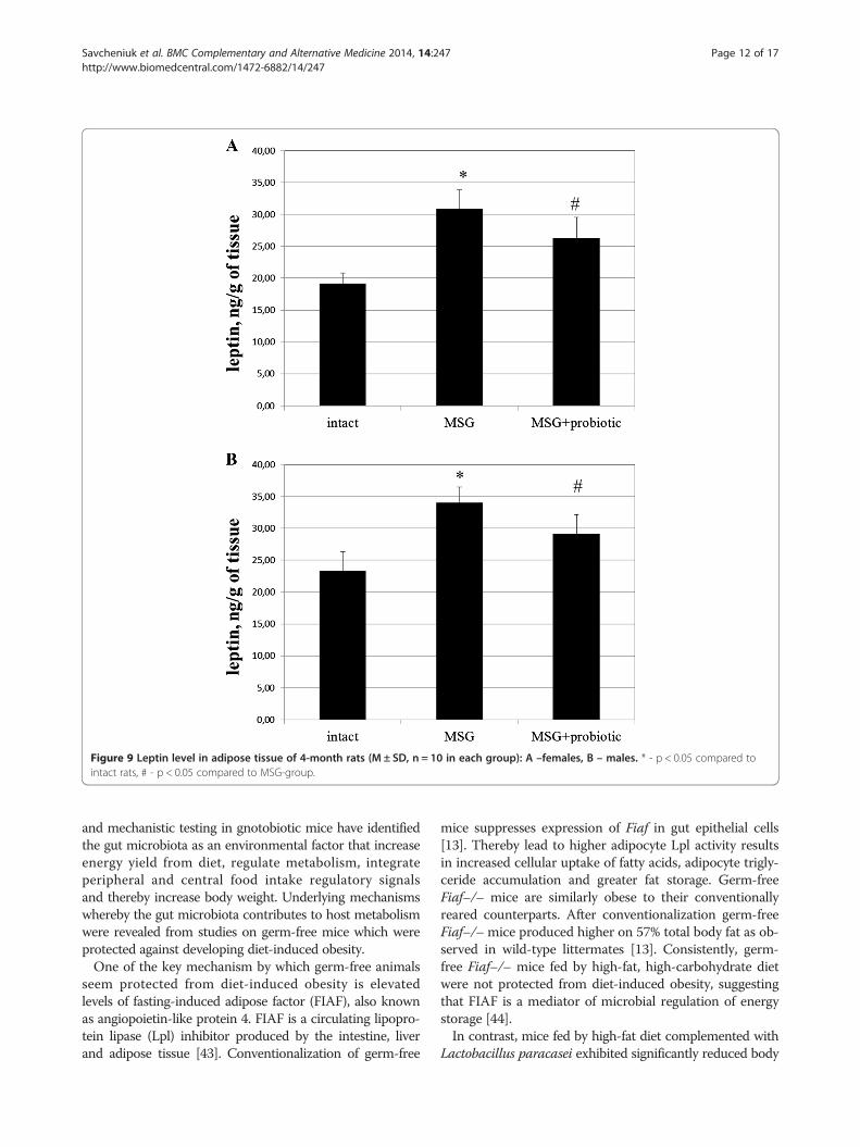
Figure 9 Leptin level in adipose tissue of 4-month rats (M ± SD, n = 10 in each group): A –females, B – males. * - p < 0.05 compared tointact rats, # - p < 0.05 compared to MSG-group.
Savcheniuk et al. BMC Complementary and Alternative Medicine 2014, 14:247 Page 12 of 17http://www.biomedcentral.com/1472-6882/14/247
and mechanistic testing in gnotobiotic mice have identifiedthe gut microbiota as an environmental factor that increaseenergy yield from diet, regulate metabolism, integrateperipheral and central food intake regulatory signalsand thereby increase body weight. Underlying mechanismswhereby the gut microbiota contributes to host metabolismwere revealed from studies on germ-free mice which wereprotected against developing diet-induced obesity.One of the key mechanism by which germ-free animals
seem protected from diet-induced obesity is elevatedlevels of fasting-induced adipose factor (FIAF), also knownas angiopoietin-like protein 4. FIAF is a circulating lipopro-tein lipase (Lpl) inhibitor produced by the intestine, liverand adipose tissue [43]. Conventionalization of germ-free
mice suppresses expression of Fiaf in gut epithelial cells[13]. Thereby lead to higher adipocyte Lpl activity resultsin increased cellular uptake of fatty acids, adipocyte trigly-ceride accumulation and greater fat storage. Germ-freeFiaf–/– mice are similarly obese to their conventionallyreared counterparts. After conventionalization germ-freeFiaf–/–mice produced higher on 57% total body fat as ob-served in wild-type littermates [13]. Consistently, germ-free Fiaf–/– mice fed by high-fat, high-carbohydrate dietwere not protected from diet-induced obesity, suggestingthat FIAF is a mediator of microbial regulation of energystorage [44].In contrast, mice fed by high-fat diet complemented with
Lactobacillus paracasei exhibited significantly reduced body
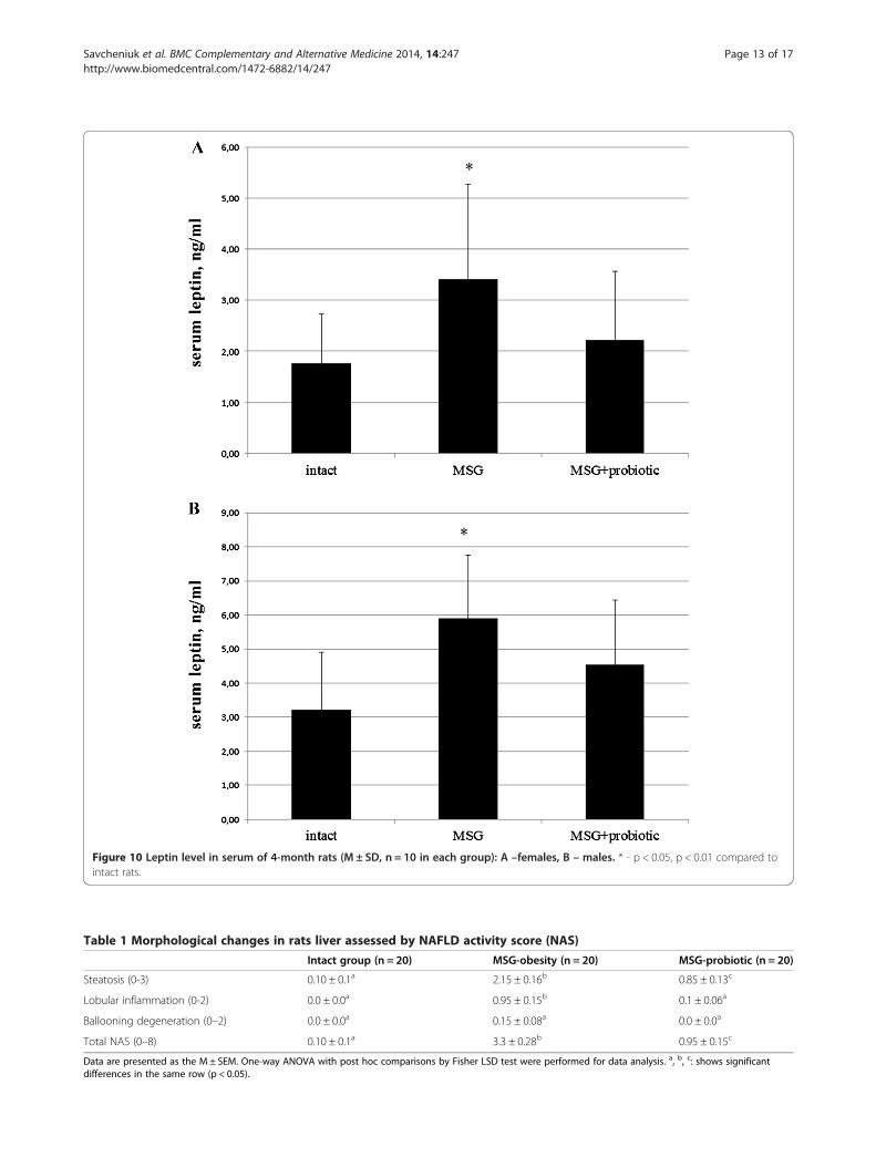
Figure 10 Leptin level in serum of 4-month rats (M ± SD, n = 10 in each group): A –females, B – males. * - p < 0.05, p < 0.01 compared tointact rats.
Table 1 Morphological changes in rats liver assessed by NAFLD activity score (NAS)
Intact group (n = 20) MSG-obesity (n = 20) MSG-probiotic (n = 20)
Steatosis (0-3) 0.10 ± 0.1a 2.15 ± 0.16b 0.85 ± 0.13c
Lobular inflammation (0-2) 0.0 ± 0.0a 0.95 ± 0.15b 0.1 ± 0.06a
Ballooning degeneration (0–2) 0.0 ± 0.0a 0.15 ± 0.08a 0.0 ± 0.0a
Total NAS (0–8) 0.10 ± 0.1a 3.3 ± 0.28b 0.95 ± 0.15c
Data are presented as the M ± SEM. One-way ANOVA with post hoc comparisons by Fisher LSD test were performed for data analysis. a, b, c: shows significantdifferences in the same row (p < 0.05).
Savcheniuk et al. BMC Complementary and Alternative Medicine 2014, 14:247 Page 13 of 17http://www.biomedcentral.com/1472-6882/14/247

Savcheniuk et al. BMC Complementary and Alternative Medicine 2014, 14:247 Page 14 of 17http://www.biomedcentral.com/1472-6882/14/247
fat, which was paralleled by increased circulating levels ofFIAF [45]. Fleissner et al. showed that germ-free mice onhigh-fat diet showed increased intestinal mRNA expressionof Fiaf with no major changes in circulating FIAF comparedwith conventionalised mice, suggesting that FIAF mechan-ism is not universally associated with gut microbiota-related fat mass development [46].Furthermore, Backhed and colleagues have also dem-
onstrated that germ-free mice have increased levels ofphosphorylated AMP-activated protein kinase (AMPK) inmuscle and liver. AMPK is a key enzyme that controlscellular energy status which activates key enzymes ofmitochondrial fatty acid oxidation, including acetyl-CoAcarboxylase (ACC) and carnitine-palmitoyltransferase I(CTP1), indicative of increased energy expenditure. Theexact pathway whereby the microbiota signals to liver orskeletal muscle AMPK is unclear but appears to be inde-pendent from FIAF [44].Another mechanism is related to energy extraction from
undigested food components. The gut microbiota thatdigest complex dietary carbohydrates produces manymonosaccharides and short-chain fatty acids (SCFAs)such as acetate, propionate, and butyrate [44] which rep-resent an important energy source. Conventionalization ofgerm-free mice doubled the density of small intestinal villicapillaries [47] and enhanced uptake of this componentsfrom the gut into the portal blood and eventually partici-pate in hepatic de novo lipogenesis promoting fat accu-mulation in the liver and adipose tissue [44]. This reactioncontrolled by carbohydrate responsive element bindingprotein (ChREBP) and sterol responsive element bindingprotein (SREBP-1) [48]. We observed that multiprobioticadministration from childhood lead to histological featuresimprovement which manifested with hepatic steatosisattenuation and decreased lobular inflammation in MSG-probiotic group. NASH we confirmed only in 20% of ratswith MSG-obesity (p = 0.035).SCFAs act in the gut as signaling molecules and are
specific ligands for at least two G protein-coupled recep-tors, GPR41 and GPR43, mainly expressed by intestinalepithelial cells [49,50]. Samuel et al. have demonstratedthat conventionally raised Gpr41–/– mice and germ-freeGpr41–/– mice colonized with only Bacteroides thetaiotao-micron and Methanobrevibacter smithii are significantlyleaner than wild-type littermates, while there are no differ-ences between wild-type or Gpr41–/– germ-free mice [51].Gpr41, which is produced by enteroendocrine cells, mightbe a regulator of host energy balance through effects thatare dependent upon the gut microbiota. Activation ofGPR41 increases production of peptide YY (PYY), anenteroendocrine cell hormone that normally inhibitsgut motility, increases intestinal transit rate and reducesextraction of energy from the diet, thus affecting peripheralglucose utilisation [51]. Recent study has shown that
Gpr43–/– mice are resistant to diet-induced obesity andinsulin resistance, at least partly due to Gpr43-regulatedenergy expenditure [52].Some lines of experimental evidence suggest that high-
fat diets may affect epithelial integrity due to changes inthe distribution and localisation of Zonula Occludens-1(ZO-1) and Occludin (two tight junction proteins) inintestinal tissue and hence lead to impaired gut perme-ability, and consequently to low-grade systemic inflam-mation [53-55]. Cani et al. demonstrated that bacteriallipopolysaccharide (LPS), which continuously produced inthe gut through lysis of gram-negative bacteria is amicrobiota-related factor which can trigger the inflamma-tory process by binding to the CD14 toll-like receptor-4(TLR-4) complex at the surface of innate immune cells[55]. Author mentioned that after 4 weeks of high-fatfeeding, mice exhibited an obese phenotype accompaniedby a change in gut microbiota composition (the reductionof Bifidobacteria and Eubacteria spp.) and a two- to three-fold increase in circulating LPS levels, which they called“metabolic endotoxemia” since LPS plasma concentrationswere much lower than those observed during septic shock[56]. In fact, in this study, continuous subcutaneouslow-rate infusion of LPS led to excessive weight gainand insulin resistance in mice. Moreover, LPS receptorCd14-/- mice tend to be resistant to this chronic inflam-matory state and were hypersensitive to insulin evenwhen they were fed a normal diet, suggesting that CD14may modulate insulin sensitivity in physiological conditions[57]. Deletion of TLR-4 prevented the high-fat diet–in-duced insulin resistance [58].They also demonstrated that modulation of gut micro-
biota, e.g. by antibiotic treatment or dietary interventionwith oligofructoses, reduced metabolic endotoxemia andthe cecal content of LPS, improved glucose intolerance,insulin sensitivity and decreased body weight gain inboth high-fat fed and ob/ob mice [59,60].In models of diabetes, probiotic intervention has been
examined for its ability to impact on metabolic biomarkersof disease. Tabuchi et al showed that Lactobacillus rhamno-sus GG improved glucose tolerance in the streptozotocin-induced rat model of diabetes possibly due to prevention ofa decrease in insulin secretion [61]. Studies using thetraditional Indian yoghurt – dahi supplemented withprobiotic strains of Lactobacillus acidophilus and L.casei have shown that this product can improve markersof diabetes, including hyperglycemia, hyperinsulinemia inhigh-fructose induced rat models of diabetes [62,63].We first studied influence of probiotic administration
for insulin sensitivity in rat model of MSG-induced obesity.Nagata et al. found that mice after neonatal treatment withMSG were observed to be obese but had no polyphagia,and were glycosuric by 29 weeks of age. The pathologicalstudy showed hypertrophy of the pancreatic islet, with

Savcheniuk et al. BMC Complementary and Alternative Medicine 2014, 14:247 Page 15 of 17http://www.biomedcentral.com/1472-6882/14/247
elevation of glucose and insulin serum concentrations at29 and 54 weeks aged compared to control mice [32].From the other hand in the early phase of obesity level ofplasma glucose can be normal but associated with hyper-insulinemia [64] which indicates that insulin resistance ispresent and high insulin levels may be compensatory dueto direct hypersecretion of β cells [65].Our data is agreed with recently reported studies [62,63]
and showed that in addition to reduction in total bodyweight administration of multiprobiotic by short coursesled to improvement of insulin sensitivity that was con-firmed by significant decreasing of hyperinsulinemiaand HOMA-IR in MSG-probiotic group compared toMSG-animals. From the other hand we observed sig-nificant increase of serum adiponectin, which define asone of the main regulators of peripheral tissues sensitivityto insulin [66], only in females compared to MSG-group.As previously reported for MSG rats and mice, leptin
mRNA expression levels and serum levels in MSG-treatedmice were significantly higher compared to that in normalcontrols [67,68]. The increased leptin production inadipose tissues due to pancreatic hypertrophy and hyperin-sulinaemia have been reported to induce the developmentof leptin resistance [69]. We mentioned that probioticshelped to restore the hormonal activity of adipose tissue.Thus, concentration of leptin under the probiotics admis-tration did not differ from the level of intact rats.
ConclusionIn summary, we studied effectiveness of short-term periodicconsumption of multiprobiotic from childhood on meta-bolic profile in adult rats with MSG-induced model ofobesity. Subcutaneous neonatal injection of MSG is ableto induce obesity without hyperphagia, which diagnosedby high Lee index and characterized by small corporalweight and naso-anal length. Obesity in rats caused byalterations in hypothalamic arcuate nucleus and impairsleptin and insulin signaling in this region resulting inhyperleptinemia and hyperinsulinemia.Our study has indicated that daily oral administration
of 2.5 ml/kg of alive multiprobiotic “Symbiter” containingconcentrated biomass of 14 probiotic bacteria of Bifidobac-terium, Lactobacillus, Lactococcus, Propionibacterium gen-era to neonatal MSG-treated rats by 2-weeks courses led tosignificant reduce of total body and VAT weight, togetherwith improvement in insulin sensitivity and prevention ofNAFLD development.
AbbreviationsAMPK: AMP-activated protein kinase; ACC: Acetyl-CoA carboxylase;ChREBP: Carbohydrate responsive element binding protein;CTP1: Carnitine-palmitoyltransferase 1; FIAF: Fasting-induced adiposefactor; FISH: Fluorescent in situ hybridization; GPR: G protein-coupledreceptors; LPS: Lipopolysaccharide; Lpl: Lipoprotein lipase; NAFLD:Non-alcoholic fatty liver disease; NASH: Non-alcoholic steatohepatitis;
SREBP-1: Sterol responsive element binding protein; SCFAs: Short-chainfatty acids; TLR-4: Toll-like receptor-4; VAT: Visceral adipose tissue.
Competing interestsThe authors declare to have no competing interests.
Authors’ contributionsTB, TF and NK designed the study. OV develop the protocol and wrote thefirst and second drafts of the manuscript. OS performed the experimentalworks. MK involved in the collection of literature, interpret the results andperformed the statistical analysis. All authors read and approved the finalmanuscript.
Authors’ informationOS - PhD student, SRL ‘Pharmacology and Experimental Pathology’, Departmentof Biological and Biomedical Technology, ESC ‘Institute of Biology’, TarasShevchenko National University of Kyiv.NK – PhD, assistant of Endocrinology Department, Bogomolets NationalMedical University.OV - PhD student, SRL ‘Pharmacology and Experimental Pathology’, Departmentof Biological and Biomedical Technology, ESC ‘Institute of Biology’, TarasShevchenko National University of Kyiv.FT - Ph.D., D.Sci., researcher of RL “Pharmacology and Experimental Pathology”Department of Biological and Biomedical Technology ESC «Institute of Biology»Taras Shevchenko National University of Kyiv.MK – PhD, Assosiate Professor of Department of Normal Physiology DanyloHalytsky Lviv National Medical University.TB - Professor, Ph.D., D.Sci., SRL ‘Pharmacology and Experimental Pathology’,Department of Biological and Biomedical Technology, ESC ‘Institute ofBiology’, Taras Shevchenko National University of Kyiv.
AcknowledgmentsThe authors express their sincere thanks to Dr. Yankovsky DmitroStanislavovych for the help, advice and financial support of this work.
Author details1Taras Shevchenko National University of Kyiv, Volodymyrska Str., 64/13, Kyiv01601, Ukraine. 2Bogomolets National Medical University, T. Shevchenkoboulevard, 13, Kyiv 01601, Ukraine. 3Danylo Halytsky Lviv National MedicalUniversity, Pekarska Str., 69, Lviv 79010, Ukraine.
Received: 11 April 2014 Accepted: 11 July 2014Published: 16 July 2014
References1. Visscher TL, Seidell JC: The public health impact of obesity. Annu Rev
Public Health 2001, 22:355–375.2. Flier JS: Obesity wars: molecular progress confronts an expanding
epidemic. Cell 2004, 116:337–350.3. Jiang J, Ahn J, Huang WY, Hayes RB: Association of obesity with
cardiovascular disease mortality in the PLCO trial. Prev Med 2013,57:60–64.
4. Jung DH, Kim JY, Kim JK, Koh SB, Park JK, Ahn SV: Relative contribution ofobesity and serum adiponectin to the development of hypertension.Diabetes Res Clin Pract 2014, 103(1):51–56.
5. Janghorbani M, Momeni F, Dehghani M: Hip circumference, height andrisk of type 2 diabetes: systematic review and meta-analysis. Obes Rev2012, 13:1172–1181.
6. Takahashi H, Hosono K, Endo H, Nakajima A: Colon epithelial proliferationand carcinogenesis in diet-induced obesity. J Gastroenterol Hepatol 2013,28(Suppl 4):41–47.
7. Ntikoudi E, Kiagia M, Boura P, Syrigos KN: Hormones of adipose tissue andtheir biologic role in lung cancer. Cancer Treat Rev 2013, 40:22–30.
8. Eichholzer M, Huang DJ, Modlasiak A, Schmid SM, Schotzau A, Rohrmann S,Guth U: Impact of body mass index on prognostically relevant breastcancer tumor characteristics. Breast Care (Basel) 2013, 8:192–198.
9. Ward KK, Roncancio AM, Shah NR, Davis MA, Saenz CC, McHale MT, PlaxeSC: The risk of uterine malignancy is linearly associated with body massindex in a cohort of US women. Am J Obstet Gynecol 2013,209:579. e571-575.

Savcheniuk et al. BMC Complementary and Alternative Medicine 2014, 14:247 Page 16 of 17http://www.biomedcentral.com/1472-6882/14/247
10. van Dorp W, Blijdorp K, Laven JS, Pieters R, Visser JA, van der Lely AJ,Neggers SJ, van den Heuvel-Eibrink MM: Decreased ovarian function isassociated with obesity in very long-term female survivors of childhoodcancer. Eur J Endocrinol 2013, 168:905–912.
11. Obesity and overweight. [http://www.who.int/mediacentre/factsheets/fs311/en/]
12. Kopelman PG, Caterson ID, Stock MJ, Dietz WH: Clinical obesity in adults andchildren: In Adults and Children. Oxford: Blackwell Publishing; 2005.
13. Backhed F, Ding H, Wang T, Hooper LV, Koh GY, Nagy A, Semenkovich CF,Gordon JI: The gut microbiota as an environmental factor that regulatesfat storage. Proc Natl Acad Sci U S A 2004, 101:15718–15723.
14. Backhed F, Ley RE, Sonnenburg JL, Peterson DA, Gordon JI: Host-bacterialmutualism in the human intestine. Science 2005, 307:1915–1920.
15. Ley RE, Backhed F, Turnbaugh P, Lozupone CA, Knight RD, Gordon JI:Obesity alters gut microbial ecology. Proc Natl Acad Sci U S A 2005,102:11070–11075.
16. Turnbaugh PJ, Backhed F, Fulton L, Gordon JI: Diet-induced obesity islinked to marked but reversible alterations in the mouse distal gutmicrobiome. Cell Host Microbe 2008, 3:213–223.
17. Turnbaugh PJ, Ley RE, Mahowald MA, Magrini V, Mardis ER, Gordon JI: Anobesity-associated gut microbiome with increased capacity for energyharvest. Nature 2006, 444:1027–1031.
18. Lee SJ, Bose S, Seo JG, Chung WS, Lim CY, Kim H: The effects ofco-administration of probiotics with herbal medicine on obesity,metabolic endotoxemia and dysbiosis: A randomized double-blindcontrolled clinical trial. Clin Nutr 2014, in press.
19. Luoto R, Collado MC, Salminen S, Isolauri E: Reshaping the gut microbiotaat an early age: functional impact on obesity risk? Ann Nutr Metab 2013,63(Suppl 2):17–26.
20. Savcheniuk OA, Virchenko OV, Falalyeyeva TM, Beregova TV, Babenko LP,Lazarenko LM, Demchenko OM, Bubnov RV, Spivak MY: The efficacy ofprobiotics for monosodium glutamate-induced obesity: dietologyconcerns and opportunities for prevention. EPMA J 2014, 5:2.
21. Timmerman HM, Koning CJ, Mulder L, Rombouts FM, Beynen AC:Monostrain, multistrain and multispecies probiotics–A comparison offunctionality and efficacy. Int J Food Microbiol 2004, 96:219–233.
22. Sanabria ER, Pereira MF, Dolnikoff MS, Andrade IS, Ferreira AT, Cavalheiro EA,Fernandes MJ: Deficit in hippocampal long-term potentiation inmonosodium glutamate-treated rats. Brain Res Bull 2002, 59:47–51.
23. Bernardis LL, Patterson BD: Correlation between ’Lee index’ and carcassfat content in weanling and adult female rats with hypothalamic lesions.J Endocrinol 1968, 40:527–528.
24. Vogeser M, Konig D, Frey I, Predel HG, Parhofer KG, Berg A: Fasting seruminsulin and the homeostasis model of insulin resistance (HOMA-IR) inthe monitoring of lifestyle interventions in obese persons. Clin Biochem2007, 40:964–968.
25. Kleiner DE, Brunt EM, Van Natta M, Behling C, Contos MJ, Cummings OW,Ferrell LD, Liu YC, Torbenson MS, Unalp-Arida A, Yeh M, McCullough AJ,Sanyal AJ, Nonalcoholic Steatohepatitis Clinical Research Network: Designand validation of a histological scoring system for nonalcoholic fattyliver disease. Hepatology 2005, 41:1313–1321.
26. Genoni G, Prodam F, Marolda A, Giglione E, Demarchi I, Bellone S, Bona G:Obesity and infection: two sides of one coin. Eur J Pediatr 2014, 173:25–32.
27. McAllister EJ, Dhurandhar NV, Keith SW, Aronne LJ, Barger J, Baskin M, BencaRM, Biggio J, Boggiano MM, Eisenmann JC, Elobeid M, Fontaine KR,Gluckman P, Hanlon EC, Katzmarzyk P, Pietrobelli A, Redden DT, Ruden DM,Wang C, Waterland RA, Wright SM, Allison DB: Ten putative contributors tothe obesity epidemic. Crit Rev Food Sci Nutr 2009, 49:868–913.
28. Ninomiya K: Natural occurrence. Food Rev Int 1998, 14:177–211.29. Kwok RHM: Chinese-restaurant syndrome. N Engl J Med 1968, 278:796.30. He K, Du S, Xun P, Sharma S, Wang H, Zhai F, Popkin B: Consumption of
monosodium glutamate in relation to incidence of overweight inChinese adults: China Health and Nutrition Survey (CHNS). Am J Clin Nutr2011, 93:1328–1336.
31. He K, Zhao L, Daviglus ML, Dyer AR, Van Horn L, Garside D, Zhu L, Guo D,Wu Y, Zhou B, Stamler J: Association of monosodium glutamate intakewith overweight in Chinese adults: the INTERMAP Study. Obesity(Silver Spring) 2008, 16:1875–1880.
32. Nagata M, Suzuki W, Iizuka S, Tabuchi M, Maruyama H, Takeda S, AburadaM, Miyamoto K: Type 2 diabetes mellitus in obese mouse model inducedby monosodium glutamate. Exp Anim 2006, 55:109–115.
33. Beas-Zarate C, Perez-Vega M, Gonzalez-Burgos I: Neonatal exposure tomonosodium L-glutamate induces loss of neurons and cytoarchitecturalalterations in hippocampal CA1 pyramidal neurons of adult rats. BrainRes 2002, 952:275–281.
34. Lorden JF, Caudle A: Behavioral and endocrinological effects of singleinjections of monosodium glutamate in the mouse. Neurobehav ToxicolTeratol 1986, 8:509–519.
35. Oida K, Nakai T, Hayashi T, Miyabo S, Takeda R: Plasma lipoproteins ofmonosodium glutamate-induced obese rats. Int J Obes 1984, 8:385–391.
36. Collison KS, Maqbool Z, Saleh SM, Inglis A, Makhoul NJ, Bakheet R, Al-JohiM, Al-Rabiah R, Zaidi MZ, Al-Mohanna FA: Effect of dietary monosodiumglutamate on trans fat-induced nonalcoholic fatty liver disease. J LipidRes 2009, 50:1521–1537.
37. Diniz YS, Faine LA, Galhardi CM, Rodrigues HG, Ebaid GX, Burneiko RC,Cicogna AC, Novelli EL: Monosodium glutamate in standard and high-fiber diets: metabolic syndrome and oxidative stress in rats. Nutrition2005, 21:749–755.
38. Ley RE, Turnbaugh PJ, Klein S, Gordon JI: Microbial ecology: human gutmicrobes associated with obesity. Nature 2006, 444:1022–1023.
39. Zhang H, DiBaise JK, Zuccolo A, Kudrna D, Braidotti M, Yu Y, ParameswaranP, Crowell MD, Wing R, Rittmann BE, Krajmalnik-Brown R: Human gutmicrobiota in obesity and after gastric bypass. Proc Natl Acad Sci U S A2009, 106:2365–2370.
40. Schwiertz A, Taras D, Schafer K, Beijer S, Bos NA, Donus C, Hardt PD:Microbiota and SCFA in lean and overweight healthy subjects. Obesity(Silver Spring) 2009, 18:190–195.
41. Duncan SH, Lobley GE, Holtrop G, Ince J, Johnstone AM, Louis P, Flint HJ:Human colonic microbiota associated with diet, obesity and weight loss.Int J Obes (Lond) 2008, 32:1720–1724.
42. Kalliomaki M, Collado MC, Salminen S, Isolauri E: Early differences in fecalmicrobiota composition in children may predict overweight. Am J ClinNutr 2008, 87:534–538.
43. Yoon JC, Chickering TW, Rosen ED, Dussault B, Qin Y, Soukas A, FriedmanJM, Holmes WE, Spiegelman BM: Peroxisome proliferator-activated receptorgamma target gene encoding a novel angiopoietin-related proteinassociated with adipose differentiation. Mol Cell Biol 2000, 20:5343–5349.
44. Backhed F, Manchester JK, Semenkovich CF, Gordon JI: Mechanismsunderlying the resistance to diet-induced obesity in germ-free mice. ProcNatl Acad Sci U S A 2007, 104:979–984.
45. Aronsson L, Huang Y, Parini P, Korach-Andre M, Hakansson J, Gustafsson JA,Pettersson S, Arulampalam V, Rafter J: Decreased fat storage by Lactobacillusparacasei is associated with increased levels of angiopoietin-like 4 protein(ANGPTL4). PLoS One 2010, 5(9):e13087.
46. Fleissner CK, Huebel N, Abd El-Bary MM, Loh G, Klaus S, Blaut M: Absenceof intestinal microbiota does not protect mice from diet-inducedobesity. Br J Nutr 2010, 104:919–929.
47. Stappenbeck TS, Hooper LV, Gordon JI: Developmental regulation ofintestinal angiogenesis by indigenous microbes via Paneth cells. ProcNatl Acad Sci U S A 2002, 99:15451–15455.
48. Denechaud PD, Dentin R, Girard J, Postic C: Role of ChREBP in hepaticsteatosis and insulin resistance. FEBS Lett 2008, 582:68–73.
49. Brown AJ, Goldsworthy SM, Barnes AA, Eilert MM, Tcheang L, Daniels D,Muir AI, Wigglesworth MJ, Kinghorn I, Fraser NJ, Pike NB, Strum JC,Steplewski KM, Murdock PR, Holder JC, Marshall FH, Szekeres PG, Wilson S,Ignar DM, Foord SM, Wise A, Dowell SJ: The Orphan G protein-coupledreceptors GPR41 and GPR43 are activated by propionate and other shortchain carboxylic acids. J Biol Chem 2003, 278:11312–11319.
50. Le Poul E, Loison C, Struyf S, Springael JY, Lannoy V, Decobecq ME, BrezillonS, Dupriez V, Vassart G, Van Damme J, Parmentier M, Detheux M: Functionalcharacterization of human receptors for short chain fatty acids and theirrole in polymorphonuclear cell activation. J Biol Chem 2003,278:25481–25489.
51. Samuel BS, Shaito A, Motoike T, Rey FE, Backhed F, Manchester JK, HammerRE, Williams SC, Crowley J, Yanagisawa M, Gordon JI: Effects of the gutmicrobiota on host adiposity are modulated by the short-chain fatty-acidbinding G protein-coupled receptor, Gpr41. Proc Natl Acad Sci U S A 2008,105:16767–16772.
52. Bjursell M, Admyre T, Goransson M, Marley AE, Smith DM, Oscarsson J,Bohlooly YM: Improved glucose control and reduced body fat mass infree fatty acid receptor 2-deficient mice fed a high-fat diet. Am J PhysiolEndocrinol Metab 2010, 300:E211–220.

Savcheniuk et al. BMC Complementary and Alternative Medicine 2014, 14:247 Page 17 of 17http://www.biomedcentral.com/1472-6882/14/247
53. Cani PD, Possemiers S, Van de Wiele T, Guiot Y, Everard A, Rottier O, GeurtsL, Naslain D, Neyrinck A, Lambert DM, Muccioli GG, Delzenne NM: Changesin gut microbiota control inflammation in obese mice through amechanism involving GLP-2-driven improvement of gut permeability.Gut 2009, 58:1091–1103.
54. de La Serre CB, Ellis CL, Lee J, Hartman AL, Rutledge JC, Raybould HE:Propensity to high-fat diet-induced obesity in rats is associated withchanges in the gut microbiota and gut inflammation. Am J PhysiolGastrointest Liver Physiol 2010, 299:G440–448.
55. Cani PD, Delzenne NM: Gut microflora as a target for energy andmetabolic homeostasis. Curr Opin Clin Nutr Metab Care 2007, 10:729–734.
56. Amar J, Iglesias MA, Poggi M, Knauf C, Bastelica D, Neyrinck AM, Fava F,Tuohy KM, Chabo C, Waget A, Delmée E, Cousin B, Sulpice T, Chamontin B,Ferrières J, Tanti JF, Gibson GR, Casteilla L, Delzenne NM, Alessi MC, BurcelinR: Metabolic endotoxemia initiates obesity and insulin resistance.Diabetes 2007, 56:1761–1772.
57. Cani PD, Bibiloni R, Knauf C, Waget A, Neyrinck AM, Delzenne NM, BurcelinR: Changes in gut microbiota control metabolic endotoxemia-inducedinflammation in high-fat diet-induced obesity and diabetes in mice.Diabetes 2008, 57:1470–1481.
58. Shi H, Kokoeva MV, Inouye K, Tzameli I, Yin H, Flier JS: TLR4 links innateimmunity and fatty acid-induced insulin resistance. J Clin Invest 2006,116:3015–3025.
59. Cani PD, Neyrinck AM, Fava F, Knauf C, Burcelin RG, Tuohy KM, Gibson GR,Delzenne NM: Selective increases of bifidobacteria in gut microfloraimprove high-fat-diet-induced diabetes in mice through a mechanismassociated with endotoxaemia. Diabetologia 2007, 50:2374–2383.
60. Membrez M, Blancher F, Jaquet M, Bibiloni R, Cani PD, Burcelin RG, CorthesyI, Mace K, Chou CJ: Gut microbiota modulation with norfloxacin andampicillin enhances glucose tolerance in mice. FASEB J 2008,22:2416–2426.
61. Tabuchi M, Ozaki M, Tamura A, Yamada N, Ishida T, Hosoda M, Hosono A:Antidiabetic effect of Lactobacillus GG in streptozotocin-induceddiabetic rats. Biosci Biotechnol Biochem 2003, 67:1421–1424.
62. Yadav H, Jain S, Sinha PR: Effect of Dahi containing Lactococcus lactis onthe progression of diabetes induced by a high-fructose diet in rats. BiosciBiotechnol Biochem 2006, 70:1255–1258.
63. Yadav H, Jain S, Sinha PR: Antidiabetic effect of probiotic dahi containingLactobacillus acidophilus and Lactobacillus casei in high fructose fedrats. Nutrition 2007, 23:62–68.
64. de Carvalho PP, Vargas AM, da Silva JL, Nunes MT, Machado UF: GLUT4protein is differently modulated during development of obesity inmonosodium glutamate-treated mice. Life Sci 2002, 71:1917–1928.
65. de Souza CT, Nunes WM, Gobatto CA, de Mello MA: Insulin secretion inmonosodium glutamate (MSG) obese rats submitted to aerobic exercisetraining. Physiol Chem Phys Med NMR 2003, 35:43–53.
66. Combs TP, Pajvani UB, Berg AH, Lin Y, Jelicks LA, Laplante M, Nawrocki AR,Rajala MW, Parlow AF, Cheeseboro L, Ding YY, Russell RG, Lindemann D,Hartley A, Baker GR, Obici S, Deshaies Y, Ludgate M, Rossetti L, Scherer PE:A transgenic mouse with a deletion in the collagenous domain ofadiponectin displays elevated circulating adiponectin and improvedinsulin sensitivity. Endocrinology 2004, 145:367–383.
67. Perello M, Gaillard RC, Chisari A, Spinedi E: Adrenal enucleation inMSG-damaged hyperleptinemic male rats transiently restores adrenalsensitivity to leptin. Neuroendocrinology 2003, 78:176–184.
68. Chen W, Wang LL, Liu HY, Long L, Li S: Peroxisome proliferator-activatedreceptor delta-agonist, GW501516, ameliorates insulin resistance,improves dyslipidaemia in monosodium L-glutamate metabolicsyndrome mice. Basic Clin Pharmacol Toxicol 2008, 103:240–246.
69. Pallett AL, Morton NM, Cawthorne MA, Emilsson V: Leptin inhibits insulinsecretion and reduces insulin mRNA levels in rat isolated pancreaticislets. Biochem Biophys Res Commun 1997, 238:267–270.
doi:10.1186/1472-6882-14-247Cite this article as: Savcheniuk et al.: Short-term periodic consumption ofmultiprobiotic from childhood improves insulin sensitivity, preventsdevelopment of non-alcoholic fatty liver disease and adiposity inadult rats with glutamate-induced obesity. BMC Complementary andAlternative Medicine 2014 14:247.
Submit your next manuscript to BioMed Centraland take full advantage of:
• Convenient online submission
• Thorough peer review
• No space constraints or color figure charges
• Immediate publication on acceptance
• Inclusion in PubMed, CAS, Scopus and Google Scholar
• Research which is freely available for redistribution
Submit your manuscript at www.biomedcentral.com/submit

Figure 1

Figure 2

Figure 3

Figure 4
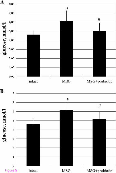
Figure 5

Figure 6

Figure 7

Figure 8

Figure 9

Figure 10


