Embed Size (px)
Citation preview
Volume 8, issue 4,JUly 2008ISSN 1567-1348
I £ cnon Genn E olut on
• c
Editor-in-Chief
M. Tibayrenc IRD Representative Office, French Embassy, 29, Thanon Sathorn Tai,Bangkok 10120, ThailandTel.: +66 2 627 2190; Fax: +66 2 627 2194; e-mail: [email protected]
Receiving Editors
A. van Belkum Department of Medical Microbiology and Infectious Diseases, Erasmus University Medical Centre,Room L333, Dr. Molewaterplein 40, Rotterdam, 3015 GD, The NetherlandsTel.: +31 10 463 5813; Fax: +31 10 463 3875; e-mail: [email protected]
F. Guhl Universidad de Los Andes, Facultad de Ciencias, Depto de Ciencias Biologicas,Calle 18A CRA. 1E Of. A-202, Apartado Aereo 4976, Bogotá, ColombiaTel.: +57 13324540; Fax: +57 12841890; e-mail: [email protected]
S. Hasnain Center for DNA Fingerprinting and Diagnostics, CDFD, ECIL Road, Nacharam, Hyderabad 500 076,India, Tel.: +91 40 7151344/346 ext. 38; Fax: +91 40 7150008/5610; e-mail: [email protected]
J.-C. Piffaretti Interlifescience, via San Gottardo 92, CH-6900 Massagno, SwitzerlandTel.: +41 91 960 05 55; Fax: +41 91 960 05 56; e-mail: [email protected]
M.-A. Shaw University of Leeds, School of Biology, Human Genetics, The Louis Compton Miall Building, Leeds LS29JT, UK, Tel.: +44 113 233 2822; Fax.: +44 113 233 2835; e-mail: [email protected]
Consulting Editor
F.J. Ayala University of California - Irvine, Department of Ecology and Evolutionary Biology, 321 Steinhaus Hall,Irvine CA 92697, USA, Tel.: +1 949 824 82 93; Fax: +1 949 824 24 74; e-mail: [email protected]
Editorial Board
A. Andremont, Paris, FranceA.-L. Banuls, Montpellier, FranceC.B. Beard, Atlanta, GA, USAS. Beverley, St. Louis, MO, USAD. Blanc, Lausanne, SwitzerlandS. Brisse, Paris, FranceY. Carlier, Brussels, BelgiumD.A. Caugant, Oslo, NorwayJ.-C. Dujardin, Antwerpen, BelgiumJ.-P. Dujardin, Montpellier, FranceD. Dykhuizen, Stony Brook, CA, USAD. Ebert, Fribourg, SwitzerlandS.F. Elena, Valencia, Spain
A.A. Escalante, Tempe, AZ, USAW.M. Fitch, Irvine, CA, USAW. Gibson, Bristol, UKJ.P. Gonzalez, Nakhonpathom, ThailandP.A.D. Grimont, Paris, FranceE. Groisman, St. Louis, MO, USAY. Gräser, Berlin, GermanyP. Keim, Flagstaff, AZ, USAA.A. Lal, New Delhi, I ndiaB. Levin, Atlanta, GA, USAG. Lucotte, Paris, FranceM.C.J. Maiden, Oxford, UKP. Majiwa, Nairobi, Kenya
D.P. Mindell, Ann Arbor, MI, USAE.R. Moxon, Oxford, UKS.T. Nichol, Atlanta, GA, USAH. Ochman, Tucson, AZ, USAM. Peeters, Montpellier, FranceS. Qari, Atlanta, GA, USAM. Raymond, Montpellier, FranceD. Relman, Palo Alto, CA, USAF. Renaud, Montpellier, FranceC.J. Schofield, London, UKP. Small, Stanford, CA, USAB. Spratt, London, UKJ. Stevens, Exeter, UK
doi:10.1016/S1567-1348(08)00099-3

Infection, Geneticsand EvolutionVolume 8 (2008)
Special Issue
8th International Meeting on Molecular Epidemiology andEvolutionary Genetics of Infectious Diseases -
Royal River Hotel, Bangkok, Thailand
30 November-2 December 2006
Edited by
Michel Tibayrenc
ELSEVIER
Vol. 8, issue 4, July 2008
CONTENTS •~ Infection, Geneticsand Evolution
www.elsevier.com!1ocatc/meegid
Abstracted/indexed in: Biological Abstracts, Chemical Abstracts, BIOBASE, EMBASE/Excerpta Medica, ISI, Photozoological Abstracts.Alsocovered in the abstract and citation database SCOPUS@ Full text availableon Scienceuirect'".
Special Issue: 8th International Meeting on Molecular Epidemiology and Evolutionary Genetics of Infectious Diseases
Editorial
MEEGID VIII Bangkok, ThailandM. Tibayrenc (Thailand)
Review
The SARS-CoV nucleocapsid protein: A protein with multifarious activitiesM. Surjit and S.K. Lal (India)
395
397
Research papers
Phylogenetic analysis of the promoter region of the CD40L gene in primates and other mammalsM.E. Steiper, S.J. Parikh and J.M. Zichello (USA) 406
The geometry of the wing of Aedes (Stegomyia) aegypti in isofemale lines through successive generationsN. Jirakanjanakit, S. Leemingsawat and J.P. Dujardin (Thailand, France) 414
Using a climate-dependent model to predict mosquito abundance: Application to Aedes (Stegomyia) africanus and Aedes(Diceromyia) furcifer (Diptera: Culicidae)B. Schaeffer, B. Mondet and S. Touzeau (France, Jndia) 422
Molecular characterization of Thai Ehrlichia canis and Anaplasma platys strains detected in dogsD. Pinyoowong, S. Jittapalapong, F. Suksawat, R.W Stich and A Thamchaipenet (Thailand, USA) 433
The identification, diversity and prevalence of trypanosomes in field caught tsetse in Tanzania using ITS-1 primers andfluorescent fragment length barcodingE.R. Adams, P.B.Hamilton, 1.1. Malele and WC. Gibson (UK, Tanzania) 439
Molecular detection of divergent trypanosomes among rodents of ThailandS. Jittapalapong, T. Inpankaew, N. Sarataphan, V. Herbreteau, J.P. Hugot, S. Morand and R.W Stich (Thailand, France, USA) 445
Association between the DQA MHC class I1 gene and Puumala virus infection in Myodes glareolus, the bank voleJ. Deter, J. Bryja, Y. Chaval, M. Galan, H. Henttonen, J. Laakkonen, L. Voutilainen, O. Vapalahti, A Vaheri, A.R. Salvador,S. Morand, J.-F. Cosson and N. Charbonnel (France, Czech Republic, Finland, Spain) 450
Differential strain-specific diagnosis of the heartwater agent: Ehrlichia ruminantiumN. Vachiery, G. Maganga, T. l.etrancois, Y. Kandassamy, F. Stachurski, H. Adakal, C. Ferraz, A. Morgat, A. Bensaid,E. Coissac, F. Boyer, J. Demaille, A Viari, D. Martinez and R. Frutos (France, Burkina-Faso, SWitzerland) 459
Promiscuous T cell epitope prediction of Candida albicans secretory aspartyl protienase family of proteinsS. Tongchusak, V. Brusic and S.C. Chaiyaroj (Thailand, USA, Australia) 467
Genomic interrogation of ancestral Mycobacterium tuberculosis from south IndiaS. Narayanan, S. Gagneux, L. Hari, AG. Tsolaki, S. Rajasekhar, P.R. Narayanan, P.M. Small, S. Holmes and K. DeRiemer(India, USA) 474
Genetic diversity of Trypanosoma evansi in beef cattle based on internal transcribed spacer regionS. Areekit, P. Singhaphan, P. Kanjanavas, S. Khuchareontaworn, T. Sriyapai, A Pakpitcharoen and K. Chansiri (Thailand) 484
Bionomics, taxonomy, and distribution of the major malaria vector taxa of Anopheles subgenus Cellia in Southeast Asia: Anupdated reviewS. Manguin, C. Garros, I. Dusfour, R.E. Harbach and M. Coosemans (France, UK, Belgium) 489
(Contents Continued on page I)
Available online at
11111111111111111111111111111111111111111111111567-1348(200807)8:4;1-M
ScienceDirectwww.sciencedirect.com

(Contents continued from outside back cover)
Discussions
Behind the scene, something else is pulling the strings: Empnasizinq parasitic manipulation in vector-borne diseasesT l.efevre and F. Thomas (France)
From population structure to genetically-engineered vectors: New ways to control vector-borne diseases?OAE. Sparagano and C.J. De l.una
Abstracts
504
520
Abstract for the 8th International Meeting on Molecular Epidemiology and Evolutionary Genetics in Infectious Diseases. Royal RiverHotel, Bangkok. Thailand, 30 Novernber-z December :0>006 81
www.elsevier.com/locate/meegid
Available online at www.sciencedirect.com
Infection, Genetics and Evolution 8 (2008) 395–396
Editorial
MEEGID VIII Bangkok, Thailand§
The 8th session of the international congress, Molecular environment of the Royal River Hotel, enjoying the legendary
Epidemiology and Evolutionary Genetics of Infectious Dis-
eases, was held at the Royal River Hotel, Bangkok, Thailand,
30th November–2nd December 2006.
The MEEGID congresses were born in 1996 at the Centers
for Disease Control (CDC) in Atlanta. My CDC counterpart,
Altaf Lal (chief, malaria section), and I felt that molecular
epidemiology was fast gaining in topicality at that time and
fully deserved to be supported by a specific congress. Initially,
it only covered pathogens, already original in its scope in that
all pathogens were considered at MEEGID I, whether they be
parasites, bacteria or viruses, of medical, veterinary or
agronomical relevance. Soon came the vision that pathogen
specialists needed to cooperate with entomologists and host
geneticists to bring out a global picture of the entire infectious
diseases transmission chain. This concept proved to be so
successful in the MEEGIDs that followed that it became the
backbone of the newborn journal Infection, Genetics and
Evolution started in 2001. Another key concept of IGE and the
MEEGID is the close interaction between basic science and
biomedical research. This original formula has since then
inspired several new journals.
Unfortunately, IGE was included in the Thompson
Scientific/ISI database only in 2007, so that impact factor will
be available only in 2009. However, an unofficial IF was
computed by Elsevier experts using the same software as the ISI
official IF: 3.554, which is excellent for such a young journal
(Straub and Tibayrenc, 2007). Thanks to Elsevier’s very active
and professional support, our journal is on the way to fulfilling
the role that I assigned to it from the very beginning: becoming
the major tribune of all infectious disease specialists using
evolutionary concepts and of all geneticists/evolutionists/
bioinformaticians interested in the fascinating models offered
by transmissible diseases.
MEEGID VIII proved again how successful the formula
was. For three days, microbiologists, virologists, parasitolo-
gists, entomologists, evolutionists, geneticists, and bioinfor-
maticians interacted in the dreamy and very professional
§ See information on the MEEGID IX congress at
http://www.th.ird.fr//site_meegid/menu.htm.
1567-1348/$ – see front matter # 2008 Elsevier B.V. All rights reserved.
doi:10.1016/j.meegid.2008.01.009
and smiling Thai hospitality.
According to its classical formula, MEEGID VIII specifi-
cally focused on those endemies that are the most relevant to the
host country and its neighbors: malaria, dengue, avian flu, and
cattle pathogens, among others.
No fewer than 26 different nationalities were present at the
congress. A strong (37%) and enthusiastic minority of Thai
scientists animated the sessions and illustrated the dynamism of
Thai science in this topical research domain. Following its
tradition, MEEGID VIII has catered particularly students, who
were exempted from registration fees and were granted a
specific symposium, chaired by students and with only student
speakers. No less than 28% of MEEGID VIII participants were
students.
A last tradition was fulfilled with the attribution of the
MEEGID medals. The medal for the best oral communication
was granted to Bruce Wilcox (University of Hawaii). Niyaz
Ahmed (University of Hyderabad, India) was awarded the prize
for the best oral communication by a scientist from a southern
country. The prize for the best communication by a student was
granted to Thierry Lefevre (IRD, Montpellier, France). Last,
the prize for the best poster communication was given to
Panatda Saenkham (Chulabhorn Research Institute, Luksi,
Bangkok).
It is my pleasure to open this special issue of Infection,
Genetics and Evolution, which features the abstracts of all the
papers delivered at the congress, together with 15 selected
papers featuring a balanced sample of MEEGID and IGE topics
(host genetics, microbiology and entomology).
The location of 2008’s MEEGID IX will be Nairobi, Kenya.
We will enjoy the hospitality and scientific expertise of our
colleagues from ICIPE (African Insect Science for Food and
Health; http://www.icipe.org/), the host institute. Apart from
IRD (http://www.ird.fr), other organizers will include CNRS
(http://www.cnrs.fr), KEMRI (http://www.kemri.org/), ILRI
(http://www.ilri.org/), CDC Kenya (http://www.cdc.gov/
malaria/cdcactivities/kenya.htm), the University of Nairobi
(http://www.uonbi.ac.ke/), CIRAD (http://www.cirad.fr/fr/)
and the French Embassy in Kenya (http://www.ambafrance-
ke.org). According to tradition, MEEGID IX will emphasize
problems that are more specifically relevant to Africa and the

Editorial / Infection, Genetics and Evolution 8 (2008) 395–396396
Indian Ocean: AIDS, malaria, tuberculosis, ebola, HIV, human
African trypanosomiasis (sleeping sickness), chikungunya, as
well as cattle and crop pathologies.
Welcome to Nairobi for another successful MEEGID!
References
Straub, B., Tibayrenc, M., 2007. Infection, genetics and evolution: a journal
with a high impact but no impact factor (as yet). Infect. Genet. Evol. 7, 145–
146.
Editor-in-Chief
Michel Tibayrenc*
IRD representative in Thailand,
IRD Representative Office, French Embassy,
29, Thanon Sathorn Tai, Bangkok 10120, Thailand
*Tel.: +66 2 627 2190; fax: +66 2 627 2194
E-mail address: [email protected]
URL:http://www.th.ird.fr
Available online 20 January 2008
www.elsevier.com/locate/meegid
Infection, Genetics and Evolution 8 (2008) 397–405
Review
The SARS-CoV nucleocapsid protein:
A protein with multifarious activities
Milan Surjit, Sunil K. Lal *
Virology Group, International Centre for Genetic Engineering & Biotechnology, Aruna Asaf Ali Road, New Delhi 110067, India
Received 15 April 2007; received in revised form 10 July 2007; accepted 11 July 2007
Available online 20 July 2007
Abstract
Ever since the discovery of SARS-CoV in the year 2003, numerous researchers around the world have been working relentlessly to understand
the biology of this virus. As in other coronaviruses, nucleocapsid (N) is one of the most crucial structural components of the SARS-CoV. Hence
major attention has been focused on characterization of this protein. Independent studies conducted by several laboratories have elucidated
significant insight into the primary function of this protein, which is to encapsidate the viral genome. In addition, many reports also suggest that this
protein interferes with different cellular pathways, thus implying it to be a key regulatory component of the virus too. In the first part of this review,
we will discuss these different properties of the N-protein in a consolidated manner. Further, this protein has also been proposed to be an efficient
diagnostic tool and a candidate vaccine against the SARS-CoV. Hence, towards the end of this article, we will discuss some recent progress
regarding the possible clinically relevant use of the N-protein.
# 2007 Published by Elsevier B.V.
Keywords: SARS; Nucleocapsid protein; Coronavirus; SARS diagnosis; SARS-CoV assembly; RNA virus
Contents
1. N-protein: structure and composition . . . . . . . . . . . . . . . . . . . . . . . . . . . . . . . . . . . . . . . . . . . . . . . . . . . . . . . . . . . . . . . . 498
2. Stability of the N-protein . . . . . . . . . . . . . . . . . . . . . . . . . . . . . . . . . . . . . . . . . . . . . . . . . . . . . . . . . . . . . . . . . . . . . . . . 498
3. Post-translational modification . . . . . . . . . . . . . . . . . . . . . . . . . . . . . . . . . . . . . . . . . . . . . . . . . . . . . . . . . . . . . . . . . . . . . 498
4. Localization of the N-protein . . . . . . . . . . . . . . . . . . . . . . . . . . . . . . . . . . . . . . . . . . . . . . . . . . . . . . . . . . . . . . . . . . . . . 498
5. Genome encapsidation: primary function of a viral capsid protein . . . . . . . . . . . . . . . . . . . . . . . . . . . . . . . . . . . . . . . . . . . . 499
5.1. Recognition and binding with the genomic RNA . . . . . . . . . . . . . . . . . . . . . . . . . . . . . . . . . . . . . . . . . . . . . . . . . . . 499
5.2. Formation of the capsid . . . . . . . . . . . . . . . . . . . . . . . . . . . . . . . . . . . . . . . . . . . . . . . . . . . . . . . . . . . . . . . . . . . . 499
6. Perturbation of host cellular process by the N-protein . . . . . . . . . . . . . . . . . . . . . . . . . . . . . . . . . . . . . . . . . . . . . . . . . . . . 400
6.1. Deregulation of host cell cycle. . . . . . . . . . . . . . . . . . . . . . . . . . . . . . . . . . . . . . . . . . . . . . . . . . . . . . . . . . . . . . . . 400
6.2. Inhibition of interferon production . . . . . . . . . . . . . . . . . . . . . . . . . . . . . . . . . . . . . . . . . . . . . . . . . . . . . . . . . . . . . 400
6.3. Up-regulation of COX2 production. . . . . . . . . . . . . . . . . . . . . . . . . . . . . . . . . . . . . . . . . . . . . . . . . . . . . . . . . . . . . 400
6.4. Up-regulation of AP1 activity . . . . . . . . . . . . . . . . . . . . . . . . . . . . . . . . . . . . . . . . . . . . . . . . . . . . . . . . . . . . . . . . 400
6.5. Induction of apoptosis in serum starved monkey kidney cells . . . . . . . . . . . . . . . . . . . . . . . . . . . . . . . . . . . . . . . . . . 400
6.6. Association with host cell proteins . . . . . . . . . . . . . . . . . . . . . . . . . . . . . . . . . . . . . . . . . . . . . . . . . . . . . . . . . . . . . 401
7. N-protein: an efficient diagnostic tool. . . . . . . . . . . . . . . . . . . . . . . . . . . . . . . . . . . . . . . . . . . . . . . . . . . . . . . . . . . . . . . . 401
8. N-protein: a suitable vaccine candidate. . . . . . . . . . . . . . . . . . . . . . . . . . . . . . . . . . . . . . . . . . . . . . . . . . . . . . . . . . . . . . . 402
9. Future perspective . . . . . . . . . . . . . . . . . . . . . . . . . . . . . . . . . . . . . . . . . . . . . . . . . . . . . . . . . . . . . . . . . . . . . . . . . . . . . 403
Acknowledgements . . . . . . . . . . . . . . . . . . . . . . . . . . . . . . . . . . . . . . . . . . . . . . . . . . . . . . . . . . . . . . . . . . . . . . . . . . . . 403
References . . . . . . . . . . . . . . . . . . . . . . . . . . . . . . . . . . . . . . . . . . . . . . . . . . . . . . . . . . . . . . . . . . . . . . . . . . . . . . . . . . 403
* Corresponding author at: Virology Group, ICGEB, P.O. Box 10504, Aruna Asaf Ali Road, New Delhi 110067, India. Tel.: +91 9818522900.
E-mail address: [email protected] (S.K. Lal).
1567-1348/$ – see front matter # 2007 Published by Elsevier B.V.
doi:10.1016/j.meegid.2007.07.004
M. Surjit, S.K. Lal / Infection, Genetics and Evolution 8 (2008) 397–405398
1. N-protein: structure and composition
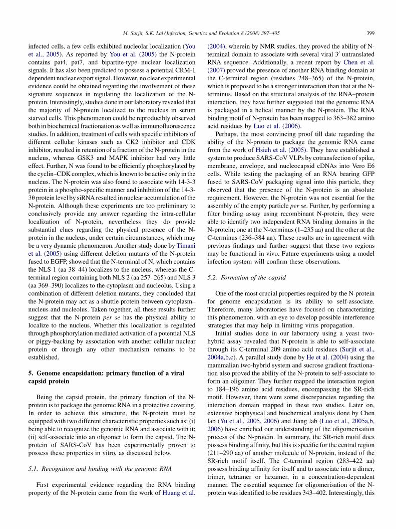
The nucleocapsid (N) protein is encoded by the 9th ORF of
SARS-CoV. The same ORF also codes for another unique
accessory protein called ORF9b, though in a different reading
frame, whose function is yet to be defined. The N-protein is a
46 kDa protein composed of 422 amino acids (Rota et al., 2003).
Its N-terminal region consists mostly of positively charged
amino acids, which is responsible for RNA binding. A lysine rich
region is present between 373 and 390 amino acids at the C-
terminus, which is predicted to be the nuclear localization signal.
Besides that, a SR-rich motif is present in the middle region
encompassing 177–207 amino acids. Biophysical studies done
by Chang et al. (2006) have suggested that this protein is
composed of two independent structural domains and a linker
region. The first domain is present at the N-terminus, inside the
putative RNA binding domain. The second domain consists of
the C-terminal region that is capable of self-association. Between
these two structural domains, there lies a highly disordered
region, which serves as a linker. This region has been reported to
interact with the membrane (M) protein and human cellular
hnRNPA1 protein (Fang et al., 2006; Luo et al., 2005a,b).
Besides, this region is also predicted to be a hot spot for
phosphorylation. Hence, in summary, the N-protein can be
classified into three distinct regions (Fig. 1), which may serve
completely different functions during different stages of the viral
life cycle. A similar mode of organization has been reported for
other coronavirus nucleocapsid proteins.
2. Stability of the N-protein
In vitro thermodynamic studies done by C. Luo et al. (2004)
and H. Luo et al. (2004) using purified recombinant N-protein
have shown it to be stable between pH 7 and 10, with maximum
conformational stability near pH 9. Further, it was observed to
undergo irreversible thermal-induced denaturation. It starts to
unfold at 35 8C and is completely denatured at 55 8C (Wang et al.,
2004). However, chemicals such as urea or guanidium chloride-
induced denaturation of the N-protein is a reversible process.
3. Post-translational modification
As in other coronavirus N-proteins, SARS-CoV N-protein
has been predicted and later experimentally proven to undergo
Fig. 1. Structure of the SARS-CoV nucleocapsid protein. A schematic diagram
showing various different domains identified to-date. The numbers 1–422
correspond to the length in amino acids of the N gene. GKEE represents the
sumoylation motif (lysine residue). KEL is the RXL motif, responsible for
binding with cyclin D and SPAR is the motif that gets phosphorylated by cyclin–
CDK complex (serine residue).
various post-translational modifications such as acetylation,
phosphorylation, and sumoylation.
Acetylation is the first modification of the N-protein to be
experimentally proven. By mass spectrometric analysis of
convalescent sera from several SARS patients, it has been
shown that the N-terminal methionine of N is removed as well
as all other methionines are oxidised and the resulting N-
terminal serine is acetylated. However, the functional relevance
of this modification, if any, remains to be elucidated (Krokhin
et al., 2003).
Another unique modification of the N-protein is its ability to
get sumoylated. Studies done by Y.H. Li et al. (2005) and F.Q.
Li et al. (2005) have clearly established that heterologously
expressed N in mammalian cells is sumoylated. Using a site-
directed mutagenesis approach, the sumoylation motif has been
mapped to the lysine residue 62, which is present in a putative
sumo-modification domain (GK62EE). Their data further
suggests that sumoylation may play a key role in modulating
homo-oligomerization, nucleolar translocation and cell-cycle
deregulatory property of the N-protein. Further experimental
support regarding sumoylation of N-protein came from another
independent study carried out by Fan et al. (2006) wherein they
have demonstrated an association between the N-protein and
Hubc9, which is a ubiquitin conjugating enzyme of the
sumoylation system. They have also mapped the interaction
domain to the SR-rich motif, which is in agreement with the
earlier report. However, they failed to detect the involvement of
the GKEE motif in mediating this interaction (Fan et al., 2006).
Initially, the SARS-CoV N-protein was predicted to be
heavily phosphorylated. Later on, from results obtained in our
laboratory as well as by other researchers, it is now clear that
the N-protein is a substrate of multiple cellular kinases. First
experimental evidence for the phosphorylation status of the N-
protein came from the study done by Zakhartchouk et al. (2005)
wherein by 32P orthophosphate labelling, they were able to
observe phosphorylation of adenovirus vector expressed N-
protein in 293T cells. Further studies done in our laboratory
clearly confirmed this observation. Majority of the N-protein
was found to be phosphorylated at its serine residues (although
the involvement of threonine and tyrosine residues could not be
detected, they may be occurring in vivo). In addition, using a
variety of biochemical assays, it was proved that, at least in
vitro, the N-protein could get phosphorylated by mitogen
activated protein kinase (MAP kinase), cyclin-dependent
kinase (CDK), glycogen synthase kinase 3 (GSK3), and casein
kinase 2 (CK2). Also, this data provides preliminary indication
regarding phosphorylation-dependent nucleo-cytoplasmic
shuttling of the N-protein (Surjit et al., 2005). Whether these
events actually bear any functional significance in vivo, remains
yet to be established.
4. Localization of the N-protein
In contrast to the N-protein of many other coronaviruses, the
SARS-CoV N-protein is predominantly distributed in the
cytoplasm, when expressed heterologously or in infected cells
(Surjit et al., 2005; You et al., 2005; Rowland et al., 2005). In
M. Surjit, S.K. Lal / Infection, Genetics and Evolution 8 (2008) 397–405 399
infected cells, a few cells exhibited nucleolar localization (You
et al., 2005). As reported by You et al. (2005) the N-protein
contains pat4, pat7, and bipartite-type nuclear localization
signals. It has also been predicted to possess a potential CRM-1
dependent nuclear export signal. However, no clear experimental
evidence could be obtained regarding the involvement of these
signature sequences in regulating the localization of the N-
protein. Interestingly, studies done in our laboratory revealed that
the majority of N-protein localized to the nucleus in serum
starved cells. This phenomenon could be reproducibly observed
both in biochemical fractionation as well as immunofluorescence
studies. In addition, treatment of cells with specific inhibitors of
different cellular kinases such as CK2 inhibitor and CDK
inhibitor, resulted in retention of a fraction of the N-protein in the
nucleus, whereas GSK3 and MAPK inhibitor had very little
effect. Further, N was found to be efficiently phosphorylated by
the cyclin–CDK complex, which is known to be active only in the
nucleus. The N-protein was also found to associate with 14-3-3
protein in a phospho-specific manner and inhibition of the 14-3-
3u protein level by siRNA resulted in nuclear accumulation of the
N-protein. Although these experiments are too preliminary to
conclusively provide any answer regarding the intra-cellular
localization of N-protein, nevertheless they do provide
substantial clues regarding the physical presence of the N-
protein in the nucleus, under certain circumstances, which may
be a very dynamic phenomenon. Another study done by Timani
et al. (2005) using different deletion mutants of the N-protein
fused to EGFP, showed that the N-terminal of N, which contains
the NLS 1 (aa 38–44) localizes to the nucleus, whereas the C-
terminal region containing both NLS 2 (aa 257–265) and NLS 3
(aa 369–390) localizes to the cytoplasm and nucleolus. Using a
combination of different deletion mutants, they concluded that
the N-protein may act as a shuttle protein between cytoplasm–
nucleus and nucleolus. Taken together, all these results further
suggest that the N-protein per se has the physical ability to
localize to the nucleus. Whether this localization is regulated
through phosphorylation mediated activation of a potential NLS
or piggy-backing by association with another cellular nuclear
protein or through any other mechanism remains to be
established.
5. Genome encapsidation: primary function of a viral
capsid protein
Being the capsid protein, the primary function of the N-
protein is to package the genomic RNA in a protective covering.
In order to achieve this structure, the N-protein must be
equipped with two different characteristic properties such as: (i)
being able to recognize the genomic RNA and associate with it;
(ii) self-associate into an oligomer to form the capsid. The N-
protein of SARS-CoV has been experimentally proven to
possess these properties in vitro, as discussed below.
5.1. Recognition and binding with the genomic RNA
First experimental evidence regarding the RNA binding
property of the N-protein came from the work of Huang et al.
(2004), wherein by NMR studies, they proved the ability of N-
terminal domain to associate with several viral 30 untranslated
RNA sequence. Additionally, a recent report by Chen et al.
(2007) proved the presence of another RNA binding domain at
the C-terminal region (residues 248–365) of the N-protein,
which is proposed to be a stronger interaction than that at the N-
terminus. Based on the structural analysis of the RNA–protein
interaction, they have further suggested that the genomic RNA
is packaged in a helical manner by the N-protein. The RNA
binding motif of N-protein has been mapped to 363–382 amino
acid residues by Luo et al. (2006).
Perhaps, the most convincing proof till date regarding the
ability of the N-protein to package the genomic RNA came
from the work of Hsieh et al. (2005). They have established a
system to produce SARS-CoV VLPs by cotransfection of spike,
membrane, envelope, and nucleocapsid cDNAs into Vero E6
cells. While testing the packaging of an RNA bearing GFP
fused to SARS-CoV packaging signal into this particle, they
observed that the presence of the N-protein is an absolute
requirement. However, the N-protein was not essential for the
assembly of the empty particle per se. Further, by performing a
filter binding assay using recombinant N-protein, they were
able to identify two independent RNA binding domains in the
N-protein; one at the N-terminus (1–235 aa) and the other at the
C-terminus (236–384 aa). These results are in agreement with
previous findings and further suggest that these two regions
may be functional in vivo. Future experiments using a model
infection system will confirm these observations.
5.2. Formation of the capsid
One of the most crucial properties required by the N-protein
for genome encapsidation is its ability to self-associate.
Therefore, many laboratories have focused on characterizing
this phenomenon, with an eye to develop possible interference
strategies that may help in limiting virus propagation.
Initial studies done in our laboratory using a yeast two-
hybrid assay revealed that N-protein is able to self-associate
through its C-terminal 209 amino acid residues (Surjit et al.,
2004a,b,c). A parallel study done by He et al. (2004) using the
mammalian two-hybrid system and sucrose gradient fractiona-
tion also proved the ability of the N-protein to self-associate to
form an oligomer. They further mapped the interaction region
to 184–196 amino acid residues, encompassing the SR-rich
motif. However, there were some discrepancies regarding the
interaction domain mapped in these two studies. Later on,
extensive biophysical and biochemical analysis done by Chen
lab (Yu et al., 2005, 2006) and Jiang lab (Luo et al., 2005a,b,
2006) have enriched our understanding of the oligomerisation
process of the N-protein. In summary, the SR-rich motif does
possess binding affinity, but this is specific for the central region
(211–290 aa) of another molecule of N-protein, instead of the
SR-rich motif itself. The C-terminal region (283–422 aa)
possess binding affinity for itself and to associate into a dimer,
trimer, tetramer or hexamer, in a concentration-dependent
manner. The essential sequence for oligomerisation of the N-
protein was identified to be residues 343–402. Interestingly, this
M. Surjit, S.K. Lal / Infection, Genetics and Evolution 8 (2008) 397–405400
region also encompasses the RNA binding motif of the N-
protein, which prompts us to speculate that there might be
mutual interplay between RNA binding and oligomerisation
activities of the N-protein. Further, the oligomerisation was
observed to be independent of electrostatic interactions and
addition of single strand DNA to the reaction mixture
containing tetramers of the N-protein promoted oligomerisa-
tion. Thus, it has been proposed that once the tetramer is formed
by protein–protein interaction between nucleocapsid mole-
cules, binding with genomic RNA prompts further assembly of
the complete nucleocapsid structure.
6. Perturbation of host cellular process by the N-
protein
Besides being the capsid protein of the virus, the N-protein
of many coronaviruses is known to double up as a regulatory
protein. The N-protein of the SARS-CoV too has been shown to
modulate the host cellular machinery in vitro, thereby
indicating its possible regulatory role during its viral life
cycle. Some of the major cellular processes perturbed by
heterologous expression of the N-protein are discussed below.
6.1. Deregulation of host cell cycle
Two groups have reported the ability of the N-protein to
interfere with the host cell cycle in vitro. Work done by Y.H. Li
et al. (2005) and F.Q. Li et al. (2005) proved that mutation of the
sumoylation motif in the N-protein leads to cell cycle arrest.
Work done in our laboratory has shown the inhibition of S phase
progress in cells expressing the N-protein (Surjit et al., 2006).
Further, we have observed down-regulation of S phase specific
gene products like cyclin E and CDK2 in SARS-CoV infected
cell lysate, which suggest that the observed phenomenon may
be relevant in vivo. In an attempt to further characterize the
mechanism of cell cycle blockage induced by the N-protein,
several biochemical and mutational analysis were carried out.
The results thus obtained demonstrated that the N-protein
directly inhibits the activity of the cyclin–CDK complex,
resulting in hypophosphorylation of retinoblastoma protein
with a concomitant down-regulation of E2F1-mediated
transactivation. Analysis of RXL and CDK phosphorylation
mutant N-protein identified the mechanism of inhibition of
CDK4 and CDK2 activity to be different. Whereas the N-
protein could directly bind to cyclin D and inhibit the activity of
CDK4–cyclin D complex; inhibition of CDK2 activity
appeared to be achieved in two different ways: indirectly by
down-regulation of protein levels of CDK2, cyclin E, and cyclin
A, and by direct binding of N-protein to CDK2–cyclin
complex. Nevertheless, the mechanism of cell cycle deregula-
tion in vivo, if any, remains to be understood.
6.2. Inhibition of interferon production
Production of interferon is one of the primary host defense
mechanism. However, SARS-CoV infection does not result in
IFN production. Nevertheless, pretreatment of cells with IFN
blocks SARS-CoV infection (Spiegel et al., 2005; Zheng et al.,
2004). Based on this observation, Palese lab has studied the IFN
inhibitory property of different SARS-CoV proteins, which
revealed that ORF3b, ORF6 as well as the N-protein have the
ability to independently inhibit IFN production through
different mechanisms. The N-protein was found to inhibit
the activity of IRF3 and NFkB in host cells, resulting in
inhibition of IFN synthesis. IRF3 activity was also blocked by
3b and ORF6 proteins, but inhibition of NFkB activity was a
property unique to the N-protein. In addition, 3b and ORF6
proteins were able to block STAT1 activity through different
mechanisms (Kopecky-Bromberg et al., 2007). All these data
suggest that SARS-CoV may employ multiple factors to check
the activity of host immune system and N-protein may be one of
the major partners in this process. It may be possible that these
different factors act independently during different stages of
viral life cycle. In that case, regulatory activity of the N-protein
will be as indispensible as its structural activity.
6.3. Up-regulation of COX2 production
Another major proinflammatory factor-induced during viral
infection is the cyclo oxygenase-2 protein. Using 293T cells
expressing the N-protein, Yan et al. (2006) have shown that
expression of the N-protein leads to upregulation of COX2
protein production in a transcriptional manner. They have
further demonstrated that the N-protein directly binds to the
NFkB response element present in the COX2 promoter through
a 68 aa residue binding domain (136–204 aa) and activates its
transcription.
Although the N-protein is known to associate with stretches
of nucleic acids, till date there is no other documentation or
prediction of its sequence specific DNA binding activity (as a
transcription factor). In such a scenario, the above observation,
if reproducible in vivo, may really be a unique property of the
N-protein and may further add to the established regulatory
functions of the N-protein.
6.4. Up-regulation of AP1 activity
Exogenously expressed N-protein has been reported to
enhance the DNA binding activity of c-fos, ATF-2, CREB-1,
and fos B in an ELISA-based assay, thus suggesting an increase
in AP1 activity in these cells (He et al., 2003). Mechanistic
details and functional significance of this phenomenon remains
to be elucidated.
6.5. Induction of apoptosis in serum starved monkey kidney
cells
Earlier work done in our laboratory has shown that N-
protein, when expressed in COS-1 monkey kidney cells,
induces apoptosis in the absence of growth factors. Attempts to
understand the mechanism of programmed cell death revealed
that the N-protein down modulated the activity of pro-survival
factors such as extracellular regulated kinase, Akt, and bcl 2,
and upregulated the activity of pro-apoptotic factors like
M. Surjit, S.K. Lal / Infection, Genetics and Evolution 8 (2008) 397–405 401
caspase-3 and caspase-7. This phenomenon was also associated
with reorganization of the actin cytoskeleton (Surjit et al.,
2004a,b,c). However, this phenomenon was not observed in
another cell line of epithelial lineage (huh7). Recently, Zhang
et al. (2007) have reported that serum starvation apoptosis of N
expressing COS-1 cells involves activation of mitochondrial
pathway. It remains to be studied whether this phenomenon is
actually recapitulated in vivo.
6.6. Association with host cell proteins
C. Luo et al. (2004) and H. Luo et al. (2004) have reported
the interaction between hnRNPA1 and N-protein by using a
variety of biochemical and genetic assays. The interaction
was found to be mediated through the middle region (161–
210 aa) of N-protein. If relevant in vivo, this interaction may
play a significant role in regulation of the viral RNA
synthesis.
Another interesting study done by C. Luo et al. (2004) and
H. Luo et al. (2004) have reported association between the N-
protein and human cyclophylin A. By SPR analysis they have
shown it to be a high affinity interaction. Although the
significance of this interaction is not known in vivo, they have
proposed that this interaction might be crucial for viral
infection. Notable is the fact that HIV-1 gag also binds with
human cyclophylin A and this interaction is crucial for HIV
infection (Gamble et al., 1996).
In summary, although several regulatory roles have been
proposed for the SARS-CoV N-protein using a variety of in
vitro experimental systems, no clear evidence exists for their
occurrence in vivo. In the absence of a suitable in vivo
experimental system, all these functions remain speculative.
7. N-protein: an efficient diagnostic tool
One of the most essential steps to limit the outbreak of any
infectious disease is the ability to diagnose the causative agent,
at the earliest possible time, which can be achieved by detecting
some of the markers that are specifically expressed by the
pathogen or by identifying some of the host factors that are
specifically produced during infection. N-protein, being one of
the predominantly expressed proteins at the early stage of
SARS-CoV infection, against which a strong antibody response
is initiated by the host; has been proposed to be an attractive
diagnostic tool.
In serum of SARS-CoV patients, the N-protein has been
detected as early as day 1 of infection by ELISA using
monoclonal antibodies against it (Che et al., 2004). Further, a
comparative study to detect SARS-CoV specific IgG, SARS-
CoV RNA, and the N-protein during early stages of infection
has demonstrated that detection efficiency of the N-protein is
significantly higher than the other two markers (Y.H. Li et al.,
2005; F.Q. Li et al., 2005).
Researchers have been mainly focussing on two different
strategies by which nucleocapsid can be used as a diagnostic
tool (i) development of efficient monoclonal antibodies against
the N-protein, (ii) production of recombinantly expressed,
highly purified N-protein for detection of N-specific antibody in
the host.
Using a phage display approach, Flego et al. (2005) have
identified human antibody fragments that recognise distinct
epitopes of the N-protein. These may help to develop efficient
reagents to detect N-protein in the infected host. Further,
several laboratories have been trying to develop efficient
monoclonal antibodies against the major immunodominant
epitopes of the N-protein, that can be used in ELISA to detect
SARS-CoV at an early stage of infection (Shang et al., 2005;
Liu et al., 2003; He et al., 2005; Woo et al., 2005). In another
interesting study, Liu et al. (2005) have developed an
immunofluorescence assay using antirabbit N-antibody that
can specifically detect N-protein from throat wash sample of
SARS-CoV patients at day 2 of illness.
Several other workers have focused on economical
production of highly purified recombinant N-protein using a
variety of heterologous expression systems that can be used in
ELISA to detect N-specific antibody in the patient sample. N-
protein has been produced in abundant quantity using codon
optimised gene in Escherichia coli (Das and Suresh, 2006).
Saijo et al. (2005) have successfully expressed recombinant N-
protein using a baculovirus expression system, which was
found to be 92% efficient in neutralizing antibody assay. In
another study, Liu et al. (2004), have expressed full length N-
protein using yeast expression system. However diagnostic use
of recombinant N-protein has been a problematic issue because
of several reasons as discussed below.
Bacterially expressed N-protein has been reported to
produce false sero-positivity owing to interference of bacte-
rially derived antigens (Leung et al., 2006; Yip et al., 2007). In
addition, several studies have shown cross-reactivity between
full-length N-protein of SARS and polyclonal antisera of group
1 animal coronaviruses, which may lead to faulty detection
(Sun and Meng, 2004). Another study done by Woo et al., have
also reported crossreactivity of full-length recombinant N-
protein with antisera of H CoV-OC43 and H CoV-229E infected
patients, thus giving false positive results. They were able to
minimise this false positivity by further verifying the ELISA
results with Western blot assay using recombinant N and spike
protein of SARS-CoV (Woo et al., 2004).
Later on, studies done by Qiu et al. (2005) and Bussmann
et al. (2006), showed that recombinantly expressed C-terminal
of the N-protein acts more specifically in detecting SARS-CoV
specific antisera in comparision to full-length N-protein. It is
noteworthy that this region is predicted to encompass major
antigenic sites of the N-protein.
Also, several reports have been published dealing with the
detection of N specific IgM by ELISA or indirect immuno-
fluorescent assay (Chang et al., 2004; Hsueh et al., 2004; Woo
et al., 2004). However, in these studies, IgM antibodies became
detectable later than IgG antibodies, which is in contrast to the
phenomena observed in most other pathogens.
A recent report published by Yu et al. (2007), have attempted
to solve this problem by using a truncated N-protein (122–422
aa) as an antigen in IgM ELISA. They found the IgM response
to appear 3 days before detection of IgG response, which is in
M. Surjit, S.K. Lal / Infection, Genetics and Evolution 8 (2008) 397–405402
agreement with the results obtained from other known
pathogens. Further, their results showed 100% specificity
and sensitivity of the truncated protein in detecting N-specific
IgM from patients with laboratory confirmed SARS cases in
comparison to healthy volunteers. The authors have suggested
that the IgM capture ELISA using this truncated N-protein may
be more effective in serodiagnosis of SARS-CoV at an earlier
time.
In another interesting report, Woo et al. (2005) have carried
out comparative studies to evaluate the relative diagnostic
efficacy of recombinantly expressed N- and S-proteins. They
observed sensitivity of recombinant N-IgG ELISA to be
significantly higher than that of recombinant S-IgG ELISA.
The reverse was true in case of IgM ELISA using recombinant
N- and S-proteins. Based on this data, they have suggested the
practice of an ELISA for detection of IgM against both S- and
N-protein instead of N alone (Woo et al., 2005).
Taken together, all these data does support the notion that the
N-protein may be used as an efficient diagnostic tool for
detection of SARS-CoV infection. Nevertheless, production
scale-up and further validation of specificity using patient
samples will determine the possible clinical use of these
reagents.
8. N-protein: a suitable vaccine candidate
One of the most clinically relevant uses of the N-protein can
be its use as a protective vaccine against SARS-CoV infection.
N-protein is one of the major antigens of the SARS-CoV. Also,
N-protein analysed from different patient samples shows least
variation in the gene sequence (Tong et al., 2004), therefore
indicating it to be a stable protein, which is a primary
requirement for an efficient vaccine candidate.
Earlier studies carried in Collins lab, Rao lab, and Li lab
have clearly shown that anti-serum to the N-protein does not
contain neutralizing antibodies against SARS-CoV (Buchholz
et al., 2004; Pang et al., 2004, and Liang et al., 2005). This may
be attributed to the localization of N-protein inside the viral
envelope, which will not be accessible to the antibody during
infection. It is noteworthy that the most effective SARS-CoV
structural protein that can induce neutralizing antibody
production is the S-protein (Buchholz et al., 2004). The S
antibody could block viral infection with 100% efficiency. On
the other hand, although unable to induce humoral immunity,
expression of N-protein-induced significant cytotoxic T
lymphocyte (CTL) response (Buchholz et al., 2004; Gao
et al., 2003; Zhu et al., 2004). Induction of N specific CTLs will
help limit the infection by lysing virus infected cells. This will
also limit the spread of virus. Thus, N-based vaccines may
further augment the protection efficiency when co-adminis-
tered with S-based vaccine. Several laboratories have been
exploring various strategies to evaluate the potential of N-
protein as a vaccine candidate.
In an elegant work done by Kim et al. (2004), calreticulin
fused N-protein expressing vaccinia virus has been shown to
generate potent N-specific humoral and T-cell immune
responses in mice. As reported by the authors, fusion with
calreticulin specifically enhanced the efficiency and signifi-
cantly reduced the titre of challenging vector (vaccinia virus).
The authors have proposed that N-protein may be the logical
choice as a target antigen in the event of S antibody dependent
enhancement (ADE) of infection. However, ADE phenomenon
has not been observed during spike-mediated vaccination
(Buchholz et al., 2004). Another study done by Wang et al.
(2005) has attempted to use plasmid DNA expressing S-, M-,
and N-proteins as an efficient vaccine candidate. Although they
report the production of some B-cell and T-cell responses
against N-protein, however stringent immune response was
obtained for the S- and M-proteins, thus scaling down the
choice of N-protein as a suitable candidate vaccine (Wang et al.,
2005). A similar plasmid mediated vaccination approach has
also been reported by Zhao et al. (2004) wherein they have
immunised mice with the DNA construct (pCI vector)
expressing the N-protein. They too have reported the generation
of a robust B-cell and T-cell immune response in animals.
Another group of workers have also reported successful use of
the N-protein as a DNA vaccine. They immunised mice by
intra-mucosal injection of the N-protein expressing plasmid
vector and were able to obtain specific humoral and T-cell
responses (Zhu et al., 2004).
The N-protein has also been reported to be of potential
interest as a peptide-based vaccine. A systematic study done by
Liu et al. (2006) has revealed the immunodominant epitopes of
the N-protein which could efficiently stimulate immune
response. They have also deduced some conserved immuno-
dominant epitopes in mouse, monkey, and humans, which may
help in design of the vaccine.
A recent report published by Gao’s laboratory provides
further evidence regarding the efficiency of an N-based vaccine
(Zhao et al., 2007). By using overlapping synthetic peptides
spanning the N-protein, they have identified dominant helper T-
cell epitopes in the nucleocapsid protein of SARS-CoV.
Immunization of mice with peptides emcompassing these
dominant TH-cell epitopes resulted in strong cellular immunity
in vivo. Priming with the helper peptides significantly
accelerated the immune response induced by the N-protein.
Further, by fusing with a conserved neutralizing epitope from
the spike protein of SARS-CoV, two of the TH-cell epitope
bearing peptides assisted in the production of higher titre
neutralizing antibodies in vivo, in comparision to spike epitope
alone or its mixture with TH epitope of N. Thus, it is practically
possible to generate a better immune response by using a fusion
of N- and S-protein. However, the TH epitopes identified in
their report is specific to mouse. Therefore, TH epitopes
identified in that study will not be useful for human.
Nevertheless, their data provides useful information for the
design of peptide-based anti-SARS-CoV vaccines.
Another interesting study conducted by Pei et al. (2005)
reports the possible use of the N-protein as a mucosal vaccine
candidate. They expressed the N-protein in Lactobacillus lactis,
which is a food grade bacteria, and challenged the mice either
orally or intra-mucosally. As preliminary evidence, they were
able to observe significant N-specific IgG in the sera of orally
challenged animals.
M. Surjit, S.K. Lal / Infection, Genetics and Evolution 8 (2008) 397–405 403
9. Future perspective
It is a significant achievement for the research community
that within a short span of time, we have been able to obtain
more or less a clear understanding regarding the structural and
functional properties of the N-protein. However, it is a fact
worth mentioning that all the studies done here were performed
with in vitro experiments, using recombinantly expressed N-
protein, in isolation. So at present, all we can conclude is that,
the N-protein per se has the physical ability to perform the
above described functions, in other words N-protein does bear
the necessary signature sequence or motifs or conformation to
perform these functions under suitable circumstances. Whether
a similar event is recapitulated in vivo during viral infection,
will be dependent on several criteria: (i) net effect of other viral
factors on the activity of N-protein, (ii) net translation and turn
over rate of N-protein, (iii) a conducive intra-cellular milieu,
and (iv) net modulation of an already skewed cellular pathway
by other viral factors. Hence, it will be interesting to re-evaluate
the properties of N-protein in a SARS-CoV infection model.
However, owing to the limited user friendliness and accessi-
bility of an infection system, probably we still have to resort to
in vitro systems for further analysis of the characteristics of N-
protein. One of the better experimental system has already been
established by the Chang lab (Hsieh et al., 2005), wherein all
the structural proteins were co-expressed to form VLP in 293T
cells. If this system can be further improved to optimise the rate
of synthesis of these different proteins to a near in vivo level, it
will at least enable us to study the net effect of the N-protein
with respect to other viral proteins. Further establishment of a
replicon system may also be helpful. In addition, some of the
interesting preliminary observations reported by several
laboratories need to be elaborately analysed. To begin with,
the reported interaction of the N-protein with genomic RNA
packaging signal needs to be further characterised and mapped.
Since the oligomerisation domain and the RNA binding regions
of the N-protein overlap with each other, the suggested
possibility of regulated genome incorporation and capsid
assembly should be further characterised with the aid of a
replicon system or a particle assembly system. In addition, the
reported ability of the N-protein to modulate different cellular
pathways should be further characterised in the particle
assembly system or at least in the presence of other viral
accessory proteins.
The most unique and significant property of the N-protein
revealed by preliminary studies is its ability to act as sequence
specific DNA binding factor. It has been shown to bind NFkB
response element of COX2 promoter and enhance COX2 gene
expression. This activity may be further empowering the N-
protein to manipulate the entire gene expression programme of
the infected cell. Therefore, studies should be initiated to
elaborately analyse this phenomenon. It seems to deserve so
much attention because another study done by Palese lab has
proved the ability of the N-protein to inhibit NFkB activity,
which results in inhibition of IFN synthesis. Further, Liao et al.
(2005) have reported the activation of NFkB by N-protein in
Vero E6 cells and He et al. (2005) failed to detect any change in
NFkB activity in the same cells. Therefore it needs to be
clarified whether N enhances NFkB activity and if yes; whether
upregulation of COX2 transcription by direct DNA binding is a
property specific to that promoter or it is a global phenomenon.
In such a scenario, there may be complicated crosstalk between
the ability of N-protein to deregulate the expression of COX2
and IFN in infected cells.
Lastly, the N-protein is known to be the most abundantly
expressed protein of the SARS-CoV. Therefore, any informa-
tion generated from the analysis of this protein, whether in vivo
or ex vivo, will definitely help to increase our understanding of
the biology of SARS-CoV and may someday help design better
protective tools against it.
Acknowledgements
The authors wish to thank Ms. Alisha Lal for helping out in
typing and formatting this review. We apologize to all those
colleagues whose work we might have missed to cite in this
article.
References
Buchholz, U.J., Bukreyev, A., Yang, L., Lamirande, E.W., Murphy, B.R.,
Subbarao, K., Collins, P.L., 2004. Contributions of the structural proteins
of severe acute respiratory syndrome coronavirus to protective immunity.
Proc. Natl. Acad. Sci. U.S.A. 101, 9804–9809.
Bussmann, B.M., Reiche, S., Jacob, L.H., Braun, J.M., Jassoy, C., 2006.
Antigenic and cellular localisation analysis of the severe acute respiratory
syndrome coronavirus nucleocapsid protein using monoclonal antibodies.
Virus Res. 122, 119–126.
Chang, W.T., Kao, C.L., Chung, M.Y., Chen, S.C., et al., 2004. SARS exposure
and emergency department workers. Emerg. Infect. Dis. 10, 1117–1119.
Chang, C.K., Sue, S.C., Yu, T.H., et al., 2006. Modular organization of SARS
coronavirus nucleocapsid protein. J. Biomed. Sci. 13, 59–72.
Che, X.Y., Hao, W., Wang, Y., et al., 2004. Nucleocapsid protein as early
diagnostic marker for SARS. Emerg. Infect. Dis. 10, 1947–1949.
Chen, C.Y., Chang, C.K., Chang, Y.W., et al., 2007. Structure of the SARS
coronavirus nucleocapsid protein RNA-binding dimerization domain sug-
gests a mechanism for helical packaging of viral RNA. J. Mol. Biol. 368,
1075–1086.
Das, D., Suresh, M.R., 2006. Copious production of SARS-CoV nucleocapsid
protein employing codon optimized synthetic gene. J. Virol. Methods 137,
343–346.
Fan, Z., Zhuo, Y., Tan, X., et al., 2006. SARS-CoV nucleocapsid protein binds
to hUbc9, a ubiquitin conjugating enzyme of the sumoylation system. J.
Med. Virol. 78, 1365–1373.
Fang, X., Ye, L.B., Zhang, Y., et al., 2006. Nucleocapsid amino acids 211 to
254, in particular, tetrad glutamines, are essential for the interaction
between the nucleocapsid and membrane proteins of SARS-associated
coronavirus. J. Microbiol. 44, 577–580.
Flego, M., Di Bonito, P., Ascione, et al., 2005. Generation of human antibody
fragments recognizing distinct epitopes of the nucleocapsid (N) SARS-CoV
protein using a phage display approach. BMC Infect. Dis. 5, 73.
Gamble, T.R., Vajdos, F.F., Yoo, S., et al., 1996. Crystal structure of human
cyclophilin A bound to the amino-terminal domain of HIV-1 capsid. Cell 87,
1285–1294.
Gao, W., Tamin, A., Soloff, A., D’Aiuto, L., Nwanegbo, E., Robbins, P.D.,
Bellini, W.J., Barratt-Boyes, S., Gambotto, A., 2003. Effects of a SARS-
associated coronavirus vaccine in monkeys. Lancet 362, 1895–1896.
He, R., Leeson, A., Andonov, A., et al., 2003. Activation of AP-1 signal
transduction pathway by SARS coronavirus nucleocapsid protein. Biochem.
Biophys. Res. Commun. 311, 870–876.
M. Surjit, S.K. Lal / Infection, Genetics and Evolution 8 (2008) 397–405404
He, R., Dobie, F., Ballantine, M., et al., 2004. Analysis of multimerization of
the SARS coronavirus nucleocapsid protein. Biochem. Biophys. Res.
Commun. 316, 476–483.
He, Q., Du, Q., Lau, S., et al., 2005. Characterization of monoclonal antibody
against SARS coronavirus nucleocapsid antigen and development of an
antigen capture ELISA. J. Virol. Methods 127, 46–53.
Hsieh, P.K., Chang, S.C., Huang, C.C., et al., 2005. Assembly of severe acute
respiratory syndrome coronavirus RNA packaging signal into virus-like
particles is nucleocapsid dependent. J. Virol. 79, 13848–13855.
Hsueh, P.R., Huang, L.M., Chen, P.J., et al., 2004. Chronological evolution of
IgM, IgA, IgG and neutralisation antibodies after infection with SARS-
associated coronavirus. Clin. Microbiol. Infect. 10, 1062–1066.
Huang, Q., Yu, L., Petros, A.M., et al., 2004. Structure of the N-terminal RNA-
binding domain of the SARS CoV nucleocapsid protein. Biochemistry 43,
6059–6063.
Kim, T.W., Lee, J.H., Hung, C.F., et al., 2004. Generation and characterization
of DNA vaccines targeting the nucleocapsid protein of severe acute respira-
tory syndrome coronavirus. J. Virol. 78, 4638–4645.
Kopecky-Bromberg, S.A., Martinez-Sobrido, L., Frieman, M., et al., 2007.
Severe acute respiratory syndrome coronavirus open reading frame (ORF)
3b, ORF 6, and nucleocapsid proteins function as interferon antagonists. J.
Virol. 81, 548–557.
Krokhin, O., Li, Y., Andonov, A., et al., 2003. Mass spectrometric character-
ization of proteins from the sars virus: a preliminary report. Mol. Cell
Proteomics 2, 346–356.
Leung, D.T., van Maren, W.W., Chan, F.K., Chan, W.S., Lo, A.W., Ma, C.H.,
Tam, F.C., To, K.F., Chan, P.K., Sung, J.J., Lim, P.L., 2006. Extremely low
exposure of a community to severe acute respiratory syndrome coronavirus:
false seropositivity due to use of bacterially derived antigens. J. Virol. 80,
8920–8928.
Li, F.Q., Xiao, H., Tam, J.P., et al., 2005. Sumoylation of the nucleocapsid
protein of severe acute respiratory syndrome coronavirus. FEBS Lett. 579,
2387–2396.
Li, Y.H., Li, J., Liu, X.E., et al., 2005. Detection of the nucleocapsid
protein of severe acute respiratory syndrome coronavirus in serum:
comparison with results of other viral markers. J. Virol. Methods 130,
45–50.
Liang, M.F., Du, R.L., Liu, J.Z., et al., 2005. SARS patients-derived human
recombinant antibodies to S and M proteins efficiently neutralize SARS-
coronavirus infectivity. Biomed. Environ. Sci. 18, 363–374.
Liao, Q.J., Ye, L.B., Timani, K.A., et al., 2005. Activation of NF-kappaB by the
full-length nucleocapsid protein of the SARS coronavirus. Acta Biochim.
Biophys. Sin. (Shanghai) 37, 607–612.
Liu, G., Hu, S., Hu, Y., et al., 2003. The C-terminal portion of the nucleocapsid
protein demonstrates SARS-CoVantigenicity. Genom. Proteom. Bioinform.
1, 193–197.
Liu, R.S., Yang, K.Y., Lin, J., et al., 2004. High-yield expression of recombi-
nant SARS coronavirus nucleocapsid protein in methylotrophic yeast Pichia
pastoris. World J. Gastroenterol. 10, 3602–3607.
Liu, I.J., Chen, P.J., Yeh, S.H., et al., 2005. Immunofluorescence assay for
detection of the nucleocapsid antigen of the severe acute respiratory
syndrome (SARS)-associated coronavirus in cells derived from throat wash
samples of patients with SARS. J. Clin. Microbiol. 43, 2444–2448.
Liu, S.J., Leng, C.H., Lien, S.P., et al., 2006. Immunological characterizations
of the nucleocapsid protein based SARS vaccine candidates. Vaccine 24,
3100–3108.
Luo, C., Luo, H., Zheng, S., et al., 2004. Nucleocapsid protein of SARS
coronavirus tightly binds to human cyclophilin A. Biochem. Biophys. Res.
Commun. 321, 557–565.
Luo, H., Ye, F., Sun, T.Y., et al., 2004. In vitro biochemical and thermodynamic
characterization of nucleocapsid protein of SARS. Biophys. Chem. 112,
15–25.
Luo, H., Ye, F., Chen, K., et al., 2005a. SR-rich motif plays a pivotal role in
recombinant SARS coronavirus nucleocapsid protein multimerization.
Biochemistry 44, 15351–15358.
Luo, H., Chen, Q., Chen, J., et al., 2005b. The nucleocapsid protein of SARS
coronavirus has a high binding affinity to the human cellular heterogeneous
nuclear ribonucleoprotein A1. FEBS Lett. 579, 2623–2628.
Luo, H., Chen, J., Chen, K., et al., 2006. Carboxyl terminus of severe acute
respiratory syndrome coronavirus nucleocapsid protein: self-association
analysis and nucleic acid binding characterization. Biochemistry 45,
11827–11835.
Pang, H., Liu, Y., Han, X., Xu, Y., Jiang, F., Wu, D., Kong, X., Bartlam, M., Rao,
Z., 2004. Protective humoral responses to severe acute respiratory syn-
drome-associated coronavirus: implications for the design of an effective
protein-based vaccine. J. Gen. Virol. 85, 3109–3113.
Pei, H., Liu, J., Cheng, Y., et al., 2005. Expression of SARS-coronavirus
nucleocapsid protein in Escherichia coli and Lactococcus lactis for
serodiagnosis and mucosal vaccination. Appl. Microbiol. Biotechnol. 68,
220–227.
Qiu, M., Wang, J., Wang, H., Chen, Z., Dai, E., Guo, Z., Wang, X., Pang, X.,
Fan, B., Wen, J., Wang, J., Yang, R., 2005. Use of the COOH portion
of the nucleocapsid protein in an antigen-capturing enzyme-linked immu-
nosorbent assay for specific and sensitive detection of severe
acute respiratory syndrome coronavirus. Clin. Diagn. Lab. Immunol.
12, 474–476.
Rota, P.A., Oberste, M.S., Monroe, S.S., et al., 2003. Characterization of a
novel coronavirus associated with severe acute respiratory syndrome.
Science 300, 1394–1399.
Rowland, R.R., Chauhan, V., Fang, Y., et al., 2005. Intracellular localization of
the severe acute respiratory syndrome coronavirus nucleocapsid protein:
absence of nucleolar accumulation during infection and after expression as a
recombinant protein in vero cells. J. Virol. 79, 11507–11512.
Saijo, M., Ogino, T., Taguchi, F., et al., 2005. Recombinant nucleocapsid
protein-based IgG enzyme-linked immunosorbent assay for the serological
diagnosis of SARS. J. Virol. Methods 125, 181–186.
Shang, B., Wang, X.Y., Yuan, J.W., et al., 2005. Characterization and
application of monoclonal antibodies against N protein of SARS-corona-
virus. Biochem. Biophys. Res. Commun. 336, 110–117.
Spiegel, M., Pichlmair, A., Martinez-Sobrido, L., et al., 2005. Inhibition of beta
interferon induction by severe acute respiratory syndrome coronavirus
suggests a two-step model for activation of interferon regulatory factor
3. J. Virol. 79, 2079–2086.
Sun, Z.F., Meng, X.J., 2004. Antigenic cross-reactivity between the nucleo-
capsid protein of severe acute respiratory syndrome (SARS) coronavirus
and polyclonal antisera of antigenic group I animal coronaviruses: implica-
tion for SARS diagnosis. J. Clin. Microbiol. 42 (5), 2351–2352.
Surjit, M., Liu, B., Chow, V.T., et al., 2004a. The nucleocapsid protein of the
SARS coronavirus is capable of self-association through a C-terminal 209
amino acid interaction domain. Biochem. Biophys. Res. Commun. 317,
1030–1036.
Surjit, M., Liu, B., Jameel, S., et al., 2004b. The SARS coronavirus nucleo-
capsid protein induces actin reorganization and apoptosis in COS-1 cells in
the absence of growth factors. Biochem. J. 383, 13–18.
Surjit, M., Liu, B., Kumar, P., et al., 2004c. The nucleocapsid protein of the
SARS coronavirus is capable of self-association through a C-terminal 209
amino acid interaction domain. Biochem. Biophys. Res. Commun. 317,
1030–1036.
Surjit, M., Kumar, R., Mishra, R.N., et al., 2005. The severe acute respiratory
syndrome coronavirus nucleocapsid protein is phosphorylated and localizes
in the cytoplasm by 14-3-3-mediated translocation. J. Virol. 79, 11476–
11486.
Surjit, M., Liu, B., Chow, V.T., et al., 2006. The nucleocapsid protein of severe
acute respiratory syndrome-coronavirus inhibits the activity of cyclin-
cyclin-dependent kinase complex and blocks S phase progression in mam-
malian cells. J. Biol. Chem. 281, 10669–10681.
Timani, K.A., Liao, Q., Ye, L., et al., 2005. Nuclear/nucleolar localization
properties of C-terminal nucleocapsid protein of SARS coronavirus. Virus
Res. 114, 23–34.
Tong, S., Lingappa, J.R., Chen, Q., et al., 2004. Direct sequencing of SARS-
coronavirus S and N genes from clinical specimens shows limited variation.
J. Infect. Dis. 190, 1127–1131.
Wang, Y., Wu, X., Wang, Y., et al., 2004. Low stability of nucleocapsid protein
in SARS virus. Biochemistry 43, 11103–11108.
Wang, Z., Yuan, Z., Matsumoto, M., et al., 2005. Immune responses with DNA
vaccines encoded different gene fragments of severe acute respiratory
M. Surjit, S.K. Lal / Infection, Genetics and Evolution 8 (2008) 397–405 405
syndrome coronavirus in BALB/c mice. Biochem. Biophys. Res. Commun.
327, 130–135.
Woo, P.C.Y., Lau, S.K.P., Wong, B.H.L., Chan, K.H., Chu, C.M., Tsoi, H.W.,
Huang, Y., Peiris, J.S.M., Yuen, K.Y., 2004. Longitudinal profile of immu-
noglobulin G (IgG), IgM, and IgA antibodies against the severe acute
respiratory syndrome (SARS) coronavirus nucleocapsid protein in patients
with pneumonia due to the SARS coronavirus. Clin. Diagn. Lab. Immunol.
11, 665–668.
Woo, P.C., Lau, S.K., Wong, B.H., et al., 2005. Differential sensitivities of
severe acute respiratory syndrome (SARS) coronavirus spike polypeptide
enzyme-linked immunosorbent assay (ELISA) and SARS coronavirus
nucleocapsid protein ELISA for serodiagnosis of SARS coronavirus pneu-
monia. J. Clin. Microbiol. 43, 3054–3058.
Yan, X., Hao, Q., Mu, Y., et al., 2006. Nucleocapsid protein of SARS-CoV
activates the expression of cyclooxygenase-2 by binding directly to reg-
ulatory elements for nuclear factor-kappa B and CCAAT/enhancer binding
protein. Int. J. Biochem. Cell Biol. 38, 1417–1428.
Yip, C.W., Hon, C.C., Zeng, F., Chow, K.Y., Chan, K.H., Peiris, J.S., Leung,
F.C., 2007. Naturally occurring anti-Escherichia coli protein antibodies in
the sera of healthy humans cause analytical interference in a recombinant
nucleocapsid protein-based enzyme-linked immunosorbent assay for ser-
odiagnosis of severe acute respiratory syndrome. Clin. Vaccine Immunol.
14, 99–101.
You, J., Dove, B.K., Enjuanes, L., et al., 2005. Subcellular localization of the
severe acute respiratory syndrome coronavirus nucleocapsid protein. J. Gen.
Virol. 86, 3303–3310.
Yu, I.M., Gustafson, C.L., Diao, J., et al., 2005. Recombinant severe
acute respiratory syndrome (SARS) coronavirus nucleocapsid protein
forms a dimer through its C-terminal domain. J. Biol. Chem. 280,
23280–23286.
Yu, I.M., Oldham, M.L., Zhang, J., et al., 2006. Crystal structure of the severe
acute respiratory syndrome (SARS) coronavirus nucleocapsid protein
dimerization domain reveals evolutionary linkage between corona- and
arteriviridae. J. Biol. Chem. 281, 17134–17139.
Yu, F., Le, M.Q., Inoue, S., et al., 2007. Recombinant truncated nucleocapsid
protein as antigen in a novel immunoglobulin M capture enzyme-linked
immunosorbent assay for diagnosis of severe acute respiratory syndrome
coronavirus infection. Clin. Vaccine Immunol. 14, 146–149.
Zakhartchouk, A.N., Viswanathan, S., Mahony, J.B., et al., 2005. Severe acute
respiratory syndrome coronavirus nucleocapsid protein expressed by an
adenovirus vector is phosphorylated and immunogenic in mice. J. Gen.
Virol. 86, 211–215.
Zhang, L., Wei, L., Jiang, D., Wang, J., Cong, X., Fei, R., 2007. SARS-CoV
nucleocapsid protein induced apoptosis of COS-1 mediated by the mito-
chondrial pathway. Artif. Cells Blood Substit. Immobil. Biotechnol. 35,
237–253.
Zhao, P., Cao, J., Zhao, L.J., et al., 2004. Immune responses against SARS-
coronavirus nucleocapsid protein induced by DNA vaccine. Virology 331,
128–135.
Zhao, J., Huang, Q., Wang, W., Zhang, Y., Lv, P., Gao, X.-M., 2007. Identifica-
tion and characterization of dominant helper T-cell epitopes in the nucleo-
capsid protein of severe acute respiratory syndrome coronavirus. J. Virol. 81
(11), 6079–6088.
Zheng, B., He, M.L., Wong, K.L., et al., 2004. Potent inhibition of SARS-
associated coronavirus (SCOV) infection and replication by type I inter-
ferons (IFNalpha/beta) but not by type II interferon (IFN-gamma). J.
Interferon Cytokine Res. 24, 388–390.
Zhu, M.S., Pan, Y., Chen, H.Q., et al., 2004. Induction of SARS-nucleoprotein-
specific immune response by use of DNA vaccine. Immunol. Lett. 92,
237–243.
www.elsevier.com/locate/meegid
Infection, Genetics and Evolution 8 (2008) 406–413
Phylogenetic analysis of the promoter region of the
CD40L gene in primates and other mammals§
Michael E. Steiper a,b,*, Sonia J. Parikh a, Julia M. Zichello a
a Department of Anthropology, Hunter College of the City University of New York, 695 Park Avenue, New York, NY 10021, United Statesb Departments of Anthropology and Biology, The Graduate Center of the City University of New York, 365 Fifth Avenue,
New York, NY 10016, United States
Received 18 October 2006; received in revised form 12 December 2006; accepted 14 December 2006
Available online 28 December 2006
Abstract
CD40L is a type II membrane protein comprised of 261 amino acids. CD40L plays a crucial role in the immune system where it is primarily
expressed on activated T cells and triggers immunoglobulin class switching. The genetic disease X-linked hypergammaglobulinemia (HIGM1,
XHIGM or XHIM) is caused by mutations in the CD40L gene. Individuals with HIGM1 are susceptible to recurrent infections to pathogens and a
relationship has been shown to exist with malaria [Sabeti, P., Usen, S., Farhadian, S., Jallow, M., Doherty, T., Newport, M., Pinder, M., Ward, R.,
Kwiatkowski, D., 2002a. CD40L association with protection from severe malaria. Genes Immun. 3, 286–291]. In this paper, we phylogenetically
examine the promoter region of CD40L in primates and other mammals via phylogenetic shadowing. This analysis revealed several regions of the
CD40L promoter that were highly constrained and thereby inferred to be functional. These constrained regions confirmed many known regulatory
sites. In addition, a novel, highly constrained upstream region was also identified which had an NF-AT recognition motif. These analyses also
showed that the different mammal groups do not share an exactly similar set of promoter binding sites and taxon-specific promoters have evolved.
# 2007 Elsevier B.V. All rights reserved.
Keywords: Evolution; XHIM; Hyper IgM; NF-AT; XHIGM; Phylogenetic shadowing; Immune system; Gene regulation; CD154
1. Introduction
Differences in gene regulation have been hypothesized to be
crucial to the evolutionary process (Carroll, 2005; King and
Wilson, 1975). While direct functional studies on the evolution
of gene regulation are becoming increasingly common,
bioinformatical studies of regulatory elements have proven
useful both for confirming known regulatory elements and
generating target regions for study. Generally, these bioinfor-
matical methods, known as phylogenetic footprinting (Gumu-
cio et al., 1992, 1993; Tagle et al., 1988), examine sequence
alignments of two or more species and derive regions that have
been conserved over phylogenetic distance. Genetic regions
§ Note: Nucleotide sequence data reported in this paper are available in the
GenBank under the accession numbers: EF055456–EF055471.
* Corresponding author at: Department of Anthropology, Hunter College of
the City University of New York, 695 Park Avenue, New York, NY 10021,
United States. Fax: +1 212 772 5423.
E-mail address: [email protected] (M.E. Steiper).
1567-1348/$ – see front matter # 2007 Elsevier B.V. All rights reserved.
doi:10.1016/j.meegid.2006.12.004
that remain conserved, despite mutational pressure over
evolutionary time, are forwarded as candidate functional
elements. Recently, a related method, phylogenetic shadowing
(Boffelli et al., 2003), has been introduced. The shadowing
method examines alignments of closely related taxa and has
proven useful in confirming sequence conservation among
known sequence elements, such as exons (Boffelli et al., 2003;
Ovcharenko et al., 2004). Furthermore, this method also has
identified conserved upstream genetic regions which were
subsequently determined to be important in gene regulation in
vitro, in the case of the APO(A) gene (Boffelli et al., 2003).
In this paper, we utilize phylogenetic shadowing to examine
the promoter region of CD40L in 18 primates and 4 other
mammals. Also known as CD154, gp39, TNFSF5 or TRAP,
CD40L is an X-linked, type II membrane protein, comprised of
261 amino acids and encoded by 5 exons (Allen et al., 1993;
Gauchat et al., 1993; Hollenbaugh et al., 1992; Padayachee
et al., 1992; Villa et al., 1994). CD40L plays a crucial role in the
immune system, where it is primarily expressed on activated T
cells and triggers immunoglobulin class switching upon
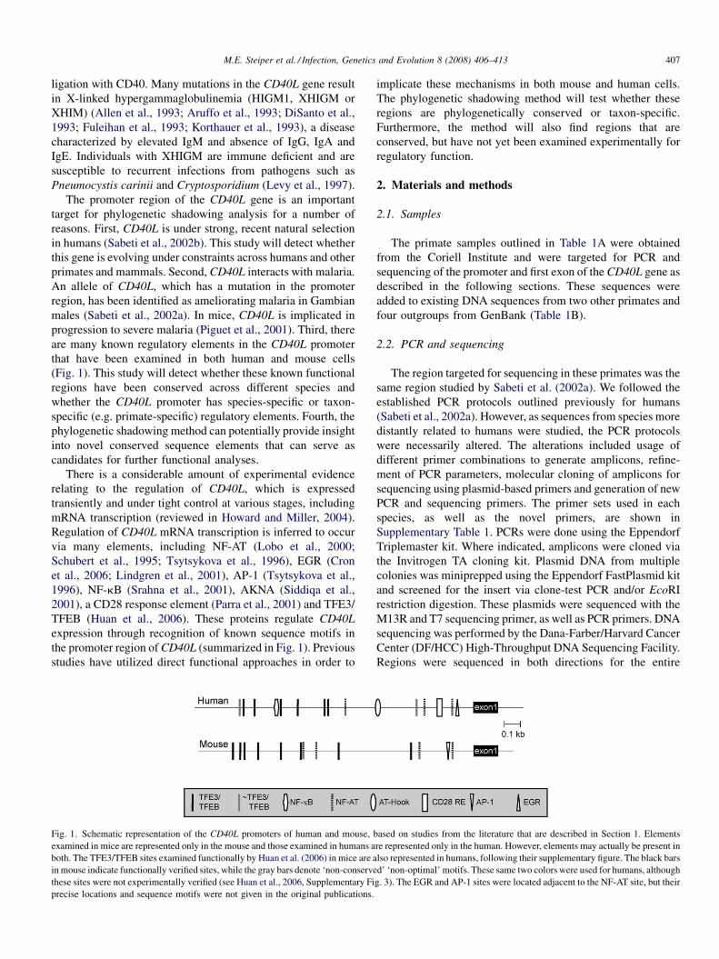
M.E. Steiper et al. / Infection, Genetics and Evolution 8 (2008) 406–413 407
ligation with CD40. Many mutations in the CD40L gene result
in X-linked hypergammaglobulinemia (HIGM1, XHIGM or
XHIM) (Allen et al., 1993; Aruffo et al., 1993; DiSanto et al.,
1993; Fuleihan et al., 1993; Korthauer et al., 1993), a disease
characterized by elevated IgM and absence of IgG, IgA and
IgE. Individuals with XHIGM are immune deficient and are
susceptible to recurrent infections from pathogens such as
Pneumocystis carinii and Cryptosporidium (Levy et al., 1997).
The promoter region of the CD40L gene is an important
target for phylogenetic shadowing analysis for a number of
reasons. First, CD40L is under strong, recent natural selection
in humans (Sabeti et al., 2002b). This study will detect whether
this gene is evolving under constraints across humans and other
primates and mammals. Second, CD40L interacts with malaria.
An allele of CD40L, which has a mutation in the promoter
region, has been identified as ameliorating malaria in Gambian
males (Sabeti et al., 2002a). In mice, CD40L is implicated in
progression to severe malaria (Piguet et al., 2001). Third, there
are many known regulatory elements in the CD40L promoter
that have been examined in both human and mouse cells
(Fig. 1). This study will detect whether these known functional
regions have been conserved across different species and
whether the CD40L promoter has species-specific or taxon-
specific (e.g. primate-specific) regulatory elements. Fourth, the
phylogenetic shadowing method can potentially provide insight
into novel conserved sequence elements that can serve as
candidates for further functional analyses.
There is a considerable amount of experimental evidence
relating to the regulation of CD40L, which is expressed
transiently and under tight control at various stages, including
mRNA transcription (reviewed in Howard and Miller, 2004).
Regulation of CD40L mRNA transcription is inferred to occur
via many elements, including NF-AT (Lobo et al., 2000;
Schubert et al., 1995; Tsytsykova et al., 1996), EGR (Cron
et al., 2006; Lindgren et al., 2001), AP-1 (Tsytsykova et al.,
1996), NF-kB (Srahna et al., 2001), AKNA (Siddiqa et al.,
2001), a CD28 response element (Parra et al., 2001) and TFE3/
TFEB (Huan et al., 2006). These proteins regulate CD40L
expression through recognition of known sequence motifs in
the promoter region of CD40L (summarized in Fig. 1). Previous
studies have utilized direct functional approaches in order to
Fig. 1. Schematic representation of the CD40L promoters of human and mouse, b
examined in mice are represented only in the mouse and those examined in humans a
both. The TFE3/TFEB sites examined functionally by Huan et al. (2006) in mice are a
in mouse indicate functionally verified sites, while the gray bars denote ‘non-conserve
these sites were not experimentally verified (see Huan et al., 2006, Supplementary Fi
precise locations and sequence motifs were not given in the original publications.
implicate these mechanisms in both mouse and human cells.
The phylogenetic shadowing method will test whether these
regions are phylogenetically conserved or taxon-specific.
Furthermore, the method will also find regions that are
conserved, but have not yet been examined experimentally for
regulatory function.
2. Materials and methods
2.1. Samples
The primate samples outlined in Table 1A were obtained
from the Coriell Institute and were targeted for PCR and
sequencing of the promoter and first exon of the CD40L gene as
described in the following sections. These sequences were
added to existing DNA sequences from two other primates and
four outgroups from GenBank (Table 1B).
2.2. PCR and sequencing
The region targeted for sequencing in these primates was the
same region studied by Sabeti et al. (2002a). We followed the
established PCR protocols outlined previously for humans
(Sabeti et al., 2002a). However, as sequences from species more
distantly related to humans were studied, the PCR protocols
were necessarily altered. The alterations included usage of
different primer combinations to generate amplicons, refine-
ment of PCR parameters, molecular cloning of amplicons for
sequencing using plasmid-based primers and generation of new
PCR and sequencing primers. The primer sets used in each
species, as well as the novel primers, are shown in
Supplementary Table 1. PCRs were done using the Eppendorf
Triplemaster kit. Where indicated, amplicons were cloned via
the Invitrogen TA cloning kit. Plasmid DNA from multiple
colonies was miniprepped using the Eppendorf FastPlasmid kit
and screened for the insert via clone-test PCR and/or EcoRI
restriction digestion. These plasmids were sequenced with the
M13R and T7 sequencing primer, as well as PCR primers. DNA
sequencing was performed by the Dana-Farber/Harvard Cancer
Center (DF/HCC) High-Throughput DNA Sequencing Facility.
Regions were sequenced in both directions for the entire
ased on studies from the literature that are described in Section 1. Elements
re represented only in the human. However, elements may actually be present in
lso represented in humans, following their supplementary figure. The black bars
d’ ‘non-optimal’ motifs. These same two colors were used for humans, although
g. 3). The EGR and AP-1 sites were located adjacent to the NF-AT site, but their
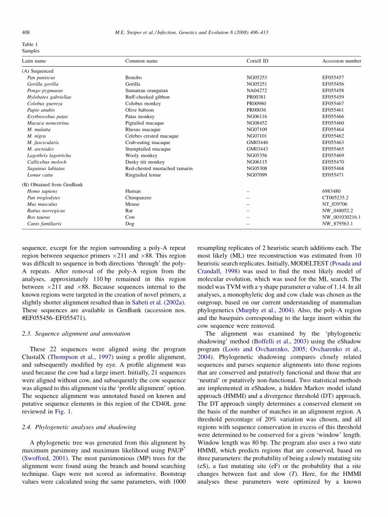
Table 1
Samples
Latin name Common name Coriell ID Accession number
(A) Sequenced
Pan paniscus Bonobo NG05253 EF055457
Gorilla gorilla Gorilla NG05251 EF055456
Pongo pygmaeus Sumatran orangutan NA04272 EF055458
Hylobates gabriellae Buff-cheeked gibbon PR00381 EF055459
Colobus guereza Colobus monkey PR00980 EF055467
Papio anubis Olive baboon PR00036 EF055461
Erythrocebus patas Patas monkey NG06116 EF055466
Macaca nemestrina Pigtailed macaque NG08452 EF055460
M. mulatta Rhesus macaque NG07109 EF055464
M. nigra Celebes crested macaque NG07101 EF055462
M. fascicularis Crab-eating macaque GM03446 EF055463
M. arctoides Stumptailed macaque GM03443 EF055465
Lagothrix lagotricha Wooly monkey NG05356 EF055469
Callicebus moloch Dusky titi monkey NG06115 EF055470
Saguinus labiatus Red-chested mustached tamarin NG05308 EF055468
Lemur catta Ringtailed lemur NG07099 EF055471
(B) Obtained from GenBank
Homo sapiens Human – 6983480
Pan troglodytes Chimpanzee – CT005235.2
Mus musculus Mouse – NT_039706
Rattus norvegicus Rat – NW_048052.2
Bos taurus Cow – NW_001030216.1
Canis familiaris Dog – NW_879563.1
M.E. Steiper et al. / Infection, Genetics and Evolution 8 (2008) 406–413408
sequence, except for the region surrounding a poly-A repeat
region between sequence primers �211 and �88. This region
was difficult to sequence in both directions ‘through’ the poly-
A repeats. After removal of the poly-A region from the
analyses, approximately 110 bp remained in this region
between �211 and �88. Because sequences internal to the
known regions were targeted in the creation of novel primers, a
slightly shorter alignment resulted than in Sabeti et al. (2002a).
These sequences are available in GenBank (accession nos.
#EF055456–EF055471).
2.3. Sequence alignment and annotation
These 22 sequences were aligned using the program
ClustalX (Thompson et al., 1997) using a profile alignment,
and subsequently modified by eye. A profile alignment was
used because the cow had a large insert. Initially, 21 sequences
were aligned without cow, and subsequently the cow sequence
was aligned to this alignment via the ‘profile alignment’ option.
The sequence alignment was annotated based on known and
putative sequence elements in this region of the CD40L gene
reviewed in Fig. 1.
2.4. Phylogenetic analyses and shadowing
A phylogenetic tree was generated from this alignment by
maximum parsimony and maximum likelihood using PAUP*
(Swofford, 2001). The most parsimonious (MP) trees for the
alignment were found using the branch and bound searching
technique. Gaps were not scored as informative. Bootstrap
values were calculated using the same parameters, with 1000
resampling replicates of 2 heuristic search additions each. The
most likely (ML) tree reconstruction was estimated from 10
heuristic search replicates. Initially, MODELTEST (Posada and
Crandall, 1998) was used to find the most likely model of
molecular evolution, which was used for the ML search. The
model was TVM with a g shape parameter a value of 1.14. In all
analyses, a monophyletic dog and cow clade was chosen as the
outgroup, based on our current understanding of mammalian
phylogenetics (Murphy et al., 2004). Also, the poly-A region
and the basepairs corresponding to the large insert within the
cow sequence were removed.
The alignment was examined by the ‘phylogenetic
shadowing’ method (Boffelli et al., 2003) using the eShadow
program (Loots and Ovcharenko, 2005; Ovcharenko et al.,
2004). Phylogenetic shadowing compares closely related
sequences and parses sequence alignments into those regions
that are conserved and putatively functional and those that are
‘neutral’ or putatively non-functional. Two statistical methods
are implemented in eShadow, a hidden Markov model island
approach (HMMI) and a divergence threshold (DT) approach.
The DT approach simply determines a conserved element on
the basis of the number of matches in an alignment region. A
threshold percentage of 20% variation was chosen, and all
regions with sequence conservation in excess of this threshold
were determined to be conserved for a given ‘window’ length.
Window length was 80 bp. The program also uses a two state
HMMI, which predicts regions that are conserved, based on
three parameters: the probability of being a slowly mutating site
(eS), a fast mutating site (eF) or the probability that a site
changes between fast and slow (T). Here, for the HMMI
analyses these parameters were optimized by a known
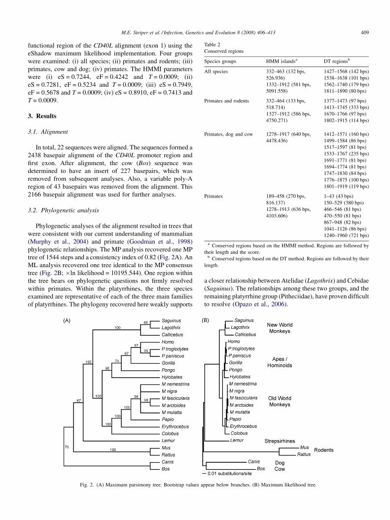
Table 2
Conserved regions
Species groups HMM islandsa DT regionsb
All species 332–463 (132 bps,
526.936)
1427–1568 (142 bps)
1332–1912 (581 bps,
5091.558)
1538–1638 (101 bps)
1562–1740 (179 bps)
1811–1890 (80 bps)
Primates and rodents 332–464 (133 bps,
518.714)
1377–1473 (97 bps)
1327–1912 (586 bps,
4750.271)
1413–1745 (333 bps)
1670–1766 (97 bps)
1802–1915 (114 bps)
Primates, dog and cow 1278–1917 (640 bps,
4478.436)
1412–1571 (160 bps)
1499–1584 (86 bps)
1517–1597 (81 bps)
1533–1767 (235 bps)
1691–1771 (81 bps)
1694–1774 (81 bps)
1747–1830 (84 bps)
1776–1875 (100 bps)
1801–1919 (119 bps)
Primates 189–458 (270 bps,
816.137)
1–43 (43 bps)
1278–1913 (636 bps,
4103.606)
150–529 (380 bps)
466–546 (81 bps)
470–550 (81 bps)
867–948 (82 bps)
1041–1126 (86 bps)
1240–1960 (721 bps)
a Conserved regions based on the HMMI method. Regions are followed by
their length and the score.b Conserved regions based on the DT method. Regions are followed by their
length.
M.E. Steiper et al. / Infection, Genetics and Evolution 8 (2008) 406–413 409
functional region of the CD40L alignment (exon 1) using the
eShadow maximum likelihood implementation. Four groups
were examined: (i) all species; (ii) primates and rodents; (iii)
primates, cow and dog; (iv) primates. The HMMI parameters
were (i) eS = 0.7244, eF = 0.4242 and T = 0.0009; (ii)
eS = 0.7281, eF = 0.5234 and T = 0.0009; (iii) eS = 0.7949,
eF = 0.5678 and T = 0.0009; (iv) eS = 0.8910, eF = 0.7413 and
T = 0.0009.
3. Results
3.1. Alignment
In total, 22 sequences were aligned. The sequences formed a
2438 basepair alignment of the CD40L promoter region and
first exon. After alignment, the cow (Bos) sequence was
determined to have an insert of 227 basepairs, which was
removed from subsequent analyses. Also, a variable poly-A
region of 43 basepairs was removed from the alignment. This
2166 basepair alignment was used for further analyses.
3.2. Phylogenetic analysis
Phylogenetic analyses of the alignment resulted in trees that
were consistent with our current understanding of mammalian
(Murphy et al., 2004) and primate (Goodman et al., 1998)
phylogenetic relationships. The MP analysis recovered one MP
tree of 1544 steps and a consistency index of 0.82 (Fig. 2A). An
ML analysis recovered one tree identical to the MP consensus
tree (Fig. 2B; �ln likelihood = 10195.544). One region within
the tree bears on phylogenetic questions not firmly resolved
within primates. Within the platyrrhines, the three species
examined are representative of each of the three main families
of platyrrhines. The phylogeny recovered here weakly supports
Fig. 2. (A) Maximum parsimony tree. Bootstrap values a
a closer relationship between Atelidae (Lagothrix) and Cebidae
(Saguinus). The relationships among these two groups, and the
remaining platyrrhine group (Pitheciidae), have proven difficult
to resolve (Opazo et al., 2006).
ppear below branches. (B) Maximum likelihood tree.
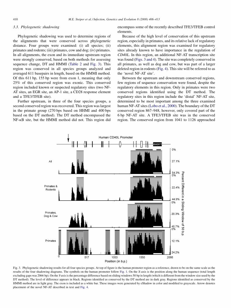
M.E. Steiper et al. / Infection, Genetics and Evolution 8 (2008) 406–413410
3.3. Phylogenetic shadowing
Phylogenetic shadowing was used to determine regions of
the alignments that were conserved across phylogenetic
distance. Four groups were examined: (i) all species; (ii)
primates and rodents; (iii) primates, cow and dog; (iv) primates.
In all alignments, the exon and its immediate upstream region
were strongly conserved, based on both methods for assessing
sequence change, DT and HMMI (Table 2 and Fig. 3). This
region was conserved in all species groups analyzed and
averaged 611 basepairs in length, based on the HMMI method.
Of this 611 bp, 155 bp were from exon 1, meaning that only
25% of this conserved region was exonic. This conserved
region included known or suspected regulatory sites (two NF-
AT sites, an EGR site, an AP-1 site, a CD28 response element
and a TFE3/TFEB site).
Further upstream, in three of the four species groups, a
second conserved region was recovered. This region was largest
in the primate group (270 bps based on HHMI and 400 bps
based on the DT method). The DT method encompassed the
NF-kB site, but the HMMI method did not. This region did
Fig. 3. Phylogenetic shadowing results for all four species groups. At top of figure is
results of the four shadowing diagrams. The symbols on the human promoter follo
excluding gaps was 2066 bp). On the Y-axis is the percentage difference based on slid
DT method). The level of difference appears in black. Regions identified as conserv
HMMI method are in light gray. The exon is included as a white bar. These images
placement of the novel NF-AT described in text and Fig. 4.
encompass some of the recently described TFE3/TFEB control
elements.
Because of the high level of conservation of this upstream
region, especially in primates, and its relative lack of regulatory
elements, this alignment region was examined for regulatory
sites already known to have importance in the regulation of
CD40L. In this region, an additional NF-AT transcription site
was found (Figs. 3 and 4). The site was completely conserved in
all primates, as well as dog and cow, but was part of a larger
deleted region in rodents (Fig. 4). This site will be referred to as
the ‘novel NF-AT site’.
Between the upstream and downstream conserved regions,
few regions of sequence conservation were found, despite the
regulatory elements in this region. Only in primates were two
conserved regions identified using the DT method. The
regulatory sites in this region include the ‘distal’ NF-AT site,
determined to be most important among the three examined
human NF-AT sites (Lobo et al., 2000). The boundary of the DT
conserved region 867–948, however, only covered part of the
6 bp NF-AT site. A TFE3/TFEB site was in the conserved
region. The conserved region from 1041 to 1126 approached
the human promoter region as a reference, drawn to be on the same scale as the
w Fig. 1. On the X-axis is the position along the human sequence (total length
ing windows 50 bp in length (which is different from the window size used by the
ed by the DT method are in dark gray. Regions identified as conserved by the
were generated by eShadow in color and modified to grayscale. Arrow denotes
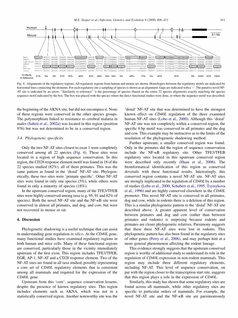
Fig. 4. Alignments of the regulatory regions. All regulatory regions from human and mouse are shown. Homologies between the regulatory motifs are indicated by
horizontal lines connecting the elements. For each regulatory site a sampling of species is shown as an alignment. Gaps are indicated with a ‘-’. The putative novel NF-
AT site is indicated by an arrow. ‘‘Similarity to reference’’ is the percentage of species (based on the entire 22 species alignment) exactly matching the species
sequence motif indicated by the box. The box was placed with the species where the direct functional studies were done, or where the sequence motif was described.
M.E. Steiper et al. / Infection, Genetics and Evolution 8 (2008) 406–413 411
the beginning of the AKNA site, but did not encompass it. None
of these regions were conserved in the other species groups.
The polymorphism linked to resistance to cerebral malaria in
males (Sabeti et al., 2002a) was located in this region (position
976) but was not determined to be in a conserved region.
3.4. Phylogenetic specificity
Only the two NF-AT sites closest to exon 1 were completely
conserved among all 22 species (Fig. 4). These sites were
located in a region of high sequence conservation. In this
region, the CD28 response element motif was found in 18 of the
22 species studied (82%), all of them primates. This was the
same pattern as found in the ‘distal’ NF-AT site. Phylogen-
etically, these two sites were ‘primate specific’. Other NF-AT
sites were found in only one species (5%), while others were
found in only a minority of species (18%).
In the upstream conserved region, many of the TFE3/TFEB
sites were highly conserved among taxa (e.g. 95, 91 and 82% of
species). Both the novel NF-AT site and the NF-kB site were
conserved in almost all primates, and dog, and cow, but were
not recovered in mouse or rat.
4. Discussion
Phylogenetic shadowing is a useful technique that can assist
in understanding gene regulation in silico. At the CD40L gene,
many functional studies have examined regulatory regions in
both human and mice cells. Many of these functional regions
are conserved, particularly those in the vicinity immediately
upstream of the first exon. This region includes TFE3/TFEB,
EGR, AP-1, NF-AT and a CD28 response element. Two of the
NF-AT sites are found in all taxa studied, possibly representing
a core set of CD40L regulatory elements that is consistent
among all mammals and required for the expression of the
CD40L gene.
Upstream from this ‘core’, sequence conservation lessens,
despite the presence of known regulatory sites. This region
includes elements such as AKNA, which was not in a
statistically conserved region. Another noteworthy site was the
‘distal’ NF-AT site that was determined to have the strongest
known effect on CD40L regulation of the three examined
human NF-AT sites (Lobo et al., 2000). Although this ‘distal’
NF-AT site was not completely within a conserved region, the
specific 6 bp motif was conserved in all primates and the dog
and cow. This example may be instructive as to the limits of the
resolution of the phylogenetic shadowing method.
Further upstream, a smaller conserved region was found.
Only in the primates did the region of sequence conservation
include the NF-kB regulatory site. Other TFE3/TFEB
regulatory sites located in this upstream conserved region
were described only recently (Huan et al., 2006). The
bioinformatical identification of this region as conserved
dovetails with these functional results. Interestingly, this
conserved region contains a novel NF-AT site. NF-AT sites
are strongly implicated in the regulation of CD40L by a number
of studies (Lobo et al., 2000; Schubert et al., 1995; Tsytsykova
et al., 1996) and are highly conserved elsewhere in the CD40L
promoter. This novel NF-AT site is conserved in all primates,
dog and cow, while in rodents there is a deletion of this region.
This is a similar phylogenetic pattern to the ‘distal’ NF-AT site
described above. A greater apparent level of conservation
between primates and dog and cow (rather than between
primates and rodents) is surprising because rodents and
primates are closer phylogenetic relatives. Parsimony suggests
that these three NF-AT sites were lost in rodents. This
phylogenetic pattern has also been found in the regulatory sites
of other genes (Perry et al., 2006), and may perhaps hint at a
more general phenomenon affecting the rodent lineage.
This evidence strongly suggests that the upstream conserved
region is worthy of additional study to understand its role in the
regulation of CD40L expression in non-rodent mammals. This
region may include three different regulatory elements,
including NF-AT. This level of sequence conservation, on
par with the region closer to the transcription start site, suggests
that this region plays a role in the expression of CD40L.
Similarly, this study has shown that some regulatory sites are
found across all mammals, while other regulatory sites are
specific to particular orders of mammals. For example, the
novel NF-AT site and the NF-kB site are parsimoniously
M.E. Steiper et al. / Infection, Genetics and Evolution 8 (2008) 406–413412
reconstructed as having been lost in rodents. Other elements,
such as the ‘distal’ NF-AT site and the CD28 response element,
are primate specific. Other sites are completely conserved
across all examined orders. This highlights the importance of
comparative bioinformatics in understanding potential for
translating the results of studies in one organism (e.g. mice) and
others (e.g. humans). Furthermore, this reveals that certain
elements may be critical to CD40L regulation in all mammals,
while other elements may be taxon specific. These specificities
may have evolved by natural selection in response to unique
sets of selective pressures facing different orders of mammals,
such as pathogens.
This study also has relevance to human health, as
mutations in the CD40L gene are implicated in XHIGM.
CD40L mutations which lead to XHIGM listed in the CD40L
mutation database and elsewhere point towards mutations
within the coding region of the gene, e.g. frameshift,
nonsense, missense and splice site mutations (Etzioni and
Ochs, 2004; Vihinen, 2006). These different mutations can
present with different phenotypes (Lopez-Granados et al.,
2003). In 15% of HIGM patients, however, a genetic etiology
is unknown (Etzioni and Ochs, 2004). Based on the strong
conservation in the promoter of the gene, one hypothesis is
that some of these patients have mutations within the
promoter of CD40L that lead to an HIGM phenotype.
Additional sequencing and analysis of these unclassified cases
would enable a test of this hypothesis. The extreme
conservation of many of these promoter regions and their
role in gene function suggests that changes in these sequences
could disrupt CD40L expression.
Acknowledgements
The authors thank Weigang Qiu and Derrick Brazill for
comments on this manuscript. This research was supported by
Grant NIGMS S06-060654. The infrastructure of the Anthro-
pological Genetics Lab at Hunter College was supported by
Grant Number RR03037 from the National Center for Research
Resources (NCRR), a component of the National Institutes of
Health (NIH). The contents of this publication are solely the
responsibility of the authors and do not necessarily represent
the official views of NCRR or NIH.
Appendix A. Supplementary data
Supplementary data associated with this article can be found,
in the online version, at: doi:10.1016/j.meegid.2006.12.004.
References
Allen, R.C., Armitage, R.J., Conley, M.E., Rosenblatt, H., Jenkins, N.A.,
Copeland, N.G., Bedell, M.A., Edelhoff, S., Disteche, C.M., Simoneaux,
D.K., et al., 1993. CD40 ligand gene defects responsible for X-linked
hyper-IgM syndrome. Science 259, 990–993.
Aruffo, A., Farrington, M., Hollenbaugh, D., Li, X., Milatovich, A., Nonoyama,
S., Bajorath, J., Grosmaire, L.S., Stenkamp, R., Neubauer, M., et al., 1993.
The CD40 ligand, gp39, is defective in activated T cells from patients with
X-linked hyper-IgM syndrome. Cell 72, 291–300.
Boffelli, D., McAuliffe, J., Ovcharenko, D., Lewis, K.D., Ovcharenko, I.,
Pachter, L., Rubin, E.M., 2003. Phylogenetic shadowing of primate
sequences to find functional regions of the human genome. Science 299,
1391–1394.
Carroll, S.B., 2005. Evolution at two levels: on genes and form. PLoS Biol. 3,
e245.
Cron, R.Q., Bandyopadhyay, R., Genin, A., Brunner, M., Kersh, G.J., Yin, J.,
Finkel, T.H., Crow, M.K., 2006. Early growth response-1 is required for
CD154 transcription. J. Immunol. 176, 811–818.
DiSanto, J.P., Bonnefoy, J.Y., Gauchat, J.F., Fischer, A., de Saint Basile, G.,
1993. CD40 ligand mutations in X-linked immunodeficiency with hyper-
IgM. Nature 361, 541–543.
Etzioni, A., Ochs, H.D., 2004. The hyper IgM syndrome—an evolving story.
Pediatr. Res. 56, 519–525.
Fuleihan, R., Ramesh, N., Loh, R., Jabara, H., Rosen, R.S., Chatila, T., Fu, S.M.,
Stamenkovic, I., Geha, R.S., 1993. Defective expression of the CD40 ligand
in X chromosome-linked immunoglobulin deficiency with normal or ele-
vated IgM. Proc. Natl. Acad. Sci. U.S.A. 90, 2170–2173.
Gauchat, J.F., Aubry, J.P., Mazzei, G., Life, P., Jomotte, T., Elson, G., Bonnefoy,
J.Y., 1993. Human CD40-ligand: molecular cloning, cellular distribution
and regulation of expression by factors controlling IgE production. FEBS
Lett. 315, 259–266.
Goodman, M., Porter, C.A., Czelusniak, J., Page, S.L., Schneider, H., Shoshani,
J., Gunnell, G., Groves, C.P., 1998. Toward a phylogenetic classification of
primates based on DNA evidence complemented by fossil evidence. Mol.
Phylogenet. Evol. 9, 585–598.
Gumucio, D.L., Heilstedt-Williamson, H., Gray, T.A., Tarle, S.A., Shelton,
D.A., Tagle, D.A., Slightom, J.L., Goodman, M., Collins, F.S., 1992.
Phylogenetic footprinting reveals a nuclear protein which binds to silencer
sequences in the human gamma and epsilon globin genes. Mol. Cell. Biol.
12, 4919–4929.
Gumucio, D.L., Shelton, D.A., Bailey, W.J., Slightom, J.L., Goodman, M.,
1993. Phylogenetic footprinting reveals unexpected complexity in trans
factor binding upstream from the epsilon-globin gene. Proc. Natl. Acad. Sci.
U.S.A. 90, 6018–6022.
Hollenbaugh, D., Grosmaire, L.S., Kullas, C.D., Chalupny, N.J., Braesch-
Andersen, S., Noelle, R.J., Stamenkovic, I., Ledbetter, J.A., Aruffo, A.,
1992. The human T cell antigen gp39, a member of the TNF gene family, is
a ligand for the CD40 receptor: expression of a soluble form of gp39 with B
cell co-stimulatory activity. EMBO J. 11, 4313–4321.
Howard, L.M., Miller, S.D., 2004. Immunotherapy targeting the CD40/CD154
costimulatory pathway for treatment of autoimmune disease. Autoimmunity
37, 411–418.
Huan, C., Kelly, M.L., Steele, R., Shapira, I., Gottesman, S.R., Roman, C.A.,
2006. Transcription factors TFE3 and TFEB are critical for CD40 ligand
expression and thymus-dependent humoral immunity. Nat. Immunol. 7,
1082–1091.
King, M.C., Wilson, A.C., 1975. Evolution at two levels in humans and
chimpanzees. Science 188, 107–116.
Korthauer, U., Graf, D., Mages, H.W., Briere, F., Padayachee, M., Malcolm, S.,
Ugazio, A.G., Notarangelo, L.D., Levinsky, R.J., Kroczek, R.A., 1993.
Defective expression of T-cell CD40 ligand causes X-linked immunodefi-
ciency with hyper-IgM. Nature 361, 539–541.
Levy, J., Espanol-Boren, T., Thomas, C., Fischer, A., Tovo, P., Bordigoni, P.,
Resnick, I., Fasth, A., Baer, M., Gomez, L., Sanders, E.A., Tabone, M.D.,
Plantaz, D., Etzioni, A., Monafo, V., Abinun, M., Hammarstrom, L.,
Abrahamsen, T., Jones, A., Finn, A., Klemola, T., DeVries, E., Sanal,
O., Peitsch, M.C., Notarangelo, L.D., 1997. Clinical spectrum of X-linked
hyper-IgM syndrome. J. Pediatr. 131, 47–54.
Lindgren, H., Axcrona, K., Leanderson, T., 2001. Regulation of transcriptional
activity of the murine CD40 ligand promoter in response to signals through
TCR and the costimulatory molecules CD28 and CD2. J. Immunol. 166,
4578–4585.
Lobo, F.M., Xu, S., Lee, C., Fuleihan, R.L., 2000. Transcriptional activity of the
distal CD40 ligand promoter. Biochem. Biophys. Res. Commun. 279, 245–
250.
Loots, G.G., Ovcharenko, I., 2005. Dcode.org anthology of comparative
genomic tools. Nucleic Acids Res. 33, W56–W64.
M.E. Steiper et al. / Infection, Genetics and Evolution 8 (2008) 406–413 413
Lopez-Granados, E., Cambronero, R., Ferreira, A., Fontan, G., Garcia-Rodri-
guez, M.C., 2003. Three novel mutations reflect the variety of defects
causing phenotypically diverse X-linked hyper-IgM syndrome. Clin. Exp.
Immunol. 133, 123–131.
Murphy, W.J., Pevzner, P.A., O’Brien, S.J., 2004. Mammalian phylogenomics
comes of age. Trends Genet. 20, 631–639.
Opazo, J.C., Wildman, D.E., Prychitko, T., Johnson, R.M., Goodman, M.,
2006. Phylogenetic relationships and divergence times among New
World monkeys (Platyrrhini primates). Mol. Phylogenet. Evol. 40,
274–280.
Ovcharenko, I., Boffelli, D., Loots, G.G., 2004. eShadow: a tool for comparing
closely related sequences. Genome Res. 14, 1191–1198.
Padayachee, M., Feighery, C., Finn, A., McKeown, C., Levinsky, R.J., Kinnon,
C., Malcolm, S., 1992. Mapping of the X-linked form of hyper-IgM
syndrome (HIGM1) to Xq26 by close linkage to HPRT. Genomics 14,
551–553.
Parra, E., Mustelin, T., Dohlsten, M., Mercola, D., 2001. Identification of a
CD28 response element in the CD40 ligand promoter. J. Immunol. 166,
2437–2443.
Perry, G.H., Verrelli, B.C., Stone, A.C., 2006. Molecular evolution of the
primate developmental genes MSX1 and PAX9. Mol. Biol. Evol. 23, 644–
654.
Piguet, P.F., Kan, C.D., Vesin, C., Rochat, A., Donati, Y., Barazzone, C., 2001.
Role of CD40-CD40L in mouse severe malaria. Am. J. Pathol. 159, 733–
742.
Posada, D., Crandall, K.A., 1998. MODELTEST: testing the model of DNA
substitution. Bioinformatics 14, 817–818.
Sabeti, P., Usen, S., Farhadian, S., Jallow, M., Doherty, T., Newport, M., Pinder,
M., Ward, R., Kwiatkowski, D., 2002a. CD40L association with protection
from severe malaria. Genes Immun. 3, 286–291.
Sabeti, P.C., Reich, D.E., Higgins, J.M., Levine, H.Z., Richter, D.J., Schaffner,
S.F., Gabriel, S.B., Platko, J.V., Patterson, N.J., McDonald, G.J., Ackerman,
H.C., Campbell, S.J., Altshuler, D., Cooper, R., Kwiatkowski, D., Ward, R.,
Lander, E.S., 2002b. Detecting recent positive selection in the human
genome from haplotype structure. Nature 419, 832–837.
Schubert, L.A., King, G., Cron, R.Q., Lewis, D.B., Aruffo, A., Hollenbaugh, D.,
1995. The human gp39 promoter. Two distinct nuclear factors of activated T
cell protein-binding elements contribute independently to transcriptional
activation. J. Biol. Chem. 270, 29624–29627.
Siddiqa, A., Sims-Mourtada, J.C., Guzman-Rojas, L., Rangel, R., Guret, C.,
Madrid-Marina, V., Sun, Y., Martinez-Valdez, H., 2001. Regulation of
CD40 and CD40 ligand by the AT-hook transcription factor AKNA. Nature
410, 383–387.
Srahna, M., Remacle, J.E., Annamalai, K., Pype, S., Huylebroeck, D., Boo-
gaerts, M.A., Vandenberghe, P., 2001. NF-kappaB is involved in the
regulation of CD154 (CD40 ligand) expression in primary human T cells.
Clin. Exp. Immunol. 125, 229–236.
Swofford, D.L., 2001. PAUP*. Phylogenetic Analysis Using Parsimony (* and
Other Methods). Version 4. Sinauer Associates, Sunderland, MA.
Tagle, D.A., Koop, B.F., Goodman, M., Slightom, J.L., Hess, D.L., Jones, R.T.,
1988. Embryonic epsilon and gamma globin genes of a prosimian primate
(Galago crassicaudatus). Nucleotide and amino acid sequences, develop-
mental regulation and phylogenetic footprints. J. Mol. Biol. 203, 439–455.
Thompson, J.D., Gibson, T.J., Plewniak, F., Jeanmougin, F., Higgins, D.G.,
1997. The CLUSTAL_X windows interface: flexible strategies for multiple
sequence alignment aided by quality analysis tools. Nucleic Acids Res. 25,
4876–4882.
Tsytsykova, A.V., Tsitsikov, E.N., Geha, R.S., 1996. The CD40L promoter
contains nuclear factor of activated T cells-binding motifs which require
AP-1 binding for activation of transcription. J. Biol. Chem. 271, 3763–3770.
Vihinen, M., 2006. CD40Lbase v.2.04.
Villa, A., Notarangelo, L.D., Di Santo, J.P., Macchi, P.P., Strina, D., Frattini, A.,
Lucchini, F., Patrosso, C.M., Giliani, S., Mantuano, E., et al., 1994.
Organization of the human CD40L gene: implications for molecular defects
in X chromosome-linked hyper-IgM syndrome and prenatal diagnosis. Proc.
Natl. Acad. Sci. U.S.A. 91, 2110–2114.
www.elsevier.com/locate/meegid
Infection, Genetics and Evolution 8 (2008) 414–421
The geometry of the wing of Aedes (Stegomyia) aegypti in isofemale
lines through successive generations
Nuananong Jirakanjanakit a, Somjai Leemingsawat b, Jean Pierre Dujardin c,*a Center for Vaccine Development, Institute of Science and Technology for Research and Development, Mahidol University, Salaya,
Puttamonthon, Nakhonpathom, Thailandb Department of Medical Entomology, Faculty of Tropical Medicine, Mahidol University, Rajvithee, Bangkok, Thailand
c UMR IRD-CNRS 2724, Avenue Agropolis, 911, BP 64501, 34394 Montpellier, France
Received 1 February 2007; received in revised form 3 May 2007; accepted 4 May 2007
Available online 22 May 2007
Abstract
Under a common laboratory environment, three isofemale lines of Aedes (Stegomyia) aegypti were used to score metric properties (size and
shape) of the wings during 10 generations. Since the number of generations was much higher than the number of founders in each line, genetic drift
was expected to occur.
Size tended to slightly increase with time, but its variation among the successive generations did not show any detectable information specific to
each line. Shape could discriminate among lines in females. Males of lines A and B were not discriminated before generation 8, after which they
became completely separated.
For each line at each generation, the variance of size and the metric disparity index (as an estimate of shape variance) were higher in females
than in males. From one generation to another, the within line shape variance decreased in both sexes, while shape similarity progressively and
consistently decreased between males of lines A and B. At each generation, in both sexes, shape variance of the pooled lines was higher than that of
each line separately, a pattern not observed for size variance.
In conclusion, shape, as a metric property, was closer to a genetic character than size: (i) it showed modifications compatible with the hypothesis
of genetic drift and (ii) its variation was related to the complexity of the sample.
# 2007 Elsevier B.V. All rights reserved.
Keywords: Aedes aegypti; Isofemale lines; Geometric morphometrics; Metric disparity
1. Introduction
Aedes (Stegomyia) aegypti is the main vector of arthropod-
borne viruses in the tropical and subtropical regions. It
originated from Africa and expanded around the tropical world
with a pantropical distribution in 1930 (Rodhain, 1996). Known
as the yellow fever mosquito in Africa and South America, it is
the principal vector of dengue worldwide (Christophers, 1960).
To fight this latter disease, vector control and entomological
surveillance remain the most important issues.
Many laboratory lines were reared to perform tests on
sensitivity to control measures (Brogdon et al., 1999; Ganesh
et al., 2002), on vectorial capacity (Mourya et al., 2001; Bennett
* Corresponding author.
E-mail address: [email protected] (J.P. Dujardin).
1567-1348/$ – see front matter # 2007 Elsevier B.V. All rights reserved.
doi:10.1016/j.meegid.2007.05.004
et al., 2005), or to improve our knowledge about the biology or
the taxonomy of the mosquito vector (Cook et al., 2005).
Isofemale lines were used to assess the genetic basis for dengue
susceptibility among females (Miller and Mitchell, 1991;
Tardieux et al., 1991). In the present study, laboratory lines
were used to explore the morphometric variability of the wings
Ae. aegypti.
Wings are bi-dimensional rigid structures well suited for
morphometric work. For Ae. aegypti, they have been used to
demonstrate the power of geometric morphometrics in
distinguishing among laboratory lines from different geo-
graphic origins in Thailand. Although reared in the same
insectarium, laboratory lines did not show any evidence of
morphological convergence, since on the basis of shape only,
97% of the wings were assigned to their correct origin
(Jirakanjanakit and Dujardin, 2005). However, it was not clear
which factor could cause the discriminating power of the wing,
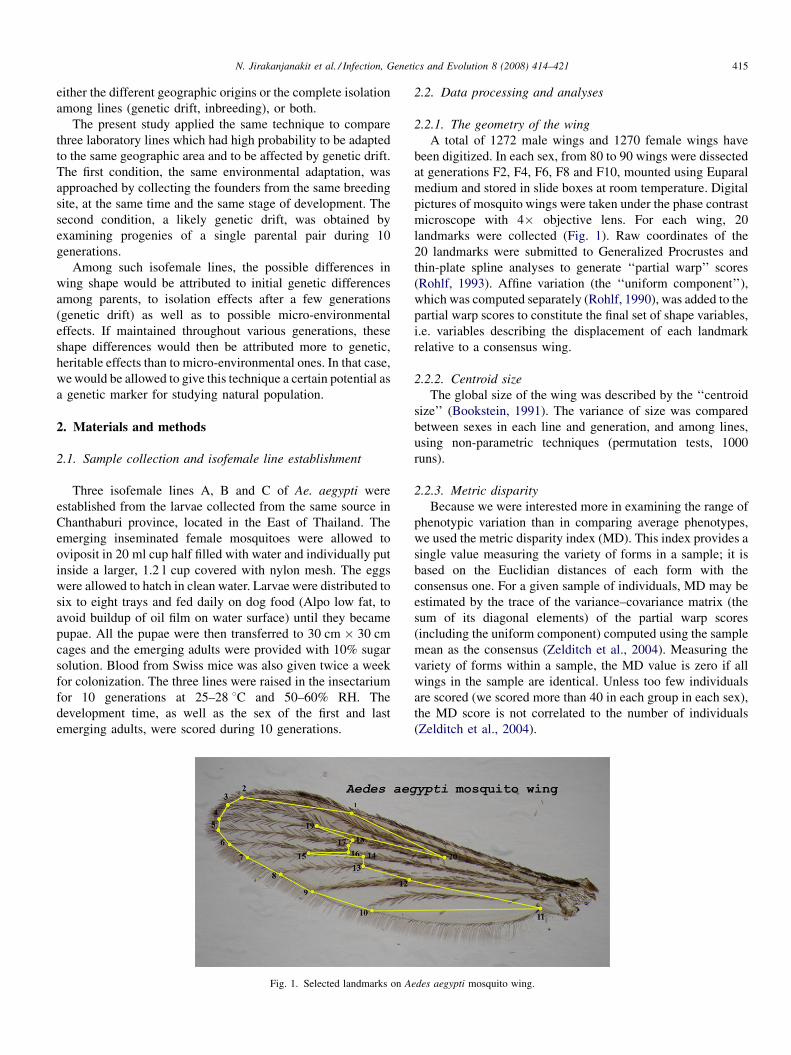
N. Jirakanjanakit et al. / Infection, Genetics and Evolution 8 (2008) 414–421 415
either the different geographic origins or the complete isolation
among lines (genetic drift, inbreeding), or both.
The present study applied the same technique to compare
three laboratory lines which had high probability to be adapted
to the same geographic area and to be affected by genetic drift.
The first condition, the same environmental adaptation, was
approached by collecting the founders from the same breeding
site, at the same time and the same stage of development. The
second condition, a likely genetic drift, was obtained by
examining progenies of a single parental pair during 10
generations.
Among such isofemale lines, the possible differences in
wing shape would be attributed to initial genetic differences
among parents, to isolation effects after a few generations
(genetic drift) as well as to possible micro-environmental
effects. If maintained throughout various generations, these
shape differences would then be attributed more to genetic,
heritable effects than to micro-environmental ones. In that case,
we would be allowed to give this technique a certain potential as
a genetic marker for studying natural population.
2. Materials and methods
2.1. Sample collection and isofemale line establishment
Three isofemale lines A, B and C of Ae. aegypti were
established from the larvae collected from the same source in
Chanthaburi province, located in the East of Thailand. The
emerging inseminated female mosquitoes were allowed to
oviposit in 20 ml cup half filled with water and individually put
inside a larger, 1.2 l cup covered with nylon mesh. The eggs
were allowed to hatch in clean water. Larvae were distributed to
six to eight trays and fed daily on dog food (Alpo low fat, to
avoid buildup of oil film on water surface) until they became
pupae. All the pupae were then transferred to 30 cm � 30 cm
cages and the emerging adults were provided with 10% sugar
solution. Blood from Swiss mice was also given twice a week
for colonization. The three lines were raised in the insectarium
for 10 generations at 25–28 8C and 50–60% RH. The
development time, as well as the sex of the first and last
emerging adults, were scored during 10 generations.
Fig. 1. Selected landmarks on A
2.2. Data processing and analyses
2.2.1. The geometry of the wing
A total of 1272 male wings and 1270 female wings have
been digitized. In each sex, from 80 to 90 wings were dissected
at generations F2, F4, F6, F8 and F10, mounted using Euparal
medium and stored in slide boxes at room temperature. Digital
pictures of mosquito wings were taken under the phase contrast
microscope with 4� objective lens. For each wing, 20
landmarks were collected (Fig. 1). Raw coordinates of the
20 landmarks were submitted to Generalized Procrustes and
thin-plate spline analyses to generate ‘‘partial warp’’ scores
(Rohlf, 1993). Affine variation (the ‘‘uniform component’’),
which was computed separately (Rohlf, 1990), was added to the
partial warp scores to constitute the final set of shape variables,
i.e. variables describing the displacement of each landmark
relative to a consensus wing.
2.2.2. Centroid size
The global size of the wing was described by the ‘‘centroid
size’’ (Bookstein, 1991). The variance of size was compared
between sexes in each line and generation, and among lines,
using non-parametric techniques (permutation tests, 1000
runs).
2.2.3. Metric disparity
Because we were interested more in examining the range of
phenotypic variation than in comparing average phenotypes,
we used the metric disparity index (MD). This index provides a
single value measuring the variety of forms in a sample; it is
based on the Euclidian distances of each form with the
consensus one. For a given sample of individuals, MD may be
estimated by the trace of the variance–covariance matrix (the
sum of its diagonal elements) of the partial warp scores
(including the uniform component) computed using the sample
mean as the consensus (Zelditch et al., 2004). Measuring the
variety of forms within a sample, the MD value is zero if all
wings in the sample are identical. Unless too few individuals
are scored (we scored more than 40 in each group in each sex),
the MD score is not correlated to the number of individuals
(Zelditch et al., 2004).
edes aegypti mosquito wing.

N. Jirakanjanakit et al. / Infection, Genetics and Evolution 8 (2008) 414–421416
Within each generation, the values of MD for the sum of
individuals over the three lines were computed (Table 2, ABC
rows). Within each line, as well as for the total of them, the
individuals of generations F2, F4, F6, F8 and F10 were pooled
into one group, and their metric disparities were computed
(Table 2, rightmost column). All these analyses were performed
separately on each sex. Statistical comparisons made use of
bootstrap methods as described by Zelditch et al. (2004).
2.2.4. Allometry
The residual allometry within lines was estimated by multi-
variate regression of PW on size, separately in each sex, and
statistical significance estimated by 1000 runs permutation tests
(Good, 2000). The contribution of size to the discrimination
among lines was examined by the linear regression of the two
canonical factors derived from shape variation, against size
variation. To explore a possible allometric effect in MD
Fig. 2. UPGMA tree of male Ae. aegypti at different generations of isofema
comparisons, a regression of MD against size and variance of
size was performed within each sex, as well as mixing both sexes.
2.2.5. Euclidian distances, classification trees and
reclassification method
The average shape of each generation for each line was
computed from the partial warps, and the Euclidian distances
between these mean shapes (see Fig. 4) allowed the
construction of an UPGMA tree (Figs. 2 and 3).
The individual shapes (i.e. sets of partial warps) were
classified according to their Mahalanobis distances to the mean
shape of each isofemale line (Table 3).
2.3. Software
Landmarks were scored using tpsDig (Rohlf, 2003). We
used our own modules (under General Public License) to
le lines A, B and C based on Euclidian distances between mean shapes.
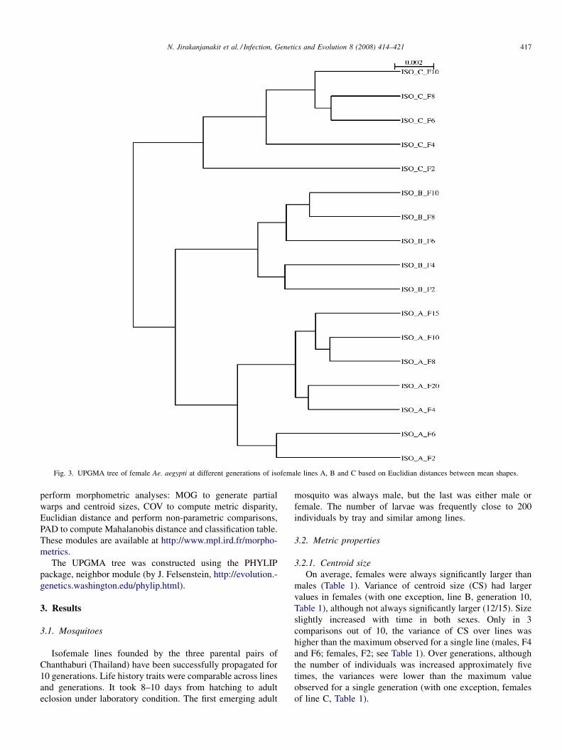
Fig. 3. UPGMA tree of female Ae. aegypti at different generations of isofemale lines A, B and C based on Euclidian distances between mean shapes.
N. Jirakanjanakit et al. / Infection, Genetics and Evolution 8 (2008) 414–421 417
perform morphometric analyses: MOG to generate partial
warps and centroid sizes, COV to compute metric disparity,
Euclidian distance and perform non-parametric comparisons,
PAD to compute Mahalanobis distance and classification table.
These modules are available at http://www.mpl.ird.fr/morpho-
metrics.
The UPGMA tree was constructed using the PHYLIP
package, neighbor module (by J. Felsenstein, http://evolution.-
genetics.washington.edu/phylip.html).
3. Results
3.1. Mosquitoes
Isofemale lines founded by the three parental pairs of
Chanthaburi (Thailand) have been successfully propagated for
10 generations. Life history traits were comparable across lines
and generations. It took 8–10 days from hatching to adult
eclosion under laboratory condition. The first emerging adult
mosquito was always male, but the last was either male or
female. The number of larvae was frequently close to 200
individuals by tray and similar among lines.
3.2. Metric properties
3.2.1. Centroid size
On average, females were always significantly larger than
males (Table 1). Variance of centroid size (CS) had larger
values in females (with one exception, line B, generation 10,
Table 1), although not always significantly larger (12/15). Size
slightly increased with time in both sexes. Only in 3
comparisons out of 10, the variance of CS over lines was
higher than the maximum observed for a single line (males, F4
and F6; females, F2; see Table 1). Over generations, although
the number of individuals was increased approximately five
times, the variances were lower than the maximum value
observed for a single generation (with one exception, females
of line C, Table 1).
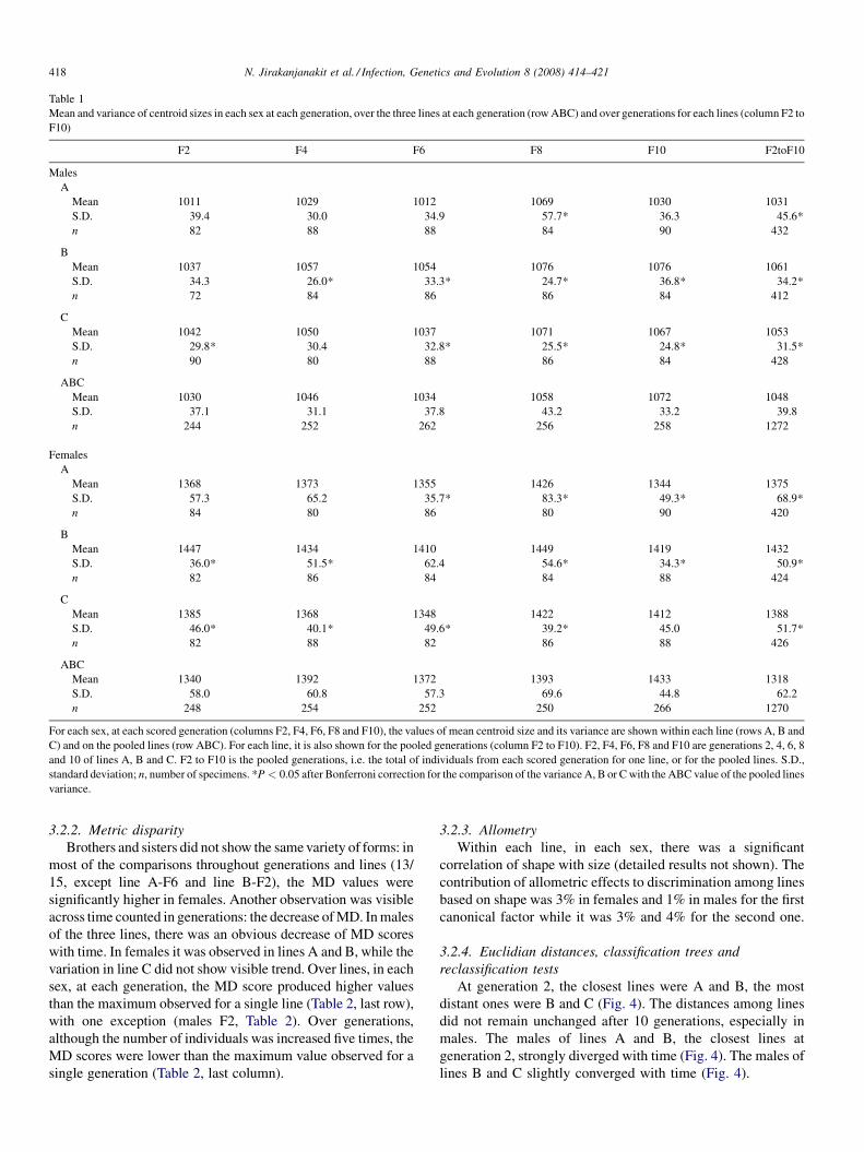
Table 1
Mean and variance of centroid sizes in each sex at each generation, over the three lines at each generation (row ABC) and over generations for each lines (column F2 to
F10)
F2 F4 F6 F8 F10 F2toF10
Males
A
Mean 1011 1029 1012 1069 1030 1031
S.D. 39.4 30.0 34.9 57.7* 36.3 45.6*
n 82 88 88 84 90 432
B
Mean 1037 1057 1054 1076 1076 1061
S.D. 34.3 26.0* 33.3* 24.7* 36.8* 34.2*
n 72 84 86 86 84 412
C
Mean 1042 1050 1037 1071 1067 1053
S.D. 29.8* 30.4 32.8* 25.5* 24.8* 31.5*
n 90 80 88 86 84 428
ABC
Mean 1030 1046 1034 1058 1072 1048
S.D. 37.1 31.1 37.8 43.2 33.2 39.8
n 244 252 262 256 258 1272
Females
A
Mean 1368 1373 1355 1426 1344 1375
S.D. 57.3 65.2 35.7* 83.3* 49.3* 68.9*
n 84 80 86 80 90 420
B
Mean 1447 1434 1410 1449 1419 1432
S.D. 36.0* 51.5* 62.4 54.6* 34.3* 50.9*
n 82 86 84 84 88 424
C
Mean 1385 1368 1348 1422 1412 1388
S.D. 46.0* 40.1* 49.6* 39.2* 45.0 51.7*
n 82 88 82 86 88 426
ABC
Mean 1340 1392 1372 1393 1433 1318
S.D. 58.0 60.8 57.3 69.6 44.8 62.2
n 248 254 252 250 266 1270
For each sex, at each scored generation (columns F2, F4, F6, F8 and F10), the values of mean centroid size and its variance are shown within each line (rows A, B and
C) and on the pooled lines (row ABC). For each line, it is also shown for the pooled generations (column F2 to F10). F2, F4, F6, F8 and F10 are generations 2, 4, 6, 8
and 10 of lines A, B and C. F2 to F10 is the pooled generations, i.e. the total of individuals from each scored generation for one line, or for the pooled lines. S.D.,
standard deviation; n, number of specimens. *P < 0.05 after Bonferroni correction for the comparison of the variance A, B or C with the ABC value of the pooled lines
variance.
N. Jirakanjanakit et al. / Infection, Genetics and Evolution 8 (2008) 414–421418
3.2.2. Metric disparity
Brothers and sisters did not show the same variety of forms: in
most of the comparisons throughout generations and lines (13/
15, except line A-F6 and line B-F2), the MD values were
significantly higher in females. Another observation was visible
across time counted in generations: the decrease of MD. In males
of the three lines, there was an obvious decrease of MD scores
with time. In females it was observed in lines A and B, while the
variation in line C did not show visible trend. Over lines, in each
sex, at each generation, the MD score produced higher values
than the maximum observed for a single line (Table 2, last row),
with one exception (males F2, Table 2). Over generations,
although the number of individuals was increased five times, the
MD scores were lower than the maximum value observed for a
single generation (Table 2, last column).
3.2.3. Allometry
Within each line, in each sex, there was a significant
correlation of shape with size (detailed results not shown). The
contribution of allometric effects to discrimination among lines
based on shape was 3% in females and 1% in males for the first
canonical factor while it was 3% and 4% for the second one.
3.2.4. Euclidian distances, classification trees and
reclassification tests
At generation 2, the closest lines were A and B, the most
distant ones were B and C (Fig. 4). The distances among lines
did not remain unchanged after 10 generations, especially in
males. The males of lines A and B, the closest lines at
generation 2, strongly diverged with time (Fig. 4). The males of
lines B and C slightly converged with time (Fig. 4).
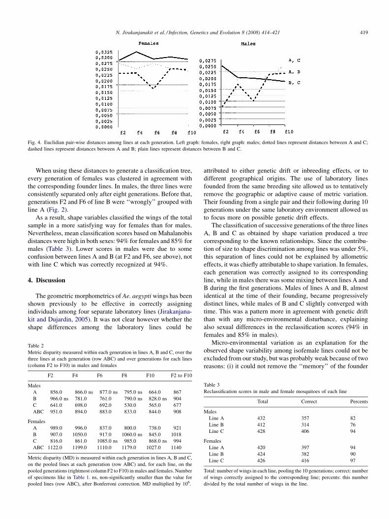
Fig. 4. Euclidian pair-wise distances among lines at each generation. Left graph: females, right graph: males; dotted lines represent distances between A and C;
dashed lines represent distances between A and B; plain lines represent distances between B and C.
N. Jirakanjanakit et al. / Infection, Genetics and Evolution 8 (2008) 414–421 419
When using these distances to generate a classification tree,
every generation of females was clustered in agreement with
the corresponding founder lines. In males, the three lines were
consistently separated only after eight generations. Before that,
generations F2 and F6 of line B were ‘‘wrongly’’ grouped with
line A (Fig. 2).
As a result, shape variables classified the wings of the total
sample in a more satisfying way for females than for males.
Nevertheless, mean classification scores based on Mahalanobis
distances were high in both sexes: 94% for females and 85% for
males (Table 3). Lower scores in males were due to some
confusion between lines A and B (at F2 and F6, see above), not
with line C which was correctly recognized at 94%.
4. Discussion
The geometric morphometrics of Ae. aegypti wings has been
shown previously to be effective in correctly assigning
individuals among four separate laboratory lines (Jirakanjana-
kit and Dujardin, 2005). It was not clear however whether the
shape differences among the laboratory lines could be
Table 2
Metric disparity measured within each generation in lines A, B and C, over the
three lines at each generation (row ABC) and over generations for each lines
(column F2 to F10) in males and females
F2 F4 F6 F8 F10 F2 to F10
Males
A 856.0 866.0 ns 877.0 ns 795.0 ns 664.0 867
B 966.0 ns 781.0 761.0 790.0 ns 828.0 ns 904
C 641.0 698.0 692.0 530.0 565.0 677
ABC 951.0 894.0 883.0 833.0 844.0 908
Females
A 989.0 996.0 837.0 800.0 738.0 921
B 907.0 1050.0 917.0 1060.0 ns 845.0 1018
C 816.0 861.0 1085.0 ns 985.0 868.0 ns 994
ABC 1122.0 1199.0 1110.0 1179.0 1027.0 1140
Metric disparity (MD) is measured within each generation in lines A, B and C,
on the pooled lines at each generation (row ABC) and, for each line, on the
pooled generations (rightmost column F2 to F10) in males and females. Number
of specimens like in Table 1. ns, non-significantly smaller than the value for
pooled lines (row ABC), after Bonferroni correction. MD multiplied by 106.
attributed to either genetic drift or inbreeding effects, or to
different geographical origins. The use of laboratory lines
founded from the same breeding site allowed us to tentatively
remove the geographic or adaptive cause of metric variation.
Their founding from a single pair and their following during 10
generations under the same laboratory environment allowed us
to focus more on possible genetic drift effects.
The classification of successive generations of the three lines
A, B and C as obtained by shape variation produced a tree
corresponding to the known relationships. Since the contribu-
tion of size to shape discrimination among lines was under 5%,
this separation of lines could not be explained by allometric
effects, it was chiefly attributable to shape variation. In females,
each generation was correctly assigned to its corresponding
line, while in males there was some mixing between lines A and
B during the first generations. Males of lines A and B, almost
identical at the time of their founding, became progressively
distinct lines, while males of B and C slightly converged with
time. This was a pattern more in agreement with genetic drift
than with any micro-environmental disturbance, explaining
also sexual differences in the reclassification scores (94% in
females and 85% in males).
Micro-environmental variation as an explanation for the
observed shape variability among isofemale lines could not be
excluded from our study, but was probably weak because of two
reasons: (i) it could not remove the ‘‘memory’’ of the founder
Table 3
Reclassification scores in male and female mosquitoes of each line
Total Correct Percents
Males
Line A 432 357 82
Line B 412 314 76
Line C 428 406 94
Females
Line A 420 397 94
Line B 424 382 90
Line C 426 416 97
Total: number of wings in each line, pooling the 10 generations; correct: number
of wings correctly assigned to the corresponding line; percents: this number
divided by the total number of wings in the line.
N. Jirakanjanakit et al. / Infection, Genetics and Evolution 8 (2008) 414–421420
after 10 generations and (ii) at each generation, several trays
were used to produce the progeny of each isofemale line (David
et al., 2005). On the other hand, according to many studies
performed on Drosophila, such micro-environmental contribu-
tion (the ‘‘common environmental effect’’, or CEE) is likely to
be low (David et al., 2005).
In agreement with these considerations, our data provided
additional arguments for a genetic basis of shape variation by
examining the variance of metric properties.
If individuals are added having similar morphotypes to the
existing ones, even if they are many, the variance of metric
properties is not expected to increase. Within a given line
founded by a single parental pair, there is no reason a priori for
new genetic variants to appear in 10 generations. On the
contrary, due to genetic drift, genetic variation is expected to
decrease. Thus, combining 10 generations of a given line into a
single group, the resulting variance of centroid size or MD
score was expected to show values similar as or lower than
values scored for a single generation. Our results were in
agreement with that expectation (Tables 1 and 2, rightmost
column).
If individuals added to a given sample represent new
morphotypes, even if they are very few, the metric variance of
this sample will increase. We thus expected the metric variation
computed from pooled lines to show significantly higher values
than for single lines. This was confirmed at each generation for
shape variation (with 25/36 values found significantly higher,
Table 2, rows ABC), much less so for size variation (only 5/36
values significantly higher, Table 1, rows ABC).
Contrary to what was suggested previously (Jirakanjanakit
and Dujardin, 2005), size did not show here any trend of
decreasing with time; it actually slightly increased with time.
However, the time is not comparable in both studies: size was
shown to decrease after more than 17–564 generations
(Jirakanjanakit and Dujardin, 2005), while we examined only
10 generations. This should be verified, as the previous results
could actually be reflecting different micro-environmental
conditions (different laboratories) instead of true inbreeding
effect.
Females were consistently larger than males (Table 1) and
they showed larger variance of size (Table 1) and also of shape
(Table 2). Since the variance of size was often (12/15) larger in
females, the sexual dimorphism observed for metric disparity
could partially be explained by an allometric effect. Actually, a
significant correlation was found on the total sample between
MD scores and mean values and/or variances of size
(determination coefficients 30% and 18%, respectively).
Nevertheless, no such correlation could be detected within
each sex.
Thus, shape showed more consistent evidence for genetic
determinism than size. Notably, it was able to show patterns
suggestive of genetic drift under the same laboratory
environment, allowing to distinguish individuals belonging
to recently isolated groups. Our data suggest that the metric
disparity index could be used in natural conditions to evaluate
the relative complexity of collecting sites. They also confirmed
that the wing shape could be used to monitor laboratory lines
(Jirakanjanakit and Dujardin, 2005). However, the present
demonstration does not mean that shape can be used on natural
populations to assess their current level of isolation. Natural
populations are not genetically as simple as isofemale lines, and
many factors – in addition to isolation and/or bottlenecks – are
liable to affect the development of the insect, hence its final
shape, exaggerating or blurring its expected relationship to
isolation levels.
Acknowledgements
The authors gratefully thank Dr. Christian Bellec (IRD-DSF
program) on the support for training, kind suggestion and
encouragement. Many thanks to Napaporn Kuatrakool and
Sadanun Boonsatien for their help in mosquito rearing and wing
slides preparation, and to Pornpimol Rongnaparut (Mahidol
University, Department of Biochemistry) for critical reading.
This research work was supported by Faculty of Graduate
Studies, Mahidol University, academic year 2006.
References
Bennett, K.E., Flick, D., Fleming, K.H., Jochim, R., Beaty, B.J., Black, W.C.,
2005. Quantitative trait loci that control dengue-2 virus dissemination in the
mosquito Aedes aegypti. Gene 170, 185–194.
Bookstein, F.L., 1991. Morphometric Tools for Landmark Data: Geometry and
Biology. Cambridge University Press, Cambridge, 435 pp.
Brogdon, W.G., McAllister, J.C., Corwin, A.M., Cordon-Rosales, C., 1999.
Independent selection of multiple mechanisms for pyrethroid resistance in
Guatemalan Anopheles albimanus (Diptera: Culicidae). J. Econ. Entomol.
92, 298–302.
Christophers, S.R., 1960. Aedes aegypti (L.). The Yellow Fever Mosquito.
Cambridge University Press, London, 739 pp.
Cook, S., Diallo, M., Sall, A.A., Cooper, A., Holmes, E.C., 2005. Mitochondrial
markers for molecular identification of Aedes mosquitoes (Diptera: Culi-
cidae) involved in transmission of arboviral disease in West Africa. J. Med.
Entomol. 42, 19–28.
David, J.R., Gibert, P., Legout, H., Petavy, G., Morreteau, B., 2005. Review:
isofemale line in Drosophila: an empirical approach to quantitative trait
analysis in natural population. Heredity 94, 3–12.
Ganesh, K.N., Vijayan, V.A., Urmila, J., Gopalan, N., Prakash, S., 2002. Role
of esterases and monooxygenase in the deltamethrin resistance in Ano-
pheles stephensi Giles (1908), at Mysore. Indian J. Exp. Biol. 40, 583–
588.
Good, P., 2000. Permutation Tests: A Practical Guide to Re-sampling Methods
for Testing Hypotheses. Springer, New York.
Jirakanjanakit, N., Dujardin, J.P., 2005. Discrimination of Aedes aegypti
(diptera; culicidae) laboratory lines based on wing geometry. Southeast
Asian J. Trop. Med. Public Health 36, 858–861.
Miller, B.R., Mitchell, C.J., 1991. Genetic selection of a flavivirus-refractory
strain of the yellow fever mosquito Aedes aegypti. Am. J. Trop. Med. Hyg.
45, 399–407.
Mourya, D.T., Gokhale, Basu, A., Barde, P.V., Sapkal, G.N., Padbidri, V.S.,
Gore, M.M., 2001. Horizontal and vertical transmission of dengue virus
type 2 in highly and lowly susceptible strains of Aedes aegypti mosquitoes.
Acta Virol. 45, 67–71.
Rodhain, F., 1996. Ecology of Aedes aegypti in Africa and Asia. Bull. Soc.
Pathol. Exot. 89, 103–106.
Rohlf, F.J., 1990. Rotational fit (Procrustes) methods. In: Rohlf, F.J., Bookstein,
F.L. (Eds.), Proceedings of the Michigan Morphometrics Workshop. Special
Publication Number 2. The University of Michigan Museums, Ann Arbor,
MI, pp. 227–236.
Rohlf, F.J., 1993. Morphometric spaces, shape components and the effects of
linear transformations. In: Marcus, L.F., Corti, M., Loy, A., Naylor,

N. Jirakanjanakit et al. / Infection, Genetics and Evolution 8 (2008) 414–421 421
G.J.P., Slice, D. (Eds.), Advances in Morphometrics, Proceedings of the
1993 NATO-ASI on Morphometrics, NATO ASI, Series A, Life Sciences.
Plenum Publ., New York, pp. 117–129.
Rohlf, F.J., 2003. TpsDig ver 1. 34. Available at http://morph.bio.sunysb.edu./
morph/index/html. Department of Ecology and Evolution, State University
of New York.
Tardieux, I., Poupel, O., Lapchin, L., Rodhain, F., 1991. Analysis of inheritance
of oral susceptibility of Aedes aegypti (Diptera: Culicidae) to dengue-2 virus
using isofemale lines. J. Med. Entomol. 28, 518–521.
Zelditch, M.L., Donald, L., Swiderski, H., Sheets, D., Fink, W.L., 2004.
Geometric Morphometrics for Biologists: A Primer. Elsevier/Academic
Press, Paris, 443 pp.
www.elsevier.com/locate/meegid
Infection, Genetics and Evolution 8 (2008) 422–432
Using a climate-dependent model to predict mosquito abundance:
Application to Aedes (Stegomyia) africanus and
Aedes (Diceromyia) furcifer (Diptera: Culicidae)
Brigitte Schaeffer a,*, Bernard Mondet b, Suzanne Touzeau a
a INRA, UR341 Mathematiques et Informatique Appliquees, F-78350 Jouy-en-Josas Cedex, Franceb Institut Francais de Pondichery, CP 33, 11 Saint-Louis Street, Pondicherry 605001, India
Received 15 February 2007; received in revised form 2 July 2007; accepted 5 July 2007
Available online 10 July 2007
Abstract
Mosquitoes, acting as vectors, are involved in the transmission of viruses. Thus, their abundances, which strongly depend on the weather and
environment, are closely linked to major disease outbreaks. The aim of this paper is to provide a tool to predict vector abundance.
In order to describe the dynamics of mosquito populations, we developed a matrix model integrating climate fluctuations. The population is
structured in five stages: two egg stages (immature and mature), one larval stage and two female flying stages (nulliparous and parous). The water
availability in breeding sites was considered as the main environmental factor affecting the mosquito life-cycle. Thus, the model represents the
evolution of the mosquito abundance in each stage over time, in connection with water availability.
The model was used to simulate the abundance trends over 3 years of two mosquito species, Aedes africanus (Theobald) and Aedes furcifer
(Edwards), vectors of the yellow fever virus in Ivory Coast. As both these species breed in tree holes, the water dynamics in the tree hole was
reproduced from daily rainfall data. The results we obtained showed a good match between the simulated populations and the field data over the
time period considered.
# 2007 Elsevier B.V. All rights reserved.
Keywords: Mosquito vector population; Mathematical model; Climate dependency
1. Introduction
Yellow fever, a vector-borne disease, remains a major public
health problem both in Africa and South America. It is due to
infection by a Flavivirus and causes a viral hemorrhagic fever in
humans responsible for 30,000 deaths annually (Tomori, 2004).
Furthermore, global warming may increase the outbreak risk of
such infectious diseases associated with hot weather or rainfall
pattern modifications. Chevalier et al. (2004) showed that
geographical distributions of several arthropod-borne zoonoses
have dramatically expanded over the last years. Although one
knows that climate influences the abundance of vectors, it is
still difficult to appreciate the effect of climate variations on the
* Corresponding author at: INRA, Unite de Mathematiques & Informatique
Appliquees, Centre de Recherche de Jouy-en-Josas, 78352 Jouy-en-Josas
Cedex, France. Tel.: +33 1 34 65 22 18; fax: +33 1 34 65 22 17.
E-mail address: [email protected] (B. Schaeffer).
1567-1348/$ – see front matter # 2007 Elsevier B.V. All rights reserved.
doi:10.1016/j.meegid.2007.07.002
emergence and re-emergence of arbovirus diseases. That is
mainly because the epidemiological process is driven by
numerous tangled mechanisms, making analyses fairly com-
plex. Breaking up the process and focusing on its different
elements, in particular on the biology of the vectors which is the
key of the virus transmission, might help to apprehend the
global process.
Mathematical models combined with computer simulations
are powerful tools for describing and understanding complex
biological phenomena. Indeed, modelling allows to test
assumptions, explain some events, or compare alternate
strategies in decision-making. Thus, models have been widely
used for studying complex biological processes such as
population dynamics (e.g. Jarry et al., 1996; Ghosh and
Pugliese, 2004; Awerbuch-Friedlander et al., 2005; Mazaris
and Matsinos, 2006). For mosquito populations in particular,
Moon (1976) developed a dynamic model, but that model did
not consider environmental factors. Focks et al. (1993)
proposed a dynamic life table model for Aedes aegypti taking
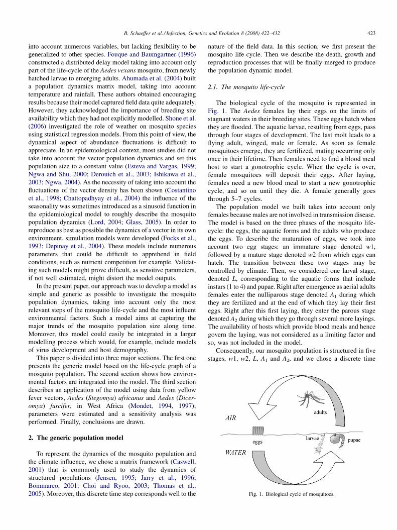
Fig. 1. Biological cycle of mosquitoes.
B. Schaeffer et al. / Infection, Genetics and Evolution 8 (2008) 422–432 423
into account numerous variables, but lacking flexibility to be
generalized to other species. Fouque and Baumgartner (1996)
constructed a distributed delay model taking into account only
part of the life-cycle of the Aedes vexans mosquito, from newly
hatched larvae to emerging adults. Ahumada et al. (2004) built
a population dynamics matrix model, taking into account
temperature and rainfall. These authors obtained encouraging
results because their model captured field data quite adequately.
However, they acknowledged the importance of breeding site
availability which they had not explicitly modelled. Shone et al.
(2006) investigated the role of weather on mosquito species
using statistical regression models. From this point of view, the
dynamical aspect of abundance fluctuations is difficult to
appreciate. In an epidemiological context, most studies did not
take into account the vector population dynamics and set this
population size to a constant value (Esteva and Vargas, 1999;
Ngwa and Shu, 2000; Derouich et al., 2003; Ishikawa et al.,
2003; Ngwa, 2004). As the necessity of taking into account the
fluctuations of the vector density has been shown (Costantino
et al., 1998; Chattopadhyay et al., 2004) the influence of the
seasonality was sometimes introduced as a sinusoid function in
the epidemiological model to roughly describe the mosquito
population dynamics (Lord, 2004; Glass, 2005). In order to
reproduce as best as possible the dynamics of a vector in its own
environment, simulation models were developed (Focks et al.,
1993; Depinay et al., 2004). These models include numerous
parameters that could be difficult to apprehend in field
conditions, such as nutrient competition for example. Validat-
ing such models might prove difficult, as sensitive parameters,
if not well estimated, might distort the model outputs.
In the present paper, our approach was to develop a model as
simple and generic as possible to investigate the mosquito
population dynamics, taking into account only the most
relevant steps of the mosquito life-cycle and the most influent
environmental factors. Such a model aims at capturing the
major trends of the mosquito population size along time.
Moreover, this model could easily be integrated in a larger
modelling process which would, for example, include models
of virus development and host demography.
This paper is divided into three major sections. The first one
presents the generic model based on the life-cycle graph of a
mosquito population. The second section shows how environ-
mental factors are integrated into the model. The third section
describes an application of the model using data from yellow
fever vectors, Aedes (Stegomya) africanus and Aedes (Dicer-
omya) furcifer, in West Africa (Mondet, 1994, 1997);
parameters were estimated and a sensitivity analysis was
performed. Finally, conclusions are drawn.
2. The generic population model
To represent the dynamics of the mosquito population and
the climate influence, we chose a matrix framework (Caswell,
2001) that is commonly used to study the dynamics of
structured populations (Jensen, 1995; Jarry et al., 1996;
Bommarco, 2001; Choi and Ryoo, 2003; Thomas et al.,
2005). Moreover, this discrete time step corresponds well to the
nature of the field data. In this section, we first present the
mosquito life-cycle. Then we describe the death, growth and
reproduction processes that will be finally merged to produce
the population dynamic model.
2.1. The mosquito life-cycle
The biological cycle of the mosquito is represented in
Fig. 1. The Aedes females lay their eggs on the limits of
stagnant waters in their breeding sites. These eggs hatch when
they are flooded. The aquatic larvae, resulting from eggs, pass
through four stages of development. The last molt leads to a
flying adult, winged, male or female. As soon as female
mosquitoes emerge, they are fertilized, mating occurring only
once in their lifetime. Then females need to find a blood meal
host to start a gonotrophic cycle. When the cycle is over,
female mosquitoes will deposit their eggs. After laying,
females need a new blood meal to start a new gonotrophic
cycle, and so on until they die. A female generally goes
through 5–7 cycles.
The population model we built takes into account only
females because males are not involved in transmission disease.
The model is based on the three phases of the mosquito life-
cycle: the eggs, the aquatic forms and the adults who produce
the eggs. To describe the maturation of eggs, we took into
account two egg stages: an immature stage denoted w1,
followed by a mature stage denoted w2 from which eggs can
hatch. The transition between these two stages may be
controlled by climate. Then, we considered one larval stage,
denoted L, corresponding to the aquatic forms that include
instars (1 to 4) and pupae. Right after emergence as aerial adults
females enter the nulliparous stage denoted A1 during which
they are fertilized and at the end of which they lay their first
eggs. Right after this first laying, they enter the parous stage
denoted A2 during which they go through several more layings.
The availability of hosts which provide blood meals and hence
govern the laying, was not considered as a limiting factor and
so, was not included in the model.
Consequently, our mosquito population is structured in five
stages, w1, w2, L, A1 and A2, and we chose a discrete time
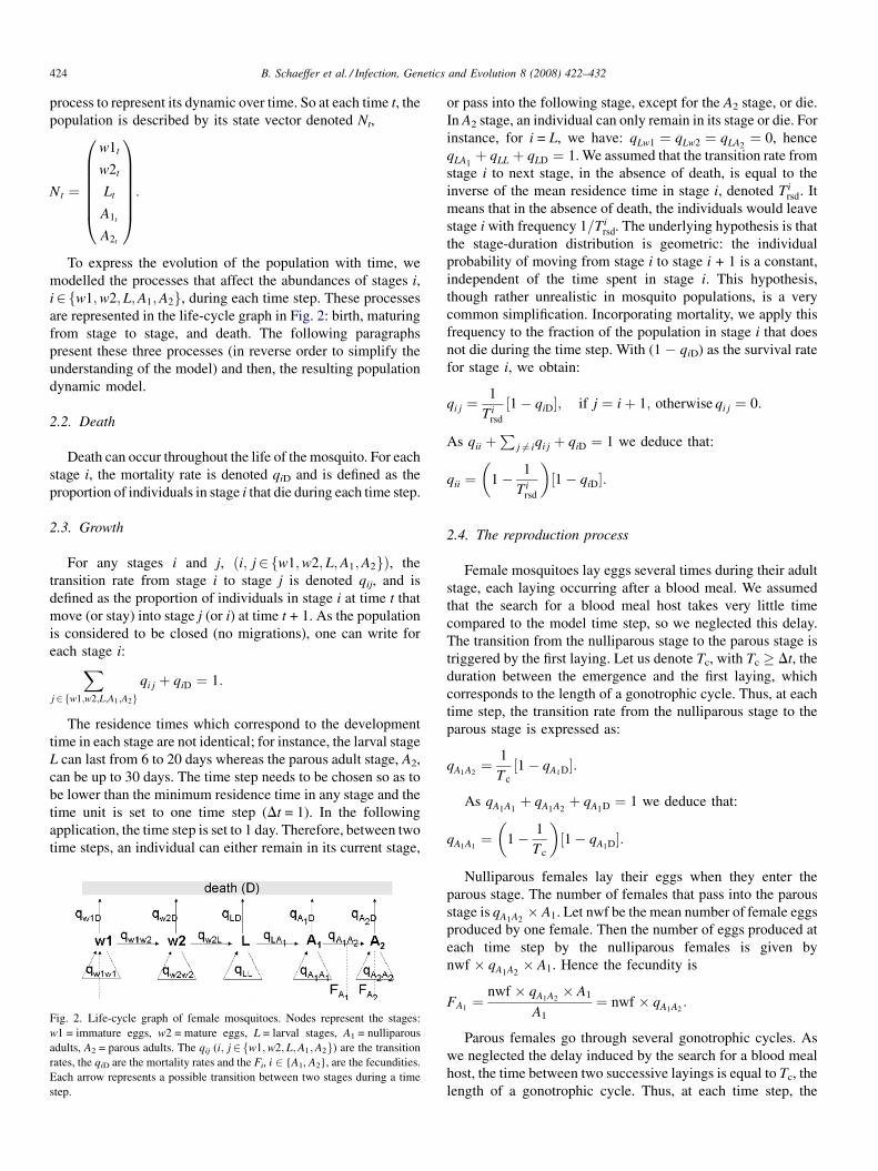
B. Schaeffer et al. / Infection, Genetics and Evolution 8 (2008) 422–432424
process to represent its dynamic over time. So at each time t, the
population is described by its state vector denoted Nt,
Nt ¼
w1t
w2t
Lt
A1t
A2t
0BBBBBB@
1CCCCCCA:
To express the evolution of the population with time, we
modelled the processes that affect the abundances of stages i,
i2fw1;w2; L;A1;A2g, during each time step. These processes
are represented in the life-cycle graph in Fig. 2: birth, maturing
from stage to stage, and death. The following paragraphs
present these three processes (in reverse order to simplify the
understanding of the model) and then, the resulting population
dynamic model.
2.2. Death
Death can occur throughout the life of the mosquito. For each
stage i, the mortality rate is denoted qiD and is defined as the
proportion of individuals in stage i that die during each time step.
2.3. Growth
For any stages i and j, ði; j2fw1;w2; L;A1;A2gÞ, the
transition rate from stage i to stage j is denoted qij, and is
defined as the proportion of individuals in stage i at time t that
move (or stay) into stage j (or i) at time t + 1. As the population
is considered to be closed (no migrations), one can write for
each stage i:Xj2fw1;w2;L;A1;A2g
qi j þ qiD ¼ 1:
The residence times which correspond to the development
time in each stage are not identical; for instance, the larval stage
L can last from 6 to 20 days whereas the parous adult stage, A2,
can be up to 30 days. The time step needs to be chosen so as to
be lower than the minimum residence time in any stage and the
time unit is set to one time step (Dt = 1). In the following
application, the time step is set to 1 day. Therefore, between two
time steps, an individual can either remain in its current stage,
Fig. 2. Life-cycle graph of female mosquitoes. Nodes represent the stages:
w1 = immature eggs, w2 = mature eggs, L = larval stages, A1 = nulliparous
adults, A2 = parous adults. The qij (i; j2fw1;w2;L;A1;A2g) are the transition
rates, the qiD are the mortality rates and the Fi, i 2 {A1, A2}, are the fecundities.
Each arrow represents a possible transition between two stages during a time
step.
or pass into the following stage, except for the A2 stage, or die.
In A2 stage, an individual can only remain in its stage or die. For
instance, for i = L, we have: qLw1 ¼ qLw2 ¼ qLA2¼ 0, hence
qLA1þ qLL þ qLD ¼ 1. We assumed that the transition rate from
stage i to next stage, in the absence of death, is equal to the
inverse of the mean residence time in stage i, denoted Tirsd. It
means that in the absence of death, the individuals would leave
stage i with frequency 1=Tirsd. The underlying hypothesis is that
the stage-duration distribution is geometric: the individual
probability of moving from stage i to stage i + 1 is a constant,
independent of the time spent in stage i. This hypothesis,
though rather unrealistic in mosquito populations, is a very
common simplification. Incorporating mortality, we apply this
frequency to the fraction of the population in stage i that does
not die during the time step. With (1 � qiD) as the survival rate
for stage i, we obtain:
qi j ¼1
Tirsd
½1� qiD�; if j ¼ iþ 1; otherwise qi j ¼ 0:
As qii þP
j 6¼ iqi j þ qiD ¼ 1 we deduce that:
qii ¼�
1� 1
Tirsd
�½1� qiD�:
2.4. The reproduction process
Female mosquitoes lay eggs several times during their adult
stage, each laying occurring after a blood meal. We assumed
that the search for a blood meal host takes very little time
compared to the model time step, so we neglected this delay.
The transition from the nulliparous stage to the parous stage is
triggered by the first laying. Let us denote Tc, with Tc � Dt, the
duration between the emergence and the first laying, which
corresponds to the length of a gonotrophic cycle. Thus, at each
time step, the transition rate from the nulliparous stage to the
parous stage is expressed as:
qA1A2¼ 1
Tc
½1� qA1D�:
As qA1A1þ qA1A2
þ qA1D ¼ 1 we deduce that:
qA1A1¼�
1� 1
Tc
�½1� qA1D�:
Nulliparous females lay their eggs when they enter the
parous stage. The number of females that pass into the parous
stage is qA1A2� A1. Let nwf be the mean number of female eggs
produced by one female. Then the number of eggs produced at
each time step by the nulliparous females is given by
nwf � qA1A2� A1. Hence the fecundity is
FA1¼
nwf � qA1A2� A1
A1
¼ nwf � qA1A2:
Parous females go through several gonotrophic cycles. As
we neglected the delay induced by the search for a blood meal
host, the time between two successive layings is equal to Tc, the
length of a gonotrophic cycle. Thus, at each time step, the
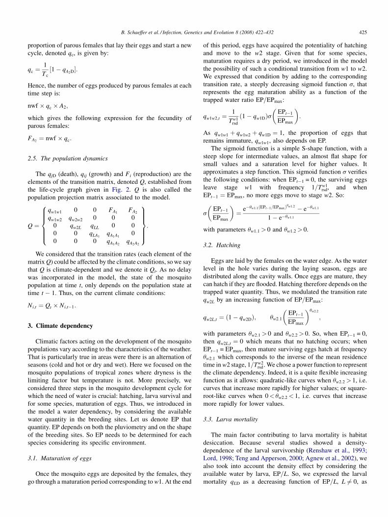
B. Schaeffer et al. / Infection, Genetics and Evolution 8 (2008) 422–432 425
proportion of parous females that lay their eggs and start a new
cycle, denoted qc, is given by:
qc ¼1
Tc
½1� qA2D�:
Hence, the number of eggs produced by parous females at each
time step is:
nwf � qc � A2;
which gives the following expression for the fecundity of
parous females:
FA2¼ nwf � qc:
2.5. The population dynamics
The qjD (death), qij (growth) and Fi (reproduction) are the
elements of the transition matrix, denoted Q, established from
the life-cycle graph given in Fig. 2. Q is also called the
population projection matrix associated to the model.
Q ¼
qw1w1 0 0 FA1FA2
qw1w2 qw2w2 0 0 0
0 qw2L qLL 0 0
0 0 qLA1qA1A1
0
0 0 0 qA1A2qA2A2
8>>>><>>>>:
9>>>>=>>>>;:
We considered that the transition rates (each element of the
matrix Q) could be affected by the climate conditions, so we say
that Q is climate-dependent and we denote it Qt. As no delay
was incorporated in the model, the state of the mosquito
population at time t, only depends on the population state at
time t � 1. Thus, on the current climate conditions:
Ni;t ¼ Qt � Ni;t�1:
3. Climate dependency
Climatic factors acting on the development of the mosquito
populations vary according to the characteristics of the weather.
That is particularly true in areas were there is an alternation of
seasons (cold and hot or dry and wet). Here we focused on the
mosquito populations of tropical zones where dryness is the
limiting factor but temperature is not. More precisely, we
considered three steps in the mosquito development cycle for
which the need of water is crucial: hatching, larva survival and
for some species, maturation of eggs. Thus, we introduced in
the model a water dependency, by considering the available
water quantity in the breeding sites. Let us denote EP that
quantity. EP depends on both the pluviometry and on the shape
of the breeding sites. So EP needs to be determined for each
species considering its specific environment.
3.1. Maturation of eggs
Once the mosquito eggs are deposited by the females, they
go through a maturation period corresponding to w1. At the end
of this period, eggs have acquired the potentiality of hatching
and move to the w2 stage. Given that for some species,
maturation requires a dry period, we introduced in the model
the possibility of such a conditional transition from w1 to w2.
We expressed that condition by adding to the corresponding
transition rate, a steeply decreasing sigmoid function s, that
represents the egg maturation ability as a function of the
trapped water ratio EP=EPmax:
qw1w2;t ¼1
Tw1rsd
ð1� qw1DÞs�
EPt�1
EPmax
�:
As qw1w1 þ qw1w2 þ qw1D ¼ 1, the proportion of eggs that
remains immature, qw1w1, also depends on EP.
The sigmoid function is a simple S-shape function, with a
steep slope for intermediate values, an almost flat shape for
small values and a saturation level for higher values. It
approximates a step function. This sigmoid function s verifies
the following conditions: when EPt�1 = 0, the surviving eggs
leave stage w1 with frequency 1=Tw1rsd, and when
EPt�1 ¼ EPmax, no more eggs move to stage w2. So:
s
�EPt�1
EPmax
�¼ e�uw1:1ðEPt�1=EPmaxÞuw1:2 � e�uw1:1
1� e�uw1:1
with parameters uw1:1 > 0 and uw1:2 > 0.
3.2. Hatching
Eggs are laid by the females on the water edge. As the water
level in the hole varies during the laying season, eggs are
distributed along the cavity walls. Once eggs are mature, they
can hatch if they are flooded. Hatching therefore depends on the
trapped water quantity. Thus, we modulated the transition rate
qw2L by an increasing function of EP=EPmax:
qw2L;t ¼ ð1� qw2DÞ; uw2:1
�EPt�1
EPmax
�uw2:2
;
with parameters uw2:1 > 0 and uw2:2 > 0. So, when EPt�1 = 0,
then qw2L;t ¼ 0 which means that no hatching occurs; when
EPt�1 = EPmax, then mature surviving eggs hatch at frequency
uw2:1 which corresponds to the inverse of the mean residence
time in w2 stage, 1=Tw2rsd . We chose a power function to represent
the climate dependency. Indeed, it is a quite flexible increasing
function as it allows: quadratic-like curves when uw2:2 > 1, i.e.
curves that increase more rapidly for higher values; or square-
root-like curves when 0< uw2:2 < 1, i.e. curves that increase
more rapidly for lower values.
3.3. Larva mortality
The main factor contributing to larva mortality is habitat
desiccation. Because several studies showed a density-
dependence of the larval survivorship (Renshaw et al., 1993;
Lord, 1998; Teng and Apperson, 2000; Agnew et al., 2002), we
also took into account the density effect by considering the
available water by larva, EP=L. So, we expressed the larval
mortality qLD as a decreasing function of EP=L, L 6¼ 0, as
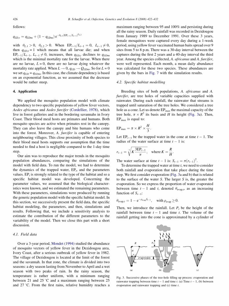
Fig. 3. Successive phases of the tree-hole filling up process: evaporation and
rainwater trapping between time t � 1 and time t. (a) Time t � 1, (b) between
evaporation and rainwater trapping and (c) time t.
B. Schaeffer et al. / Infection, Genetics and Evolution 8 (2008) 422–432426
follows:
qLD;t ¼ qLDm þ ð1� qLDmÞ e�uL:1ðEPt�1=Lt�1ÞuL:2
with uL.1 > 0, uL.2 > 0. When EPt�1=Lt�1 ¼ 0, Lt�1 6¼ 0,
then qLD,t = 1 which means that all larvae die; and when
EPt�1=Lt�1, Lt�1 6¼ 0, increases, then qLD,t declines to qLDm
which is the minimal mortality rate for the larvae. When there
are no larvae, L = 0, there are no larvae dying whatever the
mortality rate applied. When L! 0, qLD! QLDm. So for L = 0
we set qLD = qLDm. In this case, the climate dependency is based
on an exponential function, as we assumed that the decrease
would be rather steep.
4. Application
We applied the mosquito population model with climate
dependency to two specific populations of yellow fever vectors,
Aedes africanus and Aedes furcifer (Cordellier, 1978), which
live in forest galleries and in the bordering savannahs in Ivory
Coast. Their blood meal hosts are primates and humans. Both
mosquito species are active when primates rest in the canopy.
They can also leave the canopy and bite humans who come
into the forest. Moreover, A. furcifer is capable of entering
neighbouring villages. This close proximity of both species to
their blood meal hosts supports our assumption that the time
needed to find a host is negligible compared to the 1-day time
step.
Our aim was to reproduce the major trends in the mosquito
population abundances, comparing the simulations of the
model with field data. To run the model, we had to determine
the dynamics of the trapped water, EPt, and the parameters
values. EPt is strongly related to the type of the habitat and so a
specific habitat model was developed. Concerning the
parameter values, we assumed that the biological character-
istics were known, and we estimated the remaining parameters.
With these parameters, simulations were produced by running
the generic population model with the specific habitat model. In
this section, we successively present the field data, the specific
habitat modeling, the parameters, and then, simulations and
results. Following that, we include a sensitivity analysis to
estimate the contribution of the different parameters to the
variability of the model. Then we close this part with a short
discussion.
4.1. Field data
Over a 3-year period, Mondet (1994) studied the abundance
of mosquito vectors of yellow fever in the Dezidougou area,
Ivory Coast, after a serious outbreak of yellow fever in 1982.
The village of Dezidougou is located at the limit of the forest
and the savannah. In that zone, the climate is divided into two
seasons: a dry season lasting from November to April and a wet
season with two peaks of rain. In the rainy season, the
temperature is rather uniform, with a minimum ranging
between 21 and 25 8C and a maximum ranging between 25
and 27 8C. From the first rains, relative humidity reaches a
maximum ranging between 95 and 100% and persisting during
all the rainy season. Daily rainfall was recorded in Dezidougou
from January 1989 to December 1991. Over these 3 years,
female mosquitoes were captured every day during a 1-week
period, using yellow fever vaccinated human baits spread over 9
sites from 5 to 8 p.m. There was a 30-day interval between the
captures during the first 2 years and a 40-day interval the third
year. Among the species collected, A. africanus and A. furcifer,
were well represented. Each month, a mean daily abundance
was calculated for these two species. These abundances are
given by the bars in Fig. 7 with the simulation results.
4.2. Specific habitat modelling
Breeding sites of both populations, A. africanus and A.
furcifer, are tree holes of variable capacities supplied with
rainwater. During each rainfall, the rainwater that streams is
trapped until saturation of the tree holes. We considered a tree
hole as a cone. Let us denote EPmax the maximal capacity of the
tree hole, p � R2 its basis and H its height (Fig. 3a). Then,
EPmax is equal to:
EPmax ¼ p� R2 � H
3:
Let EPt�1 be the trapped water in the cone at time t � 1. The
radius of the water surface at time t � 1 is:
rt�1 ¼ffiffiffiffiffiffiffiffiffiffiffiffiffiffiffiffiffiffiffiffiK
3EPt�1
p;
3
rwhere K ¼ R
H:
The water surface at time t � 1 is: St�1 ¼ pðrt�1Þ2.
To determine the trapped water at time t, we need to consider
both rainfall and evaporation that take place during the time
step. We first consider evaporation (Fig. 3a and b) that is related
to the surface of the water S. The larger S is, the greater the
evaporation. So we express the proportion of water evaporated
between time t � 1 and t, denoted uevap,t, as an increasing
function of St�1:
uevap;t ¼ 1� e�revapSt�1 ; with revap� 0:
Then, we introduce the rainfall. Let Pt be the height of the
rainfall between time t � 1 and time t. The volume of the
rainfall getting into the cone is approximated by a cylinder of
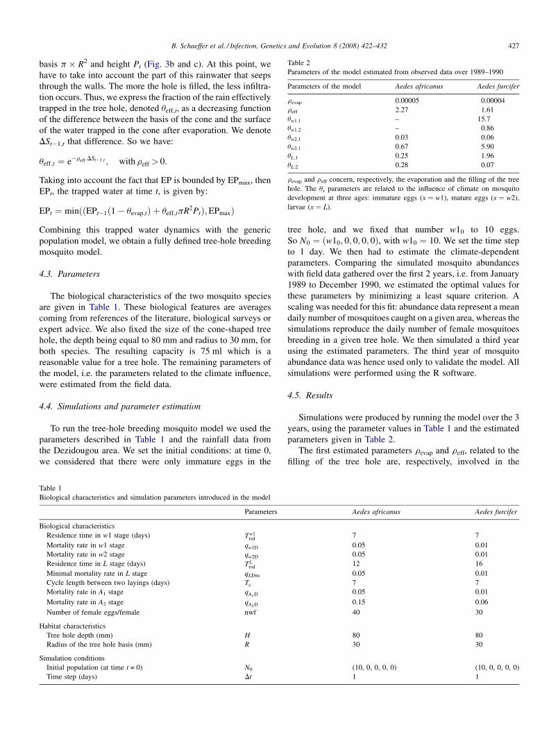
Table 2
Parameters of the model estimated from observed data over 1989–1990
Parameters of the model Aedes africanus Aedes furcifer
revap 0.00005 0.00004
reff 2.27 1.61
uw1:1 – 15.7
uw1:2 – 0.86
uw2:1 0.03 0.06
uw2:1 0.67 5.90
uL.1 0.25 1.96
uL.2 0.28 0.07
revap and reff concern, respectively, the evaporation and the filling of the tree
hole. The ux parameters are related to the influence of climate on mosquito
development at three ages: immature eggs (x ¼ w1), mature eggs (x ¼ w2),
larvae (x ¼ L).
B. Schaeffer et al. / Infection, Genetics and Evolution 8 (2008) 422–432 427
basis p � R2 and height Pt (Fig. 3b and c). At this point, we
have to take into account the part of this rainwater that seeps
through the walls. The more the hole is filled, the less infiltra-
tion occurs. Thus, we express the fraction of the rain effectively
trapped in the tree hole, denoted ueff,t, as a decreasing function
of the difference between the basis of the cone and the surface
of the water trapped in the cone after evaporation. We denote
DSt�1,t that difference. So we have:
ueff;t ¼ e�reff DSt�1;t ; with reff > 0:
Taking into account the fact that EP is bounded by EPmax, then
EPt, the trapped water at time t, is given by:
EPt ¼ minððEPt�1ð1� uevap;tÞ þ ueff;tpR2PtÞ;EPmaxÞ
Combining this trapped water dynamics with the generic
population model, we obtain a fully defined tree-hole breeding
mosquito model.
4.3. Parameters
The biological characteristics of the two mosquito species
are given in Table 1. These biological features are averages
coming from references of the literature, biological surveys or
expert advice. We also fixed the size of the cone-shaped tree
hole, the depth being equal to 80 mm and radius to 30 mm, for
both species. The resulting capacity is 75 ml which is a
reasonable value for a tree hole. The remaining parameters of
the model, i.e. the parameters related to the climate influence,
were estimated from the field data.
4.4. Simulations and parameter estimation
To run the tree-hole breeding mosquito model we used the
parameters described in Table 1 and the rainfall data from
the Dezidougou area. We set the initial conditions: at time 0,
we considered that there were only immature eggs in the
Table 1
Biological characteristics and simulation parameters introduced in the model
Parameters
Biological characteristics
Residence time in w1 stage (days) Tw1rsd
Mortality rate in w1 stage qw1D
Mortality rate in w2 stage qw2D
Residence time in L stage (days) TLrsd
Minimal mortality rate in L stage qLDm
Cycle length between two layings (days) Tc
Mortality rate in A1 stage qA1D
Mortality rate in A2 stage qA2D
Number of female eggs/female nwf
Habitat characteristics
Tree hole depth (mm) H
Radius of the tree hole basis (mm) R
Simulation conditions
Initial population (at time t = 0) N0
Time step (days) Dt
tree hole, and we fixed that number w10 to 10 eggs.
So N0 ¼ ðw10; 0; 0; 0; 0Þ, with w10 ¼ 10. We set the time step
to 1 day. We then had to estimate the climate-dependent
parameters. Comparing the simulated mosquito abundances
with field data gathered over the first 2 years, i.e. from January
1989 to December 1990, we estimated the optimal values for
these parameters by minimizing a least square criterion. A
scaling was needed for this fit: abundance data represent a mean
daily number of mosquitoes caught on a given area, whereas the
simulations reproduce the daily number of female mosquitoes
breeding in a given tree hole. We then simulated a third year
using the estimated parameters. The third year of mosquito
abundance data was hence used only to validate the model. All
simulations were performed using the R software.
4.5. Results
Simulations were produced by running the model over the 3
years, using the parameter values in Table 1 and the estimated
parameters given in Table 2.
The first estimated parameters revap and reff, related to the
filling of the tree hole are, respectively, involved in the
Aedes africanus Aedes furcifer
7 7
0.05 0.01
0.05 0.01
12 16
0.05 0.01
7 7
0.05 0.01
0.15 0.06
40 30
80 80
30 30
(10, 0, 0, 0, 0) (10, 0, 0, 0, 0)
1 1
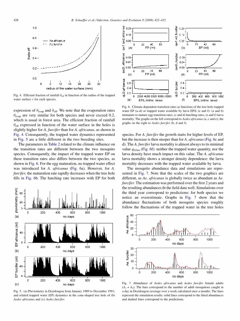
Fig. 4. Efficient fraction of rainfall ueff in function of the radius of the trapped
water surface r for each species.
Fig. 6. Climate-dependent transition rates as functions of the tree hole trapped
water EP (a–d) or trapped water available by larva EP/L (e and f): (a and b)
immature to mature egg transition rates, (c and d) hatching rates, (e and f) larva
mortality. The graphs on the left correspond to Aedes africanus (a, c and e); the
graphs on the right to Aedes furcifer (b, d and f).
B. Schaeffer et al. / Infection, Genetics and Evolution 8 (2008) 422–432428
expression of uevap and ueff. We note that the evaporation rates
uevap are very similar for both species and never exceed 0.2,
which is usual in forest area. The efficient fraction of rainfall
ueff expressed in function of the water surface in the holes is
slightly higher for A. furcifer than for A. africanus, as shown in
Fig. 4. Consequently, the trapped water dynamics represented
in Fig. 5 are a little different in the two breeding sites.
The parameters in Table 2 related to the climate influence on
the transition rates are different between the two mosquito
species. Consequently, the impact of the trapped water EP on
these transition rates also differs between the two species, as
shown in Fig. 6. For the egg maturation, no trapped water effect
was introduced for A. africanus (Fig. 6a). However, for A.
furcifer, the maturation rate rapidly decreases when the tree hole
fills in Fig. 6b. The hatching rate increases with EP for both
Fig. 5. (a) Pluviometry in Dezidougou from January 1989 to December 1991;
and related trapped water (EP) dynamics in the cone-shaped tree hole of (b)
Aedes africanus and (c) Aedes furcifer.
species. For A. furcifer the growth starts for higher levels of EP,
but the increase is then steeper than for A. africanus (Fig. 6c and
d). The A. furcifer larva mortality is almost always to its minimal
value qLDm (Fig. 6f): neither the trapped water quantity, nor the
larva density have much impact on this value. The A. africanus
larva mortality shows a stronger density dependence: the larva
mortality decreases with the trapped water available by larva.
The mosquito abundance data and simulations are repre-
sented in Fig. 7. Note that the scales of the two graphics are
different, as Ae. africanus is globally twice as abundant as Ae.
furcifer. The estimation was performed over the first 2 years and
the resulting abundances fit the field data well. Simulations over
the third year correspond to predictions: for both species we
notice an overestimate. Graphs in Fig. 7 show that the
abundance fluctuations of both mosquito species roughly
follow the fluctuations of the trapped water in the tree holes
Fig. 7. Abundance of Aedes africanus and Aedes furcifer female adults
(A1 + A2). The bars correspond to the number of adult mosquitoes caught in
a day in Dezidougou (average over a week calculated once a month). The lines
represent the simulation results: solid lines correspond to the fitted abundances
and dashed lines correspond to the predictions.
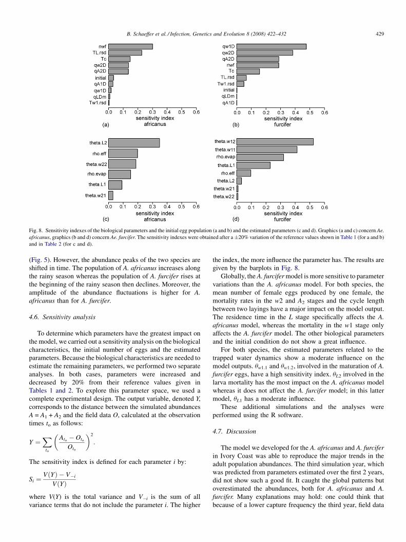
Fig. 8. Sensitivity indexes of the biological parameters and the initial egg population (a and b) and the estimated parameters (c and d). Graphics (a and c) concern Ae.
africanus, graphics (b and d) concern Ae. furcifer. The sensitivity indexes were obtained after a�20% variation of the reference values shown in Table 1 (for a and b)
and in Table 2 (for c and d).
B. Schaeffer et al. / Infection, Genetics and Evolution 8 (2008) 422–432 429
(Fig. 5). However, the abundance peaks of the two species are
shifted in time. The population of A. africanus increases along
the rainy season whereas the population of A. furcifer rises at
the beginning of the rainy season then declines. Moreover, the
amplitude of the abundance fluctuations is higher for A.
africanus than for A. furcifer.
4.6. Sensitivity analysis
To determine which parameters have the greatest impact on
the model, we carried out a sensitivity analysis on the biological
characteristics, the initial number of eggs and the estimated
parameters. Because the biological characteristics are needed to
estimate the remaining parameters, we performed two separate
analyses. In both cases, parameters were increased and
decreased by 20% from their reference values given in
Tables 1 and 2. To explore this parameter space, we used a
complete experimental design. The output variable, denoted Y,
corresponds to the distance between the simulated abundances
A = A1 + A2 and the field data O, calculated at the observation
times to as follows:
Y ¼X
to
�Ato� Oto
Oto
�2
:
The sensitivity index is defined for each parameter i by:
Si ¼VðYÞ � V�i
VðYÞ
where V(Y) is the total variance and V�i is the sum of all
variance terms that do not include the parameter i. The higher
the index, the more influence the parameter has. The results are
given by the barplots in Fig. 8.
Globally, the A. furcifer model is more sensitive to parameter
variations than the A. africanus model. For both species, the
mean number of female eggs produced by one female, the
mortality rates in the w2 and A2 stages and the cycle length
between two layings have a major impact on the model output.
The residence time in the L stage specifically affects the A.
africanus model, whereas the mortality in the w1 stage only
affects the A. furcifer model. The other biological parameters
and the initial condition do not show a great influence.
For both species, the estimated parameters related to the
trapped water dynamics show a moderate influence on the
model outputs. uw1:1 and uw1:2, involved in the maturation of A.
furcifer eggs, have a high sensitivity index. uL2 involved in the
larva mortality has the most impact on the A. africanus model
whereas it does not affect the A. furcifer model; in this latter
model, uL1 has a moderate influence.
These additional simulations and the analyses were
performed using the R software.
4.7. Discussion
The model we developed for the A. africanus and A. furcifer
in Ivory Coast was able to reproduce the major trends in the
adult population abundances. The third simulation year, which
was predicted from parameters estimated over the first 2 years,
did not show such a good fit. It caught the global patterns but
overestimated the abundances, both for A. africanus and A.
furcifer. Many explanations may hold: one could think that
because of a lower capture frequency the third year, field data
B. Schaeffer et al. / Infection, Genetics and Evolution 8 (2008) 422–432430
did not catch the abundance peaks, or that the mosquitoes that
year experienced a mortality increase . . ..For both species, the most influent parameters are those that
relate to the production and survival of eggs. However, the
initial condition has very little impact, which implies that the
population development does not only depend on the egg
survival from the previous rainy season. Each year, the
abundances of each species show a distinct pattern. These
different trends are explained by the fact that the eggs of A.
furcifer need a dry season to mature: eggs laid during the rainy
season become mature during the next dry season and are
therefore accumulated in this stage, until new rainfalls. The
sensitivity analysis showed that this maturation process is
critical in the population development. The very low larva
mortality rate obtained for A. furcifer suggests that the larvae
need very little water to survive. Therefore, once the larvae
hatch, which only occurs when sufficient water is available in
the tree hole, they are quite resilient to environmental
fluctuations. A. africanus larvae seem to be more affected by
larva density and water availability, which is supported by the
sensitivity analysis (uL2 has a high index). It could explain why
the adult abundance fluctuations observed seem to follow more
closely the trapped water evolution.
No field data were available to define the tree holes. From
expert advice we chose a conical shape as one of the simplest
representation of a rather deep and narrow hole. Moreover, we
were able to fix the hole capacity to a reasonable value (75 ml).
Keeping the same fixed capacity and varying the shape of the
cone (width and depth), the results were not modified. The only
differences observed were the estimations of the parameters
controlling the filling of the tree hole, revap and reff, which
fluctuated slightly. These two parameters have a moderate
impact on the adult population abundances. However, reff has
different values for both species, which leads us to assume
that t may not have the same type of breeding sites. The
retention of rainwater in the tree hole appears higher for A.
furcifer: it could be due to a different shape of the tree hole or a
different species of tree. Indeed, various types of breeding sites
are available in the wild and mosquitoes might show
preferences. It is the case for Aedes usambara, vector of the
Chikungunya virus, which was caught inside bamboo
plantations in Ivory Coast (Mondet and Montange, 1993).
So to simulate the abundance of A. usambara, we would have to
consider the breeding habitat as a cylinder-shaped hole to
represent the EP process.
Our results suggest that the field populations of A. africanus
and A. furcifer are well simulated by a stage-structured matrix
model incorporating pluviometry influence. In that area, as far
as we know, pluviometry is the only environmental limiting
factor in the Aedes population development. Other factors, such
as temperature, are not considered as limiting factors for both
species in that area, and thus were not introduced in the model.
However, they may also interact with pluviometry and
modulate the mosquito population abundance. Nevertheless,
this simple pluviometry-based model allowed us to capture the
major trends in the population fluctuations, which was our aim
in this application.
In Dezidougou the yellow fever is not endemic. The virus
migrates from the southern forests during the rainy season,
carried by primate and mosquito populations along the forest
galleries. So the virus usually appears in savannah at the end of
the rainy season. Both data and model agree on the following
trend: the population of A. africanus is higher at the end of the
rainy season whereas the population of A. furcifer shows a peak
at the beginning of the season. Therefore, high A. africanus
abundances increase the risk of a yellow fever epidemic in the
monkey population of the Dezidougou area. But YF virus can
survive in some A. furcifer eggs during the dry season. These
females born of contaminated eggs will be capable to generate a
human YF outbreak at the beginning of the following rainy
season, especially with abundant rainfalls that lead to a high
peak of A. furcifer.
5. Conclusions
We have presented a generic mosquito population model that
includes several features: various development stages, climate
and density dependency. Results obtained by applying the
model to field data have shown that it allows a rather good
assessment of the mosquito abundance trends. Indeed, by
taking into account the rain profile in the demographic process,
we have reproduced over 2 years, and predicted over 1 year, the
population evolution of two species of mosquitoes, that differ
by their ecological and biological features. The model fitted the
data quite accurately during the first 2 years; it overestimated
the abundances the third year, but reproduced the global
patterns. In this deterministic model, pluviometry fluctuations
were sufficient to capture the major trends in the population
abundances. However, we may introduce stochastic factors to
account for the biological variability. As we introduced the
effect of trapped water, which is the main factor under inter-
tropical climates for tree-hole breeding mosquitoes, we can also
introduce other factors, such as the temperature for example.
We can also reduce or expand the number of stages. Although,
the generic nature of the model allows the incorporation of
many other factors, we have to keep in mind the parsimony rule
before refining the model.
Applying the model to real abundance and rainfall data, we
have shown its predictive capacity once parameters are
estimated. Therefore, this model could prove useful to estimate
the abundance evolution of disease vectors, according to
different climate change scenarios. Thus, such a model is a
valuable tool to assess the risk of an epidemiological outbreak.
Appendix A. Filling up process of the tree hole
Let us consider a tree hole as an inverted cone with pR2 its
basis and H its depth.
At each time step, the rainwater trapped in the tree hole, EPt
is updated. The filling up process is composed of two successive
phases: evaporation and rainwater trapping (Fig. 3).
The volume of the water that evaporates between t � 1 and
t is uevap,t � EPt�1, where uevap,t is the evaporation rate. This
rate depends on the water surface at t � 1, St�1 ¼ pr2t�1. The
B. Schaeffer et al. / Infection, Genetics and Evolution 8 (2008) 422–432 431
larger St�1, the greater the evaporation. Moreover, uevap,t being a
fraction, it is bounded between 0 and 1. So we express the
evaporation rate, uevap,t, between time t � 1 and time t as
uevap;t ¼ 1� e�revapSt�1 , with revap � 0.
Let us denote EPint the trapped water quantity remaining in
the tree hole after the evaporation process is applied between
t � 1 and t:
EPint ¼ EPt�1ð1� e�revapSt�1Þ:
This volume can also be expressed as EPint ¼ ð1=3Þpr2inthint
where rint and hint are, respectively, the radius and the height of
the water remaining in the hole.
Let us fix K ¼ R=H. Using the cone property R=H ¼rint=hint we can write that:
EPint ¼1
3p
r3int
K:
We deduce that:
rint ¼�
3K
pEPint
�1=3
¼�
3K
pEPt�1ðe�revapSt�1Þ
�1=3
The second phase concerns the rainwater trapping. Let Pt be the
rainfall height between time t � 1 and time t. The rainwater that
comes into the tree hole is approximated by a cylinder pR2Pt.
Because of infiltration, only a fraction ueff,t of this water is
trapped. ueff,t depends on DSt�1,t, the normalized difference
between the basis of the cone, pR2, and the trapped water
surface in the cone after evaporation, pr2int:
DSt�1;t ¼pR2 � pr2
int
pR2¼ R2 � r2
int
R2:
The bigger this surface, the more infiltration there is. Moreover,
ueff,t being a fraction, it is bounded between 0 and 1. So we
express ueff,t as:
ueff;t ¼ e�reff DSt�1;t ; with reff� 0:
After evaporation and rainwater trapping, the volume of water
in the tree hole is:
EPt ¼ EPint þ ðueff;t � pR2PtÞ
¼ EPt�1ð1� uevap;tÞ þ ðueff;t � pR2PtÞ:
Finally, EPt, the trapped water at time t, is bounded by the tree
hole capacity EPmax ¼ p� R2 � ðH=3Þ. So, we obtain:
EPt ¼ minððEPt�1ð1� uevap;tÞ þ ueff;tpR2PtÞ;EPmaxÞ:
References
Agnew, P., Hide, M., Sidobre, C., Michalakis, Y., 2002. A minimalist approach
to the effects of density-dependent competition on insect life-history traits.
Ecol. Entomol. 27, 396–402.
Ahumada, J.A., Lapointe, D., Samuel, M.D., 2004. Modeling the population
dynamics of Culex quinquefasciatus (Diptera: Culicidae), along an eleva-
tion gradient in Hawaii. J. Med. Entomol. 41, 1157–1170.
Awerbuch-Friedlander, T., Levins, R., Predescu, M., 2005. The role of season-
ality in the dynamics of deer tick populations. Bull. Math. Biol. 67, 467–
486.
Bommarco, R., 2001. Using matrix models to explore the influence of tem-
perature on population growth of arthropod pests. Agric. Forest Entomol. 3,
275–283.
Caswell, H., 2001. Matrix Population Models: Construction, Analysis and
Interpretation, second ed. Sinauer Ass. Inc. Publishers.
Chattopadhyay, J., Sarkar, R.R., Chaki, S., Bhattacharya, S., 2004. Effects of
environmental fluctuations on the occurrence of malignant malaria—a
model based study. Ecol. Model. 177, 179–192.
Chevalier, V., de la Rocque, S., Baldet, T., Vial, L., Roger, F., 2004. Epide-
miological processes involved in the emergence of vector-borne diseases:
West Nile fever, Rift Valley fever, Japanese encephalitis and Crimean-
Congo haemorrhagic fever. Revue Scientifique Et Technique De L Office
International Des Epizooties 23, 535–555.
Choi, W.I., Ryoo, M.I., 2003. A matrix model for predicting seasonal fluctua-
tions in field populations of Paronychiurus kimi (Collembola: Onychirui-
dae). Ecol. Model. 162, 259–265.
Cordellier, R., 1978. Les vecteurs potentiels sauvages dans l’epidemiologie de
la fievre jaune en Afrique de l’Ouest. Travaux et documents ORSTOM, vol.
81, Paris, 1978, 258 pp.
Costantino, R.F., Cushing, J.M., Dennis, B., Desharnais, R.A., 1998. Resonant
population cycles in temporally fluctuating habitats. Bull. Math. Biol. 60,
247–273.
Depinay, J.M.O., Mbogo, C.M., Killeen, G., Knols, B., Beier, J., Carlson, J.,
Dushoff, J., Billingsley, P., Mwambi, H., Githure, J., Toure, A.M., McKen-
zie, F.E., 2004. A simulation model of African Anopheles ecology and
population dynamics for the analysis of malaria transmission. Malaria J. 3.
Derouich, M., Boutayeb, A., Twizell, E.H., 2003. A model of dengue fever.
BioMed. Eng. Online 2, 4.
Esteva, L., Vargas, C., 1999. A model for dengue disease with variable human
population. J. Math. Biol. 38, 220–240.
Focks, D.A., Haile, D.G., Daniels, E., Mount, G.A., 1993. Dynamic life table
model for Aedes-Aegypti (Diptera, Culcidae)—analysis of the literature and
model development. J. Med. Entomol. 30, 1003–1017.
Fouque, F., Baumgartner, J., 1996. Simulating development and survival of
Aedes vexans (Diptera: Culicidae) preimaginal stages under field condi-
tions. J. Med. Entomol. 33, 32–38.
Ghosh, M., Pugliese, A., 2004. Seasonal population dynamics of ticks, and its
influence on infection transmission: a semi-discrete approach. Bull. Math.
Biol. 66, 1659–1684.
Glass, K., 2005. Ecological mechanisms that promote arbovirus survival: a
mathematical model of Ross River virus transmission. Trans. R. Soc. Trop.
Med. Hyg. 99, 252–260.
Ishikawa, H., Ishii, A., Nagai, N., Ohmae, H., Harada, M., Suguri, S., Leafasia,
J., 2003. A mathematical model for the transmission of Plasmodium vivax
malaria. Parasitol. Int. 52, 81–93.
Jarry, M., Khaladi, M., Gouteux, J.P., 1996. A matrix model for studying tsetse
fly populations. Entomol. Exp. Appl. 78, 51–60.
Jensen, A.L., 1995. Simple density-dependent matrix model for population
projection. Ecol. Model. 77, 43–48.
Lord, C.C., 1998. Density dependence in larval Aedes albopictus (Diptera:
Culicidae). J. Med. Entomol. 35, 825–829.
Lord, C.C., 2004. Seasonal population dynamics and behaviour of insects in
models of vector-borne pathogens. Physiol. Entomol. 29, 214–222.
Mazaris, A.D., Matsinos, Y.G., 2006. An individual based model of sea turtles:
Investigating the effect of temporal variability on population dynamics.
Ecol. Model. 194, 114–124.
Mondet, B., 1994. Age physiologique et biologie des Aedes (Diptera: Culicidae)
vecteurs potentiels de fievre jaune en Cote d’Ivoire. Implications epide-
miologiques. Thesis. Paris-Sud University.
Mondet, B., 1997. Importance of Aedes (Diceromyia) furcifer Edwards, 1913
(Diptera: Culicidae) among the arbovirus potential vectors, in the epide-
miology of the yellow fever, in the sub-Sudanese savannas of Ivory Coast.
Annales de la Societe Entomologique de France 33, 47–54.
Mondet, B., Montange, L., 1993. Presence of Aedes (Stegomyia) usambara
Mattingly, 1953 (Diptera: Culicidae) in the Ivory Coast, seasonal variations

B. Schaeffer et al. / Infection, Genetics and Evolution 8 (2008) 422–432432
and epidemiological position. Annales de la Societe Entomologique de
France 29, 261–267.
Moon, T.E., 1976. Astatistical model of the dynamics of a mosquito vector
(Culex tarsalis) population. Biometrics 32, 355–368.
Ngwa, G.A., 2004. Modelling the dynamics of endemic malaria in growing
populations. Dis. Cont. Dyn. Syst. Ser. B 4, 1173–1202.
Ngwa, G.A., Shu, W.S., 2000. A mathematical model for endemic malaria with
variable human and mosquito populations. Math. Comput. Model. 32, 747–
763.
Renshaw, M., Service, M.W., Birley, M.H., 1993. Density-dependent regulation
of Aedes-Cantans (Diptera, Culicidae) in natural and artificial populations.
Ecol. Entomol. 18, 223–233.
Shone, S.M., Curriero, F.C., Lesser, C.R., Glass, G.E., 2006. Characterizing
population dynamics of Aedes sollicitans (Diptera: Culicidae) using meteor-
ological data. J. Med. Entomol. 43, 393–402.
Teng, H.J., Apperson, C.S., 2000. Development and survival of immature Aedes
albopictus and Aedes triseriatus (Diptera: Culicidae) in the laboratory:
effects of density, food, and competition on response to temperature. J. Med.
Entomol. 37, 40–52.
Thomas, L., Buckland, S.T., Newman, K.B., Harwood, J., 2005. A unified
framework for modelling wildlife population dynamics. Aust. N. Z. J. Stat.
47, 19–34.
Tomori, O., 2004. Yellow fever: the recurring plague. Crit. Rev. Clin. Lab. Sci.
41, 391–427.
www.elsevier.com/locate/meegid
Available online at www.sciencedirect.com
Infection, Genetics and Evolution 8 (2008) 433–438
Molecular characterization of Thai Ehrlichia canis and
Anaplasma platys strains detected in dogs§
Danai Pinyoowong a, Sathaporn Jittapalapong b, Fanan Suksawat c,Roger W. Stich d, Arinthip Thamchaipenet a,*
a Department of Genetics, Faculty of Science, Kasetsart University, Thailandb Department of Parasitology, Faculty of Veterinary Medicine, Kasetsart University, Thailandc Department of Medicine, Faculty of Veterinary Medicine, Khon Kaen University, Thailand
d Department of Veterinary Pathobiology, University of Missouri, Columbia, MO, USA
Received 31 March 2007; accepted 12 June 2007
Available online 19 June 2007
Abstract
Canine monocytic ehrlichiosis caused by Ehrlichia canis is of veterinary importance worldwide. In Thailand, there has been little information
available on E. canis and its phylogeny. The objective of this study was to characterize and establish molecular structure and phylogeny of Thai
Ehrlichia and Anaplasma strains. Genus-specific primers for Ehrlichia and Anaplasma were used to amplify the 16S rRNA gene from naturally
infected canine blood samples, and these amplicon sequences were compared with other sequences from GenBank. Both homology and secondary
structure analysis of 16S rRNA sequences indicated that they were novel E. canis and A. platys strains. Phylogenetic analysis revealed that the Thai
E. canis strain was closely related and formed a single cluster with E. canis from different countries. A. platys found in this study showed close
relationship with earlier report of A. platys from Thailand. To our knowledge this report represents the first molecular characterization of the nearly
complete 16S rRNA gene from E. canis in dogs from Thailand.
# 2007 Elsevier B.V. All rights reserved.
Keywords: Ehrlichia canis; Anaplasma platys; 16S rDNA; Phylogenetic tree; Dogs; Bangkok; Thailand
1. Introduction
Canine monocytic ehrlichiosis (CME) and canine cyclic
thrombocytopenia (CCT) are caused by Ehrlichia canis and
Anaplasma platys, respectively. These bacteria are classified in
the rickettsial family Anaplasmataceae, which includes obligate
intracellular prokaryotic parasites that reside within a para-
sitophorous vacuole (Dumler et al., 2001). In canine hosts, E.
canis is infective for monocytes while A. platys infect platelets
(Greene and Harvey, 1990). Rhipicephalus sanguineus ticks
are considered the primary vector of both pathogens (Groves
et al., 1975; Greene and Harvey, 1990). Globally distributed,
well-characterized pathogens such as E. canis offer unique
opportunities to study coevolution and interaction between
§ This study was presented in part as an abstract at the MEEGID VIII
Congress, Bangkok, Thailand, 30 November to 2 December 2006.
* Corresponding author. Tel.: +66 2 562 5444x4208; fax: +66 2 579 5528.
E-mail address: [email protected] (A. Thamchaipenet).
1567-1348/$ – see front matter # 2007 Elsevier B.V. All rights reserved.
doi:10.1016/j.meegid.2007.06.002
tick-borne pathogens and their vertebrate and invertebrate hosts
(Stich et al., in press).
Although CME and CCTare considered enzootic throughout
Thailand, these conclusions are based on diagnoses that rely on
clinical signs, haematological abnormalities and microscopic
examination of peripheral blood. Thus, these diagnoses are
often ambiguous and may fail to identify the pathogen species
involved. Molecular diagnostic methods allow direct detection
of these etiologic agents and sequence analysis facilitates their
comparison to geographically diverse strains. To our knowl-
edge, detailed genetic and phylogenetic information about E.
canis and A. platys in Thailand are limited to a single report of
an A. platys 16S rRNA gene (16S rDNA) sequence (Suksawat
et al., 2001).
In this study, we investigated diagnosed cases of canine
ehrlichiosis from a private laboratory in Bangkok, to confirm
the presence of E. canis and A. platys and to compare Thai
strains to those from other regions. PCR and 16S rDNA
sequence analysis were used to characterize the molecular
D. Pinyoowong et al. / Infection, Genetics and Evolution 8 (2008) 433–438434
features of these infections, which allowed identification of a
novel strain of A. platys that differed from that of the previous
report of Suksawat et al. (2001). Moreover, E. canis from this
country was genetically verified for the first time.
2. Materials and methods
2.1. DNA extraction from canine blood
Blood samples were collected with EDTA from dogs
diagnosed with clinical ehrlichiosis and submitted to a private
laboratory in Bangkok. The blood was chilled until arrival at the
laboratory and then stored at �20 8C. Total DNA was isolated
from 100 ml of thawed blood using the phenol/chloroform
extraction method of Sambrook and Russell (2001).
2.2. Primers designed for 16S rDNA amplification
Primers for amplification of Ehrlichia and Anaplasma
16S rDNA were designed from nucleotide sequences deposited
in GenBank database (DQ342324, AF414873, AF414870,
AF414869, AB211163, U23503, CR767821, U96436,
AB196302 and AF318946). All the sequences were aligned
for the maximum homology by ClustalW Version 8.1 (Thompson
et al., 1994). Conserved regions were selected and specific
oligonucleotide primers named ATT062F (50CCTGGCTCA–
GAACGAACGCT30) and ATT062R (50GATCCAGCCGCA-
GGTTCACC30) were derived.
2.3. 16S rDNA amplification and sequencing
DNA isolated from dog blood was used as a template to
amplify the majority of the 16S rRNA gene from Ehrlichia and
Anaplasma spp. by PCR. Amplifications were generated using
3–6 ml of genomic DNA with primers ATT062F and ATT062R
for each 20 ml PCR reaction mixture in a Peltier thermal cycler
(MJ Research, Watertown, MA, USA), by 30 cycles of 30 s at
94.0 8C, 30 s at 64.0 8C, and 1 min at 72.0 8C, preceded by
4 min at 94.0 8C and followed by 4 min at 72.0 8C. PCR
products were examined by 0.8% agarose gel electrophoresis
and ethidium bromide staining. Amplicons were purified with
QIAquick PCR purification kits (QIAGEN) according to the
manufacturer’s protocol. Direct sequencing was carried on
the PCR product using the same PCR primers, ATT062F
and ATT062R. In order to cover both strands of the PCR
fragment completely, another set of primers, ATT066F
(50CCCTGGTAGTCCACGCTG30) and ATT067R (50CAGC-
GTGGACTACCAGGG30), were designed for annealing in the
middle of the 16S rDNA amplicons and used for sequencing
towards 30 and 50 ends, respectively.
2.4. Phylogenetic analysis
Multiple sequence alignments of amplicons and 16S rDNA
sequences from GenBank were performed using the ClustalW
Version 1.8 (Thompson et al., 1994). Phylogenetic trees
were inferred using neighbor-joining (NJ) analysis by MEGA
software Version 3.1 (Kumar et al., 2004). The distance matrix of
nucleotide divergences was calculated according to Kimura’s
two-parameter model furnished by MEGA. A bootstrap re-
sampling technique of 1000 replications was performed to
statistically support the reliabilities of the nodes on the trees. The
differences between nucleotide positions were confirmed by
DnaSP Version 4.10 (Rozas et al., 2003). Neorickettsia sennetsu
was used as an outgroup.
2.5. Nucleotide sequences accession numbers
The 16S rDNA sequences from this study were deposited at
GenBank database under accession numbers EF139458 for E.
canis-Bangkok and EF139459 for A. platys-Bangkok. Other
Anaplasmataceae 16S rDNA sequences from Genbank
(and their accession numbers) included 10 E. canis strains
reported from China (AF162860), Japan (AF536827), Peru
(DQ915970), South Africa (U54805), Spain (AY394465),
Venezuela (AF373613, AF373612) and the USA (M73226,
M73221), eight A. platys strains from China (AF156784),
France (AF303467), Japan (AY077619, AF536828), Spain
(AY530806), Thailand (AF286699), Venezuela (AF287153)
and the USA (M82801), E. chaffeensis (U23503), E. ewingii
(U96436), E. muris (NC_007354, AB196302), E. ruminantium
(CR767821), A. bovis (AB211163), A. centrale (AF414869), A.
marginale (AF414873), A. ovis (AF414870), A. phagocyto-
philum (DQ342324) and N. sennetsu (M73219).
3. Results
3.1. Amplification and sequencing of 16S rDNA
The identity of agents associated with diagnosed CME and
CCT are often assumed based on clinical diagnosis and
microscopic examination of peripheral blood. Blood samples
from dogs diagnosed with clinical ehrlichiosis were screened to
compare these agents to previously reported strains. Template
was prepared and assayed by PCR with primers ATT062F and
ATT062R, which were designed for specific amplification of
Ehrlichia and Anaplasma 16S rDNA. Approximately 1.5 kb
amplicons corresponding to the expected size of targeted 16S
rRNA gene fragments were obtained (data not shown).
Amplicons from two individual canine blood samples were
purified and directly sequenced with the same primers. The
nearly complete 16S rRNA gene sequences of 1478 (51% AT)
and 1481 (52% AT) bp were identical to consensus 16S rDNA
sequences for E. canis and A. platys, respectively. These
sequences were named E. canis-Bangkok and A. platys-
Bangkok and deposited as new 16S rDNA sequences in
GenBank.
3.2. Molecular characterization of E. canis from Thai dogs
E. canis and A. platys are distributed globally, and it is
generally assumed that all strains primarily utilize dogs and
rhipicephaline ticks as vertebrate and invertebrate hosts.
However, closely related anaplasmal pathogens, including
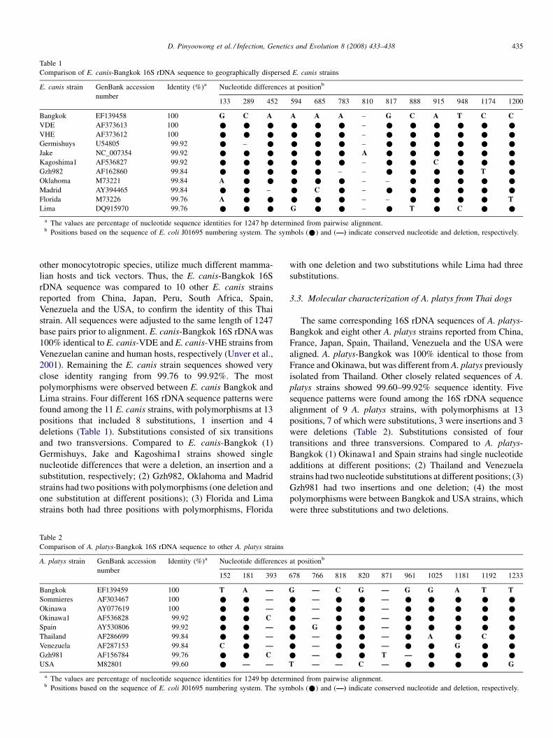
Table 1
Comparison of E. canis-Bangkok 16S rDNA sequence to geographically dispersed E. canis strains
E. canis strain GenBank accession
number
Identity (%)a Nucleotide differences at positionb
133 289 452 594 685 783 810 817 888 915 948 1174 1200
Bangkok EF139458 100 G C A A A A – G C A T C C
VDE AF373613 100 * * * * * * – * * * * * *
VHE AF373612 100 * * * * * * – * * * * * *
Germishuys U54805 99.92 * – * * * * – * * * * * *
Jake NC_007354 99.92 * * * * * * A * * * * * *
Kagoshima1 AF536827 99.92 * * * * * * – * * C * * *
Gzh982 AF162860 99.84 * * * * * – – * * * * T *
Oklahoma M73221 99.84 A * * * * * – – * * * * *
Madrid AY394465 99.84 * * – * C * – * * * * * *
Florida M73226 99.76 A * * * * * – – * * * * T
Lima DQ915970 99.76 * * * G * * – * T * C * *
a The values are percentage of nucleotide sequence identities for 1247 bp determined from pairwise alignment.b Positions based on the sequence of E. coli J01695 numbering system. The symbols (*) and (—) indicate conserved nucleotide and deletion, respectively.
D. Pinyoowong et al. / Infection, Genetics and Evolution 8 (2008) 433–438 435
other monocytotropic species, utilize much different mamma-
lian hosts and tick vectors. Thus, the E. canis-Bangkok 16S
rDNA sequence was compared to 10 other E. canis strains
reported from China, Japan, Peru, South Africa, Spain,
Venezuela and the USA, to confirm the identity of this Thai
strain. All sequences were adjusted to the same length of 1247
base pairs prior to alignment. E. canis-Bangkok 16S rDNA was
100% identical to E. canis-VDE and E. canis-VHE strains from
Venezuelan canine and human hosts, respectively (Unver et al.,
2001). Remaining the E. canis strain sequences showed very
close identity ranging from 99.76 to 99.92%. The most
polymorphisms were observed between E. canis Bangkok and
Lima strains. Four different 16S rDNA sequence patterns were
found among the 11 E. canis strains, with polymorphisms at 13
positions that included 8 substitutions, 1 insertion and 4
deletions (Table 1). Substitutions consisted of six transitions
and two transversions. Compared to E. canis-Bangkok (1)
Germishuys, Jake and Kagoshima1 strains showed single
nucleotide differences that were a deletion, an insertion and a
substitution, respectively; (2) Gzh982, Oklahoma and Madrid
strains had two positions with polymorphisms (one deletion and
one substitution at different positions); (3) Florida and Lima
strains both had three positions with polymorphisms, Florida
Table 2
Comparison of A. platys-Bangkok 16S rDNA sequence to other A. platys strains
A. platys strain GenBank accession
number
Identity (%)a Nucleotide differences
152 181 393
Bangkok EF139459 100 T A —
Sommieres AF303467 100 * * —
Okinawa AY077619 100 * * —
Okinawa1 AF536828 99.92 * * C
Spain AY530806 99.92 * * —
Thailand AF286699 99.84 * * —
Venezuela AF287153 99.84 C * —
Gzh981 AF156784 99.76 * * C
USA M82801 99.60 * — —
a The values are percentage of nucleotide sequence identities for 1249 bp determb Positions based on the sequence of E. coli J01695 numbering system. The sym
with one deletion and two substitutions while Lima had three
substitutions.
3.3. Molecular characterization of A. platys from Thai dogs
The same corresponding 16S rDNA sequences of A. platys-
Bangkok and eight other A. platys strains reported from China,
France, Japan, Spain, Thailand, Venezuela and the USA were
aligned. A. platys-Bangkok was 100% identical to those from
France and Okinawa, but was different from A. platys previously
isolated from Thailand. Other closely related sequences of A.
platys strains showed 99.60–99.92% sequence identity. Five
sequence patterns were found among the 16S rDNA sequence
alignment of 9 A. platys strains, with polymorphisms at 13
positions, 7 of which were substitutions, 3 were insertions and 3
were deletions (Table 2). Substitutions consisted of four
transitions and three transversions. Compared to A. platys-
Bangkok (1) Okinawa1 and Spain strains had single nucleotide
additions at different positions; (2) Thailand and Venezuela
strains had two nucleotide substitutions at different positions; (3)
Gzh981 had two insertions and one deletion; (4) the most
polymorphisms were between Bangkok and USA strains, which
were three substitutions and two deletions.
at positionb
678 766 818 820 871 961 1025 1181 1192 1233
G — C G — G G A T T
* — * * — * * * * *
* — * * — * * * * *
* — * * — * * * * *
* G * * — * * * * *
* — * * — * A * C *
* — * * — * * G * *
* — * * T — * * * *
T — — C — * * * * G
ined from pairwise alignment.
bols (*) and (—) indicate conserved nucleotide and deletion, respectively.
D. Pinyoowong et al. / Infection, Genetics and Evolution 8 (2008) 433–438436
3.4. 16S rRNA secondary structures
16S rRNA is not subject to the same selective influences on
function as mRNA (i.e., 16S rRNA function relies on structure
rather than codon usage), thus the effects of nucleotide changes
on predicted secondary structures could be more informative
than primary sequence variation alone. The positions of 16S
rDNA sequences defined in Tables 1 and 2 were correlated with
the E. coli J01695 numbering system (Konings and Gutell, 1995).
Comparison of E. canis-Bangkok and A. platys-Bangkok 16S
rRNA predicted secondary structures to that of the E. coli J01695
indicated that both had conserved tetra loops that generally
constrained the rRNA architecture (Woese et al., 1990). Nucleo-
tides at positions 289, 452, 594, 888, 915, 948 and 1200 of E.
canis-Bangkok were common among bacteria while positions
133, 685, 783, 810, 817 and 1174 were different from other
eubacteria. At position 948, we observed T (U) in most samples
except E. canis-Lima that carried C, the latter of which is similar
to those of alphaproteobacteria (Woese, 1987). In A. platys-
Bangkok, nucleotide differences at positions 181, 678, 871 and
1025 were within the common structure of eubacteria. At posi-
tion 393, eubacteria generally carried A (Woese, 1987), but most
of A. platys had a single deletion except for A. platys-Okinawa1
and A. platys-Gzh981 that carried a C. At position 1233, T was
observed in most samples except for A. platys USA, which had a
G that is similar to those of alphaproteobacteria (Woese, 1987).
3.5. Phylogenetic analysis of Ehrlichia and Anaplasma
Ehrlichia and Anaplasma 16S rDNA sequences were used to
generate a phylogenetic tree using the neighbor-joining method
by MEGA software (Version 3.1). In addition to E. canis and A.
platys strains, closely related species included the tick-borne
anaplasmal parasites E. chaffeensis, E. ewingii, E. muris, E.
ruminantium, A. bovis, A. centrale, A. marginale, A. ovis and A.
phagocytophilum. A biologically divergent member of the
Anaplasmataceae, N. sennetsu, was used as the outgroup. The
resultant phylogenetic tree revealed that E. canis-Bangkok and
A. platys-Bangkok were grouped tightly within the other E.
canis and A. platys strains, respectively (Fig. 1). This analysis
revealed that (1) Ehrlichia and Anaplasma were divided into
clearly defined clades; (2) E. canis strains from different
geographic regions were always grouped in a clade independent
from E. chaffeensis, E. ewingii, E. muris and E. ruminantium;
(3) E. ewingii showed the closet relationship to E. canis while
E. ruminantium was the most distant; (4) A. platys strains from
different countries constantly grouped in a clade independent
from A. bovis, A. centrale, A. marginale, A. ovis and A.
phagocytophilum; (5) A. phagocytophilum had the closet
relationship to A. platys while A. centrale, A. marginale and A.
ovis were the most distant; (6) A. marginale clustered in a
branch linked to A. centrale and A. ovis.
4. Discussion
Understanding the epidemiology of these diseases and
infectious cycles of anaplasmal organisms is expected to
facilitate understanding of similar diseases, including zoonoses
that may share similar etiologic agents or vectors. These results
support previous studies that indicated E. canis and A. platys
are uniform worldwide, suggesting that biological observations
with different strains and from different regions could be
laterally applicable. However, it is important to note that these
observations are based on the highly conserved 16S rRNA gene
and that further work with more divergent sequences is needed
to confirm whether these strains are indeed uniform. Moreover,
it is important to remember that potential heterogeneity of
vertebrate and invertebrate hosts from different locations is
likely to influence the biology of these organisms. Thus, global
analyses of evolutionary patterns among E. canis and A. platys
will likely require parallel analyses of such patterns among
their reservoirs and vectors. For example, R. sanguineus, which
is considered an important vector of both pathogens described
in this report, is considered highly host-specific in some regions
but reportedly feeds on diverse medium to large mammalian
hosts (including humans) in other areas. Susceptibility of these
tick strains to various pathogens, including E. canis and A.
platys, could explain reports suggesting E. canis infections of
ruminants and humans (Stich et al., in press; Yu et al., 2007).
Alternatively, similarities among geographically diverse E.
canis strains could be reflective of relatively recent global
spread of this pathogen with its vertebrate and invertebrate
hosts (Yu et al., 2007).
This report describes molecular analysis of nearly complete
16S rDNA sequences of E. canis and A. platys from Bangkok,
Thailand. The sequence alignments and phylogenetic tree
suggested low diversity within E. canis and A. platys strains
based on the close similarity amongst their 16S rDNA
sequences from all geographic regions tested, and these
conclusions are consistent with other reports (de la Fuente et al.,
2006; Aguirre et al., 2006; Yu et al., 2007). Genetic
polymorphisms from comparison of 16S rRNA sequences
based on the E. coli J01695 numbering system (Konings and
Gutell, 1995) also indicated that E. canis-Bangkok and A.
platys-Bangkok were structurally conserved in 16S rRNA
architecture. Therefore, we are confident that all polymorph-
isms observed in these experiments are consistent with these
two species. However, although nucleotide differences at many
positions indicated that E. canis-Bangkok and A. platys-
Bangkok shared some structure with other bacteria, other
nucleotides were different from most eubacteria.
Close similarity among 16S rDNA of A. platys from
different locations worldwide supported the hypothesis that A.
platys strains are not geographically segregated (Huang et al.,
2005). Interestingly, the sequence of A. platys-Bangkok
reported from this study had two nucleotide substitutions
compared to a previously characterized A. platys strain from
Thailand (Suksawat et al., 2001) at positions 1025 and 1192
(Table 2) that appeared through G/A and T/C transitions,
respectively, suggesting that there might be at least two A.
platys strains enzootic to Thailand.
The phylogenetic tree separated two major clusters of
Ehrlichia spp. and Anaplasma spp. As expected, E. canis-
Bangkok was within the Ehrlichia clade and A. platys-Bangkok

Fig. 1. Phylogenetic tree based on Ehrlichia and Anaplasma 16S rDNA. Sequences from the Ehrlichia and Anaplasma genera were compared with the neighbor-
joining method with distance matrix calculation by Kimura-two parameters, operated by MEGA software (Version 3.1), using N. sennetsu as the outgroup. Scale bar
indicates the number of mutations per sequence position. The numbers at the nodes represent the percentage of 1000 bootstrap re-samplings.
D. Pinyoowong et al. / Infection, Genetics and Evolution 8 (2008) 433–438 437
was within its respective clade. Within these two clusters,
E. canis and A. platys strains grouped mostly in multiple
connected branches. Although they were from geographically
diverse regions, little genetic diversity was observed, suggest-
ing slow and homogeneous evolution (Keysary et al., 1996).
These results were in concordance with previous reports of
slight genetic variation among 16S rDNA from different E.
canis strains (Unver et al., 2003; Parola et al., 2003; Aguirre
et al., 2004) and A. platys strains (Unver et al., 2003; Huang
et al., 2005; Martin et al., 2005; de la Fuente et al., 2006).
Notably, the E. canis-Bangkok 16S rDNAwas identical to VDE
and VHE that were respectively isolated from a dog and a
human in Venezuela (Unver et al., 2001), which suggests little
differentiation among E. canis between these geographic
locations and tempts speculation about potential similarities in
the epidemiology of these strains, but further analyses of
less conserved sequences are needed to test this idea. This
phylogenetic analysis also suggested that the E. canis was more
closely related to E. ewingii than to E. chaffeensis, E. muris and
E. ruminantium, and that A. platys was more closely related to
A. phagocytophilum than to A. bovis, A. centrale, A. marginale
and A. ovis, which both corroborate an earlier report (Yu et al.,
2001).
In conclusion, our primers detected both Ehrlichia and
Anaplasma in canine blood, which resulted in new 16S rDNA
sequences from E. canis and A. platys infections of Thai dogs.
D. Pinyoowong et al. / Infection, Genetics and Evolution 8 (2008) 433–438438
Although the 16S rDNA sequences were highly conserved
among geographically diverse strains of these organisms,
additional analyses of genes more subject to selective pressure
from host environments (e.g., outer membrane protein gene
families) could help elucidate the diversity and evolution of
strains from different geographic areas. Current efforts include
examination of additional canine blood samples to determine
the presence and genetic diversity of Ehrlichia and Anaplasma
spp. found in Thailand.
Acknowledgements
We give special thanks to Veterinary Diagnostic Laboratory
(VDL) center, Bangkok for dog blood samples. This research
was supported by the Graduate School, Kasetsart University.
References
Aguirre, E., Sainz, A., Dunner, S., Amusategui, I., Lopez, L., Rodriguez-
Franco, F., Luaces, I., Cortes, O., Tesouro, M.A., 2004. First isolation and
molecular characterization of Ehrlichia canis in Spain. Vet. Parasitol. 125,
365–372.
Aguirre, E., Tesouro, M.A., Ruiz, L., Amusategui, I., Sainz, A., 2006. Genetic
characterization of Anaplasma (Ehrlichia) platys in dogs in Spain. J. Vet.
Med. B Infect. Dis. Vet. Public Health 53, 197–200.
de la Fuente, J., Torina, A., Naranjo, V., Nicosia, S., Alongi, A., Mantia, F.L.,
Kocan, K.M., 2006. Molecular characterization of Anaplasma platys strains
from dogs in Sicily. Italy Vet. Res. 2, 24–29.
Dumler, J.S., Barbet, A.F., Bekker, C.P., Dasch, G.A., Palmer, G.H., Ray, S.C.,
Rikihisa, Y., Rurangirwa, F.R., 2001. Reorganization of genera in the
families Rickettsiaceae and Anaplasmataceae in the order Rickettsiales:
unification of some species of Ehrlichia with Anaplasma, Cowdria with
Ehrlichia and Ehrlichia with Neorickettsia, descriptions of six new species
combinations and designation of Ehrlichia equi and ‘HGE agent’ as
subjective synonyms of Ehrlichia phagocytophila. Int. J. Syst. Evol.
Microbiol. 51, 2145–2165.
Greene, C.E., Harvey, J.W., 1990. Canine ehrlichiosis. In: Greene, C.E. (Ed.),
Clinical Microbiology and Infectious Diseases of the Dog and Cat. W.B.
Saunders, Philadelphia, pp. 137–148.
Groves, M.G., Dennis, G.L., Amyx, H.L., Huxsoll, D.L., 1975. Transmission of
Ehrlichia canis to dogs by tick (Rhipicephalus sanguineus). Am. J. Vet. Res.
36, 937–940.
Huang, H., Unver, A., Perez, M.J., Orellana, N.G., Rikihisa, Y., 2005. Pre-
valence and molecular analysis of Anaplasma platys in dogs in Lara,
Venezuela. Braz. J. Microbiol. 36, 211–216.
Keysary, A., Waner, T., Rosner, M., Warner, C.K., Dawson, J.E., Zass, R.,
Biggie, K.L., Harrus, S., 1996. The first isolation, in vitro propagation, and
genetic characterization of Ehrlichia canis in Israel. Vet. Parasitol. 62, 331–
340.
Konings, D.A.M., Gutell, R.R., 1995. A comparison of thermodynamic foldings
with comparatively derived structures of 16S and 16S-like rRNAs. RNA 1,
559–574.
Kumar, S., Tamura, K., Nei, M., 2004. MEGA3: integrated software for
molecular evolutionary genetics analysis and sequence alignment. Brief.
Bioinform. 5, 150–163.
Martin, A.R., Brown, G.K., Dunstan, R.H., Roberts, T.K., 2005. Anaplasma
platys: an improved PCR for its detection in dogs. Exp. Parasitol. 109, 176–
180.
Parola, P., Cornet, J.P., Sanogo, Y.O., Miller, R.S., Thien, H.V., Gonzalez, J.P.,
Raoult, D., Telford III, S.R., Wongsrichanali, C., 2003. Detection of
Ehrlichia spp., Anaplasma spp., Rickettsia spp., and other eubacteria in
ticks from the Thai-Myanmar border and Vietnam. J. Clin. Microbiol. 41,
1600–1608.
Rozas, J., Sanchez-Delbarrio, J.C., Messeguer, X., Rozas, R., 2003. DnaSP,
DNA polymorphism analysis by the coalescent and other methods. Bioin-
formatics 19, 2496–2497.
Sambrook, J., Russell, D.W., 2001. In: Molecular Cloning: A Laboratory
Manual. third ed. Cold Spring Harbor Laboratory Press, New York.
Stich, R.W., Schaefer, J.J., Bremer, W.G., Needham, G.R., Jittapalapong, S.,
in press. Host surveys, ixodid tick biology and transmission scenarios as
related to the tick-borne pathogen, Ehrlichia canis. Vet. Parasitol., in
press.
Suksawat, J., Pitulie, C., Arraga-Alvarado, C., Madrigal, K., Hancock, S.I.,
Breitschwerdt, E.B., 2001. Coinfection with three Ehrlichia species in dogs
from Thailand and Venezuela with emphasis on consideration of the 16S
ribosomal DNA secondary structure. J. Clin. Microbiol. 39, 90–93.
Thompson, J.D., Higgins, D.G., Gibson, T.J., 1994. CLUSTALW: improving
the sensitivity of progressive multiple sequence alignment through sequence
weighting, position-specific gap penalties and weight matrix choice.
Nucleic Acids Res. 22, 4673–4680.
Unver, A., Perez, M., Orellana, N., Huang, H., Rikihisa, Y., 2001. Molecular and
antigenic comparison of Ehrlichia canis isolates from dogs, ticks, and a
human in Venezuela. J. Clin. Microbiol. 39, 2788–2793.
Unver, A., Rikihisa, Y., Kawahara, M., Yamamoto, S., 2003. Analysis of 16S
rRNA gene sequence of Ehrlichia canis, Anaplasma platys, and Wol-
bachia species from canine blood in Japan. Ann. N.Y. Acad. Sci. 990,
692–698.
Woese, C.R., 1987. Bacterial evolution. Microbiol. Rev. 51, 221–271.
Woese, C.R., Winker, S., Gutell, R.R., 1990. Architecture of ribosomal RNA:
constraints on the sequence of ‘‘tetra-loops’’. Proc. Natl. Acad. Sci. U.S.A.
87, 8467–8471.
Yu, X.J., Zhang, X.F., McBride, J.W., Zhang, Y., Walker, D.H., 2001. Phylo-
genetic relationships of Anaplasma marginale and ‘Ehrlichia platys’ to
other Ehrlichia species determined by GroEL amino acid sequences. Int. J.
Syst. Evol. Microbiol. 51, 1143–1146.
Yu, X.J., McBride, J.W., Walker, D.H., 2007. Restriction and expansion of
Ehrlichia strain diversity. Vet. Parasitol. 143, 337–346.
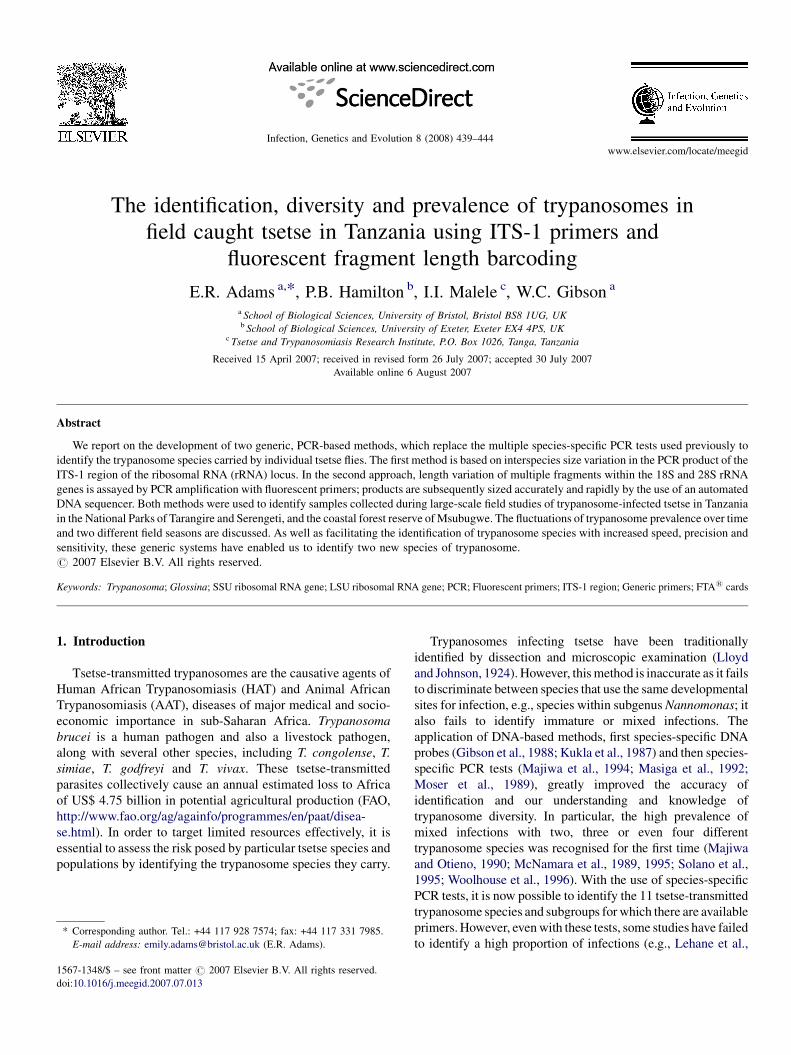
www.elsevier.com/locate/meegid
Infection, Genetics and Evolution 8 (2008) 439–444
The identification, diversity and prevalence of trypanosomes in
field caught tsetse in Tanzania using ITS-1 primers and
fluorescent fragment length barcoding
E.R. Adams a,*, P.B. Hamilton b, I.I. Malele c, W.C. Gibson a
a School of Biological Sciences, University of Bristol, Bristol BS8 1UG, UKb School of Biological Sciences, University of Exeter, Exeter EX4 4PS, UK
c Tsetse and Trypanosomiasis Research Institute, P.O. Box 1026, Tanga, Tanzania
Received 15 April 2007; received in revised form 26 July 2007; accepted 30 July 2007
Available online 6 August 2007
Abstract
We report on the development of two generic, PCR-based methods, which replace the multiple species-specific PCR tests used previously to
identify the trypanosome species carried by individual tsetse flies. The first method is based on interspecies size variation in the PCR product of the
ITS-1 region of the ribosomal RNA (rRNA) locus. In the second approach, length variation of multiple fragments within the 18S and 28S rRNA
genes is assayed by PCR amplification with fluorescent primers; products are subsequently sized accurately and rapidly by the use of an automated
DNA sequencer. Both methods were used to identify samples collected during large-scale field studies of trypanosome-infected tsetse in Tanzania
in the National Parks of Tarangire and Serengeti, and the coastal forest reserve of Msubugwe. The fluctuations of trypanosome prevalence over time
and two different field seasons are discussed. As well as facilitating the identification of trypanosome species with increased speed, precision and
sensitivity, these generic systems have enabled us to identify two new species of trypanosome.
# 2007 Elsevier B.V. All rights reserved.
Keywords: Trypanosoma; Glossina; SSU ribosomal RNA gene; LSU ribosomal RNA gene; PCR; Fluorescent primers; ITS-1 region; Generic primers; FTA1 cards
1. Introduction
Tsetse-transmitted trypanosomes are the causative agents of
Human African Trypanosomiasis (HAT) and Animal African
Trypanosomiasis (AAT), diseases of major medical and socio-
economic importance in sub-Saharan Africa. Trypanosoma
brucei is a human pathogen and also a livestock pathogen,
along with several other species, including T. congolense, T.
simiae, T. godfreyi and T. vivax. These tsetse-transmitted
parasites collectively cause an annual estimated loss to Africa
of US$ 4.75 billion in potential agricultural production (FAO,
http://www.fao.org/ag/againfo/programmes/en/paat/disea-
se.html). In order to target limited resources effectively, it is
essential to assess the risk posed by particular tsetse species and
populations by identifying the trypanosome species they carry.
* Corresponding author. Tel.: +44 117 928 7574; fax: +44 117 331 7985.
E-mail address: [email protected] (E.R. Adams).
1567-1348/$ – see front matter # 2007 Elsevier B.V. All rights reserved.
doi:10.1016/j.meegid.2007.07.013
Trypanosomes infecting tsetse have been traditionally
identified by dissection and microscopic examination (Lloyd
and Johnson, 1924). However, this method is inaccurate as it fails
to discriminate between species that use the same developmental
sites for infection, e.g., species within subgenus Nannomonas; it
also fails to identify immature or mixed infections. The
application of DNA-based methods, first species-specific DNA
probes (Gibson et al., 1988; Kukla et al., 1987) and then species-
specific PCR tests (Majiwa et al., 1994; Masiga et al., 1992;
Moser et al., 1989), greatly improved the accuracy of
identification and our understanding and knowledge of
trypanosome diversity. In particular, the high prevalence of
mixed infections with two, three or even four different
trypanosome species was recognised for the first time (Majiwa
and Otieno, 1990; McNamara et al., 1989, 1995; Solano et al.,
1995; Woolhouse et al., 1996). With the use of species-specific
PCR tests, it is now possible to identify the 11 tsetse-transmitted
trypanosome species and subgroups for which there are available
primers. However, even with these tests, some studies have failed
to identify a high proportion of infections (e.g., Lehane et al.,
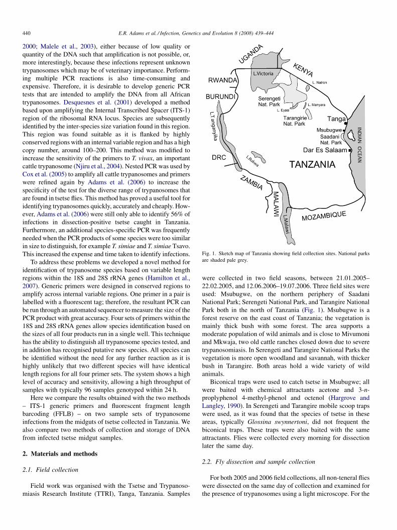
Fig. 1. Sketch map of Tanzania showing field collection sites. National parks
are shaded pale grey.
E.R. Adams et al. / Infection, Genetics and Evolution 8 (2008) 439–444440
2000; Malele et al., 2003), either because of low quality or
quantity of the DNA such that amplification is not possible, or,
more interestingly, because these infections represent unknown
trypanosomes which may be of veterinary importance. Perform-
ing multiple PCR reactions is also time-consuming and
expensive. Therefore, it is desirable to develop generic PCR
tests that are intended to amplify the DNA from all African
trypanosomes. Desquesnes et al. (2001) developed a method
based upon amplifying the Internal Transcribed Spacer (ITS-1)
region of the ribosomal RNA locus. Species are subsequently
identified by the inter-species size variation found in this region.
This region was found suitable as it is flanked by highly
conserved regions with an internal variable region and has a high
copy number, around 100–200. This method was modified to
increase the sensitivity of the primers to T. vivax, an important
cattle trypanosome (Njiru et al., 2004). Nested PCR was used by
Cox et al. (2005) to amplify all cattle trypanosomes and primers
were refined again by Adams et al. (2006) to increase the
specificity of the test for the diverse range of trypanosomes that
are found in tsetse flies. This method has proved a useful tool for
identifying trypanosomes quickly, accurately and cheaply. How-
ever, Adams et al. (2006) were still only able to identify 56% of
infections in dissection-positive tsetse caught in Tanzania.
Furthermore, an additional species-specific PCR was frequently
needed when the PCR products of some species were too similar
in size to distinguish, for example T. simiae and T. simiae Tsavo.
This increased the expense and time taken to identify infections.
To address these problems we developed a novel method for
identification of trypanosome species based on variable length
regions within the 18S and 28S rRNA genes (Hamilton et al.,
2007). Generic primers were designed in conserved regions to
amplify across internal variable regions. One primer in a pair is
labelled with a fluorescent tag; therefore, the resultant PCR can
be run through an automated sequencer to measure the size of the
PCR product with great accuracy. Four sets of primers within the
18S and 28S rRNA genes allow species identification based on
the sizes of all four products run in a single well. This technique
has the ability to distinguish all trypanosome species tested, and
in addition has recognised putative new species. All species can
be identified without the need for any further reaction as it is
highly unlikely that two different species will have identical
length regions for all four primer sets. The system shows a high
level of accuracy and sensitivity, allowing a high throughput of
samples with typically 96 samples genotyped within 24 h.
Here we compare the results obtained with the two methods
– ITS-1 generic primers and fluorescent fragment length
barcoding (FFLB) – on two sample sets of trypanosome
infections from the midguts of tsetse collected in Tanzania. We
also compare two methods of collection and storage of DNA
from infected tsetse midgut samples.
2. Materials and methods
2.1. Field collection
Field work was organised with the Tsetse and Trypanoso-
miasis Research Institute (TTRI), Tanga, Tanzania. Samples
were collected in two field seasons, between 21.01.2005–
22.02.2005, and 12.06.2006–19.07.2006. Three field sites were
used: Msubugwe, on the northern periphery of Saadani
National Park; Serengeti National Park, and Tarangire National
Park both in the north of Tanzania (Fig. 1). Msubugwe is a
forest reserve on the east coast of Tanzania; the vegetation is
mainly thick bush with some forest. The area supports a
moderate population of wild animals and is close to Mivumoni
and Mkwaja, two old cattle ranches closed down due to severe
trypanosomiasis. In Serengeti and Tarangire National Parks the
vegetation is more open woodland and savannah, with thicker
bush in Tarangire. Both areas hold a wide variety of wild
animals.
Biconical traps were used to catch tsetse in Msubugwe; all
were baited with chemical attractants acetone and 3-n-
proplyphenol 4-methyl-phenol and octenol (Hargrove and
Langley, 1990). In Serengeti and Tarangire mobile scoop traps
were used, as it was found that the species of tsetse in these
areas, typically Glossina swynnertoni, did not frequent the
biconical traps. These traps were also baited with the same
attractants. Flies were collected every morning for dissection
later the same day.
2.2. Fly dissection and sample collection
For both 2005 and 2006 field collections, all non-teneral flies
were dissected on the same day of collection and examined for
the presence of trypanosomes using a light microscope. For the
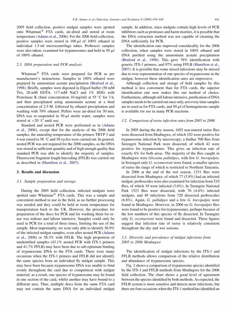
E.R. Adams et al. / Infection, Genetics and Evolution 8 (2008) 439–444 441
2005 field collection, positive midgut samples were spotted
onto Whatman1 FTA cards, air-dried and stored at room
temperature (Adams et al., 2006). For the 2006 field collection,
positive samples were stored in 100 ml of 100% ethanol in
individual 1.5 ml microcentrifuge tubes. Proboscis samples
were also taken, examined for trypanosomes and held in 50 ml
of 100% ethanol.
2.3. DNA preparation and PCR analysis
Whatman1 FTA cards were prepared for PCR as per
manufacturer’s instructions. Samples in 100% ethanol were
prepared by ammonium acetate precipitation (Bruford et al.,
1998). Briefly, samples were digested in Digsol buffer (50 mM
Tris, 20 mM EDTA, 117 mM NaCl and 1% SDS) with
Proteinase K (final concentration 10 mg/ml) at 55 8C for 3 h,
and then precipitated using ammonium acetate at a final
concentration of 2.5 M, followed by ethanol precipitation and
washing with 70% ethanol. Pellets were air-dried for 30 min,
DNA was re-suspended in 50 ml sterile water, samples were
stored at �20 8C until use.
Standard and nested PCR were performed as in (Adams
et al., 2006), except that for the analysis of the 2006 field
samples, the annealing temperature of the primers TRYP 3 and
4 was raised to 58 8C and 30 cycles were carried out. In general,
nested PCR was not required for the 2006 samples, as the DNA
was stored in sufficient quantity and of high enough quality that
standard PCR was able to identify the majority of samples.
Fluorescent fragment length barcoding (FFLB) was carried out
as described in (Hamilton et al., 2007).
3. Results and discussion
3.1. Sample preparation and storage
During the 2005 field collection, infected midguts were
spotted onto Whatman1 FTA cards. This was a simple and
convenient method to use in the field, as no further processing
was needed and they could be held at room temperature for
transportation back to the UK. However, the procedure for
preparation of the discs for PCR and for washing them for re-
use was tedious and labour intensive. Samples could only be
used in PCR for a total of three times, limiting the use of the
sample. Most importantly, we were only able to identify 56.9%
of the infected midgut samples, even after nested PCR (Adams
et al., 2006) or 58.3% with FFLB. The high proportion of
unidentified samples (43.1% nested PCR with ITS-1 primers
and 41.7% FFLB) may have been due to sub-optimum binding
of trypanosome DNA to the FTA cards. There were many
occasions when the ITS-1 primers and FFLB did not identify
the same species from an individual fly midgut sample. This
may have been because trypanosome DNA was unable to bind
evenly throughout the card due to competition with midgut
material; as a result, one species of trypanosome may be found
in one section of the card, whilst another may have bound to a
different area. Thus, multiple discs from the same FTA card
may not contain the same DNA for an individual midgut
sample. In addition, since midguts contain high levels of PCR
inhibitors such as proteases and haem moeties, it is possible that
the DNA extraction method was not capable of cleaning the
DNA sufficiently for PCR.
The identification rate improved considerably for the 2006
collection, when samples were stored in 100% ethanol and
DNA purified using the ammonium acetate precipitation
(Bruford et al., 1998). This gave 78% identification with
generic ITS-1 primers, and 97% using FFLB (Hamilton et al.,
2007). It is possible that some mixed infections may be missed
due to over-representation of one species of trypanosome in the
midgut, however these identification rates are impressive.
Although collection and storage of field samples by this
method is less convenient than for FTA cards, the superior
identification rate now makes this our method of choice.
Furthermore, although still labour intensive, purification of DNA
samples needs to be carried out once only, not every time samples
are re-used as for FTA cards, and 50 ml of homogeneous sample
is available for use in many PCR reactions.
3.2. Comparison of tsetse infection rates from 2005 to 2006
In 2005 during the dry season, 1692 non-teneral tsetse flies
were dissected from Msubugwe, of which 102 were positive for
trypanosome infection by microscopy; a further 700 flies from
Serengeti National Park were dissected, of which 42 were
positive for trypanosomes. This gives an infection rate of
exactly 6% for both areas. The majority of the flies caught in
Msubugwe were Glossina pallidipes, with few G. brevipalpis;
in Serengeti only G. swynnertoni were found, a smaller species
of tsetse the range of which is restricted to Northern Tanzania.
In 2006 at the end of the wet season, 1331 flies were
dissected from Msubugwe, of which 77 (5.8%) had an infected
midgut; proboscides were also examined for infection from 519
flies, of which 19 were infected (3.6%). In Tarangire National
Park 1523 flies were dissected, with 70 (4.6%) infected
midguts, and 49 infections from 720 dissected proboscides
(6.8%). Again, G. pallidipes and a few G. brevipalpis were
found in Msubugwe. However, in 2006 no G. brevipalpis flies
were found to be positive for trypanosomes, perhaps because of
the low numbers of this species of fly dissected. In Tarangire
only G. swynnertoni were found and dissected. These figures
suggest that infection rate of tsetse is relatively consistent
throughout the dry and wet seasons.
3.3. Diversity and prevalence of midgut infections from
2005 to 2006 Msubugwe
The identification of midgut infections by the ITS-1 and
FFLB methods allows comparison of the relative distribution
and abundance of trypanosome species.
Fig. 2 shows a comparison of trypanosome species identified
by the ITS-1 and FFLB methods from Msubugwe for the 2006
field collection. The chart shows a good level of agreement
between the species identified by both methods. As expected, the
FFLB system is more sensitive and detects more infections, but
there are four occasions when the ITS-1 method has identified an
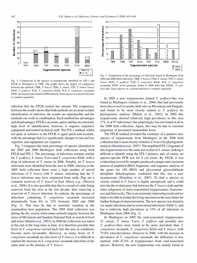
Fig. 2. Comparison of the species of trypanosome identified by ITS-1 and
FFLB in Msubugwe in 2006. The graph shows the degree of congruency
between the methods. TBR, T. brucei; TSM, T. simiae; TST, T. simiae Tsavo;
DGG, T. godfreyi; TCK, T. congolense Kilifi; TCS, T. congolense savannah;
NEW, novel genotype found in 2006 field trip. Each species in a mixed infection
is counted separately.
Fig. 3. Comparison of the percentage of infections found in Msubugwe from
2005 and 2006 field collections. TBR, T. brucei; TSM, T. simiae; TST, T. simiae
Tsavo; DGG, T. godfreyi; TCK, T. congolense Kilifi; TCS, T. congolense
savannah; NEW, novel genotype found in 2006 field trip; DGGL, T. god-
freyi-like. Each species in a mixed infection is counted separately.
E.R. Adams et al. / Infection, Genetics and Evolution 8 (2008) 439–444442
infection that the FFLB system has missed. The congruency
between the results shows that both methods are accurate in their
identification of infection, the results are reproducible and the
methods can work in combination. Each method has advantages
and disadvantages: FFLB is accurate, quick and has an extremely
high level of identification; however, it requires expensive
equipment and trained technical staff. The ITS-1 method, whilst
not quite as sensitive as the FFLB, is again quick and accurate,
with the advantage that it is significantly cheaper to run and less
expertise and equipment are required.
Fig. 3 compares the total percentage of species identified in
the 2005 and 2006 Msubugwe field collections using both
FFLB and ITS-1. The percentage of infections remains similar
for T. godfreyi, T. simiae Tsavo and T. congolense Kilifi, with a
drop of infections of T. simiae in 2006. Notably, no T. brucei
infections were identified from the area in 2006, whereas in the
2005 field collection there were a high number of mixed
infections of T. brucei with T. simiae, indicating that the T.
brucei infections may have originated from suids. Pigs are a
common reservoir of T. brucei in East Africa, e.g., (Waiswa
et al., 2006). It is also possible that this is a result of cattle being
removed from the area in the last decade, thus removing a
reservoir of T. brucei infection. In contrast, the percentage of
infections containing T. congolense savannah increased
dramatically from 6% to 33% between 2005 and 2006
(Fig. 3). This may be due to seasonal variation in the
mammalian host population. The 2005 field collection was
during the dry season when many animals migrate between the
areas of Mivumoni and Saadani National Park in search of food
and water (Malele et al., 2003); in 2006 the collection was taken
at the end of the long rains in June. It is therefore possible that
hosts of T. congolense moved back into the area as conditions
become more favourable. However, as many hosts of T.
congolense savannah are also hosts of T. brucei, it is difficult to
explain the increase in T. congolense savannah infections at the
same time as the absence of T. brucei.
In 2005 a new trypanosome named T. godfreyi-like was
found in Msubugwe (Adams et al., 2006) that had previously
been discovered in nearby field sites at Mivumoni and Pangani,
and found to be most closely related to T. godfreyi by
phylogenetic analysis (Malele et al., 2003). In 2005 this
trypanosome showed relatively high prevalence in this area
(7%, 6 of 87 infections), but surprisingly was not found at all in
the 2006 field collection. Again, this may be due to seasonal
migration of presumed mammalian hosts.
The FFLB method revealed the existence of a putative new
species of trypanosome from Msubugwe in the 2006 field
collection that is most closely related to T. brucei by phylogenetic
analysis (Hamilton et al., 2007). The amplified ITS-1 fragment of
this trypanosome was the same size as that of T. simiae, making it
difficult to identify using the ITS-1 primers, and, of course, no
species-specific PCR test for it yet exists. By FFLB, it was
evident that several fly samples produced a unique and consistent
pattern of amplified rRNA fragments, and sequence analysis of
the genes for 18S rRNA and glycosomal glyceraldehyde
phosphate dehydrogenase, confirmed that this was a new
trypanosome (Hamilton et al., 2007). To find a species so
closely related to T. brucei is highly unexpected, and it could
provide the evolutionary link between the T. brucei clade and the
other subgenera of tsetse-transmitted trypanosomes, Nannomo-
nas and Duttonella. This is an extremely interesting result and we
hope to be able to isolate the living specimen in the near future for
further biological characterisation. The new species was found in
six single infections and in seven mixed infections (Table 1), and
has a relatively high prevalence at 15% of all infections in
Msubugwe from 2006 (Fig. 3).
In Msubugwe in 2005 the suid-associated trypanosomes
(T. simiae, T. simiae Tsavo, T. godfreyi and possibly also
T. godfreyi-like) were found to be more prevalent than T.
congolense savannah, T. congolense Kilifi and T. brucei, with
75.8% total prevalence. However in 2006, with the increase in
prevalence of T. congolense savannah, the difference is less
marked, with 47.0% of trypanosomes from suid-associated
species. However, the new trypanosome was mainly found in
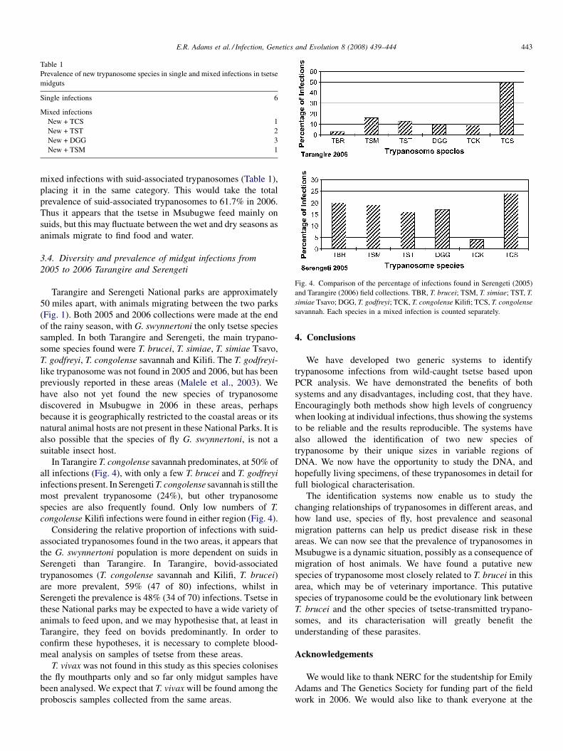
Fig. 4. Comparison of the percentage of infections found in Serengeti (2005)
and Tarangire (2006) field collections. TBR, T. brucei; TSM, T. simiae; TST, T.
simiae Tsavo; DGG, T. godfreyi; TCK, T. congolense Kilifi; TCS, T. congolense
savannah. Each species in a mixed infection is counted separately.
Table 1
Prevalence of new trypanosome species in single and mixed infections in tsetse
midguts
Single infections 6
Mixed infections
New + TCS 1
New + TST 2
New + DGG 3
New + TSM 1
E.R. Adams et al. / Infection, Genetics and Evolution 8 (2008) 439–444 443
mixed infections with suid-associated trypanosomes (Table 1),
placing it in the same category. This would take the total
prevalence of suid-associated trypanosomes to 61.7% in 2006.
Thus it appears that the tsetse in Msubugwe feed mainly on
suids, but this may fluctuate between the wet and dry seasons as
animals migrate to find food and water.
3.4. Diversity and prevalence of midgut infections from
2005 to 2006 Tarangire and Serengeti
Tarangire and Serengeti National parks are approximately
50 miles apart, with animals migrating between the two parks
(Fig. 1). Both 2005 and 2006 collections were made at the end
of the rainy season, with G. swynnertoni the only tsetse species
sampled. In both Tarangire and Serengeti, the main trypano-
some species found were T. brucei, T. simiae, T. simiae Tsavo,
T. godfreyi, T. congolense savannah and Kilifi. The T. godfreyi-
like trypanosome was not found in 2005 and 2006, but has been
previously reported in these areas (Malele et al., 2003). We
have also not yet found the new species of trypanosome
discovered in Msubugwe in 2006 in these areas, perhaps
because it is geographically restricted to the coastal areas or its
natural animal hosts are not present in these National Parks. It is
also possible that the species of fly G. swynnertoni, is not a
suitable insect host.
In Tarangire T. congolense savannah predominates, at 50% of
all infections (Fig. 4), with only a few T. brucei and T. godfreyi
infections present. In Serengeti T. congolense savannah is still the
most prevalent trypanosome (24%), but other trypanosome
species are also frequently found. Only low numbers of T.
congolense Kilifi infections were found in either region (Fig. 4).
Considering the relative proportion of infections with suid-
associated trypanosomes found in the two areas, it appears that
the G. swynnertoni population is more dependent on suids in
Serengeti than Tarangire. In Tarangire, bovid-associated
trypanosomes (T. congolense savannah and Kilifi, T. brucei)
are more prevalent, 59% (47 of 80) infections, whilst in
Serengeti the prevalence is 48% (34 of 70) infections. Tsetse in
these National parks may be expected to have a wide variety of
animals to feed upon, and we may hypothesise that, at least in
Tarangire, they feed on bovids predominantly. In order to
confirm these hypotheses, it is necessary to complete blood-
meal analysis on samples of tsetse from these areas.
T. vivax was not found in this study as this species colonises
the fly mouthparts only and so far only midgut samples have
been analysed. We expect that T. vivax will be found among the
proboscis samples collected from the same areas.
4. Conclusions
We have developed two generic systems to identify
trypanosome infections from wild-caught tsetse based upon
PCR analysis. We have demonstrated the benefits of both
systems and any disadvantages, including cost, that they have.
Encouragingly both methods show high levels of congruency
when looking at individual infections, thus showing the systems
to be reliable and the results reproducible. The systems have
also allowed the identification of two new species of
trypanosome by their unique sizes in variable regions of
DNA. We now have the opportunity to study the DNA, and
hopefully living specimens, of these trypanosomes in detail for
full biological characterisation.
The identification systems now enable us to study the
changing relationships of trypanosomes in different areas, and
how land use, species of fly, host prevalence and seasonal
migration patterns can help us predict disease risk in these
areas. We can now see that the prevalence of trypanosomes in
Msubugwe is a dynamic situation, possibly as a consequence of
migration of host animals. We have found a putative new
species of trypanosome most closely related to T. brucei in this
area, which may be of veterinary importance. This putative
species of trypanosome could be the evolutionary link between
T. brucei and the other species of tsetse-transmitted trypano-
somes, and its characterisation will greatly benefit the
understanding of these parasites.
Acknowledgements
We would like to thank NERC for the studentship for Emily
Adams and The Genetics Society for funding part of the field
work in 2006. We would also like to thank everyone at the
E.R. Adams et al. / Infection, Genetics and Evolution 8 (2008) 439–444444
TTRI, especially Dr. Msangi and Fussah, Matembo and
Mashenga for help with fieldwork and dissection, TANAPA HQ
in Arusha and Tarangire National Park Management for the
permit to trap tsetse in the park. Thanks to Tim Colborn for
assistance with artwork.
References
Adams, E.R., Malele, I.I., Msangi, A.R., Gibson, W.C., 2006. Trypanosome
identification in wild tsetse populations in Tanzania using generic
primers to amplify the ribosomal RNA ITS-1 region. Acta Trop. 100,
103–109.
Bruford, M.W., Hanotte, O., Brookfield, J.F.Y., Burke, T., 1998. Multilocus and
single-locus DNA fingerprinting. In: Hoelzel, A.R. (Ed.), Molecular
Genetic Analysis of Populations: A Practical Approach. IRL Press,
Oxford, pp. 287–336.
Cox, A., Tilley, A., McOdimba, F., Fyfe, J., Eisler, M., Hide, G., Welburn, S.,
2005. A PCR based assay for detection and differentiation of African
trypanosome species in blood. Exp. Parasitol. 111, 24–29.
Desquesnes, M., McLaughlin, G., Zoungrana, A., Davila, A.M.R., 2001.
Detection and identification of Trypanosoma of African livestock through
a single PCR based on internal transcribed spacer 1 of rDNA. Int. J.
Parasitol. 31, 610–614.
Gibson, W.C., Dukes, P., Gashumba, J.K., 1988. Species-specific DNA
probes for the identification of trypanosomes in tsetse. Parasitology 97,
63–73.
Hamilton, P.B., Adams, E.R., Malele, I.I., Gibson, W.C., 2007. A novel high
throughput technique for species identification, reveals a new species of
tsetse-transmitted trypanosome related to Trypanosoma brucei. Infect.
Genet. Evol., submitted.
Hargrove, J.W., Langley, P.A., 1990. Sterilizing tsetse in the field: a successful
field trial. Bull. Entomol. Res. 80, 397–403.
Kukla, B.A., Majiwa, P.A.O., Young, C.J., Moloo, S.K., Ole-Moiyoi, O.K.,
1987. Use of species-specific DNA probes for the detection and identifica-
tion of trypanosome infections in tsetse flies. Parasitology 95, 1–26.
Lehane, M.J., Msangi, A.R., Whitaker, C.J., Lehane, S.M., 2000. Grouping of
trypanosome species in mixed infections in Glossina pallidipes. Parasitol-
ogy 120, 583–592.
Lloyd, L., Johnson, W.B., 1924. The trypanosome infections of tsetse flies in
Northern Nigeria and a new method of estimation. Bull. Entomol. Res. 14,
265–288.
Majiwa, P.A.O., Otieno, L.H., 1990. Recombinant DNA probes reveal simul-
taneous infection of tsetse flies with different trypanosome species. Mol.
Biochem. Parasitol. 40, 245–254.
Majiwa, P.A.O., Thatthi, R., Moloo, S.K., Nyeko, J.H.P., Otieno, L.H., Maloo,
S., 1994. Detection of trypanosome infections in the saliva of tsetse flies and
buffy coat samples from antigenaemic but aparasitaemic cattle. Parasitology
108, 313–322.
Malele, I., Craske, L., Knight, C., Ferris, V., Njiru, Z., Hamilton, P., Lehane, S.,
Lehane, M., Gibson, W., 2003. Identification of new trypanosome species
from wild tsetse flies in Tanzania. Infect. Genet. Evol. 3, 271–279.
Masiga, D.K., Smyth, A.J., Hayes, P.J., Bromidge, T.J., Gibson, W.C., 1992.
Sensitive detection of trypanosomes in tsetse flies by DNA amplification.
Int. J. Parasitol. 22, 909–918.
McNamara, J., Dukes, P., Snow, W.F., Gibson, W.C., 1989. Use of DNA probes
to identify Trypanosoma congolense and T. simiae in tsetse from The
Gambia. Acta Trop. 46, 55–61.
McNamara, J.J., Laveissiere, C., Masiga, D.K., 1995. Multiple trypanosome
infections in wild tsetse in Cote d’Ivoire detected by PCR analysis. Acta
Trop. 59, 85–92.
Moser, D.R., Cook, G.A., Ochs, D.E., Bailey, C.P., McKane, M.R., Donelson,
J.E., 1989. Detection of Trypanosoma congolense and T. brucei subspecies
by DNA amplification using the polymerase chain reaction. Parasitology 99,
57–66.
Njiru, Z.K., Makumi, J.N., Okoth, S., Ndungu, J.M., Gibson, W.C., 2004.
Identification of trypanosomes in Glossina pallidipes and G. longipennis in
Kenya. Infect. Genet. Evol. 4, 29–35.
Solano, P., Argiro, L., Reifenberg, J.M., Yao, Y., Duvallet, G., 1995. Field
application of the polymerase chain reaction (PCR) to the detection and
characterization of trypanosomes in Glossina longipalpis (Diptera: Glossi-
nidae) in Cote d’Ivoire. Mol. Ecol. 4, 781–785.
Waiswa, C., Olaho-Mukani, W., Katunguka-Rwakishaya, E., 2006. Domestic
animals as reservoirs for sleeping sickness in three endemic foci in south-
eastern Uganda. Ann. Trop. Med. Parasitol. 97, 149–155.
Woolhouse, M.E.J., McNamara, J.J., Hargrove, J.W., Bealby, K.A., 1996.
Distribution and abundance of trypanosome (subgenus Nannomonas) infec-
tions of the tsetse fly Glossina pallidipes in southern Africa. Mol. Ecol. 5,
11–18.
www.elsevier.com/locate/meegid
Infection, Genetics and Evolution 8 (2008) 445–449
Molecular detection of divergent trypanosomes among
rodents of Thailand
S. Jittapalapong a, T. Inpankaew a, N. Sarataphan b,V. Herbreteau c, J.P. Hugot d, S. Morand e,*, R.W. Stich f
a Department of Parasitology, Kasetsart University, Bangkok, Thailandb National Institute of Animal Health, Pathumthani, Thailand
c Laboratoire Espace, Sante, Territoire, Universite Paris X, Nanterre, Franced OSEB, UMR 5202 CNRS, MNHN, Paris, France
e Institut des Sciences de l’Evolution, CNRS-UM2, Universite Montpellier 2, 34095 Montpellier, Francef Department of Veterinary Pathobiology, University of Missouri, Columbia, MO, USA
Received 19 April 2007; received in revised form 17 August 2007; accepted 21 August 2007
Available online 25 August 2007
Abstract
Herpetosoma is a homogenous subgenus of several dozen named species that are often described as morphologically indistinguishable T. lewisi-
like parasites. These trypanosomes normally infect rodents and utilize fleas as vectors. Although this trypanosome subgenus is considered non-
pathogenic to normal hosts, some of them are on rare occasion reported in association with human disease. Recently, a T. lewisi-like infection was
detected in a sick Thai infant, thus the objective of this study was to investigate the prevalence of T. lewisi infections among different rodents
indigenous to Thailand in order to identify possible sources of human cases. Blood was collected from a total of 276 rodents trapped from urban and
rural areas of three Thai provinces between 2006 and 2007. These samples were processed for DNA isolation and tested with a PCR assay universal
for the genus Trypanosoma, followed by internal transcribed spacer 1 (ITS-1) sequence analysis to identify infections in positive samples.
Herpetosoma known as T. lewisi-like trypanosomes were present among Rattus (14.3%) and Bandicota (18.0%) rodent species and salivarian
trypanosomes closely related to T. evansi were detected in Leopoldamys (20%) and Rattus (2.0%) species. Herpetosoma were prevalent among
rodents associated with both human and sylvatic habitats, while three of the four salivaria-positive rodents were from a forest biotope. A
Herpetosoma ITS-1 sequence amplified from one of these samples was 97.9% identical to that reported for T. lewisi in an experimentally infected
rat and 96.4% identical to the sequence amplified from blood from a Thai infant. Habitats where rodents were collected significantly affect rodent
infection, at least for T. lewisi, suggesting that the degree of anthropization may influence the transmission of Trypanosoma spp. These results
suggest that multiple Herpetosoma species or strains are enzootic to Thailand, and that Rattus and Bandicota species are possible sources of human
exposure to these parasites.
# 2007 Published by Elsevier B.V.
Keywords: Trypanosoma; Herpetosoma; Salivaria; Rodents; Habitat; Internal transcribed spacer 1 (ITS-1)
1. Introduction
The etiologic agents of many serious infectious diseases
utilize invertebrate hosts during a portion of their life cycle.
Most of these agents are adapted to hematophagous arthropods
that share their vertebrate hosts, and identification of arthropod
vectors and vertebrate reservoirs is usually key to sustained
* Corresponding author.
E-mail address: [email protected] (S. Morand).
1567-1348/$ – see front matter # 2007 Published by Elsevier B.V.
doi:10.1016/j.meegid.2007.08.002
control of vector-borne diseases. Trypanosomes are flagellated
protozoan parasites, some of which can cause distinct zoonoses,
the most notable of which include the Leishmaniases,
American trypanosomiasis (Chagas’ disease) and African
trypanosomiasis (sleeping sickness). Like most vector-borne
zoonotic agents, trypanosomes that infect humans utilize a
broad vertebrate host range and a relatively narrow range of
invertebrate vectors. However, there are rare yet notable
exceptions to the latter generalization.
The genus Trypanosoma can be divided into two major
groups that infect vertebrates, the salivaria and the stercoraria
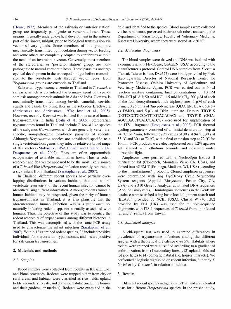
S. Jittapalapong et al. / Infection, Genetics and Evolution 8 (2008) 445–449446
(Hoare, 1972). Members of the salivaria or ‘anterior station’
group are frequently pathogenic to vertebrate hosts. These
organisms usually undergo cyclical development in the anterior
part of the insect, midgut, prior to biological transmission via
vector salivary glands. Some members of this group are
mechanically transmitted by inoculation during vector feeding
and some others are completely adapted to vertebrates without
the need of an invertebrate vector. Conversely, most members
of the stercoraria, or ‘posterior station’ group, are non-
pathogenic to natural vertebrate hosts. These parasites undergo
cyclical development in the arthropod hindgut before transmis-
sion to the vertebrate hosts through vector feces. Both
Trypanosoma groups are enzootic to Thailand.
Salivarian trypanosome enzootic to Thailand is T. evansi, a
salivaria, which is considered the primary agent of trypano-
somiasis among domestic animals in Asia and India. T. evansi is
mechanically transmitted among bovids, camelids, cervids,
equids and canids by biting flies in the suborder Brachycera
(Shrivastava and Shrivastava, 1974; Joshi et al., 2005).
However, recently T. evansi was isolated from a case of human
trypanosomiasis in India (Joshi et al., 2005). Stercorarian
trypanosomes found in Thailand include T. lewisi-like species
of the subgenus Herpetosoma, which are generally vertebrate-
specific, non-pathogenic flea-borne parasites of rodents.
Although Herpetosoma species are considered specific to a
single vertebrate host genus, they infect a relatively broad range
of flea vectors (Molyneux, 1969; Linardi and Botelho, 2002;
Desquesnes et al., 2002). Fleas are often opportunistic
ectoparasites of available mammalian hosts. Thus, a rodent
reservoir and flea vector appeared to be the most likely source
of a T. lewisi-like (Herpetosoma) infection recently reported in
a sick infant from Thailand (Sarataphan et al., 2007).
In Thailand, different rodent species have partially over-
lapping distributions in various habitats, thus the natural
vertebrate reservoir(s) of the recent human infection cannot be
identified using current information. Although rodents found in
human habitats may be suspected, given the rarity of human
trypanosomiasis in Thailand, it is also plausible that the
aforementioned human infection was a Trypanosoma sp.
naturally infecting rodents spp. not normally associated with
humans. Thus, the objective of this study was to identify the
rodent reservoirs of trypanosomes among different biotopes in
Thailand. This was accomplished with the same PCR assay
used to characterize the infant infection (Sarataphan et al.,
2007). Within 12 examined rodent species, 38 included positive
individuals for stercorarian trypanosomes, and 4 were positive
for salivarian trypanosomes.
2. Materials and methods
2.1. Samples
Blood samples were collected from rodents in Kalasin, Loei
and Phrae provinces. Rodents were trapped either from city or
rural areas, and habitats were classified as rice fields, upland
fields, secondary forests, and domestic habitat (including houses
and their gardens, or markets). Rodents were examined in the
field and identified to the species. Blood samples were collected
via heart puncture, preserved in citrate salt tubes, and sent to the
Department of Parasitology, Faculty of Veterinary Medicine,
Kasetsart University, where they were stored at �20 8C.
2.2. Molecular diagnostics
The blood samples were thawed and DNA was isolated with
a commercial kit (FlexiGene, QIAGEN, USA) according to the
manufacturer’s protocol. Control DNA samples from T. evansi
(Tansui, Taiwan isolate, D89527) were kindly provided by Prof.
Ikuo Igarashi, Director of National Research Center for
Protozoan Disease, Obihiro University of Agriculture and
Veterinary Medicine, Japan. PCR was carried out in 50 ml
reaction mixture containing final concentrations of 10 mM
Tris–HCl pH 8.3, 50 mM KCl, 1.5 mM MgCl2, 200 mM of each
of the four deoxyribonucleotide triphosphates, 1 mM of each
primer, 0.25 units of Taq polymerase (QIAGEN, USA), 5% (v/
v) DMSO, and 5 mL of DNA template. Primers TRYP1S
(CGTCCCTGCCATTTGTACACAC) and TRYP1R (GGA-
AGCCAAGTCATCCATCG) were used for amplification of
the ITS-1 fragment (Desquesnes et al., 2002). PCR thermal
cycling parameters consisted of an initial denaturation step at
94 8C for 2 min, followed by 35 cycles of 30 s at 94 8C, 30 s at
55 8C and 30 s at 72 8C, with a final extension step at 72 8C for
10 min. PCR products were electrophoresed on a 1.2% agarose
gel, stained with ethidium bromide and observed under
ultraviolet light.
Amplicons were purified with a NucleoSpin Extract II
purification kit (Clonetech, Mountain View, CA, USA), and
cloned into pGEM-T (Promega, Madison, WI, USA) according
to the manufacturers’ protocols. Cloned amplicon sequences
were determined with Taq DyeDeoxy Cycle Sequencing
System reagents (Applied Biosystems, Foster City, CA,
USA) and a 310 Genetic Analyzer automated DNA sequencer
(Applied Biosystems). Homologous sequences in the GenBank
database were searched using basic local alignment search tool
(BLAST) provided by NCBI (USA). Clustal W (V. 1.82)
provided by EBI (UK) was used for multiple-sequence
alignments with ITS-1 sequences of T. lewisi from an infected
rat and T. evansi from Taiwan.
2.3. Statistical analysis
A chi-square test was used to examine differences in
prevalence of trypanosome infections among the different
species with a theoretical prevalence over 5%. Habitats where
rodent were trapped were classified according to a gradient of
anthropization: from (1) secondary forests, (2) upland fields and
(3) rice fields to (4) domestic habitat (i.e. houses, markets). We
performed a logistic regression on rodent infection, either by T.
lewisi or by T. evansi, in relation to habitat.
3. Results
Different rodent species indigenous to Thailand are potential
hosts for different Herpetosoma species. In the present study,
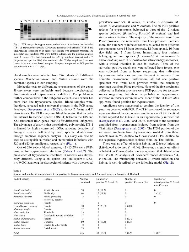
Fig. 1. PCR assays for trypanosomes rodent blood. Amplicons that included
ITS-1 of trypanosome-specific rDNAwere generated with primers TRYP1S and
TRYP1R and visualized on an agarose gel stained with ethidium bromide. The
molecular size standards (M) were 100 bp ladders, and the positive controls
were T. evansi (Te) that contained the 520 bp amplicon (arrow) and a T.
Herpetosoma species (TH) that contained the 623 bp amplicon (chevron).
Lanes 1–14 are rodent blood samples. Samples interpreted as PCR-positive
are indicated with a ‘‘+’’ sign.
S. Jittapalapong et al. / Infection, Genetics and Evolution 8 (2008) 445–449 447
blood samples were collected from 276 rodents of 12 different
species. Bandicota savilei and Rattus exulans were the
dominant species in our sampling.
Molecular tests to differentiate trypanosomes of the genus
Trypanosoma were preferably used because morphological
discrimination of trypanosomes is difficult. The problem is
further compounded in the subgenus Herpetosoma which has
more than one trypanosome species. Blood samples were,
therefore, screened using universal primers in the PCR assay
developed Desquesnes et al. (2002) to detect T. lewisi and T.
vivax in rats. The PCR assay amplified the region that includes
the internal transcribed spacer 1 (IST-1) between the 18S and
5.8S ribosomal RNA genes (rDNA) for differential diagnosis.
The advantage of assay is that the relatively polymorphic ITS-1
is flanked by highly conserved rDNA, allowing detection of
divergent species followed by more specific identification
through amplicon sequence analysis. This assay can also be
used to distinguish salivarian and stercorarian infections with
520 and 623 bp amplicons, respectively (Fig. 1).
Out of 276 rodent blood samples, 42 (15.2%) were PCR-
positive for trypanosome infections (Tables 1 and 2). The
prevalence of trypanosome infections in rodents was statisti-
cally different, using a chi-square test (chi-square = 123.1,
p < 0.0001), among the six species of rodents with a theoretical
Table 1
Species and number of rodents found to be positive to Trypanosoma lewisi and T.
Rodent species Biotope Number
examined
N
po
Bandicota indica Ricefields, wet 58 10
Bandicota savilei Fields, dry 64 12
Berylmys bowersi Fields, proximity
to forests, uplands
3 0
Berylmys berdmorei Forests 1 0
Leopoldamys edwardsi Forests 10 2
Maxomys surifer Forests 12 0
Mus cervicolor Ricefields 13 0
Mus cookii Grasslands, upland ricefields 17 0
Rattus andamanensis Forests 3 0
Rattus exulans Domestic 80 14
Rattus losea Ricefields, other fields 2 0
Rattus tanezumi Peri-domestic 13 0
Total 276 38
prevalence over 5%: B. indica, B. savilei, L. edwardsi, M.
cookii, R. andamanensis and R. exulans. The 36 PCR-positive
rodents for trypanosomes belonged to three of the 12 rodent
species collected (B. indica, B.sarilei, B exulans) and had
stercorarian infections. The majority of the rodents were from
Phrae province, the remainder from Loei province. Further-
more, the numbers of infected rodents collected from different
environments were 14 from domestic, 12 from upland, 10 from
rice field and 2 from forest. Interestingly, four rodents
belonging to three species (L. edwardsi, R. andamanensis
and R. exulans) were PCR-positive for salivarian trypanosomes,
with a mixed infection in one R. exulans. Three of the
salivarian-positive specimens were collected from forests, one
was collected from a house suggesting that salivarian
trypanosome infections are less frequent in rodents from
domestic environment, Furthermore, all but one positive
specimen was from Loei province while the remaining
specimen was from Phrae province. None of the five specimens
collected in Kalasin province were PCR-positive for trypano-
somes suggesting that there is probably no trypanosome
infection in rodents within the area. None of the 31 tested Mus
spp. were found positive for trypanosomes.
Amplicons were sequenced to confirm the identity of the
parasites detected with PCR. The ITS-1 portion of the sequence
representative of the stercorarian amplicon was 97.9% identical
to that reported for T. lewisi in an experimentally infected rat
(Desquesnes et al., 2002) and 96.4% identical to the sequence
amplified from trypanosomes isolated from rodents from the
Thai infant (Sarataphan et al., 2007). The ITS-1 portion of the
salivarian amplicon from trypanosomes isolated from these
rodents was 96.5% identical to T. evansi and 41.1% identical to
the sequence trypanosomes isolated from the Thai infant.
There was no effect of rodent habitat on T. lewisi infection
(Likelihood ratio test, P = 0.46). However, a significant effect
of habitat on T. evansi infection was observed (Likelihood ratio
test, P = 0.02; analysis of deviance: model deviance = 5.4,
P = 0.02). The relationship between T. evansi infection and
habitat is well described by the following model (Fig. 2):
evansi in several biotopes of Thailand
umber of
sitive T. lewisi (%)
Number of
positive T. evansi (%)
Number of
mixed positive T. lewisi
and T. evansi
(17.2) 0 0
(18.8) 0 1 (1.6)
0 0
0 0
(20.0) 2 (20.0)
0
0
0
1 (33.3)
(17.5) 1 (1.3)
0
0
(13.8) 4 (1.5) 1 (0.4)
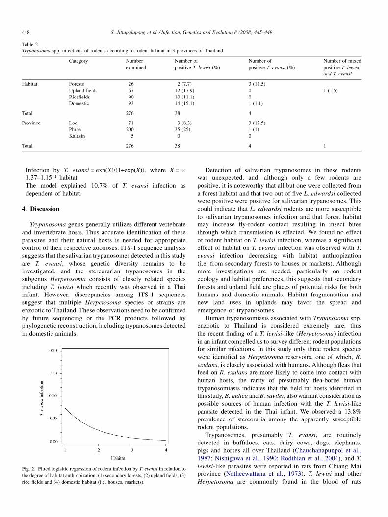
Table 2
Trypanosoma spp. infections of rodents according to rodent habitat in 3 provinces of Thailand
Category Number
examined
Number of
positive T. lewisi (%)
Number of
positive T. evansi (%)
Number of mixed
positive T. lewisi
and T. evansi
Habitat Forests 26 2 (7.7) 3 (11.5)
Upland fields 67 12 (17.9) 0 1 (1.5)
Ricefields 90 10 (11.1) 0
Domestic 93 14 (15.1) 1 (1.1)
Total 276 38 4
Province Loei 71 3 (8.3) 3 (12.5)
Phrae 200 35 (25) 1 (1)
Kalasin 5 0 0
Total 276 38 4 1
S. Jittapalapong et al. / Infection, Genetics and Evolution 8 (2008) 445–449448
� I
F
th
ri
nfection by T. evansi = exp(X)/(1+exp(X)), where X = �1.37–1.15 * habitat.
� T
he model explained 10.7% of T. evansi infection asdependent of habitat.
4. Discussion
Trypanosoma genus generally utilizes different vertebrate
and invertebrate hosts. Thus accurate identification of these
parasites and their natural hosts is needed for appropriate
control of their respective zoonoses. ITS-1 sequence analysis
suggests that the salivarian trypanosomes detected in this study
are T. evansi, whose genetic diversity remains to be
investigated, and the stercorarian trypanosomes in the
subgenus Herpetosoma consists of closely related species
including T. lewisi which recently was observed in a Thai
infant. However, discrepancies among ITS-1 sequences
suggest that multiple Herpetosoma species or strains are
enzootic to Thailand. These observations need to be confirmed
by future sequencing or the PCR products followed by
phylogenetic reconstruction, including trypanosomes detected
in domestic animals.
ig. 2. Fitted logisitic regression of rodent infection by T. evansi in relation to
e degree of habitat anthropization: (1) secondary forests, (2) upland fields, (3)
ce fields and (4) domestic habitat (i.e. houses, markets).
Detection of salivarian trypanosomes in these rodents
was unexpected, and, although only a few rodents are
positive, it is noteworthy that all but one were collected from
a forest habitat and that two out of five L. edwardsi collected
were positive were positive for salivarian trypanosomes. This
could indicate that L. edwardsi rodents are more susceptible
to salivarian trypanosomes infection and that forest habitat
may increase fly-rodent contact resulting in insect bites
through which transmission is effected. We found no effect
of rodent habitat on T. lewisi infection, whereas a significant
effect of habitat on T. evansi infection was observed with T.
evansi infection decreasing with habitat anthropization
(i.e. from secondary forests to houses or markets). Although
more investigations are needed, particularly on rodent
ecology and habitat preferences, this suggests that secondary
forests and upland field are places of potential risks for both
humans and domestic animals. Habitat fragmentation and
new land uses in uplands may favor the spread and
emergence of trypanosomes.
Human trypanosomiasis associated with Trypanosoma spp.
enzootic to Thailand is considered extremely rare, thus
the recent finding of a T. lewisi-like (Herpetosoma) infection
in an infant compelled us to survey different rodent populations
for similar infections. In this study only three rodent species
were identified as Herpetosoma reservoirs, one of which, R.
exulans, is closely associated with humans. Although fleas that
feed on R. exulans are more likely to come into contact with
human hosts, the rarity of presumably flea-borne human
trypanosomiasis indicates that the field rat hosts identified in
this study, B. indica and B. savilei, also warrant consideration as
possible sources of human infection with the T. lewisi-like
parasite detected in the Thai infant. We observed a 13.8%
prevalence of stercoraria among the apparently susceptible
rodent populations.
Trypanosomes, presumably T. evansi, are routinely
detected in buffaloes, cats, dairy cows, dogs, elephants,
pigs and horses all over Thailand (Chauchanapunpol et al.,
1987; Nishigawa et al., 1990; Rodthian et al., 2004), and T.
lewisi-like parasites were reported in rats from Chiang Mai
province (Natheewattana et al., 1973). T. lewisi and other
Herpetosoma are commonly found in the blood of rats
S. Jittapalapong et al. / Infection, Genetics and Evolution 8 (2008) 445–449 449
worldwide, and members of this subgenus are generally
considered to be non-pathogenic and rarely found in
humans (Hoare, 1972). The results of this survey confirmed
that some wild rodents could represent highly prevalent
reservoirs of T. lewisi-like parasites. Our results show that
habitats where we collected rodents significantly affect
rodent infection, at least by T. lewisi, suggesting that the
degree of anthropization may influence the transmission of
Trypanosoma spp.
In summary, various rodents from different habitats
appear to present risks for human exposure to trypanosomes
in Thailand. The results of this study suggest that Rattus
and Bandicota species are possible sources of human
exposure to stercoraria, and that multiple trypanosomes
species or strains within the Herpetosoma subgenus is
enzootic to Thailand. Remaining questions include whether
trypanosomes detected in these rodents are infective to flea
and human populations in the same region. Further work is
also needed to isolate and characterize these parasites in an
experimental system and to test the vector competence of
indigenous fleas.
Acknowledgements
The authors gratefully acknowledge Kasetsart Univer-
sity Research Development Institution (KURDI) for
partial funding for publication and presentation of this
work and the French program ANR-Sante-Environne-
ment (no. 00121 0505) for supporting the fieldwork. We
also thank Arthorn Boonsaner (National Park, Wildlife and
Plant Conservation Department, Thailand, Bangkok), Khom-
son Satchasataporn and Kittiponk Chaisiri (Faculty of Veter-
inary Technology, Kasetsart University) for their precious
assistance.
References
Chauchanapunpol, I., Withuragoon, C., Suvarnvasi, P., Nopphakhun, W.,
Panphan, S., Sakdeechumphol, K., 1987. Study on Trypanosoma evansi
infection in dairy cattle. In: The proceeding of the Sixth Annual Conference
of Livestock Development in Thailand. Department of Livestock develop-
ment, Ministry of Agriculture, 18–20 January, 1986, pp. 13–20.
Desquesnes, M., Ravel, S., Cuny, G., 2002. PCR identification of Trypanosoma
lewisi, a common parasite of laboratory rats. Kinetoplastid Biol. Dis. 1, 2.
Hoare, C.A., 1972. The Trypanosomes of Mammals. A Zoological Monograph.
Blackwell Scientific Publications, Oxford and Edinburgh, p. 749.
Joshi, P.P., Shegokar, V.R., Powar, R.M., Herder, S., Katti, R., Salkar, H.R.,
Dani, V.S., Bhargava, A., Jannin, J., Truc, P., 2005. Human trypanosomiasis
caused by Trypanosoma evansi in India: the first case report. Am. J. Trop.
Med. Hyg. 73, 491–495.
Linardi, P.M., Botelho, J.R., 2002. Prevalence of Trypanosoma lewisi in Rattus
novegicus from Belo Horizonte, State of Minas Gerais. Br. Mem. Inst.
Oswaldo Cruz. Rio de Janeiro 97, 411–414.
Molyneux, D.H., 1969. Intracellular stages of Trypanosoma lewisi in fleas and
attempts to find such stages in other trypanosome species. Parasitology 59,
737–744.
Natheewattana, N., Hongsbhanich, L., Khamboonruang, C., Thitasut, P., 1973.
Preliminary study of a Trypanosoma lewisi-like parasite of rats in Chiang
Mai. Thailand. Southeast Asian J. Trop. Med. Public Health 4, 322–323.
Nishigawa, H., Sarataphan, N., Tuntasuvan, D., Neramitmansuk, P., 1990.
Serological survey of Trypansomiasis and babesiosis in cattle and buffaloes
in Thailand. In: The proceedings of the seventh FAVA Congress, Pattaya,
Thailand, November 4–7, p. 1990.
Rodthian, P., Hinon, W., Uthaiwan, W., Withuragoon, P., Taritsanarom, U.,
Chaisert, S., Khaihong, S., Sarataphan, N., Muangyai, M., 2004. A case
report: Trypanosoma evansi in draft elephants in Lamphang. In: The 19th
Annual Conference of Livestock Development, Pathumthani. Thailand,
May 26–30.
Sarataphan, N., Vongpakorn, M., Nuansrichay, B., Autarkool, N., Keowkarn-
kah, T., Rodtian, P., Stich, R.W., Jittapalapong, S., 2007. Diagnosis of a
Trypanosoma lewisi-like infection in a sick infant from Thailand. J. Med.
Microbiol. 56, 1118–1121.
Shrivastava, K.K., Shrivastava, G.P., 1974. Two cases of Trypanosoma (Her-
petosoma) species infection of man in India. Trans. Royal Soc. Trop. Med.
Hyg. 68, 143–144.
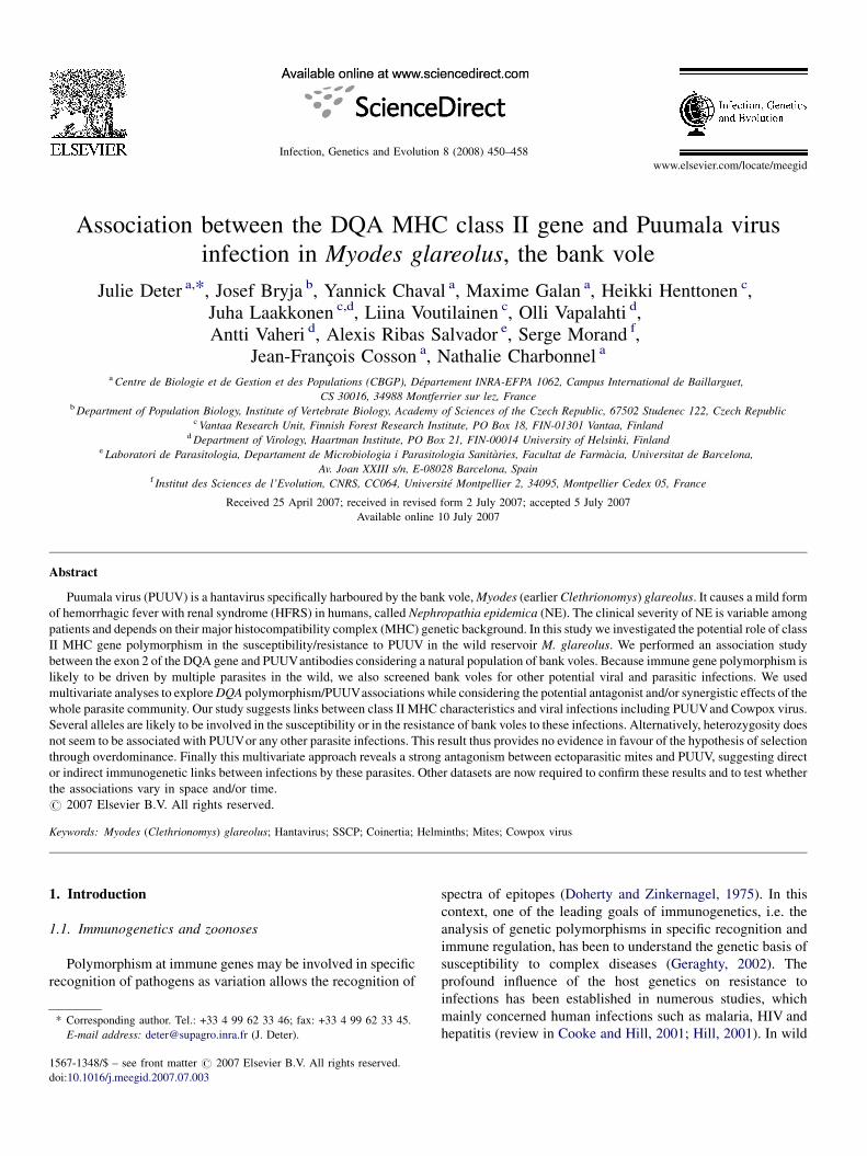
www.elsevier.com/locate/meegid
Infection, Genetics and Evolution 8 (2008) 450–458
Association between the DQA MHC class II gene and Puumala virus
infection in Myodes glareolus, the bank vole
Julie Deter a,*, Josef Bryja b, Yannick Chaval a, Maxime Galan a, Heikki Henttonen c,Juha Laakkonen c,d, Liina Voutilainen c, Olli Vapalahti d,Antti Vaheri d, Alexis Ribas Salvador e, Serge Morand f,
Jean-Francois Cosson a, Nathalie Charbonnel a
a Centre de Biologie et de Gestion et des Populations (CBGP), Departement INRA-EFPA 1062, Campus International de Baillarguet,
CS 30016, 34988 Montferrier sur lez, Franceb Department of Population Biology, Institute of Vertebrate Biology, Academy of Sciences of the Czech Republic, 67502 Studenec 122, Czech Republic
c Vantaa Research Unit, Finnish Forest Research Institute, PO Box 18, FIN-01301 Vantaa, Finlandd Department of Virology, Haartman Institute, PO Box 21, FIN-00014 University of Helsinki, Finland
e Laboratori de Parasitologia, Departament de Microbiologia i Parasitologia Sanitaries, Facultat de Farmacia, Universitat de Barcelona,
Av. Joan XXIII s/n, E-08028 Barcelona, Spainf Institut des Sciences de l’Evolution, CNRS, CC064, Universite Montpellier 2, 34095, Montpellier Cedex 05, France
Received 25 April 2007; received in revised form 2 July 2007; accepted 5 July 2007
Available online 10 July 2007
Abstract
Puumala virus (PUUV) is a hantavirus specifically harboured by the bank vole, Myodes (earlier Clethrionomys) glareolus. It causes a mild form
of hemorrhagic fever with renal syndrome (HFRS) in humans, called Nephropathia epidemica (NE). The clinical severity of NE is variable among
patients and depends on their major histocompatibility complex (MHC) genetic background. In this study we investigated the potential role of class
II MHC gene polymorphism in the susceptibility/resistance to PUUV in the wild reservoir M. glareolus. We performed an association study
between the exon 2 of the DQA gene and PUUVantibodies considering a natural population of bank voles. Because immune gene polymorphism is
likely to be driven by multiple parasites in the wild, we also screened bank voles for other potential viral and parasitic infections. We used
multivariate analyses to explore DQA polymorphism/PUUVassociations while considering the potential antagonist and/or synergistic effects of the
whole parasite community. Our study suggests links between class II MHC characteristics and viral infections including PUUVand Cowpox virus.
Several alleles are likely to be involved in the susceptibility or in the resistance of bank voles to these infections. Alternatively, heterozygosity does
not seem to be associated with PUUVor any other parasite infections. This result thus provides no evidence in favour of the hypothesis of selection
through overdominance. Finally this multivariate approach reveals a strong antagonism between ectoparasitic mites and PUUV, suggesting direct
or indirect immunogenetic links between infections by these parasites. Other datasets are now required to confirm these results and to test whether
the associations vary in space and/or time.
# 2007 Elsevier B.V. All rights reserved.
Keywords: Myodes (Clethrionomys) glareolus; Hantavirus; SSCP; Coinertia; Helminths; Mites; Cowpox virus
1. Introduction
1.1. Immunogenetics and zoonoses
Polymorphism at immune genes may be involved in specific
recognition of pathogens as variation allows the recognition of
* Corresponding author. Tel.: +33 4 99 62 33 46; fax: +33 4 99 62 33 45.
E-mail address: [email protected] (J. Deter).
1567-1348/$ – see front matter # 2007 Elsevier B.V. All rights reserved.
doi:10.1016/j.meegid.2007.07.003
spectra of epitopes (Doherty and Zinkernagel, 1975). In this
context, one of the leading goals of immunogenetics, i.e. the
analysis of genetic polymorphisms in specific recognition and
immune regulation, has been to understand the genetic basis of
susceptibility to complex diseases (Geraghty, 2002). The
profound influence of the host genetics on resistance to
infections has been established in numerous studies, which
mainly concerned human infections such as malaria, HIV and
hepatitis (review in Cooke and Hill, 2001; Hill, 2001). In wild
J. Deter et al. / Infection, Genetics and Evolution 8 (2008) 450–458 451
animal populations, immunogenetics provides key insight into
the relative influence of genetic variation and environmental
factors on host-pathogen interactions. A number of zoonoses,
i.e. infections transmitted from animals to humans (Taylor
et al., 2001), has (re-)emerged during the past 15 years (Kallio-
Kokko et al., 2005). Therefore, an important application of
immunogenetics concerns the assessment of emergent or re-
emergent disease risks in natural populations. (i) The study of
immune genes may explain why hosts differ in their
susceptibility to different parasites. The reasons why hosts
differ in their susceptibility/resistance to different parasites
could rely on the degree of matching between immune genes
and parasite antigens. Immunogenetics may thus contribute to
the identification of unknown zoonotic agents, which is
essential for understanding zoonoses epidemiology (Mills
and Childs, 1998). (ii) Natural population studies of immune
gene polymorphism may provide key insight into the factors
determining the appearance, spread and distribution of
resistance/immuno-modulating alleles within populations and
across geographical landscapes. Such information is essential
to study the spatial and temporal variations in disease risk or
incidence.
1.2. Puumala virus and hemorrhagic fever with renal
syndrome
Hantaviruses are one of the three main emerging infectious
agents in Europe (Kallio-Kokko et al., 2005). They are specific
rodent-borne viruses belonging to the Bunyaviridae family, and
are transmitted to humans primarily from aerosols of rodent
excreta. Puumala virus (PUUV) is a hantavirus, which causes a
mild form of haemorrhagic fever with renal syndrome (HFRS)
in humans, called Nephropathia epidemica (NE). The mortality
rate ranges between 0.1 and 0.4% (Vapalahti et al., 2003).
Infections are frequent in Northern Europe, European Russia
and parts of Central Western Europe (Vapalahti et al., 2003),
with about 6000 cases reported per year. In France, it has been
classified as an important emerging disease by the Institut de
Veille Sanitaire (INVS, Capek, 2006).
The specific reservoir host of PUUV is the bank vole Myodes
(earlier Clethrionomys) glareolus. In this rodent, PUUV
produces a chronic, long-lasting infection. Host and virus
have geographical ranges that do not completely overlap. Many
parts of Europe, such as the Mediterranean peninsulas and
Britain remain blank of PUUV infections although M. glareolus
is present. Two recent studies provided arguments in favour of
selection acting on bank voles and mediated by PUUV. First,
Kallio et al. (2006a,b) observed that PUUV infection could
affect bank vole fecundity (earlier breeding). Second, experi-
mental infections of M. glareolus laboratory colonies with
PUUV revealed that some bank voles could not be successfully
infected. Neither antibodies, nor PUUV RNA could be detected
in these individuals reared in PUUV-contaminated beddings,
providing evidence of variability in vole susceptibility to
PUUV. This variability was not significantly explained by age,
sex, maturation status or individual weight (Kallio et al.,
2006a,b). This suggests that immunogenetic factors could be
involved not only in the disease progression (Klein et al.,
2004a) but also in controlling infection itself. The absence of
HFRS in some parts of M. glareolus geographic distribution
could therefore be partly explained by selected vole
immunogenetic factors associated with resistance to PUUV
infections.
1.3. Immunogenetic factors associated with Puumala virus
infections
Association studies have provided evidence for the role of
immunogenetics in the course of hantavirus infections in
rodents. Using the hantavirus Seoul and the rat Rattus
norvegicus, Klein et al. (2004a,b) characterized immunologic
pathways that differ between males and females in response to
infection. They showed that most of the 192 identified immune-
related genes that encode for these immune responses were
differentially expressed in infected females compared with
infected males. Several MHC genes were associated with these
sex differences in gene expression. Alternatively, the con-
siderable variability in the clinical severity of NE among
humans depends on MHC genes (Mustonen et al., 1996, 1998,
2004; Plyusnin et al., 1997). The HLA-B227 haplotype is
associated with a mild course of NE, whereas the extended
haplotypes HLA-B8-DR3 and HLA-DRB1-0301 are associated
with severe clinical courses. The complete MHC class II region
has been sequenced in rats (Gunther and Walter, 2001; Hurt
et al., 2004) and mice (Blake et al., 2003). It has a similar
regional organization as the human MHC, and orthologous
relationships exist between class II regions in all mammals
(Takahata and Nei, 1990). Moreover, in laboratory mouse and
rats, the DQA gene (called RT1.B in rats, H2-A in mice) is
tightly linked to DRB genes (Blake et al., 2003; Hurt et al.,
2004). As DRB is at least quadriduplicated in bank voles
(Axtner and Sommer, 2007), what makes genotyping difficult
to perform, DQA was a relevant candidate to look for
associations between MHC class II genes and PUUV infection.
In this study we investigate the role of the class II MHC DQA
gene polymorphism on susceptibility/resistance to PUUV by
the mean of an association study conducted in a natural
population of Myodes glareolus, the specific reservoir of this
hantavirus. Vole sampling was performed within the second
most important French endemic area of HFRS (Jura, Franche
Comte), which has had an unusually high number of human
cases in 2005 (230 in France, 38 in the Jura, INVS).
2. Materials and methods
2.1. Trapping design
The main study area is located around Mignovillard (46880N,
68130E and elevation 850 m) in Jura, France. It consists of a 4-
km2 site, half of which is composed of wooded meadows and
the other half of forests (man-made spruce or semi-natural
forests). Bank voles were sampled in September 2004, July and
September 2005 using French Agricultural Research Institute
(INRA) live traps, which were fitted out with dormitory boxes,
J. Deter et al. / Infection, Genetics and Evolution 8 (2008) 450–458452
baited with hay and a piece of apple. Twenty 100-m trap-lines
composed of 34 traps placed at 3-m intervals were equally
distributed in open (meadows and hedges, nine lines) and
wooded (five lines in forests and six lines in meadows in border
of forest) areas. For each trapping session, these lines were
checked twice a day during three consecutive nights.
2.2. Infection screening
Once trapped, rodents were killed by cervical dislocation as
recommended by Mills et al. (1995). For each individual, blood
samples were taken from the heart or the thoracic cavity on a 1-
cm2 Whatman1 blotting paper. In the laboratory, each piece of
1-cm2 paper with dried blood was placed in 1 ml phosphate-
buffered saline. These diluted blood samples were screened for
IgG antibodies to Puumala virus (PUUV) using immunofluor-
escence assays (IFA) as described in Lundkvist et al. (1991)
(see also Klingstrom et al., 2004; Kallio-Kokko et al., 2005). As
PUUV produces a long-lasting, chronic infection in bank voles,
the detection of antibodies for PUUV is strongly related with
the presence of the virus except for very young voles because of
possible maternal antibodies.
Because immune gene polymorphism is likely to be driven
by multiple parasites in the wild (Chantrey et al., 1999; Wegner
et al., 2003), we also screened bank voles for other potential
viral and parasitic infections. M. glareolus is considered as one
of the main reservoir of Cowpox virus (CPXV, Chantrey et al.,
1999). Moreover, recent serological surveys have revealed the
potential role of M. glareolus as a reservoir of the Lymphocytic
Choriomeningitis virus (LCMV) (Kallio-Kokko et al., 2006).
We thus used vole blood samples to screen for IgG antibodies
against CPXV and LCMV by IFA as described in (Kallio-
Kokko et al., 2005, 2006; Laakkonen et al., 2006). On the field,
the presence of ectoparasitic mites was visually inspected and
noted. Whole fresh brains were squashed and checked
microscopically for the presence of Frenkelia cysts (Coccidia,
Apicomplexa: Sarcocystidae). Finally, we looked for the
presence of larval cestodes in the liver and in the body cavity
of voles. The digestive tracts (stomach, intestines) were stored
in 95% ethanol before being dissected in the laboratory. All the
helminths detected were carefully counted under the micro-
scope and then identified in Barcelona.
2.3. MHC class II gene genotyping
Genomic DNA was extracted from toe tissues by Puregene
DNA purification kit1 (Gentra Systems) according to
manufacturer’s instructions and finally eluted in 400-ml of
water. We amplified the complete exon 2 of DQA (called
RT1.Ba in rat) homologue of the MHC class II as described in
Bryja et al. (2005, 2006). A single PCR (PCR1 in Bryja et al.
(2006)) was performed on an Epgradient1 thermocycler
(Eppendorf) as it allowed the amplification of all bank vole
alleles. Capillary electophoreses-single strand conformation
polymorphism (CE-SSCP) were performed on a MegaBACE
1000 DNA Analysis system1 (Amersham Biosciences) as
described in Bryja et al. (2006). All alleles detected following
the CE-SSCP analyses were cloned and sequenced using a
minimal subset of animals exhibiting the diverse SSCP patterns
observed. Briefly, the DQA gene was amplified as described
above, but using non-labelled primers. The PCR products were
cloned and purified by the QIAquick PCR purification kit1
(Promega) as described in Bryja et al. (2006). Eight clones were
subsequently isolated for each individual selected. The plasmid
DNA was extracted using QIAprep Spin Miniprep kit1
(Qiagen). The DQA insert was sequenced using DYEnamic
ET terminator cycle sequencing kit1 (Amersham Biosciences)
and primer SP6. Sequences were performed on MegaBACE
1000 DNA analysis system1 (Amersham Biosciences). They
were edited and aligned using BioEdit Sequence Alignment
Editor 7.0.5.2 (Hall, 1999). A new sequence variant was
considered as a new allele when it met the criteria summarised
by Kennedy et al. (2002). Paup version 4.0b10 for Microsoft
Windows 95/NT was employed to construct a phylogenetic tree
of the DQA amino acid sequences based on 247 bp of the exon 2
(base pairs no. 3-249 of the complete exon), applying the
neighbour-joining (NJ) algorithm with Kimura’s two-para-
meter distances for nucleotides, and the Poisson correction for
amino acids. Bootstrap analyses (5000 replicates) were
performed to determine the reliability of the branching. R.
norvegicus (GenBank accession no. AJ554214) and Mus
musculus (no. K01924) sequences were used as outgroups in
the phylogenetic analyses.
2.4. Statistical analyses
Seroprevalence (percentage of seropositive animals) for
each virus and prevalence (percentage of infected animals) for
each parasite were calculated and 95% confidence intervals
were estimated by bootstraps using the software Quantitative
Parasitology 3.0 (Rozsa et al., 2000).
We explored the immunologic factors (DQA alleles and
heterozygosis) potentially associated with PUUV infection
status using multivariate analyses, which affords the integration
of other viruses, ectoparasites, coccidia and helminths. This
gave the opportunity to explore at the same time the antagonist
and/or synergistic effects of other parasites on DQA
polymorphism/PUUV associations. We performed a co-inertia
analysis (ACO) between (i) a genetic matrix including the
presence/absence of each DQA allele and the DQA hetero-
zygosis for each individual, and (ii) a parasitological matrix
including the abundance (when available) or presence of each
parasite species and the presence of antibodies against each
virus for each individual. The ACO links the independent
multivariate analyses of the genetic matrix (correspondence
analysis, COA) and of the parasitological matrix (normalised
principal component analysis, PCA). It considers all types of
variables and it is robust to correlations between the variables of
a matrix (Dray et al., 2003). The ACO gives co-inertia axes that
maximize the covariance between the row coordinates of the
two matrices (Dray et al., 2003; Thioulouse et al., 2004). The
genetic and parasitological results can be compared by
superimposing both kinds of variables on the ACO factor
map. Therefore, particular associations were visually detected
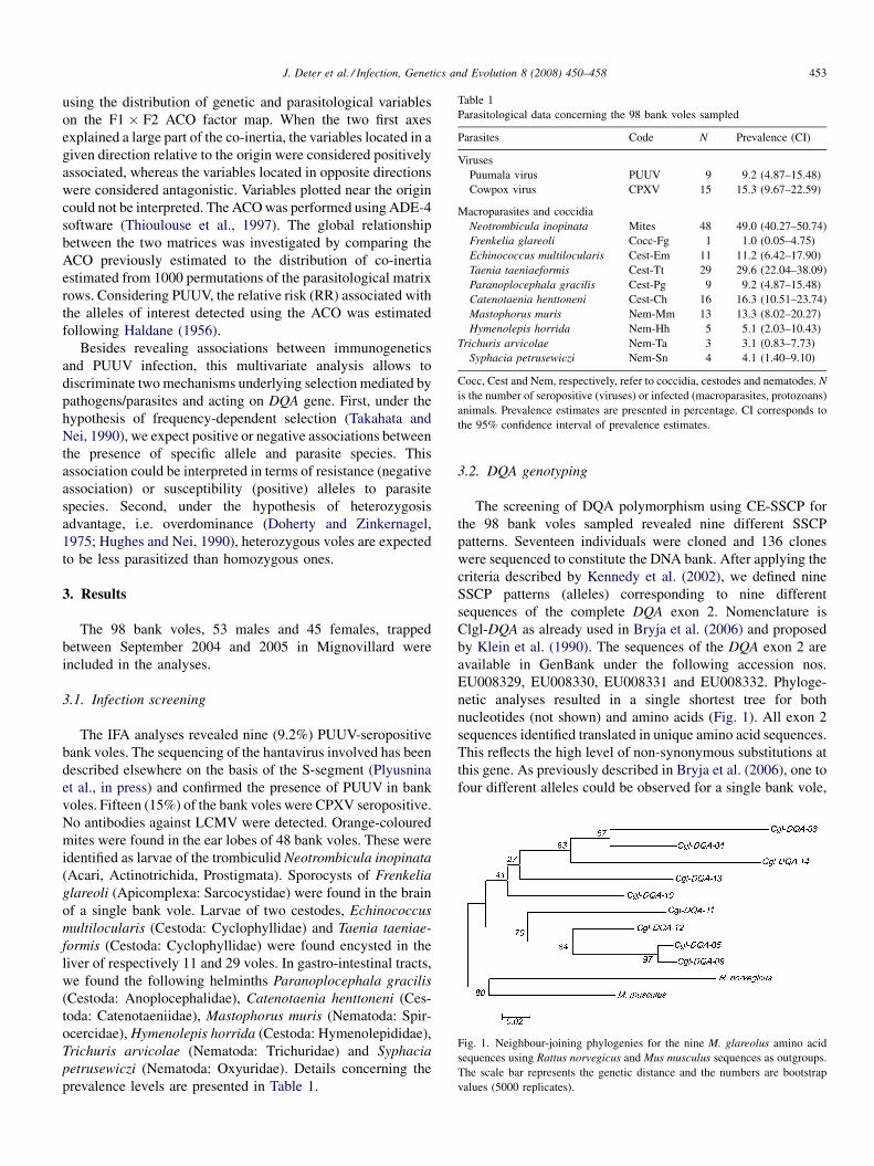
Table 1
Parasitological data concerning the 98 bank voles sampled
Parasites Code N Prevalence (CI)
Viruses
Puumala virus PUUV 9 9.2 (4.87–15.48)
Cowpox virus CPXV 15 15.3 (9.67–22.59)
Macroparasites and coccidia
Neotrombicula inopinata Mites 48 49.0 (40.27–50.74)
Frenkelia glareoli Cocc-Fg 1 1.0 (0.05–4.75)
Echinococcus multilocularis Cest-Em 11 11.2 (6.42–17.90)
Taenia taeniaeformis Cest-Tt 29 29.6 (22.04–38.09)
Paranoplocephala gracilis Cest-Pg 9 9.2 (4.87–15.48)
Catenotaenia henttoneni Cest-Ch 16 16.3 (10.51–23.74)
Mastophorus muris Nem-Mm 13 13.3 (8.02–20.27)
Hymenolepis horrida Nem-Hh 5 5.1 (2.03–10.43)
Trichuris arvicolae Nem-Ta 3 3.1 (0.83–7.73)
Syphacia petrusewiczi Nem-Sn 4 4.1 (1.40–9.10)
Cocc, Cest and Nem, respectively, refer to coccidia, cestodes and nematodes. N
is the number of seropositive (viruses) or infected (macroparasites, protozoans)
animals. Prevalence estimates are presented in percentage. CI corresponds to
the 95% confidence interval of prevalence estimates.
Fig. 1. Neighbour-joining phylogenies for the nine M. glareolus amino acid
sequences using Rattus norvegicus and Mus musculus sequences as outgroups.
The scale bar represents the genetic distance and the numbers are bootstrap
values (5000 replicates).
J. Deter et al. / Infection, Genetics and Evolution 8 (2008) 450–458 453
using the distribution of genetic and parasitological variables
on the F1 � F2 ACO factor map. When the two first axes
explained a large part of the co-inertia, the variables located in a
given direction relative to the origin were considered positively
associated, whereas the variables located in opposite directions
were considered antagonistic. Variables plotted near the origin
could not be interpreted. The ACO was performed using ADE-4
software (Thioulouse et al., 1997). The global relationship
between the two matrices was investigated by comparing the
ACO previously estimated to the distribution of co-inertia
estimated from 1000 permutations of the parasitological matrix
rows. Considering PUUV, the relative risk (RR) associated with
the alleles of interest detected using the ACO was estimated
following Haldane (1956).
Besides revealing associations between immunogenetics
and PUUV infection, this multivariate analysis allows to
discriminate two mechanisms underlying selection mediated by
pathogens/parasites and acting on DQA gene. First, under the
hypothesis of frequency-dependent selection (Takahata and
Nei, 1990), we expect positive or negative associations between
the presence of specific allele and parasite species. This
association could be interpreted in terms of resistance (negative
association) or susceptibility (positive) alleles to parasite
species. Second, under the hypothesis of heterozygosis
advantage, i.e. overdominance (Doherty and Zinkernagel,
1975; Hughes and Nei, 1990), heterozygous voles are expected
to be less parasitized than homozygous ones.
3. Results
The 98 bank voles, 53 males and 45 females, trapped
between September 2004 and 2005 in Mignovillard were
included in the analyses.
3.1. Infection screening
The IFA analyses revealed nine (9.2%) PUUV-seropositive
bank voles. The sequencing of the hantavirus involved has been
described elsewhere on the basis of the S-segment (Plyusnina
et al., in press) and confirmed the presence of PUUV in bank
voles. Fifteen (15%) of the bank voles were CPXV seropositive.
No antibodies against LCMV were detected. Orange-coloured
mites were found in the ear lobes of 48 bank voles. These were
identified as larvae of the trombiculid Neotrombicula inopinata
(Acari, Actinotrichida, Prostigmata). Sporocysts of Frenkelia
glareoli (Apicomplexa: Sarcocystidae) were found in the brain
of a single bank vole. Larvae of two cestodes, Echinococcus
multilocularis (Cestoda: Cyclophyllidae) and Taenia taeniae-
formis (Cestoda: Cyclophyllidae) were found encysted in the
liver of respectively 11 and 29 voles. In gastro-intestinal tracts,
we found the following helminths Paranoplocephala gracilis
(Cestoda: Anoplocephalidae), Catenotaenia henttoneni (Ces-
toda: Catenotaeniidae), Mastophorus muris (Nematoda: Spir-
ocercidae), Hymenolepis horrida (Cestoda: Hymenolepididae),
Trichuris arvicolae (Nematoda: Trichuridae) and Syphacia
petrusewiczi (Nematoda: Oxyuridae). Details concerning the
prevalence levels are presented in Table 1.
3.2. DQA genotyping
The screening of DQA polymorphism using CE-SSCP for
the 98 bank voles sampled revealed nine different SSCP
patterns. Seventeen individuals were cloned and 136 clones
were sequenced to constitute the DNA bank. After applying the
criteria described by Kennedy et al. (2002), we defined nine
SSCP patterns (alleles) corresponding to nine different
sequences of the complete DQA exon 2. Nomenclature is
Clgl-DQA as already used in Bryja et al. (2006) and proposed
by Klein et al. (1990). The sequences of the DQA exon 2 are
available in GenBank under the following accession nos.
EU008329, EU008330, EU008331 and EU008332. Phyloge-
netic analyses resulted in a single shortest tree for both
nucleotides (not shown) and amino acids (Fig. 1). All exon 2
sequences identified translated in unique amino acid sequences.
This reflects the high level of non-synonymous substitutions at
this gene. As previously described in Bryja et al. (2006), one to
four different alleles could be observed for a single bank vole,
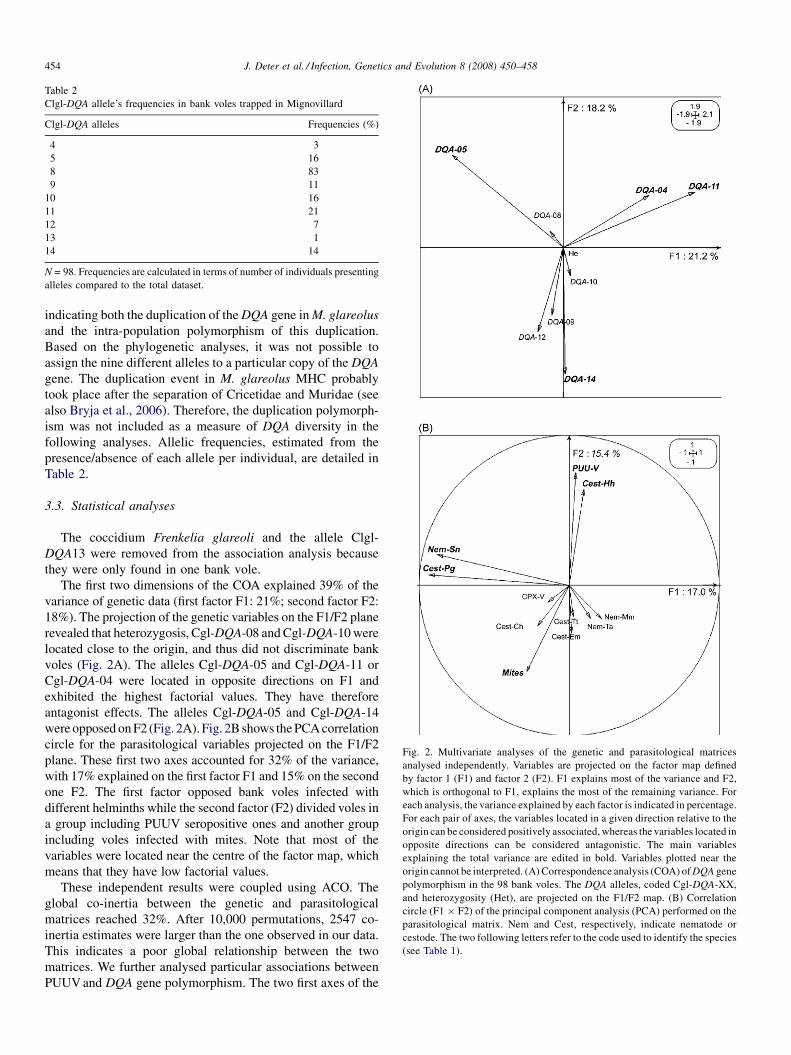
Table 2
Clgl-DQA allele’s frequencies in bank voles trapped in Mignovillard
Clgl-DQA alleles Frequencies (%)
4 3
5 16
8 83
9 11
10 16
11 21
12 7
13 1
14 14
N = 98. Frequencies are calculated in terms of number of individuals presenting
alleles compared to the total dataset.
Fig. 2. Multivariate analyses of the genetic and parasitological matrices
analysed independently. Variables are projected on the factor map defined
by factor 1 (F1) and factor 2 (F2). F1 explains most of the variance and F2,
which is orthogonal to F1, explains the most of the remaining variance. For
each analysis, the variance explained by each factor is indicated in percentage.
For each pair of axes, the variables located in a given direction relative to the
origin can be considered positively associated, whereas the variables located in
opposite directions can be considered antagonistic. The main variables
explaining the total variance are edited in bold. Variables plotted near the
origin cannot be interpreted. (A) Correspondence analysis (COA) of DQA gene
polymorphism in the 98 bank voles. The DQA alleles, coded Cgl-DQA-XX,
and heterozygosity (Het), are projected on the F1/F2 map. (B) Correlation
circle (F1 � F2) of the principal component analysis (PCA) performed on the
parasitological matrix. Nem and Cest, respectively, indicate nematode or
cestode. The two following letters refer to the code used to identify the species
(see Table 1).
J. Deter et al. / Infection, Genetics and Evolution 8 (2008) 450–458454
indicating both the duplication of the DQA gene in M. glareolus
and the intra-population polymorphism of this duplication.
Based on the phylogenetic analyses, it was not possible to
assign the nine different alleles to a particular copy of the DQA
gene. The duplication event in M. glareolus MHC probably
took place after the separation of Cricetidae and Muridae (see
also Bryja et al., 2006). Therefore, the duplication polymorph-
ism was not included as a measure of DQA diversity in the
following analyses. Allelic frequencies, estimated from the
presence/absence of each allele per individual, are detailed in
Table 2.
3.3. Statistical analyses
The coccidium Frenkelia glareoli and the allele Clgl-
DQA13 were removed from the association analysis because
they were only found in one bank vole.
The first two dimensions of the COA explained 39% of the
variance of genetic data (first factor F1: 21%; second factor F2:
18%). The projection of the genetic variables on the F1/F2 plane
revealed that heterozygosis, Cgl-DQA-08 and Cgl-DQA-10 were
located close to the origin, and thus did not discriminate bank
voles (Fig. 2A). The alleles Cgl-DQA-05 and Cgl-DQA-11 or
Cgl-DQA-04 were located in opposite directions on F1 and
exhibited the highest factorial values. They have therefore
antagonist effects. The alleles Cgl-DQA-05 and Cgl-DQA-14
were opposed on F2 (Fig. 2A). Fig. 2B shows the PCA correlation
circle for the parasitological variables projected on the F1/F2
plane. These first two axes accounted for 32% of the variance,
with 17% explained on the first factor F1 and 15% on the second
one F2. The first factor opposed bank voles infected with
different helminths while the second factor (F2) divided voles in
a group including PUUV seropositive ones and another group
including voles infected with mites. Note that most of the
variables were located near the centre of the factor map, which
means that they have low factorial values.
These independent results were coupled using ACO. The
global co-inertia between the genetic and parasitological
matrices reached 32%. After 10,000 permutations, 2547 co-
inertia estimates were larger than the one observed in our data.
This indicates a poor global relationship between the two
matrices. We further analysed particular associations between
PUUV and DQA gene polymorphism. The two first axes of the
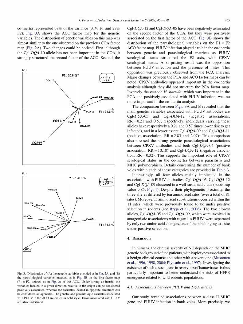
J. Deter et al. / Infection, Genetics and Evolution 8 (2008) 450–458 455
co-inertia represented 58% of the variance (31% F1 and 27%
F2). Fig. 3A shows the ACO factor map for the genetic
variables. The distribution of genetic variables on this map was
almost similar to the one observed on the previous COA factor
map (Fig. 2A). Two changes could be noticed. First, although
the Cgl-DQA-10 allele has not been important in the COA, it
strongly structured the second factor of the ACO. Second, the
Fig. 3. Distribution of (A) the genetic variables encoded as in Fig. 2A, and (B)
the parasitological variables encoded as in Fig. 2B on the first factor map
(F1 � F2, defined as in Fig. 2) of the ACO. Under strong co-inertia, the
variables located in a given direction relative to the origin can be considered
positively associated, whereas the variables located in opposite directions can
be considered antagonistic. The genetic and parasitologic variables associated
with PUUV in the ACO are edited in bold style. Those associated with CPXV
are also underlined.
Cgl-DQA-12 and Cgl-DQA-05 have been negatively associated
on the second factor of the COA, but they were positively
associated on the first factor of the ACO. Fig. 3B shows the
distribution of the parasitological variables on the F1 � F2
ACO factor map. PUUV infection played a role in the co-inertia
between genetic and parasitological matrices as PUUV
serological status structured the F2 axis, with CPXV
serological status. A surprising result was the opposition
between PUUV infection and the presence of mites. This
opposition was previously observed from the PCA analysis.
Major changes between the PCA and ACO factor maps can be
noted. CPXV antibodies appeared important in the co-inertia
analysis although they did not structure the PCA factor map.
Inversely the cestode H. horrida, which was important in the
PCA and positively associated with PUUV infection, was no
more important in the co-inertia analysis.
The comparison between Figs. 3A and B revealed that the
main genetic variables associated with PUUV antibodies are
Cgl-DQA-05 and Cgl-DQA-12 (negative associations,
RR = 0.21 and 0.57, respectively: individuals carrying these
alleles have respectively a 0.21 and 0.57 times lower risk to get
infected), and in a lesser extent Cgl-DQA-09 and Cgl-DQA-11
(positive association, RR = 2.83 and 2.07). This comparison
also stressed the strong genetic–parasitological associations
between CPXV antibodies and both Cgl-DQA-04 (positive
association, RR = 10.18) and Cgl-DQA-12 (negative associa-
tion, RR = 0.32). This supports the important role of CPXV
serological status in the co-inertia between parasitism and
MHC polymorphism. Details concerning the number of bank
voles within each of these categories are provided in Table 3.
Interestingly, all four alleles mainly implicated in the
association with PUUV antibodies, Cgl-DQA-05, Cgl-DQA-12
and Cgl-DQA-09 clustered in a well-sustained clade (bootstrap
value >85, Fig. 1). Despite their phylogenetic proximity, the
three alleles differed by ten amino acid sites (over a total of 81
sites). Moreover, 5 amino acid substitutions occurred within the
11 sites, which were previously found to be under positive
selection in rodents (see Bryja et al., 2006). The two closest
alleles, Cgl-DQA-05 and Cgl-DQA-09, which were involved in
antagonistic associations with regard to PUUV, were separated
by only two amino acid changes, one of them belonging to a site
under positive selection.
4. Discussion
In humans, the clinical severity of NE depends on the MHC
genetic background of the patients, with haplotypes associated to
a benign clinical course and other with a severe one (Mustonen
et al., 1996, 1998, 2004; Plyusnin et al., 1997). Investigating the
existence of such associations in reservoirs of hantaviruses is thus
particularly important to better understand the risks of HFRS
emergence related to wild rodents populations.
4.1. Associations between PUUV and DQA alleles
Our study revealed associations between a class II MHC
gene and PUUV infection in bank voles. More precisely, we
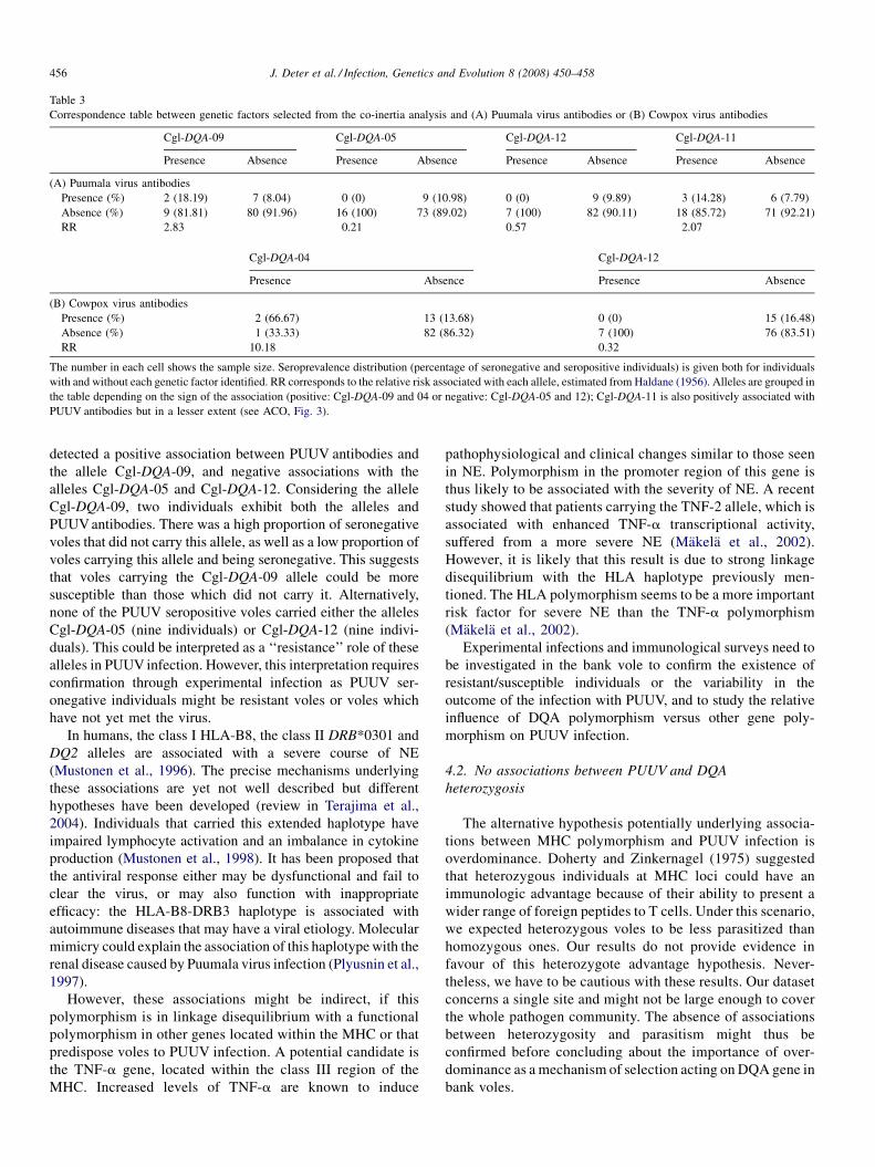
Table 3
Correspondence table between genetic factors selected from the co-inertia analysis and (A) Puumala virus antibodies or (B) Cowpox virus antibodies
Cgl-DQA-09 Cgl-DQA-05 Cgl-DQA-12 Cgl-DQA-11
Presence Absence Presence Absence Presence Absence Presence Absence
(A) Puumala virus antibodies
Presence (%) 2 (18.19) 7 (8.04) 0 (0) 9 (10.98) 0 (0) 9 (9.89) 3 (14.28) 6 (7.79)
Absence (%) 9 (81.81) 80 (91.96) 16 (100) 73 (89.02) 7 (100) 82 (90.11) 18 (85.72) 71 (92.21)
RR 2.83 0.21 0.57 2.07
Cgl-DQA-04 Cgl-DQA-12
Presence Absence Presence Absence
(B) Cowpox virus antibodies
Presence (%) 2 (66.67) 13 (13.68) 0 (0) 15 (16.48)
Absence (%) 1 (33.33) 82 (86.32) 7 (100) 76 (83.51)
RR 10.18 0.32
The number in each cell shows the sample size. Seroprevalence distribution (percentage of seronegative and seropositive individuals) is given both for individuals
with and without each genetic factor identified. RR corresponds to the relative risk associated with each allele, estimated from Haldane (1956). Alleles are grouped in
the table depending on the sign of the association (positive: Cgl-DQA-09 and 04 or negative: Cgl-DQA-05 and 12); Cgl-DQA-11 is also positively associated with
PUUV antibodies but in a lesser extent (see ACO, Fig. 3).
J. Deter et al. / Infection, Genetics and Evolution 8 (2008) 450–458456
detected a positive association between PUUV antibodies and
the allele Cgl-DQA-09, and negative associations with the
alleles Cgl-DQA-05 and Cgl-DQA-12. Considering the allele
Cgl-DQA-09, two individuals exhibit both the alleles and
PUUV antibodies. There was a high proportion of seronegative
voles that did not carry this allele, as well as a low proportion of
voles carrying this allele and being seronegative. This suggests
that voles carrying the Cgl-DQA-09 allele could be more
susceptible than those which did not carry it. Alternatively,
none of the PUUV seropositive voles carried either the alleles
Cgl-DQA-05 (nine individuals) or Cgl-DQA-12 (nine indivi-
duals). This could be interpreted as a ‘‘resistance’’ role of these
alleles in PUUV infection. However, this interpretation requires
confirmation through experimental infection as PUUV ser-
onegative individuals might be resistant voles or voles which
have not yet met the virus.
In humans, the class I HLA-B8, the class II DRB*0301 and
DQ2 alleles are associated with a severe course of NE
(Mustonen et al., 1996). The precise mechanisms underlying
these associations are yet not well described but different
hypotheses have been developed (review in Terajima et al.,
2004). Individuals that carried this extended haplotype have
impaired lymphocyte activation and an imbalance in cytokine
production (Mustonen et al., 1998). It has been proposed that
the antiviral response either may be dysfunctional and fail to
clear the virus, or may also function with inappropriate
efficacy: the HLA-B8-DRB3 haplotype is associated with
autoimmune diseases that may have a viral etiology. Molecular
mimicry could explain the association of this haplotype with the
renal disease caused by Puumala virus infection (Plyusnin et al.,
1997).
However, these associations might be indirect, if this
polymorphism is in linkage disequilibrium with a functional
polymorphism in other genes located within the MHC or that
predispose voles to PUUV infection. A potential candidate is
the TNF-a gene, located within the class III region of the
MHC. Increased levels of TNF-a are known to induce
pathophysiological and clinical changes similar to those seen
in NE. Polymorphism in the promoter region of this gene is
thus likely to be associated with the severity of NE. A recent
study showed that patients carrying the TNF-2 allele, which is
associated with enhanced TNF-a transcriptional activity,
suffered from a more severe NE (Makela et al., 2002).
However, it is likely that this result is due to strong linkage
disequilibrium with the HLA haplotype previously men-
tioned. The HLA polymorphism seems to be a more important
risk factor for severe NE than the TNF-a polymorphism
(Makela et al., 2002).
Experimental infections and immunological surveys need to
be investigated in the bank vole to confirm the existence of
resistant/susceptible individuals or the variability in the
outcome of the infection with PUUV, and to study the relative
influence of DQA polymorphism versus other gene poly-
morphism on PUUV infection.
4.2. No associations between PUUV and DQA
heterozygosis
The alternative hypothesis potentially underlying associa-
tions between MHC polymorphism and PUUV infection is
overdominance. Doherty and Zinkernagel (1975) suggested
that heterozygous individuals at MHC loci could have an
immunologic advantage because of their ability to present a
wider range of foreign peptides to T cells. Under this scenario,
we expected heterozygous voles to be less parasitized than
homozygous ones. Our results do not provide evidence in
favour of this heterozygote advantage hypothesis. Never-
theless, we have to be cautious with these results. Our dataset
concerns a single site and might not be large enough to cover
the whole pathogen community. The absence of associations
between heterozygosity and parasitism might thus be
confirmed before concluding about the importance of over-
dominance as a mechanism of selection acting on DQA gene in
bank voles.
J. Deter et al. / Infection, Genetics and Evolution 8 (2008) 450–458 457
4.3. Antagonist infections with mites and PUUV
Analysing PUUV infections jointly with the whole helminth
community, viruses and protozoan infections gave us the
opportunity to detect antagonist and/or synergistic effects of
parasites on the PUUV/MHC associations observed. In this
study, the multivariate approach developed on parasitological
variables revealed a strong opposition between mites and
PUUV infections. Among the nine PUUV seropositive voles,
only two were infected by mites. Alternatively, among the
PUUV seronegative voles, the proportions of individuals
infected and non-infected by mites were similar (respectively,
51% and 48%). This contrast between mites and PUUV
infections was still observed when analysing the co-structure
between parasitological and genetic variables. This suggests an
antagonist role of immunogenetics in resistance/susceptibility
to mites and PUUV. To our knowledge, this is the first time that
this antagonism is highlighted. It is quite surprising as a role of
trombiculid mites in hantavirus transmission has even been
suggested (Song, 1999; Houck et al., 2001). Several explana-
tions might be envisaged and tested experimentally: (i) one of
the immune response developed against PUUV infection and
associated with MHC susceptibility factor in voles might
prevent from further mite infestations, (ii) one of the immune
pathway associated with MHC factor of resistance to PUUV
infection might not prevent from further mite infestations, and
(iii) trombiculid mites are vectors of microparasites, which
were not detected in this study. The immune response
developed against these microparasites might prevent further
infestations with PUUV. This hypothesis is worth investigating
as trombiculid mites are known vectors of bacteria such as
Borrelia sp. (Kampen et al., 2004) or Rickettsia sp. (Frances
et al., 2000).
4.4. Associations between Cowpox virus and DQA
polymorphism
Surprisingly, our results show that CPXV plays an important
role in the co-structure between parasitological and MHC data
in bank voles. The two main associations detected in this study
concerned the allele Cgl-DQA-04, which is rare but found in
over 60% of CPXV-seropositive individuals, and the allele Cgl-
DQA-12, which was never found among the 15 CPXV-
seropositive individuals. This might suggest that this allele is
associated with resistance to CPXV.
Orthopoxviruses, along with a number of other viruses,
share mechanisms capable of modulating the host immune
response. Many orthopoxviruses possess a protein that is
capable of down-regulating MHC class I, which is responsible
for presenting all intracellular antigens (Dasgupta et al., 2007).
Biochemical and functional analyses have also revealed that
poxviruses infection directly interfered with ligand binding to
class II molecules (Ping et al., 2005). Therefore, down-
regulation of MHC class I trafficking and disruption of MHC
class II-mediated antigen presentation may be some of the
multiple strategies orthopoxviruses have evolved to escape host
immune surveillance. This modulation of the host defence
mechanisms seems to rely on homologues of immune
molecules encoded by orthopoxviruses (Johnston and McFad-
den, 2003). Combining immunogenetics of genes involved in
host immunity and virus evasion could help us understanding
these associations.
5. Conclusions
Our study provides evidence for the role of immunogenetics
in viral infections. Several alleles are suggested for either
susceptibility or resistance to these infections. However, these
results have to be taken cautiously as they concern few
seropositive individuals. Moreover, as demonstrated for
passerines and malaria parasites, links between host immuno-
genetics and resistance can result from local adaptation
processes (Bonneaud et al., 2006). The associations detected
might thus involve population-specific alleles and conse-
quently, vary in space and/or time. Further investigations are
needed to confirm the associations observed and to test the
existence of such spatio-temporal fluctuations.
Acknowledgements
We thank Patrice Cadet for help in the statistical analyses,
Audrey Scala and Florian Holon for help in field work and
Stanislas Kaluz for mite identification. This research was
partially funded by EU grant GOCE-2003-010284 EDEN and
the paper is catalogued by the EDEN Steering Committee as
EDEN 044 (http://www.eden-fp6project.net/).
References
Axtner, J., Sommer, S., 2007. Gene duplication, allelic diversity, selection
processes and adaptive value of MHC class II DRB genes of the bank vole,
Clethrionomys glareolus. Immunogenetics 59, 417–426.
Blake, J.A., Richardson, J.E., Bult, C.J., Kadin, J.A., Eppig, J.T., Mouse
Genome Database Group, 2003. MGD: the mouse genome database.
Nucleic Acids Res. 31, 193–195.
Bonneaud, C., Perez-Tris, J., Federici, P., Chastel, O., Sorci, G., 2006. Major
histocompatibility alleles associated with local resistance to malaria in a
passerine. Evolution 60, 383–389.
Bryja, J., Galan, M., Charbonnel, N., Cosson, J.F., 2005. Analysis of major
histocompatibility complex class II gene in water voles using capillary
electrophoresis-single stranded conformation polymorphism. Mol. Ecol.
Notes 5, 173–176.
Bryja, J., Galan, M., Charbonnel, N., Cosson, J.F., 2006. Duplication, balancing
selection and trans-species evolution explain the high levels of polymorph-
ism of the DQA MHC class II gene in voles (Arvicolinae). Immunogenetics
58, 191–202.
Capek, I., 2006. Definition de priorites et actions realisees dans le domaine
des zoonoses non alimentaires, 2000–2005. Bull. Epid. Hebdo. 27–28,
196–199.
Chantrey, J., Meyer, H., Baxby, D., Begon, M., Bown, K., Feore, S.M., Jones, T.,
Montgomery, W.I., Bennett, M., 1999. Cowpox: reservoir hosts and geo-
graphic range. Epidemiol. Infect. 122, 455–460.
Cooke, G.S., Hill, A.V.S., 2001. Genetics of susceptibility to human infectious
disease. Nat. Rev. Genet. 2, 967–977.
Dasgupta, A., Hammarlund, E., Slifka, M.K., Frueh, K., 2007. Cowpox virus
evades CTL recognition and inhibits the intracellular transport of MHC
class I molecules. J. Immunol. 178, 1654–1661.
Doherty, P.C., Zinkernagel, R.M., 1975. A biological role for the major
histocompatibility antigens. Lancet 1, 1406–1409.
J. Deter et al. / Infection, Genetics and Evolution 8 (2008) 450–458458
Dray, S., Chessel, D., Thioulouse, J., 2003. Procrustean co-inertia analysis for
the linking of multivariate datasets. Ecoscience 10, 110–119.
Frances, S.P., Watcharapichat, P., Phulsuksombati, D., Tanskul, P., 2000.
Transmission of Orientia tsutsugamushi, the aetiological agent for scrub
typhus, to co-feeding mites. Parasitology 120, 601–607.
Geraghty, D.E., 2002. Genetic diversity and genomics of the immune response.
Immunol. Rev. 190, 5.
Gunther, E., Walter, L., 2001. The major histocompatibility complex of the rat
(Rattus norvegicus). Immunogenetics 53, 520–542.
Haldane, S., 1956. The estimation and significance of the logarithm of a ratio of
frequencies. Ann. Hum. Genet. 20, 309–311.
Hall, T., 1999. BioEdit: a user-friendly biological sequence alignment editor
and analysis program for Windows 95/98/NT. Nucleic Acids Symp. Ser. 41,
95–98.
Hill, A.V.S., 2001. The genomics and genetics of human infectious disease
susceptibility. Annu. Rev. Genomics Hum. Genet. 2, 373–400.
Houck, M.A., Qin, H., Roberts, H.R., 2001. Hantavirus transmission: potential
role of ectoparasites. Vector-Borne Zoonotic Dis. 1, 75–79.
Hughes, A.L., Nei, M., 1990. Evolutionary relationships of class II major-
histocompatibility-complex genes in mammals. Mol. Biol. Evol. 7,
191–514.
Hurt, P., Walter, L., Sudbrak, R., Klages, S., Muller, I., Shiina, T., Inoko, H.,
Lehrach, H., Gunther, E., Reinhardt, R., Himmelbauer, H., 2004. The
genomic sequence and comparative analysis of the rat major histocompat-
ibility complex. Gen. Res. 14, 631–639.
Johnston, J.B., McFadden, G., 2003. Poxvirus immunomodulatory strategies:
current perspectives. J. Virol. 77, 6093–6100.
Kallio-Kokko, H., Laakkonen, J., Rizzoli, A., Tagliapietra, V., Cattadori, I.,
Perkins, S.E., Hudson, P.J., Cristofolini, A., Versini, W., Vapalahti, O.,
Vaheri, A., Henttonen, H., 2006. Hantavirus and arenavirus antibody
prevalence in rodents and humans in Trentino, Northern Italy. Epidemiol.
Infect. 134, 830–836.
Kallio-Kokko, H., Uzcategui, N., Vapalahti, O., Vaheri, A., 2005. Viral zoo-
noses in Europe. FEMS Microbiol. Rev. 29, 1051–1077.
Kallio, E.R., Klingstrom, J., Gustafsson, E., Manni, T., Vaheri, A., Henttonen,
H., Vapalahti, O., Lundkvist, A., 2006a. Prolonged survival of Puumala
hanatvirus outside the host: evidence for indirect transmission via the
environment. J. Gen. Virol. 87, 2127–2134.
Kallio, E.R., Poikonen, A., Vakeri, A., Vapalahti, O., Henttonen, H., Koskela,
E., Mappes, T., 2006b. Maternal antibodies postpone hantavirus infection
and enhance individual breeding success. Proc. Roy. Soc. B 273, 2771–
2776.
Kampen, H., Scholer, A., Metzen, M., Oehme, R., Hartelt, K., Kimmig, P.,
Maier, W.A., 2004. Neotrombicula autumnalis (Acari, Trombiculidae) as a
vector for Borrelia burgdorferi sensu lato? Exp. Appl. Acarol. 33, 93–102.
Kennedy, L.J., Ryvar, R., Gaskell, R.M., Addie, D.D., Willoughby, K., Carter,
S.D., Thomson, W., Ollier, W.E.R., Radford, A.D., 2002. Sequence analysis
of MHC DRB alleles in domestic cats from the United Kingdom. Immu-
nogenetics 54, 348–352.
Klein, J., Bontrop, R.E., Dawkins, R.L., Erlich, H.A., Gyllensten, U.B., Heise,
E.R., Jones, P.P., Parham, P., Wakeland, E.K., Watkins, D.I., 1990. Nomen-
clature for the major histocompatibility complex of different species: a
proposal. Immunogenetics 31, 217–219.
Klein, S.L., Cernetich, A., Hilmer, S., Hoffman, E.P., Scott, A.L., Glass, G.E.,
2004a. Differential expression of immunoregulatory genes in male and
female Norway rats following infection with Seoul virus. J. Med. Virol. 74,
180–190.
Klein, S.L., Zink, M.C., Glass, G.E., 2004b. Seoul virus infection increases
aggressive behaviour in male Norway rats. Anim. Behav. 67, 421–429.
Klingstrom, J., Maljkovic, I., Zuber, B., Rollman, E., Kjerrstrom, A., Lundkvist,
A., 2004. Vaccination of C57/BL6 mice with Dobrava hantavirus nucleo-
capsid protein in Freund’s adjuvant induced partial protection against
challenge. Vaccine 22, 4029–4034.
Laakkonen, J., Kallio-Kokko, H., Oktem, M.A., Blasdell, K., Plyusnina, A.,
Niernimaa, J., Karatas, A., Plyusnin, A., Vaheri, A., Henttonen, H., 2006.
Serological survey for viral pathogens in Turkish rodents. J. Wildl. Dis. 42,
672–676.
Lundkvist, A., Fatouros, A., Niklasson, B., 1991. Antigenic variation of
European haemorrhagic fever with renal syndrome virus strains character-
ized using bank vole monoclonal antibodies. J. Gen. Virol. 72, 2097–2103.
Makela, S., Mustonen, J., Ala-Houhala, I., Hurme, M., Partanen, J., Vapalahti,
O., Vaheri, A., Pasternack, A., 2002. Human leukocyte antigen-B8-DR3 is a
more important risk factor for severe puumala hantavirus infection than
the tumor necrosis factor-alpha(-308) G/A polymorphism. J. Infect. Dis.
843–846.
Mills, J.N., Childs, J., Ksiazek, T.G., Peters, C.J., Velleca, W.M., 1995. Methods
for Trapping and Sampling Small Mammals for Virologic Testing. Centers
for Disease Control and Prevention, Atlanta.
Mills, J.N., Childs, J.E., 1998. Ecologic studies of rodent reservoirs: their
relevance for human health. Emerg. Infect. Dis. 4, 529–537.
Mustonen, J., Huttunen, N.P., Partanen, J., Baer, M., Paakkala, A., Vapalahti, O.,
Uhari, M., 2004. Human leukocyte antigens B8-DRB1*03 in pediatric
patients with Nephropathia epidemica caused by puumala hantavirus.
Pediatr. Infect. Dis. J. 23, 959–961.
Mustonen, J., Vaheri, A., Clement, J., 1996. Third international conference on
haemorrhagic fever with renal syndrome (HFRS) and hantaviruses.
Nephrol. Dialysis Transpl. 11, 730–733.
Mustonen, J., Vapalahti, I., Henttonen, H., Pasternack, A., Vaheri, A., 1998.
Epidemiology of hantavirus infections in Europe. Nephrol. Dialysis
Transpl. 13, 2729–2731.
Ping, L., Wang, N., Zhou, D., Yee, C.S.K., Chang, C.-H., Brutkiewicz, R.R.,
Blum, J.S., 2005. Disruption of MHC class II-restricted antigen presentation
by vaccinia virus. J. Immunol. 175, 6481–6488.
Plyusnin, A., Horling, J., Kanerva, M., Mustonen, J., Cheng, Y., Partanen, J.,
Vapalahti, O., Kukkonen, S.K.J., Niemimaa, J., Henttonen, H., Niklasson,
B., Lundkvist, A., Vaheri, A., 1997. Puumala hantavirus genome in patients
with Nephropathia epidemica: correlation of PCR positivity with HLA
haplotype and link to viral sequences in local rodents. J. Clin. Microbiol. 35,
1090–1096.
Plyusnina A., Deter J., Charbonnel N., Cosson J.-F., and Plyusnin A., in press.
Puumala and Tula hantaviruses in France. Virus Research.
Rozsa, L., Reiczigel, J., Majoros, G., 2000. Quantifying parasites in samples of
hosts. J. Parasitol. 86, 228–232.
Song, G., 1999. Epidemiological progresses of hemorrhagic fever with renal
syndrome in China. Chin. Med. J. 112, 472–477.
Takahata, N., Nei, M., 1990. Allelic genealogy under overdominant and
frequency-dependent selection and polymorphism of major histocompat-
ibility complex loci. Genetics 124, 967–978.
Taylor, L.H., Latham, S.M., Woolhouse, M.E.J., 2001. Risk factors for human
disease emergence. Philos. Trans. Roy. Soc. Lond. B: Biol. Sci. 356, 983–989.
Terajima, M., Vapalahti, O., Van Epps, H.L., Vaheri, A., Ennis, F.A., 2004.
Immune responses to Puumala virus infection and the pathogenesis of
Nephropathia epidemica. Microbes Infect. 6, 238–245.
Thioulouse, J., Chessel, D., Doledec, S., Olivier, J.M., 1997. ADE-4: a multi-
variate analysis and graphical display software. Stat. Comput. 7, 75–83.
Thioulouse, J., Simier, M., Chessel, D., 2004. Simultaneous analysis of a
sequence of paired ecological tables. Ecology 85, 272–283.
Vapalahti, O., Mustonen, J., Lundkvist, A., Henttonen, H., Plyusnin, A., Vaheri,
A., 2003. Hantavirus infections in Europe. Lancet Infect. Dis. 3, 753–754.
Wegner, K.M., Reusch, T.B.H., Kalbe, M., 2003. Multiple parasites are driving
major histocompatibility complex polymorphism in the wild. J. Evol. Biol.
224–232.
www.elsevier.com/locate/meegid
Infection, Genetics and Evolution 8 (2008) 459–466
Differential strain-specific diagnosis of the heartwater agent:
Ehrlichia ruminantium
Nathalie Vachiery a, Gael Maganga a, Thierry Lefrancois a, Yane Kandassamy a,Frederic Stachurski a, Hassane Adakal b, Conchita Ferraz c, Anne Morgat d,e,
Albert Bensaid a,1, Eric Coissac d, Frederic Boyer d, Jacques Demaille c,Alain Viari d, Dominique Martinez a, Roger Frutos a,*
a CIRAD TA A-15/G, Campus International de Baillarguet, 34398, Montpellier Cedex 05, Franceb Cirdes, 01, BP. 454, Bobo-Dioulasso 01, Burkina-Faso
c Centre de Sequencage Genomique, IGH-CNRS-UPR 1142, 141 rue de la Cardonille, 34396 Montpellier Cedex 05, Franced Inria Rhone-Alpes—Projet HELIX, 655, Av. de l’Europe, 38330 Montbonnot-Saint Martin, France
e Swiss Institute of Bioinformatics, Swiss-Prot Group, 1 rue Michel, Servet, CH-1211 Geneva 4, Switzerland
Received 2 May 2007; received in revised form 8 June 2007; accepted 11 June 2007
Available online 19 June 2007
Abstract
Ehrlichia ruminantium is the causative agent of heartwater, a major tick-borne disease of livestock in Africa introduced in the Caribbean and
threatening to emerge and spread in the American mainland. Complete genome sequencing was done for two isolates of E. ruminantium of
differing phenotype, isolates Gardel (Erga) from Guadeloupe Island and Welgevonden (Erwe) originating from South Africa and maintained in
Guadeloupe. The type strain of E. ruminantium (Erwo), previously isolated and sequenced in South Africa; is identical to Erwe with respect to
target genes. They make the Erwe/Erwo complex. Comparative analysis of the genomes shows the presence of 49 unique CDS and 28 truncated
CDS differentiating Erga from Erwe/Erwo. Three regions of accumulated differences (RAD) acting as mutational hot spots were identified in E.
ruminantium. Ten CDS, six unique CDS and four truncated CDS corresponding to major genomic changes (deletions or extensive mutations) were
considered as targets for differential diagnosis on four isolates of E. ruminantium: Erga, Erwe/Erwo, Senegal and Umpala. Pairs of PCR primers
were developed for each target gene. PCR analysis of the target genes generated strain-specific patterns on Erga and Erwe/Erwo as predicted by
comparative genomics, but also for isolates Senegal and Umpala. The target genes identified by bacterial comparative genomics are shown to be
highly efficient for strain-specific PCR diagnosis of E. ruminantium and further vaccine management tools.
# 2007 Elsevier B.V. All rights reserved.
Keywords: Ehrlichia ruminantium; Heartwater; Comparative genomics; Diagnosis
1. Introduction
Ehrlichia ruminantium is a tick-borne Rickettsiale respon-
sible for cowdriosis or heartwater in ruminants all over Sub-
Saharan Africa and in the Caribbean (Provost and Bezuidenh-
out, 1987). Heartwater is not only responsible for high
* Corresponding author at: CIRAD TA30/G, Campus International de Bail-
larguet, 34398 Montpellier Cedex 5, France. Tel.: +33 4 67 59 39 62;
fax: +33 4 67 59 39 60.
E-mail address: [email protected] (R. Frutos).1 Current address: Centre de Recerca en Sanitat Animal, Campus de Belle-
terra, Edifici V, 08193 Bellaterra, Barcelona, Spain.
1567-1348/$ – see front matter # 2007 Elsevier B.V. All rights reserved.
doi:10.1016/j.meegid.2007.06.001
economical losses in Africa (Mukhebi et al., 1999), but also
threatens the American mainland because of the presence of
tick species capable of transmitting the disease (Barre et al.,
1987; Burridge et al., 2002). The current commercial vaccine
is based on the high-risk method of injection of infected
blood followed by treatment with antibiotics (Bezuidenhout,
1989). Attenuated and DNA vaccines were developed with
good protection in challenged experiments (Collins et al.,
2003a,b; Jongejan, 1991; Mahan et al., 2003; Martinez et al.,
1994, 1996), they had a limited efficacy in the field (Collins
et al., 2003a,b; Jongejan, 1991; Mahan et al., 2003; Martinez
et al., 1994, 1996). However, a more recent vaccinal strain has
not yet been tested in the wild (Zweygarth et al., 2005). map1
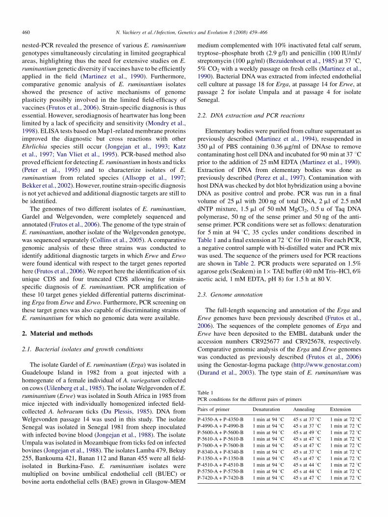
Table 1
PCR conditions for the different pairs of primers
Pairs of primer Denaturation Annealing Extension
P-4350-A + P-4350-B 1 min at 94 8C 45 s at 37 8C 1 min at 72 8CP-4990-A + P-4990-B 1 min at 94 8C 45 s at 37 8C 1 min at 72 8CP-5600-A + P-5600-B 1 min at 94 8C 45 s at 49 8C 1 min at 72 8CP-5610-A + P-5610-B 1 min at 94 8C 45 s at 47 8C 1 min at 72 8CP-7600-A + P-7600-B 1 min at 94 8C 45 s at 47 8C 1 min at 72 8CP-8340-A + P-8340-B 1 min at 94 8C 45 s at 37 8C 1 min at 72 8CP-1350-A + P-1350-B 1 min at 94 8C 45 s at 47 8C 1 min at 72 8CP-4510-A + P-4510-B 1 min at 94 8C 45 s at 44 8C 1 min at 72 8CP-5750-A + P-5750-B 1 min at 94 8C 45 s at 44 8C 1 min at 72 8CP-7420-A + P-7420-B 1 min at 94 8C 45 s at 47 8C 1 min at 72 8C
N. Vachiery et al. / Infection, Genetics and Evolution 8 (2008) 459–466460
nested-PCR revealed the presence of various E. ruminantium
genotypes simultaneously circulating in limited geographical
areas, highlighting thus the need for extensive studies on E.
ruminantium genetic diversity if vaccines have to be efficiently
applied in the field (Martinez et al., 1990). Furthermore,
comparative genomic analysis of E. ruminantium isolates
showed the presence of active mechanisms of genome
plasticity possibly involved in the limited field-efficacy of
vaccines (Frutos et al., 2006). Strain-specific diagnosis is thus
essential. However, serodiagnosis of heartwater has long been
limited by a lack of specificity and sensitivity (Mondry et al.,
1998). ELISA tests based on Map1-related membrane proteins
improved the diagnostic but cross reactions with other
Ehrlichia species still occur (Jongejan et al., 1993; Katz
et al., 1997; Van Vliet et al., 1995). PCR-based method also
proved efficient for detecting E. ruminantium in hosts and ticks
(Peter et al., 1995) and to characterize isolates of E.
ruminantium from related species (Allsopp et al., 1997;
Bekker et al., 2002). However, routine strain-specific diagnosis
is not yet achieved and additional diagnostic targets are still to
be identified.
The genomes of two different isolates of E. ruminantium,
Gardel and Welgevonden, were completely sequenced and
annotated (Frutos et al., 2006). The genome of the type strain of
E. ruminantium, another isolate of the Welgevonden genotype,
was sequenced separately (Collins et al., 2005). A comparative
genomic analysis of these three strains was conducted to
identify additional diagnostic targets in which Erwe and Erwo
were found identical with respect to the target genes reported
here (Frutos et al., 2006). We report here the identification of six
unique CDS and four truncated CDS allowing for strain-
specific diagnosis of E. ruminantium. PCR amplification of
these 10 target genes yielded differential patterns discriminat-
ing Erga from Erwe and Erwo. Furthermore, PCR screening on
these target genes was also capable of discriminating strains of
E. ruminantium for which no genomic data were available.
2. Material and methods
2.1. Bacterial isolates and growth conditions
The isolate Gardel of E. ruminantium (Erga) was isolated in
Guadeloupe Island in 1982 from a goat injected with a
homogenate of a female individual of A. variegatum collected
on cows (Uilenberg et al., 1985). The isolate Welgevonden of E.
ruminantium (Erwe) was isolated in South Africa in 1985 from
mice injected with individually homogenized infected field-
collected A. hebraeum ticks (Du Plessis, 1985). DNA from
Welgevonden passage 14 was used in this study. The isolate
Senegal was isolated in Senegal 1981 from sheep inoculated
with infected bovine blood (Jongejan et al., 1988). The isolate
Umpala was isolated in Mozambique from ticks fed on infected
bovines (Jongejan et al., 1988). The isolates Lamba 479, Bekuy
255, Bankouma 421, Banan 112 and Banan 455 were all field-
isolated in Burkina-Faso. E. ruminantium isolates were
multiplied on bovine umbilical endothelial cell (BUEC) or
bovine aorta endothelial cells (BAE) grown in Glasgow-MEM
medium complemented with 10% inactivated fetal calf serum,
tryptose–phosphate broth (2.9 g/l) and penicillin (100 IU/ml)/
streptomycin (100 mg/ml) (Bezuidenhout et al., 1985) at 37 8C,
5% CO2 with a weekly passage on fresh cells (Martinez et al.,
1990). Bacterial DNA was extracted from infected endothelial
cell culture at passage 18 for Erga, at passage 14 for Erwe, at
passage 2 for isolate Umpala and at passage 4 for isolate
Senegal.
2.2. DNA extraction and PCR reactions
Elementary bodies were purified from culture supernatant as
previously described (Martinez et al., 1994), resuspended in
350 ml of PBS containing 0.36 mg/ml of DNAse to remove
contaminating host cell DNA and incubated for 90 min at 37 8Cprior to the addition of 25 mM EDTA (Martinez et al., 1990).
Extraction of DNA from elementary bodies was done as
previously described (Perez et al., 1997). Contamination with
host DNAwas checked by dot blot hybridization using a bovine
DNA as positive control and probe. PCR was run in a final
volume of 25 ml with 200 ng of total DNA, 2 ml of 2.5 mM
dNTP mixture, 1.5 ml of 50 mM MgCl2, 0.5 u of Taq DNA
polymerase, 50 ng of the sense primer and 50 ng of the anti-
sense primer. PCR conditions were set as follows: denaturation
for 5 min at 94 8C, 35 cycles under conditions described in
Table 1 and a final extension at 72 8C for 10 min. For each PCR,
a negative control sample with bi-distilled water and PCR mix
was used. The sequence of the primers used for PCR reactions
are shown in Table 2. PCR products were separated on 1.5%
agarose gels (Seakem) in 1� TAE buffer (40 mM Tris–HCl, 6%
acetic acid, 1 mM EDTA, pH 8) for 1.5 h at 80 V.
2.3. Genome annotation
The full-length sequencing and annotation of the Erga and
Erwe genomes have been previously described (Frutos et al.,
2006). The sequences of the complete genomes of Erga and
Erwe have been deposited to the EMBL databank under the
accession numbers CR925677 and CR925678, respectively.
Comparative genomic analysis of the Erga and Erwe genomes
was conducted as previously described (Frutos et al., 2006)
using the Genostar-Iogma package (http://www.genostar.com)
(Durand et al., 2003). The type stain of E. ruminantium was
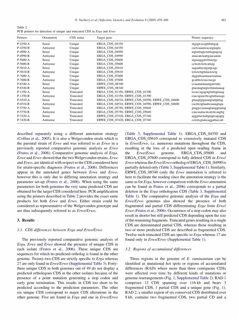
Table 2
PCR primers for detection of unique and truncated CDS in Erga and Erwe
Primers Orientation CDS status Target gene Primer sequence
P-4350-A Sense Unique ERGA_CDS_04350 atgagtcacagttttattgag
P-4350-B Antisense Unique ERGA_CDS_04350 cactcaaaatcacaagaagta
P-4990-A Sense Unique ERGA_CDS_04990 atgtatttagtctatttagtagctg
P-4990-B Antisense Unique ERGA_CDS_04990 ataacatctaattgaacaatatc
P-5600-A Sense Unique ERGA_CDS_05600 atgaaaggatctttatctgc
P-5600-B Antisense Unique ERGA_CDS_05600 ccttcttcttcttcattatg
P-5610-A Sense Unique ERGA_CDS_05610 aagaattacatgatgcagc
P-5610-B Antisense Unique ERGA_CDS_05610 tcttctcttgttatactctctg
P-7600-A Sense Unique ERGA_CDS_07600 atggatttaaataaactaataaa
P-7600-B Antisense Unique ERGA_CDS_07600 gcattttctctacctacga
P-8340-A Sense Unique ERWE_CDS_08340 ccaaatatataaatgatctattc
P-8340-B Antisense Unique ERWE_CDS_08340 gtacatagtatgtctttatataaaag
P-1350-A Sense Truncated ERGA_CDS_01350, ERWE_CDS_01390 tccaccagagatgttatttgtaaag
P-1350-B Antisense Truncated ERGA_CDS_01350, ERWE_CDS_01390 caacagaactttcagtattaaaagc
P-4510-A Sense Truncated ERGA_CDS_04510, ERWE_CDS_04590, ERWE_CDS_04600 gttaagtgtgaaatgtattgtttag
P-4510-B Antisense Truncated ERGA_CDS_04510, ERWE_CDS_04590, ERWE_CDS_04600 cactttctgttaattcaaaagtaga
P-5750-A Sense Truncated ERGA_CDS_05750, ERWE_CDS_05840 gtaggccaaaaagtataggtaatag
P-5750-B Antisense Truncated ERGA_CDS_05750, ERWE_CDS_05840 caacaaatacatcatcttcaagttg
P-7420-A Sense Truncated ERWE_CDS_07420, ERGA_CDS_07340 agggttacttattgtagtcagagtg
P-7420-B Antisense Truncated ERWE_CDS_07420, ERGA_CDS_07340 cctcttcgtatacaggattaccatt
N. Vachiery et al. / Infection, Genetics and Evolution 8 (2008) 459–466 461
described separately using a different annotation strategy
(Collins et al., 2005). It is also a Welgevonden strain which is
the parental strain of Erwe and was referred to as Erwo in a
previously reported comparative genomic analysis as Erwo
(Frutos et al., 2006). Comparative genomic analysis of Erga,
Erwe and Erwo showed that the two Welgevonden strains, Erwe
and Erwo, are identical with respect to the CDS considered here
for strain-specific diagnosis (Frutos et al., 2006). Differences
appear in the annotated genes between Erwe and Erwo,
however this is only due to differing annotation strategy and
parameter set-up (Frutos et al., 2006). When using the same
parameters for both genomes the very same predicted CDS are
obtained for the target CDS considered here. PCR amplification
using the primers described in Table 2 provides the exact same
products for both Erwe and Erwo. Either strain could be
considered as representative of the Welgevonden genotype and
are thus subsequently referred to as Erwe/Erwo.
3. Results
3.1. CDS differences between Erga and Erwe/Erwo
The previously reported comparative genomic analysis of
Erga, Erwe and Erwo showed the presence of unique CDS in
each isolate (Frutos et al., 2006). These unique CDS are
sequences for which no predicted ortholog is found in the other
genome. Twenty-two CDS are strictly specific to Erga whereas
27 are only found in Erwe/Erwo (Supplemental Table 3). Forty-
three unique CDS in both genomes out of 49 do not display a
predicted orthologous CDS in the other isolates because of the
presence of a point mutation generating a stop codon and
early gene termination. This results in CDS too short to be
predicted according to the prediction parameters. The other
six unique CDS correspond to major CDS alterations in the
other genome. Five are found in Erga and one in Erwe/Erwo
(Table 3, Supplemental Table 1). ERGA_CDS_04350 and
ERGA_CDS_05610 correspond to extensively mutated CDS
in Erwe/Erwo, i.e. numerous mutations throughout the CDS,
resulting in the loss of a predicted open reading frame in
the Erwe/Erwo genome. ERGA_CDS_05600 and
ERGA_CDS_07600 correspond to fully deleted CDS in Erwe/
Erwo whereas the Erwe/Erwo ortholog of ERGA_CDS_04990 is
partially deleted only (Table 3, Supplemental Table 1). Similarly,
ERWE_CDS_08340 (only the Erwe annotation is referred to
here to facilitate the reading since the annotation strategy is the
same as for Erga, however comparison with the Erwo annotation
can be found in Frutos et al., 2006) corresponds to a partial
deletion in the Erga orthologous CDS (Table 3, Supplemental
Table 1). The comparative genomic analysis of the Erga and
Erwe/Erwo genomes also showed the presence of both
fragmented and partial CDS differentiating Erga from Erwe/
Erwo (Frutos et al., 2006). Occurrence of a stop codon may also
result in shorter but still predicted CDS depending upon the size
of the remaining fragments. Truncated genes resulting in a single
CDS are denominated partial CDS, whereas those resulting in
two or more predicted CDS are described as fragmented CDS.
Twelve such truncated CDS are specific to Erga whereas 17 are
found only in Erwe/Erwo (Supplemental Table 1).
3.2. Regions of accumulated differences
Three regions in the genome of E. ruminantium can be
identified as mutational hot spots or regions of accumulated
differences (RAD) where more than three contiguous CDSs
were affected over time by different kinds of mutations or
genome rearrangements (Fig. 1, Supplemental Table 2). RAD 1
comprises 12 CDS spanning over 116 kb and bears 3
fragmented CDS, 1 partial CDS and a unique gene (Fig. 1).
RAD 2, a smaller region of six conserved CDS distributed over
8 kb, contains two fragmented CDS, two partial CD and a
Table 3
Target CDS for strain-specific differential diagnostic of E. ruminantium
Target gene in Erga Status Size (size
of deletion)
Function Target gene in
Erwe/Erwo
Status Size (size
of deletion)
Function
ERGA_CDS_04350 Unique gene 186 bp (NA) Unkown None Extensive mutations NA NA
ERGA_CDS_04990 Unique gene 270 bp (N.A.) Similarity to rpoB None Partial deletion NA NA
ERGA_CDS_05600 Unique gene 630 bp (NA) Similarity to cysC None Full deletion NA NA
ERGA_CDS_05610 Unique gene 828 bp (NA) Unknown None Extensive mutations N.A N.A
ERGA_CDS_07600 Unique gene 303 bp (NA) Unknown None Full deletion N.A N.A
ERGA_CDS_01350 Full-length gene 3252 bp (NA) Unknown ERWE_CDS_01390/
Erum1430
Partial deletiona 2856 bp (396 bp) Unknown
ERGA_CDS_04510 Full-length gene 3570 bp (NA) Unknown ERWE_CDS_04590/
Not predictedb
Partial deletionc 873 bp (585 bp) Unknown
ERWE_CDS_04600/
Erum4400
Partial deletionc 1470 bp Unknown
ERGA_CDS_05750 Full-length gene 1836 bp (NA) Unknown ERWE_CDS_05840/
Erum5570
Partial deletiona 1659 bp (177 bp) Unknown
ERGA_CDS_07340 Partial deletiona 3522 bp (600 bp) Unknown ERWE_CDS_07420/Erum Full-length gene 4122 bp (NA) Unknown
None Partial deletion NA NA ERWE_CDS_08340/
Not predictedb
Unique gene 225 bp (NA) Unknown
N.A.: not applicable.a The truncated CDS is a partial CDS.b The CDS are not predicted in the Erwo published genome because of differing prediction parameters. Adjusting the parameters to those used for prediction of
CDS in Erwe leads to identical results in Erwe and Erwo.c The truncated CDS is a fragmented CDS.
N. Vachiery et al. / Infection, Genetics and Evolution 8 (2008) 459–466462
unique gene (Fig. 1). RAD 3 is in the same size range as RAD 2
with 5.7 kb and bears more extensive genome rearrangements
than the other two RAD with only one conserved orthologous
gene out of six CDS (Fig. 1). RAD3 bears large unique CDS
coming from deletions in the other isolate, a partial CDS and
remaining pseudogenes (Fig. 1).
3.3. Target genes for differential diagnosis
Most of the unique, fragmented and partial CDS differ-
entiating Erga from Erwe/Erwo are affected by a single or very
few point mutations resulting in stops codons. They are not
suited for differential diagnosis since most of the homologous
Fig. 1. Representation of the regions of accumulated differences (RAD). Mutants a
shown in white. Lines connect orthologs.
sequence is still present in the other isolates although not
predicted as a coding sequence. However, 10 genes bear large
deletions or extensive mutations and thus represent targets of
choice for differential diagnosis (Table 3). Two CDS are
associated to the full-length deletion of the orthologous gene,
six display partial deletions and two are characterized by
extensive mutations (Table 3). ERGA_CDS_5610 whose
ortholog is deleted in Erwe/Erwo, falls into this group of
extensively mutated genes, due to remaining homology with the
nucleotide sequence of Erwe/Erwo. In silico analysis of the
specificity of PCR primers designed to amplify each of the 10
target genes showed that the 4 truncated genes and the 6 unique
CDS could be differentially discriminated. PCR primers were
re shown in grey, unique CDS are shown in black and conserved orthologs are
Table 4
Strain-specific differential PCR screening of E. ruminantium
Primer combinations Strain
Erga Erwe Erwoa Umpala Senegal Bankouma 421 Bekuy 255 Lamba 479 Banan 112 Banan 455
P-4350-A + P-4350-B 172 None None 172 172 Multibands 172 172 172 Multibands
P-4990-A + P-4990-B 217 None None 515 500 +
900
280 + 500 +
1200
500 + 900 500 + 900 500 +
900
500 + 1200
P-5600-A + P-5600-B 508 None None 508 None 560 508 + 1900 508 + 1900 None 508
P-5610-A + P-5610-B 642 None None 642 642 642a 642 642 642 642
P-7600-A + P-7600-B 238 None None 238 None 238 238 238 238 238
P-8340-A + P-8340- None 127 127 None 127 None 127 127 127 None
P-1350-A + P-1350-B 2791 2395 2395 2791 + 500 2395 2395 2395 2395 2395 None
P-4510-A + P-4510-B 552 +
1071 +
480
492 492 1200 + 500b 492 492 492 492 492 552 + 1071 +
480
P-5750-A + P-5750-B 1361 1178 1178 1361 1000 1000 1000 1000 1000 1178 + 1361
P-7420-A + P-7420-B 1095 +
300
1691 1691 820 820 820 820 820 820 1095
a Expected sizes were calculated in silico from the Erwo type-strain genome (Collins et al., 2005).b Presence of multiple bands is observed.
N. Vachiery et al. / Infection, Genetics and Evolution 8 (2008) 459–466 463
thus developed to detect the targeted unique CDS based on
presence–absence or size difference of the target sequences
(Table 2).
3.4. PCR strain-specific differential diagnosis of
E. ruminantium
Results of strain-specific differential PCR screening of E.
ruminantium are summarized in Table 4. Primers developed to
discriminate Erga and Erwe/Erwo upon differential detection
of truncated CDS yielded the expected results. The primer pairs
P-1350-A and -B, P-4510-A and -B, P-5750-A and -B and P-
7420-A and -B yielded PCR products of the respective expected
size of 2791 pb, 552 and 1071 pb, 1361 and 1095 pb on Erga
Fig. 2. Strain-specific differential diagnostic of E. ruminantium by PCR detection of
and -B on 2: senegal; 3: umpala; 4: Erga; 5: Erwe; 6: negative control sample; prim
control sample; 12: molecular weight marker (l EcoRI–HindIII); 13: molecular weig
16: Erga; 17: Erwe/Erwo; 18: negative control sample; primers P-7420-A and -B on
molecular weight marker (l EcoRI–HindIII).
and 2395 pb, 492 pb, 1178 pb and 1691 pb on Erwe/Erwo,
respectively (Table 4, Fig. 2). An additional band of 480 bp is
observed on Erga with the pair P-4510-A and -B. This
additional band is most likely due to a single low specificity
response occurring in Erga. Similar results were obtained using
primers specific to unique CDS (Table 4, Fig. 3). All primers for
unique CDS yielded the expected PCR products and differential
response in both Erga and Erwe/Erwo (Table 4). The PCR
primers were further tested firstly on two other strains obtained
from other groups and conserved in the laboratory, i.e. Umpala
and Senegal (Table 4, Figs. 2 and 3). Secondly, the analysis was
extended to five more strains, i.e. Bankouma 421, Bekuy 255,
Lamba 479, Banan 112 and Banan 455, which were field-
isolated by two of the authors (F. Stachurski and H. Adakal) in
truncated CDS. 1: Molecular weight marker (100-bp ladder); primers P-4510-A
ers P-5750-A and -B on 7: senegal; 8: umpala; 9: Erga; 10: Erwe; 11: negative
ht marker (100-bp ladder); primers P-1350-A and -B on 14: senegal; 15: umpala;
19: senegal; 20: umpala; 21: Erga; 22: Erwe; 23: negative control sample; 24:
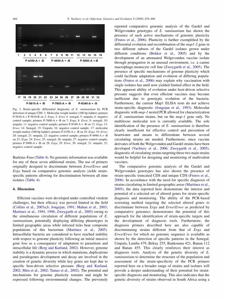
Fig. 3. Strain-specific differential diagnostic of E. ruminantium by PCR
detection of unique CDS. 1: Molecular weight marker (100-bp ladder); primers
P-5610-A + P-5610-B on 2: Erga; 3: Erwe 4: senegal; 5: umpala; 6: negative
control sample; primers P-7600-A + -B on 7: Erga; 8: Erwe; 9: senegal; 10:
umpala; 11: negative control sample; primers P-8340-A + -B on 12: Erga; 13:
Erwe; 14: Senegal; 15: Umpala; 16: negative control sample; 17: molecular
weight marker (100-bp ladder); primers P-4350-A + -B on 18: Erga; 19: Erwe;
20: senegal; 21: umpala; 22: negative control sample; primers P-4990-A + -B
on 23: Erga; 24: Erwe; 25: senegal; 26: umpala; 27: negative control sample;
primers P-5600-A + -B on 28: Erga; 29: Erwe; 30: senegal; 31: umpala; 32:
negative control sample.
N. Vachiery et al. / Infection, Genetics and Evolution 8 (2008) 459–466464
Burkina–Faso (Table 4). No genomic information was available
for any of these seven additional strains. The use of primers
originally designed to discriminate between Erwe/Erwo and
Erga based on comparative genomic analysis yields strain-
specific patterns allowing for discrimination between all nine
isolates (Table 4).
4. Discussion
Efficient vaccines were developed under controlled virulent
challenges, but their efficacy was proved limited in the field
(Collins et al., 2003a,b; Jongejan, 1991; Mahan et al., 2003;
Martinez et al., 1994, 1996; Zweygarth et al., 2005) owing to
the simultaneous circulation of different populations of E.
ruminantium, potentially displaying differing phenotypes in
limited geographic areas while infected ticks bear composite
populations of this bacterium (Martinez et al., 2005).
Intracellular bacteria are considered to have reached stability
with respect to genome plasticity following an initial massive
gene loss as a consequence of adaptation to parasitism and
intracellular life (Berg and Kurland, 2002). However, genome
stability is a dynamic process in which mutations, duplications
and pseudogenes development and decay are involved in the
creation of genetic diversity while key genes are kept due to
specific host-driven selective pressure (Berg and Kurland,
2002; Mira et al., 2002; Tamas et al., 2002). The potential and
mechanisms for genome plasticity remains and might be
expressed following environmental changes. The previously
reported comparative genomic analysis of the Gardel and
Welgevonden genotypes of E. ruminantium has shown the
presence of such active mechanisms of genomic plasticity
(Frutos et al., 2006). Plasticity is further exemplified by the
differential evolution and recombination of the map1-2 gene in
two different subsets of the Gardel isolates grown under
different conditions (Bekker et al., 2005) and by the
development of an attenuated Welgevonden vaccine isolate
through propagation in an unusual environment, i.e. a canine
macrophage-monocyte cell line (Zweygarth et al., 2005). The
presence of specific mechanisms of genome plasticity which
could facilitate adaptation and evolution of differing popula-
tions (Frutos et al., 2006) may explain why vaccination with
single isolates has until now yielded limited effect in the field.
This apparent ability of evolution under host-driven selective
pressure suggests that even efficient vaccines may become
inefficient due to genotypic evolution of the bacteria.
Furthermore, the current Map1 ELISA tests do not achieve
strain-specific diagnostic (Jongejan et al., 1993). Molecular
diagnostic with map-1 nested PCR allowed for characterization
of E. ruminantium strains, but on the map-1 gene only. No
multilocus molecular test is currently available. The sole
identification of the presence of E. ruminantium is therefore
clearly insufficient for effective control and prevention of
heartwater and means to differentiate between several
circulating strains are needed. Furthermore, since vaccine
derivates of both the Welgevonden and Gardel strains have been
developed (Vachiery et al., 2006; Zweygarth et al., 2005),
diagnostic of circulating strains targeting these two main strains
would be helpful for designing and monitoring of multivalent
vaccines.
The comparative genomic analysis of the Gardel and
Welgevonden genotypes has also shown the presence of
strain-specific truncated CDS and unique CDS (Frutos et al.,
2006). In accordance with the need for specific diagnosis of
strains circulating in limited geographic areas (Martinez et al.,
2005), the data reported here demonstrate the interest and
potential of a selected set of altered genes for strain-specific
diagnosis and monitoring. The ability of the PCR-based
screening method targeting the selected altered genes to
discriminate between Erga and Erwe/Erwo as predicted by
comparative genomics demonstrates the potential of this
approach for the identification of strain-specific targets and
the development of diagnosis tools. Furthermore, the
diagnosis primers described here are also capable of
discriminating strains different from that of Erga and
Erwe/Erwo for which no genomic sequence is available as
shown by the detection of specific patterns in the Senegal,
Umpala, Lamba 479, Bekuy 255, Bankouma 421, Banan 112
and Banan 455. This clearly reinforces their interest as
diagnosis tools. Analysis of the genetic diversity of E.
ruminantium to determine the structure of the population and
assessment of the strain-specificity of the PCR primers
reported here on a broader range of strains and isolates will
provide a deeper understanding of their potential for strain-
specific diagnosis and monitoring. This also indicates that the
genetic diversity of strains observed in South Africa using a
N. Vachiery et al. / Infection, Genetics and Evolution 8 (2008) 459–466 465
different method (Allsopp and Allsopp, 2007) is also observed
in West Africa.
The target CDS are associated to extensive mutations or
deletions leading either in the affected strain to a complete loss
or to a truncated CDS. These mutations correspond to the
description of ‘‘major genomic accidents’’ which are con-
sidered to affect preferentially non-selected genes (Berg and
Kurland, 2002). According to this hypothesis, these target CDS
might be non-functional and dispensable remnants left over
following adaptation to intracellular parasitism and genome
shrinkage. As an obligate intracellular bacterium with a very
low probability of transversal intraspecific genetic exchange, E.
ruminantium corresponds to model of a population fragmented
into numerous patches developing independently. In such a
model, the introduction of novel sequence rarely occurs (Berg
and Kurland, 2002) and major genomic accidents are likely to
affect CDS no longer under selective pressure. If the target CDS
considered here are non-functional remnants, the expected
evolution of isolated populations suggests that they might be
stably maintained in the strain (i.e. patch) as non-selected and
non-detrimental remnants evolving through time by regular
gene decay (Berg and Kurland, 2002). According to this theory,
they are expected to reflect the particular genomic history of a
given strain and thus to act as strain-specific markers. The
results reported here would tend to support this hypothesis.
Interestingly, this is also apparently in contradiction with the
high genomic plasticity described in E. ruminantium both in
terms of genomic size plasticity and mutations (Frutos et al.,
2006), creation of pseudogenes (Collins et al., 2005) and
tandem repeats (Collins et al., 2005; Frutos et al., 2006). This
discrepancy is however most likely only apparent since these
various studies did not address the same aspects. A high rate of
substitution was observed between Erga and Erwe/Erwo but it
was associated to a high level of conservation at the protein
level, indicative of selection pressures for conservation of the
proteins structure and function (Frutos et al., 2006, in press).
Similarly, active movement of large tandem repeats was also
observed, but was associated to genome size plasticity in non-
coding regions (Frutos et al., 2006, in press). Collins et al.
(2005) also reported the creation of pseudogenes through
duplication and fusion, however this occurred in regions
orthologs in all the sequenced genomes. In contrast, the CDS
targeted here for diagnosis were specifically selected because
they were associated to large deletions and extensive mutations.
This leads to another specific trait worth investigating
further which is the presence of regions of accumulated
differences or RAD in E. ruminantium. Phenotypic strain
differences like pathogenesis or host range were often related to
large insertion–deletion events or to large regions accumulating
mutations (Brosh et al., 2001; Carroll et al., 2004; Fitzgerald
and Musser, 2001; Fitzgerald et al., 2001). However, the regions
identified in the genome of E. ruminantium are instead
mutational hot spots where contiguous CDSs were affected by
different kinds of mutations or genome rearrangements over
time. The target CDS located in these regions (ERGA_CDS-
5600 and ERGA-CDS-5610) are unique CDS and accordingly
only a variation in presence–absence was observed depending
upon the strain. Beyond their potential for strain-specific
diagnosis, further investigations should be conducted to
determine if a differential in gene expression exists in these
regions. Combining the strain-specific unique features reported
here with diagnostic methods providing multiplex analyses
such as DNA arrays or PCR, more accurate and reliable means
of discrimination could thus be developed not only to
implement relevant survey, risk assessment and epidemiolo-
gical analysis but also to ensure monitoring and use of
appropriate vaccines.
Acknowledgements
The Welgevonden isolate was kindly provided by Dr. Durr
Bezuidenhout (Onderstepoort Veterinary Institute, South
Africa), in 1988, as an exchange for the Gardel isolate from
Cirad. The Umpala and Senegal isolates were kindly provided
by Dr. Frans Jongejan (Faculty of Veterinary Medicine,
University of Utrecht, The Netherlands). This work was
supported by Cirad-Cnrs grant #751745/00 and by the EC grant
Inco-Dev #FP6-003713: ‘‘Epigenevac’’.
Appendix A. Supplementary data
Supplementary data associated with this article can be found,
in the online version, at doi:10.1016/j.meegid.2007.06.001.
References
Allsopp, M.T.E.P., Allsopp, B.A., 2007. Extensive genetic recombination
occurs in the field between different genotypes of Ehrlichia ruminantium.
Vet. Microbiol. 124, 58–65.
Allsopp, M.T., Visser, E.S., Du Plessis, J.L., Vogel, S.W., Allsopp, B.A., 1997.
Different organisms associated with heartwater as shown by analysis of 16S
ribosomal RNA gene sequences. Vet. Parasitol. 71, 283–300.
Barre, N., Uilenberg, G., Morel, P.C., Camus, E., 1987. Danger of introducing
heartwater onto the American mainland: potential role of indigenous and
exotic Amblyomma ticks. Onderstepoort J. Vet. Res. 54, 406–416.
Bekker, C.P.J., De Vos, S., Taoufik, A., Sparagano, O.A.E., Jongejan, F., 2002.
Simultaneous detection of Anaplasma and Ehrlichia species in ruminants
and detection of Ehrlichia ruminantium in Amblyomma variegatum ticks by
reverse line blot hybridization. Vet. Microbiol. 89, 223–238.
Bekker, C.P.J., Postigo, M., Taoufik, A., Bell-Sakyi, L., Ferraz, C., Martinez, D.,
Jongejan, F., 2005. Transcription analysis of the major antigenic protein 1
multigene family of three in vitro cultured Ehrlichia ruminantium isolates.
J. Bacteriol. 187, 4782–4791.
Berg, O.G., Kurland, C.G., 2002. Evolution of microbial genomes: sequence
acquisition and loss. Mol. Biol. Evol. 19, 2265–2276.
Bezuidenhout, J.D., 1989. Cowdria vaccines. In: Wright, I.G. (Ed.), Veterinary
Protozoan and Hemoparasite Vaccines. CRC Press Inc., Boca Raton, pp.
31–42.
Bezuidenhout, J.D., Paterson, C.L., Barnard, B.J., 1985. In vitro cultivation of
Cowdria ruminantium. Onderstepoort J. Vet. Res. 52, 113–120.
Brosh, R., Pym, A.S., Gordon, S.V., Cole, S.T., 2001. The evolution of
mycobacterial pathogenicity: clues from comparative genomics. Trends
Microbiol. 9, 452–458.
Burridge, M.J.J., Simmons, L.A., Peter, T.F., Mahan, S.M., 2002. Increasing
risks of introduction of heartwater onto the American mainland associated
with animal movements. Ann. N. Y. Acad. Sci. 969, 269–274.
Carroll, I.M., Khan, A.A., Ahmed, N., 2004. Revisiting the pestilence of
Helicobacter pylori: insights into geographical genomics and pathogen
evolution. Infect. Genet. Evol. 4, 81–90.
N. Vachiery et al. / Infection, Genetics and Evolution 8 (2008) 459–466466
Collins, N.E., Pretorius, A., Van Kleef, M., Brayton, K.A., Allsopp, B.A.,
Zweygarth, E., Allsopp, B.A., 2003a. Development of improved attenuated
and nucleic acid vaccines for heartwater. Dev. Biol. 114, 121–136.
Collins, N.E., Pretorius, A., Van Kleef, M., Brayton, K.A., Zweygarth, E.,
Allsopp, B.A., 2003b. Development of improved vaccines for heartwater.
Ann. N. Y. Acad. Sci. 990, 474–484.
Collins, N.E., Liebenberg, J., De Villiers, E.P., Brayton, K.A., Louw, E.,
Pretorius, A., Faber, F.E., Van Heerden, H., Josemans, A., Van Kleef,
M., Steyn, H.C., van Strijp, M.F., Zweygarth, E., Jongejan, J., Maillard,
J.C., Berthier, D., Botha, M., Joubert, F., Corton, C.H., Thomson, N.R.,
Allsopp, T.M., Allsopp, B.A., 2005. The genome of the heartwater agent
Ehrlichia ruminantium contains multiple tandem repeats of actively vari-
able number. Proc. Natl. Acad. Sci. U.S.A. 102, 838–843.
Du Plessis, J.L., 1985. A method for determining the Cowdria ruminantium
infection rate of Amblyomma hebraeum: effects in mice injected with tick
homogenates. Onderstepoort J. Vet. Res. 52, 55–61.
Durand, P., Medigue, C., Morgat, A., Vandenbrouck, Y., Viari, A., Rechenmann,
F., 2003. Integration of data and methods for genome analysis. Curr. Opin.
Drug Discov. Dev. 6, 346–352.
Fitzgerald, J.R., Musser, J.M., 2001. Evolutionary genomics of pathogenic
bacteria. Trends Microbiol. 9, 547–553.
Fitzgerald, R.J., Sturdevant, D.E., Mackie, S.M., Gill, S.R., Musser, J.M., 2001.
Evolutionary genomics of Staphylococcus aureus: insight into the origin of
methicillin-resistant strains and the toxic shock syndrome epidemic. Proc.
Natl. Acad. Sci. U.S.A. 98, 8821–8826.
Frutos, R., Viari, A., Ferraz, C., Morgat, A., Eychenie, S., Kandassamy, Y.,
Chantal, I., Bensaid, A., Coissac, E., Vachiery, N., Demaille, J., Martinez,
D., 2006. Comparative genomic analysis of three strains of Ehrlichia
ruminantium reveals an active process of genome size plasticity. J. Bacter-
iol. 188, 2533–2542.
Frutos, R., Viari, A., Vachiery, N., Boyer, F., Martinez, D., in press. Ehrlichia
ruminantium: genomic and evolutionary features. Trends Parasitol., in
press.
Jongejan, F., 1991. Protective immunity to heartwater (Cowdria ruminantium
infection) is acquired after vaccination with in vitro-attenuated Rickettsiae.
Infec. Immun. 59, 729–731.
Jongejan, F., De Vries, N., Nieuwenhuijs, J., Wassink, L.A., Van Vliet,
A.H.M., 1993. The immunodominant 32-kilodalton protein of Cowdria
ruminantium is conserved within the genus Ehrlichia. Rev. Elev. Med.
Vet. Trop. 46, 145–152.
Jongejan, F., Uilenberg, G., Franssen, F.F.J., Gueye, A., Nieuwenhuijs, J., 1988.
Antigenic differences between stocks of Cowdria ruminantium. Res. Vet.
Sci. 44, 186–189.
Katz, J.B., DeWald, R., Dawson, J.E., Camus, E., Martinez, D., Mondry, R.,
1997. Development and evaluation of a recombinant antigen, monoclonal
antibody-based competitive ELISA for heartwater serodiagnosis. J. Vet.
Diagn. Invest. 9, 130–135.
Mahan, S.M., Barbet, A.F., Burridge, M.J., 2003. Development of improved
vaccines for heartwater. Dev. Biol. 114, 137–145.
Martinez, D., Maillard, J.C., Coisne, S., Sheikboudou, C., Bensaid, A., 1994.
Protection of goats against heartwater acquired by immunisation with
inactivated elementary bodies of Cowdria ruminantium. Vet. Immunol.
Immunopathol. 41, 153–163.
Martinez, D., Perez, J.M., Sheikboudou, C., Debus, A., Bensaid, A., 1996.
Comparative efficacy of Freund’s and Montanide ISA50 adjuvants for the
immunisation of goats against heartwater with inactivated Cowdria rumi-
nantium. Vet. Parasitol. 67, 175–184.
Martinez, D., Swinkels, J., Camus, E., Jongejan, J., 1990. Comparison between
3 antigens for the serodiagnosis of heartwater disease by indirect immuno-
fluorescence. Rev. Elev. Med. Vet. Pays Trop. 43, 159–166.
Martinez, D., Vachiery, N., Stachurski, F., Kandassamy, Y., Raliniaina, M.,
Aprelon, R., Gueye, A., 2005. Nested PCR for the detection and genotyping
of Ehrlichia ruminantium. Use in genetic diversity analysis. Ann. N. Y.
Acad. Sci. 1026, 106–113.
Mira, A., Klasson, L., Andersson, S.G.E., 2002. Microbial genome evolution:
sources of variability. Curr. Opin. Microbiol. 5, 506–512.
Mondry, R., Martinez, D., Camus, E., Liebisch, A., Katz, J.B., Dewald, R., Van
Vliet, A.H., Jongejan, J., 1998. Validation and comparison of three enzyme-
linked immunosorbent assays for the detection of antibodies to Cowdria
ruminantium infection. Ann. N. Y. Acad. Sci. 849, 262–272.
Mukhebi, A.W., Chamboko, T., O’Callaghan, C.J., Peter, T.F., Kruska, R.L.,
Medley, G.F., Mahan, S.M., Perry, B.D., 1999. An assessment of the
economic impact of heartwater (Cowdria ruminantium infection) and its
control in Zimbabwe. Prev. Vet. Med. 39, 173–189.
Perez, J.M., Martinez, D., Debus, A., Sheikboudou, C., Bensaid, A., 1997.
Detection of genomic polymorphisms among isolates of the intracellular
bacterium Cowdria ruminantium by random amplified polymorphic DNA
and Southern blotting. FEMS Microbiol. Lett. 154, 73–79.
Peter, T.F., Deem, S.L., Barbet, A.F., Norval, R.A., Simbi, B.H., Kelly, P.J.,
Mahan, S.M., 1995. Development and evaluation of PCR assay for detection
of low levels of Cowdria ruminantium infection in Amblyomma ticks not
detected by DNA probe. J. Clin. Microbiol. 33, 166–172.
Provost, A., Bezuidenhout, J.D., 1987. The historical background and global
importance of heartwater. Onderstepoort J. Vet. Res. 54, 165–169.
Tamas, I., Klasson, L., Canback, B., Naslund, A.K., Eriksson, A.S., Wernegreen,
J.J., Sandstrom, J.P., Moran, N.A., Andersson, S.G.E., 2002. 50 million years
of genomic stasis in endosymbiotic bacteria. Science 296, 2376–2379.
Uilenberg, G., Camus, E., Barre, N., 1985. Some observations on a stock of
Cowdria ruminantium from Guadeloupe (French West Indies). Rev. Elev.
Med. Vet. Pays Trop. 34, 34–42.
Vachiery, N., Lefrancois, T., Esteves, I., Molia, S., Sheikboudou, C., Kandas-
samy, Y., Martinez, D., 2006. Optimisation of the inactivated vaccine dose
against heartwater and in vitro quantification of Ehrlichia ruminantium
challenge material. Vaccine 24, 4747–4756.
Van Vliet, A.H., Van der Zeijst, B.A., Camus, E., Mahan, S.M., Martinez, D.,
Jongejan, F., 1995. Use of a specific immunogenic region on the Cowdria
ruminantium MAP1 protein in a serological assay. J. Clin. Microbiol. 33,
2405–2410.
Zweygarth, E., Josemans, A.I., Van Strijp, M.F., Lopez-Rebollar, L., Van Kleef,
M., Allsopp, B.A., 2005. An attenuated Ehrlichia ruminantium (Welge-
vonden stock) vaccine protects small ruminants against virulent heartwater
challenge. Vaccine 23, 1695–1702.
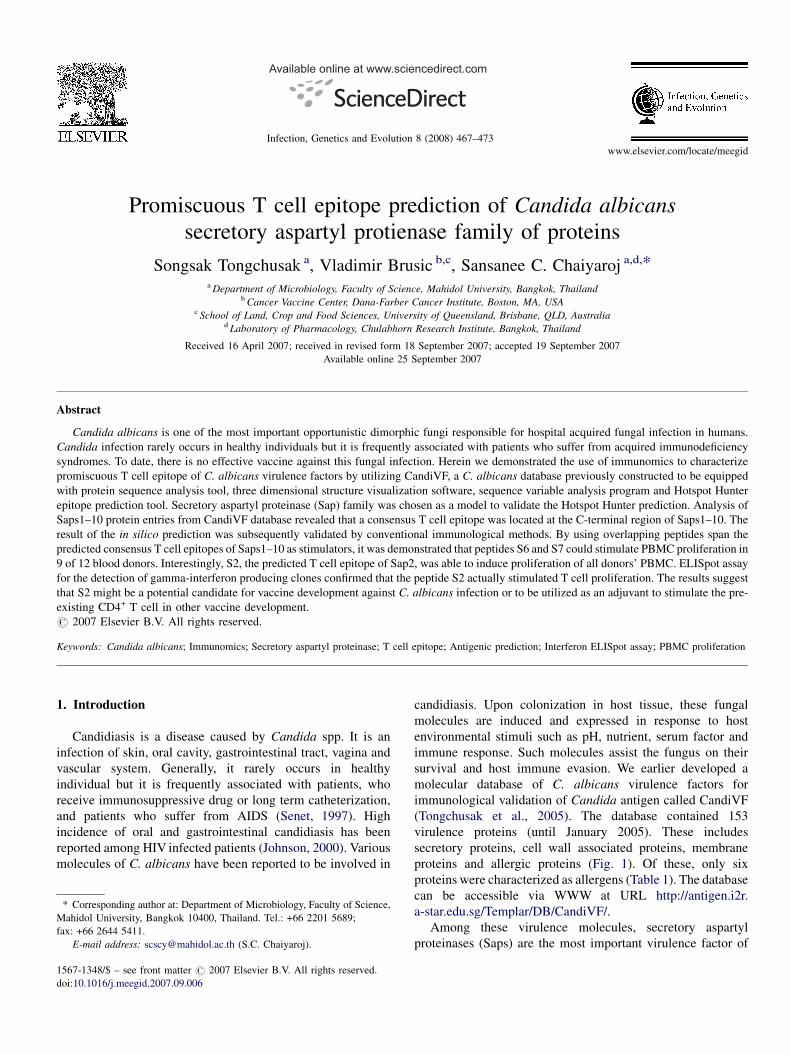
www.elsevier.com/locate/meegid
Available online at www.sciencedirect.com
Infection, Genetics and Evolution 8 (2008) 467–473
Promiscuous T cell epitope prediction of Candida albicans
secretory aspartyl protienase family of proteins
Songsak Tongchusak a, Vladimir Brusic b,c, Sansanee C. Chaiyaroj a,d,*a Department of Microbiology, Faculty of Science, Mahidol University, Bangkok, Thailand
b Cancer Vaccine Center, Dana-Farber Cancer Institute, Boston, MA, USAc School of Land, Crop and Food Sciences, University of Queensland, Brisbane, QLD, Australia
d Laboratory of Pharmacology, Chulabhorn Research Institute, Bangkok, Thailand
Received 16 April 2007; received in revised form 18 September 2007; accepted 19 September 2007
Available online 25 September 2007
Abstract
Candida albicans is one of the most important opportunistic dimorphic fungi responsible for hospital acquired fungal infection in humans.
Candida infection rarely occurs in healthy individuals but it is frequently associated with patients who suffer from acquired immunodeficiency
syndromes. To date, there is no effective vaccine against this fungal infection. Herein we demonstrated the use of immunomics to characterize
promiscuous T cell epitope of C. albicans virulence factors by utilizing CandiVF, a C. albicans database previously constructed to be equipped
with protein sequence analysis tool, three dimensional structure visualization software, sequence variable analysis program and Hotspot Hunter
epitope prediction tool. Secretory aspartyl proteinase (Sap) family was chosen as a model to validate the Hotspot Hunter prediction. Analysis of
Saps1–10 protein entries from CandiVF database revealed that a consensus T cell epitope was located at the C-terminal region of Saps1–10. The
result of the in silico prediction was subsequently validated by conventional immunological methods. By using overlapping peptides span the
predicted consensus T cell epitopes of Saps1–10 as stimulators, it was demonstrated that peptides S6 and S7 could stimulate PBMC proliferation in
9 of 12 blood donors. Interestingly, S2, the predicted T cell epitope of Sap2, was able to induce proliferation of all donors’ PBMC. ELISpot assay
for the detection of gamma-interferon producing clones confirmed that the peptide S2 actually stimulated T cell proliferation. The results suggest
that S2 might be a potential candidate for vaccine development against C. albicans infection or to be utilized as an adjuvant to stimulate the pre-
existing CD4+ T cell in other vaccine development.
# 2007 Elsevier B.V. All rights reserved.
Keywords: Candida albicans; Immunomics; Secretory aspartyl proteinase; T cell epitope; Antigenic prediction; Interferon ELISpot assay; PBMC proliferation
1. Introduction
Candidiasis is a disease caused by Candida spp. It is an
infection of skin, oral cavity, gastrointestinal tract, vagina and
vascular system. Generally, it rarely occurs in healthy
individual but it is frequently associated with patients, who
receive immunosuppressive drug or long term catheterization,
and patients who suffer from AIDS (Senet, 1997). High
incidence of oral and gastrointestinal candidiasis has been
reported among HIV infected patients (Johnson, 2000). Various
molecules of C. albicans have been reported to be involved in
* Corresponding author at: Department of Microbiology, Faculty of Science,
Mahidol University, Bangkok 10400, Thailand. Tel.: +66 2201 5689;
fax: +66 2644 5411.
E-mail address: [email protected] (S.C. Chaiyaroj).
1567-1348/$ – see front matter # 2007 Elsevier B.V. All rights reserved.
doi:10.1016/j.meegid.2007.09.006
candidiasis. Upon colonization in host tissue, these fungal
molecules are induced and expressed in response to host
environmental stimuli such as pH, nutrient, serum factor and
immune response. Such molecules assist the fungus on their
survival and host immune evasion. We earlier developed a
molecular database of C. albicans virulence factors for
immunological validation of Candida antigen called CandiVF
(Tongchusak et al., 2005). The database contained 153
virulence proteins (until January 2005). These includes
secretory proteins, cell wall associated proteins, membrane
proteins and allergic proteins (Fig. 1). Of these, only six
proteins were characterized as allergens (Table 1). The database
can be accessible via WWW at URL http://antigen.i2r.
a-star.edu.sg/Templar/DB/CandiVF/.
Among these virulence molecules, secretory aspartyl
proteinases (Saps) are the most important virulence factor of
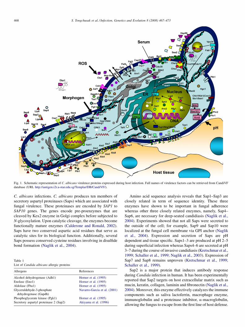
Fig. 1. Schematic representation of C. albicans virulence proteins expressed during host infection. Full names of virulence factors can be retrieved from CandiVF
database (URL http://antigen.i2r.a-star.edu.sg/Templar/DB/CandiVF/).
S. Tongchusak et al. / Infection, Genetics and Evolution 8 (2008) 467–473468
C. albicans infections. C. albicans produces ten members of
secretory aspartyl proteinases (Saps) which are associated with
fungal virulence. These proteinases are encoded by SAP1 to
SAP10 genes. The genes encode pre-proenzymes that are
cleaved by Kex2 enzyme in Golgi complex before subjected to
N-glycosylation. Upon catalytic cleavage, the enzymes become
functionally mature enzymes (Calderone and Ronald, 2002).
Saps have two conserved aspartic acid residues that serve as
catalytic sites for its biological function. Additionally, several
Saps possess conserved cysteine residues involving in disulfide
bond formation (Naglik et al., 2004).
Table 1
List of Candida albicans allergic proteins
Allergens References
Alcohol dehydrogenase (Adh1) Horner et al. (1995)
Enolase (Eno1) Horner et al. (1995)
Aldolase (Fba1) Horner et al. (1995)
Glyceraldehyde-3-phosphate
dehydrogenase (Gapdh)
Navarro-Garcia et al. (2001)
Phosphoglycerate kinase (Pgk1) Horner et al. (1995)
Secretory aspartyl proteinase 2 (Sap2) Akiyama et al. (1996)
Amino acid sequence analysis reveals that Sap1–Sap3 are
closely related in term of sequence identity. These three
enzymes have shown to be important in fungal adherence
whereas other three closely related enzymes, namely, Sap4–
Sap6, are necessary for deep-seated candidiasis (Naglik et al.,
2004). Experiments showed that not all Saps were secreted to
the outside of the cell; for example, Sap9 and Sap10 were
localized at the fungal cell membrane via GPI anchor (Naglik
et al., 2004). Expression and secretion of Saps are pH
dependent and tissue specific. Saps1–3 are produced at pH 2–5
during superficial infection whereas Saps4–6 are secreted at pH
3–7 during the course of invasive candidiasis (Kretschmar et al.,
1999; Schaller et al., 1999; Naglik et al., 2003). Expression of
Sap7 and Sap8 remains unproven (Kretschmar et al., 1999;
Schaller et al., 1999).
Sap2 is a major protein that induces antibody response
during Candida infection in human. It has been experimentally
reported that Sap2 targets on host extracellular matrix such as
mucin, keratin, collagen, laminin and fibronectin (Naglik et al.,
2004). Moreover, this enzyme effectively catalyzes the immune
components such as saliva lactoferrin, macrophage enzyme,
immunoglobulin and a proteinase inhibitor, a-macroglobulin,
allowing the fungus to escape from the first line of host defense.
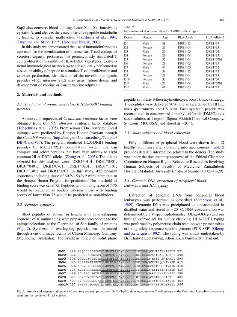
Table 2
Information of donors and their HLA-DRB1 allelic types
Donor Gender Age HLA-Allele 1 HLA-Allele 2
D1 Male 28 DRB1*12 DRB1*13
D2 Female 26 DRB1*04 DRB1*15
D3 Male 27 DRB1*03 DRB1*08
D4 Female 29 DRB1*04 DRB1*15
D5 Female 35 DRB1*04 DRB1*0701
D6 Female 29 DRB1*03 DRB1*15
D7 Male 47 DRB1*03 DRB1*12
D8 Male 39 DRB1*12 DRB1*12
D9 Female 28 DRB1*04 DRB1*14
D10 Female 33 DRB1*04 DRB1*04
D11 Male 24 DRB1*04 DRB1*0701
D12 Male 42 DRB1*03 DRB1*15
S. Tongchusak et al. / Infection, Genetics and Evolution 8 (2008) 467–473 469
Sap2 also converts blood clotting factor X to Xa, inactivates
cystatin A, and cleaves the vasoconstrictive peptide endothelin
1, leading to vascular malfunction (Tsushima et al., 1994;
Tsushima and Mine, 1995; Hube and Naglik, 2001).
In this study we demonstrated the use of immunoinformatics
approach for the identification of a consensus T cell epitope of
secretory aspartyl protinases that promiscuously stimulated T
cell proliferation via multiple HLA-DRB1 supertypes. Conven-
tional immunological methods were subsequently performed to
assess the ability of peptide to stimulate T cell proliferation and
cytokine production. Identification of the novel immunogenic
peptides of C. albicans Sap2 may assist future design and
development of vaccine or cancer vaccine adjuvant.
2. Materials and methods
2.1. Prediction of promiscuous class II HLA-DRB1 binding
peptides
Amino acid sequences of C. albicans virulence factor were
obtained from Candida albicans virulence factor database
(Tongchusak et al., 2005). Promiscuous CD4+ restricted T cell
epitopes were predicted by Hotspot Hunter Program through
the CandiVF website (http://antigen.i2r.a-star.edu.sg/Templar/
DB-/CandiVF/). The program identified HLA-DRB1 binding
peptides by MULTIPRED computation system that can
compute and select peptides that have high affinity to eight
common HLA-DRB1 alleles (Zhang et al., 2005). The alleles
selected for this analysis were: DRB1*0101; DRB1*0301;
DRB1*0401; DRB1*0701; DRB1*0801; DRB1*1101;
DRB1*1301; and DRB1*1501. In this study, 452 primary
sequences including those of SAP1–SAP10 were submitted to
the Hotspot Hunter Program for prediction. The threshold of
binding score was set at 75. Peptides with binding score of�75
would be predicted as binders whereas those with binding
scores of lower than 75 would be predicted as non-binders.
2.2. Peptides synthesis
Short peptides of 20-mer in length, with an overlapping
sequence of 10 amino acids, were prepared corresponding to the
epitope selections at the C-terminal of Sap family of proteins
(Fig. 2). Synthesis of overlapping peptides was performed
through a custom made facility of Chiron Mimotope Company
(Melbourne, Australia). The synthesis relied on solid phase
Fig. 2. Amino acid sequence alignment of secretory aspartyl proteinases, Sap1–Sa
represent the predicted T cell epitopes.
peptide synthesis 9-fluorenylmethoxycarbonyl (fmoc) strategy.
The peptides were delivered 90% pure as ascertained by HPLC,
mass spectrometry and UV scan. Each synthetic peptide was
reconstituted in concentrated dimethyl sulfoxide (DMSO) as a
stock solution at 1 mg/ml (Sigma–Aldrich Chemical Company,
St. Louis, MO, USA) and stored at �20 8C.
2.3. Study subjects and blood collection
Fifty milliliters of peripheral blood were drawn from 12
healthy volunteers after obtaining informed consent. Table 2
provides detailed information related to the donors. The study
was under the documentary approval of the Ethical Clearance
Committee on Human Rights Related to Researches Involving
Human Subjects of Faculty of Medicine, Ramathibodi
Hospital, Mahidol University (Protocol Number ID 05-48-29).
2.4. Genomic DNA extraction of peripheral blood
leukocytes and HLA typing
Extraction of genomic DNA from peripheral blood
leukocytes was performed as described (Sambrook et al.,
1989). Genomic DNA was precipitated and resuspended in
distilled water and stored at �20 8C. DNA concentration was
determined by UV spectrophotometry (OD260/OD280) and run
through agarose gel for quality checking. HLA-DRB1 typing
was performed by polymerase chain reaction with primer mixes
utilizing allele sequence specific primers (PCR-SSP) (Olerup
and Zetterquist, 1992). The typing was kindly undertaken by
Dr. Chanvit Leelayuwat, Khon Kaen University, Thailand.
p10, showing consensus T cell epitope at the C-termini. Underlined sequences
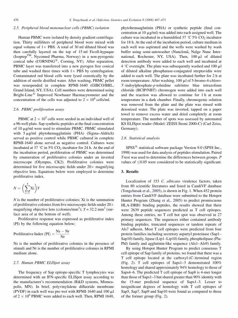
S. Tongchusak et al. / Infection, Genetics and Evolution 8 (2008) 467–473470
2.5. Peripheral blood mononuclear cells (PBMC) isolation
Human PBMC were isolated by density gradient centrifuga-
tion. Thirty milliliters of peripheral blood were mixed with
equal volume of 1� PBS. A total of 30 ml diluted blood was
then carefully layered on the top of 15 ml Ficoll-hypaque
(IsoprepTM, Nycomed Pharma, Norway) in a non-pyrogenic
conical tube (CORNING1, Corning, NY). After separation,
PBMC layer was transferred into a new pyrogen free conical
tube and washed three times with 1� PBS by centrifugation.
Contaminated red blood cells were lysed osmotically by the
addition of sterile distilled water. After washing, PBMC pellet
was resuspended in complete RPMI-1640 (GIBCO/BRL,
Grand Island, NY, USA). Cell numbers were determined using
Bright-Line1 Improved Newbauer Haemacytometer and the
concentration of the cells was adjusted to 2 � 106 cells/ml.
2.6. PBMC proliferation assay
PBMC at 2 � 105 cells were seeded in an individual well of
a 96-well plate. Sap synthetic peptides at the final concentration
of 10 mg/ml were used to stimulate PBMC. PBMC stimulated
with 5 mg/ml phytohemagglutinin (PHA) (Sigma–Aldrich)
served as positive control while PBMC cultured in complete
RPMI-1640 alone served as negative control. Cultures were
incubated at 37 8C in 5% CO2 incubator for 24 h. At the end of
the incubation period, proliferation of PBMC was determined
by enumeration of proliferative colonies under an inverted
microscope (Olympus, CK2). Proliferative colonies were
determined for five microscopic fields under 20� magnifying
objective lens. Equations below were employed to determine
proliferative index.
N ¼�X5
i¼1
Xi
�Y
N is the number of proliferative colonies; Xi is the summation
of proliferative colonies from five microscopic fields under 20�magnifying objective lens (colonies/mm2); Y = 32.2 mm2 (sur-
face area of at the bottom of well).
Proliferative response was expressed as proliferative index
(PI) by the following equation below;
Proliferative Index ðPIÞ ¼ Ns� Nr
Nr
Ns is the number of proliferative colonies in the presence of
stimuli and Nr is the number of proliferative colonies in RPMI
medium alone.
2.7. Human PBMC ELISpot assay
The frequency of Sap epitope-specific T lymphocytes was
determined with an IFN-specific ELISpot assay according to
the manufacturer’s recommendation (R&D systems, Minnea-
polis, MN). In brief, polyvinylidene difluoride membrane
(PVDF) in each well was pre-wet with RPMI-1640 and 100 ml
of 2 � 106 PBMC were added to each well. Then, RPMI 1640,
phytohemaaglutinin (PHA) or synthetic peptide (final con-
centration at 10 mg/ml) was added into each assigned well. The
culture was incubated in a humidified 37 8C 5% CO2 incubator
for 18 h. At the end of the incubation period, culture medium in
each well was aspirated and the wells were washed by wash
buffer using semi-autowasher (Nunclona, Nalge Nunc Inter-
national, Rochester, NY, USA). Then, 100 ml of diluted
detection antibody were added to each well and incubated at
4 8C overnight. The plate was subsequently washed and 100 ml
of diluted alkaline phosphatase-conjugated streptavidin were
added to each well. The plate was incubated further for 2 h at
room temperature. After washing, 100 ml of 5-bromo-4-chloro-
30-indoylphosphate-p-toluidine salt/nitro blue tetrazolium
chloride (BCIP/NBT) chromogen were added into each well
and the reaction was allowed to continue for 1 h at room
temperature in a dark chamber. Finally, chromogenic solution
was removed from the plate and the plate was rinsed with
deionized water. The plate was inverted, tapped on a paper
towel to remove excess water and dried completely at room
temperature. The number of spots was assessed by automated
KS ELISpot reader (Model: ZEISS Stemi 2000-C) (Carl Zeiss,
Germany).
2.8. Statistical analysis
SPSS1 statistical software package Version 9.0 (SPSS Inc.,
1998) was used for data analysis of peptides stimulation. Paired
T-test was used to determine the differences between groups. P
values of �0.05 were considered to be statistically significant.
3. Results
Localization of 153 C. albicans virulence factors, taken
from 80 scientific literatures and listed in CandiVF database
(Tongchusak et al., 2005), is shown in Fig. 1. When 452 protein
entries from CandiVF database were submitted to the Hotspot
Hunter Program (Zhang et al., 2005) to predict promiscuous
HLA-DRB1 binding peptides, the results showed that there
were 2639 peptide sequences predicted as T cell epitopes.
Among these entries, no T cell hot spot was observed in 27
primary sequences. The sequences either contained antibody
binding peptides, truncated sequences or tandem repeats of
Als7 adhesin. Most T cell epitopes were predicted from four
protein families including secretory aspartyl proteinase (Sap1–
Sap10) family, lipase (Lip1–Lip10) family, phospholipase (Pla-
Pld) family and agglutinin-like sequence (Als1–Als9) family.
By using Hotspot Hunter Program to predict consensus T
cell epitope of Sap family of proteins, we found that there was a
T cell epitope located at the carboxyl-(C-)terminal region
(Fig. 2). T cell epitopes of Saps1–3 demonstrated 100%
homology and shared approximately 94% homology to those of
Saps4–6. The predicted T cell epitope of Sap8 is 4-mer longer
than those of Saps1–3 but shared greater than 90% identity with
the 15-mer predicted sequence of Saps1–3. Lesser to
insignificant degrees of homology with T cell epitopes of
Sap5, Sap7, Sap9 and Sap10 were found as compared to those
of the former group (Fig. 2).
Fig. 3. Degrees of PBMC proliferation of 12 donors upon stimulation with overlapping peptides of Sap. Values are means � S.D. of three determinations.
S. Tongchusak et al. / Infection, Genetics and Evolution 8 (2008) 467–473 471
Among the proteins of Sap family, Sap2 is the common
antigen expressed in all cases of candidiasis. Thus, 11
overlapping peptides were constructed based on the Sap2
amino acid sequence. Peptides S6–S10 span the predicted
consensus T cell epitope of Sap sequence. These peptides were
used to stimulate PBMC from 12 HLA-DRB1 blood donors
(including HLA-DRB1*04/04, *04/0701, *04/0701, *04/15,
*04/15, *03/15, *03/08, *03/15, *03/12, *04/14, *12/13 and
*12/12) (Table 2). Fig. 3 showed different degrees of
proliferative response to Sap peptides of all 12 donors.
Peptides S6 (QLLFDVNDANILGDNFLRSA) and S7
(QLLFDVNKANILGDNFLRSA), which lie in the predicted
area, were able to significantly induce PBMC activation of 9 of
12 donors. Notably, these two peptides contain almost identical
amino acid sequences except for one amino acid at position 8
(D for S6, K for S7) from the amino-(N-)terminal. There were
two donors (HLA-DRB1*03/08 and *04/14) that responded to
only one of the two peptides. The other two donors did not show
any response to peptides S1 and S3–S10. The latter two non-
responders to the consensus T cell epitope (S6–S10) possess
HLA-DRB1*12/12 and 04/0701.
Interestingly, peptide S2 (DSNLSGDVVFNFSKNAKISV),
that lays upstream of the conserved T cell epitope of Sap family,
was able to stimulate PBMC of all donors (Fig. 3). The ability
of S2 peptide to stimulate T cell was confirmed by ELISpot
assay based on interferon secretion by PBMC. S2 enabled to
activate gamma-interferon release from PBMC of all donors
(Fig. 4). The response to this peptide was significant as the
number of interferon producing clones exceeded 3500 spots/
106 PBMC (Fig. 4).
Three-dimensional structure data of Sap2 were gathered
from PDB and homology modeling assignment (Fig. 5). The
resolution Sap2 three-dimensional structure was 2.10 A. From
the structural analysis, peptide S2 displays two b-sheets with –
DVVFNFS-(b261–b267) and –AKIS-(b270–b273) sequences.
The constituted hydrophobic amino acids possess side chains
that point down towards the inner part of Sap2 molecule
(Fig. 5).
4. Discussion
Our strategy of peptide-based vaccines is to identify
promiscuous, as well as consensus, T cell epitope of Sap
family of proteins. The epitope should be a relevant
representative of large proportion of the human population.
The majority of publicly available programs usually predicted
the peptides that specifically bound to an individual HLA
molecule and has not been properly assessed for predictive
accuracy. MULTIPRED was developed based on the fact that a
peptide could bind to multiple MHC allelic variants and closely
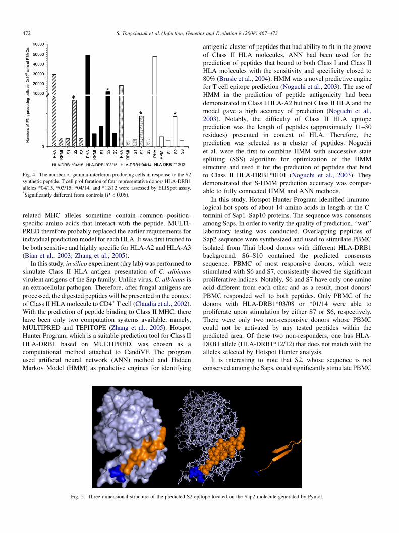
Fig. 4. The number of gamma-interferon producing cells in response to the S2
synthetic peptide. T cell proliferation of four representative donors HLA-DRB1
alleles *04/15, *03/15, *04/14, and *12/12 were assessed by ELISpot assay.*Significantly different from controls (P < 0.05).
S. Tongchusak et al. / Infection, Genetics and Evolution 8 (2008) 467–473472
related MHC alleles sometime contain common position-
specific amino acids that interact with the peptide. MULTI-
PRED therefore probably replaced the earlier requirements for
individual prediction model for each HLA. It was first trained to
be both sensitive and highly specific for HLA-A2 and HLA-A3
(Bian et al., 2003; Zhang et al., 2005).
In this study, in silico experiment (dry lab) was performed to
simulate Class II HLA antigen presentation of C. albicans
virulent antigens of the Sap family. Unlike virus, C. albicans is
an extracellular pathogen. Therefore, after fungal antigens are
processed, the digested peptides will be presented in the context
of Class II HLA molecule to CD4+ T cell (Claudia et al., 2002).
With the prediction of peptide binding to Class II MHC, there
have been only two computation systems available, namely,
MULTIPRED and TEPITOPE (Zhang et al., 2005). Hotspot
Hunter Program, which is a suitable prediction tool for Class II
HLA-DRB1 based on MULTIPRED, was chosen as a
computational method attached to CandiVF. The program
used artificial neural network (ANN) method and Hidden
Markov Model (HMM) as predictive engines for identifying
Fig. 5. Three-dimensional structure of the predicted S2 epit
antigenic cluster of peptides that had ability to fit in the groove
of Class II HLA molecules. ANN had been used for the
prediction of peptides that bound to both Class I and Class II
HLA molecules with the sensitivity and specificity closed to
80% (Brusic et al., 2004). HMM was a novel predictive engine
for T cell epitope prediction (Noguchi et al., 2003). The use of
HMM in the prediction of peptide antigenicity had been
demonstrated in Class I HLA-A2 but not Class II HLA and the
model gave a high accuracy of prediction (Noguchi et al.,
2003). Notably, the difficulty of Class II HLA epitope
prediction was the length of peptides (approximately 11–30
residues) presented in context of HLA. Therefore, the
prediction was selected as a cluster of peptides. Noguchi
et al. were the first to combine HMM with successive state
splitting (SSS) algorithm for optimization of the HMM
structure and used it for the prediction of peptides that bind
to Class II HLA-DRB1*0101 (Noguchi et al., 2003). They
demonstrated that S-HMM prediction accuracy was compar-
able to fully connected HMM and ANN methods.
In this study, Hotspot Hunter Program identified immuno-
logical hot spots of about 14 amino acids in length at the C-
termini of Sap1–Sap10 proteins. The sequence was consensus
among Saps. In order to verify the quality of prediction, ‘‘wet’’
laboratory testing was conducted. Overlapping peptides of
Sap2 sequence were synthesized and used to stimulate PBMC
isolated from Thai blood donors with different HLA-DRB1
background. S6–S10 contained the predicted consensus
sequence. PBMC of most responsive donors, which were
stimulated with S6 and S7, consistently showed the significant
proliferative indices. Notably, S6 and S7 have only one amino
acid different from each other and as a result, most donors’
PBMC responded well to both peptides. Only PBMC of the
donors with HLA-DRB1*03/08 or *01/14 were able to
proliferate upon stimulation by either S7 or S6, respectively.
There were only two non-responsive donors whose PBMC
could not be activated by any tested peptides within the
predicted area. Of these two non-responders, one has HLA-
DRB1 allele (HLA-DRB1*12/12) that does not match with the
alleles selected by Hotspot Hunter analysis.
It is interesting to note that S2, whose sequence is not
conserved among the Saps, could significantly stimulate PBMC
ope located on the Sap2 molecule generated by Pymol.
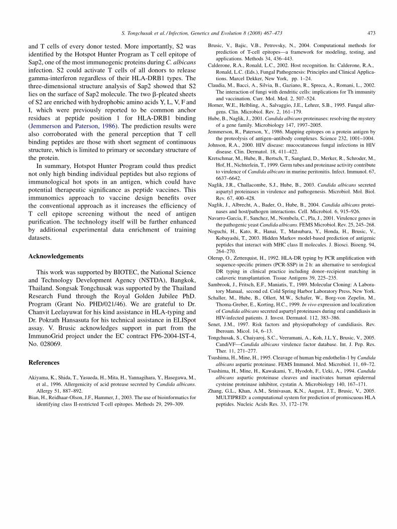
S. Tongchusak et al. / Infection, Genetics and Evolution 8 (2008) 467–473 473
and T cells of every donor tested. More importantly, S2 was
identified by the Hotspot Hunter Program as T cell epitope of
Sap2, one of the most immunogenic proteins during C. albicans
infection. S2 could activate T cells of all donors to release
gamma-interferon regardless of their HLA-DRB1 types. The
three-dimensional structure analysis of Sap2 showed that S2
lies on the surface of Sap2 molecule. The two b-pleated sheets
of S2 are enriched with hydrophobic amino acids Y, L, V, F and
I, which were previously reported to be common anchor
residues at peptide position 1 for HLA-DRB1 binding
(Jemmerson and Paterson, 1986). The prediction results were
also corroborated with the general perception that T cell
binding peptides are those with short segment of continuous
structure, which is limited to primary or secondary structure of
the protein.
In summary, Hotspot Hunter Program could thus predict
not only high binding individual peptides but also regions of
immunological hot spots in an antigen, which could have
potential therapeutic significance as peptide vaccines. This
immunomics approach to vaccine design benefits over
the conventional approach as it increases the efficiency of
T cell epitope screening without the need of antigen
purification. The technology itself will be further enhanced
by additional experimental data enrichment of training
datasets.
Acknowledgements
This work was supported by BIOTEC, the National Science
and Technology Development Agency (NSTDA), Bangkok,
Thailand. Songsak Tongchusak was supported by the Thailand
Research Fund through the Royal Golden Jubilee PhD.
Program (Grant No. PHD/021/46). We are grateful to Dr.
Chanvit Leelayuwat for his kind assistance in HLA-typing and
Dr. Pokrath Hansasuta for his technical assistance in ELISpot
assay. V. Brusic acknowledges support in part from the
ImmunoGrid project under the EC contract FP6-2004-IST-4,
No. 028069.
References
Akiyama, K., Shida, T., Yasueda, H., Mita, H., Yannagihara, Y., Hasegawa, M.,
et al., 1996. Allergenicity of acid protease secreted by Candida albicans.
Allergy 51, 887–892.
Bian, H., Reidhaar-Olson, J.F., Hammer, J., 2003. The use of bioinformatics for
identifying class II-restricted T-cell epitopes. Methods 29, 299–309.
Brusic, V., Bajic, V.B., Petrovsky, N., 2004. Computational methods for
prediction of T-cell epitopes—a framework for modeling, testing, and
applications. Methods 34, 436–443.
Calderone, R.A., Ronald, L.C., 2002. Host recognition. In: Calderone, R.A.,
Ronald, L.C. (Eds.), Fungal Pathogenesis: Principles and Clinical Applica-
tions. Marcel Dekker, New York, pp. 1–24.
Claudia, M., Bacci, A., Silvia, B., Gaziano, R., Spreca, A., Romani, L., 2002.
The interaction of fungi with dendritic cells: implications for Th immunity
and vaccination. Curr. Mol. Med. 2, 507–524.
Horner, W.E., Helbling, A., Salvaggio, J.E., Lehrer, S.B., 1995. Fungal aller-
gens. Clin. Microbiol. Rev. 2, 161–179.
Hube, B., Naglik, J., 2001. Candida albicans proteinases: resolving the mystery
of a gene family. Microbiology 147, 1997–2005.
Jemmerson, R., Paterson, Y., 1986. Mapping epitopes on a protein antigen by
the proteolysis of antigen–antibody complexes. Science 232, 1001–1004.
Johnson, R.A., 2000. HIV disease: mucocutaneous fungal infections in HIV
disease. Clin. Dermatol. 18, 411–422.
Kretschmar, M., Hube, B., Bertsch, T., Sanglard, D., Merker, R., Schroder, M.,
Hof, H., Nichterlein, T., 1999. Germ tubes and proteinase activity contribute
to virulence of Candida albicans in murine peritonitis. Infect. Immunol. 67,
6637–6642.
Naglik, J.R., Challacombe, S.J., Hube, B., 2003. Candida albicans secreted
aspartyl proteinases in virulence and pathogenesis. Microbiol. Mol. Biol.
Rev. 67, 400–428.
Naglik, J., Albrecht, A., Bader, O., Hube, B., 2004. Candida albicans protei-
nases and host/pathogen interactions. Cell. Microbiol. 6, 915–926.
Navarro-Garcia, F., Sanchez, M., Nombela, C., Pla, J., 2001. Virulence genes in
the pathogenic yeast Candida albicans. FEMS Microbiol. Rev. 25, 245–268.
Noguchi, H., Kato, R., Hanai, T., Matsubara, Y., Honda, H., Brusic, V.,
Kobayashi, T., 2003. Hidden Markov model-based prediction of antigenic
peptides that interact with MHC class II molecules. J. Biosci. Bioeng. 94,
264–270.
Olerup, O., Zetterquist, H., 1992. HLA-DR typing by PCR amplification with
sequence-specific primers (PCR-SSP) in 2 h: an alternative to serological
DR typing in clinical practice including donor–recipient matching in
cadaveric transplantation. Tissue Antigens 39, 225–235.
Sambrook, J., Fritsch, E.F., Maniatis, T., 1989. Molecular Cloning: A Labora-
tory Manual, second ed. Cold Spring Harbor Laboratory Press, New York.
Schaller, M., Hube, B., Ollert, M.W., Schafer, W., Borg-von Zepelin, M.,
Thoma-Greber, E., Korting, H.C., 1999. In vivo expression and localization
of Candida albicans secreted aspartyl proteinases during oral candidiasis in
HIV-infected patients. J. Invest. Dermatol. 112, 383–386.
Senet, J.M., 1997. Risk factors and physiopathology of candidiasis. Rev.
Iberoam. Micol. 14, 6–13.
Tongchusak, S., Chaiyaroj, S.C., Veeramani, A., Koh, J.L.Y., Brusic, V., 2005.
CandiVF—Candida albicans virulence factor database. Int. J. Pep. Res.
Ther. 11, 271–277.
Tsushima, H., Mine, H., 1995. Cleavage of human big endothelin-1 by Candida
albicans aspartic proteinase. FEMS Immunol. Med. Microbiol. 11, 69–72.
Tsushima, H., Mine, H., Kawakami, Y., Hyodoh, F., Ueki, A., 1994. Candida
albicans aspartic proteinase cleaves and inactivates human epidermal
cysteine proteinase inhibitor, cystatin A. Microbiology 140, 167–171.
Zhang, G.L., Khan, A.M., Srinivasan, K.N., August, J.T., Brusic, V., 2005.
MULTIPRED: a computational system for prediction of promiscuous HLA
peptides. Nucleic Acids Res. 33, 172–179.
www.elsevier.com/locate/meegid
Available online at www.sciencedirect.com
Infection, Genetics and Evolution 8 (2008) 474–483
Genomic interrogation of ancestral Mycobacterium tuberculosis
from south India
Sujatha Narayanan a,b,*, Sebastien Gagneux b,1, Lalitha Hari a, Anthony G. Tsolaki b,2,Suganthi Rajasekhar a, P.R. Narayanan a, Peter M. Small b,1,3,
Susan Holmes b, Kathryn DeRiemer b,4
a Tuberculosis Research Centre (TRC), Mayor VR Ramanathan Road, Chetput, Chennai 600031, Indiab Division of Infectious Diseases and Geographic Medicine, Stanford University Medical Center, 300 Pasteur Drive,
Stanford, CA 94305-5107, USA
Received 27 March 2007; received in revised form 19 September 2007; accepted 24 September 2007
Available online 29 September 2007
Abstract
Mycobacterium tuberculosis is a very important global pathogen. One quarter of the world’s TB cases occur in India. The tuberculosis strains
isolated from south Indian patients exhibit certain phenotypic characteristics like low virulence in guinea-pigs, resistance to isoniazid, thiophene-2-
carboxylic acid hydrazide (TCH) and para-amino salicylic acid (PAS), and enhanced susceptibility to H2O2. Besides this, a large percentage of the
isolates harbor only a single copy of IS 6110 which makes these strains distinct. Hence, we have studied the genotypic characteristics of these
strains by using advanced techniques like Deletion Micro array, deletion PCR, allelic discrimination RT-PCR using several lineage specific markers
and KatG G1388T (non-synonymous) polymorphism along with spoligotyping. The analysis of 1215 tuberculosis patient isolates from south India
revealed that 85.2% belonged to the ancestral lineage of M. tuberculosis. Comparative whole-genome hybridization identified six new genomic
regions within this lineage that were variably deleted.
# 2007 Elsevier B.V. All rights reserved.
Keywords: Tuberculosis; Spoligotyping; Genomic deletion; Large sequence polymorphism; Lineage; Vaccine
1. Introduction
Mycobacterium tuberculosis is a very important global
pathogen. Without HIV, the tuberculosis (TB) epidemic would
now be in decline everywhere. India has the highest number of
incident TB cases in the world. With effective DOTS
* Corresponding author at: Tuberculosis Research Centre (TRC), Mayor VR
Ramanathan Road, Chetput, Chennai 600031, India.
Tel.: +91 44 2836 9627/9600; fax: +91 44 2836 2528/2529.
E-mail address: [email protected] (S. Narayanan).1 Present address: Institute for Systems Biology, 1441 North 34th Street,
Seattle, WA 98103, USA.2 Present address: Center for Molecular Biology and Infection (CMMI),
Division of Investigative Sciences, Flowers Building, South Kensington Cam-
pus, Imperial College, London SW7 2AZ, United Kingdom.3 Present address: Bill and Melinda Gates Foundation, P.O. Box 23350,
Seattle, WA 981012, USA.4 Present address: School of Medicine, University of California, One Shields
Avenue, Davis, CA 95616, USA.
1567-1348/$ – see front matter # 2007 Elsevier B.V. All rights reserved.
doi:10.1016/j.meegid.2007.09.007
implementation tuberculosis is expected to decline but as
HIV continues to spread, the decline of TB is far from reality.
Although one fourth of the global tuberculosis burden stems
from India there are a few studies which define the tuberculosis
genogroup (Bhanu et al., 2002; Das et al., 2005; Mistry et al.,
2002; Narayanan et al., 1997; Kulkarni et al., 2005; Singh et al.,
2004; Radhakrishnan et al., 2001). There has been a population
based study from south India on transmission dynamics and risk
factors associated with transmission (Narayanan et al., 2002).
The information obtained in this study suffers from a lack of
portability which hinders comparison between global studies.
In addition, the south Indian isolates included by others in the
previous studies have been limited in number (Guitierrez et al.,
2006; Singh et al., 2007). To avoid the problem in portability of
data and limitation in number we have used spoligotyping to
genotype the isolates from a population based study.
Spoligotyping is a PCR based genotyping method that
allows to assess the M. tuberculosis genetic biodiversity and
provides enough information about the epidemiologically
S. Narayanan et al. / Infection, Genetics and Evolution 8 (2008) 474–483 475
important clones in various settings (Cowan et al., 2004;
Streicher et al., 2004). The establishment of an international
database is an advantage to this method because it is very
informative regarding the endemicity or the ubiquitous strains
(Filliol et al., 2002, 2003).
The tuberculosis strains isolated from south Indian patients
exhibit certain phenotypic characteristics like low virulences in
guinea pigs, resistance to isoniazid (Mitchison et al., 1960;
Middlebrook and Cohn, 1953), thiophene-2-carboxylic acid
hydrazide (TC4) and para-amino salicylic acid (PAS) (Joseph
et al., 1964) enhanced susceptibility to H2O2 and a majority of
them belonging to intermediate phage type (Bhatia, 1961;
Grange et al., 1978). Besides this a large percentage of the
isolates harbor only a single copy of IS6110 (Das et al., 1995)
which make these strains distinct. Hence, we wanted to study
the genotypic characteristics of these M. tuberculosis strains by
using advanced techniques like Deletion Microarray, deletion
PCR, allelic discrimination RT-PCR using several lineage
specific markers and KatG G1388T (non-synonymous) poly-
morphism (Sreevatsan et al., 1997) along with spoligotyping
and IS6110 RFLP which is considered the gold standard for
genotyping techniques.
The deletion micro array is an approach complementary to
comparative genomics. This involves the interrogation of
unsequenced genomes by DNA-microarray to identify
sequences present in a fully sequenced isolate but absent from
interrogated isolate (Kato-Maeda et al., 2001; Tsolaki et al.,
2004). Nucleotide sequences provide robust, portable and
comparable data for studying population variation. The
mutational processes that generate these variations are under-
stood and sequence data have been successfully used in the
study of evolution, bacterial epidemiology and population
biology (Baker et al., 2004).
2. Materials and methods
2.1. IS6110 RFLP & spoligotyping
We used a standardized international protocol for IS6110
RFLP genotyping (van Embden et al., 1993). Spoligotyping
was performed as previously described (Kamerbeek et al.,
1997). All of the spoligotype patterns were coded using the
octal code system (Dale et al., 2001) with M. bovis P3 and
H37Rv as positive controls and autoclaved milli Q water as
negative control. The DR region was amplified using the DRa
(50biotinylated) and DRb primers. The amplified product was
hybridized to a set of 43 immobilized oligonucleotides, each
corresponding to a unique spacer sequence within the DR locus.
Hybridization was detected by enhanced chemiluminescence
(ECL Enhanced Chemo-Luminescence Detection Kit; Amer-
sham, Hongkong) followed by exposure to X-ray film
according to the, manufacturer’s recommendations. The results
were documented in the form of binary code (x and 0) according
to the hybridization (respectively positive or negative) for each
spacer nucleotide probe and entered into excel format.
We then referred to a standardized international database of
spoligotype patterns, SpolDB4 (Brudey et al., 2006) to
determine whether each pattern had been previously reported.
The updated SpolDB4 version contained 39,295 patterns
distributed into 1939 shared types (patterns reported at least
twice that grouped 36,925 clinical isolates) and 337 orphan
patterns from more than 120 countries. The database provides
detailed information about the country of origin and the
geographic distribution within eight regions of the world.
2.2. Identification of large sequence polymorphism
We selected 25 isolates for comparative whole-genome
hybridization using an Affymetrix DNA chip (Santa Clara,
California, USA) following procedures previously described. We
identified putative deletions in the experimental strains relative to
the sequenced reference strain H37Rv using DelScan software
(AbaSci, San Pablo, California, USA), and confirmed the
putative deletions by PCR and direct sequencing. We used
phylogenetically informative large sequence polymorphisms
(LSPs) or genomic deletions, to screen one isolate per
spoligotype patterns (102 isolates) by PCR or multiplex real-
time PCR. The screening results from the clustered isolates were
extrapolated to the remaining isolates of their respective clusters.
We used previously published studies of genomic deletions
(Tsolaki et al., 2004) to identify phylogenetically important
genomic deletions and screen for them using PCR. For the
detection of LSPs by multiplex real-time PCR, we designed a
series of assays based on different TaqMan primer/probe
combinations. The presence or absence of RD105 and RD239
was evaluated using following primers and probes: RD105-F,
50-AACGAACTGCGCACTGAACTC-30; RD105-R, 50-TCC-
CGCACCGGTTGAG-30; RD105-probe, 50-FAM-AGAGTG-
GACAGTTTCG-MGBNFQ-30; RD239-F0 50-CGAGCTCAA-
TCCGAACGAAA-30; RD239-R, 50-CCGGGCTTGGCTTTA-
ACTG-30; RD239-probe, 50-VIC1-CCAGGTGCTTGCCATG-
MGBNFQ-30. Samples with no increase in fluorescence of
either FAM or VIC1 were considered deleted for the
corresponding region of interest. Appropriate positive and
negative controls were run on each plate. Isolates with an
increase in both FAM and VIC1 signal were considered not
deleted for either RD and analyzed further. Similarly, the
presence or absence of TbD1 and RD9 was evaluated with the
following primers and probes: TbD1-F, 50-CCGATTGACCA-
CAGCTCGAT-30; TBD1-R, 50-CTGGCCGACGCTTTGC-30;TbD1-probe, 50-FAM-CCGTTTCAGATCAGC-30; RD9-F, 50-TGGTGGCGGTAGGTTTCAC 30; RD9-R, 50-ATGACCCG-
CGCGATGT-30; RD9-probe, 50-VIC1-TTCGACCCCAAGAC.
2.3. Real-time PCR
The Euro-American lineage was defined based on a
characteristic seven base pair deletion in pks15/1 (Constant
et al., 2002) or the ctg to cgg substitution at codon 463 of katGi.
These two markers are known to be equivalent based on
previous studies (Baker et al., 2004; Gutacker et al., 2002;
Marmiesse et al., 2004). The katG463 SNP was analyzed by
allelic discrimination using a TaqMan multiplex real-time PCR
assay (Applied Biosystems, Foster City, CA, USA). The
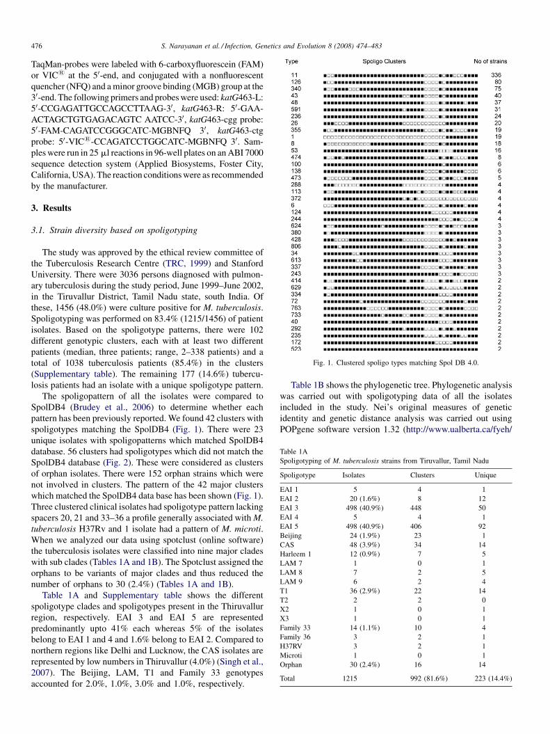
Fig. 1. Clustered spoligo types matching Spol DB 4.0.
Table 1A
Spoligotyping of M. tuberculosis strains from Tiruvallur, Tamil Nadu
Spoligotype Isolates Clusters Unique
EAI 1 5 4 1
EAI 2 20 (1.6%) 8 12
EAI 3 498 (40.9%) 448 50
EAI 4 5 4 1
EAI 5 498 (40.9%) 406 92
Beijing 24 (1.9%) 23 1
CAS 48 (3.9%) 34 14
Harleem 1 12 (0.9%) 7 5
LAM 7 1 0 1
LAM 8 7 2 5
LAM 9 6 2 4
T1 36 (2.9%) 22 14
T2 2 2 0
X2 1 0 1
X3 1 0 1
Family 33 14 (1.1%) 10 4
Family 36 3 2 1
H37RV 3 2 1
Microti 1 0 1
Orphan 30 (2.4%) 16 14
Total 1215 992 (81.6%) 223 (14.4%)
S. Narayanan et al. / Infection, Genetics and Evolution 8 (2008) 474–483476
TaqMan-probes were labeled with 6-carboxyfluorescein (FAM)
or VIC1 at the 50-end, and conjugated with a nonfluorescent
quencher (NFQ) and a minor groove binding (MGB) group at the
30-end. The following primers and probes were used: katG463-L:
50-CCGAGATTGCCAGCCTTAAG-30, katG463-R: 50-GAA-
ACTAGCTGTGAGACAGTC AATCC-30, katG463-cgg probe:
50-FAM-CAGATCCGGGCATC-MGBNFQ 30, katG463-ctg
probe: 50-VIC1-CCAGATCCTGGCATC-MGBNFQ 30. Sam-
ples were run in 25 ml reactions in 96-well plates on an ABI 7000
sequence detection system (Applied Biosystems, Foster City,
California, USA). The reaction conditions were as recommended
by the manufacturer.
3. Results
3.1. Strain diversity based on spoligotyping
The study was approved by the ethical review committee of
the Tuberculosis Research Centre (TRC, 1999) and Stanford
University. There were 3036 persons diagnosed with pulmon-
ary tuberculosis during the study period, June 1999–June 2002,
in the Tiruvallur District, Tamil Nadu state, south India. Of
these, 1456 (48.0%) were culture positive for M. tuberculosis.
Spoligotyping was performed on 83.4% (1215/1456) of patient
isolates. Based on the spoligotype patterns, there were 102
different genotypic clusters, each with at least two different
patients (median, three patients; range, 2–338 patients) and a
total of 1038 tuberculosis patients (85.4%) in the clusters
(Supplementary table). The remaining 177 (14.6%) tubercu-
losis patients had an isolate with a unique spoligotype pattern.
The spoligopattern of all the isolates were compared to
SpolDB4 (Brudey et al., 2006) to determine whether each
pattern has been previously reported. We found 42 clusters with
spoligotypes matching the SpolDB4 (Fig. 1). There were 23
unique isolates with spoligopatterns which matched SpolDB4
database. 56 clusters had spoligotypes which did not match the
SpolDB4 database (Fig. 2). These were considered as clusters
of orphan isolates. There were 152 orphan strains which were
not involved in clusters. The pattern of the 42 major clusters
which matched the SpolDB4 data base has been shown (Fig. 1).
Three clustered clinical isolates had spoligotype pattern lacking
spacers 20, 21 and 33–36 a profile generally associated with M.
tuberculosis H37Rv and 1 isolate had a pattern of M. microti.
When we analyzed our data using spotclust (online software)
the tuberculosis isolates were classified into nine major clades
with sub clades (Tables 1A and 1B). The Spotclust assigned the
orphans to be variants of major clades and thus reduced the
number of orphans to 30 (2.4%) (Tables 1A and 1B).
Table 1A and Supplementary table shows the different
spoligotype clades and spoligotypes present in the Thiruvallur
region, respectively. EAI 3 and EAI 5 are represented
predominantly upto 41% each whereas 5% of the isolates
belong to EAI 1 and 4 and 1.6% belong to EAI 2. Compared to
northern regions like Delhi and Lucknow, the CAS isolates are
represented by low numbers in Thiruvallur (4.0%) (Singh et al.,
2007). The Beijing, LAM, T1 and Family 33 genotypes
accounted for 2.0%, 1.0%, 3.0% and 1.0%, respectively.
Table 1B shows the phylogenetic tree. Phylogenetic analysis
was carried out with spoligotyping data of all the isolates
included in the study. Nei’s original measures of genetic
identity and genetic distance analysis was carried out using
POPgene software version 1.32 (http://www.ualberta.ca/fyeh/
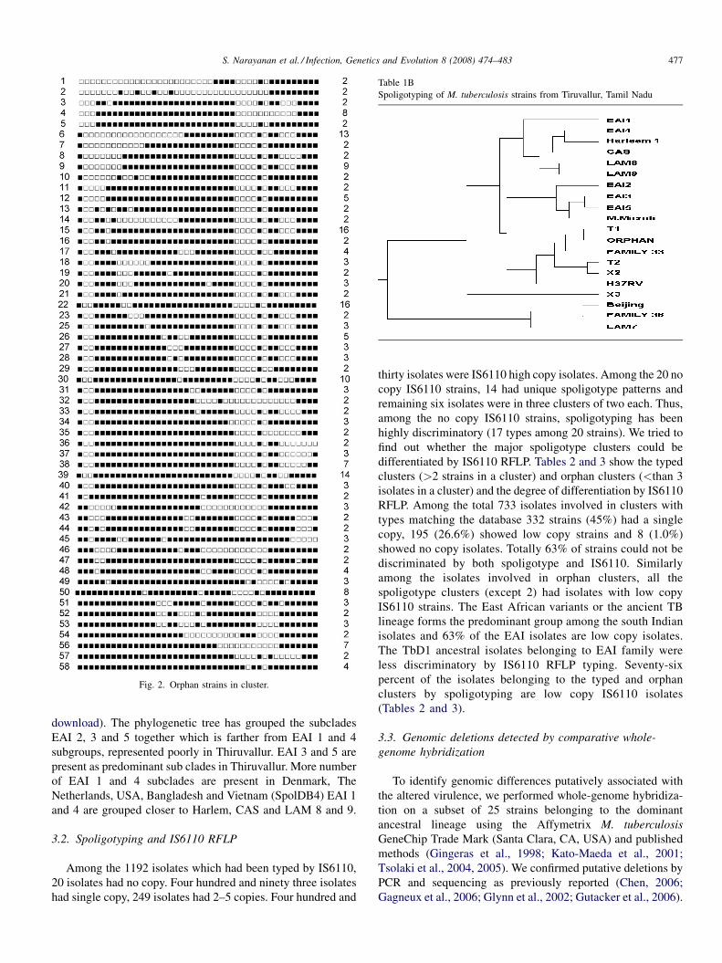
Fig. 2. Orphan strains in cluster.
Table 1B
Spoligotyping of M. tuberculosis strains from Tiruvallur, Tamil Nadu
S. Narayanan et al. / Infection, Genetics and Evolution 8 (2008) 474–483 477
download). The phylogenetic tree has grouped the subclades
EAI 2, 3 and 5 together which is farther from EAI 1 and 4
subgroups, represented poorly in Thiruvallur. EAI 3 and 5 are
present as predominant sub clades in Thiruvallur. More number
of EAI 1 and 4 subclades are present in Denmark, The
Netherlands, USA, Bangladesh and Vietnam (SpolDB4) EAI 1
and 4 are grouped closer to Harlem, CAS and LAM 8 and 9.
3.2. Spoligotyping and IS6110 RFLP
Among the 1192 isolates which had been typed by IS6110,
20 isolates had no copy. Four hundred and ninety three isolates
had single copy, 249 isolates had 2–5 copies. Four hundred and
thirty isolates were IS6110 high copy isolates. Among the 20 no
copy IS6110 strains, 14 had unique spoligotype patterns and
remaining six isolates were in three clusters of two each. Thus,
among the no copy IS6110 strains, spoligotyping has been
highly discriminatory (17 types among 20 strains). We tried to
find out whether the major spoligotype clusters could be
differentiated by IS6110 RFLP. Tables 2 and 3 show the typed
clusters (>2 strains in a cluster) and orphan clusters (<than 3
isolates in a cluster) and the degree of differentiation by IS6110
RFLP. Among the total 733 isolates involved in clusters with
types matching the database 332 strains (45%) had a single
copy, 195 (26.6%) showed low copy strains and 8 (1.0%)
showed no copy isolates. Totally 63% of strains could not be
discriminated by both spoligotype and IS6110. Similarly
among the isolates involved in orphan clusters, all the
spoligotype clusters (except 2) had isolates with low copy
IS6110 strains. The East African variants or the ancient TB
lineage forms the predominant group among the south Indian
isolates and 63% of the EAI isolates are low copy isolates.
The TbD1 ancestral isolates belonging to EAI family were
less discriminatory by IS6110 RFLP typing. Seventy-six
percent of the isolates belonging to the typed and orphan
clusters by spoligotyping are low copy IS6110 isolates
(Tables 2 and 3).
3.3. Genomic deletions detected by comparative whole-
genome hybridization
To identify genomic differences putatively associated with
the altered virulence, we performed whole-genome hybridiza-
tion on a subset of 25 strains belonging to the dominant
ancestral lineage using the Affymetrix M. tuberculosis
GeneChip Trade Mark (Santa Clara, CA, USA) and published
methods (Gingeras et al., 1998; Kato-Maeda et al., 2001;
Tsolaki et al., 2004, 2005). We confirmed putative deletions by
PCR and sequencing as previously reported (Chen, 2006;
Gagneux et al., 2006; Glynn et al., 2002; Gutacker et al., 2006).
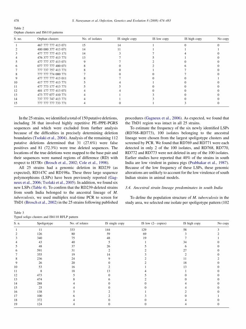
Table 2
Orphan clusters and IS6110 patterns
S. no. Orphan clusters No. of isolates IS single copy IS low copy IS high copy No copy
1 467 777 777 413 071 15 14 1 0 0
2 400 000 377 413 071 14 11 1 1 1
3 477 777 777 413 171 14 3 7 4 0
4 476 377 777 413 771 13 7 5 1 0
5 477 777 377 413 071 9 7 2 0 0
6 077 777 777 400 071 8 0 2 6 0
7 777 737 757 413 771 8 0 1 7 0
8 777 777 774 000 771 7 0 0 7 0
9 477 777 777 413 011 8 7 0 0 1
10 417 777 777 413 771 5 4 1 0 0
11 477 773 177 413 771 5 5 0 0 0
12 401 177 777 413 071 4 4 0 0 0
13 473 777 077 410 771 4 1 3 0 0
14 737 777 747 413 771 4 1 3 0 0
15 777 777 777 733 771 4 0 1 3 0
S. Narayanan et al. / Infection, Genetics and Evolution 8 (2008) 474–483478
In the 25 strains, we identified a total of 150 putative deletions,
including 38 that involved highly repetitive PE-/PPE-PGRS
sequences and which were excluded from further analysis
because of the difficulties in precisely determining deletion
boundaries (Tsolaki et al., 2004). Analysis of the remaining 112
putative deletions determined that 31 (27.6%) were false
positives and 81 (72.3%) were true deleted sequences. The
locations of the true deletions were mapped to the base pair and
their sequences were named regions of difference (RD) with
respect to H37Rv (Brosch et al., 2002; Cole et al., 1998).
All 25 strains had a genomic deletion in RD239 (as
expected), RD147C and RD198a. These three large sequence
polymorphisms (LSPs) have been previously reported (Gag-
neux et al., 2006; Tsolaki et al., 2005). In addition, we found six
new LSPs (Table 4). To confirm that the RD239-deleted strains
from south India belonged to the ancestral lineage of M.
tuberculosis, we used multiplex real-time PCR to screen for
TbD1 (Brosch et al., 2002) in the 25 strains following published
Table 3
Typed soligo clusters and IS6110 RFLP pattern
S. n. Spoligotype No. of iolates IS single copy
1 11 333 144
2 126 80 59
3 340 75 48
4 43 40 5
5 48 37 26
6 591 31 2
7 355 19 14
8 236 24 9
9 26 20 0
10 53 16 2
11 8 18 13
12 473 5 0
13 474 8 6
14 288 4 0
15 25 4 0
16 138 5 2
17 100 6 2
18 372 4 0
19 124 4 0
procedures (Gagneux et al., 2006). As expected, we found that
the TbD1 region was intact in all 25 strains.
To estimate the frequency of the six newly identified LSPs
(RD768–RD773), 100 isolates belonging to the ancestral
lineage were chosen from the largest spoligotype clusters and
screened by PCR. We found that RD769 and RD771 were each
detected in only 2 of the 100 isolates, and RD768, RD770,
RD772 and RD773 were not deleted in any of the 100 isolates.
Earlier studies have reported that 40% of the strains in south
India are low virulent in guinea pigs (Prabhakar et al., 1987).
Because of the low frequency of these LSPs, these genomic
alterations are unlikely to account for the low virulence of south
Indian strains in animal models.
3.4. Ancestral strain lineage predominates in south India
To define the population structure of M. tuberculosis in the
study area, we selected one isolate per spoligotype pattern (102
IS low (2– copies) IS high copy No copy
129 58 3
69 3 2
19 7 1
1 34 0
5 6 0
2 27 0
3 2 0
2 11 2
2 18 0
1 13 0
4 1 0
5 0 0
2 0 0
0 4 0
0 4 0
1 2 0
3 0 1
0 4 0
0 4 0
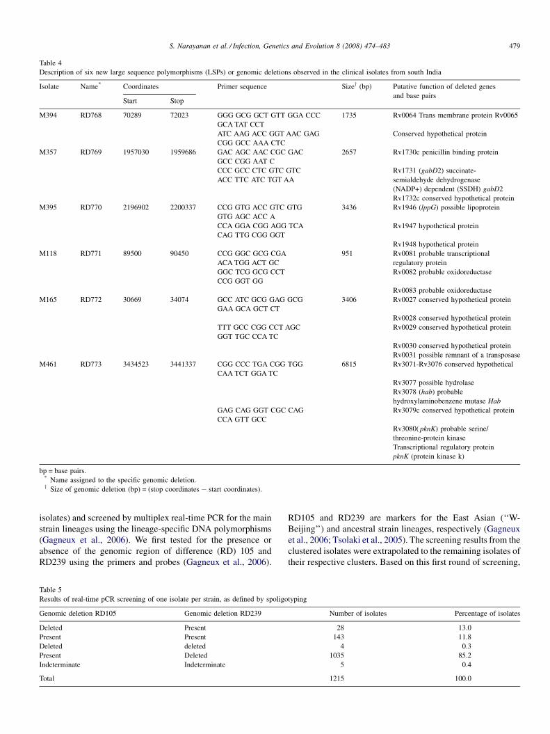
Table 4
Description of six new large sequence polymorphisms (LSPs) or genomic deletions observed in the clinical isolates from south India
Isolate Name* Coordinates Primer sequence Sizey (bp) Putative function of deleted genes
and base pairsStart Stop
M394 RD768 70289 72023 GGG GCG GCT GTT GGA CCC
GCA TAT CCT
1735 Rv0064 Trans membrane protein Rv0065
ATC AAG ACC GGT AAC GAG
CGG GCC AAA CTC
Conserved hypothetical protein
M357 RD769 1957030 1959686 GAC AGC AAC CGC GAC
GCC CGG AAT C
2657 Rv1730c penicillin binding protein
CCC GCC CTC GTC GTC
ACC TTC ATC TGT AA
Rv1731 (gabD2) succinate-
semialdehyde dehydrogenase
(NADP+) dependent (SSDH) gabD2
Rv1732c conserved hypothetical protein
M395 RD770 2196902 2200337 CCG GTG ACC GTC GTG
GTG AGC ACC A
3436 Rv1946 (lppG) possible lipoprotein
CCA GGA CGG AGG TCA
CAG TTG CGG GGT
Rv1947 hypothetical protein
Rv1948 hypothetical protein
M118 RD771 89500 90450 CCG GGC GCG CGA
ACA TGG ACT GC
951 Rv0081 probable transcriptional
regulatory protein
GGC TCG GCG CCT
CCG GGT GG
Rv0082 probable oxidoreductase
Rv0083 probable oxidoreductase
M165 RD772 30669 34074 GCC ATC GCG GAG GCG
GAA GCA GCT CT
3406 Rv0027 conserved hypothetical protein
Rv0028 conserved hypothetical protein
TTT GCC CGG CCT AGC
GGT TGC CCA TC
Rv0029 conserved hypothetical protein
Rv0030 conserved hypothetical protein
Rv0031 possible remnant of a transposase
M461 RD773 3434523 3441337 CGG CCC TGA CGG TGG
CAA TCT GGA TC
6815 Rv3071-Rv3076 conserved hypothetical
Rv3077 possible hydrolase
Rv3078 (hab) probable
hydroxylaminobenzene mutase Hab
GAG CAG GGT CGC CAG
CCA GTT GCC
Rv3079c conserved hypothetical protein
Rv3080( pknK) probable serine/
threonine-protein kinase
Transcriptional regulatory protein
pknK (protein kinase k)
bp = base pairs.* Name assigned to the specific genomic deletion.y Size of genomic deletion (bp) = (stop coordinates � start coordinates).
S. Narayanan et al. / Infection, Genetics and Evolution 8 (2008) 474–483 479
isolates) and screened by multiplex real-time PCR for the main
strain lineages using the lineage-specific DNA polymorphisms
(Gagneux et al., 2006). We first tested for the presence or
absence of the genomic region of difference (RD) 105 and
RD239 using the primers and probes (Gagneux et al., 2006).
Table 5
Results of real-time pCR screening of one isolate per strain, as defined by spoligo
Genomic deletion RD105 Genomic deletion RD239
Deleted Present
Present Present
Deleted deleted
Present Deleted
Indeterminate Indeterminate
Total
RD105 and RD239 are markers for the East Asian (‘‘W-
Beijing’’) and ancestral strain lineages, respectively (Gagneux
et al., 2006; Tsolaki et al., 2005). The screening results from the
clustered isolates were extrapolated to the remaining isolates of
their respective clusters. Based on this first round of screening,
typing
Number of isolates Percentage of isolates
28 13.0
143 11.8
4 0.3
1035 85.2
5 0.4
1215 100.0
S. Narayanan et al. / Infection, Genetics and Evolution 8 (2008) 474–483480
we found that 85.2% (1035/1215) of the isolates had a deletion
in RD239, 2.3% (28/1215) of isolates had a deletion in RD105,
11.8% (143/1215) of isolates had no deletion, and the genomic
deletions in 0.7% (9/1215) of isolates remained undetermined
(Table 5). We performed a second round of screening on the
isolates without a confirmed deletion in RD105 or RD239 using
a TaqMan allelic discrimination assay for the katG463 ctg to
cgg single nucleotide polymorphism, which defines the Euro-
American lineage (Principal Genetic Groups 2 and 3) (Gagneux
et al., 2006). We found 6.8% (83/1215) of isolates belonged to
this lineage. Finally, close inspection of the spoligotypes of the
remaining isolates (Supplementary table) revealed that 4.4%
(54/1215) belonged to the CAS lineage or the Delhi genogroup
(Baker et al., 2004; Brudey et al., 2006).
4. Discussion
For decades there has been a belief that the outcome of a
person’s exposure to and infection with M. tuberculosis
depends only on individual host characteristics such as age,
gender and genetic background. However, recent studies
increasingly suggest that bacterial factors also contribute to
the differences in outcomes seen in the human host population.
Hence, it becomes imperative to characterize the clinical
isolates of M. tuberculosis from a large population based study
in order to design vaccine and new drugs for the population.
One quarter of the world’s TB cases occur in India and the
major lineage which has been identified in this study is likely to
be responsible for a high percentage of the TB cases.
This study describes the spoligotyping of 1215 isolates of M.
tuberculosis from Tiruvallur area in south India where the
largest BCG trial was conducted. The total population of the
study area has been 580,000. The incidence of smear positive
TB is 76 per 100,000 in the population. We have compared the
spoligotyping with the widely accepted IS6110 RFLP typing
from a 3-year population based study. In addition to these two
methods Deletion Micro array based large sequence poly-
morphism (LSP), deletion PCR and RT-PCR were used as large
scale screening methods to delineate the phylogeny and
evolutionary characteristics of the south Indian isolates of M.
tuberculosis. To our knowledge this is the first study from India
which combines deletion micro array along with the widely
used genotyping tools in a population based study.
The genetic diversity observed in our study has been 22.5%
which is corroborating with the fact that endemic area might
have relatively few circulating strains (Hermans et al., 1995).
Ours is a population based study and restricted to southern most
region of the country. Hence, this data contradicts the study of
(Singh et al., 2007) which included seven different regions of
India and showed a large number of circulating strains. This
highlights the rich diversity among different regions of India.
Spoligotyping has been a very useful technique to
differentiate the M. tuberculosis isolates into different
geographical clades. It is not a highly discriminatory marker.
Studies from parts of the globe have highlighted the
predominant spoligotypes responsible for most of the
pulmonary TB cases (Brudey et al., 2003; Dale et al., 1999;
Puusteinen et al., 2003). These studies reveal that different
spoligotypes occur at variable frequencies in different countries
and continents.
The CAS or Delhi type corresponding to ST26 which
predominates in north India (Bhanu et al., 2002; Singh et al.,
2004; Kulkarni et al., 2005; Guitierrez et al., 2006) represents
1.02 % of isolates SpolDB4 database. This type has been
reported from 34 countries. None of the orphan strains
identified by Kulkarni et al. (2005) or Singh et al. (2007) were
found in our study. Spoligotyping has categorically identified
the major clade present in south India to East African lineage
with 28,32 and 34 spacers missing. This clearly shows the
difference in origin and evolution of M. tuberculosis in north
and south of India. In our study, the EAI family corresponding
to ubiquitous spoligotype ST11, is present as a large group
among the clusters matching the SpolDB4. 336 isolates among
1215 isolates belong to this group. This family is present in
other countries like Bangladesh, Malaysia, Indonesia, Pakistan,
UK, Denmark, Netherlands, France, New Zealand and USA.
The family W-Beijing which has been reported from several
countries has a distinct genotype has been associated with rapid
transmission across large parts of different continents,
accounted to only 1.9% in Thiruvallur. The other clades have
been represented by a fewer percentage in Thiruvallur
(Tables 1A and 1B).
Sixteen strains of type 53 were seen in the present study from
Tiruvallur. This type is linked to be a major clade of European
descent in a multicentric study (Kremer et al., 1999). The
presence or absence of spacer31 discriminates between the two
highly prevalent spoligotypes worldwide, ST 50 and ST 53 with
its absence being linked to IS6110 insertional events. While
Mumbai study (Kulkarni et al., 2005) showed the presence of
one strain of ST 50, we have seen ST 53 in the south Indian
region.
In spite of the endemicity of the disease a large proportion of
the patients were infected with the tuberculosis isolates having
spoligotype that had not been described in the global database,
of more than 39,000 tuberculosis isolates. But there has been a
major cluster of isolates constituting 85% of the total isolates
from this endemic area. Twelve major clusters ST 1, 8, 11, 126,
340, 43, 48, 591, 236, 26, 355, 53 comprised 86%. Except 1 and
53 all the remaining clusters are variants of EAI with the
characteristic spoligotype pattern with absence of spacers 28–
32 and 34 which may be considered as a major type in the
Tiruvallur district.
Overall the spoligotyping analysis showed diversity in
clinical isolates especially among the strains with no copy of
IS6110. The 20 no copy isolates were differentiated to 17
different spoligotypes. Thirty percent of the IS6110 low copy
strains were differentiated by spoligotyping. Spoligotyping
alone is not sufficient as a secondary marker for IS6110 low
copy strains, instead this genotyping method has been an
excellent tool to define the geno clade prevalent in the
Tiruvallur region of south India in comparison with the other
regions of the globe. This region has been predominated by
IS6110 low copy strains. 63% the EAI variants are comprised of
single and low copy IS6110 isolates. As Table 5 shows that
S. Narayanan et al. / Infection, Genetics and Evolution 8 (2008) 474–483 481
major spoligo clusters could not be differentiated by IS6110
typing because of the predominance of low copy isolates. This
warrants the use of other secondary markers like VNTR, MIRU
besides spoligotyping, and other PCR based methods to study
the transmission dynamics of M. tuberculosis in the region.
Even though the discriminating capacity of MIRU is not greater
than IS6110, it has been shown to be efficient to discriminate
the low IS6110 strains (Sun et al., 2004).
The excellent congruence observed between all the
independent sets of genetic markers used here lends strong
support to the assignment of different prevalent lineages. Our
LSP followed by large scale screening by RT-PCR and PCR has
confirmed the prevalent genotypic clade in this region. The
presence of TbD1 in a large percentage of isolates indicates that
south India could have been the initial focus of the ancient
lineage. Another characteristic feature of these strains is the
deletion of RD239 region which is also a characteristic feature
of the Manila strains (Tsolaki et al., 2004). Aggregation of 236–
239 deletion is confined to the so-called ‘‘Manila’’ clade
(Tsolaki et al., 2004), and is likely to result from a genetic event
that occurred in the ancestor of these isolates. The spoligotyp-
ing of 41 of the 48 M. tuberculosis isolates from metropolitan
Manila yielded characteristic identical patterns lacking
hybridization to eight spacers (Bhanu et al., 2002) the
remaining 35 spacers showed a positive hybridization signal.
A similar systematic association has been observed in strains
from Singapore (Sun et al., 2004) and from Bangladesh (Sola
et al., 2001). It remains to be tested whether the deletion of
TbD1 from the genome of ancient strains conferred or not some
selective advantage to the descendent strains enhancing their
epidemic potential. Due to trade links the probable route of
migration to and fro from south India to East Africa could have
been through East Asian countries like Philippines.
This study is population based and has been spread over 3
years. There have also been studies from this region describing
the prevalence of low copy IS6110 strains in the past (Das et al.,
1995) In spite of the presence of all the nine clades in this region
the only lineage which has been predominating in this region is
the EAI clade. Recently, there have been reports on the notion
that M. tuberculosis has adapted to human population. In this
region the ancient genotype or the EAI strain has adapted to the
host during several centuries and thus has a capacity for higher
transmission. The prevalence of such a high number in this part
of the continent and also its prevalence for a long time raise the
question whether these have a unique transmission dynamics
and are they suppressing the spread of other genotypes. In the
largest BCG trial conducted in the same region earlier, BCG did
not afford any protection. It is known that genetic variation of
the strains is a method of vaccine escape and has been
demonstrated in several bacterial species (Van Loo and Mooi,
2002; Mbelle et al., 1999). Further investigation in this line
would be helpful to ascertain the role of these highly prevalent
strains on the host immunity.
Starting 50 years ago, studies using the guinea pig and other
animal models have repeatedly reported that strains of M.
tuberculosis from south India were less virulent than other
strains (Mitchison et al., 1960; Singh and Sharma, 1964;
Prabhakar et al., 1987). Furthermore, BCG vaccine trails in
south India showed no significant efficacy (TRC, 1999).
Considering the global phylogeography of M. tuberculosis and
the dominance of the ancestral lineage in south India, BCG
failure in this part of the world could partially be due to strain-
specific effects (Brewer and Colditz, 1995). Based on our
approach, which was designed to detect genomic regions that
are absent relative to the laboratory reference strain, H37Rv,
deletion array was performed.
Deletion micro array followed by large scale screening by
PCR had identified six new deletions in the genome of only
1–2% of the south Indian isolates. The phenotypic character-
istics lost or gained by these deletions have to be explored.
However, considering that this strain lineage is ancestral to all
M. tuberculosis strains that have been sequenced thus far
(Gagneux et al., 2006), this lineage could harbor unique sets of
genes that are absent (i.e. deleted) in other M. tuberculosis and
thus not detectable using H37Rv as a reference.
Acknowledgments
We thank the patients and staff of the Tiruvallur District and
the Tuberculosis Research Centre, Chennai, India. This work
was funded in part by a grant from the United States Agency of
International development provided through the World Health
Organisation, partly by the NIH through NIAID/TRC ICER
programme and NIH Grant 5R03AI063576-02.
Appendix A. Supplementary data
Supplementary data associated with this article can be
found, in the online version, at doi:10.1016/j.meegid.2007.
09.007.
References
Baker, L., Brown, T., Maiden, M.C., Drobniewski, F., 2004. Silent nucleotide
polymorphisms and a phylogeny for Mycobacterium tuberculosis. Emerg.
Infect. Dis. 10, 1568–1577.
Bhanu, N.V., van Soolingen, D., van Embden, J.D., Dar, L., Pandey, R.M., Seth,
P., 2002. Predominace of a novel Mycobacterium tuberculosis genotype in
the Delhi region of India. Tuberculosis 82, 105–112.
Bhatia, A.L., 1961. Characteristics of Indian tubercle bacilli. Ind. J. Chest Dis.
3, 147–153.
Brewer, T.F., Colditz, G.A., 1995. Relationship between bacille Calmette-
Guerin (BCG) strains and the efficacy of BCG vaccine in the prevention
of tuberculosis. Clin. Infect. Dis. 20, 126–135.
Brosch, R., Gordon, S.V., Marmiesse, M., Brodin, P., Buchrieser, C., Eiglmeier,
K., Garnier, T., Gutierrez, C., Hewinson, G., Kremer, K., Parsons, L.M.,
Pym, A.S., Samper, S., van Soolingen, D., Cole, S.T., 2002. A new
evolutionary scenario for the Mycobaterium tuberculosis complex. Proc.
Natl. Acad. Sci. U.S.A. 99, 3684–3689.
Brudey, K., Filliol, I., Sola, C., Bebear, C., Elia-Pasquet, S., Texier-Maugein, J.,
Rastogi, N., 2003. Molecular characterization and biodiversity of Myco-
bacterium tuberculosis in the Antilles-Guiana region and comparative
analysis in a metropolitan region, Aquitaine. Pathol. Biol. 51, 282–289.
Brudey, K., Driscoll, J.R., Rigouts, L., Prodinger, W.M., Gori, A., Al-Hajoj,
S.A., Allix, C., Aristimuno, L., Arora, J., Baumanis, V., Binder, L., Cafrune,
P., Cataldi, A., Cheong, S., Diel, R., Ellermeier, C., Evans, J.T., Fauville-
Dufaux, M., Ferdinand, S., Garcia de Viedma, D., Garzelli, C., Gazzola, L.,
Gomes, H.M., Guttierez, M.C., Hawkey, P.M., van Helden, P.D., Kadival,
S. Narayanan et al. / Infection, Genetics and Evolution 8 (2008) 474–483482
G.V., Kreiswirth, B.N., Kremer, K., Kubin, M., Kulkarni, S.P., Liens, B.,
Lillebaek, T., Ho, M.L., Martin, C., Martin, C., Mokrousov, I., Narvskaia,
O., Ngeow, Y.F., Naumann, L., Niemann, S., Parwati, I., Rahim, Z.,
Rasolofo-Razanamparany, V., Rasolonavalona, T., Rossetti, M.L., Rusch-
Gerdes, S., Sajduda, A., Samper, S., Shemyakin, I., Singh, U., Somoskovi,
A., Skuce, R., van Soolingen, D., Streicher, E., Suffys, P.N., Tortoli, E.,
Tracevska, T., Vincent, V., Victor, T.C., Warren, R., Yap, S.F., Zaman, K.,
Portaels, F., Rastogi, N., Sola, C., 2006. Mycobacterium tuberculosis
complex genetic diversity: mining the fourth international spoligotyping
database (SpolDB4) for classification, population genetics and epidemiol-
ogy. BMC Microbiol. 6, 6–23.
Chen, I., 2006. Thinking big about global health. Cell 124, 661–663.
Cole, S.T., Brosch, R., Parkhill, J., Garnier, T., Churcher, C., Harris, D., Gordon,
S.V., Eiglmeier, K., Gas, S., Barry III, C.E., Tekaia, F., Badcock, K.,
Basham, D., Brown, D., Chillingworth, T., Connor, R., Davies, R., Devlin,
K., Feltwell, T., Gentles, S., Hamlin, N., Holroyd, S., Hornsby, T., Jagels, K.,
Krogh, A., McLean, J., Moule, S., Murphy, L., Oliver, K., Osborne, J.,
Quail, M.A., Rajandream, M.-A., Rogers, J., Rutter, S., Seeger, K., Skelton,
J., Squares, R., Squares, S., Sulston, J.E., Taylor, K., Whitehead, S.C.,
Barrell, B.G., 1998. Deciphering the biology of Mycobacterium tubercu-
losis from the complete genome sequence. Nature 393, 537–544.
Constant, P., Perez, E., Malaga, W., Laneelle, M.A., Saurel, O., Daffe, M.,
Guilhot, C., 2002. Role of the pks15/1 gene in the biosynthesis of phe-
nolglycolipids in the Mycobacterium tuberculosis complex. Evidence that
all strains synthesize glycosylated p-hydroxybenzoic methly esters and that
strains devoid of phenolglycolipids harbor a frameshift mutation in the
pks15/1 gene. J. Biol. Chem. 277, 38148–38158.
Cowan, L.S., Diem, L., Brake, M.C., Crawford, J.T., 2004. Transfer of a
Mycobacterium tuberculosis genotyping method, Spoligotyping, from a
reverse line-blot hybridization, membrane-based assay to the Luminex
multianalyte profiling system. J. Clin. Microbiol. 42, 474–477.
Dale, J.W., Nor, R.M., Ramayah, S., Tang, T.H., Zainuddin, Z.F., 1999.
Molecular epidemiology of tuberculosis in Malaysia. J. Clin. Microbiol.
37, 1265–1268.
Dale, J.W., Brittain, D., Cataldi, A.A., Cousins, D., Crawford, J.T., Driscoll, J.,
Heersma, H., Lillebaek, T., Quitugua, T., Rastogi, N., Skuce, R.A., Sola, C.,
van Soolingen, D., Vincent, V., 2001. Spacer oligonucleotide typing of
bacteria of the Mycobacterium tuberculosis complex: recommendations for
standardised nomenclature. Int. J. Tuberc. Lung Dis. 5, 216–219.
Das, S., Paramasivan, C.N., Lowrie, D.B., Prabhakar, R., Narayanan, P.R., 1995.
IS6110 restriction fragment length polymorphism typing of clinical isolates
of Mycobacterium tuberculosis from patients with pulmonary tuberculosis
in Madras, South India. Tuberc. Lung Dis. 76, 550–554.
Das, S.D., Narayanan, S., Hari, L., Hoti, S.L., Thangathurai, R.K., Charles, N.,
Jaggarajamma, K., Narayanan, P.R., 2005. Differentiation of highly pre-
valent IS6110 single-copy strains of Mycobacterium tuberculosis from a
rural community in South India with an ongoing DOTS programme. Infect.
Genet. Evol. 5, 67–77.
Filliol, I., Driscoll, J.R., Van Soolingen, D., Kreiswirth, B.N., Kremer, K.,
Valetudie, G., Anh, D.D., Barlow, R., Banerjee, D., Bifani, P.J., Brudey, K.,
Cataldi, A., Cooksey, R.C., Cousins, D.V., Dale, J.W., Dellagostin, O.A.,
Drobniewski, F., Engelmann, G., Ferdinand, S., Gascoyne-Binzi, D., Gor-
don, M., Gutierrez, M.C., Haas, W.H., Heersma, H., Kallenius, G., Kassa-
Kelembho, E., Koivula, T., Ly, H.M., Makristathis, A., Mammina, C.,
Martin, G., Mostrom, P., Mokrousov, I., Narbonne, V., Narvskaya, O.,
Nastasi, A., Niobe-Eyangoh, S.N., Pape, J.W., Rasolofo-Razanamparany,
V., Ridell, M., Rossetti, M.L., Stauffer, F., Suffys, P.N., Takiff, H., Texier-
Maugein, J., Vincent, V., De Waard, J.H., Sola, C., Rastogi, N., 2002. Global
distribution of Mycobacterium tuberculosis spoligotypes. Emerg. Infect.
Dis. 8, 1347–1349.
Filliol, I., Driscoll, J.R., van Soolingen, D., Kreiswirth, B.N., Kremer, K.,
Valetudie, G., Dang, D.A., Barlow, R., Banerjee, D., Bifani, P.J., Brudey, K.,
Cataldi, A., Cooksey, R.C., Cousins, D.V., Dale, J.W., Dellagostin, O.A.,
Drobniewski, F., Engelmann, G., Ferdinand, S., Gascoyne-Binzi, D., Gor-
don, M., Gutierrez, M.C., Haas, W.H., Heersma, H., Kassa-Kelembho, E.,
Ho, M.L., Makristathis, A., Mammina, C., Martin, G., Mostrom, P.,
Mokrousov, I., Narbonne, V., Narvskaya, O., Nastasi, A., Niobe-Eyangoh,
S.N., Pape, J.W., Rasolofo-Razanamparany, V., Ridell, M., Rossetti, M.L.,
Stauffer, F., Suffys, P.N., Takiff, H., Texier-Maugein, J., Vincent, V., de
Waard, J.H., Sola, C., Rastogi, N., 2003. Snapshot of moving and expanding
clones of Mycobacterium tuberculosis and their global distribution assessed
by spoligotyping in an international study. J. Clin. Microbiol. 41, 1963–
1970.
Gagneux, S., DeRiemer, K., Van, T., Kato-Maeda, M., de Jong, B.C.,
Narayanan, S., Nicol, M., Niemann, S., Kremer, K., Gutierrez, M.C.,
Hilty, M., Hopewell, P.C., Small, P.M., 2006. Variable host-pathogen
compatibility in Mycobacterium tuberculosis. Proc. Natl. Acad. Sci.
U.S.A. 103, 2869–2873.
Gingeras, T.R., Ghandour, G., Wang, E., Berno, A., Small, P.M., Drobniewski,
F., Alland, D., Desmond, E., Holodniy, M., Drenkow, J., 1998. Simulta-
neous genotyping and species identification using hybridization pattern
recognition analysis of generic Mycobacterium DNA arrays. Genome
Res. 8, 435–448.
Glynn, J.R., Whiteley, J., Bifani, P.J., Kremer, K., van Soolingen, D., 2002.
Worldwide occurrence of Beijing/W strains of Mycobacterium tuberculosis:
a systematic review. Emerg. Infect. Dis. 8, 843–849.
Grange, J.M., Aber, V.R., Allen, B.W., Mitchison, D.A., Goren, M.B., 1978.
The correlation of bacteriophage types of Mycobacterium tuberculosis with
guinea pig virulence and in vitro indicators of virulence. Tubercle 45, 354–
359.
Guitierrez, M.C., Ahmed, N., Willery, E., Narayanan, S., Hasnain, S.E.,
Chauhan, D.S., Katoch, V.M., Vincent, V., Locht, C., Supply, P., 2006.
Predominance of ancestral lineages of Mycobacteirum tuberculosis in India.
Emerg. Infect. Dis. 12, 1367–1374.
Gutacker, M.M., Smoot, J.C., Migliaccio, C.A., Ricklefs, S.M., Hua, S.,
Cousins, D.V., Graviss, E.A., Shashkina, E., Kreiswirth, B.N., Musser,
J.M., 2002. Genome-wide analysis of synonymous single nucleotide poly-
morphisms in Mycobacterium tuberculosis complex organisms: resolution
of genetic relationships among closely related microbial strains. Genetics
162, 1533–1543.
Gutacker, M.M., Mathema, B., Soini, H., Shashkina, E., Kreiswirth, B.N.,
Graviss, E.A., Musser, J.M., 2006. Single-nucleotide polymorphism-based
population genetic analysis of Mycobacterium tuberculosis strains from 4
geographic sites. J Infect Dis. 193, 121–128.
Hermans, P.W., Messadi, F., Guebrexabher, H., van Soolingen, D., de Haas, P.E.,
Heersma, H., de Neeling, H., Ayoub, A., Portaels, F., Frommel, D., 1995.
Analysis of the population structure of Mycobacterium tuberculosis in
Ethiopia, Tunisia, and The Netherlands: usefulness of DNA typing for
global tuberculosis epidemiology. J. Infect. Dis. 171, 1504–1513.
Joseph, S., Mitchison, D.A., Ramachandran, K., Selkon, J.B., Subbaiah, T.V.,
1964. Virulence in the guinea-pig and sensitivity to pas and thiacetazone of
tubercle bacilli from south Indian patients with pulmonary tuberculosis.
Tubercle 45, 354–359.
Kamerbeek, J., Schouls, L., Kolk, A., van Agterveld, M., van Soolingen, D.,
Kuijper, S., Bunschoten, A., Molhuizen, H., Shaw, R., Goyal, M., van
Embden, J., 1997. Simultaneous detection and strain differentiation of
Mycobacterium tuberculosis for diagnosis and epidemiology. J. Clin.
Microbiol. 35, 907–914.
Kato-Maeda, M., Rhee, J.T., Gingeras, T.R., Salamon, H., Drenkow, J., Smit-
tipat, N., Small, P., 2001. Comparing genomes within the species Myco-
bacterium tuberculosis. Genome Res. 11, 1796–1799.
Kremer, K., van Soolingen, D., Frothingham, R., Haas, W.H., Hermans, P.W.,
Martin, C., Palittapongarnpim, P., Plikaytis, B.B., Riley, L.W., Yakrus,
M.A., Musser, J.M., van Embden, J.D., 1999. Comparison of methods
based on different molecular epidemiological markers for typing of
Mycobacterium tuberculosis complex strains: interlaboratory study
of discriminatory power and reproducibility. J. Clin. Microbiol. 37,
2607–2618.
Kulkarni, S., Sola, C., Filliol, I., Rastogi, N., Kadival, G., 2005. Spoligotyping
of Mycobacterium tuberculosis isolates from patients with pulmonary
tuberculosis in Mumbai, India. Res. Microbiol. 156, 588–596.
Marmiesse, M., Brodin, P., Buchrieser, C., Gutierrez, C., Simoes, N., Vincent,
V., Glaser, P., Cole, S.T., Brosch, R., 2004. Macro-array and bioinformatic
analyses reveal mycobacterial ‘core’ genes, variation in the ESAT-6 gene
family and new phylogenetic markers for the Mycobacterium tuberculosis
complex. Microbiology 150, 483–496.
S. Narayanan et al. / Infection, Genetics and Evolution 8 (2008) 474–483 483
Mbelle, N., Huebner, R.E., Wasas, A.D., Kimura, A., Chang, I., Klugman, K.P.,
1999. Immunogenicity and impact on nasopharyngeal carriage of a non-
avalent pneumococcal conjugate vaccine. J. Infect. Dis. 180, 1171–1176.
Middlebrook, G., Cohn, M.L., 1953. Some observations on the pathogenicity of
isoniazid-resistant variants of tubercle bacilli. Science 118, 297–299.
Mistry, N.F., Iyer, A.M., D’souza, D.T., Taylor, G.M., Young, D.B., Antia, N.H.,
2002. Spoligotyping of Mycobacterium tuberculosis isolates from multiple-
drug-resistant tuberculosis patients from Bombay, India. J. Clin. Microbiol.
40, 2677–2680.
Mitchison, D.A., Wallace, J.G., Bhatia, A.L., Selkon, J.B., Subbaiah, T.V.,
Lancaster, M.C., 1960. A comparison of the virulence in guinea-pigs of
South Indian and British tubercle bacilli. Tubercle 41, 1–22.
Narayanan, S., Sahadevan, R., Narayanan, P.R., Krishnamurthy, P.V., Para-
masivan, C.N., Prabhakar, R., 1997. Restriction fragment length poly-
morphism of Mycobacterium tuberculosis strains from various regions of
India, using direct repeat probe. Indian J. Med. Res. 106, 447–454.
Narayanan, S., Das, S., Garg, R., Hari, L., Rao, V.B., Frieden, T.R., Narayanan,
P.R., 2002. Molecular epidemiology of tuberculosis in a rural area of high
prevalence in South India: implications for disease control and prevention. J.
Clin. Microbiol. 40, 4785–4788.
Prabhakar, R., Venkataraman, P., Vallishayee, R.S., Reeser, P., Musa, S.,
Hashim, R., Kim, Y., Dimmer, C., Wiegeshaus, E., Edwards, M.L.,
1987. Virulence for guinea pigs of tubercle bacilli isolated from the sputum
of participants in the BCG trial, Chingleput District, South India. Tubercle
68, 3–17.
Puusteinen, K., Marjamaki, M., Rastogi, N., Sola, C., Filliol, I., Ruutu, P.,
Holmstrom, P., Viljanen, M.K., Soini, J., 2003. Characterization of Finnish
Mycobaterium tuberculosis isolates by spoligotyping. J. Clin. Microbiol. 41,
1525–1528.
Radhakrishnan, I., Manju, Y.K., Kumar, R.A., Mundayoor, S., Harris, 2001.
Implications of low frequency of IS6110 in fingerprinting field isolates of
Mycobacterium tuberculosis from Kerala, India. J. Clin. Microbiol. 39,
1683.
Singh, B., Sharma, M.D., 1964. Niacin production and virulence in the guinea-
pig of Indian tubercle bacilli. Tubercle 45, 239–240.
Singh, U.B., Suresh, N., Bhanu, N.V., Arora, J., Pant, H., Sinha, S., Aggarwal,
R.C., Singh, S., Pande, J.N., Sola, C., Rastogi, N., Seth, P., 2004. Pre-
dominant tuberculosis spoligotypes, Delhi India. Emerg. Infect. Dis. 10,
1138–1142.
Singh, U.B., Arora, J., Suresh, N., Pant, H., Rana, T., Sola, C., Rastogi, N.,
Pande, J.N., 2007. Genetic biodiversity of Mycobacterium tuberculosis
isolates from patients with pulmonary tuberculosis in India. Infect. Genet.
Evol. 7, 441–448.
Sola, C., Filliol, E., Legrand, E., Mokrousov, I., Rastogi, N., 2001. Myco-
bacterium tuberculosis phylogeny reconstruction based on combined
numerical analysis with IS1081, IS6110, VNTR and DR-based spoligo-
typing suggests the existence of two new phylogeographical clades. J.
Mol. Evol. 53, 680–689.
Sreevatsan, S., Pan, X., Stockbauer, K.E., Connell, N.D., Kreiswirth, B.N.,
Whittam, T.S., Musser, J.M., 1997. Restricted structural gene polymorph-
ism in the Mycobacterium tuberculosis complex indicates evolutionarily
recent global dissemination. Proc. Natl. Acad. Sci. U.S.A. 94, 9869–
9874.
Streicher, E.M., Warren, R.M., Kewley, C., Simpson, J., Rastogi, N., Sola, C.,
van der Spuy, G.D., van Helden, P.D., Victor, T.C., 2004. Genotypic and
phenotypic characterization of drug-resistant Mycobacterium tuberculosis
isolates from rural districts of the Western Cape Province of South Africa. J.
Clin. Microbiol. 42, 891–894.
Sun, Y.J., Lee, A.S., Ng, S.T., Ravindran, S., Kremer, K., Bellamy, R., Wong,
S.Y., van Soolingen, D., Supply, P., Paton, N.I., 2004. Characterization of
ancestral Mycobacterium tuberculosis by multiple genetic markers and
proposal of genotyping strategy. J. Clin. Microbiol. 42, 5058–5064.
Tuberculosis Research Centre (ICMR) Chennai, 1999. Fifteen-year follow up of
trial of BCG vaccines in south India for tuberculosis prevention. Indian J.
Med. Res. 110, 56–69.
Tsolaki, A.G., Hirsh, A.E., DeRiemer, K., Enciso, J.A., Wong, M.Z., Hannan,
M., Goguet de la Salmoniere, Y.-O.L., Aman, K., Kato-Maeda, M., Small,
P.M., 2004. Functional and evolutionary genomics of Mycobacterium
tuberculosis: insights from genomic deletions in 100 strains. Proc. Natl.
Acad. Sci. U.S.A. 101, 4865–4870.
Tsolaki, A.G., Gagneux, S., Pym, A.S., Goguet de la Salmoniere, Y.-O.L.,
Kreiswirth, B.N., van Soolingen, D., Small, P.M., 2005. Genomic deletions
classify the Beijing/W strains as a distinct genetic lineage of Mycobacter-
ium tuberculosis. J. Clin. Microbiol. 43, 3185–3191.
van Embden, J.D., Cave, M.D., Crawford, J.T., Dale, J.W., Eisenach, K.D.,
Gicquel, B., Hermans, P., Martin, C., McAdam, R., Shinnick, T.M., 1993.
Strain identification of Mycobacterium tuberculosis by DNA fingerprinting:
recommendations for a standardized methodology. J. Clin. Microbiol. 31,
406–409.
Van Loo, I.H., Mooi, F.R., 2002. Changes in the Dutch Bordetella pertussis
population in the first 20 years after the introduction of whole-cell vaccines.
Microbiol. 148, 2011–2018.
www.elsevier.com/locate/meegid
Available online at www.sciencedirect.com
Infection, Genetics and Evolution 8 (2008) 484–488
Genetic diversity of Trypanosoma evansi in beef cattle based
on internal transcribed spacer region§
Supatra Areekit, Pirom Singhaphan, Pornpimon Kanjanavas, Sintawee Khuchareontaworn,Thayat Sriyapai, Arda Pakpitcharoen, Kosum Chansiri *
Department of Biochemistry, Faculty of Medicine, Srinakharinwirot University, Sukhumvit 23, Bangkok 10110, Thailand
Received 17 October 2007; accepted 17 October 2007
Available online 1 November 2007
Abstract
This study was focused on genetic diversity of Trypanosoma evansi which is a widely distributed haemoflagellate of veterinary importance that
infects a variety of larger mammals including horses, mules, camels, buffalo, cattle and deer. The genetic diversity of T. evansi of beef cattle
LAM19 was accomplished by using phylogenetic analysis based on internal transcribed spacer region (ITS). Blood sample was collected from a
naturally infected beef cattle LAM 19 and parasitemia was raised by mouse inoculation. The parasites were collected and isolated by using DE 52
DEAE cellulose anion exchange column prior to DNA extraction. Upon PCR amplification of ITS region, the product of 1300 bp in size was
obtained. The ITS nucleotide sequences were analyzed and revealed that it could demonstrate the genetic diversity of T. evansi of beef cattle
LAM19. Based on the ITS tree, beef cattle LAM 19 T. evansi were categorized into two main groups where the genetic diversity occurred within
Group 1. The data could be applicable for the survey of parasite dynamics, epidemiological studies as well as prevention and control of the disease.
# 2007 Elsevier B.V. All rights reserved.
Keywords: Internal transcribed spacer; ITS; Cattle
1. Introduction
Trypanosomes are protozoan parasites that cause major
disease in human and other animals. Trypanosomes (genus
Trypanosoma) are widespread blood parasites of vertebrates,
usually transmitted by arthropod or leech vectors. Several
Trypanosome species are agents of disease in humans and/or
livestock particularly in the tropics. In Thailand, T. evansi are
widespread where they cause considerable loss in productivity
in domestic animals, such as horses (Boonyawong et al., 1975),
beef cattle (Chaichanapunpol et al., 1985; Tuntasuvan et al.,
1997), dairy cattle (Trisanarom et al., 1987).
The finding that trypanosomes exhibit considerable intra-
species diversity has led to the proposal that these parasites
have a clonal population structure (Tibayrenc et al., 1990;
Tibayrenc and Ayala, 1991; Tibayrenc et al., 1991a,b). The
§ GenBank accession numbers of ITS regions of T. evansi isolated from beef
cattle LAM19 were EF545993-EF546014. The accession numbers of ITS
regions of other organisms used in this study were indicated in the text.
* Corresponding author. Tel.: +66 2260 2122x4605; fax: +66 2260 0125.
E-mail address: [email protected] (K. Chansiri).
1567-1348/$ – see front matter # 2007 Elsevier B.V. All rights reserved.
doi:10.1016/j.meegid.2007.10.008
clonal theory has important implications for the diagnosis and
treatment of trypanosomiasis and for the development of anti-
trypanosome drugs and vaccine. Genetically diverse clonets are
likely to differ in their pathological and other biological
properties. In fact, there is evidence for these in T. cruzi (Morel
et al., 1980; Engman et al., 1987; Aymerich and Goldenberg,
1989), T. brucei (Stevens and Godfrey, 1992) and T. congolense
(Majiwa and Webster, 1987). It is clear that much work remains
to be done to understand the molecular basis for these
differences. It will be particularly important to correlate
molecular variation at the gene level with changes in
pathophysiological properties of the parasites.
The internal transcribed spacer (ITS region) has been used for
phylogenetic analysis, evaluation of the evolutionary process, as
well as for determination of taxonomic identities. It contains two
regions (ITS1 and ITS2) that are located between the repeating
array of nuclear 18S, 5.8S and 28S ribosomal RNA genes (Lodish
et al., 2001), a locus that has 100–200 copies per genome.
Beltrame-Botelho and co-worker described the sequences of
both ITS spacers flanking the T. rangeli and 5.8S rDNA. These
sequences could be used to identify both intra- and inter-specific
variability. Most of the data indicated that the ITS-2 spacer is
S. Areekit et al. / Infection, Genetics and Evolution 8 (2008) 484–488 485
more variable than the ITS-1 spacer and the 5.8S rDNA when
performing inter-specific comparisons among the studied
trypanosomatids (Beltrame-Botelho et al., 2005). In 1999, the
phylogenetic analysis of ITS sequences was used for studying the
polymorphism of T. rangeli strains isolated from different hosts
and geographic areas (southern Brazil, Central America, and
northern South America) (Grisard et al., 1999). In 2004,
Sarataphan and colleagues have reported the 4 group-specific
genotypes within T. evansi isolates from six different hosts
(elephant, horse, buffalo, cattle, pig and deer) based on PCR-
RFLP of the open reading frame 2 (ORF2) of glucose transporter
genes located on chromosome X of T. brucei (Sarataphan et al.,
2004). However, the data were not sufficient for demonstration of
the genetic diversity and relationship of T. evansi within each
single host. In 2007, Khuchareontaworn and colleagues
demonstrated the genetic diversity of T. evansi isolated from a
buffalo based on ITS 1 and ITS2 regions. They reported that ITS
2 tree was able to classify T. evansi into 4 groups whereas ITS 1
tree showed lower efficacy in discrimination (Khuchareontaworn
et al., 2007).
Here in, ITS region of T. evansi from beef cattle was
analyzed and the genetic diversity was examined. The
phylogenetic trees were constructed to support the evidence
of genetic diversity of the parasite.
2. Materials and methods
2.1. Parasite collection
A naturally T. evansi infected blood sample from beef cattle
namely LAM19 was collected during 1999 from a farm in
Lampraya Klang district, Saraburi province, Thailand. The
infection was examined by using thin blood smear technique.
The 3.0 ml of infected blood were collected from cattle and
preserved in the phosphate saline glucose buffer (PSG), pH 8.0
(50 mM Na2HPO4�H2O, 2 mM NaH2PO4�2H2O, 36 mM NaCl
and 1.5% glucose) and stored in the liquid nitrogen tank
(�196 8C) until use.
2.2. Mouse inoculation and parasite isolation
Infected blood in the liquid nitrogen tank was thawed at room
temperature and 0.1 ml was injected to a mouse using 1 ml
Tuberculin syringe and needle gauge No 27. Parasitemia was
dailychecked bywet bloodsmear, starting fromthe thirddayafter
injection. At the highest parasitemia (108 cells/ml) which was
usually in the 3rd to 5th day of injection, blood was withdrawn
from euthanized mouse by cardiac punctured. An anion exchange
column (DE 52 DEAE cellulose) was used to purify parasite from
the horde blood cells according to the method of described by
Chao (Choa et al., 1984). The eluent with infect protozoa was
collected and proceeding to parasite lysis and DNA extraction.
2.3. DNA extraction and purification
Pellet of parasites was resuspended in PSG buffer in the
presence of SDS (final concentration was 2%) and proteinase K
(final concentration was 1 mg/ml). The solution was incubated
at 42 8C for 14 h. Parasite DNA was extracted by conventional
phenol/chloroform which responded for deproteinization of the
aqueous solution containing the desired nucleic acid. The
purified DNAs were precipitated by the addition of 2 volumes
of cold absolute ethanol. The pellet was dried, dissolved in
sterile distilled water and kept at 4 8C until use.
2.4. PCR amplification
PCR amplifications of purified DNA from blood samples
were performed by a pair of primers used for amplification of
ITS regions including 5.8S rDNA were ITS/F, forward primer,
50 GGT GAT CGG ACC GTC GCT CGT CT 30 and reverse
primer ITS/R, 50 CCT CTT CGC TCG CCG CTG ACT G 30.The primers were designed based on the nucleotide sequence
comparison of T. evansi ITS1 and ITS2 (Accession number
D89527) in GenBank database. All reactions were manipu-
lated in 25 ml volume containing 50 ng of genomic DNA
samples. The PCR amplification of ITS region will contain 1X
PCR buffer, 2 mM of each primer, 400 mM of dNTP, 6 mM
MgCl2 and 1.5 units of proof reading Taq DNA polymerase
(Gibco BRL1). Sterile distilled water was used to make
volume to 25 ml. A PCR cycle was performed using Peltier
Thermal Cycler (MJ Research, PTC-200) for 30 cycles. Each
cycle consisted of denaturation at 94 8C for 1 min, annealing
at 60 8C for 1 min and extension at 72 8C for 1 min. PCR
products were analyzed by using electrophoresis in 1.2%
agarose gel at 110 V approximately 45 min prior to staining
by ethidium bromide and the gel was observed under
ultraviolet light.
2.5. Cloning and sequencing of PCR fragment
The PCR fragment was eluted from the gel and purified by
QIAGEN DNA purification kit (QIAGEN1). The purified
200 ng of PCR fragment were further ligated to the pGEM1 –
T Easy vector by using protocol of pGEM1 – T Easy Vector
Systems (Invitrogen, Promega). The reaction was consisted of
4 ml of PCR product, 1 ml of 2X Rapid Ligation Buffer (60 mM
Tris–HCl, pH 7.8, 20 mM MgCl2, 20 mM ATP 10% PEG), 1 ml
of the pGEM1 – T Easy vector and 3 units of T4 DNA ligase.
The reaction was mixed, and incubated for 24 h at 4 8C prior to
transformation by electroporation using BioRad MicroPulser
(BIO-RAD MicroPulserTM). The transformants were then
spread on the LB-ampicillin agar plates containing X-gal and
IPTG (40 ml of 20 mg/ml X-gal and 20 ml of 200 mg/ml IPTG)
and incubated for 16 h at 37 8C. White colonies were randomly
selected and DNAs were purified by using QIAGEN plasmid
purification kit (QIAGEN1). DNA sequencing was performed
in both forward and reverse direction based on the Big Dye
Terminator Cycle Sequencing procedure (Macrogen). The
nucleotide sequence data were analyzed using software of
ABI PRISM Model 3100 version 3.7 and BLAST (http://
www.ncbi.nlm.nih.gov/BLAST/) in comparison to ITS
regions of the other T. evansi that have been previously
reported in GenBank.
Table 1
GenBank accession numbers of ITS regions of Trypanosoma spp.
Parasites names Specific host/lab host Origin GenBank accession numbers
T. evansi clone No.1.1,3.1 Beef cattle Saraburi, Thailand EF546003—EF546004
T. evansi clone No.3 Beef cattle Saraburi, Thailand EF545993
T. evansi clone No.4 Beef cattle Saraburi, Thailand EF545996
T. evansi clone No.6 Beef cattle Saraburi, Thailand EF546005
T. evansi clone No.8 Beef cattle Saraburi, Thailand EF545994
T. evansi clone No.8.1,11,12.1 Beef cattle Saraburi, Thailand EF546006—EF546008
T. evansi clone No.12 Beef cattle Saraburi, Thailand EF545997
T. evansi clone No.13 Beef cattle Saraburi, Thailand EF546009
T. evansi clone No.16 Beef cattle Saraburi, Thailand EF545998
T. evansi clone No.16.1,17,24,27 Beef cattle Saraburi, Thailand EF546010—EF546013
T. evansi clone No.28.1 Beef cattle Saraburi, Thailand EF546014
T. evansi clone No.29,38,42 Beef cattle Saraburi, Thailand EF546000—EF546002
T. brucei Da1972 Homo sapiens Daloa, Ivory Coast AF306774
T. brucei TH2 Homo sapiens Koudougou, Ivory Coast AF306777
T. brucei KP2 Glossina palpalis Kouassi-Perita, Ivory Coast AF306773
T. brucei NW2 Homo sapiens Uganda AF306776
T. brucei STIB215 Lion Serengeti, Tanzania AF306771
T. brucei B8/18 Pig Nsukka, Nigeria AF306772
T. brucei H3 Lion Luangwa Valley, Zambia AF306770
T. brucei Strain 427 Unknown – X05682
T. evansi Strain Tansui-Taiwan Unknown Taiwan D89527
T. congolense Strain riverine/forest-type Rattus norvegicus – U22319
S. Areekit et al. / Infection, Genetics and Evolution 8 (2008) 484–488486
2.6. Phylogenetic analysis
The 21 white colonies containing PCR products of ITS
region (1300 bp) amplified from beef cattle LAM 19 were
selected, sequenced and analyzed. Three sets of nucleotide
sequence data, ITS1 (341 bp), ITS2 (587 bp) and ITS
(ITS1+ITS2+5.8S rDNA), were separately aligned against
those of T. brucei and other reported T. evansi by using Clustal
X software Version 1.83 (multiple sequence alignment)
program (Thompson et al., 1997) and refined using the manual
method (Table 1). The phylogenetic trees were constructed
according to PAUP program version 4.0 (Swofford, 2002) using
T. congolense as the outgroup. The cladograms were created by
means of maximum likelihood (complete ITS region) and
maximum parsimony (ITS1 and ITS2) methods. Bootstrap
values were replicated 1000 times and computed with PAUP
program version 4.0.
3. Results
Upon PCR amplification of T. evansi genomic DNA of beef
cattle LAM 19, the product of 1300 bp in size was obtained.
After ligation and transformation processes of this PCR
product, 21 positive white colonies were randomly selected.
BLAST data of this ITS nucleotides sequences revealed that
they were corresponded to those of T. evansi Taiwan stock
(D89527.1) with the homology of 91%.
Initially, the individual two data sets of ITS 1 (341 bp) and ITS
2 (587 bp) regions were employed for phylogenetic tree
construction in comparison to those of T. evansi and
Trypanosoma spp. from GenBank. However, both trees showed
low bootstrap values among clades that were significantly unable
to discriminate the genetic diversity of ITS regions in T. evansi of
beef cattle LAM 19 (data not shown). On contrary, the
phylogenetic tree inferred from the complete ITS nucleotide
sequences (1300 bp) clearly showed the genetic diversity of the
parasites. According to the tree, two main groups (Group 1 and
Group 2) of ITS regions in T. evansi of beef cattle LAM 19 were
clearly separate with bootstrap value 89% (Fig. 1). Group 1 was
further divided into 6 subgroups, subgroup 1a–1f. Subgroup 1a
contained ITS regions of T. brucei (X05682) and T. brucei of
lions (AF306770 and AF306771) with bootstrap value 61%.
Subgroup 1b, 1c, 1e and 1f consisted of 5, 5, 2 and 3 ITS clones of
T. evansi of beef cattle LAM 19 with bootstrap value 84, 70, 91
and 89%, respectively. Subgroup 1d included ITS regions of T.
brucei of Homo sapiens (AF306774, AF306776 and AF306777),
pig (AF306772), Glossina palpalis (AF306773), and one ITS
clone of T. evansi of beef cattle LAM 19 with bootstrap value
81%. Finally, Group 2 was categorized in the same clade as ITS
of T. congolense encompassing two ITS clones of T. evansi of
beef cattle LAM 19 with bootstrap value 59%.
4. Discussion
Previously, Khuchareontaworn and colleques (Khuchareon-
taworn et al., 2007) suggested that ITS 2 region was informative
for genetic diversity exploitation in water buffalo B18 (four
groups were detected). However, this study revealed that ITS 2
tree inferring from nucleotide sequences of 341 bp failed to
elucidate the genetic diversity of T. evansi from beef cattle
LAM19 since the tree formed the low bootstrap value (�50%)
indicating the high feasibility of tree collapse. Similarly, the
ITS 1 nucleotide sequences (587 bp) could not support the tree
and also showed the low bootstrap values at 50–55%. Hence,
the tree construction based on merely either ITS 1 or ITS2 data
could be affected by their limited nucleotide sequence length as
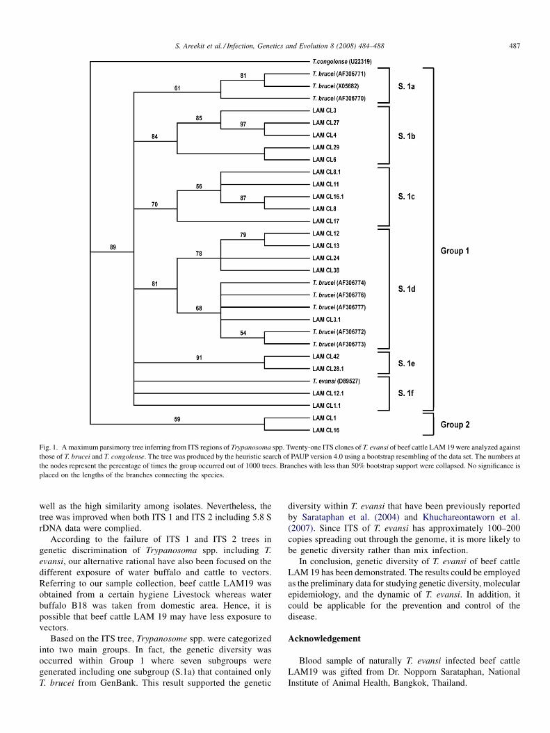
Fig. 1. A maximum parsimony tree inferring from ITS regions of Trypanosoma spp. Twenty-one ITS clones of T. evansi of beef cattle LAM 19 were analyzed against
those of T. brucei and T. congolense. The tree was produced by the heuristic search of PAUP version 4.0 using a bootstrap resembling of the data set. The numbers at
the nodes represent the percentage of times the group occurred out of 1000 trees. Branches with less than 50% bootstrap support were collapsed. No significance is
placed on the lengths of the branches connecting the species.
S. Areekit et al. / Infection, Genetics and Evolution 8 (2008) 484–488 487
well as the high similarity among isolates. Nevertheless, the
tree was improved when both ITS 1 and ITS 2 including 5.8 S
rDNA data were complied.
According to the failure of ITS 1 and ITS 2 trees in
genetic discrimination of Trypanosoma spp. including T.
evansi, our alternative rational have also been focused on the
different exposure of water buffalo and cattle to vectors.
Referring to our sample collection, beef cattle LAM19 was
obtained from a certain hygiene Livestock whereas water
buffalo B18 was taken from domestic area. Hence, it is
possible that beef cattle LAM 19 may have less exposure to
vectors.
Based on the ITS tree, Trypanosome spp. were categorized
into two main groups. In fact, the genetic diversity was
occurred within Group 1 where seven subgroups were
generated including one subgroup (S.1a) that contained only
T. brucei from GenBank. This result supported the genetic
diversity within T. evansi that have been previously reported
by Sarataphan et al. (2004) and Khuchareontaworn et al.
(2007). Since ITS of T. evansi has approximately 100–200
copies spreading out through the genome, it is more likely to
be genetic diversity rather than mix infection.
In conclusion, genetic diversity of T. evansi of beef cattle
LAM 19 has been demonstrated. The results could be employed
as the preliminary data for studying genetic diversity, molecular
epidemiology, and the dynamic of T. evansi. In addition, it
could be applicable for the prevention and control of the
disease.
Acknowledgement
Blood sample of naturally T. evansi infected beef cattle
LAM19 was gifted from Dr. Nopporn Sarataphan, National
Institute of Animal Health, Bangkok, Thailand.
S. Areekit et al. / Infection, Genetics and Evolution 8 (2008) 484–488488
References
Aymerich, S., Goldenberg, S., 1989. The karyotype of Trypanosoma cruzi Dm
28c: comparison with other T. cruzi strains and trypanosomatids. Exp.
Parasitol. 69 (2), 107–115.
Beltrame-Botelho, I.T., Gaspar-Silva, D., Steindel, M., Davila, A.M., Grisard,
E.C., 2005. Internal transcribed spacers (ITS) of Trypanosoma rangeli
ribosomal DNA (rDNA): a useful marker for inter-specific differentiation.
Infect. Genet. Evol. 5 (1), 17–28.
Boonyawong, T., Chantaraprateep, P., Muangyai, M., 1975. Surra in horse. Thai
J. Vet. Med. 5, 665–672.
Chaichanapunpol, I., Sisrrangkamanont, T., Trongwongsa, L., Suvanwasi, P.,
1985. Observations on trypanosomiasis in a native bull. Kasetsart Vet. 6, 1–9.
Chao, D., Dusanic, D.G., 1984. Comparative studies of the isolation of
metacyclic trypomastigotes of Trypanosoma cruzi by DEAE ion exchange
chromatography. Zhonghua Min Guo Wei Sheng Wu Ji Mian Yi Xue Za Zhi
17, 146–152.
Engman, D.M., Reddy, L.V., Donelson, J.E., Kirchhoff, L.V., 1987. Trypano-
soma cruzi exhibits inter- and intra-strain heterogeneity in molecular
karyotype and chromosomal gene location. Mol. Biochem. Parasitol. 22
(2–3), 115–123.
Grisard, E.C., Campbell, D.A., Romanha, A.J., 1999. Mini-exon gene sequence
polymorphism among Trypanosoma rangeli strains isolated from distinct
geographical regions. Parasitology 118, 375–382.
Khuchareontaworn, S., Singhaphan, P., Viseshakul, N., Chansiri, K., 2007.
Genetic diversity of Trypanosoma evansi in buffalo based on internal
transcribed spacer regions. J. Vet. Med. Sci. 69 (5), 487–493.
Lodish, H., Berk, A., Zipursky, S.L., Mastsudaira, P., Balimore, D., Darnell, J.,
2001. Molecular cell biology, fourth ed. W.H.Freeman and Company, New
York.
Majiwa, P.A., Webster, P., 1987. A repetitive deoxyribonucleic acid sequence
distinguishes Trypanosoma simiae from T. congolense. Parasitology 95 (3),
543–558.
Morel, C., Chiari, E., Camargo, E.P., Mattei, D.M., Romanha, A.J., Simpson, L.,
1980. Strains and clones of Trypanosoma cruzi can be characterized by
pattern of restriction endonuclease products of kinetoplast DNA minicir-
cles. Proc. Natl. Acad. Sci. USA 77 (11), 6810–6814.
Sarataphan, N., Boonchit, S., Sirivan, C., Indrakamhang, P., 2004. Genetic
diversity of Trypanosoma evansi in Thailand based on a repeated DNA
coding sequence marker.In: Livestock Development Proceeding. The
Goden Jubilee Museum of Agriculture, Pathumthani.
Stevens, J.R., Godfrey, D.G., 1992. Numerical taxonomy of Trypanozoon
based on polymorphisms in a reduced range of enzymes. Parasitology
104 (1), 75–86.
Swofford, D.L., 2002. PAUP*. Phylogenetic Analysis Using Parsimony
(*and Other Methods), 4.0b10 edn. Sinauer Associates, Sunderland,
Massachusetts.
Thompson, J.D., Gibson, T.J., Plewniak, F., Jeanmougin, F., Hoiggins, D.G.,
1997. The CLUSTALX windows interface: flexible strategies for multiple
sequence alignment aided by quality analysis tools. Nucleic Acids Res. 25.
Tibayrenc, M., Kjellberg, F., Ayala, F.J., 1990. A clonal theory of parasitic
protozoa: the population structures of Entamoeba, Giardia, Leishmania,
Naegleria, Plasmodium, Trichomonas, and Trypanosoma and their medical
and taxonomical consequences. Proc. Natl. Acad. Sci. USA 87 (7), 2414–
2418.
Tibayrenc, M., Ayala, F.J., 1991. Towards a population genetics of micro-
organisms: The clonal theory of parasitic protozoa. Parasitol. Today 7 (9),
228–232.
Tibayrenc, M., Kjellberg, F., Arnaud, J., Oury, B., Breniere, S.F., Darde, M.L.,
et al., 1991a. Are eukaryotic microorganisms clonal or sexual? A
population genetics vantage. Proc. Natl. Acad. Sci. USA 88 (12),
5129–5133.
Tibayrenc, M., Kjellberg, F., Ayala, F.J., 1991b. The clonal theory of parasitic
protozoa. BioScience 41, 767–774.
Trisanarom, A., Markmee, S., Ped-Usorn, C., 1987. Trypanosoma evansi
infection in Chaing Mai dairy cattle. In: Proceeding of the 6th Annual
Veterinary, 18–19 May, Conference of Thai Veterinary Medicine Associa-
tion, Thailan, pp. 1–12.
Tuntasuvan, D., Sarataphan, N., Nishikawa, H., 1997. Cerebral trypanosomiasis
in native cattle. Vet. Parasitol. 73 (3–4), 357–363.
www.elsevier.com/locate/meegid
Available online at www.sciencedirect.com
Infection, Genetics and Evolution 8 (2008) 489–503
Bionomics, taxonomy, and distribution of the major malaria vector taxa
of Anopheles subgenus Cellia in Southeast Asia: An updated review
S. Manguin a,*, C. Garros a,1, I. Dusfour a,2, R.E. Harbach b, M. Coosemans c,d
a Institut de Recherche pour le Developpement (IRD), Centre de Biologie et de Gestion des Populations, Montpellier, Franceb Department of Entomology, The Natural History Museum, Cromwell Road, London SW7 5BD, UK
c Institute of Tropical Medicine (ITM), Antwerp, Belgiumd Faculty of Biomedical Sciences, University of Antwerp, Belgium
Received 27 April 2007; received in revised form 22 November 2007; accepted 23 November 2007
Available online 21 February 2008
Abstract
There is high diversity of Anopheles mosquitoes in Southeast Asia and the main vectors of malaria belong to complexes or groups of species that
are difficult or impossible to distinguish due to overlapping morphological characteristics. Recent advances in molecular systematics have
provided simple and reliable methods for unambiguous species identification. This review summarizes the latest information on the seven
taxonomic groups that include principal malaria vectors in Southeast Asia, i.e. the Minimus, Fluviatilis, Culicifacies, Dirus, Leucosphyrus, and
Sundaicus Complexes, and the Maculatus Group. Main issues still to be resolved are highlighted. The growing knowledge on malaria vectors in
Southeast Asia has implications for vector control programs, the success of which is highly dependant on precise information about the biology and
behavior of the vector species. Acquisition of this information, and consequently the application of appropriate, sustainable control measures,
depends on our ability to accurately identify the specific vectors.
# 2007 Elsevier B.V. All rights reserved.
Keywords: Malaria; Vectors; Minimus Complex; Fluviatilis Complex; Culicifacies Complex; Dirus Complex; Leucosphyrus Complex; Sundaicus Complex;
Maculatus Group; Asia
1. Introduction
Southeast Asia, including the Mekong area, encompasses 15
countries3 that experience a high burden of vector-borne
diseases, among which malaria remains the most important.
Some 2.5 million cases of malaria are reported annually, but it is
estimated that as many as 100 million cases may actually occur
* Corresponding author at: Institut de Recherche pour le Developpement
(IRD), UMR Centre de Biologie et de Gestion des Populations (CBGP),
Campus International de Baillarguet, CS 30016, 34988 Montferrier sur Lez
Cedex, France. Tel.: +33 499 62 33 27; fax: +33 499 62 33 45.
E-mail address: [email protected] (S. Manguin).1 Present address: University Catholic of Louvain, Ecology and Biogeogra-
phy Unit, Place Croix du Sud 4-5, 1348 Louvain-la-Neuve, Belgium.2 Present address: Uniformed Services University of the Health Sciences
(USUHS), Department of Preventive Medicine and Biometrics, 4301 Jones
Bridge Road, Bethesda, MD 20814, USA.3 Bangladesh, Bhutan, Cambodia, southern China (Yunnan), DPR Korea,
India, Indonesia, Laos, Maldives, Myanmar, Nepal, Sri Lanka, Thailand,
Timor-Leste, Vietnam.
1567-1348/$ – see front matter # 2007 Elsevier B.V. All rights reserved.
doi:10.1016/j.meegid.2007.11.004
in the region each year (WHO, 2007a). This region accounts for
30% of the global malaria morbidity and about 8% of the global
mortality, with approximately 26,000 deaths per year (WHO,
2007b).
Major progress in malaria control was achieved during the
last decade, especially in Bhutan, Cambodia, Laos, Sri Lanka,
Thailand, and Vietnam. However, a high rate of malaria still
occurs in hilly forested areas and some coastal foci where it is a
fatal disease that is endemic in poor rural areas (Trung et al.,
2004).
The epidemiology of vector-borne diseases is strongly
linked to the biodiversity of known or potential insect vectors
such as Anopheles mosquitoes that may transmit malarial
pathogens. Nowadays one must consider the whole anopheline
community present in an area, instead of focusing on just one
vector species. Ecological, demographic, and climatic changes
influence the composition of anopheline communities and
consequently have an impact on malaria transmission. This is
quite true in Asia where the biodiversity and specific richness
of Anopheles species is high compared to the other regions
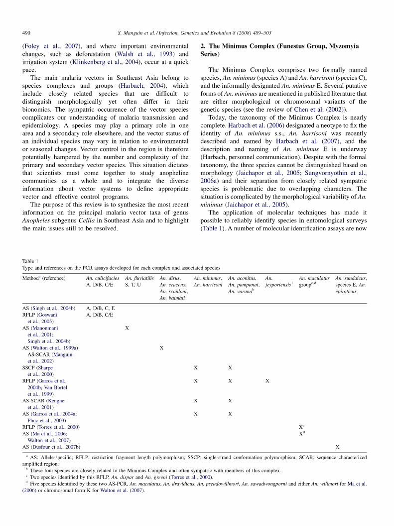
S. Manguin et al. / Infection, Genetics and Evolution 8 (2008) 489–503490
(Foley et al., 2007), and where important environmental
changes, such as deforestation (Walsh et al., 1993) and
irrigation system (Klinkenberg et al., 2004), occur at a quick
pace.
The main malaria vectors in Southeast Asia belong to
species complexes and groups (Harbach, 2004), which
include closely related species that are difficult to
distinguish morphologically yet often differ in their
bionomics. The sympatric occurrence of the vector species
complicates our understanding of malaria transmission and
epidemiology. A species may play a primary role in one
area and a secondary role elsewhere, and the vector status of
an individual species may vary in relation to environmental
or seasonal changes. Vector control in the region is therefore
potentially hampered by the number and complexity of the
primary and secondary vector species. This situation dictates
that scientists must come together to study anopheline
communities as a whole and to integrate the diverse
information about vector systems to define appropriate
vector and effective control programs.
The purpose of this review is to synthesize the most recent
information on the principal malaria vector taxa of genus
Anopheles subgenus Cellia in Southeast Asia and to highlight
the main issues still to be resolved.
Table 1
Type and references on the PCR assays developed for each complex and associate
Methoda (reference) An. culicifacies
A, D/B, C/E
An. fluviatilis
S, T, U
An. dirus,
An. cracens,
An. scanloni,
An. baimaii
An
An
AS (Singh et al., 2004b) A, D/B, C, E
RFLP (Goswani
et al., 2005)
A, D/B, C/E
AS (Manonmani
et al., 2001;
Singh et al., 2004b)
X
AS (Walton et al., 1999a)
AS-SCAR (Manguin
et al., 2002)
X
SSCP (Sharpe
et al., 2000)
X
RFLP (Garros et al.,
2004b; Van Bortel
et al., 1999)
X
AS-SCAR (Kengne
et al., 2001)
X
AS (Garros et al., 2004a;
Phuc et al., 2003)
X
RFLP (Torres et al., 2000)
AS (Ma et al., 2006;
Walton et al., 2007)
AS (Dusfour et al., 2007b)
a AS: Allele-specific; RFLP: restriction fragment length polymorphism; SSCP
amplified region.b These four species are closely related to the Minimus Complex and often symc Two species identified by this RFLP, An. dispar and An. greeni (Torres et al.,d Five species identified by these two AS-PCR, An. maculatus, An. dravidicus, A
(2006) or chromosomal form K for Walton et al. (2007).
2. The Minimus Complex (Funestus Group, Myzomyia
Series)
The Minimus Complex comprises two formally named
species, An. minimus (species A) and An. harrisoni (species C),
and the informally designated An. minimus E. Several putative
forms of An. minimus are mentioned in published literature that
are either morphological or chromosomal variants of the
genetic species (see the review of Chen et al. (2002)).
Today, the taxonomy of the Minimus Complex is nearly
complete. Harbach et al. (2006) designated a neotype to fix the
identity of An. minimus s.s., An. harrisoni was recently
described and named by Harbach et al. (2007), and the
description and naming of An. minimus E is underway
(Harbach, personnel communication). Despite with the formal
taxonomy, the three species cannot be distinguished based on
morphology (Jaichapor et al., 2005; Sungvornyothin et al.,
2006a) and their separation from closely related sympatric
species is problematic due to overlapping characters. The
situation is complicated by the morphological variability of An.
minimus (Jaichapor et al., 2005).
The application of molecular techniques has made it
possible to reliably identify species in entomological surveys
(Table 1). A number of molecular identification assays are now
d species
. minimus,
. harrisoni
An. aconitus,
An. pampanai,
An. varunab
An.
jeyporiensis1
An. maculatus
groupc,d
An. sundaicus,
species E, An.
epiroticus
X
X X
X
X
Xc
Xd
X
: single-strand conformation polymorphism; SCAR: sequence characterized
patric with members of this complex.
2000).
n. pseudowillmori, An. sawadwongporni and either An. willmori for Ma et al.
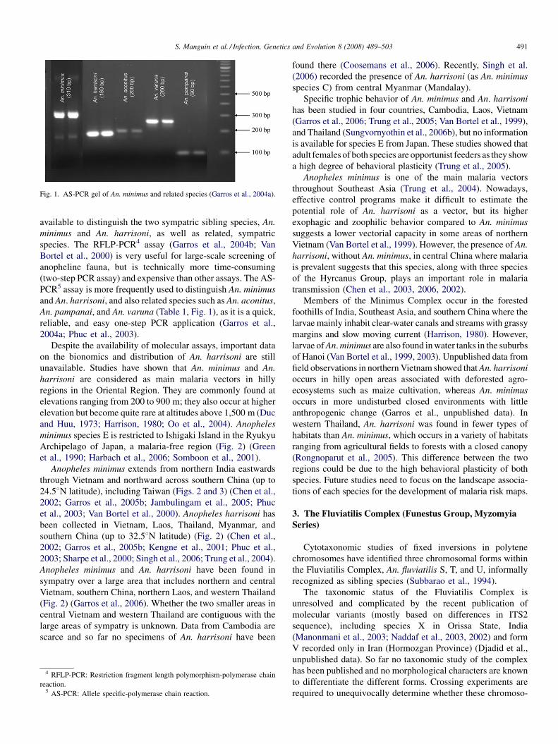
Fig. 1. AS-PCR gel of An. minimus and related species (Garros et al., 2004a).
S. Manguin et al. / Infection, Genetics and Evolution 8 (2008) 489–503 491
available to distinguish the two sympatric sibling species, An.
minimus and An. harrisoni, as well as related, sympatric
species. The RFLP-PCR4 assay (Garros et al., 2004b; Van
Bortel et al., 2000) is very useful for large-scale screening of
anopheline fauna, but is technically more time-consuming
(two-step PCR assay) and expensive than other assays. The AS-
PCR5 assay is more frequently used to distinguish An. minimus
and An. harrisoni, and also related species such as An. aconitus,
An. pampanai, and An. varuna (Table 1, Fig. 1), as it is a quick,
reliable, and easy one-step PCR application (Garros et al.,
2004a; Phuc et al., 2003).
Despite the availability of molecular assays, important data
on the bionomics and distribution of An. harrisoni are still
unavailable. Studies have shown that An. minimus and An.
harrisoni are considered as main malaria vectors in hilly
regions in the Oriental Region. They are commonly found at
elevations ranging from 200 to 900 m; they also occur at higher
elevation but become quite rare at altitudes above 1,500 m (Duc
and Huu, 1973; Harrison, 1980; Oo et al., 2004). Anopheles
minimus species E is restricted to Ishigaki Island in the Ryukyu
Archipelago of Japan, a malaria-free region (Fig. 2) (Green
et al., 1990; Harbach et al., 2006; Somboon et al., 2001).
Anopheles minimus extends from northern India eastwards
through Vietnam and northward across southern China (up to
24.58N latitude), including Taiwan (Figs. 2 and 3) (Chen et al.,
2002; Garros et al., 2005b; Jambulingam et al., 2005; Phuc
et al., 2003; Van Bortel et al., 2000). Anopheles harrisoni has
been collected in Vietnam, Laos, Thailand, Myanmar, and
southern China (up to 32.58N latitude) (Fig. 2) (Chen et al.,
2002; Garros et al., 2005b; Kengne et al., 2001; Phuc et al.,
2003; Sharpe et al., 2000; Singh et al., 2006; Trung et al., 2004).
Anopheles minimus and An. harrisoni have been found in
sympatry over a large area that includes northern and central
Vietnam, southern China, northern Laos, and western Thailand
(Fig. 2) (Garros et al., 2006). Whether the two smaller areas in
central Vietnam and western Thailand are contiguous with the
large areas of sympatry is unknown. Data from Cambodia are
scarce and so far no specimens of An. harrisoni have been
4 RFLP-PCR: Restriction fragment length polymorphism-polymerase chain
reaction.5 AS-PCR: Allele specific-polymerase chain reaction.
found there (Coosemans et al., 2006). Recently, Singh et al.
(2006) recorded the presence of An. harrisoni (as An. minimus
species C) from central Myanmar (Mandalay).
Specific trophic behavior of An. minimus and An. harrisoni
has been studied in four countries, Cambodia, Laos, Vietnam
(Garros et al., 2006; Trung et al., 2005; Van Bortel et al., 1999),
and Thailand (Sungvornyothin et al., 2006b), but no information
is available for species E from Japan. These studies showed that
adult females of both species are opportunist feeders as they show
a high degree of behavioral plasticity (Trung et al., 2005).
Anopheles minimus is one of the main malaria vectors
throughout Southeast Asia (Trung et al., 2004). Nowadays,
effective control programs make it difficult to estimate the
potential role of An. harrisoni as a vector, but its higher
exophagic and zoophilic behavior compared to An. minimus
suggests a lower vectorial capacity in some areas of northern
Vietnam (Van Bortel et al., 1999). However, the presence of An.
harrisoni, without An. minimus, in central China where malaria
is prevalent suggests that this species, along with three species
of the Hyrcanus Group, plays an important role in malaria
transmission (Chen et al., 2003, 2006, 2002).
Members of the Minimus Complex occur in the forested
foothills of India, Southeast Asia, and southern China where the
larvae mainly inhabit clear-water canals and streams with grassy
margins and slow moving current (Harrison, 1980). However,
larvae of An. minimus are also found in water tanks in the suburbs
of Hanoi (Van Bortel et al., 1999, 2003). Unpublished data from
field observations in northern Vietnam showed that An. harrisoni
occurs in hilly open areas associated with deforested agro-
ecosystems such as maize cultivation, whereas An. minimus
occurs in more undisturbed closed environments with little
anthropogenic change (Garros et al., unpublished data). In
western Thailand, An. harrisoni was found in fewer types of
habitats than An. minimus, which occurs in a variety of habitats
ranging from agricultural fields to forests with a closed canopy
(Rongnoparut et al., 2005). This difference between the two
regions could be due to the high behavioral plasticity of both
species. Future studies need to focus on the landscape associa-
tions of each species for the development of malaria risk maps.
3. The Fluviatilis Complex (Funestus Group, Myzomyia
Series)
Cytotaxonomic studies of fixed inversions in polytene
chromosomes have identified three chromosomal forms within
the Fluviatilis Complex, An. fluviatilis S, T, and U, informally
recognized as sibling species (Subbarao et al., 1994).
The taxonomic status of the Fluviatilis Complex is
unresolved and complicated by the recent publication of
molecular variants (mostly based on differences in ITS2
sequence), including species X in Orissa State, India
(Manonmani et al., 2003; Naddaf et al., 2003, 2002) and form
V recorded only in Iran (Hormozgan Province) (Djadid et al.,
unpublished data). So far no taxonomic study of the complex
has been published and no morphological characters are known
to differentiate the different forms. Crossing experiments are
required to unequivocally determine whether these chromoso-
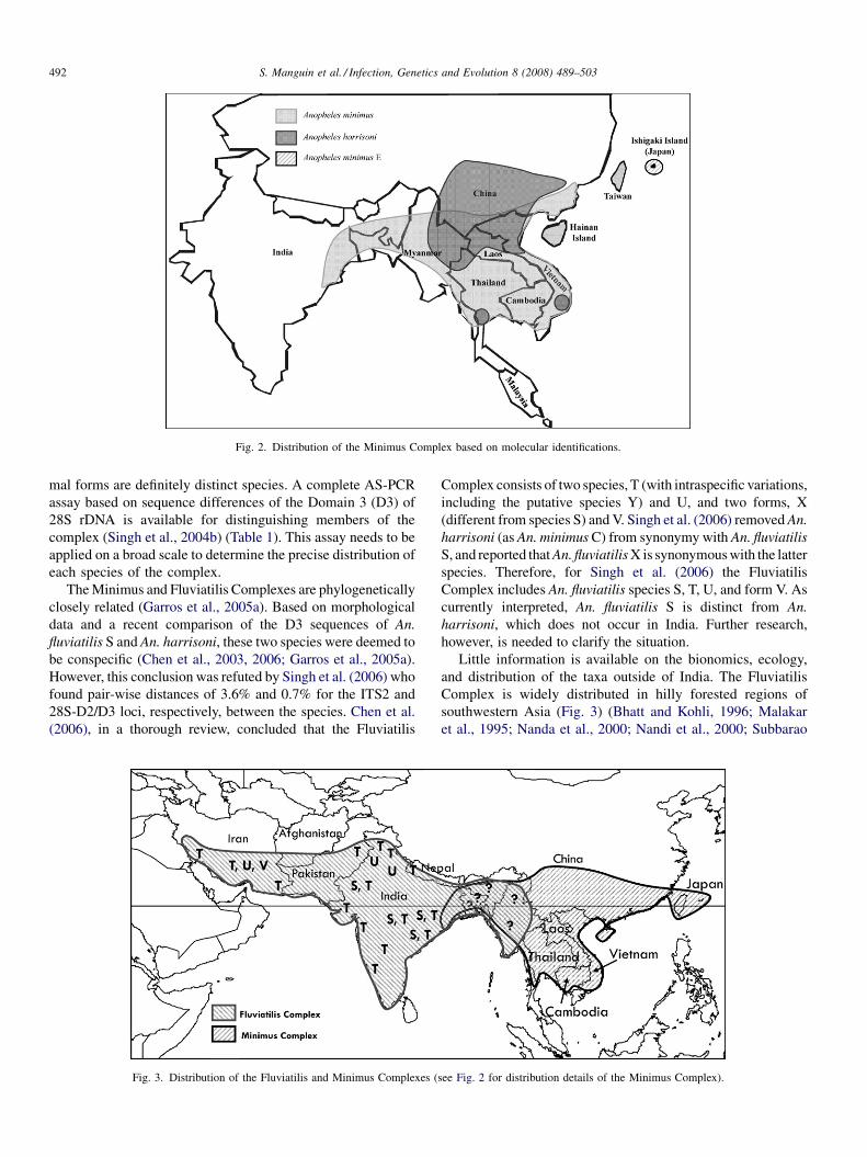
Fig. 2. Distribution of the Minimus Complex based on molecular identifications.
S. Manguin et al. / Infection, Genetics and Evolution 8 (2008) 489–503492
mal forms are definitely distinct species. A complete AS-PCR
assay based on sequence differences of the Domain 3 (D3) of
28S rDNA is available for distinguishing members of the
complex (Singh et al., 2004b) (Table 1). This assay needs to be
applied on a broad scale to determine the precise distribution of
each species of the complex.
The Minimus and Fluviatilis Complexes are phylogenetically
closely related (Garros et al., 2005a). Based on morphological
data and a recent comparison of the D3 sequences of An.
fluviatilis S and An. harrisoni, these two species were deemed to
be conspecific (Chen et al., 2003, 2006; Garros et al., 2005a).
However, this conclusion was refuted by Singh et al. (2006) who
found pair-wise distances of 3.6% and 0.7% for the ITS2 and
28S-D2/D3 loci, respectively, between the species. Chen et al.
(2006), in a thorough review, concluded that the Fluviatilis
Fig. 3. Distribution of the Fluviatilis and Minimus Complexes (
Complex consists of two species, T (with intraspecific variations,
including the putative species Y) and U, and two forms, X
(different from species S) and V. Singh et al. (2006) removed An.
harrisoni (as An. minimus C) from synonymy with An. fluviatilis
S, and reported that An. fluviatilis X is synonymous with the latter
species. Therefore, for Singh et al. (2006) the Fluviatilis
Complex includes An. fluviatilis species S, T, U, and form V. As
currently interpreted, An. fluviatilis S is distinct from An.
harrisoni, which does not occur in India. Further research,
however, is needed to clarify the situation.
Little information is available on the bionomics, ecology,
and distribution of the taxa outside of India. The Fluviatilis
Complex is widely distributed in hilly forested regions of
southwestern Asia (Fig. 3) (Bhatt and Kohli, 1996; Malakar
et al., 1995; Nanda et al., 2000; Nandi et al., 2000; Subbarao
see Fig. 2 for distribution details of the Minimus Complex).
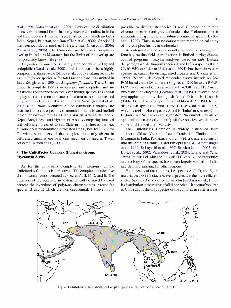
S. Manguin et al. / Infection, Genetics and Evolution 8 (2008) 489–503 493
et al., 1994; Vatandoost et al., 2004). However, the distribution
of the chromosomal forms has only been well studied in India
and Iran. Species T has the largest distribution, which includes
India, Nepal, Pakistan, and Iran (Chen et al., 2006). Species U
has been recorded in northern India and Iran (Chen et al., 2006;
Raeisi et al., 2005). The Fluviatilis and Minimus Complexes
overlap in India to Myanmar, but the limits of the overlap are
not precisely known (Fig. 3).
Anopheles fluviatilis S is mainly anthropophilic (90%) and
endophilic (Nanda et al., 2000), and is known to be a highly
competent malaria vector (Nanda et al., 2005), ranking second to
An. culicifacies species A for total malaria cases transmitted in
India (Singh et al., 2004a). Anopheles fluviatilis T and U are
primarily zoophilic (99%), exophagic, and exophilic, and are
regarded as poor or non-vectors, even though species T is known
to play a role in the maintenance of malaria in mountainous and
hilly regions of India, Pakistan, Iran, and Nepal (Naddaf et al.,
2003; Rao, 1984). Members of the Fluviatilis Complex are
restricted to forest, especially in mountainous, hilly, and foothill
regions of southwestern Asia (Iran, Pakistan, Afghanistan, India,
Nepal, Bangladesh, and Myanmar). A study comparing forested
and deforested areas of Orissa State in India showed that An.
fluviatilis S is predominant in forested areas (98% for S; 2% for
T), whereas members of the complex are nearly absent in
deforested areas where only one specimen of species T was
collected (Nanda et al., 2000).
4. The Culicifacies Complex (Funestus Group,
Myzomyia Series)
As for the Fluviatilis Complex, the taxonomy of the
Culicifacies Complex is unresolved. The complex includes five
chromosomal forms, denoted as species A, B, C, D, and E. The
members of the complex are cytogenetically defined by fixed
paracentric inversions of polytene chromosomes, except for
species B and E which are homosequential. However, it is
Fig. 4. Distribution of the Culicifacies Complex
possible to distinguish species B and C based on mitotic
chromosomes in semi-gravid females: the Y-chromosome is
acrocentric in species B and submetacentric in species E (Kar
et al., 1999). Thus, so far no comparative morphological study
of the complex has been undertaken.
As cytogenetic analyses can only be done on semi-gravid
females, routine field identification is limited during disease
control programs. Isozyme analyses based on Ldh (Lactate
dehydrogenase) distinguish species A and D from species B and
C with 95% confidence (Adak et al., 1994), but the main vector,
species E, cannot be distinguished from B and C (Kar et al.,
1999). Recently developed molecular assays include an AS-
PCR based on the D3 domain (Singh et al., 2004c) and a RFLP-
PCR based on cytochrome oxidase II (COII) and ITS2 using
two restriction enzymes (Goswani et al., 2005). However, these
two applications only distinguish A and D from B, C and E
(Table 1). In the latter group, an additional RFLP-PCR can
distinguish species E from B and C (Goswani et al., 2005),
which is useful where species A and B (India) or species B and
E (India and Sri Lanka) are sympatric. No currently available
application can directly identify all five species, which raises
some doubt about their validity.
The Culicifacies Complex is widely distributed from
southern China, Vietnam, Laos, Cambodia, Thailand, and
Myanmar to India, Pakistan, and Iran, with a western extension
into the Arabian Peninsula and Ethiopia (Fig. 4) (Amerasinghe
et al., 1999; Kobayashi et al., 1997; Rowland et al., 2002; Van
Bortel et al., 2002; Vatandoost et al., 2004; Zhang and Yang,
1996). In parallel with the Fluviatilis Complex, the bionomics
and ecology of the species have been largely studied in India,
and data are missing for other regions.
Four species of the complex, i.e. species A, C, D, and E, are
malaria vectors in India; however, species E is the most efficient
vector. Species B is a poor or non-vector (Subbarao et al., 1988).
Its distribution is thewidest of all the species—it occurs from Iran
to China and is the only species of the complex in eastern areas,
(grey) and each of the five species (A to E).
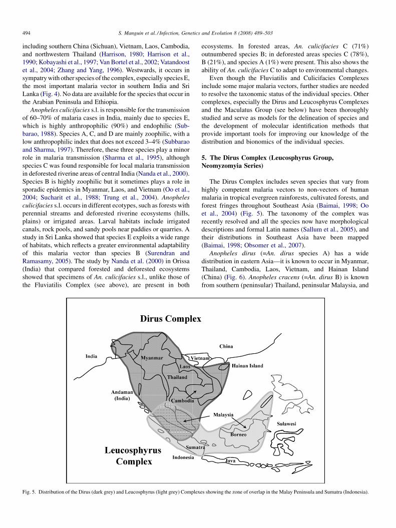
S. Manguin et al. / Infection, Genetics and Evolution 8 (2008) 489–503494
including southern China (Sichuan), Vietnam, Laos, Cambodia,
and northwestern Thailand (Harrison, 1980; Harrison et al.,
1990; Kobayashi et al., 1997; Van Bortel et al., 2002; Vatandoost
et al., 2004; Zhang and Yang, 1996). Westwards, it occurs in
sympatry with other species of the complex, especially species E,
the most important malaria vector in southern India and Sri
Lanka (Fig. 4). No data are available for the species that occur in
the Arabian Peninsula and Ethiopia.
Anopheles culicifacies s.l. is responsible for the transmission
of 60–70% of malaria cases in India, mainly due to species E,
which is highly anthropophilic (90%) and endophilic (Sub-
barao, 1988). Species A, C, and D are mainly zoophilic, with a
low anthropophilic index that does not exceed 3–4% (Subbarao
and Sharma, 1997). Therefore, these three species play a minor
role in malaria transmission (Sharma et al., 1995), although
species C was found responsible for local malaria transmission
in deforested riverine areas of central India (Nanda et al., 2000).
Species B is highly zoophilic but it sometimes plays a role in
sporadic epidemics in Myanmar, Laos, and Vietnam (Oo et al.,
2004; Sucharit et al., 1988; Trung et al., 2004). Anopheles
culicifacies s.l. occurs in different ecotypes, such as forests with
perennial streams and deforested riverine ecosystems (hills,
plains) or irrigated areas. Larval habitats include irrigated
canals, rock pools, and sandy pools near paddies or quarries. A
study in Sri Lanka showed that species E exploits a wide range
of habitats, which reflects a greater environmental adaptability
of this malaria vector than species B (Surendran and
Ramasamy, 2005). The study by Nanda et al. (2000) in Orissa
(India) that compared forested and deforested ecosystems
showed that specimens of An. culicifacies s.l., unlike those of
the Fluviatilis Complex (see above), are present in both
Fig. 5. Distribution of the Dirus (dark grey) and Leucosphyrus (light grey) Complex
ecosystems. In forested areas, An. culicifacies C (71%)
outnumbered species B; in deforested areas species C (78%),
B (21%), and species A (1%) were present. This also shows the
ability of An. culicifacies C to adapt to environmental changes.
Even though the Fluviatilis and Culicifacies Complexes
include some major malaria vectors, further studies are needed
to resolve the taxonomic status of the individual species. Other
complexes, especially the Dirus and Leucosphyrus Complexes
and the Maculatus Group (see below) have been thoroughly
studied and serve as models for the delineation of species and
the development of molecular identification methods that
provide important tools for improving our knowledge of the
distribution and bionomics of the individual species.
5. The Dirus Complex (Leucosphyrus Group,
Neomyzomyia Series)
The Dirus Complex includes seven species that vary from
highly competent malaria vectors to non-vectors of human
malaria in tropical evergreen rainforests, cultivated forests, and
forest fringes throughout Southeast Asia (Baimai, 1998; Oo
et al., 2004) (Fig. 5). The taxonomy of the complex was
recently resolved and all the species now have morphological
descriptions and formal Latin names (Sallum et al., 2005), and
their distributions in Southeast Asia have been mapped
(Baimai, 1998; Obsomer et al., 2007).
Anopheles dirus (=An. dirus species A) has a wide
distribution in eastern Asia—it is known to occur in Myanmar,
Thailand, Cambodia, Laos, Vietnam, and Hainan Island
(China) (Fig. 6). Anopheles cracens (=An. dirus B) is known
from southern (peninsular) Thailand, peninsular Malaysia, and
es showing the zone of overlap in the Malay Peninsula and Sumatra (Indonesia).
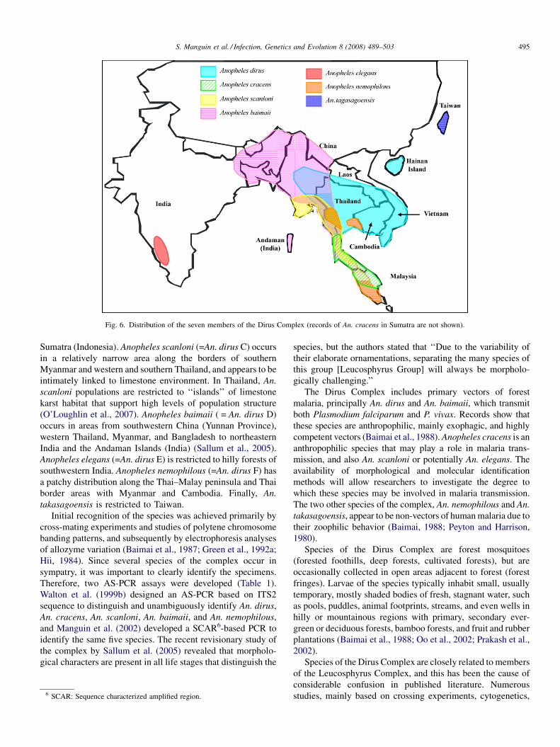
Fig. 6. Distribution of the seven members of the Dirus Complex (records of An. cracens in Sumatra are not shown).
S. Manguin et al. / Infection, Genetics and Evolution 8 (2008) 489–503 495
Sumatra (Indonesia). Anopheles scanloni (=An. dirus C) occurs
in a relatively narrow area along the borders of southern
Myanmar and western and southern Thailand, and appears to be
intimately linked to limestone environment. In Thailand, An.
scanloni populations are restricted to ‘‘islands’’ of limestone
karst habitat that support high levels of population structure
(O’Loughlin et al., 2007). Anopheles baimaii ( = An. dirus D)
occurs in areas from southwestern China (Yunnan Province),
western Thailand, Myanmar, and Bangladesh to northeastern
India and the Andaman Islands (India) (Sallum et al., 2005).
Anopheles elegans (=An. dirus E) is restricted to hilly forests of
southwestern India. Anopheles nemophilous (=An. dirus F) has
a patchy distribution along the Thai–Malay peninsula and Thai
border areas with Myanmar and Cambodia. Finally, An.
takasagoensis is restricted to Taiwan.
Initial recognition of the species was achieved primarily by
cross-mating experiments and studies of polytene chromosome
banding patterns, and subsequently by electrophoresis analyses
of allozyme variation (Baimai et al., 1987; Green et al., 1992a;
Hii, 1984). Since several species of the complex occur in
sympatry, it was important to clearly identify the specimens.
Therefore, two AS-PCR assays were developed (Table 1).
Walton et al. (1999b) designed an AS-PCR based on ITS2
sequence to distinguish and unambiguously identify An. dirus,
An. cracens, An. scanloni, An. baimaii, and An. nemophilous,
and Manguin et al. (2002) developed a SCAR6-based PCR to
identify the same five species. The recent revisionary study of
the complex by Sallum et al. (2005) revealed that morpholo-
gical characters are present in all life stages that distinguish the
6 SCAR: Sequence characterized amplified region.
species, but the authors stated that ‘‘Due to the variability of
their elaborate ornamentations, separating the many species of
this group [Leucosphyrus Group] will always be morpholo-
gically challenging.’’
The Dirus Complex includes primary vectors of forest
malaria, principally An. dirus and An. baimaii, which transmit
both Plasmodium falciparum and P. vivax. Records show that
these species are anthropophilic, mainly exophagic, and highly
competent vectors (Baimai et al., 1988). Anopheles cracens is an
anthropophilic species that may play a role in malaria trans-
mission, and also An. scanloni or potentially An. elegans. The
availability of morphological and molecular identification
methods will allow researchers to investigate the degree to
which these species may be involved in malaria transmission.
The two other species of the complex, An. nemophilous and An.
takasagoensis, appear to be non-vectors of human malaria due to
their zoophilic behavior (Baimai, 1988; Peyton and Harrison,
1980).
Species of the Dirus Complex are forest mosquitoes
(forested foothills, deep forests, cultivated forests), but are
occasionally collected in open areas adjacent to forest (forest
fringes). Larvae of the species typically inhabit small, usually
temporary, mostly shaded bodies of fresh, stagnant water, such
as pools, puddles, animal footprints, streams, and even wells in
hilly or mountainous regions with primary, secondary ever-
green or deciduous forests, bamboo forests, and fruit and rubber
plantations (Baimai et al., 1988; Oo et al., 2002; Prakash et al.,
2002).
Species of the Dirus Complex are closely related to members
of the Leucosphyrus Complex, and this has been the cause of
considerable confusion in published literature. Numerous
studies, mainly based on crossing experiments, cytogenetics,
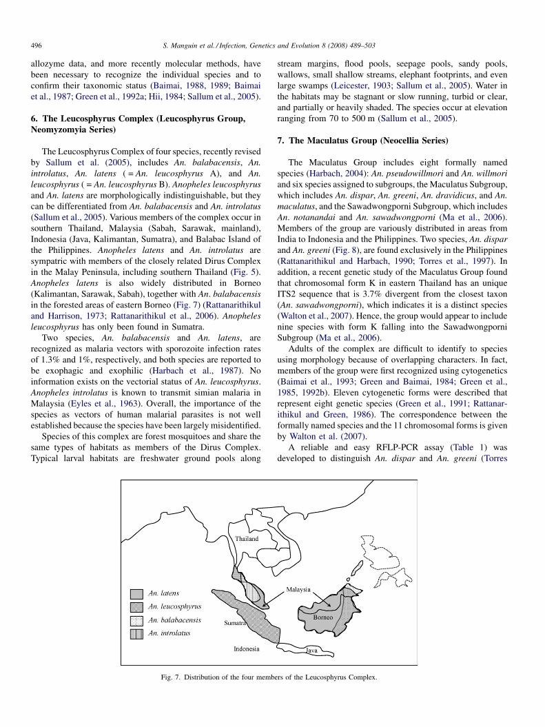
S. Manguin et al. / Infection, Genetics and Evolution 8 (2008) 489–503496
allozyme data, and more recently molecular methods, have
been necessary to recognize the individual species and to
confirm their taxonomic status (Baimai, 1988, 1989; Baimai
et al., 1987; Green et al., 1992a; Hii, 1984; Sallum et al., 2005).
6. The Leucosphyrus Complex (Leucosphyrus Group,
Neomyzomyia Series)
The Leucosphyrus Complex of four species, recently revised
by Sallum et al. (2005), includes An. balabacensis, An.
introlatus, An. latens ( = An. leucosphyrus A), and An.
leucosphyrus ( = An. leucosphyrus B). Anopheles leucosphyrus
and An. latens are morphologically indistinguishable, but they
can be differentiated from An. balabacensis and An. introlatus
(Sallum et al., 2005). Various members of the complex occur in
southern Thailand, Malaysia (Sabah, Sarawak, mainland),
Indonesia (Java, Kalimantan, Sumatra), and Balabac Island of
the Philippines. Anopheles latens and An. introlatus are
sympatric with members of the closely related Dirus Complex
in the Malay Peninsula, including southern Thailand (Fig. 5).
Anopheles latens is also widely distributed in Borneo
(Kalimantan, Sarawak, Sabah), together with An. balabacensis
in the forested areas of eastern Borneo (Fig. 7) (Rattanarithikul
and Harrison, 1973; Rattanarithikul et al., 2006). Anopheles
leucosphyrus has only been found in Sumatra.
Two species, An. balabacensis and An. latens, are
recognized as malaria vectors with sporozoite infection rates
of 1.3% and 1%, respectively, and both species are reported to
be exophagic and exophilic (Harbach et al., 1987). No
information exists on the vectorial status of An. leucosphyrus.
Anopheles introlatus is known to transmit simian malaria in
Malaysia (Eyles et al., 1963). Overall, the importance of the
species as vectors of human malarial parasites is not well
established because the species have been largely misidentified.
Species of this complex are forest mosquitoes and share the
same types of habitats as members of the Dirus Complex.
Typical larval habitats are freshwater ground pools along
Fig. 7. Distribution of the four membe
stream margins, flood pools, seepage pools, sandy pools,
wallows, small shallow streams, elephant footprints, and even
large swamps (Leicester, 1903; Sallum et al., 2005). Water in
the habitats may be stagnant or slow running, turbid or clear,
and partially or heavily shaded. The species occur at elevation
ranging from 70 to 500 m (Sallum et al., 2005).
7. The Maculatus Group (Neocellia Series)
The Maculatus Group includes eight formally named
species (Harbach, 2004): An. pseudowillmori and An. willmori
and six species assigned to subgroups, the Maculatus Subgroup,
which includes An. dispar, An. greeni, An. dravidicus, and An.
maculatus, and the Sawadwongporni Subgroup, which includes
An. notanandai and An. sawadwongporni (Ma et al., 2006).
Members of the group are variously distributed in areas from
India to Indonesia and the Philippines. Two species, An. dispar
and An. greeni (Fig. 8), are found exclusively in the Philippines
(Rattanarithikul and Harbach, 1990; Torres et al., 1997). In
addition, a recent genetic study of the Maculatus Group found
that chromosomal form K in eastern Thailand has an unique
ITS2 sequence that is 3.7% divergent from the closest taxon
(An. sawadwongporni), which indicates it is a distinct species
(Walton et al., 2007). Hence, the group would appear to include
nine species with form K falling into the Sawadwongporni
Subgroup (Ma et al., 2006).
Adults of the complex are difficult to identify to species
using morphology because of overlapping characters. In fact,
members of the group were first recognized using cytogenetics
(Baimai et al., 1993; Green and Baimai, 1984; Green et al.,
1985, 1992b). Eleven cytogenetic forms were described that
represent eight genetic species (Green et al., 1991; Rattanar-
ithikul and Green, 1986). The correspondence between the
formally named species and the 11 chromosomal forms is given
by Walton et al. (2007).
A reliable and easy RFLP-PCR assay (Table 1) was
developed to distinguish An. dispar and An. greeni (Torres
rs of the Leucosphyrus Complex.
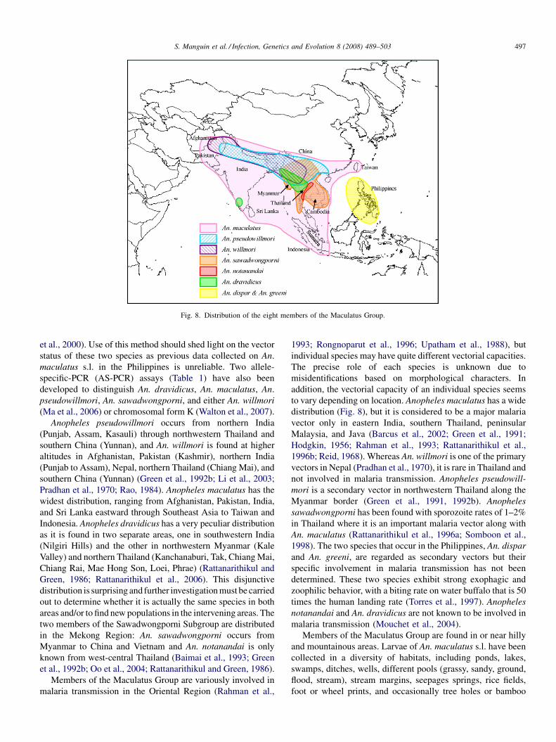
Fig. 8. Distribution of the eight members of the Maculatus Group.
S. Manguin et al. / Infection, Genetics and Evolution 8 (2008) 489–503 497
et al., 2000). Use of this method should shed light on the vector
status of these two species as previous data collected on An.
maculatus s.l. in the Philippines is unreliable. Two allele-
specific-PCR (AS-PCR) assays (Table 1) have also been
developed to distinguish An. dravidicus, An. maculatus, An.
pseudowillmori, An. sawadwongporni, and either An. willmori
(Ma et al., 2006) or chromosomal form K (Walton et al., 2007).
Anopheles pseudowillmori occurs from northern India
(Punjab, Assam, Kasauli) through northwestern Thailand and
southern China (Yunnan), and An. willmori is found at higher
altitudes in Afghanistan, Pakistan (Kashmir), northern India
(Punjab to Assam), Nepal, northern Thailand (Chiang Mai), and
southern China (Yunnan) (Green et al., 1992b; Li et al., 2003;
Pradhan et al., 1970; Rao, 1984). Anopheles maculatus has the
widest distribution, ranging from Afghanistan, Pakistan, India,
and Sri Lanka eastward through Southeast Asia to Taiwan and
Indonesia. Anopheles dravidicus has a very peculiar distribution
as it is found in two separate areas, one in southwestern India
(Nilgiri Hills) and the other in northwestern Myanmar (Kale
Valley) and northern Thailand (Kanchanaburi, Tak, Chiang Mai,
Chiang Rai, Mae Hong Son, Loei, Phrae) (Rattanarithikul and
Green, 1986; Rattanarithikul et al., 2006). This disjunctive
distribution is surprising and further investigation must be carried
out to determine whether it is actually the same species in both
areas and/or to find new populations in the intervening areas. The
two members of the Sawadwongporni Subgroup are distributed
in the Mekong Region: An. sawadwongporni occurs from
Myanmar to China and Vietnam and An. notanandai is only
known from west-central Thailand (Baimai et al., 1993; Green
et al., 1992b; Oo et al., 2004; Rattanarithikul and Green, 1986).
Members of the Maculatus Group are variously involved in
malaria transmission in the Oriental Region (Rahman et al.,
1993; Rongnoparut et al., 1996; Upatham et al., 1988), but
individual species may have quite different vectorial capacities.
The precise role of each species is unknown due to
misidentifications based on morphological characters. In
addition, the vectorial capacity of an individual species seems
to vary depending on location. Anopheles maculatus has a wide
distribution (Fig. 8), but it is considered to be a major malaria
vector only in eastern India, southern Thailand, peninsular
Malaysia, and Java (Barcus et al., 2002; Green et al., 1991;
Hodgkin, 1956; Rahman et al., 1993; Rattanarithikul et al.,
1996b; Reid, 1968). Whereas An. willmori is one of the primary
vectors in Nepal (Pradhan et al., 1970), it is rare in Thailand and
not involved in malaria transmission. Anopheles pseudowill-
mori is a secondary vector in northwestern Thailand along the
Myanmar border (Green et al., 1991, 1992b). Anopheles
sawadwongporni has been found with sporozoite rates of 1–2%
in Thailand where it is an important malaria vector along with
An. maculatus (Rattanarithikul et al., 1996a; Somboon et al.,
1998). The two species that occur in the Philippines, An. dispar
and An. greeni, are regarded as secondary vectors but their
specific involvement in malaria transmission has not been
determined. These two species exhibit strong exophagic and
zoophilic behavior, with a biting rate on water buffalo that is 50
times the human landing rate (Torres et al., 1997). Anopheles
notanandai and An. dravidicus are not known to be involved in
malaria transmission (Mouchet et al., 2004).
Members of the Maculatus Group are found in or near hilly
and mountainous areas. Larvae of An. maculatus s.l. have been
collected in a diversity of habitats, including ponds, lakes,
swamps, ditches, wells, different pools (grassy, sandy, ground,
flood, stream), stream margins, seepages springs, rice fields,
foot or wheel prints, and occasionally tree holes or bamboo
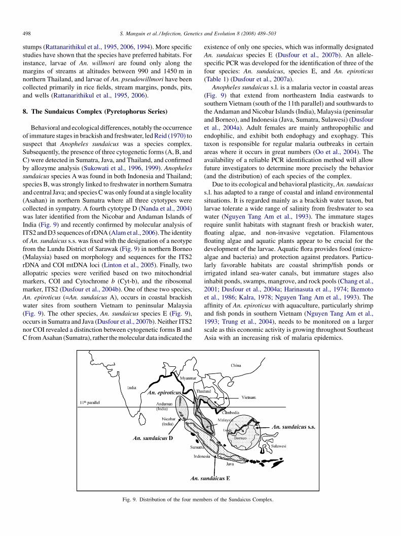
S. Manguin et al. / Infection, Genetics and Evolution 8 (2008) 489–503498
stumps (Rattanarithikul et al., 1995, 2006, 1994). More specific
studies have shown that the species have preferred habitats. For
instance, larvae of An. willmori are found only along the
margins of streams at altitudes between 990 and 1450 m in
northern Thailand, and larvae of An. pseudowillmori have been
collected primarily in rice fields, stream margins, ponds, pits,
and wells (Rattanarithikul et al., 1995, 2006).
8. The Sundaicus Complex (Pyretophorus Series)
Behavioral and ecological differences, notably the occurrence
of immature stages in brackish and freshwater, led Reid (1970) to
suspect that Anopheles sundaicus was a species complex.
Subsequently, the presence of three cytogenetic forms (A, B, and
C) were detected in Sumatra, Java, and Thailand, and confirmed
by allozyme analysis (Sukowati et al., 1996, 1999). Anopheles
sundaicus species A was found in both Indonesia and Thailand;
species B, was strongly linked to freshwater in northern Sumatra
and central Java; and species C was only found at a single locality
(Asahan) in northern Sumatra where all three cytotypes were
collected in sympatry. A fourth cytotype D (Nanda et al., 2004)
was later identified from the Nicobar and Andaman Islands of
India (Fig. 9) and recently confirmed by molecular analysis of
ITS2 and D3 sequences of rDNA (Alam et al., 2006). The identity
of An. sundaicus s.s. was fixed with the designation of a neotype
from the Lundu District of Sarawak (Fig. 9) in northern Borneo
(Malaysia) based on morphology and sequences for the ITS2
rDNA and COI mtDNA loci (Linton et al., 2005). Finally, two
allopatric species were verified based on two mitochondrial
markers, COI and Cytochrome b (Cyt-b), and the ribosomal
marker, ITS2 (Dusfour et al., 2004b). One of these two species,
An. epiroticus (=An. sundaicus A), occurs in coastal brackish
water sites from southern Vietnam to peninsular Malaysia
(Fig. 9). The other species, An. sundaicus species E (Fig. 9),
occurs in Sumatra and Java (Dusfour et al., 2007b). Neither ITS2
nor COI revealed a distinction between cytogenetic forms B and
C from Asahan (Sumatra), rather the molecular data indicated the
Fig. 9. Distribution of the four mem
existence of only one species, which was informally designated
An. sundaicus species E (Dusfour et al., 2007b). An allele-
specific PCR was developed for the identification of three of the
four species: An. sundaicus, species E, and An. epiroticus
(Table 1) (Dusfour et al., 2007a).
Anopheles sundaicus s.l. is a malaria vector in coastal areas
(Fig. 9) that extend from northeastern India eastwards to
southern Vietnam (south of the 11th parallel) and southwards to
the Andaman and Nicobar Islands (India), Malaysia (peninsular
and Borneo), and Indonesia (Java, Sumatra, Sulawesi) (Dusfour
et al., 2004a). Adult females are mainly anthropophilic and
endophilic, and exhibit both endophagy and exophagy. This
taxon is responsible for regular malaria outbreaks in certain
areas where it occurs in great numbers (Oo et al., 2004). The
availability of a reliable PCR identification method will allow
future investigators to determine more precisely the behavior
(and the distribution) of each species of the complex.
Due to its ecological and behavioral plasticity, An. sundaicus
s.l. has adapted to a range of coastal and inland environmental
situations. It is regarded mainly as a brackish water taxon, but
larvae tolerate a wide range of salinity from freshwater to sea
water (Nguyen Tang Am et al., 1993). The immature stages
require sunlit habitats with stagnant fresh or brackish water,
floating algae, and non-invasive vegetation. Filamentous
floating algae and aquatic plants appear to be crucial for the
development of the larvae. Aquatic flora provides food (micro-
algae and bacteria) and protection against predators. Particu-
larly favorable habitats are coastal shrimp/fish ponds or
irrigated inland sea-water canals, but immature stages also
inhabit ponds, swamps, mangrove, and rock pools (Chang et al.,
2001; Dusfour et al., 2004a; Harinasuta et al., 1974; Ikemoto
et al., 1986; Kalra, 1978; Nguyen Tang Am et al., 1993). The
affinity of An. epiroticus with aquaculture, particularly shrimp
and fish ponds in southern Vietnam (Nguyen Tang Am et al.,
1993; Trung et al., 2004), needs to be monitored on a larger
scale as this economic activity is growing throughout Southeast
Asia with an increasing risk of malaria epidemics.
bers of the Sundaicus Complex.
S. Manguin et al. / Infection, Genetics and Evolution 8 (2008) 489–503 499
9. Implications for vector control
Malaria vector control programs in Southeast Asia are
largely based on the use of insecticides for indoor residual
spraying (IRS) and, to a lesser extent, insecticide treated nets
(ITNs). IRS programs were first organized in the 1950s and
implemented for decades in the Southeast Asian countries.
Later, ITNs were required to supplement the IRS campaigns.
Nowadays, IRS is mainly used to control focal epidemics
(WHO, 2007c). Until recently, coverage of ITN and IRS only
reached 10–20% of the populations at risk, but with the recent
support of the Global Fund to the malaria endemic countries,
the use of ITNs has increased significantly. However, more
training and resources are required. The current trend in
Southeast Asian is to rely on the increasing use of ITN based on
several actions: (i) expansion to a maximum coverage of ITN,
including re-treatments; (ii) promotion of long-lasting ITNs to
avoid re-treatments; (iii) lowering the cost of bed nets to a
minimum through tax exemption; and (iv) increased access to
nets through the commercial sector and targeted subsidies for
the poorest people (WHO, 2007d).
These actions will most likely improve the malaria situation in
Southeast Asia but they should be closely associated with a better
knowledge of the targeted malaria vectors. The success of vector
control programs is highly dependant on a thorough knowledge
of behavior and bionomics of the vector, which must be precisely
identified to ensure the application of appropriate control
measures (Trung et al., 2005). Precise information about
ecological changes, the behavioral plasticity of most Anopheles
species as mentioned above, and the accurate identification of the
vector species is required for the development of effective vector
control strategies that are based principally on IRS and ITN. For
instance, IRS is more efficient where the vector is endophilic and
bites late at night, such as An. minimus and An. epiroticus,
whereas ITNs seem more effective than other techniques for the
control of exophagic and exophilic mosquitoes such as An. dirus
s.l. (Trung et al., 2005). For the protection of people at
occupational risk, such as forest workers, the use of treated
hammock nets needs to be implemented, although their efficacy
against this vector is sometimes questionable (Trung et al., 2004).
Rapid ecological changes, such as those occurring in Southeast
Asia, especially deforestation, are modifying the cohort of
malaria vector species, as noted for the Minimus and Dirus
Complexes, and surveillance needs to be done on a regular basis
because vector control programs are highly dependant on the
vector species and its behavior. This is necessary for appro-
priately targeting the species involved in malaria transmission.
Vector control should also be adapted to seasonal variations. In
hilly forested areas of Southeast Asia, it is common knowledge
that malaria transmission is perennial due to the presence of
species of the Dirus and Minimus Complexes, the first being
present mainly during the rainy season and the latter during the
drier periods of the year (Harbach et al., 1987; Ismail et al., 1978;
Phan, 1998; Rattanarithikul et al., 1996a).
Beside the use of chemical insecticides through IRS and
ITN, efforts to minimize this dependency have been undertaken
while searching and developing eco-friendly alternative
methods for the control of vector mosquitoes. Instead of
controlling adult mosquitoes, these alternative methods target
immature stages, particularly larvae. Application of environ-
mental management and biological control need to be utilized
wherever it is cost effective and feasible (WHO, 2007d).
Nowadays, biological control methods are once again receiving
much research focus for malaria vector control. Larvivorous
fish have been used for over 100 years for controlling mosquito
densities and malaria incidence in many countries, including
India, Malaysia, Papua New Guinea, and Thailand (Rozendaal,
1997). Gambusia affinis and Poecilia reticulata are the most
successful and effective larvivorous fish for vector control
(Ghosh and Dash, 2007). In India, remarkable results have been
achieved for the control of malaria vectors like An. culicifacies
species A that breed predominantly in ponds and wells (Ghosh
et al., 2005). Larvivorous fish have also been used to control An.
sundaicus s.l. in Indonesia (Ikemoto et al., 1986) and An. dirus
s.l. in gem pits in Thailand (Kitthawee et al., 1993). In certain
areas where An. dirus s.l. utilizes small habitats, for example
wells in Myanmar, this biological control method should be
applied and may give good results (Oo et al., 2004). However,
the effectiveness of this strategy is questionable (Meek, 1995),
particularly in large wetlands where its efficacy has not been
demonstrated. Its implementation requires some baseline
knowledge of vector biology, and should be included as part
of an integrated malaria control program (Ghosh and Dash,
2007; Meek, 1995). Another potential biological control agent,
tested in Japan, involves the use of copepod species as predators
in rice fields during the summer (Dieng et al., 2003). Results
showed that copepods are efficient biological control agents
against mosquito larvae. However, the reduction of larval
densities is temporary if not properly managed; hence, the
method only has a limited effect on malaria transmission
(Subbarao and Sharma, 1997).
Various biolarvicides have also been thoroughly investigated,
especially strains of the bacteria Bacillus sphaericus and B.
thuringiensis var. israelensis H-14 (Bti), which are highly
effective against mosquito larvae at very low doses and safe to
other non-target organisms. Formulations of B. sphaericus have
been used against An. stephensi, An. subpictus, and An. culici-
facies s.l., but repeated applications in the same habitat resulted in
the development of resistance in the larvae of the targeted species
(Mittal, 2003). Therefore, B. sphaericus has limited prospects for
the control of malaria vectors. Bti formulations have a broader
spectrum of activity against Aedes, Culex, and Anopheles species,
but it was found less effective against Anopheles due to many
limitations (exposure to sunlight reduces efficacy, weekly
application required in most habitats, etc.) (Mittal, 2003).
Biological control may have an impact on malaria vectors in
certain specific situations but in most cases it has proven to be
too tedious for general use because the types of larval habitats
of the main malaria vectors are not conducive of this kind of
strategy (Meek, 1995). However, biological control can still be
considered within an integrated vector management strategy
based on selective application of various control measures
determined by the eco-epidemiological situation of malaria.
For instance, in certain rural communities, biological control
S. Manguin et al. / Infection, Genetics and Evolution 8 (2008) 489–503500
may be a helpful supplement to IRS or ITN, particularly during
the dry season when larvae of vector species are concentrated in
relatively few habitats (Walker and Lynch, 2007).
Environmental management for larval control is another
option, especially against An. sundaicus s.l. Some success in
controlling this taxon was achieved in Malaysia by building
bunds and digging drains for excluding brackish water
(Moorhouse and Wharton, 1965). More recently, larvicide by
clearance of algae has been used successfully in Indonesia
against this malaria vector species (Kirnowardoyo, 1988;
Soekirno et al., 1983). In Malaysia, larvae of An. maculatus s.l.
have been controlled by periodic flushing of streams using
small dams fitted with siphons (Williamson and Scharff, 1936),
or by drainage (Moorhouse and Wharton, 1965). These
larvicidal methods are opportunities to complement adulticid-
ing along with other components of integrated vector
management, and have a direct bearing on concerns about
insecticide resistance, environmental impact, rising costs of
IRS, and logistical constraints (Walker and Lynch, 2007).
The lesson learned over the years is that malaria control is
too complex to be addressed by a single approach (Shiff, 2002).
It is important to tailor the strategy to the prevailing malaria
vector species, as well as ecological and epidemiological
conditions (Mouchet and Carnevale, 1998). We now understand
the ecological conditions that affect and regulate the distribu-
tion and abundance of mosquito populations (Gillies, 2001),
and reliable and easy molecular methods have been developed
to supplement morphological identification of closely related
and isomorphic species (Table 1). Therefore, combined
sustainable and appropriate vector control measures in relation
to the targeted vector species and prophylaxis must be
implemented to achieve the goal of the revised strategy of
the Southeast Asian Regional Committee, which aims to reduce
the level of malaria morbidity and mortality recorded in 2000
by 50% before 2010 (WHO, 2007a).
References
Adak, T., Subbarao, S.K., Sharma, V.P., Rao, S.R., 1994. Lactate dehydrogenase
allozyme differentiation of species in the Anopheles culicifacies complex.
Med. Vet. Entomol. 8, 137–140.
Alam, M.T., Das, M.K., Ansari, M.A., Sharma, Y.D., 2006. Molecular identi-
fication of Anopheles (Cellia) sundaicus from the Andaman and Nicobar
islands of India. Acta Trop. 97, 10–18.
Amerasinghe, P.H., Amerasinghe, F.P., Konradsen, F., Fonseka, K.T., Wirtz,
R.A., 1999. Malaria vectors in a traditional dry zone village in Sri Lanka.
Am. J. Trop. Med. Hyg. 60, 421–429.
Baimai, V., 1988. Population cytogenetics of the malaria vector Anopheles
leucosphyrus group. Southeast Asian J. Trop. Med. Public Health 19, 667–
680.
Baimai, V., 1989. Speciation and species complexes of the Anopheles malaria
vector in Thailand. In: Proceeding of the 3rd conference on malaria
research, Thailand, 18–20 October, pp. 146–162.
Baimai, V., 1998. Heterochromatin accumulation and karyotypic evolution in
some Dipteran insects. Zoolog. Stud. 37, 75–88.
Baimai, V., Andre, R.G., Harrison, B.A., Kijchalao, U., Panthusiri, L., 1987.
Crossing and chromosomal evidence for two additional sibling species
within the taxon Anopheles dirus Peyton and Harrison (Diptera: Culicidae)
in Thailand. In: Proceedings of the Entomological Society of Washington,
vol. 89. pp. 157–166.
Baimai, V., Kijchalao, U., Sawadwongporn, P., Green, C.A., 1988. Geographic
distribution and biting behaviour of four species of the Anopheles dirus
complex (Diptera: Culicidae) in Thailand. Southeast Asian J. Trop. Med.
Public Health 19, 151–161.
Baimai, V., Kijchalao, U., Rattanarithikul, R., Green, C.A., 1993. Metaphase
karyotypes of Anopheles of Thailand and Southeast Asia. II. Maculatus
Group, Neocellia Series, subgenus Cellia. Mosq. Syst. 25, 116–123.
Barcus, M.J., Laihad, F., Sururi, M., Sismadi, P., Marwoto, H., Bangs, M.J.,
Baird, K.J., 2002. Epidemic malaria in the Menoreh hills of Central Java.
Am. J. Trop. Med. Hyg. 66, 287–292.
Bhatt, R.M., Kohli, V.K., 1996. Biting rhythms of some anophelines in Central
Gujarat. Indian J. Malariol. 33, 180–190.
Chang, M.S., Linton, Y.M., Harbach, R.E., 2001. The Anopheles sundaicus
species complex—a preliminary study of larval ecology from two sites in
Sarawak, Malaysia. Mekong Malaria Forum 8, 91–95.
Chen, B., Harbach, R.E., Butlin, R.K., 2002. Molecular and morphological
studies on the Anopheles minimus group of mosquitoes in southern China:
taxonomic review, distribution and malaria vector status. Med. Vet. Ento-
mol. 16, 253–265.
Chen, B., Butlin, R.K., Harbach, R.E., 2003. Molecular phylogenetics of the
Oriental members of the Myzomyia Series of Anopheles subgenus Cellia
(Diptera: Culicidae) inferred from nuclear and mitochondrial DNA
sequences. Syst. Entomol. 28, 1–13.
Chen, B., Butlin, R.K., Pedro, P.M., Wang, X.Z., Harbach, R.E., 2006.
Molecular variation, systematics and distribution of the Anopheles fluviatilis
complex in southern Asia. Med. Vet. Entomol. 20, 33–43.
Coosemans, M., Thuan, L.K., Socheat, D., Phompida, S., Baimai, V., Manguin,
S., Harbach, R.E., Hemingway, J. 2006. Monitoring insecticide resistance
and mapping malaria vectors in Southeast Asia: a prerequisite for sustain-
able malaria vector control. (INCO report, IC4-CT-2002-10041), p. 202.
Dieng, H., Boots, M., Tuno, N., Tsuda, Y., Takagi, M., 2003. Life history effects
of prey choice by copepods: implications for biocontrol of vector mosqui-
toes. J. Am. Mosq. Control Assoc. 19, 67–73.
Duc, N.H., Huu, H.V., 1973. Distribution of Anopheles minimus in Northern
Vietnam. Ann. Sci. Works 134–140.
Dusfour, I., Harbach, R.E., Manguin, S., 2004a. Bionomics and systematics of
the oriental Anopheles sundaicus complex in relation to malaria transmis-
sion and vector control. Am. J. Trop. Med. Hyg. 71, 518–524.
Dusfour, I., Linton, Y.M., Cohuet, A., Harbach, R.E., Baimai, V., Trung, H.D.,
Seng, C.M., Matusop, A., Manguin, S., 2004b. Molecular evidence of
speciation between island and continental populations of Anopheles (Cellia)
sundaicus (Diptera: Culicidae), a principal malaria vector taxon in South-
east Asia. J. Med. Entomol. 41, 287–295.
Dusfour, I., Blondeau, J., Harbach, R.E., Vythilingham, I., Baimai, V., Trung,
H.D., Sochanta, T., Bangs, M.J., Manguin, S., 2007a. Polymerase chain
reaction identification of three members of the Anopheles sundaicus
(Diptera: Culicidae) complex, malaria vectors in Southeast Asia. J. Med.
Entomol. 44, 723–731.
Dusfour, I., Michaux, J.R., Harbach, R.E., Manguin, S., 2007b. Speciation and
phylogeography of the Southeast Asian Anopheles sundaicus complex.
Infect. Genet. Evol. 7, 484–493.
Eyles, D.E., Warren, M., Guinn, E., Wharton, R.H., Ramachandran, C.P., 1963.
Identification of Anopheles balabacensis introlatus as a vector of monkey
Malaria in Malaya. Bull. World Health Organ. 28, 134–135.
Foley, D.H., Rueda, L.M., Wilkerson, R.C., 2007. Insight into global mosquito
biogeography from country species records. J. Med. Entomol. 44, 554–567.
Garros, C., Koekemoer, L.L., Coetzee, M., Coosemans, M., Manguin, S., 2004a.
A single multiplex assay to identify major malaria vectors within the
African Anopheles funestus and the Oriental An. minimus groups. Am. J.
Trop. Med. Hyg. 70, 583–590.
Garros, C., Koekemoer, L.L., Kamau, L., Awolola, T.S., Van Bortel, W.,
Coetzee, M., Coosemans, M., Manguin, S., 2004b. Restriction fragment
length polymorphism method for the identification of major African and
Asian malaria vectors within the Anopheles funestus and An. minimus
groups. Am. J. Trop. Med. Hyg. 70, 260–265.
Garros, C., Harbach, R.E., Manguin, S., 2005a. Morphological assessment and
molecular phylogenetics of the Funestus and Minimus Groups of Anopheles
(Cellia). J. Med. Entomol. 42, 522–536.
S. Manguin et al. / Infection, Genetics and Evolution 8 (2008) 489–503 501
Garros, C., Harbach, R.E., Manguin, S., 2005b. Systematics and biogeogra-
phical implications of the phylogenetic relationships between members of
the Funestus and Minimus Groups of Anopheles (Diptera: Culicidae). J.
Med. Entomol. 42, 7–16.
Garros, C., Van Bortel, W., Trung, H.D., Coosemans, M., Manguin, S., 2006.
Review of the Minimus Complex of Anopheles, main malaria vector in
Southeast Asia: from taxonomic issues to vector control strategies. Trop.
Med. Int. Health 11, 102–114.
Ghosh, S.K., Dash, A.P., 2007. Larvivorous fish against malaria vectors: a new
outlook. Trans. R. Soc. Trop. Med. Hyg. 101, 1063–1064.
Ghosh, S.K., Tiwari, S.N., Sathyanarayan, T.S., Sampath, T.R., Sharma, V.P.,
Nanda, N., Joshi, H., Adak, T., Subbarao, S.K., 2005. Larvivorous fish in
wells target the malaria vector sibling species of the Anopheles culicifacies
complex in villages in Karnataka, India. Trans. R. Soc. Trop. Med. Hyg. 99,
101–105.
Gillies, M.T., 2001. In: Wernsdorfer, W., Mc Gregor, I. (Eds.),Vector Behaviour
and Bionomics, Malaria. Churchill Livingstone, Edinburgh, UK, pp. 453–
485.
Goswani, G., Raghavendra, K., Nanda, N., Gakhar, S.K., Subbarao, S.K., 2005.
PCR-RFLP of mitochondrial cytochrome oxidase subunit II and ITS2 of
ribosomal DNA: markers for the identification of members of the Anopheles
culicifacies complex (Diptera: Culicidae). Acta Trop. 95, 92–99.
Green, C.A., Baimai, V., 1984. Polytene chromosomes and their use in species
studies of malaria vectors as exemplified by the Anopheles maculatus
complex. In: Chopra, V.L., Joshi, B.C., Sharma, R.P., Bansal, H.C.
(Eds.), Genetics: New Frontiers Proceedings of the XV International
Congress of Genetics. Oxford and IBH Publ. Comp., New Delhi.
Green, C.A., Baimai, V., Harrison, B.A., Andre, R.G., 1985. Cytogenetic
evidence for a complex of species within the taxon Anopheles maculatus
(Diptera Culicidae). Biol. J. Linnean. Soc. 24, 321–328.
Green, C.A., Gass, R.F., Munstermann, L.E., Baimai, V., 1990. Population-
genetic evidence for two species in Anopheles minimus in Thailand. Med.
Vet. Entomol. 4, 25–34.
Green, C.A., Rattanarithikul, R., Ponparit, S., Sawadwongporn, P., Baimai, V.,
1991. A newly recognised vector of human malarial parasites in the Oriental
region, Anopheles (Cellia) pseudowillmori (Theobald 1910). Trans. R. Soc.
Trop. Med. Hyg. 85, 35–36.
Green, C.A., Munstermann, L.E., Tan, S.G., Panyim, S., Baimai, V., 1992a.
Population genetic evidences for species A, B, C, and D of the Anopheles
dirus complex in Thailand and enzyme electromorphs for their identifica-
tion. Med. Vet. Entomol. 6, 29–36.
Green, C.A., Rattanarithikul, R., Charoensub, A., 1992b. Population genetic
confirmation of species status of the malaria vectors Anopheles willmori and
An. pseudowillmori in Thailand and chromosome phylogeny of the Macu-
latus Group of mosquitoes. Med. Vet. Entomol. 6, 335–341.
Harbach, R.E., 2004. The classification of genus Anopheles (Diptera: Culici-
dae): a working hypothesis of phylogenetic relationships. Bull. Entomol.
Res. 94, 537–553.
Harbach, R.E., Gingrich, J.B., Pang, L.W., 1987. Some entomological observa-
tions on malaria transmission in a remote village in northwestern Thailand.
J. Am. Mosq. Control Assoc. 3, 296–301.
Harbach, R.E., Parkin, E., Chen, B., Butlin, R.K., 2006. Anopheles (Cellia)
minimus Theobald (Diptera: Culicidae): neotype designation, characteriza-
tion, and systematics. Proc. Entomol. Soc. Wash. 108, 198–209.
Harbach, R.E., Garros, C., Manh, N.D., Manguin, S., 2007. Formal taxonomy of
species C of the Anopheles minimus sibling species complex (Diptera:
Culicidae). Zootaxa 1654, 41–54.
Harinasuta, C., Guptavanij, P., Vasuvat, C., 1974. Studies on the medical
ecological epidemiology in mangrove areas in Thailand. Southeast Asian
J. Trop. Med. Hyg. 32, 588–594.
Harrison, B.A., 1980. The Myzomyia Series of Anopheles (Cellia) in
Thailand, with emphasis on intra-interspecific variations (Diptera:
Culicidae) Medical entomology studies-XIII. Contr. Am. Entomol. Inst.
17, 1–195.
Harrison, B.A., Rattanarithikul, R., Peyton, E.L., Mongkolpanya, K., 1990.
Taxonomic changes, revised occurrence records and notes on the
Culicidae of Thailand and neighboring countries. Mosq. Syst. 22, 196–
227.
Hii, J.L.K., 1984. Involvement of the X-chromosome in hybrid male sterility
from crosses between species A and species B of the taxon Anopheles dirus.
Mosq. News 44, 192–196.
Hodgkin, E.P., 1956. The transmission of malaria in Malaya. Stud. Inst. Med.
Res. Malaya 27 (viii+), 1–98.
Ikemoto, T., Sumitro, S., Panjaitan, W., Shibuya, T., 1986. Laboratory and
small-scale field tests of larvicides and larvivorous fishes against Anopheles
sundaicus larvae at Perupuk village, North Sumatra. Indonesia. Jpn. J. Sanit.
Zool. 37, 105–112.
Ismail, I.A., Phinichpongse, S., Boonrassri, P., 1978. Responses of Anopheles
minimus to DDT residual spraying in a cleared forested foothill area in
central Thailand. Acta Trop. 35, 69–82.
Jaichapor, B., Kengluecha, A., Rongnoparut, P., Rueda, L.M., Sithiprasasna, R.,
2005. Morphological variations of Anopheles minimus A in Tak province,
Thailand. Southeast Asian J. Trop. Med. Hyg. 36, 609–615.
Jambulingam, P., Sahu, S.S., Manonmani, A., 2005. Reappearance of Anopheles
minimus in Singhbum hills of East-Central India. Acta Trop. 96, 31–35.
Kalra, N.L., 1978. National malaria eradication programme, India—its pro-
blems, management and research needs. J. Commun. Dis. 10, 1–20.
Kar, I., Subbarao, S.K., Eapen, A., Ravindran, J., Satyanarayana, T.S., Ragha-
vendra, K., Nanda, N., Sharma, V.P., 1999. Evidence for a new malaria
species, species E, within the Anopheles culicifacies complex (Diptera:
Culicidae). J. Med. Entomol. 36, 595–600.
Kengne, P., Trung, H.D., Baimai, V., Coosemans, M., Manguin, S., 2001.
A multiplex PCR-based method derived from random amplified poly-
morphic DNA (RAPD) markers for the identification of species of
the Anopheles minimus group in Southeast Asia. Insect Mol. Biol. 10,
427–435.
Kirnowardoyo, S., 1988. Anopheles malaria vector and control measures
applied in Indonesia. Southeast Asian J. Trop. Med. Public Health 19,
713–716.
Kitthawee, S., Edman, J.D., Upatham, E.S., 1993. Mosquito larvae and asso-
ciated macroorganisms occurring in gem pits in southern Tha Mai District,
Chanthaburi Province Thailand. Southeast Asian J. Trop. Med. Public
Health 24, 143–151.
Klinkenberg, E., Konradsen, F., Herrel, N., Mukhtar, M., van der Hoek, W.,
Amerasinghe, F.P., 2004. Malaria vectors in the changing environment of
the southern Punjab. Pakistan. Trans. R. Soc. Trop. Med. Hyg. 98, 442–449.
Kobayashi, J., Nambanya, S., Miyagi, I., Vanachone, B., Manivong, K.,
Koubouchan, T., Amano, H., Nozaki, H., Inthakone, S., Sato, Y., 1997.
Collection of anopheline mosquitoes in three villages endemic for malaria
in Khammouane, Lao PDR. Southeast Asian J. Trop. Med. Hyg. 28, 20.
Leicester, G.F., 1903. A breeding place of certain forest mosquitoes in Malaya.
J. Trop. Med. 6, 291–292.
Li, S.Z., Ma, Y.J., Zheng, Z.M., 2003. Molecular identification of Anopheles
maculatus complex (Diptera: Culicidae) from China: single stranded con-
formation polymorphism (SSCP) analysis assay. Entomotaxonomia 25,
125–130.
Linton, Y.M., Dusfour, I., Howard, T.M., Ruiz, L.F., Duc Manh, N., Ho Dinh, T.,
Sochanta, T., Coosemans, M., Harbach, R.E., 2005. Anopheles (Cellia)
epiroticus (Diptera: Culicidae), a new malaria vector species in the South-
east Asian Sundaicus Complex. Bull. Entomol. Res. 95, 329–339.
Ma, Y., Li, S., Xu, J., 2006. Molecular identification and phylogeny of the
Maculatus Group of Anopheles mosquitoes (Diptera: Culicidae) based on
nuclear and mitochondrial DNA sequences. Acta Trop. 99, 272–280.
Malakar, P., Das, S., Saha, G.K., Dasgupta, B., Hati, A.K., 1995. Indoor resting
anophelines of north Bengal. Indian J. Malariol. 32, 24–31.
Manguin, S., Kengne, P., Sonnier, L., Harbach, R.E., Baimai, V., Trung, H.D.,
Coosemans, M., 2002. SCAR markers and multiplex PCR-based identifica-
tion of isomorphic species in the Anopheles dirus complex in Southeast
Asia. Med. Vet. Entomol. 16, 46–54.
Manonmani, A., Townson, H., Adeniran, T., Jambulingam, P., Sahu, S.S.,
Vijayakumar, T., 2001. rDNA-ITS2 polymerase chain reaction assay for
the sibling species of Anopheles fluviatilis. Acta Trop. 78, 3–9.
Manonmani, A., Nanda, N., Jambulingam, P., Sahu, S., Vijayakumar, T., Ramya
Vani, J., Subbarao, S.K., 2003. Comparison of polymerase chain reaction
assay and cytotaxonomy for identification of sibling species of Anopheles
fluviatilis (Diptera: Culicidae). Bull. Entomol. Res. 93, 169–171.
S. Manguin et al. / Infection, Genetics and Evolution 8 (2008) 489–503502
Meek, S.R., 1995. Vector control in some countries of Southeast of Asia:
comparing the vectors and the strategies. Ann. Trop. Med. Parasitol. 89,
135–147.
Mittal, P.K., 2003. Biolarvicides in vector control: challenges and prospects. J.
Vector Borne Dis. 40, 20–32.
Moorhouse, D.E., Wharton, R.H., 1965. Studies on Malayan vectors of malaria;
methods of trapping and observations on biting cycles. J. Med. Entomol. 1,
359–370.
Mouchet, J., Carnevale, P., 1998. Entomological biodiversity of malaria in the
World. Res. Rev. Parasitol. 58, 189–195.
Mouchet, J., Carnevale, P., Coosemans, M., Julvez, J., Manguin, S., Richard-
Lenoble, D., Sircoulon, J., 2004. Biodiversite du paludisme dans le monde.
John Libbey Eurotext, Paris, France, 428 pages.
Naddaf, S.R., Oshagi, M.A., Vatandoost, H., Djavadian, E., Telmadarei, Z.,
Assmar, M., 2002. Use of random amplified polymorphic DNA polymerase
chain reaction (RAPD-PCR) and ITS2 PCR assays for differentiation of
populations and putative sibling species of Anopheles fluviatilis (Diptera:
Culicidae) in Iran. Iranian J. Public Health 32, 137.
Naddaf, S.R., Oshagi, M.A., Vatandoost, H., Assmar, M., 2003. Molecular
characterization of Anopheles fluviatilis species complex in the Islamic
Republic of Iran. Eastern Mediterranean Health J. 9, 257–265.
Nanda, N., Yadav, R.S., Subbarao, S.K., Joshi, H., Sharma, V.P., 2000. Studies
on Anopheles fluviatilis and Anopheles culicifacies sibling species in
relation to malaria in forested hilly and deforested riverine ecosystems
in northern Orissa, India. J. Am. Mosq. Control Assoc. 16, 199–205.
Nanda, N., Das, M.K., Wattal, S., Adak, T., Subbarao, S.K., 2004. Cytogenetic
characterization of Anopheles sundaicus (Diptera: Culicidae) population
from Car Nicobar island, India. Ann. Entomol. Soc. Am. 97, 171–176.
Nanda, N., Yadav, R.S., Sahu, S.S., Sharma, S.K., Malhotra, M.S., 2005.
Anopheles fluviatilis species S-related malaria in hyperendemic districts
of Orissa state, India. In: International Conference on Malaria, New Delhi,
India, 4–6 November.
Nandi, J., Kaul, S.M., Sharma, S.N., Lal, S., 2000. Anthropophily of anophe-
lines in Duars of west Bengal and other regions of India. J. Commun. Dis.
32, 95–99.
Nguyen Tang Am, Le Quy, R., Vu Thi, H., Ngyuen Bieh, L., 1993. Entomo-
epidemiological studies of malaria in the coastal zone of Ho Chi Minh City,
1990–1992. Sante 3, 464–473.
Obsomer, V., Defourny, P., Coosemans, M., 2007. The Anopheles dirus com-
plex: spatial distribution and environmental drivers. Malaria J. 6, http://
www.malariajournal.com/content/6/1/26.
O’Loughlin, S.M., Somboon, P., Walton, C., 2007. High levels of population
structure caused by habitat islands in the malarial vector Anopheles
scanloni. Heredity 99, 31–40.
Oo, T.T., Storch, V., Becker, N., 2002. Studies on the bionomics of Anopheles
dirus (Culicidae: Diptera) in Mudon, Mon State, Myanmar. J. Vector Ecol.
27, 44–54.
Oo, T.T., Storch, V., Becker, N., 2004. Review of the anopheline mosquitoes of
Myanmar. J. Vector Ecol. 29, 21–40.
Peyton, E.L., Harrison, B.A., 1980. Anopheles (Cellia) takasagoensis Morishita
1946, an additional species in the Balabacensis complex of Southeast Asia
(Diptera: Culicidae). Mosq. Syst. 12, 335–347.
Phan, V.T., 1998. Epidemiologie du paludisme et lutte antipaludique au
Vietnam. Editions Medicales Vietnam, Hanoi, 241 pages.
Phuc, H.K., Ball, A.J., Son, L., Hanh, N.V., Tu, N.D., Lien, N.G., Verardi, A.,
Townson, H., 2003. Multiplex PCR assay for malaria vector Anopheles
minimus and four related species in the Myzomyia Series from Southeast
Asia. Med. Vet. Entomol. 17, 423–428.
Pradhan, S.P., Shrestha, S.L., Vaidya, R.G., 1970. Malaria transmission in high
mountain valleys of west Nepal including the first record of Anopheles
willmori (James) as a third vector of malaria. J. Nepal Med. Assoc. 8, 89–97.
Prakash, A., Bhattacharyya, D.R., Mohapatra, P.K., Mahanta, J., 2002. Physico-
chemical characteristics of breeding habitats of Anopheles dirus (Diptera:
Culicidae) in Assam, India. J. Environ. Biol. 23, 95–100.
Raeisi, A., Vatandoost, H., Mousakazemi, S.H., 2005. Understanding bio-
nomics of main malaria vectors in irrigated areas in southern Baluchistan,
for implementation of impregnated bednets in Iran. In: International Con-
ference on Malaria, New Delhi, India 4–6 November.
Rahman, W.A., Abu Hassan, A., Adanan, C.R., Rashid, M.R., 1993. The
prevalence of Plasmodium falciparum and P. vivax in relation to Anopheles
maculatus densities in a Malaysian village. Acta Trop. 55, 231–235.
Rao, T.R., 1984. The Anophelines of India. Malaria Center Research. Indian
Council of Medical Research, New Delhi, India.
Rattanarithikul, R., Green, C.A., 1986. Formal recognition of the species of the
Anopheles maculatus group (Diptera: Culicidae) occurring in Thailand,
including the descriptions of two new species and a preliminary key to
females. Mosq. Syst. 18, 246–278.
Rattanarithikul, R., Harbach, R.E., 1990. Anopheles maculatus (Diptera: Culi-
cidae) from the type locality of Hong Kong and two new species of the
Maculatus Complex from the Philippines. Mosq. Syst. 22, 160–183.
Rattanarithikul, R., Harrison, B.A., 1973. An Illustrated Key to the Anopheles
larvae of Thailand. Bangkok, 42 pages.
Rattanarithikul, R., Mongkolpanya, K., Noigamol, C., Chanaimongkol, S.,
Mahapibul, P., Nakngen, S., 1994. Dry-season distribution of mosquito
larvae in the bed of the Mekong River, northwestern Thailand. J. Am. Mosq.
Control Assoc. 10, 197–201.
Rattanarithikul, R., Green, C.A., Panyim, S., Noigamol, C., Chanaimongkol, S.,
Mahapibul, P., 1995. Larval habitats of malaria vectors and other Anopheles
mosquitoes around a transmission focus in northwestern Thailand. J. Am.
Mosq. Control Assoc. 11, 428–433.
Rattanarithikul, R., Konishi, E., Linthicum, K., 1996a. Observations on noc-
turnal biting activity and host preference of Anophelines collected in
southern Thailand. J. Am. Mosq. Control Assoc. 12, 52–57.
Rattanarithikul, R., Konishi, E., Linthicum, K.J., 1996b. Detection of Plasmo-
dium vivax and Plasmodium falciparum circumsporozoite antigen in ano-
pheline mosquitoes collected in southern Thailand. Am. J. Trop. Med. Hyg.
54, 114–121.
Rattanarithikul, R., Harrison, B.A., Harbach, R.E., Panthusiri, P., Coleman,
R.E., 2006. Illustrated keys to the mosquitoes of Thailand. IV. Anopheles.
Southeast Asian J. Trop. Med. Public Health 37 (Suppl 2), 1–128.
Reid, J.A., 1968. Anopheline mosquitoes of Malaya and Borneo. Stud.
Inst.Med. Res. Malaya 31.
Reid, J.A., 1970. Systematics of malaria vectors - anopheline systematics and
malaria control, with special reference to Southeast Asia. Misc. Publ.
Entomol. Soc. Am. 7, 52–62.
Rongnoparut, P., Yaicharoen, S., Sirichotpakorn, N., Rattanarithikul, R., Lan-
zaro, G.C., Linthicum, K.J., 1996. Microsatellite polymorphism in Ano-
pheles maculatus, a malaria vector in Thailand. Am. J. Trop. Med. Hyg. 55,
589–594.
Rongnoparut, P., Ugsang, D.M., Baimai, V., Honda, K., Sithiprasasna, R., 2005.
Use of a remote sensing-based geographic information system in the
characterizing spatial patterns for Anopheles minimus A and C breeding
habitats in western Thailand. Southeast Asian J. Trop. Med. Public Health
36, 1145–1152.
Rowland, M., Mohammed, N., Rehman, H., Hewitt, S., Mendis, C., Ahmad, M.,
Kamal, M., Wirtz, R.A., 2002. Anopheline vectors and malaria transmission
in eastern Afghanistan. Trans. R. Soc. Trop. Med. Hyg. 96, 620–626.
Rozendaal, J.A., 1997. Vector Control: Methods for Use by Individuals and
Communities. WHO, Geneva, 412 pages.
Sallum, M.A., Peyton, E.L., Wilkerson, R.C., 2005. Six new species of the
Anopheles leucosphyrus group, reinterpretation of An. elegans and vector
implications. Med. Vet. Entomol. 19, 158–199.
Sharma, S.K., Nanda, N., Dutta, V.K., 1995. Studies on the bionomics of
Anopheles fluviatilis s.l. and the sibling species composition in the foothills
of Shiwalik Range India. Southeast Asian J. Trop. Med. Public Health 26,
566.
Sharpe, R.G., Harbach, R.E., Butlin, R.K., 2000. Molecular variation and
phylogeny of members of the Minimus group of Anopheles subgenus Cellia
(Diptera: Culicidae). Syst. Entomol. 25, 263–272.
Shiff, C., 2002. Integrated approach to malaria control. Clin. Microbiol. Rev. 15,
278–293.
Singh, N., Nagpal, A.C., Saxena, A., Singh, M.P., 2004a. Changing scenario of
malaria in central India, the replacement of Plasmodium vivax by Plasmo-
dium falciparum (1986–2000). Trop. Med. Int. Health 9, 364–371.
Singh, O.P., Chandra, D., Nanda, N., Raghavendra, K., Sunil, S., Sharma, S.K.,
Dua, V.K., Subbarao, S.K., 2004b. Differentiation of members of the
S. Manguin et al. / Infection, Genetics and Evolution 8 (2008) 489–503 503
Anopheles fluviatilis species complex by an allele-specific polymerase chain
reaction based on 28S ribosomal DNA sequences. Am. J. Trop. Med. Hyg.
70, 27–32.
Singh, O.P., Goswami, G., Nanda, N., Raghavendra, K., Chandra, D., Subbarao,
S.K., 2004c. An allele-specific polymerase chain reaction assay for the
differentiation of members of the Anopheles culicifacies complex. J. Biosci.
29, 275–280.
Singh, O.P., Chandra, D., Nanda, N., Sharma, S.K., Htun, P.T., Adak, T.,
Subbarao, S.K., Dash, A.P., 2006. On the conspecificity of Anopheles
fluviatilis species S with Anopheles minimus species C. J. Biosci. 31,
671–677.
Soekirno, M., Bang, Y.H., Sudomo, M., Pemayun, T.P., Fleming, G.A., 1983.
Bionomics of Anopheles sundaicus and other anophelines associated with
malaria in coastal areas of Bali, Indonesia. Document. WHO/VBC/83.885,
Geneva.
Somboon, P., Aramrattana, A., Lines, J., Webber, R., 1998. Entomological and
epidemiological investigations of malaria transmission in relation to popu-
lation movements in forest areas of north-west Thailand. Southeast Asian J.
Trop. Med. Public Health 29, 3–9.
Somboon, P., Walton, C., Sharpe, R.G., Higa, Y., Tuno, N., Tsuda, Y., Takagi,
M., 2001. Evidence for a new sibling species of Anopheles minimus from the
Ryukyu Archipelago, Japan. J. Am. Mosq. Control Assoc. 17, 98–113.
Subbarao, S.K., 1988. The Anopheles culicifacies complex and control of
malaria. Parasitol. Today 4, 72–75.
Subbarao, S.K., Sharma, V.P., 1997. Anopheles species complexes and malaria
control. Indian J. Med. Res. 106, 164–173.
Subbarao, S.K., Vasantha, K., Raghavendra, K., Sharma, V.P., Sharma, G.K.,
1988. Anopheles culicifacies: sibling species composition and its relation-
ship to malaria incidence. J. Am. Mosq. Control Assoc. 4, 29–33.
Subbarao, S.K., Nanda, N., Vasantha, K., Dua, V.K., Malhotra, M.S., Yadav,
R.S., Sharma, V.P., 1994. Cytogenetic evidence for three sibling species in
Anopheles fluviatilis. Ann. Entomol. Soc. Am. 87, 116–121.
Sucharit, S., Komalamisra, N., Leemingsawat, S., Apiwathnasorn, C., Thon-
grungkiat, S., 1988. Population genetic studies on the Anopheles minimus
complex in Thailand. Southeast Asian J. Trop. Med. Public Health 19, 717–
723.
Sukowati, S., Baimai, V., Andris, H., 1996. Sex chromosome variation in natural
populations of the Anopheles sundaicus complex from Thailand and
Indonesia. Mosq. Borne Dis. Bull. 13, 8–13.
Sukowati, S., Baimai, V., Harun, S., Dasuki, Y., Andris, H., Efriwati, M., 1999.
Isozyme evidence for three sibling species in the Anopheles sundaicus
complex from Indonesia. Med. Vet. Entomol. 13, 408–414.
Sungvornyothin, S., Garros, C., Chareonviryaphap, T., Manguin, S., 2006a.
How reliable is the humeral pale spot for identification of cryptic
species of the Minimus Complex? J. Am. Mosq. Control Assoc. 20,
185–191.
Sungvornyothin, S., Muenvorn, V., Garros, C., Prabaripai, A., Bangs, M.J.,
Manguin, S., Chareonviryaphap, T., 2006b. Trophic behavior and biting
activity of the two sibling species of the Anopheles minimus Complex in
western Thailand. J. Vector Ecol. 31, 252–261.
Surendran, S.N., Ramasamy, R., 2005. Some characteristics of the larval
breeding sites of Anopheles culicifacies species B and E in Sri Lanka. J.
Vec. Borne Dis. 42, 39–44.
Torres, E.P., Salazar, N.P., Belizario, V.Y., Saul, A., 1997. Vector abundance and
behaviour in an area of low malaria endemicity in Bataan, the Philippines.
Acta Trop. 63, 209–220.
Torres, E.P., Foley, D.H., Saul, A., 2000. Ribosomal DNA sequence
markers differentiate two species of the Anopheles maculatus (Diptera:
Culicidae) complex in the Philippines. J. Med. Entomol. 37, 933–937.
Trung, H.D., Van Bortel, W., Sochanta, T., Keokenchanh, K., Quang, N.T.,
Cong, L.D., Coosemans, M., 2004. Malaria transmission and major malaria
vectors in different geographical areas of Southeast Asia. Trop. Med. Int.
Health 9, 230–237.
Trung, H.D., Van Bortel, W., Sochanta, T., Keokenchanh, K., Briet, O., Coose-
mans, M., 2005. Behavioural heterogeneity of Anopheles species in eco-
logically different localities in Southeast Asia: a challenge for vector
control. Trop. Med. Int. Health 10, 251–262.
Upatham, E.S., Prasittisuk, C., Ratanatham, S., Green, C.A., Rojanasunan, W.,
Setakana, P., Theerasilp, N., Tremongkol, A., Viyanant, V., Pantuwatana, S.,
Andre, R.G., 1988. Bionomics of Anopheles maculatus complex and their
role in malaria transmission in Thailand. Southeast Asian J. Trop. Med.
Public Health 19, 259–269.
Van Bortel, W., Trung, H.D., Manh, N.D., Roelants, P., Verle, P., Coosemans,
M., 1999. Identification of two species within the Anopheles minimus
complex in northern Vietnam and their behavioural divergences. Trop.
Med. Int. Health 4, 257–265.
Van Bortel, W., Trung, H.D., Roelants, P., Harbach, R.E., Backeljau, T.,
Coosemans, M., 2000. Molecular identification of Anopheles minimus
s.l. beyond distinguishing the members of the species complex. Insect
Mol. Biol. 9, 335–340.
Van Bortel, W., Sochanta, T., Harbach, R.E., Socheat, D., Roelants, P., Back-
eljau, T., Coosemans, M., 2002. Presence of Anopheles culicifacies B in
Cambodia established by the PCR-RFLP assay developed for the identifica-
tion of Anopheles minimus species A and C and four related species. Med.
Vet. Entomol. 16, 329–334.
Van Bortel, W., Trung, H.D., Roelants, P., Backeljau, T., Coosemans, M., 2003.
Population genetic structure of the malaria vector Anopheles minimus A in
Vietnam. Heredity 91, 487–493.
Vatandoost, H., Shahi, B., Abai, M.R., Hanafi-Bojd, A.A., Oshaghi, M.A.,
Zamani, G., 2004. Larval habitats of main malaria vectors in Hormozgan
province and their susceptibility to different larvicides. Southeast Asian J.
Trop. Med. Hyg. 35, 1–4.
Walker, K., Lynch, M., 2007. Contributions of Anopheles larval control to
malaria suppression in tropical Africa: review of achievements and poten-
tial. Med. Vet. Entomol. 21, 2–21.
Walsh, J.F., Molyneux, D.H., Birley, M.H., 1993. Deforestation: effects on
vector-borne disease. Parasitology 106 (Suppl), S55–S75.
Walton, C., Handley, J.M., Kuvangkadilok, C., Collins, F.H., Harbach, R.E.,
Baimai, V., Butlin, R.K., 1999a. Identification of five species of the
Anopheles dirus complex from Thailand, using allele-specific polymerase
chain reaction. Med. Vet. Entomol. 13, 24–32.
Walton, C., Sharpe, R.G., Pritchard, S.J., Thelwell, N.J., Butlin, R.K., 1999b.
Molecular identification of mosquito species. Biol. J. Linnean Soc. 68, 241–
256.
Walton, C., Somboon, P., O’Loughlin, S.M., Zhang, S., Harbach, R.E., Linton,
Y.M., Chen, B., Nolan, K., Duong, S., Fong, M.Y., Vythilingam, I.,
Mohammed, Z.D., Trung, H.D., Butlin, R.K., 2007. Genetic diversity
and molecular identification of mosquito species in the Anopheles macu-
latus group using the ITS2 region of rDNA. Infect. Genet. Evol. 7, 93–102.
WHO, 2007a. Agenda items to be discussed at the Regional Committee: Note
for the Media. SEARO, WHO Southeast Asia Region SEA/07-02, http://
www.searo.who.int/EN/Section316/Section503/Section2358_13492.htm.
WHO, 2007b. Malaria: Disease burden in SEA Region. SEARO, WHO
Southeast Asia Region, http://www.searo.who.int/EN/Section10/Section21/
Section340_4018.htm.
WHO, 2007c. Malaria: Goals, objectives and strategies. SEARO, WHO
Southeast Asia Region, http://searo.who.int/EN/Section10/Section21/
Section335.htm.
WHO, 2007d. Malaria: The Mekong Roll Back Malaria initiative (Proceed-
ings). SEARO, WHO Southeast Asia Region, http://www.searo.who.int/
EN/Section10/Section21/Section342_1046.htm.
Williamson, K.B., Scharff, J.W., 1936. Anti-larval sluicing. Malaya Med. J. 11,
123–151.
Zhang, Z., Yang, C., 1996. Application of deltamethrin-impregnated bednets for
mosquito and malaria control in Yunnan, China. Southeast Asian J. Trop.
Med. Public Health 27, 367–371.
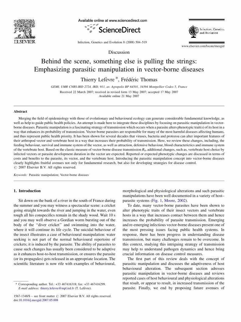
www.elsevier.com/locate/meegid
Infection, Genetics and Evolution 8 (2008) 504–519
Discussion
Behind the scene, something else is pulling the strings:
Emphasizing parasitic manipulation in vector-borne diseases
Thierry Lefevre *, Frederic Thomas
GEMI, UMR CNRS-IRD 2724, IRD, 911, av. Agropolis BP 64501, 34394 Montpellier Cedex 5, France
Received 22 March 2007; received in revised form 13 May 2007; accepted 17 May 2007
Available online 21 May 2007
Abstract
Merging the field of epidemiology with those of evolutionary and behavioural ecology can generate considerable fundamental knowledge, as
well as help to guide public health policies. An attempt is made here to integrate these disciplines by focusing on parasitic manipulation in vector-
borne diseases. Parasitic manipulation is a fascinating strategy of transmission which occurs when a parasite alters phenotypic trait(s) of its host in a
way that enhances its probability of transmission. Vector-borne parasites are responsible for many of the most harmful diseases affecting humans,
and thus represent public health priority. It has been shown for several decades that viruses, bacteria and protozoa can alter important features of
their arthropod vector and vertebrate host in a way that increases their probability of transmission. Here, we review these changes, including, the
feeding behaviour, survival and immune system of the vector, as well as attraction, defensive behaviour, blood characterictics and immune system
of the vertebrate host. Based on the classic measure of vector-borne disease transmission R0, additional changes, such as, vertebrate host choice by
infected vectors or parasite development duration in the vector are expected. Reported or expected phenotypic changes are discussed in terms of
costs and benefits to the parasite, its vector, and the vertebrate host. Introducing the parasitic manipulation concept into vector-borne diseases
clearly highlights fruitful avenues not only for fundamental research, but also for developing strategies for disease control.
# 2007 Elsevier B.V. All rights reserved.
Keywords: Parasitic manipulation; Vector-borne diseases
1. Introduction
Sit down on the bank of a river in the south of France during
the summer and you may witness a spectacular scene: a cricket
going straight towards the river and jumping in the water, even
tough all his conspecifics remain in the shady wood. Wait 10 s
and you may well observe a Gordian worm bursting out of the
body of the ‘‘diver cricket’’ and swimming into the water,
where it will continue its life cycle. The suicidal behaviour of
the insect illustrates a case of behavioural manipulation: water
seeking is not part of the normal behavioural repertoire of
crickets; it is induced by the parasite. The ability of parasites to
cause such changes has usually been considered to be adaptive
as it enhances host-to-host transmission, or ensures the parasite
(or its propagules) gets released in an appropriate location. The
scientific literature is now rife with examples of behavioural,
* Corresponding author. Tel.: +33 467416318; fax: +33 467416299.
E-mail address: [email protected] (T. Lefevre).
1567-1348/$ – see front matter # 2007 Elsevier B.V. All rights reserved.
doi:10.1016/j.meegid.2007.05.008
morphological and physiological alterations and such parasitic
manipulations have been well documented in a variety of host–
parasite systems (Fig. 1, Moore, 2002).
To date, many vector-borne parasites have been shown to
alter phenotypic traits of their insect vectors and vertebrate
hosts in a way that increases contact between them and hence
increases the probability of parasite transmission. Emerging
and re-emerging infectious vector-borne diseases present one of
the most pressing issues facing public health systems. In
response, there has been progress in understanding disease
transmission, but many challenges remain to be overcome. In
this context, studying this intriguing strategy of transmission
may help to understand pathogen dynamics and hence bring
crucial information on disease control measures.
The first part of this review deals with the concept of
parasitic manipulation and discusses the adaptiveness of host
behavioural alteration. The subsequent section adresses
parasitic manipulation in vector-borne diseases and reviews
reported cases of host behavioural and physiological alterations
that result, or appear to result, in increased transmission of the
parasite. Finally, we end by proposing future avenues of
Fig. 1. Examples of parasitic manipulation. (a) The Gordian worm (Paragordius tricuspidatus) exiting the body of a cricket (Nemobius sylvestris) (Photo P.
Goetgheluck). (b) A cockle (Austrovenus stutchburyi) parasitized by a trematode (Curtuteria australis). The parasites impair the natural burrowing behaviour of their
cockle intermediate hosts, making them more likely to be eaten by aquatic bird (final host) (Photo F. Thomas). (c) Coral polyps infected with a trematode
(Podocotyloides stenometra). The parasite induces pink, swollen nodules on the coral colony and impairs their retraction ability. Infected polyps are, therefore, both
conspicuous and vulnerable to predation by the coral-feeding butterflyfish, Chaetodon multicinctus (parasite’s definitive host) (Photo G. Aeby). (d) The sporocysts of
Leucochloridium paradoxum develops in the snail’s tentacles, where they can be seen conspicuously pulsating. This make the tentacles look to a bird, definitive host,
like caterpillars (Photo P. Vogel). (e) Metacercariae of Microphallus papillorobustus encyst in the brain of the intermediate host, Gammarus insensibilis (Photo F.
Thomas). (f) Cystacanth of Polymorphus minutus encysts in the body cavity of Gammarus pulex (Photo F. Cezilly). Infected G. pulex and infected G. insensibilis show
similar behavioural changes (i.e. negative geotaxis, aberrant evasive behaviour and in infected G. insensibilis a positive phototaxis). Since both parasites are
phylogenetically distant, these changes can be considered as a case of convergence.
T. Lefevre, F. Thomas / Infection, Genetics and Evolution 8 (2008) 504–519 505
research in order for this topic to prosper in the emerging field
of evolutionary epidemiology.
2. The ‘spread’ of parasitic manipulation
The field of host manipulation by parasites acquired a
conceptual framework with the Richard Dawkins’ book entitled
‘The Extended Phenotype’ published in 1982. In this book,
Dawkins proposes a vision of life where the unit of natural
selection is the gene and where its influence can extend outside
its ‘vehicle’, i.e. the body in which the gene resides. For
instance, a bird’s nest is a phenotype like any other, under the
control of the bird’s genes in the same way as the shape of its
beak is under the control of its genes. This vision has helped
scientists to establish a framework for research on parasitic
manipulation of infected hosts. Henceforth, host alteration may
be regarded as the expression of the genes of the parasite in the
host phenotype. In this view, genes of the parasite are selected
for their effect on host phenotype. For example, in behavioural
manipulation, one could expect that the parasite secretes
chemicals (or induces their production in the host) acting on the
central nervous system of the host to induce altered behaviour.
Recently, it has been shown that the Gordian worm mentioned
earlier alters the normal functions of the grasshopper’s central
nervous system by producing certain ‘‘effective’’ molecules. In
particular, a protein from the Wnt family was found that act
directly on the development of the central nervous system.
These proteins show important similarities with those known in
insects, suggesting a case of molecular mimicry (Biron et al.,
2005).
Dawkins’ theoretical explanation, namely, that host beha-
vioural changes can be parasite adaptations, has led researchers
to consider all behavioural changes observed in an infected
organism as beneficial for the parasite. However, other
explanations are possible. First it has been proposed that the
host may benefit from these changes. For instance, suicidal
behaviour may reduce the risk of infection for the host’s kin.
Similarly, behavioural fever and elevated body temperature
may help the host to eliminate its parasite (Hart, 1988) in
raising its body temperature. Alternatively, changes might be
pathological consequences of infection, adaptive to neither host
nor parasite, and that not all behavioural modifications
T. Lefevre, F. Thomas / Infection, Genetics and Evolution 8 (2008) 504–519506
following infection are mediated by parasites. Indeed, not all
behavioural modifications have evolved to increase the
reproductive success of the parasite.
Since it is difficult to distinguish between these different
possibilities, four criteria have been proposed by Poulin (1995) to
consider changes as adaptive in the context of transmission:
complexity; purposiveness of design (i.e. conformity between a
priori design and the alterations); convergence (similar changes
in several independent lineages) and fitness consequences. This
important paper has helped to highlight the need for a novel and
more rigorous approach to studying and interpreting potential
cases of parasitic manipulation. The first criterion, i.e. complex-
ity, is perhaps the least useful since it is difficult to judge if a trait
is complex or not. Simple host behavioural changes may indeed
rely on complex physiological modifications induced by the
parasite. Natural selection does not always favour complex
solutions over simple ones; it favours the solutions that work.
Concerning the second criterion (i.e. purposiveness of design),
the host behavioural changes are indeed often exactly those we
could expect to enhance parasite transmission. This aspect is well
illustrated when looking at different types of parasite life cycles.
First, in direct life cycles, host phenotypic alterations leading to
increased contacts among conspecifics are likely to be selected.
For instance, the rabies virus, Hantavirus, and borna disease virus
induce increased aggression and physical contacts in the host
population. Second, in complex life cycles involving trophic
transmission, many parasites alter the behaviour of their
intermediate host in a way that increases the risk of them being
preyed upon by final hosts, by, for example, changing
intermediate host microhabitat or inhibiting anti-predatory
behaviour. The third criterion proposed to assess the adaptiveness
of behavioural alteration is convergence. When a similar
phenotypic trait in several organisms has not been inherited
from their most recent common ancestor but conversely has
independently evolved, the trait is said to be a homoplasy or a
convergence. A convergent trait is likely to be an adaptation since
it results from the same selective pressures acting on
phylogenetically distant organisms. For instance, three species
of parasites (i.e. two acanthocephalans, Polymorphus minutus
and P. paradoxus and a trematode, Microphalus papillorobustus)
are known to induce in their crustacean gammarids (intermediate
hosts) similar aberrant evasive behaviour making them more
prone to be eaten by aquatic birds (definitive hosts) (Bethel and
Holmes, 1973; Helluy, 1984; Cezilly et al., 2000). As parasitism
has independently evolved in Trematoda and Acanthocephala,
the induced aberrant evasive behaviour can be seen as convergent
(Fig. 1). Conversely, between the two acanthocephalan species it
is likely that the ability to induce this aberrant evasive behaviour
was inherited from the common ancestor; it is thus less likely to
be convergence than in the latter case. The last criterion
proposed, undoubtedly the most convincing evidence in favour
of adaptation, is the demonstration of fitness benefits. For several
decades researchers have attempted to show that behavioural
modifications result in more successful transmission. Linking
behavioural modification with increased probability of transmis-
sion is difficult, that is why the case of trophic transmission by
intermediate hosts is surely the easiest to investigate. The first
study considering this conjecture was that carried out by Holmes
and Bethel (1972). In this study, a predator (definitive host) was
experimentally offered known numbers of infected and
uninfected gammarids. At the end of the experiment, numbers
of infected and uninfected individuals that survived were
counted. Holmes and Bethel showed that the behavioural
alterations induced by the parasite lead to a significantly
increased predation rate of infected gammarids. Since results
obtained in the laboratory may not reflect precisely that which
occurs naturally, a second method has been devised by
researchers in attempt to show that behavioural alteration
actually increases transmission rates. In this approach, the
prevalence of infected individuals in the intermediate host
population is compared to the prevalence of infected individuals
in the final host’s diet. An over-representation of infected preys in
the gut contents of an appropriate definitive host is considered as
evidence for an increased rate of transmission. For instance,
Moore (1983) showed that terrestrial isopods parasitized by an
acanthocephalan were more active compared to uninfected ones,
and were consequently more likely to be found in the diet of the
definitive host, the starling.
The publication of Poulin’s critical review (1995) has
marked the start of a new period during which many studies
taking into account the previous recommendations appeared in
the scientific literature. This paper has nonetheless contains one
obscure point: should we consider the changes due to
pathological consequences of infection and that are coinciden-
tally beneficial for the parasite as adaptations? Poulin answers
no: ‘‘Experiments can also serve to distinguish between
behavioural modifications that are truly adaptive and those that
are coincidentally beneficial. For instance, parasitized hosts can
make themselves more visible to predators because they have
increased energy requirements and must forage more, not
because they are manipulated by the parasite’’. In this case,
even if the link between behavioural alterations and increased
rate of transmission is demonstrated, we cannot consider the
changes as adaptive. Another situation illustrating this
restriction corresponds to what is called ‘‘fortuitous payoff
of other adaptations’’. For example, the eye fluke trematode
inducing blindness in his intermediate host fish could have been
selected to encyst in the eye of the fish to avoid the host immune
system, affording later the ability to alter the host evasive
behaviour. In this case, the manipulation can be regarded as the
consequence of traits that have evolved for other purposes. This
definition clearly looks like the definition of exaptation sensu
Gould and Vrba (1982): an exaptation is a trait that evolved for
other functions, or no function at all, but which has been co-
opted for a new use. Concerning this point, it is also possible
that more than one advantage to the eye fluke trematode could
immediately occur from encysting in the eyes and that selection
pressures for this phenotype will be much stronger if several
benefits ensue (i.e. increased transmission and immune system
avoidance). Unfortunately, we often take a simplistic approach
by only looking at a single consequence of an action at a time.
As for the confusion between these ‘‘by-products’’ (i.e.
changes coincidentally beneficial and fortuitous payoff) and
adaptation, a review written by Reeve and Sherman (1993)
Fig. 2. Two evolutionary sequences of a trait illustrating the confusion con-
cerning the label ‘‘exaptation’’. (modified after Reeve and Sherman, 1993).
T. Lefevre, F. Thomas / Infection, Genetics and Evolution 8 (2008) 504–519 507
helps to clarify the issue. This paper deals with the slippery
concept of adaptation. Reeve and Sherman stressed the fact that
it is dangerous to confuse the product of the selective process
and the process itself. For instance, exaptation and adaptation
are actually similar, because both refer to traits that exist
because they have been, and continue to be favoured over
alternative traits. Do host behavioural modifications result from
energy depletion, from the consequences of another adaptation,
or from a true manipulation by parasites? Let us imagine two
evolutionary sequences for a trait (Fig. 2). In the first sequence,
the present trait with function A would be interpreted as an
adaptation, i.e. as a trait selected for the purpose it has served in
the past and continues to serve. In the second sequence, a trait
having a new function A’ would be an adaptation if we examine
the trait between t1 and the present time, but it would be
considered as an exaptation if we examine the trait between t0and t1. Thus, since the label ‘‘exaptation’’ depends on the point
in evolutionary history at which we investigate the trait’s
function, this concept generates ambiguity, rather than
clarification. Moreover, concerning our trait of interest, that
is, host behavioural modification, it is virtually impossible to
identify its original role (no fossil records). To circumvent these
problems, Reeve and Sherman (1993) proposed an operational,
non-historical definition of adaptation, particularly ‘‘adapted’’
to behavioural ecologists interested in questions of phenotype
existence in the field of parasitic manipulation: ‘‘an adaptation
is a phenotypic variant that results in the highest fitness among
specified set of variants in a given environment’’. Considering
this definition, all hosts’ behavioural modifications that lead to
an enhancement of parasite transmission are maintained by
natural selection and thus are a parasite adaptation. To return to
the ‘‘by product’’ hypothesis, being more visible to predators
because of an increased energy requirement would be an
adaptation under this definition if it increases transmission
towards a definitive host and the parasite’s reproductive
success, when compared to an infected host no more visible to
predators. For the moment no studies provide information
concerning the link between variability in manipulative effort
and an increased rate of transmission (and subsequently the
parasite’s reproductive success), but one could expect that, all
else being equal, a parasite that manipulates more than other
parasites increases the probability of being transmitted. It could
be possible to experimentally select over several generations
the phenotypes of parasites with high, medium and low
manipulative effort and to examine the rates of transmission
between the selected phenotypes.
But why does the host not resist and why does it act in a way
that favours the parasite? Behavioural modification must be
seen as a co-evolved trait, i.e., a phenotype shared by the host
and his parasite. The host is under strong selective pressures to
resist manipulation and the parasite is under strong selective
pressure to manipulate. The changes might thus be interpreted
as an equilibrium state, nonetheless often biased toward the
parasite (e.g. in the case of trophic transmission). Under-
standing why the benefits are often balanced in favour of
parasites is easy when highlighted by the life—dinner principle
proposed by Dawkins and Krebs (1979). This principle states
that the rabbit runs faster than the fox because the rabbit is
running for his life, while the fox is only running for his dinner.
Thus, the consequences of failure are asymmetric for those
involved: one has more to lose than the other. We can apply this
principle to the case of parasitic manipulation (Barnard, 1990;
Poulin et al., 1994). Transmission is always a matter of life and
death for the parasite, while parasitic manipulation does not
necessarily have such drastic consequences for the host. The
host is descended from a long line of ancestors, only a
proportion of which ever met a manipulator parasite. Moreover,
some of the individuals subjected to parasitic manipulation may
have already reproduced and others may survive to reproduce
(depending on the type of host–parasite interaction). Host genes
coding for a lack of resistance can thus be transmitted to the
next generation. In contrast, every single parasite alive today is
descended from a long line of ancestors who have all negotiated
successfully the passage between two suitable successive hosts.
Therefore parasite genes coding for an effective transmission,
e.g. manipulation, have a greater chance to be passed on to the
next generations. This reasoning helps us to understand why the
host is more likely to capitulate. The stable compromise
resulting from this arms race is dynamic and depends on many
parameters, such as the prevalence of the parasite, the cost of
host resistance, the cost of being manipulated, the cost of
parasite manipulation and the cost of the parasite’s failure.
3. Parasitic manipulation and vector-borne diseases
Haematophageous insects, when feeding on their host can
transmit numerous blood pathogens. An increasing number of
studies demonstrate, or suggest, that vector-borne parasites are
able to manipulate several phenotypic traits of their vertebrate
hosts and vectors in ways that render parasite transmission
more probable (Molyneux and Jefferies, 1986; Moore, 1993,
2002; Hurd, 2003; Lacroix et al., 2005). The basic case
reproduction number, R0, the fundamental and classic measure
of infectious disease transmission (Macdonald, 1957) can help
establish the framework for research on parasitic manipulation
in vector-borne diseases. R0 describes the average number of
new infections arising from a single current infection, for
vector-borne disease: R0 = ma2pn/�r ln p (Dye, 1992; Garrett-
Jones and Shidrawi, 1969).
Thus, the parasite transmission depends on the number of
insect vectors per individual vertebrate host (m), the daily biting
rate of an individual vector on vertebrate hosts (a) (once to
become infected, and once to transmit, hence a2), the daily
survival rate ( p), the number of days required for the parasite to
develop (n, i.e. the extrinsic latent period), and r the daily
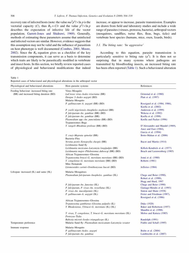
T. Lefevre, F. Thomas / Infection, Genetics and Evolution 8 (2008) 504–519508
recovery rate of infected hosts (note: the value ma2pn/�ln p is the
vectorial capacity (C), thus R0 = C/r and the value pn/�ln p
describes the expectation of infective life of the vector
population; Garrett-Jones and Shidrawi, 1969). Generally,
methods of estimating these parameters assume that uninfected
and infected vectors are similar. However, evidence suggests that
this assumption may not be valid and the influence of parasitism
on host phenotype is well documented (Combes, 2001; Moore,
2002). Since the R0 equation gives us a checklist of the key
transmission components, it can serve as a basis to determine
which traits are likely to be parasitically modified in vertebrate
and insect hosts. In this section, we briefly review reported cases
of physiological and behavioural modifications that indeed
Table 1
Reported cases of behavioural and physiological alterations in the arthropod vecto
Physiological and behavioural alterations Host–parasite systems
Feeding behaviour: increased biting rate
(BR) and increased biting duration (BD)
Virus–Mosquito
LaCrosse virus–Aedes triseria
Dengue 3–Aedes aegypti (BD
Malaria–Mosquito
P. gallinaceum–A. aegypti (B
P. yoelii nigeriensis–Anophel
P. falciparum–An. gambiae (B
P. falciparum–An. gambiae (B
Plasmodium spp.–An. punctu
Trypanosomes–Bugs
T. rangeli–Rhodnius prolixus
T. cruzi–Mepraia spinolai (B
Bacteria–Flea
Yersina pestis–Xenopsylla che
Leishmania–Sand fly
Leishmania mexicana–Lutzom
Leishmania major–Phlebotom
African Trypanosomes–Gloss
Trypanosoma brucei–G. mors
T. congolense–G. morsitans m
Mite–Nematode
Litomosoides carinii–Ornitho
Lifespan: increased (IL) and same (SL) Malaria–Mosquitoes
Plasmodium falciparum–Ano
P. falciparum–An. funestus (S
P. falciparum, P. vivax–An. te
P. vivax–An. maculipennis (S
P. gallinaceum–A. aegypti (S
African Trypanosomes–Gloss
Trypanosoma gambiense–Glo
T. Rhodesiense, T.brucei–G. m
T. vivax, T. congolense, T. br
Protozoa–Ticks
Babesia microti–Ixodes triang
Temperature preference Malaria–Sand fly: Plasmodiu
Immune response Malaria–Mosquito
P. gallinaceum–Aedes. aegyp
P. falciparum–An. gambiae
increase, or appear to increase, parasite transmission. Examples
are drawn from field and laboratory studies and include a wide
range of parasites (viruses, protozoa, bacteria), arthropod vectors
(mosquitoes, sandflies, tsetse flies, fleas, bugs, ticks) and
vertebrate host species (humans, mice, oxen, lizards, birds).
3.1. The biting rate: ‘be aggressive’
According to this equation, parasite transmission is
particularly sensitive to biting rate (a2). It is then not so
surprising that in many systems where pathogens are
transmitted by bloodfeeding insects, an increased biting rate
has been often reported (Table 1). Such a behavioural alteration
r
References
tus (BR) Grimstad et al. (1980)
) Platt et al. (1997)
R) (BD) Rossignol et al. (1984, 1986),
Koella et al. (2002)
es stephensi (BR) Anderson et al. (1999)
R) (BD) Wekesa et al. (1992)
R) Koella et al. (1998)
latus (BR) (BD) Koella and Packer (1996)
(BR) (BD) D’Alessandro and Mandel (1969),
Anez and East (1984),
Garcia et al. (1994)
R) Botto-Mahan et al. (2006)
opis (BR) Bacot and Martin (1914)
yia longipalpis (BR) Killick-Kendrick et al. (1977)
us duboscqi (BR) (BD) Beach and Leeuwenberg (1985)
ina
itans morsitans (BR) (BD) Jenni et al. (1980)
orsitans (BR) (BD) Roberts (1981)
nyssus bacoti (BD) Jefferies (1984)
pheles. gambiae (SL) Chege and Beier (1990),
Robert et al. (1990),
Hogg and Hurd, 1997
L) Chege and Beier (1990)
ssellatus (SL) Gamage-Mendis et al. (1993)
L) Sinton and Shute (1938)
L) Freier and Friedman (1987),
Rossignol et al. (1986)
ina
ssina palpalis (IL) Duke (1928)
orsitans (IL) (SL) Baker and Robertson (1957)
Maudlin et al. (1998)
ucei–G. morsitans morsitans (SL) Moloo and Kutuza (1985)
uliceps (IL) Randolph (1991)
m mexicanum–Lutzomyia vexator Fiahlo and Schall (1995)
ti Boete et al. (2004)
Lambrechts et al. (2007)
T. Lefevre, F. Thomas / Infection, Genetics and Evolution 8 (2008) 504–519 509
has indeed been shown for tsetse flies infected with African
trypanosomes, in bugs infected with Trypanosoma spp., in
sandflies infected with Leishmania spp., in fleas infected with
the plague bacterium, in mosquitoes infected with Plasmodium
spp. and viruses (Molyneux and Jefferies, 1986; Moore, 2002;
Hurd, 2003). Interestingly, in almost all cases, the infective
stages of the parasites appear to interfere with the ingestion
process of the insects by, for example, obscuring phagorecep-
tors (e.g. trypanosome-infected tsetse flies), blocking the
foregut (e.g. leishmania-infected sandflies) and reducing
apyrase activity in salivary glands (e.g. malaria-infected
mosquitoes). These different mechanisms seem to impair the
vector’s ability to fully engorge and therefore induce them to
bite vertebrate hosts several times.
An increased biting rate was first observed by Bacot and
Martin (1914) in fleas parasitized with the plague bacillus,
Yersina pestis. In this system, infected fleas probe more often
and sometimes without ingesting blood (Bibikova, 1977). The
bacteria multiply in the midgut and proventriculus causing
occlusion of the proventriculus and blockage of the gut. The
block prevents the flow of host blood from the foregut into the
midgut. As a consequence, to obtain a blood meal, a blocked
flea regurgitates the plug and injects it into the host
(Hinnebusch et al., 1998).
Infected sandflies have also difficulty in feeding and hence
will often bite a host several times before a blood meal is taken
(Table 1, Killick-Kendrick et al., 1977; Beach and Leeuwen-
berg, 1985). In this system, the parasite produces a gel-like
plug, the promastigote secretory gel (PSG) that blocks the
foregut (Rogers et al., 2002; see also Volf et al., 2004). Rogers
et al. (2004) demonstrated that the parasite, Leishmania
mexicana accumulates behind the plug and are actively
regurgitated during biting. An average of 1086 parasites was
egested per bite, and 86–98% of them were infective metacyclic
promastigotes (Rogers et al., 2004). In addition, this study
sheds light on an important structural component of the PSG,
the filamentous proteophosphoglycan (fPPG), which beyond its
role in blocking the foregut, actively participates in the success
of vertebrate host infection. First, by comparing infections
caused by a single fly bite with those caused by a syringe
inoculation, Rogers et al. (2004) found that an exacerbation
factor, facilitating mice infections, was egested along with the
plug containing metacyclic promastigotes. Then, they demon-
strated that this factor was of parasite origin, and finally that
egested fPPG was responsible for this infection exacerbation.
In tsetse flies parasitized with trypanosomes, Jenni et al.
(1980) showed that Glossina morsitans morsitans and G. austeni
infected with Trypanosoma brucei probed three times more often
and fed more voraciously than uninfected flies. Working on G. m.
morsitans infected with T. congolense, Roberts (1981) reported
that infected flies probed significantly more frequently and took
longer to engorge than uninfected flies. The increased probing
may be caused by physical interference of the parasite with
phagoreceptors in the tsetse fly labrum (Thevenaz and Hecker,
1980; Livesey et al., 1980; Molyneux and Jenni, 1981). However,
such findings have not been confirmed by studies carried out by
Moloo’s group (Moloo, 1983; Moloo and Dar, 1985; Makumi
and Moloo, 1991). Combinations of vectors and parasites from
different locations and/or parasite intensities may help explain
these contradictory results.
Malaria-infected mosquitoes bite their vertebrate hosts
longer (Rossignol et al., 1984, 1986; Wekesa et al., 1992), more
often (Rossignol et al., 1986; Wekesa et al., 1992; Koella et al.,
1998, 2002, but see Li et al., 1992) and are more persistent in
seeking out blood meals (Koella and Packer, 1996; Anderson
et al., 1999) (Table 1). These three aspects of feeding appear to
be crucial for parasite transmission since it arouses vertebrate
host defensive behaviour, leading to interrupted feeding, which
in turn, could multiply host contacts per gonotrophic cycle
(Rossignol et al., 1986; Wekesa et al., 1992). Disruption of
feeding activity, and hence multiple biting, occur in this system
probably as a result of altered apyrase activity. Apyrase is a
salivary enzyme that inhibits platelet aggregation, facilitates
blood vessel location and thus promotes blood-feeding by the
insect (Ribeiro et al., 1984). For instance, in malaria-infected
Aedes aegypti, apyrase activity is reduced to a third following
maturation of sporozoites (i.e. the mature transmissible stage of
Plasmodium spp.) and results in a longer biting period
(Rossignol et al., 1984, see also Ribeiro et al., 1985).
Triatomines infected with Trypanosoma spp. also experi-
ence difficulties in engorging (Tobie, 1965; D’Alessandro and
Mandel, 1969; Schaub, 1989; Garcia et al., 1994). It has been
shown that the biting rate of Rhodnius prolixus and R. robustus
was increased by infection with Trypanosoma rangeli
(D’Alessandro and Mandel, 1969; Anez and East, 1984;
Garcia et al., 1994) and that it probably results from reduced
apyrase activity (Garcia et al., 1994; Azambuja and Garcia,
2005). In this system, it has been also proposed that the sluggish
movements observed in infected bugs might be parasitic
manipulation of locomotory activity aiming at facilitate the
parasite transmission by predation and/or cannibalism (Schaub,
2006). Recently, Botto-Mahan et al. (2006) demonstrated that
Trypanosoma cruzi, transmitted through defecation and
responsible for Chagas disease, manipulates the behaviour of
the kissing bug vector, Mepraia spinolai, in two ways. First, it
induces an increased biting rate and second it reduces the
defecation time after feeding. As a consequence, parasite
transmission may be enhanced because more bites induce more
wounds for parasite contamination from faeces deposited
shortly afterwards (Botto-Mahan et al., 2006).
Reduced feeding success, increased biting rates and their
duration have also been reported in Aedes triseriatus infected
with La Crosse virus (Grimstad et al., 1980) and in A. aegypti
infected with dengue virus (Platt et al., 1997, but see Putnam
and Scott, 1995). Although no mechanism has been demon-
strated, Grimstad et al. (1980) and Platt et al. (1997) suggested
that infection of the salivary gland coupled with heavy infection
of the nervous system, eyes and abdominal ganglion (all
involved in host seeking and blood feeding processes) may
provide proximal explanations of altered feeding behaviour in
virus-infected mosquitoes.
Following the examples of feeding alterations described
above, two striking features must be emphasized. First,
behavioural changes often occur with precise timing. Altered
T. Lefevre, F. Thomas / Infection, Genetics and Evolution 8 (2008) 504–519510
feeding behaviour indeed often takes place only when parasites
are fully developed, i.e. once the parasites reach the infective
stage for the next host. For instance, Koella et al. (2002) (see
also Anderson et al., 1999; but see Ferguson and Read, 2004)
showed that P. gallinaceum manipulates the mosquito vector, A.
aegypti, in two different ways and in a stage-specific manner:
when ready to be transmitted to the vertebrate hosts (sporozoite
stage), the parasite increases the biting rate of his vector. In
contrast, at an earlier developmental stage (oocyst) which is not
transmissible to the vertebrate hosts, the parasite decreases the
contact between vector and vertebrate hosts, by decreasing the
natural host seeking behaviour of the insect. Since biting is
risky and could lead to the death of the insect, this change seems
to be beneficial for the parasite. Beach and Leeuwenberg (1985)
also observed that uninfected sandflies and sandflies with
immature infections probe only once or twice and are fully
engorged within 10 min, whereas mature-infected flies probe
more often and never result in complete engorgement.
Second, it appears that vector-borne parasites have evolved
slightly different mechanisms (i.e. blocking the foregut,
reducing apyrase, obscuring phagoreceptors) aimed at inducing
similar feeding alterations (i.e. interference in ingestion process
leading to new feeding attempts). Poulin (1995) (see also
Thomas and Poulin, 1998) pointed out that convergence in
manipulative processes is a likely scenario when host
alterations bare on the same function, but are derived from
different proximal mechanisms. Both the timing of feeding
alteration and its independent evolution in phylogenetically
distant organisms (i.e. convergence) are strong arguments for
parasitic manipulation. Unfortunately empirical evidence in
support of increased transmission induced by increased biting
rate is still lacking.
3.2. Longevity and survival rate: ‘have a long life’
The R0 equation describes that the longer vectors live, the
more the parasite can be transmitted. Life history theory
suggests that a reduction or suppression of reproductive effort
will result in increased lifespan (Stearns, 1992). Thus, it has
been suggested that vector-borne parasites should be able to
manipulate resource allocation of their insect vectors in a way
that changes the optimum trade-off between reproduction and
longevity, which in turn, could favour longer vector survivor-
ship and hence the parasite’s overall transmission (Hamilton
and Hurd, 2002). Globally, studies on the effects of infection on
survival reported no significant lifespan differences between
infected and uninfected vectors. However, studies on the effects
of infection on fecundity (i.e. number of produced gametes)
often report a reduction in fecundity. Since parasites use vector
resources to develop and reproduce, it has been suggested that
the observed fecundity reduction in infected vectors does not
induce a longer lifespan but the same lifespan as uninfected
insects (Hamilton and Hurd, 2002).
Parasite-induced fecundity reduction has been reported in
many insect vector—parasite associations (Hurd et al., 1995;
Hurd, 2003). The association between malaria parasites and
mosquitoes provides the most reported case of parasite-induced
fecundity reduction. Potential proximate causes for this
phenomenon in malaria have been extensively reviewed in
Hurd (2001, 2003) and will not be developed here.
Whereas parasite-induced fecundity reduction of insect
hosts seems to be a widespread phenomenon, lifespan
comparisons of infected and uninfected insect vectors have
provided much more conflicting results. Some studies found
reduced vector lifespan, while others showed no effect of
infection, and even increased lifespan (Table 1). For instance,
Ferguson and Read (2002) used a meta-analysis on several
published laboratory studies to demonstrate that overall,
malaria does reduce mosquito survival suggesting that no such
manipulation is occurring in this system. However, they also
showed that morbidity effects are more likely to be found in
unnatural vector–parasite combinations and in studies of longer
duration. As a consequence, when considering studies carried
out on natural associations, no effect of infection on longevity is
found. Concerning tsetse flies, sandflies and bugs, the picture
that emerges is also far from conclusive. For instance, greater
longevity of trypanosome-infected tsetse flies has been reported
by Duke (1928) and Baker and Robertson (1957). In contrast,
Moloo and Kutuza (1985) found no difference in longevity and
fecundity between infected and uninfected individuals, while
Makumi and Moloo (1991) reported a higher longevity for
tsetse males but the reverse for females, and no effect of
infection on fecundity. Comparison of the survival distributions
of uninfected glossina with those exposed to infection with
either Trypanosoma congolense or T. brucei showed that the
first significantly reduced glossina survival, while the second
had little or no effect on the survival (Maudlin et al., 1998). In
the sandflies, Phlebotomus papatasi and P. langeroni, infection
with Leishmania major and L. infantum results not only in a
significant reduction in fecundity but also in longevity (El
Sawaf et al., 1994). Such findings are consistent with those
reported for Rocky Mountain wood ticks, Dermacentor
andersoni, when infected with Rickettsia rickettsii, the
causative agent of Rocky Mountain spotted fever (Niebylski
et al., 1999). Reduced survival has also been found for ticks
infected with several protozoa (e.g. Gray, 1982; Watt and
Walker, 2000; but see Randolph, 1991), in mosquitoes infected
with several viruses (e.g. Faran et al., 1987; McGaw et al.,
1998; Scott and Lorenz, 1998), and finally in mosquitoes
infected with filariasis (Krishnamoorthy et al., 2004).
As conditions in the laboratory may not reflect precisely
those which occur in the field, the influence of parasitism on
survival and longevity has not yet been well addressed. Insect
vectors are often naturally exposed to adverse conditions which
are rarely considered in laboratory studies, and might act
synergistically with parasitism. In addition, one might assume
that the optimal survival rate of the insect may differ between
parasite developmental stages: immature stages are expected to
increase the vector’s survival to increase their chance of
becoming mature, while mature stages are potentially
confronted with a trade-off between increasing the vector’s
lifespan and thus survival rate and increasing biting rate (see
Section 3.1) that may decrease the survival rate since biting is
risky in natura (Koella, 1999; Schwartz and Koella, 2001). For
T. Lefevre, F. Thomas / Infection, Genetics and Evolution 8 (2008) 504–519 511
instance, it has been shown under natural conditions that
sporozoite-infected mosquitoes have higher feeding associated
mortality than those without sporozoites (Anderson et al.,
2000). This finding suggests that malaria parasites might harm
their mosquito host more than usually thought.
Unfortunately, many studies exploring the influence of
parasitism have concentrated only on either vector reproduction
or vector survival/longevity. As a consequence, studies on the
links between infection, vector reproduction and vector
longevity/survival are still rare (but see Makumi and Moloo,
1991; Hogg and Hurd, 1997) and clearly more investigation is
needed to fully explore this interesting hypothesis. In addition,
a better understanding of the effects of infection on longevity
and fecundity will be achieved after more investigations of
naturally infected vectors.
3.3. The parasite development duration: ‘become infective
early’
In vector-borne parasites, one might expect that natural
selection will favour an optimal developmental schedule for
each parasite stage that ensures transmission between
successive hosts. Once in the insect vector, a major challenge
facing the parasite is to reach its infective stage before the insect
takes its last blood meal and dies. This idea is particularly valid
in systems involving short-lived vectors, such as, mosquitoes
and sandflies. In this context, shortening the duration of
development should permit the parasite to become infective
earlier and hence to increase its probability of transmission. So,
why is this period so long? In many vector-borne pathogens,
this period is indeed as long as their insect vector’s average
lifespan. For instance, estimates for the probability that a
‘newly’ infected mosquito will live long enough to transmit
Plasmodium spp. range from 80% (Macdonald, 1956) to less
than 10% (e.g. Rodriguez et al., 1992). In fact, this duration
probably results from constraints and/or trade-offs aimed at
increasing the overall parasite transmission.
In malaria–mosquito systems, the vector injects a very
small number of sporozoites (i.e. 10–20) into the host
compared to the huge quantity available in the salivary gland
(Paul et al., 2003). Koella (1999) thus asked: ‘‘if only a few
(sporozoites) are necessary, should evolutionary pressures not
lead the parasite to produce the sporozoites earlier, if doing so
would increase the likelihood that mosquito survives the
developmental period?’’ At a first glance, we can suggest that
it results from a constraint, that is, a large number of
sporozoites are needed in salivary glands to enable the
injection of only a few. However, it has been demonstrated
that this large number is neither correlated to the number of
those injected nor to the probability of successfully infecting
the host (Ponnudurai et al., 1991; Beier et al., 1991). As a
consequence, knowing that the vector’s biting rate increases
with the sporozoite load (Koella et al., 1998), Koella (1999)
proposed a model to investigate whether the developmental
pattern could be, in fact, a mechanism that increases the
overall parasite’s transmission. The model showed that if
mortality increases no more than linearly with biting rate,
maximal parasite transmission success can occur when all
sporozoites are released from the oocyst simultaneously.
Consequently, Koella (1999) concluded that the development
time of Plasmodium within its insect may be a mechanism for
manipulating vector-biting behaviour to increase its transmis-
sion. Atlhough interesting, further work is necessary to fully
clarify this point.
Another argument in favour of an adaptive development
period over constrained development period is the case of
Plasmodium mexicanum. In Central America, this parasite is
transmitted to the fence lizard, Sceloporus occidentalis, by the
sandfly, Lutzomyia vexator. Sandfly species have the particu-
larity of having very short lifespans; that is, only about 2% live
long enough to take a second blood meal (Fiahlo and Schall,
1995). As a consequence, compared to other Plasmodium
species, P. mexicanum has evolved toward rapid development
that ensures the matching with the high vector’s mortality. In
addition, Fiahlo and Schall (1995) showed that infected
sandflies were attracted by higher temperatures compared to
uninfected counterparts. It turned out that this higher
temperature is optimal for the rapid development of the
parasite, but sub-optimal for the development of sandfly eggs,
suggesting a parasitic manipulation of host temperature
preference (Table 1).
It is well known that the development rate of vector-borne
parasites is sensitive to temperature (e.g. Garnham, 1964).
However studies like the one performed by Fiahlo and Schall
(1995) are scarce. Extending such investigations to other insect
vector–parasite systems, particularly those affecting human
welfare, is promising and could shed light on new insights into
parasitic manipulation of vector-borne diseases.
3.4. The recovery rate: ‘evading the immune response’
Escaping the immune system of both the vertebrate and
insect host is a sine qua non condition to the success of parasite
transmission. As a consequence, vector-borne parasites evolved
the ability to manipulate the immune response of their hosts
(Tables 1 and 2).
In human for instance, malaria parasites, Giardia and
African trypanosomes evolved an evasion mechanism known as
antigenic variation. These parasites have a large multigene
family of proteic variants and the evasion involves a sequential
dominance of antigenic variants. Any one-parasite individual
expresses only a single variant and only a few individuals
exhibit a new variant per generation. As a consequence,
parasites that exhibit the rare variant avoid the immune system
and those that exhibit the frequent variant are killed. This
enables the parasites to persist in the host. In addition, other
strategies have been developed by protozoan parasites (i.e.
malaria, African trypanosomes, T. cruzi and Leishmania) to
avoid and/or suppress both adaptive and innate immune
responses of their vertebrate host, such as eliminating their
protein coat, induction of blocking antibodies, molecular
mimicry, modulation of dendritic cells maturation and
alterations of memory T-cell, macrophages, and cytokines
functions (Zambrano-Villa et al., 2002; Sacks and Sher, 2002).
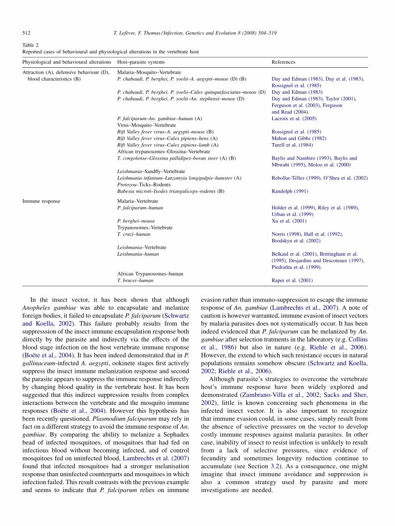
Table 2
Reported cases of behavioural and physiological alterations in the vertebrate host
Physiological and behavioural alterations Host–parasite systems References
Attraction (A), defensive behaviour (D),
blood characteristics (B)
Malaria–Mosquito–Vertebrate
P. chabaudi, P. berghei, P. yoelii–A. aegypti–mouse (D) (B) Day and Edman (1983), Day et al. (1983),
Rossignol et al. (1985)
P. chabaudi, P. berghei, P. yoelii–Culex quinquefasciatus–mouse (D) Day and Edman (1983)
P. chabaudi, P. berghei, P. yoelii–An. stephensi–mouse (D) Day and Edman (1983), Taylor (2001),
Ferguson et al. (2003), Ferguson
and Read (2004).
P. falciparum–An. gambiae–human (A) Lacroix et al. (2005)
Virus–Mosquito–Vertebrate
Rift Valley fever virus–A. aegypti–mouse (B) Rossignol et al. (1985)
Rift Valley fever virus–Culex pipiens–hens (A) Mahon and Gibbs (1982)
Rift Valley fever virus–Culex pipiens–lamb (A) Turell et al. (1984)
African trypanosomes–Glossina–Vertebrate
T. congolense–Glossina pallidipes–boran steer (A) (B) Baylis and Nambiro (1993), Baylis and
Mbwabi (1995), Moloo et al. (2000)
Leishmania–Sandfly–Vertebrate
Leishmania infantum–Lutzomyia longipalpis–hamster (A) Rebollar-Tellez (1999), O’Shea et al. (2002)
Protozoa–Ticks–Rodents
Babesia microti–Ixodes trianguliceps–rodents (B) Randolph (1991)
Immune response Malaria–Vertebrate
P. falciparum–human Holder et al. (1999), Riley et al. (1989),
Urban et al. (1999)
P. berghei–mouse Xu et al. (2001)
Trypanosomes–Vertebrate
T. cruzi–human Norris (1998), Hall et al. (1992),
Brodskyn et al. (2002)
Leishmania–Vertebrate
Leishmania–human Belkaid et al. (2001), Brittingham et al.
(1995), Desjardins and Descoteaux (1997),
Piedrafita et al. (1999).
African Trypanosomes–human
T. brucei–human Raper et al. (2001)
T. Lefevre, F. Thomas / Infection, Genetics and Evolution 8 (2008) 504–519512
In the insect vector, it has been shown that although
Anopheles gambiae was able to encapsulate and melanize
foreign bodies, it failed to encapsulate P. falciparum (Schwartz
and Koella, 2002). This failure probably results from the
suppresssion of the insect immune encapsulation response both
directly by the parasite and indirectly via the effects of the
blood stage infection on the host vertebrate immune response
(Boete et al., 2004). It has been indeed demonstrated that in P.
gallinaceum-infected A. aegypti, ookinete stages first actively
suppress the insect immune melanization response and second
the parasite appears to suppress the immune response indirectly
by changing blood quality in the vertebrate host. It has been
suggested that this indirect suppression results from complex
interactions between the vertebrate and the mosquito immune
responses (Boete et al., 2004). However this hypothesis has
been recently questioned. Plasmodium falciparum may rely in
fact on a different strategy to avoid the immune response of An.
gambiae. By comparing the ability to melanize a Sephadex
bead of infected mosquitoes, of mosquitoes that had fed on
infectious blood without becoming infected, and of control
mosquitoes fed on uninfected blood, Lambrechts et al. (2007)
found that infected mosquitoes had a stronger melanisation
response than uninfected counterparts and mosquitoes in which
infection failed. This result contrasts with the previous example
and seems to indicate that P. falciparum relies on immune
evasion rather than immuno-suppression to escape the immune
response of An. gambiae (Lambrechts et al., 2007). A note of
caution is however warranted, immune evasion of insect vectors
by malaria parasites does not systematically occur. It has been
indeed evidenced that P. falciparum can be melanized by An.
gambiae after selection tratments in the laboratory (e.g. Collins
et al., 1986) but also in nature (e.g. Riehle et al., 2006).
However, the extend to which such resistance occurs in natural
populations remains somehow obscure (Schwartz and Koella,
2002; Riehle et al., 2006).
Although parasite’s strategies to overcome the vertebrate
host’s immune response have been widely explored and
demonstrated (Zambrano-Villa et al., 2002; Sacks and Sher,
2002), little is known concerning such phenomena in the
infected insect vector. It is also important to recognize
that immune evasion could, in some cases, simply result from
the absence of selective pressures on the vector to develop
costly immune responses against malaria parasites. In other
case, inability of insect to resist infection is unlikely to result
from a lack of selective pressures, since evidence of
fecundity and sometimes longevity reduction continue to
accumulate (see Section 3.2). As a consequence, one might
imagine that insect immune avoidance and suppression is
also a common strategy used by parasite and more
investigations are needed.
T. Lefevre, F. Thomas / Infection, Genetics and Evolution 8 (2008) 504–519 513
3.5. The vertebrate host attraction: ‘be quiet and
rapturous’
Once the parasite has reached maturity in the vertebrate host,
any traits that could render its transmission from the vertebrate
to the insect more probable should have a selective advantage.
In this context, several malaria parasites, filarial worms and
trypanosomes have evolved synchronicity between the peak
number of infective stages and the peak biting time of their
vector species (Lehane, 2005). Another way to optimize
transmission would be for the parasite to respond positively to
the presence of vectors. Recently, it has been shown that
malaria parasites accelerate the growth of asexual stages and
the development of infective gametocytes (i.e. gametocytogen-
esis) when exposed to a high number of mosquitoes (Billingsley
et al., 2005). A last way to optimize transmission from
vertebrate to insect would be to induce a bias for vectors toward
hosts with mature infections. This can be achieved by
alterations of vertebrate host (i) attraction, (ii) behavioural
defences and (iii) blood characteristics (Moore, 2002).
Increased attractiveness of infected vertebrate host has been
reported in many (e.g. Mahon and Gibbs, 1982; Baylis and
Mbwabi, 1995; O’Shea et al., 2002; Ferguson et al., 2003;
Lacroix et al., 2005; Table 2) but not all studies (Freier and
Friedman, 1976; Burkot et al., 1989; Kruppa and Burchard,
1999). It has been suggested that fever and its symptoms (i.e.
increase in body temperature, in lactate production and in
hyperventilation) that accompany infections (e.g. arboviruses,
malaria) might increase attractiveness of infected individuals
(Turell et al., 1984; Nacher, 2005). However, in many cases, no
consistent differences in body temperature are found between
infected and uninfected individuals (e.g. Mahon and Gibbs,
1982; Day and Edman, 1984). Recently, through an elegant
semi-natural study, Lacroix et al. (2005) demonstrated that
people infected with transmissible stages of malaria appear to
produce something attractive to mosquitoes. First, they found
that mosquitoes are more attracted to humans harbouring the
transmissible gametocyte stages of the parasite than to
uninfected individuals and individuals harbouring non-trans-
missible asexual stages of the parasite. As the infection was
asymptomatic, a raise in body temperature could not explain
this differential attractiveness. Second, they showed that after
the clearance of the parasite, previously infected individuals
were no more attractive than other individuals. Therefore, the
observed increased attractiveness was not the consequence of
intrinsic attractiveness (and thus making these individuals
infected frequently) but rather the consequence of infection
with transmissible stages of the parasite.
At shorter distances, reducing host defensive behaviour is a
good way to facilitate the vector blood meal and hence the
transmission. In this context, Day et al. (1983) and Day and
Edman (1983) found that infection with Plasmodium chabaudi,
P. yoelli and P. berghei make mice less defensive and
consequently preferentially bitten and fed upon by mosquito
vectors. In addition, they showed that reduced defensive
behaviours coincided with peaks in gametocyte numbers and
consequently would result in increased transmission (see also
Turell et al., 1984). In this case, the general lethargy associated
with infection may be the cause of reduced vector-repellent
behaviour (Rossignol et al., 1985).
Hosts infected with malaria but also with other vector-borne
diseases (e.g. dengue, African trypanosomiasis, babesiosis,
etc.) often exhibit altered blood characteristics, including
anaemia (i.e. loss of erythrocytes), and thrombocytopenia (i.e.
loss of platelets). These modifications seem to reduce blood
viscosity that may facilitate blood ingestion, and also decrease
the vector’s mortality during the blood meal (Ribeiro et al.,
1985; Taylor and Hurd, 2001). For instance, Rossignol et al.
(1985) found that the time taken by mosquitoes to feed on mice
and hamsters experimentally infected with P. chabaudi or Rift
Valley fever was reduced by at least one minute. In the field,
tsetse flies feed more successfully on cattle infected with T.
congolense than on uninfected cattle (Baylis and Nambiro,
1993; Baylis and Mbwabi, 1995; Moloo et al., 2000). The
increased feeding success of tsetse flies may not be related to
changes in the level of anaemia, but rather because of
vasodilatation in infected cattles (Moloo et al., 2000).
3.6. The vertebrate host selection: ‘make the good choice’
Vector host choice is a very important key predictor for the
transmission intensity of vector-borne diseases. This choice
may be influenced by genetic and environmental factors such as
the innate host preference of the bloodsucking insect and the
host availability. Beside this aspect, vector-borne parasites
show some degree of vertebrate host specificity and one might
imagine that parasites acquired, during the course of evolution,
the ability to target appropriate host and/or avoid unsuitable
ones. In the context of parasitic manipulation, studying
specificity is one way to estimate the costs associated with
the changes and thus is one way to address the adaptiveness of
this strategy. The costs for a manipulative parasite can include
energetic expenses that are necessary to induce the changes and
also mortality associated with these changes. For example in
the case of trophically transmitted parasites, increased
susceptibility to predation by unsuitable hosts should be very
costly (e.g. Mouritsen and Poulin, 2003). In vector-borne
diseases, the ‘‘qualitative manipulation’’ hypothesis has been
recently proposed, according to which generalist blood-feeding
insects, once infected, would develop a feeding preference for
hosts targeted by the parasite (Lefevre et al., 2006). Alteration
of host choice could theoretically occur at inter- and/or intra-
specific level with infected vectors preferentialy feeding on
host species or on host individuals that are suitable for the
parasite.
First, maximising transmission towards a suitable host could
be achieved by parasites by inducing in the vector a sensory bias
for host traits that are correlated with optimal suitability for the
parasite. Second, the parasite may induce changes in the
generalist vector such as an alteration of microhabitat choice, in
order to spatially match the microhabitat of the suitable host.
Finally, the parasite may induce changes in the vector such as an
alteration of time activity in order to temporally match the time
rest or activity of the suitable host. These scenarios can be
T. Lefevre, F. Thomas / Infection, Genetics and Evolution 8 (2008) 504–519514
easily tested, by for example, sampling at distinct period and
comparing the proportion of infected vectors between samples.
However, if we observe a difference in habits such as
microhabitat choice, temporal activity and host choice between
infected and uninfected vectors, one might argue that infection
can be more a consequence than a cause of these habits. In other
words, an infected vector may prefer a host species that is
suitable for parasite not because of being infected but just
because of an innate or acquired preference for this host, and
thus making these vectors infected. To test between these two
alternatives, comparison of habits (e.g. host choice) between
uninfected vectors, vectors with mature infection and vectors
with immature infection could be performed in the field. For
instance, if vectors with mature infection do not exhibit the
same habits than uninfected vectors and those with immature
infection, then the observed habits are more likely to result
from a manipulative process rather than from innate or acquired
processes. In addition, comparisons between uninfected vectors
and experimentally infected ones could be also performed at
laboratory.
Qualitative manipulation deserves consideration and its
investigation would permit to adress the complexity and
multidimensionality in host manipulation by parasites. Indeed,
many studies concentrated on only one character whereas
manipulative parasites are known to modify more than a single
dimension in their host phenotype (Thomas et al., 2005; Cezilly
and Perrot-Minnot, 2005). Studies exploring the qualitative
manipulation hypothesis may yield considerable information
about the diversity of transmission strategies used by vector-
borne parasites and find, for instance, that insect vectors
infected with transmissible stages of the parasite bite more but
also bite better.
4. Conclusion and future directions
Taken together the examples presented above suggest that
many vector-borne parasites manipulate their hosts to increase
their probability of transmission. Unfortunately, we have little
evidence that such changes indeed affect transmission,
although it is reasonable to expect that it would (Moore,
2002). For example, evidence have been accumulating showing
that vector-borne parasites alter their vertebrate hosts to
improve the attraction and feeding success of their vectors (see
Section 3.5) and important epidemiological consequences of
such parasitic influences have been demonstrated by models of
vector-borne disease transmission (Dye and Hasibeder, 1986;
Kingsolver, 1987; Rossignol and Rossignol, 1988; Burkot,
1988; Randolph and Nuttall, 1994).
Whereas some authors consider these changes as examples
of parasitic manipulation (e.g. Lehane, 2005), others inter-
preted these as non-adaptive side effects of infection or
modifications that are coincidentally beneficial for the parasite
(e.g. Clements, 1999). As discussed in the first section of this
review, such notion emanates from Poulin (1995) and can
sometimes bring confusions that may lead to sentence such as:
‘‘All these potential benefits are a direct result of infection
pathology rather than any direct manipulation of the host.
Although it is feasible that increasingly virulent pathology is
selected for increased transmission to the insect vector, it is
more likely that these are simply side-effect, rather than
selected trait’’ (Paul et al., 2003). We fully agree that many of
these changes may result from indirect processes (see Thomas
et al., 2005 for discussion on indirect/direct mechanisms in
manipulative process), however if the trait that affects parasite
transmission has a genetic basis, and presents some variation,
one could expect that natural selection has not been blind, and
that consequently this trait has been selected over the course of
evolution. Could a side-effect not be a selected trait?
It has been also suggested that such changes might be
considered as examples of mutualism (Rossignol et al., 1985;
Clements, 1999): the vector benefits from the pathological
conditions induced by the parasites in the vertebrate host (e.g.
reduction of blood viscosity facilitating blood ingestion) while
the parasite gains from the choice of the vector. However, one
might argue that it is not clear whether this short-term benefit
for the vector would outweigh the long-term costs of becoming
parasitized. Indeed, it appears that parasites harm their vectors
in many ways (Lehane, 2005).
In the future, it will be useful to perfect the empirical and
theoretical study of parasitic manipulation in vector-borne
diseases to move from documenting the existence of parasitic
manipulation to asking how and why parasitic manipulation is
expressed in some instances and not others. For this we will
need a sharper quantification of the benefits and costs of
manipulating and being manipulated. Beyond potential
energetic expenses to induce phenotypic changes, costs for
a manipulative parasite can also include mortality associated to
these changes. For example, the balance between the
transmission benefit of an increased biting rate and the costs
of mortality associated with this behavioural change may
partly explain why this change is expressed in many but not all
cases. From the insect vector’s perspective, increased biting
rate is also very risky. It has been indeed shown that sporozoite-
infected mosquitoes have higher feeding associated mortality
than those without sporozoites (Anderson et al., 2000).
However theoretical analysis showed that the optimal biting
rate is higher for infective stages of malaria parasites than for
their mosquito vectors (Koella, 1999; Schwartz and Koella,
2001). Usually, selection to reduce the effects of manipulation
is expected, when the extent with which the parasite reduces its
own fitness by manipulating its host reaches a certain
threshold. Additional research is requiered to fill gaps in our
knowledge of the balance between costs and benefits of
parasitic manipulation.
As pointed out by Paul et al. (2003), the strategies used by
vector-borne parasites to optimize interaction with one host
may also be subject to selection for optimization in the other.
For instance, the ‘‘transmission enhancer’’ fPPG produced by
leishmania parasite (see Section 3.1) appears to participate in
both increased feeding rate of infected sandflies and the success
of infection within the host (Rogers et al., 2004). Recasting the
parasite in its two host-life cycle may clearly offer new research
avenues in manipulative process. For instance in malaria,
conflict of interest between immature stages (i.e. oocyst) and
T. Lefevre, F. Thomas / Infection, Genetics and Evolution 8 (2008) 504–519 515
mature ones (i.e. sporozoite) in the mosquito have been
suggested concerning the optimal biting rate of the vector
(Koella, 1999; Koella et al., 2002). Such conflicts may also
occur between different stages in the mosquito and in the
vertebrate host: when a mosquito infected with malaria feeds on
an infected vertebrate host, sporozoite stages within the
mosquito would prefer a higher blood viscosity than what is
optimal for gametocyte stages within the host. Indeed, infection
in the host may cause anemia to facilitate the insect bloodmeal,
whereas infection in the insect may cause apyrase reduction to
impair the insect bloodmeal (see Sections 3.1 and 3.5).
Recently, emphasis has been placed on the role of co-
occuring parasite species within manipulated hosts. What
should we expect if the same insect-vector harbours different
parasite species that may or may not have the same vertebrate
host? This within-host interaction creates opportunities for
either synergistic or conflicting interests between different
parasite species and has thus the potential to affect the result
of manipulation. In trophically transmitted parasites, several
scenarii have been proposed to illustrate this phenomenon and
empirical evidence begins to accumulate (Lafferty, 1999;
Thomas et al., 2005). For example, when different parasite
species share both vectors and vertebrate hosts, all parasites
may benefit from a higher transmission success if one of them
is a manipulative parasite. Non-manipulative parasites can be
simply ‘lucky passenger’ (in the vector and/or in the host)
when randomly associated with a manipulator. In trophic
transmission it has been shown that selection may even favour
non-manipulative parasites able to preferentially infect
intermediate hosts already manipulated, a strategy named
‘hitchhicking’. Mixed trypanosome infections in tsetse flies
seem to be common and hitchhicking has been hypothesized
to explain the non-random associations between the savannah
and riverine forest taxonomic groups of T. congolense (Solano
et al., 2001). The riverine-forest type of T. congolense appears
to benefit from the presence of the savannah type within the
glossina both for its development and transmission. Parasite
species may also have conflicting interests in the use of the
insect vector shared with manipulative species when it
requires different definitive hosts. Such a situation may
theoretically occur in Glossina (e.g. palpalis or morsitans
group) that are important vectors for both human and non-
human trypanosomes. For example, conflicts may be solved
by the sabotage of the manipulation. However, the prevalence
of the parasites should be high in order to exert a selection
pressure strong enough for such a strategy to evolve. Co-
occuring parasites within manipulated host is a fruitful area
for further research and we therefore encourage to examine
the entire community of parasites in manipulated insect
vectors and vertebrate hosts.
Another interesting avenue is the investigation of the
variation in the intensity of alterations. Several studies on host
manipulation by parasites indeed indicated that the level of
manipulative abilities in parasites and/or the level of resistance
in hosts show some degree of variability (Thomas et al., 2005).
Another similar situation occurs in a much more investigated
topic, that is, the variation in virulence and resistance in malaria
parasite—rodent systems (e.g. Grech et al., 2006). Exploring
variability in manipulative processes in vector-borne diseases
and integrating the approach developed to understand the
evolution of virulence would certainly provide novel informa-
tion to understand the evolution of parasitic manipulation and
may offer new insights into control measures. Intra-specific
(host and/or parasite) variation may be responsible for the
variation in manipulative processes; in an ecological context,
another cause that warrants investigations is the variation due to
seasonality. As for some trophically transmitted parasites
(Helluy and Holmes, 2005), seasonal patterns can also occur in
manipulative vector-borne parasites. For instance, the optimal
manipulative effort (sensu Poulin, 1994) to make the vertebrate
host attractive (see Section 3.5) may decrease when the vector
abundance increases.
Future research emphasizing the molecular and physiolo-
gical mechanisms of altered behaviour in vector-borne diseases
will probably provide a considerable gain to knowledge.
Fundamentally, exploring the proximate mechanisms under-
lying behavioural manipulation could partly address the
existence and magnitude of the costs of manipulation. Do
manipulative parasites produce themself the molecules (e.g.
neuromodulators) leading to the altered behaviours or do they
use instead the products of the immunity responses they induce
in the hosts? (Adamo, 2002; Thomas et al., 2005). Such
investigations require significant input from field biologists,
evolutionary ecologists, neurobiologists, biochemists and
epidemiologists. Combining the different approaches, techni-
ques, and backgrounds of such disciplines is likely to produce
information of wide interest.
Vector-borne parasites induce many changes in their insect
and vertebrate hosts in a way that renders their transmission
more probable. Studying these strategies and the interplay with
the hosts has the potential to bring important amount of
fundamental knowledge as well as define potential ‘‘Achille-
s’heels’’ for control measures.
Acknowledgements
We thank Simone Helluy, Phil Agnew, Manue Rival and two
anonymous referees for constructive comments on earlier drafts
of this paper.
References
Adamo, S., 2002. Modulating the modulators: parasites, neuromodulators and
host behavioral change. Brain Behav. Evol. 60, 370–377.
Anderson, R.A., Koella, J.C., Hurd, H., 1999. The effect of Plasmodium yoelii
nigeriensis infection on the feeding persistence of Anopheles stephensi
Liston throughout the sporogonic cycle. Proc. R. Soc. Lond. B 266, 1729–
1733.
Anderson, R.A., Knols, B.G., Koella, J.C., 2000. Plasmodium falciparum
sporozoites increase feeding-associated mortality of their mosquito hosts
Anopheles gambiae s.l. Parasitology 120, 329–333.
Anez, East, 1984. Studies on Trypanosoma rangeli Tejera, 1920. II. Its effect on
feeding behaviour of triatomine bugs. Acta Trop. 41, 93–95.
Azambuja, P., Garcia, E.S., 2005. Trypanosoma rangeli interactions within the
vector Rhodnius prolixus: a mini review. Mem. Inst. Oswaldo Cruz 100,
567–572.
T. Lefevre, F. Thomas / Infection, Genetics and Evolution 8 (2008) 504–519516
Bacot, A.W., Martin, C.J., 1914. Observations of the mechanism of the
transmission of plague by fleas. J. Hyg., Plague Supplement III 423–429.
Baker, J.R., Robertson, D.H.H., 1957. An experiment on the infectivity to
Glossina morsitans of a strain of Trypanosoma rhodesiense and of a strain of
T. brucei with some observations on the longevity of infected flies. Ann.
Trop. Med. Parasitol. 51, 121–135.
Barnard, C.J., 1990. Parasitic relationship. In: Barnard, C.J., Behnke, J.M.
(Eds.), Parasitism and Host Behaviour. Taylor and Francis, London, pp.
1–33.
Baylis, M., Nambiro, C.O., 1993. The effect of cattle infection by Trypanosoma
congolense on the attraction, and feeding success, of the tsetse fly Glossina
pallidipes. Parasitology 106, 357–361.
Baylis, M., Mbwabi, A.L., 1995. Feeding behaviour of tsetse flies (Glossina
pallidipes Austen) on Trypanosoma-infected oxen in Kenya. Parasitology
110, 297–305.
Beach, R., Leeuwenberg, J., 1985. Modification of sandfly biting behavior by
Leishmania leads to increased parasite transmission. Am. J. Trop. Med.
Hyg. 34, 278–282.
Beier, J.C., Davis, J.R., Vaughan, J.A., Noden, B.H., Beier, M.S., 1991.
Quantitation of Plasmodium falciparum sporozoites transmitted in vitro
by experimentally infected Anopheles gambiae and Anopheles stephensi.
Am. J. Trop. Med. Hyg. 44, 564–570.
Belkaid, Y., Hoffmann, K.F., Mendez, S., Kamhawi, S., Udey, M.C., Wynn,
T.A., Sacks, D.L., 2001. The role of interleukin (IL)-10 in the persistence of
Leishmania major in the skin after healing and the therapeutic potential of
anti-IL-10 receptor antibody for sterile cure. J Exp. Med. 19, 1497–1506.
Bethel, W.M., Holmes, J.C., 1973. Altered evasive behavior and responses to
light in amphipods harboring acanthocpehalan cystacanths. J. Parasitol. 59,
945–956.
Bibikova, V.A., 1977. Contemporary views on the interrelationships between
fleas and the pathogens of human and animal diseases. Annu. Rev. Entomol.
22, 23–32.
Billingsley, P.F., Snook, L.S., Johnston, V.J., 2005. Malaria parasite growth is
stimulated by mosquito probing. Biol. Lett. 1, 185–189.
Biron, D.G., Marche, L., Ponton, F., Loxdale, H., Galeotti, N., Renault, L., Joly,
C., Thomas, F., 2005. Behavioural manipulation in a grasshopper harbour-
ing hairworm: a proteomics approach. Proc. R. Soc. Lond. B 272, 2117–
2126.
Boete, C., Paul, R.E.L., Koella, J.C., 2004. Direct and indirect immunosuppres-
sion by a malaria parasite in its mosquito vector. Proc. R. Soc. Lond. B 271,
1611–1615.
Botto-Mahan, C., Cattan, P.E., Medel, R., 2006. Chagas disease parasite induces
behavioural changes in the kissing bug Mepraia spinolai. Acta Trop. 98,
219–223.
Brittingham, A., Morrison, C.J., McMaster, W.R., McGwire, B.S., Chang, K.P.,
Mosser, D.M., 1995. Role of the Leishmania surface protease gp63 in
complement fixation, cell adhesion, and resistance to complement-mediated
lysis. J. Immunol. 155, 3102–3111.
Brodskyn, C., Patricio, J., Oliveira, R., Lobo, L., Arnholdt, A., Mendonca-
Previato, L., Barral, A., Barral-Netto, M., 2002. Glycoinositolphospholipids
from Trypanosoma cruzi interfere with macrophages and dendritic cell
responses. Infect. Immun. 70, 3736–3737.
Burkot, T.R., 1988. Non-random host selection by anopheline mosquitoes.
Parasitol. Today 4, 156–162.
Burkot, T., Narara, A., Paru, R., Graves, P., Garner, P., 1989. Human host
selection by anophelines: no evidence for preferential selection of malaria
or microfilariae-infected individuals in a hyperendemic area. Parasitology
98, 337–342.
Cezilly, F., Gregoire, A., Bertin, A., 2000. Conflict between co-occurring
manipulative parasites? An experimental study of the joint influence of
two acanthocephalan parasites on the behaviour of Gammarus pulex.
Parasitology 120, 625–630.
Cezilly, F., Perrot-Minnot, M.-J., 2005. Studying adaptive changes in the
behaviour of infected hosts: a long and winding road. Behav. Proc. 68,
223–228.
Chege, G.M.M., Beier, J.C., 1990. Effect of Plasmodium falciparum on the
survival of naturally infected Afrotropical Anopheles (Diptera: Culicidae).
J. Med. Entomol. 27, 454–458.
Clements, A.N., 1999. The Biology of Mosquitoes. Sensory Reception and
Behaviour. Cambridge University Press, Cambridge.
Collins, F.H., Sakai, R.K., Vernick, K.D., Paskewitz, S., Seeley, D.C., Miller,
L.H., Collins, W.E., Campbell, C.C., Gwadz, R.W., 1986. Genetic selection
of a Plasmodium-refractory strain of the malaria vector Anopheles gambiae.
Science 234, 607–610.
Combes, C., 2001. Parasitism. The Ecology and Evolution of Intimate Inter-
actions. Chicago University Press.
D’Alessandro, A., Mandel, S., 1969. Natural Infections and Behavior of
Trypanosoma rangeli and Trypanosoma cruzi in the Vector Rhodnius
prolixus in Colombia. J. Parasitol. 55, 846–852.
Dawkins, R., Krebs, J.R., 1979. Arms races between and within species. Proc.
R. Soc. Lond. B 205, 489–511.
Dawkins, R., 1982. The Extended Phenotype. Oxford University Press, Oxford.
Day, J.F., Edman, J.D., 1983. Malaria renders mice susceptible to mosquito
feeding when gametocytes are most infective. J. Parasitol. 69, 163–170.
Day, J.F., Ebert, K.M., Edman, J.D., 1983. Feeding patterns of mosquitoes
(Diptera: Culicidae) simultaneously exposed to malarious and healthy mice,
including a method for separating blood meals from conspecific hosts. J.
Med. Entomol. 20, 120–127.
Day, J.F., Edman, J.D., 1984. Mosquito engorgement on normally defensive
hosts depends on host activity patterns. J. Med. Entomol. 21, 732–
740.
Desjardins, M., Descoteaux, A., 1997. Inhibition of phagolysosomal biogenesis
by the Leishmania lipophosphoglycan. J. Exp. Med. 185, 2061–2068.
Duke, A.L., 1928. On the effect on the longevity of G. palpalis of trypanosome
infections. Ann. Trop. Med. Parasitol. 22, 25–32.
Dye, C., Hasibeder, G., 1986. Population dynamics of mosquito-borne disease:
effects of flies which bite some people more frequently than others. Trans.
R. Soc. Trop. Med. Hyg. 80, 69–77.
Dye, C., 1992. The analysis of parasite transmission by blood-sucking insect.
Annu. Rev. Entomol. 37, 1–19.
El Sawaf, B.M., El Sattar, S.A., Shehata, M.G., Lane, R.P., Morsy, T.A., 1994.
Reduced longevity and fecundity in Leishmania-infected sand flies. Am. J.
Trop. Med. Hyg. 51, 767–770.
Faran, M.E., Turell, M.J., Romoser, W.S., Routier, R.G., Gibbs, P.H., Cannon,
T.L., Bailey, C.L., 1987. Reduced survival of adult Culex pipiens infected
with Rift Valley fever virus. Am. J. Trop. Med. Hyg. 37, 403–409.
Ferguson, H.M., Read, A.F., 2002. Why is the effect of malaria parasites on
mosquito survival still unresolved? Trends Parasitol. 18, 256–261.
Ferguson, H.M., Rivero, A., Read, A.F., 2003. The influence of malaria parasite
genetic diversity and anaemia on mosquito feeding and fecundity. Para-
sitology 127, 9–19.
Ferguson, H.M., Read, A.F., 2004. Mosquito appetite for blood is stimulated by
Plasmodium chabaudi infection in themselves and their vertebrate hosts.
Malaria J. 3, 12.
Fiahlo, R.F., Schall, J.J., 1995. Thermal ecology of a malarial parasite and its
insect vector: consequences for the parasite’s transmission success. J. Anim.
Ecol. 64, 553–562.
Freier, J.E., Friedman, S., 1976. Effect on host infection with Plasmodium
gallinaceum on the reproductive capacity of Aedes aegypti. J. Invertebr.
Pathol. 28, 161–166.
Freier, J.E., Friedman, S., 1987. Effect of Plasmodium gallinaceum infection on
the mortality and body weight of Aedes aegypti (Diptera: Culicidae). J. Med.
Entomol. 24, 6–10.
Gamage-Mendis, A.C., Rajakaruna, J., Weerasinghe, S., Mendis, C., Carter, R.,
Mendis, K.N., 1993. Infectivity of Plasmodium vivax and P. Falciparum to
Anopheles tesselatus: relationship between oocyst and sporozoite develop-
ment. Trans. R. Soc. Trop. Med. Hyg. 87, 3–6.
Garcia, E.S., Mello, C.B., Azambuja, P., Ribeiro, J.M.C., 1994. Rhodnius
prolixus: salivary anti-hemostatic components decrease with Trypanosoma
rangeli infection. Exp. Parasitol. 78, 287–293.
Garnham, P.C.C., 1964. In: Taylor, A.E.R. (Ed.), Factors Influencing the
Development of Protozoa in Their Arthropodan Hosts. Host–Parasite
Relationships in Invertebrate Hosts. Blackwell, Oxford, pp. 33–50.
Garrett-Jones, C., Shidrawi, G.R., 1969. Malaria vectorial capacity of a
population of Anopheles gambiae: an exercise in epidemiological entomol-
ogy. Bull. WHO 40, 531–545.
T. Lefevre, F. Thomas / Infection, Genetics and Evolution 8 (2008) 504–519 517
Gould, S.J., Vrba, E.S., 1982. Exaptation—a missing term in the science of
form. Paleobiology 8, 4–15.
Gray, J.S., 1982. The effects of the piroplasm Babesia bigemina on the survival
and reproduction of the blue tick, Boophilus decoloratus. J. Invertebr.
Pathol. 39, 413–415.
Grech, K., Watt, K., Read, A.F., 2006. Host–parasite interactions for virulence
and resistance in a malaria model system. J. Evol. Biol. 19, 1620–1630.
Grimstad, P.R., Ross, Q.E., Graiq, J.B., 1980. Aedes triseriatus (Diptera:
Culicidae) and La Crosse virus. II. Modification of mosquito feeding
behavior by virus infection. J. Med. Entomol. 17, 1–7.
Hall, B.F., Webster, P., Ma, A.K., Joiner, K.A., Andrews, N.W., 1992. Desia-
lylation of lysosomal membrane glycoproteins by Trypanosoma cruzi: a role
for the surface neuraminidase in facilitating parasite entry into the host cell
cytoplasm. J. Exp. Med. 1, 313–325.
Hamilton, J.G.C., Hurd, H., 2002. In: Lewis, E.E., Campbell, J.F., Sukhdeo,
M.V.K. (Eds.), The Behavioural Ecology of Parasites. CABI Publishing,
pp. 259–281.
Hart, B.L., 1988. Biological basis of the behavior of sick animals. Neurosci.
Behav. Rev. 12, 123–137.
Helluy, S., 1984. Relations hotes-parasites du trematode Microphallus papil-
lorobustus (Rankin, 1940). III. Facteurs impliques dans les modifications du
comportement des Gammarus hotes intermediaires et tests de predation.
Ann. Parasitol. Hum. Comp. 59, 41–56.
Helluy, S., Holmes, J.C., 2005. Parasitic manipulation: further considerations.
Behav. Proc. 68, 205–210.
Hinnebusch, B.J., Fischer, E.R., Schwan, T.G., 1998. Evaluation of the role of the
Yersinia pestis plasminogen activator and other plasmid-encoded factors in
temperature-dependent blockage of the flea. J. Infect. Dis. 178, 1406–1415.
Hogg, J.C., Hurd, H., 1997. The effects of natural Plasmodium falciparum
infection on the fecundity and mortality of Anopheles gambiae s.l. in north
east Tanzania. Parasitology 114, 325–331.
Holder, A.A., Guevara Patino, J.A., Uthaipibull, C., Syed, S.E., Ling, I.T., Scott-
Finnigan, T., Blackman, M.J., 1999. Merozoite surface protein 1, immune
evasion, and vaccines against asexual blood stage malaria. Parassitologia
41, 409–414.
Holmes, J.C., Bethel, W.M., 1972. Modification of intermediate host behaviour
by parasites. In: Canning, E.U., Wright, C.A. (Eds.), Behavioural Aspects
of Parasite Transmission. Academic Press, London, pp. 123–149.
Hurd, H., Hogg, J.C., Renshaw, M., 1995. Interaction between blood-feeding,
fecundity and infection in mosquitoes. Parasitol. Today 11, 411–416.
Hurd, H., 2001. Host fecundity reduction: a strategy for damage limitation?
Trends Parasitol. 17, 363–368.
Hurd, H., 2003. Manipulation of medically important insect vectors by their
parasites. Annu. Rev. Entomol. 48, 141–161.
Jefferies, D., 1984. Transmission of Disease by Haematophagous Arthropods:
Host–Parasite Interactions and Fluid Mechanics. PhD Thesis. University of
Salford.
Jenni, L., Molyneux, D.H., Livesey, J.L., Galun, R., 1980. Feeding behavior of
tsetse flies infected with salivarian trypanosomes. Nature 283, 383–385.
Killick-Kendrick, R., Leaney, A.J., Ready, P.D., Molyneux, D.H., 1977. Leish-
mania in phlebotomid sandflies. IV. The transmission of Leishmania
mexicana amazonensis to hamsters by the bite of experimentally infected
Lutzomyia longipalpis. Proc. R. Soc. B 196, 105–115.
Kingsolver, J.G., 1987. Mosquito host choice and the epidemiology of malaria.
Am. Nat. 130, 811–827.
Koella, J.C., Packer, M.J., 1996. Malaria parasites enhance blood-feeding of
their naturally infected vector Anopheles punctulatus. Parasitology 113,
105–109.
Koella, J.C., Sørensen, F.L., Anderson, R., 1998. The malaria parasite Plas-
modium falciparum increases the frequency of multiple feeding of its
mosquito vector Anopheles gambiae. Proc. R. Soc. Lond. B 265, 763–768.
Koella, J.C., 1999. An evolutionary view of the interactions between anopheline
mosquitoes and malaria parasites. Microbes Infect. 1, 303–308.
Koella, J.C., Rieu, L., Paul, R.E.L., 2002. Stage-specific manipulation of a
mosquito’s host-seeking behavior by the malaria parasite Plasmodium
gallinaceum. Behav. Ecol. 13, 816–820.
Krishnamoorthy, K., Subramanian, S., Van Oortmarssen, G.J., Habbema, J.D.F.,
Das, P.K., 2004. Vector survival and parasite infection: the effect of
Wuchereria bancrofti on its vector Culex quinquefasciatus. Parasitology
129, 43–50.
Kruppa, T.F., Burchard, G.D., 1999. Similar blackfly attraction by onchocer-
ciasis patients and individuals putatively immune to Onchocerca volvulus.
Trans. R. Soc. Trop. Med. Hyg. 93, 365–367.
Lacroix, R., Mukabana, W.R., Gouagna, L.C., Koella, J.C., 2005. Malaria
infection increases attractiveness of human to mosquitoes. PLoS
Biol. 3.
Lafferty, K.D., 1999. The evolution of trophic transmission. Parasitol. Today 15,
111–115.
Lambrechts, L., Morlais, I., Awono-Ambene, P.H., Cohuet, A., Simard, F.,
Jacques, J.-C., Bourgouin, C., Koella, J.C., 2007. Effect of infection by
Plasmodium falciparum on the melanisation immune response of Anopheles
gambiae. Am. J. Trop. Med. Hyg. 76, 475–480.
Lefevre, T., Koella, J.C., Renaud, F., Hurd, H., Biron, D.G., Thomas, F., 2006.
New prospectes for research on manipulation of insect vectors by patho-
gens. PLoS Pathogens 2, 0633–0635.
Lehane, M., 2005. The Biology of Bloodsucking in Insects, 2nd ed. Cambridge
University Press, Cambridge.
Li, X., Sina, B., Rossignol, P.A., 1992. Probing behavior and sporozoite delivery
by Anopheles stephensi infected with Plasmodium berghei. Med. Vet.
Entomol. 6, 57–61.
Livesey, J.L., Molyneux, D.H., Jenni, L., 1980. Mechanoreceptor–trypano-
some interactions in the labrum of Glossina: fluid mechanics. Acta Trop.
37, 151–161.
Macdonald, G., 1956. Epidemiological basis of malaria control. Bull. WHO 15,
613–626.
Macdonald, G., 1957. The Epidemiology and Control of Malaria. Oxford
University Press, 201 pp.
Mahon, R., Gibbs, A., 1982. Arbovirus-infected hens attract more mosquitoes.
In: Mackenzie, J.D. (Ed.), Viral Diseases in Southeast Asia and the Western
Pacific. Academic Press, Sydney, pp. 502–504.
Makumi, J.N., Moloo, S.K., 1991. Trypanosoma vivax in Glossina palpalis
gambiensis do not appear to affect feeding behaviour, longevity or repro-
ductive performance of the vector. Med. Vet. Entomol. 5, 35–42.
Maudlin, I., Welburn, S.C., Milligan, P.J., 1998. Trypanosome infections and
survival in tsetse. Parasitology 116, S23–S28.
McGaw, M.M., Chandler, L.J., Wasieloski, L.P., Blair, C.D., Beaty, B.J., 1998.
Effect of La Crosse virus infection on overwintering Aedes triseriatus. Am.
J. Trop. Med. Hyg. 58, 168–175.
Moloo, S.K., 1983. Feeding behaviour of Glossina morsitans morsitans
infected with Trypanosoma vivax, T. congolense or T. brucei. Parasitology
86, 51–56.
Moloo, S.K., Dar, F., 1985. Probing by Glossina morsitans centralis infected
with pathogenic Trypanosoma species. Trans. R. Soc. Trop. Med. Hyg. 79,
119.
Moloo, S.K., Kutuza, S.B., 1985. Survival and reproductive performance of
female Glossina morsitans morsitans when maintained on livestock
infected with salivarian trypanosomes. Ann. Trop. Med. Parasitol. 79,
223–224.
Moloo, S.K., Sabwa, C.L., Baylis, M., 2000. Feeding behaviour of Glossina
pallidipes and G. Morsitans centralis on Boran cattle infected with Try-
panosoma congolense or T. Vivax under laboratory conditions. Med. Vet.
Entomol. 14, 290–299.
Molyneux, D.H., Jenni, L., 1981. Mechanoreceptors, feeding behaviour and
trypanosomes transmission in Glossina. Trans. R. Soc. Trop. Med. Hyg. 75,
160–163.
Molyneux, D.H., Jefferies, D., 1986. Feeding behaviour of pathogen-infected
vectors. Parasitology 92, 721–736.
Moore, J., 1983. Responses of an avian predator and its isopod prey to an
acanthocephalan parasite. Ecology 64, 1000–1015.
Moore, J., 1993. Parasites and the behaviour of biting flies. J. parasitol. 79, 1–
16.
Moore, J., 2002. Parasites and the Behavior of Animals. Oxford Series in
Ecology and Evolution. Oxford University Press, Oxford, UK.
Mouritsen, K.N., Poulin, R., 2003. Parasite-induced trophic facilitation
exploited by a non-host predator: a manipulator’s nightmare. Int. J. Para-
sitol. 33, 1043–1050.
T. Lefevre, F. Thomas / Infection, Genetics and Evolution 8 (2008) 504–519518
Nacher, M., 2005. Charming the mosquito: do malaria symptoms increase the
attractiveness of the host for the vector? Med. Hypotheses 64, 788–791.
Niebylski, M.L., Peacock, M.G., Schwan, T.G., 1999. Lethal effect of Rickettsia
rickettsii on its tick vector (Dermacentor andersoni). Appl. Environ.
Microbiol. 65, 773–778.
Norris, K.A., 1998. Stable transfection of Trypanosoma cruzi epimastigotes
with the trypomastigote-specific complement regulatory protein cDNA
confers complement resistance. Infect. Immun. 66, 2460–2465.
O’Shea, B., Rebollar-Tellez, E., Ward, R.D., Hamilton, J.G.C., El Naiem, D.,
Polwart, A., 2002. Enhanced sandfly attraction to Leishmania-infected
hosts. Trans. R. Soc. Trop. Med. Hyg. 96, 117–118.
Paul, R.E.L., Ariey, F., Robert, V., 2003. The evolutionary ecology of Plas-
modium. Ecol. Lett. 6, 866–880.
Platt, K.B., Linthicum, K.J., Myint, K.S., Innis, B.L., Lerdthusnee, K., Vaughn,
D.W., 1997. Impact of dengue virus infection on feeding behavior of Aedes
aegypti. Am. J. Trop. Med. Hyg. 57, 119–125.
Piedrafita, D., Proudfoot, L., Nikolaev, A.V., Xu, D., Sands, W., Feng, G.J.,
Thomas, E., Brewer, J., Ferguson, M.A., Alexander, J., Liew, F.Y., 1999.
Regulation of macrophage IL-12 synthesis by Leishmania phosphoglycans.
Eur. J. Immunol. 29, 235–244.
Ponnudurai, T., Lensen, A.H.W., Van Gemert, G.F.A., Bolmer, J.H.E., Meu-
wissen, J.H.E.T., 1991. Feeding behaviour and sporozoite ejection by
infected Anopheles stephensi. Trans. R. Soc. Trop. Med. Hyg. 85, 175–180.
Poulin, R., 1994. The evolution of parasite manipulation of host behaviour: a
theoretical analysis. Parasitology 109, S109–S118.
Poulin, R., Brodeur, J., Moore, J., 1994. Parasite manipulation of host beha-
viour: should host always lose? Oikos 70, 479–484.
Poulin, R., 1995. ‘‘Adaptive’’ change in the behaviour of parasitized animals: a
critical review. Int. J. Parasitol. 25, 1371–1383.
Putnam, J.P., Scott, T.W., 1995. Blood feeding behavior of dengue-2 virus
infected Aedes aegypti. Am. J. Trop. Med. Hyg. 52, 225–227.
Randolph, S.E., 1991. The effect of Babesia microti on feeding and survival in
its tick vector, Ixodes trianguliceps. Parasitology 102, 9–16.
Randolph, S.E., Nuttall, P.A., 1994. Nearly right or precisely wrong? Natural
versus laboratory studies of vector-borne diseases. Parasitol. Today 10,
458–462.
Raper, J., Portela, M.P., Lugli, E., Frevert, U., Tomlinson, S., 2001. Trypano-
some lytic factors: novel mediators of human innate immunity. Curr. Opin.
Microbiol. 4, 402–408.
Rebollar-Tellez, E.A., 1999. Kairomone Mediated Behaviour of Members of
the Lutzomyia Longipalpis Complex (Diptera: Psychodidae). PhD Thesis.
Keele University, Keele, UK.
Reeve, H.K., Sherman, P.W., 1993. Adaptation and the goals of evolutionary
research. Q. Rev. Biol. 68, 1–32.
Ribeiro, J.M.C., Rossignol, P.A., Spielman, A., 1984. Role of mosquito saliva in
blood vessel location. J. Exp. Biol. 108, 1–7.
Ribeiro, J.M.C., Rossignol, P.A., Spielman, A., 1985. Aedes aegypti: model for
blood finding strategy and prediction of parasite manipulation. Exp. Para-
sitol. 60, 118–132.
Riley, E.M., Jobe, O., Blackman, M., Whittle, H.C., Greenwood, B.M., 1989.
Plasmodium falciparum schizont sonicates suppress the lymphoprolifera-
tive response to mitogens and antigens in malaria-immune adults. Infect.
Immun. 57, 3181–3188.
Riehle, M.M., Markianos, K., Niare, O., Xu, J., Li, J., Toure, A.M., Podiougou,
B., Oduol, F., Diawara, S., Diallo, M., Coulibaly, B., Ouatara, A., Kruglyak,
L., Traore, S.F., Vernick, K.D., 2006. Natural malaria infection in Anopheles
gambiae is regulated by a single genomic control region. Science 312, 577–
579.
Robert, V., Verhave, J.P., Carnevale, P., 1990. Plasmodium falciparum does not
increase the precocious mortality rate of Anopheles gambiae. Trans. R. Soc.
Trop. Med. Hyg. 84, 346–347.
Roberts, W., 1981. Probing by Glossina morsitans morsitans and transmission
of Trypanosoma (Nannomonas) congolense. Am. J. Trop. Med. Hyg. 30,
948–951.
Rodriguez, M.H., Brown, D.N., Arrendondo-Jimenez, J.J., Villarreal, C.,
Loyola, E., Frederickson, C.E., 1992. Gonotrophic cycle and survivorship
of Anopheles albimanus (Diptera: Culicidae) in southern Mexico. J. Med.
Entomol. 29, 395–399.
Rogers, M.E., Chance, M.L., Bates, P.A., 2002. The role of promastigote
secretory gel in the origin and transmission of the infective stage of
Leishmania mexicana by the sandfly Lutzomyia longipalpis. Parasitology
124, 495–507.
Rogers, M.E., Ilg, T., Nikolaev, A.V., Ferguson, M.A., Bates, P.A., 2004.
Transmission of cutaneous leishmaniasis by sand flies is enhanced by
regurgitation of fPPG. Nature 430, 463–467.
Rossignol, P.A., Ribeiro, J.M.C., Spielman, A., 1984. Increased intradermal
probing time in sporozoite-infected mosquitoes. Am. J. Trop. Med. Hyg. 33,
17–20.
Rossignol, P.A., Ribeiro, J.M.C., Jungery, M., Turell, M.J., Spielman, A.,
Bailey, C.L., 1985. Enhanced mosquito blood-finding success on parasi-
temic hosts—evidence for vector parasite mutualism. Proc. Natl. Acad. Sci.
U.S.A. 82, 7725–7727.
Rossignol, P.A., Ribeiro, J.M.C., Spielman, A., 1986. Increased biting-rate and
reduced fertility in sporozoite-infected mosquitoes. Am. J. Trop. Med. Hyg.
35, 277–279.
Rossignol, P.A., Rossignol, A.M., 1988. Simulations of enhanced malaria
transmission and host bias induced by modified vector blood location
behaviour. Parasitology 97, 363–372.
Sacks, D., Sher, A., 2002. Evasion of innate immunity by parasitic protozoa.
Nat. Immunol. 3, 1041–1047.
Schaub, G.A., 1989. Does Trypanosoma cruzi stress its vector? Parasitol. Today
10, 463–468.
Schaub, G.A., 2006. Parasitogenic alterations of vector behaviour. Int. J. Med.
Microbiol. 296, 37–40.
Scott, T.W., Lorenz, L.H., 1998. Reduction of Culiseta melanura fitness by
eastern equine encephalomyelitis virus. Am. J. Trop. Med. Hyg. 59, 341–
346.
Schwartz, A., Koella, J.C., 2001. Trade-offs, conflicts of interest and manip-
ulation in Plasmodium–mosquito interactions. Trends Parasitol. 17, 189–
194.
Schwartz, A., Koella, J.C., 2002. Melanization of Plasmodium falciparum and
C-25 Sephadex Beads by Field-Caught Anopheles gambiae (Diptera:
Culicidae) from Southern Tanzania. J. Med. Entomol. 39, 84–88.
Sinton, J.A., Shute, P.G., 1938. A report on the longevity of mosquitoes in
relation to the transmission of malaria in nature. Rep. Public Health Med.
Subj. 85, 1–45.
Solano, P., Gueguan, J.F., Reifenberg, J.M., Thomas, F., 2001. Trying to predict
and explain the presence of African trypanosomes in tsetse flies. J. Parasitol.
87, 1058–1063.
Stearns, S.C., 1992. The Evolution of Life Histories. Oxford University Press,
New York.
Taylor, P., Hurd, H., 2001. The influence of host haematocrit on the blood
feeding success of Anopheles stephensi: implications for enhanced malaria
transmission. Parasitology 122, 491–496.
Taylor, P., 2001. The Effects of Malaria Infection on the Blood Feeding
Behaviour of Anopheline Mosquitoes. PhD Thesis. Keele University, Keele,
UK.
Thevenaz, P.H., Hecker, H., 1980. Distribution and attachment of Trypanosoma
(Nannomonas) congolense in the proximal part of the proboscis of Glossina
morsitans morsitans. Acta Trop. 37, 163–175.
Thomas, F., Poulin, R., 1998. Manipulation of a mollusc by a trophically
transmitted parasite: convergent evolution or phylogenetic inheritance?
Parasitology 116, 431–436.
Thomas, F., Adamo, S., Moore, J., 2005. Parasitic manipulation: where are we
and where should we go? Behav. Proc. 68, 185–199.
Tobie, E.J., 1965. Biological factors influencing transmission of Trypanosoma
rangeli by Rhodnius prolixus. J. Parasitol. 51, 837–841.
Turell, M.J., Bailey, C.L., Rossi, C., 1984. Increased mosquito feeding on
Rift Valley fever virus-infected lambs. Am. J. Trop. Med. Hyg. 33, 1232–
1238.
Urban, B.C., Ferguson, D.J., Pain, A., Willcox, N., Plebanski, M., Austyn, J.M.,
Roberts, D.J., 1999. Plasmodium falciparum infected erythrocytes modulate
the maturation of dendritic cells. Nature 400, 73–77.
Volf, P., Hajmova, M., Sadlova, J., Votypka, J., 2004. Blocked stomodeal valve
of the insect vector: similar mechanismof transmission in two trypanoso-
matid models. Int. J. Parasitol. 34, 1221–1237.

T. Lefevre, F. Thomas / Infection, Genetics and Evolution 8 (2008) 504–519 519
Watt, D.M., Walker, A.R., 2000. Pathological effects and reduced survival in
Rhipicephalus appendiculatus ticks infected with Theileria parva protozoa.
Parasitol. Res. 86, 207–214.
Wekesa, J.W., Copeland, R.S., Mwangi, R.W., 1992. Effect of Plasmodium
falciparum on blood feeding behavior of naturally infected Anopheles
mosquitoes in western kenya. Am. J. Trop. Med. Hyg. 47, 484–488.
Xu, X., Kohsuke, S., Chiguang, F., Xiaozhong, X., Hua, S., Saho Ma, Makoto,
K., Yoshihiro, A., 2001. Down-regulation of IL-12 p40 gene in Plasmodium
berghei-infected mice. J. Immunol. 167, 235–241.
Zambrano-Villa, S., Rosales-Borjas, D., Carrero, J.C., Ortiz-Ortiz, L., 2002.
How protozoan parasites evade the immune response. Trends Parasitol. 18,
272–278.

www.elsevier.com/locate/meegid
Infection, Genetics and Evolution 8 (2008) 520–525
Discussion
From population structure to genetically-engineered vectors:
New ways to control vector-borne diseases?§
O.A.E. Sparagano *, C.J. De Luna
School of Agriculture, Food, and Rural Development, Agriculture Building, Newcastle University, Newcastle upon Tyne, NE1 7RU, UK
Received 27 March 2007; received in revised form 1 May 2007
Available online 6 May 2007
Abstract
Epidemiological studies on vectors and the pathogens they can carry (such as Borrelia burgdorferi) are showing some correlations between
infection rates and biodiversity highlighting the ‘‘dilution’’ effects on potential vectors. Meanwhile other studies comparing sympatric small rodent
species demonstrated that rodent species transmitting more pathogens are parasitized by more ectoparasite species. Studies on population structure
and size have also proven a difference on the intensity of the parasitic infection. Furthermore, preliminary results in genetic improvement in
mosquitoes (genetic markers, sexing, and genetic sterilization) will also increase performance as it has already been shown in field applications in
developing countries. Recent results have greatly improved the fitness of genetically-modified insects compared to wild type populations with new
approaches such as the post-integration elimination of transposon sequences, stabilising any insertion in genetically-modified insects. Encouraging
results using the Sterile Insect Technique highlighted some metabolism manipulation to avoid the viability of offspring from released parent insect
in the wild. Recent studies on vector symbionts would also bring a new angle in vector control capabilities, while complete DNA sequencing of
some arthropods could point out ways to block the deadly impact on animal and human populations. These new potential approaches will improve
the levels of control or even in some cases would eradicate vector species and consequently the vector-borne diseases they can transmit. In this
paper we review some of the population biology theories, biological control methods, and the genetic techniques that have been published in the
last years that are recommended to control for vector-borne diseases.
# 2007 Elsevier B.V. All rights reserved.
Keywords: Ticks; Mosquitoes; Mites; Biodiversity; Genetic engineering
1. Introduction
Vector-borne infectious diseases are a serious cause for
socio-economical losses within the agricultural industry,
especially in the topical latitudes. Each year, East Coast fever
(ECF) causes US$168 million in losses to Africa, and as a result
of the disease 1.1 million cattle annually die. Ticks and tsetse-
borne diseases cost a further US$4–5 billion per year (Eisler
et al., 2003). Forty percent of the world population is exposed to
malaria, and every 30 s a child dies from the disease in Africa.
Examples like these abound on every day life.
The most widely used control method for insects that act as
vectors for both animal and plant populations are based on
insecticides and pesticides. However, chemical resistance,
§ Related to an oral talk presented at MEEGID VIII, Bangkok, Thailand,
December 2006.
* Corresponding author. Tel.: +44 191 2225071; fax: +44 191 2226720.
E-mail address: [email protected] (O.A.E. Sparagano).
1567-1348/$ – see front matter # 2007 Elsevier B.V. All rights reserved.
doi:10.1016/j.meegid.2007.05.002
animal welfare, environmental safety, national legislations, and
the increased control by governmental agencies of these
substances have put pressure in finding alternative ways to kill
or decrease the population of vectors and in this way to control
for the spread of diseases they can transmit.
Despite every effort to control vector-borne diseases, there
are some factors that make the task even more difficult.
Examples of these factors include wildlife acting as reservoirs,
changes to the environment, insecticide resistance, vector
movement between countries, problems with vaccines in some
countries (interruption of the cold chain, funding, lack of
protection, political and social aspects), pathogen resistance to
drugs and vaccines, and variation of pathogenicity under field
conditions. Global changes that involve the movement of
animals (as pets or livestock) and human travelling have
changed the exposure, sensitivity and susceptibility between
hosts and pathogens in the presence of a constantly changing
physical, biological and socio-economical environment
(Sutherst, 2004). In this paper we discuss the population
O.A.E. Sparagano, C.J. De Luna / Infection, Genetics and Evolution 8 (2008) 520–525 521
biology effect on transmission of vector-borne diseases and we
also review the work on different biological and molecular
approaches that the scientific community has been reporting
over the last few years as alternative control methods.
2. The population biology effect
Keesing et al. (2006) stated that infectious diseases are
ecological systems that involve the interactions among small to
large networks of species. Therefore, there is a direct effect on
species diversity and disease risk as a high host diversity is
more likely to decrease disease risk. At the same time species
diversity may impact on the reduction of an encounter,
transmission reduction, susceptible host regulation, infected
host mortality and the recovery of infected individuals (Keesing
et al., 2006). Ostfeld and Keesing (2000) using a conceptual
model explained the role of biodiversity on the transmission of
Lyme disease (LD). The higher the species number that the
ixodid tick vector parasite on, the less likely it may become
infected with the spirochete (Borrelia burgdorferi) that
produces LD. This is because many of the ticks never become
infected because some hosts are inefficient to spirochete
infections. This has been called as the ‘‘dilution effect’’
(Keesing et al., 2006).
The size of the population is another factor that affects the
transmission of vectors to its hosts. Whiteman and Parker
(2004) proved that the Galapagos’ hawk, Buteo galapagensis,
was parasitized by a larger number of two lice species when
living in larger groups, when compared against birds that lived
in smaller groups. Moreover, Durden et al. (2004) proposed that
if a species acts as a reservoir of more vector-borne zoonotic
pathogens when compared with another, it will also be
parasitized by a higher number of ectoparasites. The rodent
species, Peromyscus gossypinus, or cotton mouse, is known to
be a reservoir of more vector-borne zoonotic diseases than the
golden mice Ochrotomys nuttalli. The above paper confirms the
hypothesis that the former rodent species is epidemiologically
more important for the transmission of vector-borne disease
than the latter (Durden et al., 2004).
3. New approaches against vectors
3.1. Arthropod vaccines
Evidence that vaccination with protein antigens is able to
induce significant immunity against some arthropods has been
gathered in recent years. Arthropods that feed on host blood
have been targeted for vaccination, specifically; components of
their saliva and midgut are the candidates to become antigens
for vaccination (Mejia et al., 2006; Titus et al., 2006).
Arthropod vaccination may affect the transmission of disease
by affecting vector numbers, thus they could directly influence
the incidence of the disease (Willadsen, 2004). Nevertheless, it
must be taken into account that some vectors produce multiple
infections, e.g. Amblyomma variegatum could transmit heart-
water, Crimean-Congo hemorrhagic fever (CCHF), theiler-
iosis and be associated with dermatophilosis. Ixodes
scapularis ticks could infect its host with Borrelia burgdorferi,
Babesia microti, and also produce human granulocytic
encephalitis (now within the A. phagocytophilum complex),
while Rhipicephalus appendiculatus ticks can be associated
with the transmission of Babesia bigemina, Theileria parva
(agent of East Coast fever) and the Thogoto and Nairobi sheep
viruses.
Concealed antigens have been mentioned as one of the
pathways for vaccine development, the other is the duplication
of naturally acquired immunity (Willadsen, 1999).
The most successful anti-tick vaccine work has been the one
against Rhipicephalus (Boophilus) microplus. Vaccination with
recombinant Rhipicephalus B. microplus gut antigens has
shown success in controlling tick infestations (de la Fuente
et al., 2000). The use of the gut antigens Bm86, Bm91, and
Bm95 from Rhipicephalus B. microplus is the basis of the
commercial vaccines TickGARD and GAVAC used in Australia
and Cuba, respectively (Willadsen, 2001). Another open
possibility is the development of a vaccine that protects against
several species of ticks, by using a highly conserved antigenic
molecule; therefore, cross-reactions against different species of
ticks are expected to be effective when immunising against a
single species of tick (de la Fuente and Kocan, 2006). Even
further, Mejia et al. (2006) have suggested the possibility of the
creation of a pan-arthropod vaccine that targets some glycans
attached to arthropod glycoproteins as potential antigens
synthesised from Drosophila melanogaster cells.
The sheep scab mite Psoroptes ovis has also been subject to
research in order to develop a vaccine to control for sheep scab.
P. ovis allergens produce pathogenicity and also some
immunological response (Huntley et al., 2004). This has
created the opportunity of targeting the allergen, Pso o 1, as a
candidate for the production of anti-P. ovis vaccine (Nisbet
et al., 2007). Similar to Pso o 1, the allergens produced by the
house dust mite, Dermatophagoides pteronyssinus, Der p 1 and
Der p 2, have also been involved in the production of a DNA
vaccine against the allergic reaction produced by the house dust
mite (Kwon et al., 2003; HuangFu et al., 2006).
3.2. Biological control
Biocontrol agents are an area that has not been intensively
researched and it has a lot of potential for the advancement of
vector control. Entomopathogenic fungi have proven an
effective control against vectors. Metarhizium anisopliae has
been used against adult Anopheles gambiae mosquitoes for
malaria control (Scholte et al., 2005). Fungus-infected A.
gambiae had significantly shorter life spans compared with
those of non-infected mosquitoes. Among the results reported
by Scholte et al. (2005) they found that M. anisopliae-infected
males and females lived on average for 3.70 and 3.49 days,
respectively, meanwhile uninfected males and females had
longer life spans of 5.88 and 9.30 days, respectively. Moreover,
as a direct consequence of the shorter life span of the infected
mosquitoes, up to a 75% reduction of the transmission intensity
was reported, this of course reduced the risk of contracting
malaria (Scholte et al., 2005).
O.A.E. Sparagano, C.J. De Luna / Infection, Genetics and Evolution 8 (2008) 520–525522
Isolates of Beauveria bassiana have been used to control the
poultry red mite Dermanyssus gallinae (Steenberg et al., 2006).
D. gallinae is responsible for economic losses to the poultry
industry, and it seriously affects the welfare of the bird (Chauve,
1998). Infections of B. bassiana proved to be persistent on the
mites over time, and they also proved to be virulent, reducing
the population growth of the mites where assessed on semi-
controlled experiments (Steenberg et al., 2006).
Another recent approach is the use of vector-symbionts.
Symbionts living in the alimentary tract of insects help them to
provide the nutrients that are not taken from the diet.
Genetically modified symbionts have been reintroduced into
the insects, which are refractory to the parasites they used to
transmit. An example of this technique is the work of Beard
et al. (2001). They genetically modified various triatomine
species that act as symbionts for Rhodnius prolixus that hosts
the agent of the Chagas disease, Trypanosoma cruzi. Another
example is the use of intracellular bacteria of the genus
Wolbachia. They are often associated with the reproductive
tract of arthropods. Therefore, the use of genetically modified
Wolbachia spp. as a vector-symbionts method to block the
transovarial transmission of pathogens has been described
(Riehle and Jacobs-Lorena, 2005), and this technology may
also be applied to the pathogens that reside or reproduce in
other tissues (Beard et al., 1998). Other organisms used as
entomopathogenic are some species of nematods; however, its
value as potential biological control remains to be established
(Samish et al., 2004).
Repellents, natural and synthetic, have also been used for
centuries to deter vectors from parasitizing their hosts. Kim
et al. (2004) used 56 essential oils from plant derivates to assess
the acaricidal activity against the poultry red mite Dermanyssus
gallinae. Of those 56, they found that in a filter paper contact
bioassay, 100% mortality at 0.07 mg cm�2 was observed in bay,
cade, cinnamon, clove bud, coriander, horseradish, lime dis 5F,
mustard, pennyroyal, pimento berry, spearmint, thyme red and
thyme white oils. Nevertheless, they also reported that the
mortality significantly decreased at 0.02 mg cm�2. In another
experiment conducted in South Africa the efficacy of a
synthetic repellent, diethyl-3-methylbenzamide (DEET), was
evaluated at different concentrations for the control of
Amblyomma hebraeum the principal vector of Rickettsia
africae, which causes African tick bite fever. The results
showed that DEET successfully killed the ticks (>90–77%) but
only during the first 2 h after application, so as time went by, the
efficacy was decreased significantly and after 3 h post-
application the efficacy was below 70% (Jensenius et al., 2005).
The use of traps, and other techniques used, as attractants is
another widely used technique to control for insects. Most of
the experiments were based on the use of pheromones as
attractant for vectors to be used as a trap. A method widely used
is the impregnation of a trap with the pheromones and the
subsequent adding of insecticides to kill the parasites, this could
be used on the host or on the surrounding vegetation (see
reviews by Soneshine, 2004, 2006).
Finally, the last example of biological control is the use of
predators of ectoparasites, such as other arthropods (ants,
beetles, etc.) and bird species. However, the population of
predators are not dependant on the size of the vector population
(Samish et al., 2004), and therefore the amount of success in
controlling the vector is limited. Another major disadvantage
concerning the use of this approach is that predators are only
occasional feeders, and thus large numbers of predators would
be needed to reduce the number of vectors, though, it could also
alter the surrounding environment and it will produce major
changes in the population of non-target species (Symondson
et al., 2002). Nonetheless, the use of oxpeckers (Buphagus
africanus and B. erythrorhyncus) has been noted, since these
animals can eat up to a thousand ticks per bird in less than a
week; since the population of oxpeckers have declined in recent
years due to agricultural practices, some management plans
have been put in place and there have been some attempts to
reintroduce the birds in cattle production areas in Africa
(Samish et al., 2004).
3.3. Genetically-engineered vectors
Genetic techniques rely on two different strategies of control
mechanisms, population replacement and population suppres-
sion, both ideas are based on the introduction of genetic traits
introduced into the wild by mating (Coleman and Alphey, 2004).
Among those genetic techniques identified as population
replacement, RNA interference (RNAi) is one example. RNAi
is to be considered as an acquired antiviral defence in
eukaryotic organisms in which double stranded RNA (dsRNA)
controls the process of gene expression (Sanchez-Vargas et al.,
2004). In non-mammalian cells long dsRNA are used while in
mammalian cells RNAi pathways can be triggered by synthetic
short interfering RNA (siRNA) molecules or by DNA-based
expression vectors expressing short hairpin (shRNA) molecules
(Bernstein et al., 2001). Therefore, the host can control for the
replication of viruses. This mechanism could explain why some
mosquitoes are better vectors than others. Genetically modified
mosquitoes that trigger the RNAi response against specific
viruses may therefore resist the viral infection (Sanchez-Vargas
et al., 2004). One example of this approach is the work done by
Franz et al. (2006) in which they developed transgenic lines of
the mosquito Aedes aegypti against infections of dengue virus.
Bian et al. (2005) using RNAi and reverse genetics, created a
transgenic alteration on the same species of mosquito to
increase the susceptibility to the entomopathogenic fungus
Beauveria bassiana.
Readers interested about the different RNAi pathways, their
advantages and drawbacks can refer to review papers existing
on this topic (e.g. Aljamali et al., 2002, 2003; Aigner, 2006).
Another technology that genetically modifies an organism is
the use of transposable elements (TEs) also known as ‘‘jumping
genes’’. TEs are large portions of DNA that move around the
cell, and the genomes of many organisms contain numerous
copies of TEs (Biemont and Vieira, 2006). Genomes are likely
to be composed of two main types of TEs, the transposons (or
ClassII), which are DNA-based elements and the retro-
transposons (or Class I), which are based on RNA (Biemont
and Vieira, 2005).
O.A.E. Sparagano, C.J. De Luna / Infection, Genetics and Evolution 8 (2008) 520–525 523
Recent developments in gene transfer technologies in
mosquitoes, in which several transposable elements have been
used to genetically transform mosquito species to retain
genetically desirable characteristics (Tu and Coates, 2004).
Kokoza et al. (2001) successfully modified the yellow fever
mosquito Aedes aegypti by inserting a TE (called piggyBac)
into its genome, producing transgenic lines that are able to pass
the modification to further generations. This principle has been
also used to transmit the capacity to resist infection from
undesirable pathogens (Arensburger et al., 2005). Holt et al.
(2002) sequenced the complete genome of the mosquito
Anopheles gambiae and found the presence of TEs. A
considerable amount of work has been undertaken to use
TEs as gene vectors in Anopheline mosquitoes (Catterucia
et al., 2000; Grossman et al., 2001; Perera et al., 2002). As a
result, genetically-modified Anopheles stephensi showed a
lower ability to transmit Plasmodium berghei (Ito et al., 2002).
Later, Arensburger et al. (2005) also developed an active TE in
the Anopheles gambiae genome that resists the infection of the
Plasmodium parasite, thus controlling the spread of malaria.
Recently, genetically modified Anopheles stephensi were used
to block malaria transmission (Marrelli et al., 2007) opening a
tremendous potential to block more vector-borne diseases.
3.4. Population suppression—the sterile insect technique
The sterile insect technique consists of the release of mass
numbers of vectors that are sterilized by irradiation. The
principle is based on releasing a large numbers of sterile males
that mate with wild females, thus reducing the reproductive
potential, and in this way it reduces the wild population
overtime (Coleman and Alphey, 2004). It is a widely used
method for biological pest control. It has already been
successful in eliminating some agricultural pests such as the
Mediterranean fruit fly, the screwworm fly, and the tsetse fly
(Coleman and Alphey, 2004). Although it is environmentally
friendly, this technology produces a decrease on fitness of the
sterile population when compared with their wild counterparts,
thus when they have to mate, their reduced fitness plays as a
disadvantage and the sterile insects are ineffective at doing so.
Drawbacks from irradiation techniques are now slowly
replaced by genetic changes of transgenes (Gong et al.,
2005) and similar autocidal studies using Sterile Acarine
Technique (SAT) in ticks has also produced interesting results
(de la Fuente et al., 2006c).
The ideal scenario is that the release of GM vectors into the
wild must bear a similar genetic background as their natural
counterparts, by releasing individuals with a different genetic
background, the possibility of bringing the presence of novel
alleles increase in the target population, and as a consequence
the genetic makeup will be different and a change of fitness may
be present (Zhong et al., 2006).
Could we have transgenic vectors with a reduction in
pathogen transmission and a higher mating competitiveness?
The use of genetically modified vectors is a promising
alternative in the control of vector-borne diseases. Never-
theless, caution must be exercised since the initial optimism
produced in the laboratory must be equalled to the one
produced under field conditions. Monitoring must be followed
after the release of these genetic engineered vectors to ensure
the success of them in the wild and if such improved
performance is passed onto the next mosquito generation for
instance (Riehle and Jacobs-Lorena, 2005).
Following the progress done on RNAi in many living
organisms such as Drosophila, nematodes or plants we see new
publications for ticks as well. For instance, de la Fuente et al.
(2006a, 2006c) working on the subolesin gene have reduced or
blocked the reproduction performance in Dermacentor
variabilis ticks and therefore it could also block the
transmission of transovarial pathogens between female ticks
and their offsprings. Labuda et al. (2006) working on
Rhipicephalus appendiculatus ticks reduced the viraemia due
to tick-borne encephalitis viruses (TBEV) by developing an
anti-tick vaccine base on the tick cement protein (64TRP).
RNAi experiments on mice and cattle have shown a reduction
of the infectivity of both rickettsiae to the vertebrate hosts (de la
Fuente et al., 2006b).
Going back to basics about the vector life cycle, behaviour
and physiology could also bring more future targets, while the
understanding of TEs on genomes could advance our
knowledge of their genetic organisation and future manip-
ulations. Currently, the mosquito DNA sequencing program
has improved the potential to control for vector-borne
diseases. Nevertheless, the general public has expressed
some worries on the release of genetically modified organisms
into the wild. However, the potential of these techniques to be
used as a control of vector-borne diseases is powerful yet
laborious.
References
Aigner, A., 2006. Gene silencing through RNA interference (RNAi) in vivo:
strategies based on the direct application of siRNAs. J. Biotechnol. 124, 12–
25.
Aljamali, M.N., Sauer, J.R., Essenberg, R.C., 2002. RNA interference: applic-
ability in tick research. Exp. Appl. Acarol. 28, 89–96.
Aljamali, M.N., Bior, A.D., Sauer, J.R., Essenberg, R.C., 2003. RNA inter-
ference in ticks: a study using histamine binding protein dsRNA in the
female tick Amblyomma variegatum. Insect Mol. Biol. 12, 299–305.
Arensburger, P., Kim, Y.J., Orsetti, J., Aluvihare, C., O’Brochta, D.A., Atkin-
son, P.W., 2005. An active transposable element, Herves, from the African
malaria mosquito Anopheles gambiae. Genetics 169, 697–708.
Beard, C.B., Durvasula, R.V., Richards, F.F., 1998. Bacterial symbiosis in
arthropods and the control of disease transmission. Emerg. Infect. Dis. 4,
581–591.
Beard, C.B., Dotson, E.M., Pennington, P.M., Eichler, S., Cordon-Rosales, C.,
Durvasula, R.V., 2001. Bacterial symbiosis and paratransgenic control of
vector-borne Chagas disease. Int. J. Parasitol. 31, 621–627.
Bernstein, E., Denli, A.M., Hannon, G.J., 2001. The rest is silence. RNA 7,
1509–1521.
Bian, G., Shin, S.W., Cheon, H.M., Kokoza, V., Raikhel, A.S., 2005. Transgenic
alteration of toll immune pathway in the female mosquito Aedes aegypti.
Proc. Natl. Acad. Sci. U.S.A. 102, 13568–13573.
Biemont, C., Vieira, C., 2005. What transposable elements tell us about genome
organization and evolution: the case of Drosophila. Cytogenet. Genome
Res. 110, 25–34.
Biemont, C., Vieira, C., 2006. Junk DNA as an evolutionary force. Nature 443,
521–524.
O.A.E. Sparagano, C.J. De Luna / Infection, Genetics and Evolution 8 (2008) 520–525524
Catterucia, F., Nolan, T., Loukeris, T.G., Blass, C., Savakis, C., Kafatos, F.C.,
Crisanti, A., 2000. Stable germline transformation of the malaria mosquito
Anopheles stephensi. Nature 405, 959–962.
Chauve, C., 1998. The poultry red mite Dermanyssus gallinae (De Geer, 1778):
current situation and future prospects for control. Vet. Parasitol. 79, 239–245.
Coleman, P.G., Alphey, L., 2004. Genetic control of vector populations: an
imminent prospect. Trop. Med. Int. Health 9, 433–437.
de la Fuente, J., Kocan, K.M., 2006. Strategies for development of vaccines for
control of ixodid tick species. Parasite Immunol. 28, 275–283.
de la Fuente, J., Rodriguez, M., Garcia-Garcia, J.C., 2000. Immunological
control of ticks through vaccination with Boophilus microplus gut antigens.
Ann. New York Acad. Sci. 916, 617–621.
de la Fuente, J., Almazan, C., Blas-Machado, U., Naranjo, V., Mangold, A.J.,
Blouin, E.F., Gortazar, C., Kocan, K.M., 2006a. The tick protective antigen,
4D8, is a conserved protein involved in modulation of tick blood digestion
and reproduction. Vaccine 24, 4082–4095.
de la Fuente, J., Almazan, C., Blouin, E.F., Naranjo, V., Kocan, K.M., 2006b.
Reduction of tick infections with Anaplasma marginale and A. phagocy-
tophilum by targeting the tick protective antigen subolesin. Parasitol. Res.
100, 85–91.
de la Fuente, J., Almazan, C., Naranjo, V., Blouin, E.F., Meyer, J.M., Kocan,
K.M., 2006c. Autocidal control of ticks by silencing of a single gene by
RNA interference. Biochem. Biophys. Res. Commun. 344, 332–338.
Durden, L.A., Polur, R.N., Nims, T., Banks, C.W., Oliver, J.H., 2004. Ectopar-
asites and other epifaunistic arthropods of sympatric cotton mice and golden
mice: Comparisons and implications for vector-borne zoonotic diseases. J.
Parasitol. 90, 1293–1297.
Eisler, M.C., Torr, S.J., Coleman, P.G., Machila, N., Morton, J.F., 2003.
Integrated control of vector-borne diseases of livestock-pyrethroids: pana-
cea or poison? Trends Parasitol. 19, 341–345.
Franz, A.W.E., Sanchez-Vargas, I., Adelman, Z.N., Blair, C.D., Beaty, B.J.,
James, A.A., Olson, K.E., 2006. Engineering RNA interference-based
resistance to dengue virus type 2 in genetically modified Aedes aegypti.
Proc. Natl. Acad. Sci. U.S.A. 103, 4198–4203.
Gong, P., Epton, M.J., Fu, G., Scaife, S., Hiscox, A., Condon, K.C., Condon,
G.C., Morrison, N.I., Kelly, D.W., Dafa’alla, T., Coleman, P.G., Alphey, L.,
2005. A dominant lethal genetic system for autocidal control of the
Mediterranean fruitfly. Nat. Biotecnol. 23, 453–456.
Grossman, G.L., Rafferty, C.S., Clayton, J.R., Stevens, T.K., Mukabayire, O.,
Benedict, M.Q., 2001. Germline transformation of the malaria vector,
Anopheles gambiae, with the piggyBac transposable element. Ins. Mol.
Biol. 10, 597–604.
Holt, R.A., Subramanian, G.M., Halpern, A., Sutton, G.G., Charlab, R.,
Nusskern, D.R., Wincker, P., Clark, A.G., Ribeiro, J.M., Wides, R., Salz-
berg, S.L., Loftus, B., Yandell, M., Majoros, W.H., Rusch, D.B., Lai, Z.,
Kraft, C.L., Abril, J.F., Anthouard, V., Arensburger, P., Atkinson, P.W.,
Baden, H., de Berardinis, V., Baldwin, D., Benes, V., Biedler, J., Blass, C.,
Bolanos, R., Boscus, D., Barnstead, M., Cai, S., Center, A., Chaturverdi, K.,
Christophides, G.K., Chrystal, M.A., Clamp, M., Cravchik, A., Curwen, V.,
Dana, A., Delcher, A., Dew, I., Evans, C.A., Flanigan, M., Grundschober-
Freimoser, A., Friedli, L., Gu, Z., Guan, P., Guigo, R., Hillenmeyer, M.E.,
Hladun, S.L., Hogan, J.R., Hong, Y.S., Hoover, J., Jaillon, O., Ke, Z.,
Kodira, C., Kokoza, E., Koutsos, A., Letunic, I., Levitsky, A., Liang, Y., Lin,
J.J., Lobo, N.F., Lopez, J.R., Malek, J.A., McIntosh, T.C., Meister, S.,
Miller, J., Mobarry, C., Mongin, E., Murphy, S.D., O’Brochta, D.A.,
Pfannkoch, C., Qi, R., Regier, M.A., Remington, K., Shao, H., Sharakhova,
M.V., Sitter, C.D., Shetty, J., Smith, T.J., Strong, R., Sun, J., Thomasova, D.,
Ton, L.Q., Topalis, P., Tu, Z., Unger, M.F., Walenz, B., Wang, A., Wang, J.,
Wang, M., Wang, X., Woodford, K.J., Wortman, J.R., Wu, M., Yao, A.,
Zdobnov, E.M., Zhang, H., Zhao, Q., Zhao, S., Zhu, S.C., Zhimulev, I.,
Coluzzi, M., della Torre, A., Roth, C.W., Louis, C., Kalush, F., Mural, R.J.,
Myers, E.W., Adams, M.D., Smith, H.O., Broder, S., Gardner, M.J., Fraser,
C.M., Birney, E., Bork, P., Brey, P.T., Venter, J.C., Weissenbach, J., Kafatos,
F.C., Collins, F.H., Hoffman, S.L., 2002. The genome sequence of the
malaria mosquito Anopheles gambiae. Science 298, 129–149.
HuangFu, T.Q., Lim, L.H., Chua, K.Y., 2006. Efficacy evaluation of Der p 1
DNA vaccine for allergic asthma in an experimental mouse model. Vaccine
24, 4576–7581.
Huntley, J.F., Machell, J., Nisbet, A.J., van den Broek, A., Chua, K.Y., Cheong,
N., Hales, B., Thomas, W.R., 2004. Identification of tropomyosin, para-
myosin, and apolipophorin/vitellogenin as three major allergens of the
sheep scab mite, Psoroptes ovis. Parasite Immunol. 26, 335–342.
Ito, J., Ghosh, A., Moreira, L.A., Wimmer, E.A., Jacobs-Lorena, M., 2002.
Transgenic anopheline mosquitoes impaired in transmission of a malaria
parasite. Nature 417, 452–455.
Jensenius, M., Pretorius, A.M., Clarke, F., Myrvang, B., 2005. Repellent
efficacy of four commercial DEET lotions against Amblyomma hebraeum
(Acari: Ixodidae), the principal vector of Rickettsia africae in southern
Africa. Trans. R. Soc. Trop. Med. Hyg. 99, 708–711.
Keesing, F., Holt, R.D., Ostfeld, R.S., 2006. Effects of species diversity on
disease risk. Ecol. Lett. 9, 485–498.
Kim, S.I., Yi, J.H., Tak, J.H., Ahn, Y.J., 2004. Acaricidal activity of plant
essential oils against Dermanyssus gallinae (Acari: Dermanyssidae). Vet.
Parasitol. 120, 297–304.
Kokoza, V., Ahmed, A., Wimmer, E.A., Raikhel, A.S., 2001. Efficient trans-
formation of the yellow fever mosquito Aedes aegypti using the piggyBac
transposable element vector pBac[3xP3-EGFP afm]. Insect Biochem. Mol.
Biol. 31, 1137–1143.
Kwon, S.S., Kim, N., Yoo, T.J., 2003. The effect of vaccination with DNA
encoding murine T-cell epitopes on the Der p 1 and 2 induced immuno-
globulin E synthesis. Int. Congr. 1240, 1189–1194.
Labuda, M., Trimnell, A.R., Lickova, M., Kazimirova, M., Davies, G.M.,
Lissina, O., Hails, R.S., Nuttall, P.A., 2006. An antivector vaccine protects
against a lethal vector-borne pathogen. PLOS Pathog. 2, 251–259.
Marrelli, M.T., Li, C., Rasgon, J.L., Jacobs-Lorena, M., 2007. Transgenic
malaria-resistant mosquitoes have a fitness advantage when feeding on
Plasmodium infected blood. PNAS 104, 5580–5583.
Mejia, J.S., Bishop, J.V., Titus, R.G., 2006. Is it possible to develop pan-
arthropod vaccines? Trends Parasitol. 22, 367–370.
Nisbet, A.J., MacKellar, A., McLean, K., Brennan, G.P., Huntley, J.F., 2007.
Eukaryotic expression of recombinant Pso o 1, an allergen from Psoroptes
ovis, and its localization in the mite. Parasitology 134, 83–89.
Ostfeld, R.S., Keesing, F., 2000. Biodiversity and disease risk: the case of Lyme
disease. Conserv. Biol. 14, 722–728.
Perera, O.P., Harrel, R., Handler, A.M., 2002. Germ-line transformation of the
South American malaria vector, Anopheles albimanus, with a piggyBa-
clEGFP transposon vector is routine and highly efficient. Insect Mol. Biol.
11, 291–297.
Riehle, M.A., Jacobs-Lorena, M., 2005. Using bacteria to express and display
anti-parasite molecules in mosquitoes: current and future strategies. Insect
Biochem. Mol. Biol. 35, 699–707.
Samish, M., Ginsberg, H., Glazer, I., 2004. Biological control of ticks. Para-
sitology 129 (Suppl.), S389–S403.
Sanchez-Vargas, I., Travanty, E.A., Keene, K.M., Franz, A.W.E., Beaty, B.J.,
Blair, C.D., Olson, K.E., 2004. RNA interference, arthropod-borne viruses,
and mosquitoes. Virus Res. 102, 65–74.
Scholte, E.J., Ng’habi, K., Kihonda, J., Takken, W., Paaijmans, K., Abdulla, S.,
Killeen, G.F., Knols, B.G.J., 2005. An entomopathogenic fungus for control
of adult African malaria mosquitoes. Science 308, 1641–1642.
Soneshine, D.E., 2004. Pheromones and other semiochemicals of ticks and their
use in tick control. Parasitology 129 (Suppl.), S405–S425.
Soneshine, D.E., 2006. Tick pheromones and their use in tick control. Annu.
Rev. Entomol. 51, 557–580.
Steenberg, T., Kilpinen, O., Moore, D., 2006. Fungi control of the poultry red
mite, Dermanyssus gallinae. DIAS report No. 119, 71–73.
Sutherst, R.W., 2004. Global change and human vulnerability to vector-borne
diseases. Clin. Microbiol. Rev. 17, 136–173.
Symondson, W.O.C., Sunderland, K., Greenstone, M.H., 2002. Can generalist
predators be effective biocontrol agents? Annu. Rev. Entomol. 47, 561–594.
Titus, R.G., Bishop, J.V., Mejia, J.S., 2006. The immunomodulatory factors of
arthropod saliva and the potential for these factors to serve as vaccine targets
to prevent pathogen transmission. Parasite Immunol. 28, 131–141.
Tu, Z., Coates, C., 2004. Mosquito transposable elements. Insect Biochem. Mol.
Biol. 34, 631–644.
Whiteman, N.K., Parker, P., 2004. Effects of host sociality on ectoparasites
population biology. J. Parasitol. 90, 939–947.

O.A.E. Sparagano, C.J. De Luna / Infection, Genetics and Evolution 8 (2008) 520–525 525
Willadsen, P., 1999. Immunological control of ectoparasites: past achieve-
ments and future research priorities. Genet. Anal. Biomol. Eng. 15, 131–
137.
Willadsen, P., 2001. The molecular revolution in the development of vaccines
against ectoparasites. Vet. Parasitol. 101, 353–367.
Willadsen, P., 2004. Anti-tick vaccines. Parasitologys 129 (Suppl.), S367–
S387.
Zhong, D., Temu, E.A., Guda, T., Gouagna, L., Menge, D., Pai, A., Githure, J.,
Beier, J.C., Yan, G., 2006. Dynamics of gene introgression in the African
malaria vector Anopheles gambiae. Genetics 172, 2359–2365.
www.elsevier.com/locate/meegid
Available online at www.sciencedirect.com
Infection, Genetics and Evolution 8 (2008) S1–S49
Abstracts
Abstract for the 8th International Meeting on Molecular Epidemiology and
Evolutionary Genetics in Infectious Diseases
Royal River Hotel, Bangkok, Thailand, 30 November–2 December 2006
Available online 20 January 2008
Plenary lectures
The molecular epidemiology of enteric protozoan infec-
tions—Emerging issues and paradigm shifts
R.C. Andrew Thompson
World Health Organization Collaborating Centre for the
Molecular Epidemiology of Parasitic Infections, School of
Veterinary and Biomedical Sciences, Murdoch University,
Murdoch, WA 6150, Australia.
In recent years a variety of issues have emerged concerning
the epidemiology of zoonotic protozoan infections that result
from the ingestion of environmentally resistant infective
stages. They have many features in common regarding their
transmission, which can be direct, or via water or food, and
most exhibit low host specificity. Although they have been the
subject of research for many years, recent studies have raised
fundamental questions concerning our understanding of the
epidemiology of infections with these parasites. Giardia and
Blastocystis both have wide host ranges and are genetically
very divergent yet how this variability is reflected in terms of
zoonotic potential, clinical significance and virulence is not
clear. With Cryptosporidium, many taxonomic and epidemio-
logical questions have been resolved but recent studies have
not only questioned Cryptosporidium’s phylogenetic affi-
nities, but have also revealed new aspects about its life cycle
and development. These findings will have a major impact on
both surveillance and control. In the case of Toxoplasma,
recent studies in domestic animals and wildlife have raised
questions about how the parasite is maintained in nature. In
particular, the role of vertical transmission in wildlife popula-
tions may have been underestimated. In the case of Blasto-
cystis, Entamoeba coli, Chilomastix and Dientamoeba, they
have been largely overlooked in terms of their impact on
public health yet their common, and sometimes concurrent
occurrence, has raised questions about their clinical and
zoonotic potential.
1567-1348/$ – see front matter
doi:10.1016/j.meegid.2008.01.008
These emerging issues will be discussed with emphasis on
how molecular tools and epidemiological studies can help
resolve these questions.
Leishmania and sand flies: Parasite–vector co-evolution oropportunism?
Paul Bates
Liverpool School of Tropical Medicine, Pembroke Place,
Liverpool, UK.
Leishmaniasis is an emerging and re-emerging disease in
several parts of the world. One of the principle areas of
investigation that helps us to understand a change in disease
epidemiology are the key biological factors responsible for
disease transmission. For vector-borne diseases like leishma-
niasis an understanding of vector specificity and the mechanics
of transmission are two such key factors. Recent work in several
laboratories has shown that Leishmania and sand flies provide a
range of examples from both ends of the spectrum with regard
to vector specificity. For example, Leishmania major and
Phlebotomus papatasi appear to be a very specific parasite–
vector combination, and co-evolution has driven the molecular
differentiation of a specific ligand on the surface of the parasite
that binds to a corresponding galectin on the wall of the sand fly
midgut. Thus P. papatasi is a representative member of a group
that can be called the ‘‘restricted’’ vectors of leishmaniasis. At
the other extreme lies Lutzomyia longipalpis, which transmits
L. infantum in Central and South America. There is now strong
evidence that this parasite has only very recently been intro-
duced into the Americas from Europe in last few hundred years,
probably when European colonists brought L. infantum-
infected dogs to America. Lutzomyia longipalpis was already
there and was adopted as a vector by the incoming parasites,
taking over from P. perniciosus and P. ariasi found in Southern
Europe. All of these vectors belong to a second group, the
‘‘permissive’’ vectors of leishmaniasis. Although in nature they
usually only transmit one particular species of parasite, this

Abstracts / Infection, Genetics and Evolution 8 (2008) S1–S49S2
appears to be more due to ecological constraints rather than any
intrinsic barrier, as under laboratory conditions they can sup-
port the development of many species of Leishmania. Thus this
parasite–vector combination can be regarded as a case of
evolutionary opportunism. Current work is being pursued to
investigate the molecular basis of this opportunism. Once the
parasite has established an infection in a particular sand fly it
must then overcome the challenge of transmission by bite: how
can the parasite travel against the flow of an incoming blood-
meal? Recent work has shown that a gel-like material secreted
by parasites in the sand fly gut plays a key role in promoting
transmission. The so-called promastigote secretory gel (PSG)
creates a ‘‘blocked fly’’ that cannot feed properly. This material
must be egested by regurgitation before bloodfeeding can
proceed, thereby egesting the infective parasites at the same
time. This mechanism of transmission appears to be common
amongst the Leishmania parasite–vector combinations examined
so far. It may have evolved either before or after the specialisation
of individual Leishmania species to a particular vector, thus
representing either a conserved or convergent evolutionary
response. What lies ahead for SE Asia? Leishmaniasis may
remain a relatively rare disease, but the emergence of an epi-
demic of cutaneous leishmaniasis in Sri Lanka in the past 5 years,
cases of visceral disease in Thailand, and reports of leishmaniasis
in kangaroos in Australia all illustrate that complacency is
dangerous. As the examples mentioned above show, both para-
sites and vectors have shown themselves capable of adapting to
new circumstances, either by the spread of a well-established
parasite–vector combination due to changes in ecology or the
establishment of a novel parasite–vector partnership.
Pharmacogenomics of HIV
Wasun Chantratita
Virology and Molecular Microbiology Unit, Department of
Pathology, Faculty of Medicine, Ramathibodi Hospital,
Mahidol University, Bangkok 10400, Thailand.
In post-genomics era, we have found the additive effect of
individual genetic variations in loci encoding metabolic
enzymes, drug transporters, cell surface markers, and cellular
growth and differentiation factors may play a significant role in
the variability of response and toxicity of a number of drugs.
The ‘‘one-size-fits-all’’ regimen of antiretroviral treatments
results in interpersonal variation in drug concentrations and
differences in susceptibility to drug toxicity. Many of the
antiretrovirals are metabolized by polymorphically expressed
enzymes (cytochrome P450, CYP450; glucuronyl transferase,
GT) and/or transported by drug transporters (ABC and SLC
families). The nonnucleoside reverse transcriptase inhibitors
(NNRTIs), nevirapine and efavirenz, are metabolized primarily
by CYP2B6. The associations have been identified between a
frequent CYP2B6 variant (G516T) and NNRTI pharmacoki-
netics. Greater plasma efavirenz exposure was predicted by
CYP2B6 G516T and recent data suggest that G516T also
predicts nevirapine exposure. Study the effect CYP2B6 poly-
morphism in mother-to-child HIV transmission of single-dose
Nevirapine is currently studied in Thailand. The clearest asso-
ciation between genetic variants and response relates to the
hypersensitivity reaction that occurs with abacavir. The identi-
fication that the major histocompatibility complex haplotype
acts as a strong genetic predisposing factor which can be
translated into a pharmacogenetic test. However, much more
work needs to be done to define the genetic factors determining
response to antiretroviral agents. In Thailand, pharmacoge-
nomics project was established in 2003. Study of allele fre-
quency and linkage disequilibrium of markers in drug related
genes loci are relevant to the objective of this project. We
genotyped 1536 haplotype tagging SNPs known polymorphic
sites in 182 drug related genes in 280 unrelated healthy Thai
samples, which comprises 70 samples from each of four
geographical Thai populations: North, Northeastern, Central,
and South. This data is crucial for pharmacogenomics case–
control association studies with clinical records.
Molecular epidemiology of important bacterial pathogens
in India: Ancient origins, current diversity and future
epidemics
Seyed E. Hasnain1,2, Niyaz Ahmed1
1Pathogen Evolution Group, Centre for DNA Fingerprinting
and Diagnostics, Hyderabad, India.2University of Hyderabad, Hyderabad, India.
Mycobacterium tuberculosis, leptospira and Helicobacter
pylori are some of the bacterial pathogens that trigger diseases
with a complex interplay between infection dynamics, patho-
gen biology and host immune responses. The whole genome
sequence determination has greatly facilitated our understand-
ing of these pathogens. Tuberculosis is the disease with a
highest morbidity and mortality worldwide. The disease haunts
millions of people in India with a huge death rate. The genetic
diversity and evolutionary history of the underlying M. tuber-
culosis strains are largely unknown in the context of this
country that has earned dubious distinctions for tuberculosis
prevalence. Our ongoing, large-scale analysis of hundreds of
strains of tubercle bacilli highlighted a clear predominance of
ancestral M. tuberculosis genotypes in the Indian subcontinent,
compared to other regions of the world, and support the opinion
that India is a historically ancient endemic focus of tubercu-
losis. It is hypothesized that such ‘ancient’ bacilli are relatively
‘docile’ than some of the highly ‘killer’ ones such as the highly
disseminating Beijing types which harbor inherent propensity
to acquire multiple drug resistance (MDR) and are spreading in
India through major metropolitan cities. Beijing strains are
likely to evade and replace ancestral reservoirs of M. tubercu-
losis in the country. If that happens, India will probably face
large, institutional outbreaks involving hospital wards, prisons,
schools, etc. This is perhaps a major issue that needs to be
addressed in the post-genomic scenario, with the same magni-
tude of zeal that researchers have shown towards drug dis-
covery and diagnostic or vaccine development. Leptospirosis is
another major pestilence, a worldwide zoonosis caused by the
spirochetes of the genus Leptospira. The leptospires have been
extremely diverse pathogens having more than three hundred
different strains or serovars with specific geographic distribu-
Abstracts / Infection, Genetics and Evolution 8 (2008) S1–S49 S3
tion. But this enormous inventory of serovars, based mainly on
an ever-changing surface antigen repertoire, throws an artificial
and unreliable scenario of strain diversity. It is therefore
difficult to track strains whose molecular identity keeps chan-
ging according to the host and the environmental niches they
inhabit and cross through. To address this problem, we have
developed highly sophisticated genotyping systems based on
integrated genome analysis approaches to correctly identify and
track leptospiral strains. These approaches are expected to
greatly facilitate epidemiology of leptospirosis apart from
deciphering the origins and evolution of leptospires in a
global sense. The human gastric pathogen H. pylori is pre-
sumed to be co-evolved with its human host and is again a
very highly diverse and robust pathogen. Our ‘geographic
genomics’ study tests the theory that H. pylori existed in
humans as a benign bacterium for thousands of years until it
acquired some virulence factors from the microorganisms
abundant in the human societies of the neolithic period, after
the domestication of agriculture and livestock. We found
traces of East Asian ancestry in the gene pool of Native
Peruvian strains (Amerindian?). This finding supports ancient
human migration across the Bering-strait (20,000 years BP). We
also attempted to support the idea that the major single virulence
factor of the bacterium, the cag Pathogenicity Island (cagPAI)
was acquired during different times, at different places in the
world and from a ‘local’ microbial source. We followed this
with theoretical approaches to find significant overlap among the
H. pylori population expansion time and domestication of
agriculture in the world. This study provides some new insights
into the ancient origins and diversity of H. pylori and the
significance of such diversity in the development of gastroduo-
denal pathology. Why has this bacterium survived for this long
time in humans? Does this association makes the colonization
beneficial or of low biological cost? These are the questions that
need to be answered in the near future.
Viral population size is a key element in the risk assessment
of the emergence in humans of a pandemic prone H5N1avian influenza virus
Jean-Claude Manuguerra
Laboratory for Urgent Response to Biological Threats,
Institut Pasteur, Paris, France.
The surge of the global avian influenza epizootic caused by
genotype Z H5N1 highly pathogenic avian influenza viruses
(HPAIVs) has posed numerous questions, in particular to risk
managers and policy makers. Scientific knowledge is limited on
many aspects of the ecology and environmental properties of
HPAIVs, in particular H5N1. In addition to being an animal
health issue with strong impact on human nutrition and socio-
economics consequences, the current widespread epizootic has
spilled over as a human health issue. Indeed, some 250 zoonotic
cases of H5N1 infections have been reported worldwide since the
end of 2003. Current H5N1 HPAIVs are however poorly trans-
missible from domestic birds to humans and need specific
conditions to achieve this passage. Besides, virus transmission
between humans is rare and extremely inefficient. This is due to
two probable main reasons: 1/in humans, H5N1 HPAIVs find
their preferred receptor structures (terminal sialic acid moieties
in a 2,3 bonds) in the lower respiratory tracts (particularly in
alveolar cells), 2/their optimum temperature of replication is
higher than the temperature of the upper parts of the human
respiratory tract. These facts could make it difficult for the virus
to reach its proper targets in humans during the contamination
process and could confine the virus deep in the lungs without
possibility of easy exit, necessary for virus transmission. In the
past, new virus subtypes emerged in the human population either
by reassortments between human/mammalian and avian influ-
enza viruses, as probably happened around 1957 and again
around 1968, or by accumulation of point mutations as probably
occurred with the precursor of the Spanish influenza virus.
Indeed, some residues have been pointed out as important for
the adaptation to new hosts and their accumulation could pave the
way to a virus adapted to humans: 1/amino acid (AA) 627 on PB2
is probably involved in temperature dependence, 2/AA 223 in the
haemagglutinin is involved in binding to terminal sialic acid
moieties, which vary from one host species to another and within
a host species from one tissue to another. Other determinants,
probably in the NP or NS genes may greatly contribute to viral
adaptation to their hosts. Influenza viruses are present in the form
of quasi-species, i.e. populations of viral genomes bearing point
differences between them. Viral diversity increases the prob-
ability of a group of minority viral genomes to harbour a set of
mutations directly involved in an increased viral capacity for
human-to-human transmission. Viral diversity depends both on
virus intrinsic variation capabilities and viral population size.
Influenza virus polymerase complexes are error prone and gen-
erate frequent point mutations. When a virus succeeds in chan-
ging host, its mutation rates seems generally higher in the new
host from a phylogenetic viewpoint. This is also true among birds
when an avian influenza virus (AIV) jumps from a duck species
to chickens or turkeys. In the past, the hypothesis has been raised
according to which precursor viruses would pre-exist in their
current host where they acquire the necessary set of mutations
through a hypermutation mechanism. This would be due to a
polymerase complex with an error rate higher than that of other
viruses, following the acquisition of point mutation (mutator
mutations) affecting the enzyme fidelity. In vitro studies using
avian-like influenza A (H1N1) viruses introduced in the pig
population in Germany during the early 1980s suggest that there
is no such thing as mutator mutations. Their conclusion was that
the increased viral diversity was linked more to the global size of
the virus population rather than to an enhanced mutation
capacity of the virus. Applying this to the H5N1 current
situation, it is probable that the animal host demographic
factor, especially in domestic flocks, is a critical factor in viral
diversity. For example, the poultry population increased from
1.1 billions in 1980 to 4.9 billions in 2002 in China only,
offering a possibility of vast virus populations present at any
one time in domestic poultry. Virus global maintenance in
nature is a key element to understand its population dynamics.
Data from the literature on AIV worldwide and long-term
cycles in birds and in the environment are rather limited and
there is a lot more to understand. Using a virus population
Abstracts / Infection, Genetics and Evolution 8 (2008) S1–S49S4
approach and basing it on sequence variation data, it should be
possible to estimate the risk of a set of mutations to occur and
thus the risk of viral emergence by accumulation of point
mutations.
Genome scan study of clinical malaria in Senegal and
Thailand
A. Sakuntabhai
Sakuntabhai A. Laboratoire de la genetique de la predis-
position aux maladies infectieuses, Institut Pasteur, 28 rue du
Dr Roux, 75015 Paris, France.
Malaria has exerted considerable selective pressure on the
human genome, most notably apparent in the prevalence of
hemoglobin mutations in regions endemic for malaria. Epide-
miological studies in regions of high malaria endemicity have
consistently shown that the severity of disease considerably
decreased after the first years of life, whereas parasite prevalence
and incidence remain high throughout adolescence and only
decrease slowly in adults. Knowledge of the relationship between
parasite infection and disease remains one of the major enigmas
of malaria epidemiology. No clear picture of the mechanisms
underlying naturally acquired immunity to malaria or disease
transmission have yet emerged. We carried out a human genetic
study of two well-defined cohorts in whom malaria parameters
were recorded longitudinally from two continents, Senegal and
Thailand. The major difference apart from genetic background
between the two cohorts is the presence of Plasmodium vivax in
Thailand. We first estimated genetic effect for each phenotype as
quantitative traits by mean of variance component. We found that
number of clinical malaria attacks for the three species (P.
falciparum (PF), P. vivax and P. ovale) and trophozoite density
of PF are significantly under human genetic influence. In addition
human genetic factors showed significant effect on gametocy-
togenesis of PF, which may influence transmission of the disease.
We performed genome screening linkage analysis and tested the
effect some known and candidate genes. We confirmed the
previous finding of linkage on chromosome 5q31 (Pfil1) with
parasite infection level. We found a new region on chromosome
5p15, which showed linkage to clinical PF attacks both in
Senegal and Thailand. There are genes involved in complement
activation, cytokines, etc. We planned to perform systematic
screening of this region using information from the public
database.
Defining risk to pathogenic infections: Utilizing the HAP-
MAP database and broad based screens to discover host
susceptibility genes to infectious diseases
J. Sheng1,2, J. Smith1,3, J. Murray4, T. Hodge4, D.H. Rubin1,2
1Research Medicine, VA TVHS, United States.2Division of Infectious Diseases, United States.3Division of Genetics, Department of Medicine, Vanderbilt
University, United States.4Department of Infectious Diseases, College of Veterinary
Medicine, University of Georgia, United States.
Background: The susceptibility to infection and subsequent
manifestations of disease is dependent upon the host and
pathogen. Pathogen-encoded proteins have been discovered
that regulate specific features of pathogen behavior and affect
its survival in the host. Host factors, which affect pathogen
survival and can be manipulated to enhance or inhibit infection,
have been more difficult to discover.
Methods: We have utilized a process of random insertional
mutagenesis coupled with siRNA knockdown of gene
expression to discover and validate cellular genes that play
roles in various aspects of intracellular pathogen replication.
We used the dbSNP database of NCBI to view the single
nucleotide polymorphism (SNPs) in these genes, which we then
categorized for potential function by virtue of predicted
alteration of protein structure or transcript processing. The
HAPMAP database provides an assessment of the major
haplotypes (ancestral fragments of DNA harboring a specific
series of alleles at variant positions) in a given region of the
genome (and their frequencies) in a sample of Caucasian,
Chinese, Japanese, or Yoruban populations.
Results: The human viruses, reovirus, Ebola and Marburg,
were used for selection of mutant cells, which were resistant
to lytic infection. HIV was selected as the virus of interest
to validate whether the mutant gene had broad based
association with virus replication. SNPs were found in
validated genes that were predicted to affect the coding
sequence or non-coding regions that may affect transcription
or translational efficiencies. It was found that for some of the
candidate genes, haplotype frequencies were notably differ-
ent among Caucasians compared to peoples from Asia or
Africa.
Conclusions: Mammalian genes were discovered that have
roles in infection of Marburg and Ebola viruses, reovirus and
HIV. These genes contain potentially functional genetic
variation of varying frequency across major populations.
Genetic variation of these candidate host genes may be subject
to selective pressure by pathogens and may modify suscept-
ibility and disease course following exposure to a potential
pathogen. Further analysis will help to develop genetic profiles,
which can be used to personalize medicine and target
therapeutics to at risk populations.
Today knowledge and future challenges on human fascio-
liasis in Asia: The who initiative
Santiago Mas-Coma
Departamento de Parasitologıa, Facultad de Farmacia,
Universidad de Valencia, Av. Vicent Andres Estelles s/n, 46100
Burjassot, Valencia, Spain.
Fascioliasis is an important disease caused by two trematode
species: Fasciola hepatica, present in Europe, Africa, Asia, the
Americas and Oceania, and F. gigantica, mainly distributed in
Africa and Asia. Human fascioliasis was considered a second-
ary disease until the mid-1990s. This old disease has a great
expansion power thanks to the large colonization capacities of
its causal agents and vector species, and is at present emerging
or re-emerging in many countries, including both prevalence
and intensity increases and geographical expansion. WHO
(Headquarters Geneva) decided to launch a worldwide initia-
Abstracts / Infection, Genetics and Evolution 8 (2008) S1–S49 S5
tive against human fascioliasis including two main axes: (A)
transmission and epidemiology studies; (B) control activities
by mainly treatments with triclabendazole (Egaten1), a single-
dose highly effective drug. Results obtained during the last
years have furnished numerous hitherto-unknown aspects and
new information which have given rise to a complete new
general picture of this disease, explaining why human fascio-
liasis has recently been included within the list of important
human parasitic diseases. Fascioliasis is the vector-borne dis-
ease presenting the widest latitudinal, longitudinal and altitu-
dinal distribution known. Recent studies have shown it to be an
important public health problem. Human cases have been
increasing in 51 countries of the five continents. Recent papers
estimate human infection up to 17 million people, or even
higher depending from the hitherto unknown situations in many
countries, mainly of Asia and Africa. Major health problems
are known in Andean countries, the Caribbean, northern
Africa, and western Europe. In Asia, the area of most concern
is the region around the Caspian Sea (Iran and neighbouring
countries). Moreover, data from the beginning of this new
century indicate that south-east Asian countries may also be
seriously affected, with around 500 cases in the 2002–2003
period and up to 2000 cases from the beginning of 2006 to
nowadays in Vietnam. When comparing different human
endemic areas, a large diversity of situations and environments
appear. Fascioliasis in human hypo- to hyperendemic areas
appear to present, in the different continents, a very wide
spectrum of transmission and epidemiological patterns related
to the very wide diversity of environments. This large diversity
indicates that, once in a new area where the disease is emer-
ging, studies must be performed from zero and shall comprise a
multidisciplinary approach to assess which kind of epidemio-
logical pattern are we dealing with. Within this multidiscipli-
narity, molecular epidemiology studies become crucial.
Molecular markers developed during recent years shall be
applied to both liver flukes from humans and animals and to
freshwater lymnaeid transmitting snails, in order to establish
which combined haplotypes are involved in the disease trans-
mission locally. In Asia, molecular epidemiology studies
performed in the area around the Caspian Sea show that the
transmission pattern may be very complicated due to the
overlapping of both F. hepatica and F. gigantica, the appear-
ance of intermediate fasciolid forms, and the participation of
lymnaeid vector species belonging to different groups as
Radix, Galba/Fossaria, stagnicolines and Pseudosuccinea.
A similar situation may be expected throughout other Asian
regions, as in Vietnam and neighbouring countries. The fluke-
snail host species specificity factor plays a fundamental role,
although the domestic animal fauna, mainly livestock (mainly
sheep, cattle, buffaloes, goats, donkeys and pigs) but some-
times also sylvatic herbivorous mammals as lagomorphs and
rodents, are also worth noting. Molecular techniques as DNA
target sequencing, single nucleotide polymorphisms (SNPs)
and microsatellites are of great help to assess the transmission
patterns and origins of human contamination, in the way to
establish the appropriate individual prophylaxis and general
control measures.
Human immune response gene polymorphism versus HIV-1
and dengue virus diversity in SE Asians
Henry Stephens
Centre for Nephrology, University College London, Royal
Free Campus, Rowland Hill Street, London NW3 2PF, UK.
The immune response to pathogens is dependent on the pre-
sentation of microbial peptides by human leukocyte antigens
(HLA) to T cells and natural killer (NK) cells. The genes
encoding HLA and killer immunoglobulin-like receptors (KIR)
are highly polymorphic. This diversity has functional implica-
tions and is likely to be driven by microbial selection acting on
HLA and KIR gene loci. Different ethnic groups vary in their
HLA and KIR allele profiles, which in turn can act as highly
informative correlates of ethnicity in anthropological studies.
The analysis of HLA and KIR gene profiles in ethnic Thais, has
revealed that this major ethnic group is highly representative of
the overall gene pool within the large populations of mainland
SE Asia. Thus, this geographic region is most suitable for large-
scale population-based genetic epidemiological studies of
emerging infectious diseases such as HIV-1 and dengue, which
are of increasing public health concern. HLA and KIR associa-
tion studies with HIV-1 have been performed in numerous
ethnic groups. A variety of effects have been observed parti-
cularly with HLA-B57, -B27 and -A11 molecules. There is
evidence that the diversity of HIV-1 clades and recombinants
infecting different populations is being driven by immune
responses controlled by polymorphic HLA molecules. Such
an effect may well be responsible for the prevalence of HIV-1
clade E or the CRF01_A/E recombinant in the ethnic Thai,
Cambodian Khmer and Vietnamese Kinh populations, while
HIV-1 clades B and C and recombinants thereof have seeded
predominantly into the more northern Sino–Tibetan–Burman
populations of this region. By contrast, all four of the major
dengue virus serotypes are known to circulate in mainland SE
Asian populations. There is evidence in ethnic Thais that the
outcome of exposure to dengue virus in previously exposed and
immunologically primed individuals, associates with HLA-A2,
-B5 and -B15 molecules, depending on the dengue serotype
responsible for secondary infections. Taken together, these
studies are of relevance to the design, testing, and implementa-
tion of new vaccine control programmes in populations at risk
of exposure to HIV-1 and dengue.
Drug targets and drug resistance in malaria
Worachart Sirawaraporn
Department of Biochemistry and Center for Bioinformatics
and Applied Genomics, Faculty of Science, Mahidol University,
Rama VI Road, Bangkok 10400, Thailand.
Malaria is one of the most life-threatening infectious diseases in
tropical and sub-tropical countries. The disease claims approxi-
mately 3–5 millions people each year. The emergence of drug-
resistance Plasmodium falciparum to almost all the currently
available antimalarial drugs in many regions of the world has
caused treatment of malaria increasing problematic and thus
there is an immediate need to search and identify new targets,
Abstracts / Infection, Genetics and Evolution 8 (2008) S1–S49S6
develop new and effective antimalarial agents, and understand
the molecular basis of drug resistance in malaria parasites. So
far, resistance in malaria has been found to be associated with
specific single nucleotide polymorphisms (SNPs) in the gene,
i.e. pfMDR1 (N86Y) and pfCRT (K76T) are associated with
quinoline resistance, whereas pfDHFR (N51I, C59R, S108N
and I164L) correlated with resistance of antifolate antimalarial
drugs. The DHFR of P. falciparum ( pfDHFR) represents one of
the most well-defined drug targets in malaria. Research on
pfDHFR including gene cloning, expression, generation of
mutants resistant to inhibitors and structural studies during
the past two decades has contributed tremendously towards the
understanding of antifolate binding and molecular mechanism
of antifolate resistance in malaria. Studies of malarial DHFR
will be discussed with respect to the interactions to malarial
thymidylate synthase (TS) domain. The results could provide
insights into better understanding of how effective inhibitors
could be developed in order to overcome malaria resistance.
Acknowledgements
Works in WS’s Laboratory have been supported by TDR grants.
Environmental change, infectious disease emergence, and
dengue
Bruce A. Wilcox
Department of Tropical Medicine, Medical Microbiology,
and Pharmacology, University of Hawaii Manoa, 651 Ilalo
Steet, Biosciences Building 320, Honolulu, HI 96813, USA.
Regional environmental change, driven largely by globalization
and population growth, with associated increases in resource
consumption and waste generation, plays a primary role in the
emergence of infectious disease, especially in tropical devel-
oping regions. Associated land use and transformation of
resource production (urbanization, agricultural expansion and
intensification, and natural habitat alteration), have produced
changes in ecological systems, notably in landscapes and, in
turn, their natural communities and ultimately in their patho-
gen, animal host, and human populations. Thus, the altered
‘‘host–pathogen’’ dynamics facilitate novelty, including
exchange of genetic material among pathogens, resulting in
rapid adaptation by the pathogens and more frequent generation
of novel pathogen variants. Some will be more virulent, infec-
tive, and/or capable of enhanced transmission, contributing to
disease reemergence or emergence. Factors related to public
health infrastructure and climate variability, and their interac-
tions with regional environmental change, also contribute sig-
nificantly to disease emergence. Dengue and dengue
hemorrhagic fever (DHF) is possibly the clearest case of
disease re-emergence and emergence associated with regional
environmental change, specifically urbanization. Failure to
effectively control of dengue/DHF in many regions argues
for new approaches that integrate research on the vector,
pathogen, human and environment within a defined ecosystem.
Classical ecological concepts are key to understanding popula-
tion, community, and ecosystem level dynamics influencing
disease emergence. But more recent advances and research
tools in evolutionary ecology are also fundamental to under-
standing both vector and pathogen transmission dynamics that
underlying emergence. Integrating this research with social
ecological concepts represents a promising new, transdisciplin-
ary approach to dengue control.
Symposiums
Symposium ‘‘Coevolution host pathogen 1’’
(1) Peopling of South America and South Asia insights
through Helicobacter pylori genomics
Niyaz Ahmed
Centre for DNA Fingerprinting and Diagnostics (CDFD),
Hyderabad 500076, India.
The human gastric pathogen Helicobacter pylori is co-evolved
with its host and therefore, origins and expansion of multiple
populations and sub populations of H. pylori mirror ancient
human migrations. Ancient origins of H. pylori in the New
World and in India are debatable. It is not clear how different
waves of human migrations in these large continents contrib-
uted to the evolution of strain diversity of H. pylori. We tried to
address these issues through mapping genetic origins of H.
pylori of native Peruvians (of Amerindian ancestry) and Indians
and their genomic comparison with hundreds of isolates from
different geographic regions. For this purpose, we attempted to
dissect genetic identity of strains by fluorescent amplified
fragment length polymorphism (FAFLP) analysis, multilocus
sequence typing (MLST) of the seven housekeeping genes
(atpA, efp, ureI, ppa, mutY, trpC, yphC) and the sequence
analyses of the babB adhesin and oipA genes. The whole cag
pathogenicity-island (cagPAI) from these strains was analyzed
using PCR and the geographic type of cagA phosphorylation
motif EPIYA was determined by gene sequencing. In case of
South American H. pylori populations, we observed that while
European genotype (hp-Europe) predominates in native Per-
uvian strains, approximately 20% of these strains represent a
sub-population with an Amerindian ancestry (hsp-Amerind).
All of these strains however, irrespective of their ancestral
affiliation harbored a complete, ‘western’ type cagPAI and the
motifs surrounding it. This indicates a possible acquisition of
cagPAI by the hsp-Amerind strains from the European strains,
during decades of co-colonization. Our observations therefore
suggest presence of ancestral H. pylori (hsp-Amerind) in Per-
uvian Amerindians which possibly managed to survive and
compete against the Spanish strains that arrived to the New
World about 500 years ago. We suggest that this might have
happened after native Peruvian H. pylori strains acquired
cagPAI sequences, either by new acquisition in cag-negative
strains or by recombination in cag positive Amerindian strains.
In case of Indian strains, almost all the isolates analyzed
revealed a European ancestry and belonged to MLST gen-
ogroup hp-Europe. The cagPAI harbored by Indian strains also
revealed European features upon PCR based analysis and whole
PAI sequencing. These observations therefore suggest that H.
pylori in India have ancient origins in Europe. Highly similar
MLST and cagPAI genotypes observed for ethnically and
Abstracts / Infection, Genetics and Evolution 8 (2008) S1–S49 S7
linguistically diverse Indian people might argue for a Eur-
opean-Central Asian root of population expansion in the Indian
subcontinent. Predominance of genogroup hp-Europe in India
amidst non-existence of other genogroups such as hp-Africa
and hp-East Asia, point to the fact that the strains of former type
carried a special fitness advantage in Indian stomachs, possibly
conferred by complete and intact ’western’ type cagPAIs to out-
compete endogenous strains, if any. These results also might
potentate speculations related to large-scale replacement of the
ancient indigenous people of India by outsiders, bringing first
Neolithic practices and languages from the Fertile Crescent and
Central Asia.
(2) A genomics approach to understanding host response
during dengue infection
Martin L. Hibberd
Genome Institute of Singapore, Group Leader, Infectious
Disease, Genome Institute of Singapore, #02-01 Genome
Building, 60 Biopolis Street, Singapore 138672, Singapore.
Dengue infection results in a wide clinical spectrum, ranging
from asymptomatic, through fever (DF), to the life threatening
complications hemorrhagic fever (DHF) and shock syndrome
(DSS). Although we now understand that factors such as repeat
infections and the type or magnitude of the host response are
important in determining severity, the mechanisms of these
actions remain largely unknown. Understanding this host–
pathogen interaction may enable outcome prediction and
new therapy options. Developments in biology now allow a
‘‘systems approach’’ to be applied to this problem, utilizing
whole genomes of both human and virus, in vitro and in vivo to
enable a more complete picture of their interplay to be built up.
We have developed a whole genome approach to viral sequen-
cing, to increase efficiency and enable large numbers of gen-
omes to be completed, together with a web-based interpretation
tool. We have also applied human genome expression arrays to
characterize the types of host response made to the different
viruses and also investigate the role of host variation using
human whole genome genetic association studies. These tech-
nologies have identified novel host pathways involved in viral
replication, and also host immune responses, such as the
interferon signaling pathway, that are influenced by viral
sequence and thus viral evolution.
(3) Taming of host innate response by a potential biothreatagent Burkholderia pseudomallei
Stitaya Sirisinha
Department of Microbiology, Faculty of Science Mahidol of
University, Bangkok, Thailand.
Burkholderia pseudomallei is a facultive intracellular Gram-
negative bacillus classified by CDC as a category B agent. It not
only has potential for bioterrorism, but also causes potentially
fatal septicemia in humans and animals. We have previously
shown that B. pseudomallei is able to survive and replicate in
mouse macrophage cell lines, escape into the cell cytoplasm
and induce cytoplasmic protrusion leading to direct cell-to-cell
spreading. The bacteria induces cell fusion, resulting in multi-
nucleated giant cell formation and apoptotic cell death. The
macrophages infected with B. pseudomallei exhibit reduced
and delayed formation of TNF-a and fail to stimulate iNOS
and NO production, thus allowing the bacteria to survive
intracellularly. One of the mechanisms responsible for the
depressed response is most likely associated with a failure
to induce IFN-b production required for phosphorylation of
STAT1 and induction of IRF1. The latter is one of the tran-
scription factors needed to turn on the iNOS gene. On the other
hand, we can favorably modulate host cell response by using
immunomodulating agents, e.g., CpG oligodeoxynucleotide,
that can boost up its innate immunity by enhancing iNOS
production, increasing uptake and intracellular killing capa-
city of the macrophages. B. pseudomallei may also produce
negative regulator that in turn turns off a subsequent host cell
response to these stimuli. We recently demonstrated that B.
pseudomallei could readily induce the expression of negative
regulators that interfere with host cell response to interferon-g
stimulation, thus allowing the bacteria to escape killing by the
activated phagocytes. Furthermore, we now have additional
information from DNA microarray study using Affymetrix
chips with human lung epithelial cell line infected with B.
pseudomallei. There was a down regulation of IL-6, IL-8 and
the adhesion molecule ICAM-1 when compared with cells
infected with its avirulent counterpart. Altogether, the data
strongly indicate that B. pseudomallei successfully modulate
host innate response for its own survival inside the infected
host.
(4) The human EMR1 gene is under strong balancingselection, and may have a role in susceptibility to pulmon-
ary tuberculosis
Jenny Tan, Mark Seielstad
Genome Institute of Singapore, Singapore.
One third of humanity is infected by Mycobacterium tubercu-
losis and more than two million people die from the infection
each year. And yet, despite this awful toll, only a tenth of the
infected billions will ever succumb to or even exhibit symptoms
of the disease. This bespeaks a major role for genetic variability
in determining the outcome of Tb exposure and infection.
Identifying the relevant genetic variation has stymied investi-
gators for some time, with most progress to date arising from
studies of severe Mendelian mycobacterial susceptibilities. In a
preliminary analysis of two distinct data sets comprised of (1)
�10,000 SNPs distributed throughout the human genome and
genotyped in 42 active Tuberculosis cases matched with 42
household and community controls and (2) �110,000 SNPs
genotyped in 111 active cases and 116 controls, we have seen
statistically significant associations among numerous SNPs.
One of the genes that has been strongly implicated in our study
is EMR1. This unusual gene is a member of the EGF-TM7
family of receptors that are predominantly expressed by cells of
the immune system. In the course of resequencing this gene to
search for putatively functional variants that could be involved
in the TB disease process, we observed patterns of genetic
variation that were strongly suggestive of natural selection. The
Abstracts / Infection, Genetics and Evolution 8 (2008) S1–S49S8
gene displays an elevated level of nucleotide diversity that
ranks among the very extremes of the empirical distributions
for human genes, a skew in the allele frequency spectrum
towards intermediate frequency alleles, positive Tajima’s D
values, an elevated nonsynonymous substitution rate within the
human lineage, the presence of highly divergent intermediate
haplotypes, and a level of population differentiation (Fst) that is
lower than the global average. These data suggest that the
EMR1 gene does not evolve in a neutral fashion and is more
likely to have experienced strong balancing selection. Because
proteins containing EGF-like modules are typically involved in
protein–protein interactions and the observation that 14 of the
20 nonsynonymous variations reside within the extracelullar
portion of the receptor, the target of selection is probably
directed against the EGF-like domain, and may involve recog-
nition of pathogen associated molecular patterns (PAMPs).
Biological studies that seek to identify the interacting target(s)
of the EMR1 receptor can shed further insight into the nature of
the evolution of the gene, and its involvement (if any) in
susceptibility to infections.
Symposium ‘‘Medical entomology 1’’
(5) Aedes aegypti: Experimental data supports a genetic
background for shape variation
N. Jirakanjanakit1, S. Leemingsawat2, J.P. Dujardin3
1Center for Vaccine Development, Institute of Science and
Technology for Research and Development, Mahidol Uni-
versity, Salaya, Puttamonthon, Nakhonpathom, Thailand.2Department of Medical Entomology, Faculty of Tropical
Medicine, Mahidol Unversity, Bangkok, Thailand.3IRD Montpellier, GEMI, UMR CNRS-IRD 2724, Montpel-
lier, France.
Shape variation of the wing of Aedes aegypti was examined in
three isofemale lines A, B and C under controlled laboratory
conditions during ten generations. The landmark coordinate
data were collected from cross veins and junctions for morpho-
metric analysis. To quantify shape variation, we used the
‘‘metric disparity’’ index, known to be independent of sample
sizes. Statistical comparisons were performed by non-para-
metric tests (bootstraps). It was assumed that isofemale lines
had been founded by parents having different genotypes, and
that no new genotypes appeared during the 10 generations of
follow-up. Metric disparity was scored across lines within a
given generation and across different generations within a given
line. It was shown that the metric disparity index behaved as
expected for an indicator of genetic diversity: increasing when
mixing different lines, not increasing when adding individuals
of the same line. In addition, a simple classification tree of the
total sample showed that even after 10 generations, the wings
were clustered into three groups according to the initial foun-
ders. This study suggests a genetic basis for wing geometry of
A. aegypti. The epidemiological interest of wing shape behav-
ing as a genetic character would be to help in detecting natural
patterns of population structuring at a low cost. In the same way
three experimentally isolated lines were recognized by indivi-
dual wing traits, it is expected that any isolated field population
could also be detected. Similar conclusions were obtained
previously on various old laboratory lines of A. aegypti.
Keywords: Aedes aegypti; Isofemale line; Shape variation;
Metric disparity
(6) Population structure of main malaria vectors in Asia,
members of Anopheles species complexes: Implications on
vector control
Manguin Sylvie1, Garros Claire1,3, Dusfour Isabelle1,4 Coose-
mans Marc2
1Institute of Research for Development (IRD), Centre of
Biology and Management of Populations, Montpellier, France.2Institute of Tropical Medicine (ITM), Antwerp, Belgium.3University of California, Irvine, College of Health
Sciences, Program in Public Health, 3501 Hewitt Hall, Irvine,
CA 92697-4050, USA.4Uniformed Services University of the Health Sciences
(USUHS), Department of PMB, 4301 Jones Bridge Road,
Bethesda, MD 20814, USA.
In Asia, the anopheline biodiversity is very rich and the main
malaria vectors belong to complexes in which species are
morphologically indistinguishable. Recent advances in sys-
tematics and molecular identifications have allowed a clarifica-
tion of phylogenetic relationships and a simplification of
species identification among and within the sibling species
or Anopheles groups. This is of primordial importance for
applying appropriate vector control programs. The presentation
of the latest data on the main malaria vectors in southern Asia
will highlight the importance of precisely identifying the
species, assessing relationships among members of complex,
and testing phylogenetic hypotheses involving closely related
Anopheles species to conduct adequate and efficient vector
control strategies. The Minimus Complex is widely distributed
on the Asian Continent and is composed of two species
considered as malaria vectors in SE Asia. This complex belongs
to the Funestus Group which comprises 27 closely related
species distributed in Africa and in Asia. Based on molecular
and morphological characters and a complete phylogenetic
work, a new systematic scheme was recently presented which
reflects the evolutionary relationships within species of this
group. The Sundaicus Complex is distributed along the coast of
Asia and is known as one of the main malaria vector in southern
Asia. Recent molecular works on this complex have allowed the
recognition of at least three species for which phylogeographic
evolutionary scenario will be presented along with the malaria
risk linked to specific human activities. Other Anopheles com-
plexes with major malaria vectors will be mentioned such as the
Dirus, Fluviatilis, and Culicifacies in relation to systematics
and malaria transmission.
(7) From population structure to genetically engineered
vectors: New ways to control vector-borne diseases?
Olivier Sparagano
School of Agriculture, Food and Rural Development,
Newcastle University, Agriculture Building, Newcastle-upon-
Tyne NE1 7RU, UK.
Abstracts / Infection, Genetics and Evolution 8 (2008) S1–S49 S9
Epidemiological studies on ticks and the pathogens they can
carry (such as Lyme Disease) are showing some correlations
between infection rates and biodiversity highlighting the ‘‘dilu-
tion’’ effects on potential vectors while other studies comparing
sympatric small rodent species demonstrated that rodent spe-
cies transmitting more pathogens are parasitized by more
ectoparasite species. Further studies on host dispersion also
showed some impacts on genetic diversity in the ticks with
some other comparisons between tick sex, location and genetic
flows within these ectoparasite populations. However, other
studies highlighted no evidence in gene flows in Ornithodoros
coriaceus and a far more complex situation with Ixodes uriae.
The ongoing sequencing of Ixodes scapularis (vector of the
Lyme Disease spirochaete Borrelia burgdorferi, the zoonotic
Babesia microti and the HGE agent now part of the Anaplasma
phagocytophilum complex). Furthermore, complementing
results in genetic improvement in mosquitoes (genetic markers,
sexing, genetic sterilization and fail-safe systems) will also
increase performance as it has already been shown in field
applications in developing countries. Recent results have
greatly improved the fitness of genetically modified insects
compared to wild type populations with new approaches such
as the post-integration elimination of transposon sequences,
stabilising any insertion in genetically modified insects.
Encouraging results using the Sterile Insect Technique (SIT)
highlighted some metabolism manipulation to avoid the via-
bility of offspring from released parent insect in the wild, if
necessary. Recent studies on vector symbionts would also bring
a new angle in vector control capabilities. These new potential
approaches will improve the levels of control or even in some
cases would eradicate vector species and consequently the
vector-borne diseases they can transmit. This paper will review
the work on different genetic approaches to understand host/
pathogen interface in vectors and new genetically modified
techniques used to control them.
Keywords: Ticks; Mosquitoes; Biodiversity; Genetic flow;
Genetic manipulations
(8) A comparative study of genetic lineages of dengue
vectors, Aedes aegypti and Aedes albopictus, from Thailand
and France
Walairut Tuntaprasart1, Philippe Barbazan2, Jean Paul
Gonzalez2, Sylvie Manguin3
1Faculty of Public Health, Mahidol University, 420/1
Rajvithee, Ratchathewee, Bangkok 10400, Thailand.2IRD-Mahidol University, CVVD-UR 178, Project, Emer-
ging Viral Diseases and Information System Research, Faculty
of Sciences, Mahidol University, Thailand, 25/25 Phutthamon-
thon 4 Road, Nakonpathom 73170, Thailand.3Institute of Research for Development, Center of Biology
and Management of Populations, Campus Baillarguet CS
30016, Montferrier/Lez 34988, France.
In order to compare the genetic diversity among populations of
two main vectors of Dengue in Thailand, Aedes aegypti and A.
albopictus, were collected from several sites including urban,
rural and forested areas of Bangkok, Nakhonpathom, KhonKaen,
ChiangMai and Kanchanaburi Province. Various methods were
used such as ovitrap, landing catch and/or aspirator. In addition,
A. albopictus from France was also collected and manipulated
compared with the Thai populations. All adult mosquitoes were
species identified and kept at �20 8C until processed. DNA
extraction was carried out using a classical extraction buffer
as previously described (Collins, 1987). Molecular characteriza-
tion and genetic lineages identification were done, among all
collected sample populations, by using three genetic markers
including the mitochondrial NADH dehydrogenase subunit 5
(ND5) fragment, the nuclear ribosomal DNA second internal
transcribed spacer region (ITS2) and, the mitochondrial cyto-
chrome oxidase I (COI). Aligned sequences of A. aegypti and A.
albopictus genes from six localities were compared pairwise.
The preliminary result showed marqued differences in nucleotide
composition among Aedes mosquito populations of Nakhon-
pathom Province as compared to the others. This study reveals
information on divergence of dengue vector from endemic areas
and will help to understand vector competence and efficiency in
transmitting the virus. Furthermore, it will serve as an informa-
tive knowledge on the species dispersal modalities and mean for
implementing control strategies.
Keywords: Genetics; Aedes aegypti; Aedes albopictus; Thai-
land; France
‘‘Student symposium’’
(9) Molecular characterization of Thai Ehrlichia canis andAnaplasma platys strains isolated from dogs
Danai Pinyoowong1, Sathaporn Jittapalapong2, Fanan
Suksawat3, Roger W. Stich4, Arinthip Thamchaipenet1
1Department of Genetics, Faculty of Science, Kasetsart
University, Thailand.2Department of Parasitology, Faculty of Veterinary
Medicine, Kasetsart University, Thailand.3Department of Medicine, Faculty of Veterinary Medicine,
Khon Kaen University, Thailand.4Department of Veterinary Pathobiology, University of
Missouri, Columbia, MO, USA.
Canine monocytic ehrlichiosis caused by Ehrlichia canis is
veterinary importance worldwide. In Thailand, there has been
little information available on E. canis and its phylogeny. The
objective of this study was to characterize and establish mole-
cular structure and phylogeny of Thai Ehrlichia and Anaplasma
strains. Ehrlichia-positive blood samples of dogs were
extracted for genomic DNAs. 1.5 kb PCR products of 16S
rRNA gene were obtained using designed genus-specific pri-
mers for Ehrlichia and Anaplasma. Nearly complete sequences
of the 16S rRNA gene were compared with other sequences
available in the Genbank database. Percentage of similarity as
well as secondary structure analysis of 16S rRNA sequences
indicated that they are new E. canis and A. platys strains.
Phylogenetic analysis revealed that two strains of Thai E. canis
were closely related and formed a single cluster within the
cluster amongst previously published E. canis from different
countries. A. platys found in this study showed close relation-
Abstracts / Infection, Genetics and Evolution 8 (2008) S1–S49S10
ship with earlier report of A. platys in Thailand. This report
represents the first molecular characterization of E. canis in
dogs from Thailand.
Keywords: Ehrlichia canis; Anaplasma platys; PCR; Thailand;
Dogs
(10) Leishmania braziliensis: Population structure and
reproductive modes
V. Rougeron1, T. De Meeus1, M. Hide1, E. Waleckx1,
J. Arevalo2, H. Bermudez3, A. Llanos-Cuentas2, J.-C. Dujar-
din5, A.-L. Banuls1
1Laboratoire GEMI UMR CNRS-IRD 2724, Centre IRD,
Montpellier, France.2Alexander Von Humboldt Institute of Tropical Medicine,
Universidad Peruana Cayetano Heredia, Lima, Peru.3Faculty of Medicine, Universidad Mayor San Simon, P.O.
Box 4866, Cochabamba, Bolivia.4Department Parasitology, Institute of Tropical Medicine,
Antwerp, Belgium.
Leishmaniases are severe diseases affecting humans and ani-
mals caused by protozoan parasites belonging to the Leishma-
nia genus and transmitted by female sandflies’ bites. These
parasitoses are widespread over all continents, except Antarc-
tica. Nowadays, leishmaniases still pose considerable public
health problems. At present, it is suggested and even admitted
that Leishmania species present a basically clonal population
structure associated to rare sexual recombination events. How-
ever, such a statement mostly relies on population genetics
studies that may be criticised. The markers used were little
adapted (lack of resolution or dominant markers) and clonality
was inferred from the analysis of linkage disequilibria across
loci that are far from ideal in that respect. Leishmania brazi-
liensis is an important leishmaniasis agent in South America.
The principal objective of our work was to study the population
structure and reproductive mode of this species in Peru and
Bolivia and, for the first time, using microsatellite markers. On
the whole, 124 human isolates (68 from Peru and 56 from
Bolivia) were genotyped on 12 microsatellite loci. Various
population genetics tests were applied. The results obtained
appear in contradiction with a simple clonal propagation.
Indeed, strong homozygosities found at each locus, associated
to strong linkage disequilibria across loci, advocated for an
inbred reproductive strategy. Further analyses suggest that a
significant part of the high heterozygote deficits observed in our
samples is likely the consequence of a Wahlund effect, i.e. the
coexistence of strongly differentiated genetic entities within
each sample. This work brings key information concerning the
biology of these organisms and opens new prospects on the
study of this species and other members of the genus.
(11) Clonal strains of Pseudomonas aeruginosa isolated
from patients with cystic fibrosis
Pholawat Tingpej1, Mark Elkins2, Veronica Yozghatlian1,
Barbara Rose1, Jim Manos1, Peter Bye2,3, Colin Harbour1
1Department of Infectious Diseases and Immunology, Black-
burn Building DO6, University of Sydney, 2006 NSW, Australia.
2Department of Respiratory Medicine, Royal Prince Alfred
Hospital, Sydney, Australia.3Medicine, University of Sydney, Sydney, Australia.
Cystic fibrosis (CF) is the most common genetic condition
among Caucasians. Eighty percent of people with CF are
infected with Pseudomonas aeruginosa by adulthood and most
die from complications arising from chronic lung infections. P.
aeruginosa is widespread in various environments including
hospitals. It was once generally accepted that individual CF
patients acquired P. aeruginosa from their environments and
thus each patient carried their own unrelated (or unique) strain.
Recently however, clonal (or epidemic) strains have been
reported in Europe and Australia. In this study, DNA restriction
fragment length polymorphisms (RFLP) of P. aeruginosa iso-
lates from 112 patients attending an adult CF clinic at Royal
Prince Alfred Hospital in Sydney, Australia were analysed
using pulsed-field gel electrophoresis (PFGE). Cluster analyses
were performed using computer-aided software (GelCompar
II). Isolates sharing a similarity greater than 85% defined by the
Dice coefficient with a position tolerance of 1.0% and an
optimisation of 1.0% were considered as a clone. The DNA
fingerprint of each isolate from the same clone had up to a
three-band difference, which confirmed their close relatedness.
Two major clones, AES-1 and AES-2, were isolated from 38%
and 5% of 112 patients, respectively. There is a 66% similarity
between AES-1 and AES-2 clones. The AES-1 strain had an
identical DNA banding pattern with a previously reported
Melbourne epidemic strain. The AES-2 strain was also iden-
tical to an epidemic strain reported from Brisbane known as a
Pulsotype II. AES-1 isolates were significantly more resistant to
gentamycin, amikacin and Timentin1 than non-clonal isolates
reflecting treatment difficulties. These strains have not been
detected from the environment suggesting that person-to-per-
son transmission may play a role in such cases. These results
have led us to implement a segregation policy in our clinic as
well as emphasising the important role of molecular typing in
infection control.
(12) Influenza: An idea model bridging epidemiological and
evolutionary dynamics
Zhenggang Wang, Chung-Chau Hon, Tsan-Yuk Lam, Fanya
Zeng, Frederick Chi-Ching Leung
Annual outbreaks of influenza cause substantial morbidity and
mortality, and also cause heavy economic losses. In recent years
the threat of a human influenza pandemic has increased con-
siderably as humans have become susceptible to infection by
the avian influenza virus H5N1. However, our current under-
standing of influenza and the ability to evaluate the threat are
limited. Several important issues, including the influence of
climate variability on influenza epidemic patterns and intras-
pecific and interspecific interactions between various circulat-
ing influenza types, subtypes and strains, have not yet been
sufficiently studied. In this study, we explored the immunolo-
gical dynamics and epidemiological dynamics of influenza
using our host immune unit-virus-susceptible (HVS) model.
By matching model output to epidemiological patterns identi-
Abstracts / Infection, Genetics and Evolution 8 (2008) S1–S49 S11
fied in surveillance data collected from United States, we found
that three types of interspecific competitions (between influ-
enza A and B, influenza A subtypes H1 and H3, and new and
circulating strains) are essential to depict phylogenetic patterns
of influenza. The study therefore, illustrates the population
dynamics of the emergence, circulation and elimination of new
influenza variants (subtypes or strains).
Keywords: Influenza; Co-circulation; Predator–prey model;
Epidemiological dynamics; Evolutionary dynamics
Symposium ‘‘Phylogeny of pathogens’’
(13) The evolution of gene overlap in RNA viruses
Robert Belshaw1, Oliver G. Pybus1, Andrew Rambaut2
1Department of Zoology, University of Oxford, United
Kingdom.2Institute of Evolutionary Biology, University of Edinburgh,
United Kingdom.
Two of the most striking attributes of RNA viruses are their
small genome size (the average RNA virus is only 9 kb long)
and their high mutation rate. It has been argued that these
attributes are linked, with genome size being limited by the
accumulation of deleterious mutations in a long stretch of RNA.
Genome size is also linked to mutational robustness, which can
be thought of as the local gradient of the adaptive landscape
around its peak. Sharper peaks represent less robust genomes
where mutations have a proportionately greater negative effect
on fitness. Viruses with larger genomes should evolve to be
more mutationally robust because on average they can expect to
experience more mutations per round of replication. We inves-
tigate the relative mutational robustness of RNA viruses by
measuring the amount of gene overlap. Most RNA viruses have
some nucleotides that code for more than one protein by being
in two overlapping reading frames. In such viruses, some
mutations will therefore affect more than one gene and hence
will have an increased negative effect on viral fitness. We
analysed the sequences of 700 viral species, correcting for
phylogenetic non-independence, and found that – as predicted –
gene overlap is strongly negatively correlated to genome
length. Furthermore, in the relative abundance of different
frameshifts, we find evidence for two evolutionary processes
having been at work: new genes being created in other frames
within older genes, and creeping overlap between originally
contiguous genes that happen to be in different frames. We
propose two simple evolutionary models to explain these
processes.
(14) Phylogenetic and antigenic analysis of Orientia tsutsu-gamushi isolated from scrub typhus patients in Thailand
Stuart D. Blacksell1,2, Rungnapa Luksameetanasan1,3, Thar-
eerat Kalambaheti3, Vanaporn Wutheikanun1, Wirongrong
Cheirakul1, Daniel H. Paris1, Sunee Chueasuwanchai1, Rose
McGready1, Sharon J. Peacock1,2, Nicholas P.J. Day1,2
1Wellcome Trust, Mahidol University, Oxford Tropical
Medicine Programme, Faculty of Tropical Medicine, Mahidol
University, Bangkok 10400, Thailand.
2Centre for Tropical Medicine, Nuffield Department of
Clinical Medicine, John Radcliffe Hospital, Oxford, United
Kingdom.3Department of Microbiology and Immunology, Faculty of
Tropical Medicine, Mahidol University, Bangkok 10400,
Thailand.
Orientia tsutsugamushi is the causative agent of scrub typhus
infection and is a major cause of human disease in rural areas of
South East Asia. Seventeen in vitro isolates of O. tsutsugamushi
from patients with scrub typhus disease in north-eastern and
western Thailand between 2003 and 2005 were examined to
determine phylo-temporal and phylo-geographic relationships
between the samples. Implications for diagnosis were also
investigated. Phylogenetic analysis of the entire 56 kDa-
type-specific antigen gene (�1.6 kb) found that the majority
(11/17; 64.7%) of the isolates clustered within Karp genotype,
and 4 of 17 (23.5%) isolates within the Gilliam/Kawasaki
cluster. Two isolates grouped with each of the historical Thai
isolates TA763 (Karp-like) and TA716 (Kato-like). Two-
dimensional cross-binding of patient antibody reactivity
against O. tsutsugamushi isolate antigens demonstrated rela-
tionships similar to 56 kDa gene nucleotide sequence results
with distinct differences between the binding of Gilliam/Kawa-
saki antibodies and Karp antigens. Results from 56 kDa genetic
analysis demonstrates a Karp type strain dominance similar that
reported in studies from 1960s and 1970s. There were no clear
geographical associations from this study however more iso-
lates are required to confirm this observation. The majority of
scrub typhus vaccine candidates are based on the 56 kDa
protein of Karp type strain and the results presented here
demonstrate that Karp type strain should be a major component
of a future vaccine however it is unclear what is the efficacy of
such a vaccine with other type strains?
(15) Sequence analysis of the C-terminal region of mero-
zoite surface protein-1 of Plasmodium falciparum (PfMSP-119) and P. vivax (PvMSP-119) as vaccine candidate
antigens among Iranian clinical isolates
A.A. Mehrizi1,2, N.D. Djadid1, A.H. Salmanian2, S. Zakeri1
1Malaria Research Group (MRG), Biotechnology Depart-
ment, Pasteur Institute of Iran, Pasteur Avenue, P.O.Box
1316943551, Tehran, Iran.2National Research Center for Genetic Engineering and
Biotechnology, Tehran, Iran.
In this study we analyzed the sequence variation of the C-
terminal region of merozoite surface protein-1 of Plasmodium
falciparum (Pfmsp-119) and P. vivax (Pvmsp-119) genes as the
most promising blood stage vaccine target antigens, in 70 P. vivax
and 92 P. falciparum infected blood samples collected from areas
with different malaria endemicity in Iran. The presence of
polymorphism in this region may compromise its use as a vaccine
candidate. All P. vivax samples have shown 100% conserved
sequences among northern and southern isolates, however the
MAD20 allele was found significantly among P. falciparum
clinical isolates in south. Furthermore, MAD20 allelic type
showed four different allelic forms, while the K1 allelic type
Abstracts / Infection, Genetics and Evolution 8 (2008) S1–S49S12
showed no polymorphism. These results are discussed with
regards to evaluation of these vaccine antigens in both malaria
species, and in compare with the studies that were conducted in
other areas in Southeast Asia, and Africa. Such study would
complement this information and would allow comparing the
Iranian P. falciparum and P. vivax populations with those found in
distinct and contrasting epidemiological settings.
(16) Sequence variation in the T-cell epitopes of the Plas-modium falciparum circumsporozoite protein in Iranian
clinical isolates
S. Zakeri1, M. Avazalipour1, A.A. Mehrizi1, G. Snounou2, N.D.
Djadid1
1Malaria Research Group (MRG), Biotechnology Depart-
ment, Pasteur Institute of Iran, Iran.2Parasitologie Comparee et Modeles Experimentaux,
Museum National d’Histoire Naturelle, Paris, France.
The first subunit malaria vaccine tested in humans was based on
the Plasmodium falciparum circumsporozoite protein (PfCSP).
The T-cell helper epitopes Th2R and Th3R found in the carboxyl-
terminus region included in some vaccine formulations, showed
sequence diversity which could be a potential problem for
vaccine efficacy. The aim of this study was to define the nature
and extend of Pfcsp genetic polymorphism in isolates collected
from patients in Iran. The data would complement information
obtained in other endemic settings. A total of 21 complete and 69
partial Pfcsp sequences were derived from isolates collected in
the south-eastern hypoendemic area of Iran. Although nine
different allelic forms were observed in the 21 complete
sequences, they were mainly due to variation in the repeat units
number and arrangement, whereas only two haplotypes were
noted for the combined Th2R /Th3R epitopes, for each of which
only two allelic variants were noted. Comparison of the 30-end
region of all 90 sequences revealed only one more Th2R variant,
and a total of five combined haplotypes of which three were
dominant, and two only found in a minority of samples collected
from non-Iranian patients who acquired the infection abroad.
Thus, the Pfcsp gene of the parasites circulating in Iran displays a
very low level of diversity. These results contrast with observa-
tions made in Africa, but are akin to those observed in other
regions (Papua New Guinea, Thailand and Brazil).
Symposium ‘‘Coevolution host pathogen 2’’
(17) The analysis of candidate genes and their influence on
tuberculosis susceptibility in a Canadian Aboriginal popula-
tion
Linda Larcombe1, Peter Nickerson2, Iga Dembinski3, Andrew
Lodge2, Erin Larcombe4, Leisel Milligan4, Bruce Martin, Pam
Orr1,2
1Department of Medical Microbiology, University of Man-
itoba (Presenter), Room 512, Basic Medical Sciences Building,
730 William Avenue Winnipeg, MB, Canada R3E 0W3.2Faculty of Medicine, University of Manitoba, Canada.3Canadian Blood Services, 777 William Avenue Winnipeg,
Manitoba, Canada.4Faculty of Science, University of Manitoba, Canada.
Despite the ability to treat tuberculosis and the continued
decline of mortality rates, tuberculosis continues to have a
detrimental impact on the quality of life for many Aboriginal
people in northern Canada where morbidity rates remain high.
In a small isolated community, the rate of tuberculosis is 60
times higher than that of the average Canadian Aboriginal
population. Previous research demonstrated that significant
differences exist in the frequency of cytokine gene polymorph-
isms maintained by distinct Aboriginal and Caucasian popula-
tions in Manitoba. The Dene are a discrete Aboriginal cultural
group and recent analysis has shown that this group maintains a
high frequency of cytokine gene polymorphisms (TNFa, IL-6,
IFNg, IL-10, TGFb) related to an effective Th2 immune
response but a less effective Th1 response to infectious dis-
eases. In addition, the Dene have a high frequency of gene
polymorphisms in the Vitamin D Receptor gene which may in
part, contribute to their susceptibility to tuberculosis. This
presentation will describe the analysis of a panel of purported
tuberculosis-susceptibility genes (Vitamin D receptor and cyto-
kine SNPs) from a northern Canadian Dene cohort. The Dene
have a unique history and prehistory in relation to other north-
ern Canadian Aboriginal populations and as a result they have
preserved their cultural identity and along with that, their
distinct immunogenetic profile that is well adapted to a specific
pathogen environment.
(18) Malaria Plasmodium agent induces alteration in the
head proteome of their Anopheles mosquito host
Thierry Lefevre1, Frederic Thomas1, Alex Schwartz2, Elena
Levashina2, Stephanie Blandin2, Jean-Paul Brizard3, Laure Le
Bourligu4, Edith Demettre4, Francois Renaud1, David G.
Biron1
1GEMI, UMR CNRS-IRD 2724, IRD, 911 Av. Agropolis BP
64501, 34394 Montpellier Cedex 5, France.2UPR 9022 CNRS, Avenir Group INSERM, Institut de
Biologie Moleculaire et Cellulaire, 15 rue Rene Descartes,
67084 Strasbourg Cedex, France.3UMR 5096 (UP-IRD-CNRS), IRD, 911 Av. Agropolis BP
64501, 34032 Montpellier Cedex 5, France.4LGF 2580 CNRS, 141 rue de la Cardonille, 34094
Montpellier Cedex 5, France.
Host behavioural changes induced by parasites that increase the
likelihood of parasite transmission has long captured the inter-
est of parasitologists and behavioural ecologists. For instance,
in pathogens—insect vector systems, several studies support
the idea that pathogen manipulates the behaviour of their
vectors, such as feeding behaviour, in a way that increase
the contact with the vertebrate host and hence favour the
parasites’ transmission. Despite increasing evidence of such
behavioural changes, the underlying mechanisms causing
infected vectors to act in ways that benefit pathogen transmis-
sion remain enigmatic in most cases. Here, 2D difference gel
electrophoresis coupled with mass spectrometry were
employed to analyse and compare the head proteome between
malaria (Plasmodium berghei) infected mosquitoes and unin-
fected mosquitoes (Anopheles gambiae). This proteomics
Abstracts / Infection, Genetics and Evolution 8 (2008) S1–S49 S13
approach detected 12 protein spots in two cohorts of mosqui-
toes with altered levels in the head of sporozoite infected
individuals. These proteins were subsequently identified using
mass spectrometry and functionally classified as metabolic,
synaptic, molecular chaperone, signalling, and cytoskeletal
proteins. Our results indicate an altered energy metabolism
in the head of sporozoite infected mosquitoes. Some of the up/
down regulated proteins identified such as synapse associated
protein, 14-3-3 protein, and calmodulin have previously been
shown to play critical roles in the central nervous system of
invertebrates and vertebrates. Furthermore, a heat shock
response (HSP 20) and a variation of cytoarchitecture (tropo-
myosins) have been evidenced. These proteins shed light on
potential molecular mechanisms underlying behavioural mod-
ifications and offer new insights into the study of intimate
interactions between Plasmodium and its Anopheles vector.
Keywords: Malaria; Mosquitoes; Host–parasite systems; 2D
difference gel electrophoresis; Mass spectrometry
(19) The ORF2 glycoprotein of hepatitis E virus retro-translocate from the endoplasmic reticulum to the cyto-
plasm and down-regulates NF-kB activity in human
hepatoma cells
Milan Surjit, Sunil K. Lal
Virology Group, International Centre for Genetic Engineer-
ing & Biotechnology, New Delhi 110067, India.
NF-kB is a key transcription factor that has been implicated to
play a crucial role in host survival during infection by pathogens.
Therefore, it has been a priority of many pathogens to manipulate
the cellular NF-kB activity in order to create a favorable envir-
onment for their survival inside the host. In this report, we
provide evidence for a novel mechanism of inhibition of NF-
kB activity, which is mediated by the major capsid (ORF2)
protein of the hepatitis E virus. Heterologous expression of the
ORF2 protein in human hepatoma cells was found to inhibit
IkBa ubiquitination by interfering with the assembly of the
SCFbTRCP complex, thus resulting in stabilization of the cellular
IkBa pool, with a concomitant reduction in the activity of NF-kB
and its downstream targets. NF-kB inhibitory activity exhibited
by the ORF2 protein was found to depend on its ability to retro-
translocate from the endoplasmic reticulum (ER) to the cyto-
plasm, where it was observed to be stably present. Further, retro-
translocation of the ORF2 protein was dependent upon the
glycosylation status of the protein, mediated in a p97 dependent
pathway and independent of ubiquitination of the former. The
ORF2 protein, therefore, exploits the ER associated degradation
pathway to gain access to the cytoplasm, where it interferes with
the IkBa ubiquitination machinery, leading to inhibition of host
cell NF-kB activity.
Keywords: Retro-translocation; NF-kB; ER stress; ERAD path-
way; IkBa ubiquitination; ORF2 protein of hepatitis E virus
(20) Development of a novel immunome-based Candidavaccine
Songsak Tongchusak1, Chanvit Leelayuwat2, Vladimir Brusic3,
Sansanee C. Chaiyaroj1
1Department of Microbiology, Faculty of Science, Mahidol
University, Bangkok, Thailand.2Department of Clinical Immunology, Faculty of Associated
Medical Sciences, Khon Kaen University, Khon Kean,
Thailand.3Cancer Vaccine Center, Dana-Farber Cancer Institute,
Harvard Medical School, Boston, USA.
Candida albicans is one of the most important opportunistic
dimorphic fungi responsible for hospital acquired fungal infec-
tion in humans. Generally, candidiasis rarely occurs in healthy
individuals but it is frequently associated with patients who
receive immunosuppressive drug therapy or long-term catheter-
ization and patients who suffer from AIDS (Navarro-Garcia
et al., 2001). To date, there are neither effective vaccines nor
therapeutic protocols to eradicate these fungal infections. We
therefore utilized immunomics approach to assist in the identi-
fication of the fungal immunoprotective epitopes. First, a mole-
cular database of C. albicans virulence factors called CandiVF
was developed (URL http://antigen.i2r.a-star.edu.sg/Templar/
DB/-CandiVF/). The database contained 153 virulence proteins
of C. albicans. It also provided a T-cell epitope predictive algo-
rithm called Hotspot Hunter which was previously developed by
our group. Hotspot Hunter facilitated the prediction of promis-
cuous peptides that can bind specifically to eight supertypes of
HLA-DRB1 (DRB1*0101, *0301, *0401, *0701, *0801, *1101,
*1301, *1501). In order to verify T-cell epitope prediction by
Hotspot Hunter, secretory aspartyl proteinase 2 (Sap2) was selec-
ted as a study model. Sap2 was a C. albicans common antigen
during infection and capable of inducing IgE-mediated allergic
reaction in atopic individuals (Suenobu et al., 2002). Primary
sequence analysis revealed that Sap2 contained two different
sequences, groups 1 and 2. A total 40 conventional overlapping
peptides of Sap2 (20-mer overlapped 10-mer in length) were then
synthesized. All peptides were used to stimulate peripheral blood
mononuclear cells form HLA-DRB1 specific blood donors to
determine the proliferative response. Eleven of eighteen peptides
within the prediction areas were able to induce PBMCs prolif-
eration. However, when anti-IL-2 ELISpot assay was used to
confirm the cell proliferation result, only two of eleven peptides
stimulated significant T-cell activation. Outside the predictive
areas, peptide 11 could induce proliferation of IL-2 producing
clone in one donor of HLA-DRB1*04/04. The use of immu-
nomics can assist the identification of immunoprotective eptiope
and the development of a potential peptide-based vaccine.
Keywords: Immunomome; Immunomics; T-cell epitope; Data-
base and Candida albicans
References
Navarro-Garcia, F., Sanchez, M., Nombela, C., Pla, J., 2001.
Virulence genes in the pathogenic yeast Candida albicans.
FEMS Microbiol. Rev. 25 (2), 245–268.
Suenobu, N., Kweon, M.N., Kiyono, H., 2002. Nasal vaccination
induces the ability to eliminate Candida colonization without
influencing the pre-existing antigen-specific IgE Abs: a possi-
bility for the control of Candida-related atopic dermatitis. Vac-
cine 20, 2972–2980.
Abstracts / Infection, Genetics and Evolution 8 (2008) S1–S49S14
Symposium ‘‘A revolutionary way to detectpathogens: Epidemiological and clinical Impor-tance of ultra-sensitive detection of endemic andemerging diseases’’
(21) New revolutionary ultrasensitive technique to detect
pathogens based on their capture and concentration by
ApoH coated nano-magnetic beads
Ilias Stefas1, Dorothee Misse2, Johanna Gajardo2, Alvaro Mar-
tinez2, Priscille Bosc1, Francisco Veas2
1Apo-H Technologies, Montpellier, France.2Laboratoire Immunologie Virale et Moleculaire, Viral
Emerging Diseases Unit UR 178, Institut de Recherches pour le
Developpement, Montpellier, France.
Despite remarkable advances in medical research and treatments
during the 20th century, infectious diseases remain among the
leading causes of death worldwide. The main causes for this
phenomenon are: (1) emergence of new infectious pathogens; (2)
re-emergence of old infectious diseases have reappeared after a
significant decline in incidence; and (3) persistence of intractable
infectious pathogens. Indeed, the previous decades have been
marked by several striking episodes of emerging and re-emer-
ging pathogens such as HIV, Marburg virus, Hepatitis C virus,
Hantavirus, Ebola virus, West-Nile virus, Dengue virus, Yelow
Fever virus and more recently SARS coronavirus and avian flu.
New infectious diseases continue to evolve and emerge.
According to the Center for Disease Control and Prevention
in Atlanta 70% of emerging infectious diseases in humans are
zoonotic pathogens. In order ta anticipate the measures to be
taken to prevent or control future epidemics, considerable atten-
tion has recently been directed to emerging and re-emerging
infections at national and international level (http://euro.who.int/
surveillance).
To adopt the appropriate containment measures towards
emergent pathogens, fast, sensitive and reliable diagnostics are
key element. Nucleic acid amplification is widely used for the
detection and identification of pathogens. One of the main
problems for pathogens detection in clinical but also in
environmental samples is that they generate false negative
results. This problem is mainly due to three reasons: presence of
inhibitors, absence of a universal extraction method, lack of a
rapid and reliable pathogen concentration methodology. The
above-mentioned disadvantages would be compensated by the
use of a very sensitive method consisting of a matrix-bound
APOH which has a particular property to fix a broad panel of
pathogens. Interestingly, APOH strongly interacts with various
viruses such as, HBV, HCV, orthopoxviruses, Dengue virus,
Hantavirus, H5N1, West Nile which are either endemic or
emerging diseases in South-East Asia.
(22) Capture and concentration of orthopox virus using
ApoH coated nanomagnetic beads for ultrasensitive detec-
tion
Nancy Ernst1,2, Jana Rodig1, Sonja Linke1, Elias Stefas2,
Francisco Veas3, Heinz Ellerbrok1
1Robert Koch-Institut, Centre for Biological Safety, Berlin,
Germany.2Apo-H Technologies, Montpellier, France.3Laboratoire Immunologie Virale et Moleculaire, Viral
Emerging Diseases Unit UR 178, Institut de Recherches pour le
Developpement, Montpellier, France.
Major obstacles for the detection of pathogens in clinical or
environmental samples are false negative results. This is
mainly due to the lack of a rapid and reliable pathogen
concentration methodology that allows detection of highly
diluted samples, and the inability of most of the currently
used technologies to eliminate or neutralize interfering ‘‘nat-
ural inhibitors’’ that could be present in biological samples. In
order to improve virus diagnostics we wanted to exploit the
‘‘non-self’’ recognition and binding properties of human apo-
lipoprotein H (ApoH. ApoH binds and captures pathogens
enabling their concentration from different kinds of biological
samples. We have used magnetic beads coated with ApoH
recombinant protein as a pre-treatment step for orthopox
viruses to improve the detection threshold and to increase
the sensitivity for diagnosis. With this approach virus was
concentrated, DNA was extracted and subsequently detected
and quantified by real-time PCR. After ApoH-treatment, Vac-
cinia Virus was detected from highly diluted samples where
diagnosis had been negative with a standard DNA preparation
protocol. At present the concentration and improved detection
of other viruses with an ApoH-enhanced protocol is under
investigation.
(23) ApOH-capture technology enhances Andes hantavirus
detection allowing virus concentration from plasma and
urine samples of patients with acute hantavirus cardiopul-
monary syndrome
Paula Godoy1, Elias Stefas2, Pablo Ferrer1, Valeska Vollrath1,
Pablo Vial3, Marcela Ferres1, Francisco Veas4, Marcelo Lopez-
Lastra1
1Laboratorio de Infectologıa y Virologıa Molecular, Centro
de Investigaciones Medicas, Pontificia Universidad Catolica de
Chile.2ApoH Technologies, Montpellier, France.3Facultad de Ciencias de la Salud, Universidad del
Desarrollo, Santiago, Chile.4Viral and Molecular Immunology Laboratory, U178/IRD,
Montpellier, France.
Background: Hantavirus cardiopulmonary syndrome (HCPS)
is an emerging disease caused by new world hantaviruses.
Hantaviruses (HV) are segmented RNA viruses belonging to
the genus Hantavirus in the family Bunyaviridae. Hantavirus
(HV) infections are mainly transmitted to humans by inhalation
of virus-contaminated aerosols of rodent excreta and secretions,
however, sporadic person-to-person transmissions of the Andes
hantavirus (ANDV) have been reported. Based on the knowl-
edge that human apolipoprotein H (ApoH), a constituent of
human plasma, interacts with viral proteins, we wished to asses
a possible interaction between ApoH and ANDV, the major
etiological agent HCPS in South America.
Abstracts / Infection, Genetics and Evolution 8 (2008) S1–S49 S15
Materials and methods: Blood and urine samples from
acute-HCPS patients were selected on the basis of their
availability. Samples collected as part of the research initiative
NIH/NIAID #AI 45452 were kindly supplied for this study.
Donor patients met the clinical criteria for HCPS and harbored
IgM antibodies reactive with hantavirus antigens. HV genomic
RNA was confirmed in plasma by an in-house developed RT-
PCR/hemi-nested PCR, using primers designed to partially
encompass the S segment ORF of the Andes virus, strain CHI-
7913. Samples used as negative control were collected among
the laboratory staff. ApoH-coated magnetic beads and ApoH-
coated ELISA plates used in this study were supplied by ApoH
Technologies S.A. and used following their instructions.
Results: We report that ANDV interacts with ApoH, and that
ApoH-coated magnetic beads or ApoH-coated ELISA plates
can be used to capture and concentrate virus from serum and
urine samples, allowing virus detection by both immunological
and molecular approaches. We then developed an ANDV-high
throughput screen assay and assessed ANDV in urine samples,
from 50 patients with acute ANDV-HCPS, collected during 5
days following hospitalization. 45 patients showed detectable
amounts of ANDV in urine in at least one tested sample.
Conclusions: ApoH capture assay increases the sensitivity
of virus detection by both molecular and immunological
methods. This apparent enhancement in sensitivity most
probably stems from the fact that virus is being concentrated
from a larger sample volume. Additionally, we demonstrate that
ANDV can be shed in the urine of infected individuals.
Although, our data do not necessarily predict the presence of
infectious virus in urine, the fact that ANDV is readily detected
in urine samples of acute-HCPS patients not only lends support
to the possibility that urine is a route for person-to-person
transmission of HCPS but also raises the intriguing prospect
that virus might be present in other biological secretions.
Acknowledgments
This study was supported by the Jeunes Equipes Associees a
l’IRD program to MLL, and by US PHS grant number AI 45452
to MF. MLL is member of the Nucleo Milenio en Inmunologıa e
Inmunoterapia.
(24) Elsevier reviewer workshop
Michel Tibayrenc, Bas Straub
Symposium ‘‘Epidemiology and genomics of HIV’’
(25) Host genetic polymorphisms, HIV variability resis-
tance to infection and disease progression
Nicole Ngo Giang-Huong1,2,3, Gonzague Jourdain1,2,3, Tim
Cressey1,2,3, Marc Lallemant1,2,3
1U174 ‘‘Epidemiologie Clinique, Sante Maternelle et
Infantile, and HIV in South East Asia’’, Institut de Recherche
pour le Developpement, France.2Department of Immunology and Infectious Disease,
Harvard School of Public Health, USA.3Faculty of Associated Medical Sciences, Chiang Mai
University, Thailand.
Research on host and viral factors associated with susceptibility
to HIV-1 infection, disease progression, and response to anti-
retroviral therapy has proved critical for designing efficient
interventions. HIV entry into cells results from complex inter-
actions between env gp120 with CD4 and co-receptors. CCR5,
the major receptor for cytokines, is also the main co-receptor for
macrophage-tropic R5 strains. The CCR5-delta32 homozygous
deletion and the CCR5-m303 variant, have been associated with
resistance to HIV infection, and CCR5-delta32 heterozygosity
with delayed disease progression. Also, the minor co-receptor
CCR2 64I variant has been associated with delayed progression.
The effect on progression of SDF1 polymorphisms, a ligand for
the chemokine receptor CXCR4, remains controversial. With
regards to innate or specific host immune response to HIV, while
there are conflicting results on the role of neutralizing antibodies,
studies have shown an association between HLA class I alleles
and natural resistance to infection or disease progression. Dis-
cordance between maternal and infant HLA genotypes may have
a protective effect. Response to antiretroviral drugs involves both
host genetics and HIV variation. While the ability of HIV to
mutate and escape drug pressure varies with each drug’s specific
mechanism of action, human polymorphisms have been asso-
ciated with increased toxicity of some antiretroviral drugs. Until
today, the challenge has been to demonstrate the clinical sig-
nificance of identified polymorphism following a pathogenesis
hypothesis driven approach (gene/pathway candidates). Follow-
ing completion of the human genome sequencing, numerous
SNPs have been identified whose biological significance remains
unknown, and the challenge is to discover associations between
such SNPs and characterized phenotypes. Understanding a
polymorphism functional significance becomes the next step.
Discordant couples, transmitting mothers/partners, rapid pro-
gressors under therapy or long term untreated non-progressors
are phenotypes which have been widely used. In the rapidly
evolving field of HIV medicine, defining in large/diverse
populations stable/unambiguous phenotypes is increasingly dif-
ficult.
(26) Synonymous substitution rates predict HIV disease
progression as a result of underlying replication dynamics
Philippe Lemey1, Sergei L. Kosakovsky Pond2, Alexei J.
Drummond3, Oliver G. Pybus1, Beth Shapiro1, Helena Bar-
roso4, Nuno Taveira4, Andrew Rambaut1
1Department of Zoology, University of Oxford, Oxford, UK.2Department of Pathology, University of California San
Diego, La Jolla, USA.3Department of Computer Science, University of Auckland,
Auckland, New Zealand.4Centro de Patogenese Molecular, Faculdade de Farmacia
de Lisboa, and Instituto Superior de Ciencias da Saude Egas
Moniz, Lisbon, Portugal.
To investigate HIVevolution in relationship to disease progres-
sion, we developed a new computational technique that can
estimate changes in the absolute rates of synonymous and
nonsynonymous divergence through time from molecular
sequences. This allows separating changes in generation time
Abstracts / Infection, Genetics and Evolution 8 (2008) S1–S49S16
and mutation rate from changes in selective pressure and
effective population size. Using this technique, we have iden-
tified a previously unknown association between the ‘silent’
evolutionary rate of HIV and the rate of disease progression in
infected individuals. This finding demonstrates that cellular
immune processes, which are already known to determine HIV
pathogenesis, also determine viral replication rates and there-
fore impose important constraints on HIV evolution. Humoral
immune responses, on the other hand, are the major determi-
nant of nonsynonymous rate changes through time in the
envelope gene, and our relaxed clock estimates support a
decrease in selective pressure as a consequence of immune
system collapse.
(27) The presence of anti-R7V antibodies in HIV-1 infected
patients: A novel efficient marker for the non-progression toAIDS
Chonlaphat Sukasem1,2, Chutatip Srichunrusami2, Jiraporn
Keatkla2, Montip Tiensuwan3, Wasun Chantratita2
1Department of Pathobiology, Faculty of Science, Mahidol
University, Bangkok, Thailand.2Department of Pathology, Faculty of Medicine, Ramathi-
bodi Hospital, Mahidol University, Bangkok, Thailand.3Department of Mathematics, Faculty of Science, Mahidol
University, Bangkok, Thailand.
Background: Concerning HIV life cycles, it is well known that
the HIV acquires a cellular antigen when the virus is released
from the host infected cell. The epitope is defined as a peptide,
seven amino acids (RTPKIQV; R7V). The previously study
reported that the presence of antibodies directed to the peptide
R7V in HIV infection correlated with the non-progression to
AIDS.
Objectives: T5o determine the correlation between the anti
R7V antibodies in Thai treated-naive HIV-1 infected patients
and the clinical status.
Methods: A retrospective study was carried out in 124
normal population and 128 Thai treated-naive patients, infected
with HIV-1 for more than 5 years. Presence of anti R7V
antibody was detected by the anti R7V ELASA. OD was read at
450 nM.
Results: We found that treated-naive HIV-infected patients
presented anti-R7V antibodies at higher level than in normal
population (32.8% and 0.8%, respectively; P < 0.001).
Relative to clinical status of patients, the frequency of
positive anti R7V antibodies level had significantly higher
in non-progressors and moderate progressors (100% and
49%, respectively; P < 0.001). We did not find anti R7V
antibodies in rapid progressors. These results demonstrated a
strong correlation between the presence of anti-R7V
antibodies and a good prognostic status of Thai HIV-1
infected patients.
Conclusion: This study provides the strongest evidence to
date for the presence of the anti R7V antibodies in non-
progression of HIV infection.
Keywords: Progression markers; Anti-R7V antibody; Non-
progression; HIV-1 infected patients; ELASA; AIDS
(28) Origin of HIV-1
Fran Van Heuverswyn
UMR145, Institute for Research and Development (IRD)
and University of Montpellier, Montpellier, France.
West central African chimpanzees (Pan troglodytes troglo-
dytes) are now recognized as a natural reservoir of simian
immunodeficiency virus (SIVcpz) and the immediate source
of at least two human cross-species infections: (i) HIV-1
group M, the pandemic form of human immunodeficiency
virus type 1, and (ii) HIV-1 group N, thus far identified only in
a few AIDS patients from Cameroon. A third lineage, HIV-1
group O also falls within the SIVcpzPtt radiation, but the ape
reservoir of this virus has not yet been identified. First, we
report here, the geographic distribution and the genetic
diversity of groups M- and N-like viruses in wild chimpanzee
communities in southern Cameroon and secondly the detec-
tion and molecular characterization of SIVs closely related to
HIV-1 group O in wild-living gorillas (Gorilla gorilla) in the
same country. More than 1300 ape fecal samples were col-
lected at 18 remote forest sites in Cameroon. Overall, 62%
were from chimpanzees and 21% were from gorillas. The
remainder were found to be degraded or from other primate/
mammal species following mitochondrial DNA analysis. All
were tested for HIV crossreactive antibodies using a com-
mercial HIV-1/2 confirmatory assay. Thirty tree different Pan
troglodytes troglodytes apes were found to be SIV-infected
and six samples, corresponding to three different gorillas (as
determined by microsatellite analysis) contained antibodies
reactive with the HIV-1 envelope glycoprotein (gp41). Fecal
RNA was isolated and partial pol and/or gp41 sequences were
amplified by RT-PCR. Phylogenetic analysis of these SIV
sequences showed that the 33 newly identified SIVcpz and the
3 SIVgor strains fall within the HIV-1/SIVcpzPtt radiation.
The identified SIVcpz strains were characterised by a high
genetic diversity and a phylogeographic clustering. The latter
allowed us to trace the origins of HIV-1 group M and group N
to distinct chimpanzee communities in southern Cameroon.
Phylogenetic analysis of the 3 SIVgor strains revealed a
monophyletic lineage within the SIVcpzPtt radiation which
was most closely related to HIV-1 group O. We also con-
firmed further absence of SIVcpz infection in 78 samples
from Pan troglodytes vellerosus.
These findings showed that chimpanzees likely served as the
primary reservoir of SIVs now found in chimpanzees, gorillas
and humans. HIV-1 groups M and N clearly arose by transfer of
viruses from chimpanzees to men, while the origin of HIV-1
group O is less clear. Chimpanzees could have transmitted
group O-like viruses to gorillas and to humans independently,
or they could have transmitted the virus first to gorillas, which
in turn transmitted it to humans.
Symposium ‘‘Epidemiology and evolution ofmalaria’’
(29) Molecular detection of malaria parasite Plasmodiumfalciparum in a member of Anopheles hyrcanus group from
northern Iran
Abstracts / Infection, Genetics and Evolution 8 (2008) S1–S49 S17
Saber Gholizadeh1, Sedigheh Zakeri1, Ahmad Raeisi2,
Mozaffar Mosslem3, Navid Dinparast Djadid1
1Malaria Research Group (MRG), Biotechnology Depart-
ment, Pasteur Institute of Iran, P.O. Box 1316943551, Pasteur
Avenue, Tehran, Iran.2Center for Diseases Management and Control (CDMC),
Tehran, Iran.3Guilan University of Medical Sciences, Guilan province,
Iran.
Nested polymerase chain reaction which targets the conserved
18s small-subunit RNA genes of parasite, not only permits a
malaria infection in Anopheles mosquitoes to be detected, but
also allow each plasmodium species present to be detected.
Mosquitoes were collected from Guilan province in northern
Iran. After morphological identification and dissection, head and
thorax of 197 pools (985 individual mosquitoes) of Anopheles
maculipennis complex, Anopheles sacharovi and Anopheles
hyrcanus group were used for DNA extraction. PCR amplified
a 205 bp fragment of Plasmodium falciparum in one pool of An.
hyrcanus group specimens from Fooman district. The PCR
method shows greater sensitivity and specificity and confirmed
the existence of P. falciparum in An. hyrcanus group that so far
has been considered as non-vector in Iran. This unexpected
presence of P. falciparum in Anopheles hyrcanus population
urges prompts investigation and standardization of control meth-
ods in Islamic Republic of Iran, Azerbaijan and Armenia.
Keywords: An. hyrcanus group; P. falciparum; Malaria;
Guilan; Iran
(30) Molecular epidemiology of Plasmodium falciparumresistance to antimalarial drugs in Iran
S. Noorizadeh1, M. Afshar Pad1,2, N.D. Djadid1, A. Raeisi3,
P. Gil4, A. Bjorkman4, S. Zakeri1
1Malaria Research Group (MRG), Biotechnology Depart-
ment, Pasteur Institute of Iran, P.O. Box 13164, Tehran, Iran.2Research & Science Campus, Azad Islamic University,
Tehran, Iran.3Center for Diseases Management and Control, Tehran,
Iran.4Malaria Research Laboratory, Division of Infectious
Diseases, Karolinska Institute, Stockholm, Sweden.
As chloroquine (CQ) resistance spreads across Iran, the sulfa-
doxine/pyrimethamine (SP) combination therapy with CQ are
being used as alternative first-line drugs for the treatment and
prevention of Plasmodium falciparum malaria. We determined
the genotype of three different genes, P. falciparum chloroquine
resistance transporter ( pfcrt), dihydrofolate reductase (dhfr),
and dihydropteroate synthase (dhps) in 208 clinical P. falci-
parum isolates (pre-treatment and post-treatment) from Iran,
which were collected during 2003–2005. DNAwas isolated and
analyzed by using polymerase chain reaction and restriction
fragment length polymorphism (PCR/RFLP) to detect poly-
morphisms previously shown to be associated with resistance.
The results showed that 77% of field isolates carried parasites
with double mutant alleles of pfdhfr (C59R + S108N), while
retaining a wild type mutation at position 51 and all pfdhps
(S436F/A, A437G, K540E and A581E). 18.7% of field isolates
carried parasites with double mutant alleles of pfdhfr
(C59R + S108N) and single mutant alleles of pfdhps
(A437G). Limited and stable polymorphism over the time
particularly in pfdhps revealed that the SP is still effective as
antimalaria drug in Iran. Furthermore the putative key codons
of novel candidate gene for chloroquine resistance, Pfcrt was
determined and the high levels of CQ pressure have let to strong
selection of the pfcrt76T, 220S and 326S polymorphism among
P. falciparum isolates in Iran. These results may have important
implications for the future surveillance of both CQ and SP
resistance by the use of molecular markers in Iran.
(31) Epitope mapping and analysis of sequence variation in
VAR2CSA DBL3X involved in P. falciparum placental
sequestration
Thomas S. Rask2, Madeleine Dahlback1, Pernille H. Ander-
sen2, Morten A. Nielsen1, Nicaise T. Ndam1,3, Mafalda
Resende1, Louise Turner1, Philippe Deloron3, Lars Hviid1,
Ole Lund2, Anders Gorm Pedersen2, Thor G. Theander1,
Ali Salanti1
1Centre for Medical Parasitology at University of Copenha-
gen and Copenhagen University Hospital (Rigshospitalet),
Denmark.2Center for Biological Sequence Analysis, BioCentrum-
DTU, Denmark.3Institut de Recherche pour le Developpement (IRD),
Faculte de Pharmacie, France.
Pregnancy-associated malaria (PAM) is a major health pro-
blem, which mainly affects primigravidae living in malaria
endemic areas. The syndrome is precipitated by accumulation
of infected erythrocytes in placental tissue through an interac-
tion between chondroitin sulphate A (CSA) on syncytiotropho-
blasts and a parasite encoded protein on the surface of infected
erythrocytes, believed to be VAR2CSA. Women in endemic
areas develop immunity to PAM and anti-VAR2CSA antibody
levels correlate with protection. VAR2CSA is a polymorphic
protein of approximately 3000 amino acids forming six Duffy-
binding-like (DBL) domains. For vaccine development it is
important to define the antigenic targets for protective anti-
bodies and to characterize the consequences of sequence
variation. In this study, we show that the VAR2CSA DBL3X
domain mediates binding to CSA which makes it a leading
vaccine candidate. We characterize sequence variation in the
DBL3X domain, comprising single nucleotide polymorphisms,
deletions and variable number of tandem repeats, using baye-
sian inference of selection pressure and recombination rate
analysis. Combination of these results with structural model-
ling, shows that sequence variation mainly occurs in regions
under strong diversifying selection, predicted to form flexible
surface loops. From peptide array data we show that these
regions are main targets of naturally acquired IgG and acces-
sible for antibodies reacting with native VAR2CSA on infected
erythrocytes. Interestingly, surface reactive anti-VAR2CSA
antibodies also target a conserved DBL3X region predicted
Abstracts / Infection, Genetics and Evolution 8 (2008) S1–S49S18
to form an a-helix. Finally, we identify DBL3X sequence
motifs that are more likely to occur in parasites isolated from
primi- and multigravidae, respectively. These findings
strengthen the vaccine candidacy of VAR2CSA and will be
important for choosing epitopes and variants of DBL3X to be
included in a vaccine protecting women against PAM.
Keywords: Malaria; VAR2CSA; Pregnancy; Vaccine;
PfEMP1; DBL; Selection; Recombination; pepScan; Epitope
(32) Polymorphisms in the genes of interleukin 12 and its
receptors in association with resistance to severe malarial
anemia in children residing in western Kenya
Lyna Zhang1, Donald Prather1, Jodi Vanden Eng1, Sara Craw-
ford1, Simon Kariuki2, Feiko ter Kuile3, Bernard Nahlen1, Altaf
A. Lal1, Venkatachalam Udhayakumar1, Ya Ping Shi1
1Malaria Branch, Division of Parasitic Diseases, National
Center for Zoonotic, Vector-Borne & Enteric Diseases,
Coordinating Center for Infectious Diseases, Centers for
Disease Control and Prevention, Atlanta, GA 30341, United
States.2Kenya Medical Research Institute, Kisumu, Kenya.3Liverpool School of Tropical Medicine, Liverpool L3 5QA,
UK.
Of the more than 1 million Africans who die from Plasmodium
infection each year, most are children under five years of age.
The majority of these deaths are from severe malarial anemia.
Plasmodium falciparum has been shown to drive selection of
human genetic variants for conferring protection against severe
forms of malaria, such as severe malarial anemia (SMA).
Malarial anemia is characterized by the destruction of malaria
infected red blood cells and suppression of erythropoiesis.
Recent studies in murine models of malarial anemia have
demonstrated that interleukin 12 (IL12) significantly boosts
erythropoietic responses. Furthermore, several immunological
studies conducted in Africa have shown that IL12 production
was suppressed in children with SMA compared to asympto-
matic children. For these reasons the genes encoding the two
IL12 subunits, IL12A and IL12B, and its receptors, IL12RB1
and IL12RB2, are attractive candidate genes for studying SMA.
In this study, a total of 75 tagging single nucleotide polymorph-
isms (tagSNPs) covering these four genes were examined.
Genotyping was performed with the iPlex MassARRAY tech-
nology (Sequenom) in a cohort of 940 children from the Asembo
Bay region of western Kenya, an area with intense malaria
transmission. Individuals possessing two copies of IL12A
common allele (rs2243140) at 30UTR showed increased sus-
ceptibility to SMA (Hb < 6 g/dl and the presence of P.
falciparum > 10,000/mL) ( p = 0.009, RR 1.85, 95%CI: 1.10–
3.10). Individuals possessing two copies of a rare variant in
IL12RB1 (rs429774) appeared to be strongly protected against
SMA ( p = 0.0002, RR 0.24, 95%CI: 0.08–0.72) and overall
severe anemia ( p = 0.002, RR 0.49, 95% CI: 0.29–0.82). Iden-
tification of genetic polymorphisms that influence human host
susceptibility to malaria infection and severe disease outcomes
may help us to better understand the immune response to malaria
and design novel treatments against severe malarial anemia.
Symposium ‘‘Coevolution host pathogen 3’’
(33) The development of functional apical—basolaterally
differentiated midgut cell cultures for the study of arbovirus
and Plasmodium- host interactions
Robert Butcher1, Youngyuth Yuthavong2, Buntika Areekul-
Butcher3
1National Nanotechnology Centre (NANOTEC), National
Science and Technology Development Agency (NSTDA), 111
Thailand Science Park, Paholyothin Road, Klong 1 Klong
Luang, Phathumthani 12120, Thailand.2National Centre for Genetic Engineering and Biotechnol-
ogy (BIOTEC), National Science and Technology Development
Agency (NSTDA), 113 Thailand Science Park, Paholyothin
Road, Klong 1 Klong Luang, Phathumthani 12120, Thailand.3Department of Biology, Faculty of Science, Chulalongkorn
University, Phayathai Road, Paphumwan, Bangkok 10330,
Thailand.
The intractability of studying host–pathogen interactions
within the arthropod mid-gut remains a problem in arthropod
vector–pathogen research. For example, neither currently
available cell lines nor primary mid-gut cell cultures support
Plasmodium ookinete invasion, penetration and differentiation
to oocytes whilst the absence of apical-basal differentiation
and thus the control (temporal and cellular route) of mature
infective arbovirus virion export is not correctly mimicked and
indeed may be biochemically somewhat artifactual and
affect cellular defective interfering virion function and accu-
mulation.
Using a biphasic culture system and nanofiber based
lamina hydrogels, we have established apical—basal
differentiated primary mid gut cell cultures from Anopheline
and Culicine mosquitoes. Although heterogeneous in cell
morphology, they are principally comprised of columnar
epithelial cells including vATPase+ve cells with mid-gut
specific gene expression, as determined by RT-PCR. These
cultures support Plasmodium ookinete invasion, penetration
and basal lamina differentiation to oocytes (ca. 20% of
cells) and so potentially provide the first amenable cell
culture system for the detailed proteomic and genomic
analysis of Plasmodium gallinaceum, P. falciparium and
P. vivax mid-gut invasion and subsequent differentiation.
They may also prove to be a tractable model system for
the screening of both potential resistance genes and
allelic variances between parasite and host isolates, and
the subsequent screening of the efficacy of transgenes
prior to the labour intensive genetic modification of
mosquitoes.
Moreover, following apical infection (ca. 90% of cells),
Dengue and Japanese encephalitis viruses are replicated and
baso-laterally secreted; suggesting these cell cultures may be
amenable to arbovirus–host/symbiont interaction studies as
well as virus–virus interaction within the same host tissue or
cell. To illustrate this point we present preliminary results of
inducible RNAi and defective interfering particle encoding
phagemids upon dengue replication and baso-lateral secretion,
Abstracts / Infection, Genetics and Evolution 8 (2008) S1–S49 S19
as well as interactions with natural symbionts, in these cell
cultures.
(34) Susceptibility to hantavirus infection and MHC class IIgene diversity in rodents
J.F. Cosson1, J. Bryja2, J. Deter1, M. Galan1, Y. Chaval1,
H. Henttonen3, J.P. Hugot4, J.R. Michaux1, S. Morand1,
N. Charbonnel1
1Centre de Biologie et de Gestion des Populations, CBGP,
INRA, Campus International Baillarguet, CS 30016, 34988
Montferrier sur Lez, France.2Institute of Vertebrate Biology, Academy of Sciences of the
Czech Republic, Studenec, Czech Republic.3Finnish Forest Research Institute, Vantaa, Finland.4Museum National d’Histoire Naturelle, Origine et Evolu-
tion de la Biodiversite, 55 rue Buffon, 75231 Paris, France.
Immunogenetics may provide key insight into epidemiology
and transmission ecology. It may contribute to the understand-
ing of the distribution of area of endemism and of the risk of
emerging diseases in natural populations. One of the leading
goals of immunogenetics has been to understand the associa-
tions of genetics to immune related diseases. In this context,
the Major Histocompatibility Complex (MHC) has been exten-
sively studied. It is a central component of the vertebrate
immune system. There are multiple lines of evidence support-
ing the idea that this polymorphism is maintained by some
form of balancing selection mediated by pathogens and para-
sites through frequency-dependent selection. Among MHC,
Class II genes are known for their role in Puumala Hantavirus
infections severity in humans. During this talk, we are going to
analyse the genetic diversity of two class II MHC genes (DQA
and DRB) at different evolutionary scales in rodents in relation
with Hantaviruses distribution and phylogeny. First, we ana-
lysed the phylogenetic organisation of MHC allelic forms
within vole and mice species in Europe and Southeast Asia
(two area of endemism of human disease caused by Hanta-
viruses). This revealed the existence of trans-species poly-
morphism (TSP) among voles. Additional data on mice is
needed to test whether TSP occur among mice and between
vole and mice. TSP indicates that a balancing selection acts on
these genes, probably through the mediated-selection exerted
by shared pathogens. Second, we searched for associations
between MHC haplotypes and the presence of Hantavirus in
the bank vole (Clethrionomys glareolus), the reservoir of the
Puumala Hantavirus responsible of Hemorrhagic Fever with
Renal Syndrome in humans. Voles were serologically checked
for antibodies. Associations between genetic parameters (hap-
lotypes or heterozygosity) and infection status were explored
using multivariate analyses. We detected significant associa-
tions between one MHC-haplotype and the susceptibility to
Hantavirus infection. Similar studies are under progress
among other rodent species in Europe and Southeast Asia.
Our preliminary results highlights the potential importance of
immunogenetics in understanding the emerging of rodent-born
diseases like hantavirus infection.
(35) Rodents biodiversity and associated infections in
Southeast Asia
J.P. Hugot1, Y. Suputtamongkol2, S. Jittapalapong3,
S. Morand4, J. Michaux5, J.F. Cosson5, G. Dobigny5,
V. Herbreteau6
1OSEB, UMR 5202 CNRS, MNHN, Paris, France.2Siriraj Hospital, Bangkok, Thailand.3Faculty of Veterinary Medicine, Kasetsart University,
Thailand.4ISEM, CC064, Universite Montpellier-2, France.5CBGP, CS30016, Montferrier-sur-Lez, France.6IRD, Paris, France.
We are currently involved in a Franco-Thai program devolved to
a multidisciplinary investigation of Muridae rodents (‘‘mice and
rats’’), their parasites and the pathogens that they may carry and/
or transmit to human, with a better understanding of diseases
emergence as an ultimate goal. In such a context, pathogens
circulation in the wild is a complex but pivotal phenomenon
which requires a continuum of scientific approaches to be
accurately apprehended. In particular, a rigorously comparative
study is mandatory in order to take into account the interactions
between parasites, wild and domestic hosts (uncluding human)
and their environment. This is the reason why we rely on both
concepts and techniqueq from a wide range of disciplines
including taxonomy, cytogenetics, phylogenetics, phylogeogra-
phy, population genetics, ecology, geography as well as model-
ing. With the following objectives.
Objective 1: To precisely identify and characterize the
rodent species acting as reservoirs, and to document their
ecology, geographic distribution as well as the genetic structure
of their populations. To assess parasite and pathogen diversity
in relation to their associated rodent hosts. To provide co-
phylogenies and co-phylogeographies in order to enlight the
evolutionary relationships of hosts and parasites.
Objective 2: To map and correlate the observed rodents
distributions with their species-specific environmental land-
scapes in order to extrapolate their potential ‘‘real’’ range and to
anticipate their future distributions in relation to landscape
modifications.
Objective 3: Computing epidemiological databases. Cross-
checking field data and GIS data. Definition and standardiza-
tion of a risk-scale. Finalization of maps of risks and
distributions. Atlas of Thai Muridae. Reference collection of
Thai Muridae. Education and training of students.
Following the listing of the objectives, we expose the first
results of our studies and we sketch future projects.
(36) Human macrophage variability of in vitro infection by
Leishmania donovani: A new approach to dissect human
susceptibility to visceral leishmaniasis
Frederic Vezilier1, Rachel Bras-Goncalves2, Cristina Riera3,
Salima Fatha2, Roser Fisa3, Teresa Serra4, Maria Udina5,6,
Jaume Carrio3, Enric Girona5,6, Teresa Jimenez5,6, Alejandro
Forteza5,6, Matil de Sedeiro5,6, Mallorie Hide1, Anne-Laure
Banuls1, and Bruno Bucheton1
Abstracts / Infection, Genetics and Evolution 8 (2008) S1–S49S20
1UMR CNRS/IRD 2724 Genetics and Evolution of Infectious
Diseases, Montpellier, France.2IRD UR008 Pathogenie des Trypanosomatidae, Montpel-
lier, France.3Laboratorio de Parasitologia, Facultad de Farmacia,
Universtitat de Barcelona, Spain.4Servei de Microbiologia, Hospital Universitari San Dureta,
Palma, Spain.5Fundacio Banc de Sang i Teixit de les Illes Balears, Palma,
Spain.6Institut Universitari de Investigacio en Ciencies de la Salut
(IUNICS), Palma, Spain.
In its human host, Leishmania donovani is an intracellular
parasite of macrophages and parasitism display a wide spec-
trum of clinical manifestations ranging from asymptomatic
infections to fatal visceral leishmaniasis (VL). A bulk of
evidence from mice and man now indicate that host genetic
factors play an important role in determining susceptibility to
VL. Until now studies aimed at unravelling human genetic
susceptibility to VL have focussed on the analysis of clinical
phenotypes. These clinical phenotypes probably result from the
interaction of both host and parasite genetic factors but also
environmental factors, making these phenotypes complex for
genetic dissection. We propose here a novel alternative
approach based on the study of a cellular phenotype established
in vitro. In this study we have established the in vitro macro-
phage infection status of 20 healthy blood donors and their
response to IFNg. We demonstrate the existence of an impor-
tant inter-individual variability of the macrophage permissive
phenotype (parasitic index ranging from 20 to 100 amastigotes/
100 macrophages) as well as important differences in the
individual response to IFNg (parasitic index reduced in only
half of the subjects upon IFNg activation). In a second step we
attempted to correlate the observed cellular phenotypes with
the expression of various macrophage genes implicated in
Leishmania recognition (CR1, MSF1R), macrophage activa-
tion/deactivation (IL1, IL6, IL10, IL12, RANTES, MIP1b,
TNFa) and macrophage microbicide activity (NOS2, ARG1,
NADPH p40phox). Significant correlations were found
between the ratio of NOS2/ARG1 and the p40 phox unit of
the NADPH oxidase but none of the measured variables were
explicative of the capacity to respond to IFNg. These preli-
minary results indicate that this approach provide a valuable
tool to analyse host-parasite interactions at the host cell level.
Indeed studies in mouse derived macrophages or human mono-
cytic cell line have shown that infection by Leishmania has a
profound influence on the macrophage transcriptome. However
studies are lacking that address specifically the question of the
variability of macrophage responses to Leishmania infection in
human and its relation with resistance/susceptibility to VL.
Symposium ‘‘Molecular biology and epidemiology of patho-
gens and vectors’’
(37) Detection of glutathione S-transferase e2 (gste2) gene in
Iranian and Pakistani populations of main malaria vector
Anopheles stephensi
Nooshin Bayat1,2, Saber Gholizadeh1, Sedigheh Zakeri1, Navid
Dinparast Djadid1
1Malaria Research Group (MRG), Biotechnology Depart-
ment, Pasteur Institute of Iran, P.O. Box 1316943551, Pasteur
Avenue, Tehran, Iran.2Biology Department, Science and Research Branch,
Islamic Azad University, Iran.
One of the most important genes involved with metabolic
insecticide resistance (specially DDT resistance) in Anopheles
gambiae is the epsilon class of the glutathione S-transferase
super family. In current study, PCR analysis of gste2 region
have shown nucleotide variations within populations of Ano-
pheles stephensi, the most important malaria vector in Iran and
middle east. Specimens were collected from three different
zones including; Chabahar, Sarbaz, Nikshahr, Iranshahr, Sar-
avan and Khash districts in Sistan and Baluchistan province,
Iran, which are under insecticide application for a long time;
areas that has not been treated with insecticides for a long time
(Kazeron), Fars province, Iran; and An. stephensi population
from Pakistan. The result revealed that Iranian strains collected
from Sistan and Baluchistan province were 100% identical in
GSTe2 DNA sequences except Saravan strain which has
showed 100% identity with Pakistani strain, and 99% identity
with others. Kazeroon strain was 99% identical with both
Pakistani and Iranian strains with a C! G transversion and
A! C transition in 105th and 174th nucleotides, respectively.
Pakistani and Saravan strains showed A! G transition in
position 243 and C! T transition in nucleotide number
351. The follow up study on further specimens from those
areas has detected two types of nucleotide variation in Sarbaz
samples; one type is identical to Saravnian and Pakistani
samples and the other type is similar to other Sistan and
Baluchisatn samples. However, in amino acid level, all the
sequences were 100% identical proving that the nucleotide
variation which was observed does not involve with the insec-
ticide resistance. We will further discuss the cloning results
related to gste2 in Sarbaz populations of An. stephensi.
Keywords: Anopheles stephensi; gste2; Glutathione S-trans-
ferase; DDT resistance; Malaria vector
(38) Detection of Wolbachia spp. in Iranian Culicidae species
based on 16srDNA and wsp genes
Naimatallah Daneshinia1,2, Saber Gholizadeh1, Sedigheh
Zakeri1, Navid Dinparast Djadid1
1Malaria Research Group (MRG), Biotechnology Depart-
ment, Pasteur Institute of Iran, P.O. Box 1316943551, Tehran,
Iran.2Biology Department, Science and Research Branch,
Islamic Azad University.
Wolbachia are a group of cytoplasmically inherited bacteria
that cause reproduction alterations, including parthenogenesis,
cytoplasmic incompatibility, feminization of genetic males and
male killing. Based on 16srDNA and protein-coding gene (wsp)
sequences wolbachia can be typed into six different super
groups or strains (A, B, C, D, E and F). Three A, B and E
Abstracts / Infection, Genetics and Evolution 8 (2008) S1–S49 S21
types and member of F type are found in arthropods. In this
study, we aimed to detect the wolbachia in Iranian Culicidae
(Anopheles, Aedes, and Culex spp) specimens by molecular
technique. Mosquitoes were collected from Guilan, Kerman,
Sistan and Baluchistan, Khorasan, Azarbijan, and Hormozgan
provinces in Iran. DNA extracted from 204 specimens was
amplified by using specific diagnostic PCR of wsp and
16srDNA genes that can differentiate between A and B groups
of wolbachia. The length of amplified fragments by 16srDNA B
and wsp B primers was 261 and 442 nucleotide in sequenced
specimens, respectively. Within Iranian Culicidae only B type
of wolbachia was detected, while there was no discrepancy in
the amplified products of 16srDNA B and wsp B. Therefore, we
postulate that the similarity of 100% in 16srDNA and wsp genes
could be a reason for close evolutionary relation in Wolbachia
pipientis populations circulating in Iranian Culicidae species.
Keywords: Culicidae; Wolbachia pipientis; Group B;
16srDNA; wsp genes
(39) Genetic variability of tick-borne bacterial pathogens inCentral Europe, and the development of DNA chip.
M. Derdakova1,2, D. Blaskovic3, R. Ivanova1, I. Cıglerova2,
D. Mullerova3, I. Barak3, L. Grubhoffer4, B. Pet’ko1
1Parasitological Institute, Slovak Academy of Sciences,
Kosice, Slovakia.2Institute of Zoology, Slovak Academy of Sciences,
Dubravska cesta 9, Bratislava, Slovakia.3Institute of Molecular Biology, Slovak Academy of
Sciences, Dubravska cesta 23, Slovakia.4Institute of Parasitology, Academy of Sciences of the Czech
Republic, Ceske Budejovice Czech Republic.
In the recent years due to the global warming, agricultural
changes as well as other human impact on the environment,
several tick-borne pathogenic organisms have (re)-emerged in
Europe. A total of 991 ticks from two different geographical
areas in Slovakia (Carpathians Mountain and Pannonian Plain)
and one in the Czech Republic (Czech Massif) were tested by
PCR analysis for the presence of Borrelia burgdorferi s.l. and
the members of the family Anaplasmataceae. The overall
borrelial prevalence varied between 17–36%. B. afzelii, B.
garinii, B. valaisiana, B. burgdorferi s.s. were present in all
three sites. Moreover, B. lusitaniae was present at the locality in
North-Central sub-mountain area of Slovakia where 80% of all
positive ticks belonged to this species. B. garinii (43%) was
consistently the predominant species in Carpathian basin
regions of Eastern Slovakia. The most common species in
the Czech Republic was B. afzelii (52%). The sequencing of
5S–23S rDNA revealed high intraspecific variability within the
detected species. The DNA of I. ricinus ticks was also tested for
the presence of Anaplasma/Ehrlichia spp. by PCR-SSCP ana-
lysis and sequencing of a variable 247 bp fragment of 16S
rDNA. Anaplasma phagocytophilum was detected in 4.3% of
ticks. Moreover 2.1% ticks were infected with Neoehrlichia
mikurensis, and 0.6% ticks carried an Anaplasma-like micro-
organism, recently detected in I. ricinus ticks from Northern
Africa. Furthermore, we have developed a new detection
system—microarray-based assay of bacterial tick borne patho-
gens present in Central-Europe. Our oligo-chip is a sensitive
and reliable for detection of following genera: Borreliae,
Rickettsiae, Anaplasmae and Ehrlichiae. This method is based
on detection of fluorescent signal after hybridization reaction
by using laser scanner, and has a potential of use in the large-
scale epidemiological studies as well as fast diagnostic method,
since all relevant pathogens can be detected in one step.
Keywords: Ixodes ricinus; Borrelia burgdorferi sensu lato;
Anaplasma; Ehrlichia; Oligo-chip; Genetic variability; Tick-
borne pathogens
(40) Molecular detection of feline hemoplasma in stray cats
in Bangkok, Thailand
T. Jiyipong1, P. Amavisit2, S. Tasker3, I. Peters3, R.W. Stich4,
S. Jittapalapong5
1Department of Genetic Engineering, Kasetsart University,
Thailand.2Department of Microbiology and Immunology, Faculty of
Veterinary Medicine, Kasetsart University, Bangkok, Thailand.3School of Clinical Veterinary Science, University of Bristol,
Langford, Bristol, UK.4Department of Veterinary Pathobiology, University of
Missouri, Columbia, MO, USA.5Department of Parasitology, Faculty of Veterinary Medi-
cine, Kasetsart University, Bangkok, Thailand.
The stray cat population in Thailand is a considerable problem
because they are usually poorly cared for and thought to carry
infectious agents. Such agents include the feline ‘haemo-
plasma’ species, Mycoplasma haemofelis and ‘Candidatus
M. haemominutum’, together previously known as Haemobar-
tonella felis. The larger species, M. haemofelis, is an etiologic
agent of infectious hemolytic anemia in cats, while the less
virulent M. haemominutum has only been associated with
disease in immunosuppressed cats. These pleomorphic bacteria
adhere to erythrocytes and can spread among cats by blood
transfer. Although not yet confirmed, these apparently obligate
prokaryotic parasites are thought to be naturally transmitted by
arthropod vectors that are abundant in tropical climates includ-
ing Thailand. Conventional detection of M. haemofelis and M.
haemominutum involves light microscopy of stained blood
smears, and these organisms can be difficult to detect and
differentiate from each other and stain artifacts. The objective
of this study was to investigate the distribution of these patho-
gens in stray cats from Bangkok. To accomplish this we utilized
applied molecular methods to detect and differentiate M.
haemofelis and M. haemominutum. Five hundred blood speci-
mens randomly derived from stray cats, were tested with a
PCR-RFLP assay. This technique was able to detect and
distinguish these two feline haemoplasma species based on
their 16S rRNA gene (16S rDNA) sequences. Digestion of
amplicons with Hind III yielded 76 and 117 bp fragments for
M. haemominutum, and a single 170 bp fragment for M.
haemofelis. Of the total specimens tested, 7.8% (39/500) were
positive for M. haemominutum and 5.4% (27/500) were positive
for M. haemofelis. Sequences from 170 and 193 bp amplicons
Abstracts / Infection, Genetics and Evolution 8 (2008) S1–S49S22
were 98 and 99% identical to corresponding 16S rDNA
reported for M. haemofelis and M. haemominutum, respec-
tively. To our knowledge, this is the first report of both feline
haemoplasma species in cats from Bangkok.
Keywords: Mycoplasma haemofelis; Candidatus Mycoplasma
haemominutum; PCR; Bangkok; Stray cats
Symposium ‘‘Modeling infectious diseasesdynamics and diversity’’
(42) Mechanical transmission of pathogens by tabanids:
Development of a mathematic model; consequences inepidemiology and population genetic
Marc Desquesnes
Centre de Cooperation Internationale en Recherche
Agronomique pour le Developpement (CIRAD), UR-Trypano-
somes, TA30G Campus de Baillarguet, 34398 Montpellier,
France.
It is known for long that a number of pathogens (trypanosomes,
rickettsia, bacteria, viruses) circulating in the blood of their
mammalian hosts can be mechanically transmitted from a host
to another by biting insects (Krinsky, 1976; Foil, 1989).
However, parameters of mechanical transmission are not well
known, as well as medical, epidemiological, economic and
genetic impacts. A series of experiments carried out in cattle
allowed to demonstrate mechanical transmission of trypano-
somes (T. vivax and T. congolense) by tabanids (Atylotus
agrestis and A. fuscipes) (Desquesnes and Dia, 2003a,b,
2004). High transmission power of tabanids was demonstrated
as well as pathogenicity of trypanosomes mechanically trans-
mitted. T. vivax is more easily mechanically transmitted than T.
congolense due to its high parasitaemia. Data collected daily
during the experiments allowed to develop a mathematic
model of the transmission which dynamic has important
epidemiological applications. The epidemiology of cattle try-
panosomosis due to T. vivax in the absence of tsetse (Latin
America) is very different from that observed in the presence of
cyclical vectors (Africa) (Desquesnes, 2004). Medical and
economical impacts are also different. In mixed transmission
areas, the relative impact of mechanical transmission is very
difficult to establish. In the presence of tsetse, cattle trypano-
somosis is most often highly endemic with prevalence reg-
ularly above 70%. In such conditions, addition of mechanical
transmission to cyclical one has little epidemiological impact;
however, it contributes to the predominance of T. vivax versus
T. congolense. Coming to population genetic, mechanical
transmission increases the circulation of T. vivax amongst
cattle, which tends to homogenize the parasitic genetic mate-
rial present in hosts. Transferring a very little quantity of blood,
mechanical transmission tends to clone parasites; not only, it
tends to select the most prolific parasitic sub-populations.
Consequences on genetics of mechanically transferred patho-
gens should be studied.
Keywords: Trypanosoma sp.; Tabanids; Mechanical transmis-
sion; Mathematic model; Epidemiology; Population genetic
(42) Ecological and immunological factors in tuberculosis
transmission and control
Gabriela Gomes
Instituto Gulbenkian de Ciencia Apartado 14, 2781-901
Oeiras, Portugal.
Humans are exposed to populations of environmental mycobac-
teria (EM) whose composition varies between regions. Human
populations are subject to variable potentials for Mycobacterium
tuberculosis (Mtb) transmission due to differences in demo-
graphic and socioeconomic backgrounds. The bacille Calm-
ette-Guerin (BCG) vaccine is widely used worldwide but its
efficacy has revealed great variability against pulmonary tuber-
culosis. We develop mathematical models that describe the
transmission of Mtb under constraints that are imposed by host
immunity elicited by previous exposures to EM, Mtb and BCG.
We describe how levels of tuberculosis and vaccine efficacy
depend on the hypothesized interactions among the three myco-
bacterial populations. We determine a threshold in Mtb transmis-
sion – the reinfection threshold – above which tuberculosis
endemicity is high and insensitive to both EM and BCG. By
contrast, variability rules below the reinfection threshold.
(43) Of mice and men—Asymmetric interactions betweenBordetella species
Olivier Restif1, Daniel N. Wolfe2, Elizabeth M. Goebel2,
Ottar Bjornstad2, Eric T. Harvill2
1University of Cambridge, UK.2Pennsylvania State University, USA.
Bordetella pertussis and B. parapertussis are two closely related
human pathogens causing whooping cough. B. parapertussis has
received limited interest because its symptoms are typically
milder, and its incidence allegedly lower, than those of B.
pertussis. However, some epidemiological studies suggest that
the prevalence of the two pathogens may actually be similar,
because of under-reporting of B. parapertussis cases. Sustained
coexistence of these two competing species is surprising.
Recently, experiments in mice have demonstrated that cross-
immunity between B. pertussis and B. parapertussis is not
symmetric: immunity induced by B. parapertussis infection
efficiently protects against subsequent infections by either spe-
cies, while immunity induced by B. pertussis infection does not
efficiently protect against B. parapertussis infections. Using
mathematical models, we explored the possible consequences
of this asymmetry on the coexistence of the two pathogen species
at the population level. In particular, we investigate the effects of
anti-pertussis vaccination and fitness variation on both the short-
term dynamics and the longer-term equilibrium of the system.
(44) Reciprocity between modelling and experiment to meet
the challenge of controlling antigenically diverse pathogens
Lisa White
Epidemiology and Ecology Group, Department of Biologi-
cal Sciences, University of Warwick, Coventry CV4 7AL, UK.
Previous models for antigenically diverse pathogens have pre-
dicted vaccine induced strain replacement under certain con-
Abstracts / Infection, Genetics and Evolution 8 (2008) S1–S49 S23
ditions. To assess the risk of such an event, predictive models
for the transmission dynamics of these pathogens must be
developed. When modelling the transmission dynamics, there
is a conflict between the level of detail required to capture the
interactions between strains and the sparseness of the data
available to parameterise the models. It is necessary therefore
to make simplifying assumptions about individual immune
responses to develop viable models. This presentation will
demonstrate how such assumptions impact on the population
level behaviour of antigenically diverse pathogens. I will high-
light the need for further experimental investigation to develop
and parameterise models to a point where they have predictive
value. As an example I will present a case study of human
respiratory syncytial virus in rural Kenya where models were
involved in the design of a cohort study. This example will
demonstrate the challenges and potential for the fusion of
modelling and experiment to address this problem.’’
Symposium ‘‘Molecular epidemiology of tubercu-losis and leprosy’’
(45) A bioinformatic analysis of tuberculosis cases in New
York
Jeffrey Driscoll1, Inna Vitol2, Kristin Bennett2,3, Jamie Som-
mer4, Barry Kreiswirth5
1Wadsworth Center, New York State Department of Health,
P.O. Box 22002, New Scotland Avenue, Albany, NY 12201-
2002, USA.2Computer Science Department, Rensselaer Polytechnic
Institute, Troy, NY, USA.3Mathematical Sciences Department, Rensselaer Polytech-
nic Institute, Troy, NY, USA.4Bureau of Tuberculosis Control, New York State Depart-
ment of Health, Albany, NY, USA.5Public Health Research Institute, Newark, NJ, USA.
The systematic DNA fingerprinting of human pathogens has led
to valuable insights into the dynamics of disease transmission,
evolution of strain families, and global movement of pathogens.
In the case of tuberculosis (TB), multiple laboratory methods of
varying resolutions have been developed to differentiate strains
of the Mycobacterium tuberculosis complex (MTC). The three
most commonly employed methods today are spacer oligonu-
cleotide typing (spoligotyping), mycobacterial interspersed
repetitive unit (MIRU) analysis, and insertion sequence
IS6110-based RFLP typing. Currently in New York an MTC
isolate of every newly identified case of TB undergoes spoli-
gotype, MIRU, and IS6110-based RFLP analyses. This system
of universal TB genotyping has produced a large collection of
diverse data. Bioinformatic tools are being developed which
permit analysis of TB genotype and epidemiologic databases in
toto. In this study we utilized two web-based software tool
collections to analyze New York spoligotype data: SPOT-
CLUST (Vitol et al., 2006) and SIT VIT (Brudey et al.,
2006). SIT VIT was also used for examining MIRU data. By
combining basic patient demographic data and MTC global
family assignments we examine such questions such as the
degree of recent TB infection in non-US-born persons, differ-
ences in strain families versus age at TB diagnosis, and global
MTC family evolution. In New York City epidemiologists
identified a group of M. bovis infections in US-born children
(Winters, 2005). This same group is observed by graphing age
at diagnosis versus TB strain global family. Graphs plotting
patient MTC strain family versus country of birth and time in
the US suggests compartmentalization of some strain families
from particular areas of the globe. US-born patients in New
York are only rarely observed to be infected with East Asia/
India (EAI) associated strains. Bioinformatics analyses of TB
genotyping and epidemiologic data could play an important
role developing TB control and prevention programs.
Keywords: Tuberculosis; Bioinformatics; Databases; Molecu-
lar epidemiology; Genotyping
References
Brudey, K., Driscoll, J., Rigouts, L., Prodinger, W., Gori, A.,
Al-Hajoj, S., Allix, C., Aristiumuno, L., Baumanis, V., Bouza, E.,
Cataldi, A., Cheong, S., Ellermeier, C., Evan, J., Fauville, M.,
Ferdinand, S., de Viedma, D., Garzelli, C., Gazzola, L., Gomes,
H., Guttierez, M., Hawkey, P., van Helden, P., Kadival, G.,
Kreiswirth, B., Kremer, K., Kubin, M., Kulkarni, S., Lillebaek,
T., Ly, H., Martin, C., Martin, C., Mokrousov, I., Narvskaia, O.,
Ngeow, Y., Naumann, L., Niemann, S., Parwati, I., Rahim, M.,
Rasolofo-Razanamparany, V., Rasolonavalona, T., Rossetti, M.,
Rusch-Gerdes, S., Sajduda, A., Semper, S., Seth, P., Shemyakin,
I., Singh, U., Somoskovi, A., Skuce, R., van Soolingen, D.,
Streicher, E., Suffys, P., Tortoli, E., Tracevska, T., Vincent, V.,
Victor, T., Yap, S., Portaels, F., Rastogi, N., Sola, C., 2006. An
appraisal of the geographic prevalence of major genotyping
families of Mycobacterium tuberculosis complex through the
updated SpolDB4 database. BMC Micro. 6, 23 pp.
Winters, A., Driver, C., Macaraig, M., Clark, C., Munsiff, S.,
Pichardo, C., Driscoll, J., Salfinger, M., Kreiswirth, B., Jereb,
J., LoBue, P., Lynch, M., 2005. Human tuberculosis caused by
Mycobacterium bovis—New York City, 2001–2004. MMWR
54, 605–608.
Vitol, I., Driscoll, J., Kreiswirth, B., Kurepina, N., Bennett, K.,
2006. Identifying Mycobacterium tuberculosis complex
families using spoligotypes. Infect. Genet. Evol. 6 (6), 491–504.
(46) Mycobacterium tuberculosis isolates from South India
belongs to an ancient lineage
Sujatha Narayanan1, Kathy Deriemer2, Sebastien Gagneux2,
Lalitha Hari1, Anthony Tsolaki2, Suganthi Rajasekhar1, P.R.
Narayanan1, Peter Small2
1Department of Immunology, Tuberculosis Research Center,
Chennai 600031, India.2Division of Infectious Diseases and Geographic Medicine,
Stanford University Medical Center, Stanford, CA 94305,
United States.
The magnitude of the tuberculosis problem worldwide and
the global traffic requires the application of effective
approaches to decipher the portable genotype patterns of a
particular region in comparison with the global patterns of
Abstracts / Infection, Genetics and Evolution 8 (2008) S1–S49S24
disease transmission. The genetic information resulting from
high prevalence areas such as ours would prove useful for
defining phylogenetic links that exists with TB genomes
and for constructing models of genome evolution. This will
also help in the elucidatation of the evolutionary history of
ancient tubercle bacilli. We have used a number of genotypic
tools like IS6110 RFLP, Spoligotyping, Deletion Microarray,
and Deletion PCR to understand the lineage of M. tubercu-
losis in southern India. We used a standardized international
protocol for Is6110 RFLP and spoligotyping was done
according to the protocol supplied by (Isogen Bioscience
BV, Manarssen) and instructions of the manufacturer.
All of the spoligotype patterns were coded using the octal
code system. We then referred to a standardized international
database of spoligotype patterns, SpolDB3 available
at http://www.pasteur-guadeloupe.fr/tb/spol3 www.
pasteur-guadeloupe.fr/tb/spol3) to determine whether each
pattern had been previously reported. We selected 25 isolates
for comparative whole-genome hybridization using an Affyme-
trix DNA chip (Santa Clara, California, USA). We identified
putative deletions in the experimental strains relative to the
sequenced reference strain H37Rv using DelScan software
(AbaSci, San Pablo, California, USA), and confirmed the puta-
tive deletions by direct sequencing. For the detection of LSPs by
multiplex real-time PCR, we designed a series of assays based on
different TaqMan primer/probe combinations. We also used
previously published studies of genomic deletions to identify
phylogenetically important genomic deletions and screen for
them using PCR. Our results showed that the major clade present
in Tiruvallur, S. India belong to the group which is designated as
the most ancient lineage of M. tuberculosis.
(47) The use of proteomics and bioinformatics to identify
novel antigens of Mycobacterium leprae towards the devel-
opment of a rapid diagnostic test for the early detection of
leprosy
John S. Spencer1, Hee Jin Kim1, Rocio Lopez Roa1, Maria
A.M. Marques1, Renato da Silva Filho1, Nathan A. Groat-
house1, Diana L.Williams2, Thomas H.M. Ottenhoff3, Anne-
mieke Geluk3, Sang-Nae Cho4, Robert Gelber5, Hazel M.
Dockrell6, Stewart T. Cole7, Warwick J. Britton8, Malcolm
S. Duthie9, Greg Ireton9, Maria C.V. Pessolani10, Richard A.
Slayden1, Patrick J. Brennan1
1Department of Microbiology, Immunology and Pathology,
Colorado State University, Fort Collins, CO, United States.2Laboratory Research Branch, Division of the National
Hansen’s Disease Programs, Louisiana State University, Baton
Rouge, LA, United States.3Department of Immunohematology & Blood Transfusion,
Leiden University Medical Center, Leiden, The Netherlands.4Department of Microbiology, Yonsei University College of
Medicine, Seoul, Republic of Korea.5Leonard Wood Memorial Leprosy Research Center, Cebu,
Philippines.6Department of Infectious and Tropical Diseases, London
School of Hygiene & Tropical Medicine, London, UK.
7Unite de Genetique Moleculaire Bacterienne, Institut
Pasteur, Paris, France.8Mycobacterial Research Group, Centenary Institute of
Cancer Medicine and Cell Biology, Sydney, Australia.9Infectious Disease Research Institute, Seattle, WA, United
States.10Department of Mycobacterial Diseases, Oswaldo Cruz
Institute, Rio de Janeiro, Brazil.
Comparative genomic analysis of the M. leprae genome has
identified 1614 open reading frames, and up to 165 genes with
no homologues in M. tuberculosis. Diagnosis of leprosy is a
major obstacle to disease control, and has been compromised in
the past by the lack of specific reagents. Understanding which
antigens might be useful in either cell mediated or antibody
response assays has been hampered by the fact that M. leprae is
an uncultivatable microorganism, capable of growth only in
humans, armadillos, and immunocompromised nude mice.
Nevertheless, using proteomic analysis, we have identified
256 proteins from the native subcellular fractions of M. leprae,
which represents approximately 16% of the potential open
reading frames thought to be coded by the genome. In addition,
using an oligonucleotide chip, we have further identified up to
702 genes that are thought to be transcriptionally active. Of
these, there were 41 genes that coded for proteins that are unique
to M. leprae, and 205 genes that coded for proteins that are
conserved hypotheticals, proteins which are limited to myco-
bacteria. Using the information from these studies, a picture of
those antigens that might be both specific to M. leprae and
important in evoking either cell mediated or antibody responses
in leprosy patients is unfolding. In previous work, we found that a
number of recombinant M. leprae proteins or peptides behaved
reproducibly as T cell antigens, inducing stronger IFN-g
responses from tuberculoid leprosy patients than tuberculosis
patients or endemic controls. In addition, we are examining the
potential of protein/peptide arrays to define the humoral immune
response to identify disease-state-specific antigen profiles. Those
antigens that provide specific responses in leprosy patients could
be developed into a rapid diagnostic test for the early detection of
leprosy and epidemiological surveys of the incidence of leprosy,
of which little is known.
(48) Differentiation of Mycobacterium species that cause
tuberculosis by genome analysis
S.E. Vasconcellos1, R. Huard2, S. Niemann3, A.R. Santos1,
J.L. Ho4, P.N. Suffys1
1Laboratory of Molecular Biology Applied to Mycobacteria.
FIOCRUZ, Rio de Janeiro, Brazil.2Clinical Microbiology Service, Columbia University
Medical Center, The New York Presbyterian Hospital,
University Hospital Columbia and Cornell, New York, United
States.3National Reference Center for Mycobacteria, D-23845
Borstel, Germany.4Division of International Medicine and infectious Diseases,
Department of Medicine, Joan and Sanford I Weill Medical
College, Cornell University, New York, United States.
Abstracts / Infection, Genetics and Evolution 8 (2008) S1–S49 S25
Mycobacterium tuberculosis is the principal etiologic agent of
human tuberculosis (TB) in most regions of the world while M.
africanum is frequent in certain regions in the Africa. Myco-
bacterium bovis is the principal cause of TB in cattle and causes
TB in humans upon ingestion of unpasteurized milk and
products or after prolonged contact with infectious cattle.
The other species of the M. tuberculosis complex (MTC) M.
microti, M. pinnipeddi and M. caprae are less frequently
observed and the classification within the MTBC of rare
variants such as the dassie and oryx bacilli remains to be
defined. The genetic differences between MTBC strains,
besides being the cause of host preference, can be useful for
diagnosis and better define epidemiology and appropriated
treatment, help identifying potential virulence markers and
fine-tune MTBC phylogeny. Genetic characteristics such as
deletions, insertions, inversions, duplications and Single
Nucleotide Polymorphism (SNPs) are nowadays used to help
identifying MTBC isolates to the species level. In the present
study, 47 strains isolated from different TB patients from Ghana
were characterized using either PCR-based protocols for detec-
tion of genomic deletion or PCR-RFLP for detection of earlier
or presently described SNPs. A total of 13 deletions (IS1561’,
cpf32, RD701, RD702, RD713 RD711, TbD1, TbD1,RD7,
RD8, RD9 and RD10) and 16 SNPs (hsp65540, rpoB1049/116,
katG203/463, gyrB1450, gyrA95, Rv 15101129,aroA285, PRO-
narGHJI-215, 30cfp32311,RD13174, PPE552154/2148, nat751 and
RD711390) were analyzed. Using this approach, it was possible to
differentiate all M. tuberculosis (n = 12) from M. africanum
(n = 29) strains, and to recognize M. africanum subtype Ia
(n = 9) and M. africanum Ib (n = 20). In addition, the presence
of the hereby described SNPs (nat751 and RD711390) were
analyzed in a well-characterized collection of MTBC isolates
(Huard, 2006); nat751 was only pesent in M. africanum Ia, while
RD711390 was only pesent in M. africanum Ib. This suggests that
some species or sub-species within the MTBC can be differ-
entiated looking at particular SNPs and that newly discovered
SNPs could help fine tuning the MTBC to the species level.
Symposium ‘‘Molecular Epidemiology of Parasites’’
(49) Identification of trypanosomes using molecular meth-
ods from wild caught tsetse in Tanzania
Emily Adams1, Patrick Hamilton2, Imna Malele3 Wendy Gib-
son1
1School of Biological Sciences, University of Bristol,
Woodland Road, Bristol BS8 1UG, UK.2School of Biological Sciences, University of Exeter, Exeter
EX4 4PS, UK.3Tsetse and Trypanosomiasis Research Institute, PO Box
1026, Tanga, Tanzania.
Tsetse flies transmit many species of trypanosomes in Africa,
some of which are human and livestock pathogens of major
medical and socio-economic impact. Identification of trypano-
somes is essential to assess the disease risk posed by particular
tsetse populations.
We have developed two tests to replace the multiple species-
specific PCR tests used previously to identify the trypanosome
species carried by individual tsetse flies.
1. G
eneric PCR test: interspecies size variation in the PCRproduct of the ITS-1 region of the ribosomal RNA repeat
region enables species identification.
2. F
luorescent primers: size variation within the 18S rDNAregion is used to identify species, products are sized with
greater accuracy by the use of an automated DNA sequencer.
This enables identification of species with increased
precision and sensitivity.
Using the generic systems, we have been able to identify a
new species of trypanosome. The 18s region of this
trypanosome has been sequenced and we can confirm the
new trypanosome is most closely related to Trypanosoma
godfreyi, a suid trypanosome. The easy identification of this
new trypanosome by PCR will facilitate studies of its
epidemiology.
Both methods facilitate the identification of samples quickly
and accurately, and have been used for large-scale field studies.
The results of these studies and the respective advantages and
disadvantages of the two methods will be discussed.
(50) The human African trypanosomiasis: Interactions
between the tsetse fly, its secondary symbiont Sodalis glos-sinidius, and the parasite
Anne Geiger1, Sophie Ravel1, Thierry Mateille2, Jerome
Janelle1, Delphine Patrel1, Roger Frutos1, Gerard Cuny1
1UMR 17, IRD-CIRAD, CIRAD TA 207/G, Campus
International de Baillarguet, 34398 Montpellier Cedex 5,
France.2IRD, UMR Biologie et Gestion des Populations, CBGP,
Campus International de Baillarguet, CS 30016, 34988
Montferrier-sur-Lez Cedex, France.
Tsetse flies transmit African trypanosomes, the causative
agents of sleeping sickness in human and Nagana in animals.
This disease affects many people with considerable impact on
public health and economy in sub-Saharan Africa, while
trypanosomes resistance to drugs is rising. Sodalis glossini-
dius, a symbiont of tsetse flies, is considered to be involved in
vector competence. In a former study no direct correlation
was found between the presence of S. glossinidius and the
ability of the insect to acquire Trypanosoma congolense.
However, Glossina palpalis gambiensis and Glossina morsi-
tans morsitans were shown to harbour genetically distinct
populations of S. glossinidius, suggesting that vector com-
petence for a given trypanosome species could be linked to
the presence of specific genotypes of the symbiont rather than
a mere presence /absence. In order to assess this hypothesis,
Glossina palpalis individuals were fed on blood infected with
either Trypanosoma brucei gambiense (T.b.g.) or Trypano-
soma brucei brucei (T.b.b.) species, and the genetic diversity
of S. glossinidius strains isolated from dissected flies was
investigated using AFLP markers. Correspondence between
Abstracts / Infection, Genetics and Evolution 8 (2008) S1–S49S26
occurrence of these markers and parasite establishment was
analysed using multivariate analysis. We demonstrated that the
distribution of S. glossinidius strains from T.b.g.-infected flies
differed strongly from those from T.b.b-infected individuals.
Some AFLP markers were shown to be significantly linked to
the ability of T.b.g. or T.b.b. to establish in the Glossina
midgut. This suggests the differential presence of Sodalis
genotypes to influence parasite establishment and could explain
variations in Glossina vector competence in the wild. The
markers identified may be useful to assess prevalence of
Sodalis genotype facilitating establishment of parasite species,
and to develop novel risk-management strategies.
(51) Microsatellite EmsB, a relevant tool to explore the
genetic diversity of Echinococcus multilocularis at different
geographical scales
Jenny Knapp, Jean-Mathieu Bart, Marie-Helene Guislain,
Akira Ito, Bruno Gottstein, Patrick Giraudoux, Renaud Piar-
roux
The cestode Echinococcus multilocularis is widely known in
the Northern hemisphere to infect mainly foxes and rodents
and accidentally human. However human seems not to be
equal facing the alveolar echinococcosis (AE) disease. Many
factors such as environmental conditions, immunology or
genetic susceptibility can explain the heterogeneity of infec-
tion in human. Few data existed concerning the influence of
the E. multilocularis genetic diversity. Up to now, the geno-
tracking of subspecies was impossible due to a lack of fast
evolutionary markers. Recently, a tandem repeated multi-
locus microsatellite, named EmsB, was discovered in the E.
multilocularis genome. Our objective was to assess the limit
of EmsB discriminatory power at three geographical scales:
(i) at a world scale, by studying Alaskan, European and Asian
endemic areas, with parasitic lesions from definitive and
intermediate hosts, (ii) at a continental scale by studying
adult worms circulating in so-called old and new endemic
areas in eight European countries and (iii) at a local scale
(n � 100 km2) in the French region of the Ardennes. The
hierarchical clustering analysis of the results helped us to
explore relevant questions as the spatial dynamic, the circu-
lation of the parasite between different hosts, the emergence
or the re-emergence of AE in human, to increase our knowl-
edge on the parasite transmission and to enhance the public
health management.
Keywords: Echinococcus multilocularis; Microsatellite; Dif-
ferent geographical scales; Transmission dynamic
(52) Identification of the iron superoxide dismutase gene
repertoire in Trypanosoma brucei gambiense
Gustave Simo1,2, Gerard Guny3, Tazoacha Asonganyi2, Flobert
Njiokou4, Stephane Herder3
1Medical Research Center, Institute of Medical Research
and Study of Medicinal Plants (IMPM), PO Box 6163, Yaounde,
Cameroon.2Faculty of Medicine and Biomedical Sciences, University of
Yaounde 1, Yaounde, Cameroon.
3Laboratoire de Recherche et de Coordination sur les
Trypanosomoses IRD/CIRAD, UR 177 (IRD), TA 207G, Campus
International de Baillarguet, 34398 Montpellier Cedex 5,
France.4Faculty of Science, University of Yaounde 1, Yaounde,
Cameroon.
Superoxide dismutase (SOD) forms part of the defense
mechanism that helps to protect organisms from superoxide
anions. This enzyme is one of the isoenzyme systems com-
monly used to differentiate T.b. gambiense from T.b. brucei
and T.b. rhodesiense. To understand the genetic basis of the
differences observed between SOD electrophoretic profiles of
T. brucei sub-species, we undertook the identification and the
characterization of SOD gene repertoire in T.b. gambiense.
This study was performed on seven stocks (4 T.b. gambiense
group 1 and 3 group 2) showing different SOD profiles. Four
SOD genes (soda, sodb1, sodb2 and sodc) were identified in
T.b. gambiense genome. These genes were cloned and their
predicted amino acid sequences were deduced. Few differ-
ences were observed between nucleotide sequences of the four
SOD genes of T.b. gambiense group 1 and 2 stocks. Even with
T.b. brucei, few differences were observed. Several amino
acids specific to FeSOD were found in the four SODs
sequences of T.b. gambiense. Aligning the four T.b. gambiense
protein sequences with those of others organisms, important
differences were found with MnSOD and Cu/ZnSOD, but high
similarity with FeSOD; indicating that the SODs of T.b.
gambiense are FeSOD. High similarity exists between the
proteins sequences of T.b. gambiense and T.b. brucei. Despite
the differences observed in SOD electrophoretic profiles, there
is a genetic stability of the SODs genes in T. brucei sub-species.
Symposium ‘‘Emerging viral diseases: Avian fluand SARS’’
(53) Can the evolution of avian influenza virus (AIV) be
predicted?
Prasert Auewarakul
Faculty of Medicine Siriraj Hospital, Mahidol University,
Thailand.
Because the previous influenza pandemics were caused by either
viruses of avian origin or reassortant viruses with some of their
genomes derived from avian viruses, it is believed that the H5N1
AIV, which already causes limited outbreaks in human, may
become the source of the next pandemic. Continuously evolving,
the virus may adapt to the point that it can transmit efficiently in
human population. An insight in the viral evolution may help us
to estimate the risk, understand the cause, and prevent the
emergence of a pandemic strain. Trying to understand the viral
evolution, we need to look at the viral dynamics and selection
pressures. While the evolution of human influenza viruses is
driven mainly by immunological selection pressure, it is not clear
what drives the evolution of AIV. The current H5N1 AIV has
diverged into multiple sublineages. Because these sublineages or
clades are antigenically distinct, it is likely that immunological
Abstracts / Infection, Genetics and Evolution 8 (2008) S1–S49 S27
pressure has played a role in the divergence of H5N1 AIV. It is
also likely that the immunological pressure was the result of
massive immunization in poultry, especially in China. The recent
emergence of the new Fijian-like strains further supports this
notion. In general, genomes of most AIV are in evolutionary
stasis because of they are in equilibrium with their natural host.
Once transmitted to a new host species, the virus starts rapid
evolution to adapt itself to the new host. Virus–host interaction is,
therefore, considered an important factor in the evolution of
H5N1 AIV. Genome analysis has revealed positive selection
pressure on some parts of the viral genome. Some of these sites
may eventually fix and become host-specific residues when the
virus reaches the optimum in the new hosts. Although the avian-
human inter-species barrier may involve several genetic deter-
minants, the most important one may be the receptor binding
preference. A few point mutations in the receptor-binding pocket
of hemagglutinin have been shown to increase human-type
receptor binding of H5. We have found such mutation in viral
quasispecies from a human respiratory specimen. This suggests
that there is a selection pressure driving the virus toward human-
type receptor specificity in human infection. Viral genomes may
be shaped not only by selection pressures but also mutational
bias. Recent observation has shown that avian and human
influenza viral genomes are different in their GC content. It
was suggested that this indicates a difference in mutational bias
between the two hosts. However, I would like to point it out that
this may not be entirely correct. Genome composition may also
be influenced by codon composition, which could be affected by
selection pressure. We analyzed codon volatility, which repre-
sents the propensity to change non-synonymously, and found
some differences in the genome volatility between the two virus
groups. Higher codon volatility in human viruses is likely the
result of higher degree of evolution and changes, and may explain
at least part of the difference in genome composition. We also
found that hemagglutinin and neuraminidase genes of H5N1
AIV have high codon volatility suggesting that the genes are
rapidly evolving. Such analyses may help us to identify viral
genome regions with high potential to change, and may help us to
predict the pattern of viral evolution.
(54) Avian influenza H5N1 in Russia
T.N. Ilyicheva, V.A. Evseenko, V.A. Ternovoy, A.G. Dyrimanov,
S.I. Zolotikh, A.M. Shestopalov, S.V. Netesov, I.G. Drozdov
State Research Center of Virology and Biotechnology
‘‘Vector’’, Koltsovo, Novosibirsk Region 630559, Russia.
In July of 2005 in Suzdalka village (Novosibirsk Region,
Russia) the mass mortality of poultry and wild birds was
registered. Highly pathogenic influenza viruses of H5N1 sub-
type were isolated from different organs of three dead birds.
The A/Turkey/Suzdalka/Nov-1/05 strain was isolated from
turkeys’ spleen, the A/Chicken/Suzdalka/Nov-11/05 and A/
Chicken/Suzdalka/Nov-12/05 stains—from chickens’ kidney.
This was the first case of isolation of high pathogenic influenza
viruses of H5N1 subtype in Russian Federation.
The phylogenetic analysis of hemagglutinin gene sequences
showed that these strains form a cluster (support index is equal to
99) with H5N1 strains isolated from birds during the spring
outbreak of the avian influenza virus in Qinghai Lake in China in
2005. The analysis of birds’ migration ways indicates that virus
was imported to Siberia during the spring migration in 2005. In
summer and autumn of 2005 high pathogenic influenza viruses of
H5N1 subtype spread apart all South part of Siberia, reached
Caspian Sea, countries of Caucasian region and were registered
in various countries of Black Sea basin. In winter of 2005–2006
H5N1 virus was isolated repeatedly in different European
countries, in Turkey and in the Southern part of Russian
Federation: in Dagestan and in Krasnodar Territory. In 2006
during the spring migration, H5N1 virus was isolated again in
South of Western part of Siberia.
The phylogenetic analysis of sequences of genes that code
hemagglutinin, neuraminidase, nonstructural protein (NS) and
matrix protein showed that all the viruses isolated in period
from July of 2005 till May of 2006 in territory of Russian
Federation were genetically close to H5N1 virus isolated in
2005 in Qinghai lake (Northern China) and were the products of
microevolution of this virus. The analysis of birds’ migration
ways and spreading of high pathogenic influenza viruses of
H5N1 subtype in territory of Eurasia during 2005–2006 showed
the important role of swamps and lakes of South of Western part
of Siberia in expansion of H5N1 virus to European countries,
countries of Near and Middle East and Northern Africa.
Acknowledgements
The authors show the sincere gratitude to everybody who
helped authors to carry out this work, especially Dr. Robert G.
Webster, Dr. Alexander Lipatov and Dr. Michael Callahan.
This work at the FRSI ‘‘Vector’’ was carried out under the
auspices of the Government of the Russian Federation and
financial support Russian Ministry of Public Health and
BioIndustry Initiative, USA (grants BII/RUX2-20411-NO-04
and BII/RUB2-20429-NO-05).
(55) Genetic diversity of bat SARS-like coronavirus and its
interaction with ACE2
Wuze Ren1, Wendong Li1,3, Xiuxia Qu2, Meng Yu3, Hongkui
Deng2, Shuyi Zhang4, Linfa Wang3, Zhengli Shi1
1State Key Laboratory of Virology, Wuhan Institute of
Virology, Chinese Academy of Sciences (CAS), Wuhan, Hubei
430071, China.2Department of Cell Biology and Genetics, College of Life
Sciences, Peking University, Beijing, China.3CSIRO Livestock Industries, Australian Animal Health
Laboratory, Geelong, Victoria 3220, Australia.4Institute of Zoology, CAS, Beijing 100080, China.
Bats were recently identified as natural reservoirs of SARS-
like coronavirus (SL-CoV) or SARS coronavirus-like virus.
These viruses, together with SARS coronaviruses (SARS-
CoV) isolated from human and palm civet, form a distinctive
cluster within the group 2 coronaviruses of the genus Coro-
navirus, tentatively named group 2b (G2b-CoV). The bat G2b-
CoV isolates have an identical genome organization and share
an overall genome sequence identity of 88 to 92% among
Abstracts / Infection, Genetics and Evolution 8 (2008) S1–S49S28
themselves, and between them and the human/civet isolates.
The most variable regions are located in the genes coding for
nsp3, ORF3a, spike protein and ORF8 when bat and human/
civet G2b-CoV isolates are compared. Genetic analysis
demonstrated that a diverse G2b-CoV population exist in
bat habitat. The spike protein (S protein) of coronavirus is
known responsible for receptor binding to the host cells. The
receptor binding domain (RBD) of human/civet G2b-CoVand
its receptor angiotensin-converting enzyme-2 (ACE2) were
well characterized. However, two deletion sites (5 and 12 aa,
respectively) are located in S protein of bat G2b-CoV com-
pared with that of human G2b-CoV. Thus the interaction
between bat G2b-CoV S protein and the ACE of human and
bat was investigated.
(56) Detection of avian influenza virus in lung tissues of
naturally infected chickens in Thailand by in situ hybridi-
zation
Kriangkrai Witoonsatian1,2, Theera Rukkwamsuk2, Thaweesuk
Songserm2, and Theerapol Sirinarumitr2
1Center for Agricultural Biotechnology, Kasetsart Univer-
sity, Kamphaengsaen, Nakorn-Pathom 73140, Thailand.2Faculty of Veterinary Medicine, Kasetsart University,
Kamphaengsaen Campus, Nakorn-Pathom 73140, Thailand.
In situ hybridization (ISH) was developed to detect of avian
influenza virus (AIV) infection in lung tissues of chickens in
Thailand. Each of 30 samples of AIV-infected chicken and
non-AIV-infected chicken lung tissues were used in this study.
The nonradioactive digoxigenin (DIG)-labeled 604 base pairs
(bp), 544 bp, and 274 bp cDNA probes for viral RNA encod-
ing the matrix protein, hemagglutinin, and neuraminidase,
respectively, of AIV type A H5N1 strain were generated by
the reverse transcription polymerase chain reaction (RT-
PCR). The hybrid formation was detected with anti–DIG
conjugated alkaline phosphatase and nitrobule tetrazolium/
5-bromo-4-chloro-3-indolyl phosphate (NBT/BCIP) sub-
strate. The strong positive signal typically was exhibited a
dark purple reaction product in the nucleus and cytoplasm of
the bronchial and bronchiolar epithelial cells with red back-
ground of the nuclear fat red-staining whereas the less intense
signal was detected in the interstitial and alveolar macro-
phages. In contrast, all of non-AIV-infected chicken lung
tissues were negative by ISH. Therefore, ISH developed in
this study was useful for detection of AIV in tissues taken
from naturally infected chickens.
Keywords: Avian influenza virus; Digoxigenin-labeled DNA
probe; In situ hybridization; Chickens
Symposium ‘‘Evolution of animal and humantrypanosomes’’
(57) Molecular detection of Trypanosoma lewisi-like infec-
tions in rodents of Thailand
S. Jittapalapong1, T. Inpankaew1, N. Sarataphan2, V. Herbre-
teau3, J.P. Hugot4, S. Morand5, R.W. Stich6
1Faculty of Veterinary Medicine, Kasetsart University,
Bangkok, Thailand.2National Institute of Animal Health, Pathumthani,
Thailand.3IRD, Paris, France.4OSEB, UMR 5202 CNRS, MNHN, Paris, France.5CBGP Campus International de Baillarguet FRANCE
6University of Missouri, Columbia, MO, USA.
Trypanosoma lewisi is the type species for Herpetosoma, a
homogenous subgenus of several dozen named species often
described as morphologically indistinguishable, ‘‘T. lewisi-
like’’ parasites. These parasites normally infect rodents and
utilize fleas as vectors. Although this trypanosome subgenus
is considered non-pathogenic to normal hosts, some of them
are on rare occasion reported in association with human
disease. Recently, a T. lewisi-like infection was detected in
a sick Thai infant, thus the objective of this study was to
investigate the prevalence of T. lewisi infections among
different rodents indigenous to Thailand. Blood was collected
from a total of 129 rodents trapped from urban and rural
areas of three Thai provinces (Loei, Kalasin and Phrae)
between 2005 and 2006. These samples were processed for
DNA isolation and tested with a PCR assay universal
for the genus Trypanosoma, followed by internal transcribed
spacer 1 (ITS1) sequence analysis to identify infections in
positive samples. Amplicons of approximately 623 bp,
the size consistent with the stercorarian trypanosome
group, were generated from 21.7% of all rodents tested.
Further analysis suggested that ITS1 sequences from
these amplicons were 98% identical to that reported for T.
lewisi from an experimentally infected rat. Only two of six
rodent genera tested PCR-positive for these parasites. The
highest prevalence of Herpetosoma infections was found
among rodents from Phrae province (26%). These results
suggest that, in Thailand, humans exposed to certain rodents
or their ectoparasites could be at risk of infection with T.
lewisi-like parasites. More work is warranted to identify
vectors of these trypanosomes and to compare the prevalence
of these infections among rodent and human populations in
Thailand.
Keywords: Trypanosoma lewisi; Rodents; Internal transcribed
spacer 1 (ITS1); Thailand
(58) Impact of mixed infections on Benznidazole treatment
efficacy in BALB/c mice infected withTrypanosoma cruzimajor genotypes
H.R. Martins1, R. Moreira Silva2, H.M.S. Valadares3, M.J.O.
Toledo4, G.L.L. Machado-Coelho5,1, M.T. Bahia1,6, A.O. Mar-
tins-Filho7, A.M. Macedo6, M. Lana1,4
1Nucleo de Pesquisas em Ciencias Biologicas (NUPEB),
Instituto de Ciencias Exatas e Biologicas (ICEB), UFOP,
Brazil.2Departamento de Analises Clınicas, Universidade Esta-
dual de Maringa, Preto, Brazil.3Departamento de Ciencias Biologicas, ICEB, UFOP,
Brazil.
Abstracts / Infection, Genetics and Evolution 8 (2008) S1–S49 S29
4Departamento de Analises Clınicas, Escola de Farmacia,
Universidade Federal de Ouro Preto, Brazil.5Departamento de Farmacia, Escola de Farmacia, UFOP,
Brazil.6Departamento de Bioquımica e Imunologia, Instituto de
Ciencias Biologicas, UFMG, Brazil.7Centro de Pesquisa Rene Rachou Fiocruz, MG, Brazil.
In the present study the impact of T. cruzi dual-clone infections
on benznidazol treatment efficacy compared with the respective
monoclonal infections was investigated. For this, eight clonal
stocks, two of each major genotypes 19 and 20 (T. cruzi I),
hybrid genotype 39 and 32 (T. cruzi II) were combined into 24
different mixtures. BALB/c mice were inoculated by intraper-
itoneal route, with 5000 blood trypomastigotes of each clone
and treated with oral doses of 100 mg benznidazol/kg/20 days.
The cure control was performed by fresh blood examination,
hemoculture, PCR, ELISA and detection of anti-live trypomas-
tigotes antibody. The identification of each clone from not
cured mice was performed by microsatellites assay. Cure in
dual-clonal infection was detected in 28.4% of treated animals.
Considering the cure rates of mice for all T. cruzi I (35,4%), T.
cruzi II (60%) groups and their associations were not observed
difference in relation to the expected benznidazole (BZ) sus-
ceptibility for combinations T. cruzi I + I (0%), I + II (22,1%),
except II + II (60,0% susceptible). For major genotypes, com-
binations 19 � 32 (26.7%) and 19 � 39 (25.6%) shiped their
phenotypes to resistant profile and 39 � 32 (60.8%) to suscep-
tible profile. Genotype 20 was 100% resistant to BZ in mono-
clonal infections, but the cure rates of their combinations
ranged from 0 to 24,5%. Nine out of 24 dual infections changed
their profile of BZ susceptibility: 20 + 39 (n = 2), 20 + 32
(n = 1), 19 + 39 (n = 3), 19 + 32 (n = 2) and 39 + 32 (n = 1).
Although molecular characterization had identified few mixed
infections in isolates from not cured mice, very interesting
results were observed. In some mixtures (sensitive + resistant),
the selected clone identified after BZ treatment was that pre-
viously identified as sensitive to BZ in monoclonal infections.
These results suggest that mixed infections, so current in nature,
may have important impact on chemotherapy efficacy. Further
studies to elucidate the mechanisms involved in this process are
essential for advances in the knowledgment of Chagas disease
chemotherapy.
(59) The epidemiology of surra (Trypanosoma evansi infec-tion) in SE Asia: Can molecular tools provide the answers
we need?
Simon Reid
School of Veterinary and Biomedical Sciences, Murdoch
University, Western Australia, Australia.
Trypanosomiasis caused by Trypanosoma evansi (Surra) is
endemic in SE Asia where it is a significant, but often under-
estimated cause of mortality in livestock. Recent estimates
suggest that 22% of Indonesian buffalo infected with T. evansi
either die or are sold for salvage slaughter. In the last 10–15
years a series of severe outbreaks of surra have occurred in the
Philippines. What is most puzzling is that the epidemiology
observed in these outbreaks differs from other parts of SE Asia
such as Indonesia. These differences include the observation of
fatal disease in small ruminants and cattle and the observation
of different presenting signs. In addition, there have been
reports of isolated cases of human trypanosomiasis in India.
Avariety of epidemiological tools are required in order to gain a
better understanding of the biological basis of the epidemio-
logical conundrum that we are faced with. In particular mole-
cular tools can provide us with unique methods of providing us
with insight but they must be used with caution to ensure that
the benefit from the research reaches the key stakeholders—the
farmer. This talk will describe a multidisciplinary approach to
unraveling the epidemiology of surra in the Philippines.
(60) Human infection by Trypanosoma evansi in India:
Diagnosis, treatment, genetic and epidemiological investi-
gations
Philippe Truc1, Benoit Vanhollebeke2, Wendy Gibson3,
Stephane Herder4, Philippe Poelvoorde2, Annette Pays2,
Prashant P. Joshi5, Rahul Katti6, Vijay R. Shegokar7, Rajaram
M. Powar7, Jean G. Jannin8, Etienne Pays2
1Institut de Recherche pour le Developpement, UR177,
Instituto de Combate e Controlo das Tripanossomiases (ICCT),
CP 2657, Luanda, Angola.2Laboratory of Molecular Parasitology, IBMM, Universite
Libre de Bruxelles, 12 rue des Professeurs Jeener et Brachet,
B-6041 Gosselies, Belgium.3School of Biological Sciences, University of Bristol,
Woodland Road, Bristol BS8 1UG, UK.4Institut de Recherche pour le Developpement, UR177,
LRCT, TA 207/G, Campus International de Baillarguet, 34398
Montpellier Cedex 5, France.5Department of Medicine, Government Medical College,
Nagpur, India.6Department of Microbiology, Government Medical Col-
lege, Nagpur, India.7Directorate of Health Services, Maharashtra State,
Mumbay, India.8World Health Organization, Innovative and Intensified
Disease Management, Neglected Tropical Diseases Control,
Communicable Diseases, Geneva, Switzerland.
The first case of human trypanosomiasis in Asia was evidenced
in 2004 in Seoni, India. A farmer presented a fluctuating
trypanosome parasitaemia associated with febrile episodes
for five months. Clinical and biological examinations, includ-
ing parasitological, serological and molecular biological tests
confirmed the infecting species as T. evansi (Joshi et al., 2005).
Suramin was efficient to cure the patient (Joshi et al., 2006).
The SRA gene (Xong et al., 1998) was not detected by PCR in
the T. evansi DNA, which had homogeneous kDNA minicircles
of Type A (Truc et al., 2007). The parasite appeared to be a
typical T. evansi, suggesting that the explanation for this
unusual infection may lie with the patient. Normally humans
exhibit innate immunity against T. evansi and T. brucei, and in
the latter case this immunity is known to involve apolipoprotein
L-I (apoL-I, Vanhamme et al., 2003). The serum of the patient
Abstracts / Infection, Genetics and Evolution 8 (2008) S1–S49S30
was found to be devoid of trypanolytic activity, and this was
linked to the absence of apoL-I due to frameshift mutations in
both apoL-I alleles (unpublished data). Therefore, the lack of
apoL-I was sufficient to explain the human infection by T.
evansi. Because of a mechanical transmission by insects was
suspected, a serological investigation was conducted in the
patient village in 2005. Out of 1806 individuals tested using the
Card Agglutination Test for Trypanosomiasis/Trypanosoma
evansi (Pathak et al., 1997), no trypanosomes were detected
in the blood of 60 people who were positive at a significant
serum dilution (1:4). The results indicate a frequent exposure of
the human population to T. evansi in the study area, suggesting
frequent vector transmission of parasites to humans (Shegokar
et al., 2006). Further investigations are required to evaluate the
importance of this phenomenon and the potential emergence of
a new zoonotic disease (Brun, 2005).
Keywords: Trypanosoma evansi; Human trypanosomiasis;
India; SRA gene; CATT/Trypanosoma evansi; Genetic mar-
kers; apoL1
References
Brun, R., 2005. Human Asian trypanosomiasis, a new threat
to human health? Am. J. Trop. Med. Hyg. 73, 484.
Joshi, P.P., Chaudhari, A., Shegokar, V.R., Powar, R.M.,
Dani, V.S., Somalwar, A.M., Jannin, J., Truc, P., 2006.
Treatment and follow-up of the first case of human
trypanosomiasis caused by Trypanosoma evansi in India.
Trans. R. Soc. Trop. Med. Hyg. 100 (10), 989–991.
Joshi, P.P., Shegokar, V.R., Powar, R.M., Herder, S., Katti,
R., Salkar, H.R., Dani, V.S., Bhargava, A., Jannin, J., Truc, P.,
2005. Human Trypanosomiasis caused by Trypanosoma evansi
in India: the first case report. Am. J. Trop. Med. Hyg. 73, 491–
495.
Pathak, K.M.L., Singh, Y., Van Meirvenne, N., Kapoor, M.,
1997. Evaluation of various diagnostic techniques for
Trypanosoma evansi infections in naturally infected camels.
Vet. Parasitol. 69, 49–54.
Shegokar, V.R., Powar, R.M., Joshi, P.P., Bhargava, A., Dani,
V.S., Katti, R., Zare, V.R., Khanande, V.D., Jannin, J., Truc, P.,
2006. Human trypanosomiasis caused by Trypanosoma evansi
in India: a preliminary serological survey of the local
population in the patient’s village. Am. J. Trop. Med. Hyg.
75, 869–870
Truc, P., Gibson, W.C., Herder, S., 2007. Genetic
characterization of Trypanosoma evansi isolated from a patient
in India. Infection, Genetics and Evolution 7 (2), 305–307.
Vanhamme, L., Paturiaux-Hanocq, F., Poelvoorde, P., Nolan,
D.P., Lins, L., Van Den Abbeele, J., Pays, A., Tebabi, P., Van
Xong, H., Jacquet, A., Moguilevsky, N., Dieu, M., Kane, J.P., De
Baetselier, P., Brasseur, R., Pays, E., 2003. Apolipoprotein L-I is
the trypanosome lytic factor of human serum. Nature 422, 83–87.
Xong, H.V., Vanhamme, L., Chamekh, M., Chimfwembe,
C.E., Van Den Abbeele, J., Pays, A., Van Meirvenne, N.,
Hamers, R., De Baetselier, P., Pays, E., 1998. AVSG expression
site-associated gene confers resistance to human serum in
Trypanosoma rhodesiense. Cell 95 (6), 839–1346.
Symposium ‘‘Medical entomology 2’’
(61) Structuration of tsetse (Diptera: Glossinidae) metapo-
pulations according to landscape fragmentation in Burkina
Faso
Jeremy Bouyer1,2, Sophie Ravel3, Laurence Vial1, Sophie
Thevenon1, Jean-Pierre Dujardin4, Laure Guerrini1,2, Issa
Sidibe2, Philippe Solano2,3
1Centre de Cooperation Internationale en Recherche
Agronomique pour le Developpement, Departement d’elevage
et de Medecine Veterinaire (CIRAD-EMVT), BP 5035, 34032
Montpellier, France.2Centre International de Recherche-Developpement sur
l’elevage en Zone Subhumide (CIRDES), BP 454, Bobo-
Dioulasso, Burkina Faso, France.3IRD UR 177, Laboratoire de Recherche et de Coordination
sur les Trypanosomoses IRD/CIRAD, Campus de Baillarguet,
34398 Montpellier Cedex 5, France.4GEMI, UMR CNRS IRD 2724, Centre IRD Montpellier, 911
avenue Agropolis, BP 64501, 34394 Montpellier Cedex 5,
France.
The impact of landscape fragmentation due to human and
climatic pressures on the structure of a metapopulation of
Glossina palpalis gambiensis (Diptera: Glossinidae) was ana-
lysed in the Mouhoun river basin, Burkina Faso. Allele fre-
quencies at eight microsatellite loci, and morphometric features
based on 11 wing landmarks, were compared among four
populations. The populations originated from the Mouhoun
River and one of its tributaries. The among-populations dis-
tances were 74, 61 and 81 km upstream to downstream, totaliz-
ing 216 km between the first and the fourth. Both
microsatellites and wing geometry demonstrated a structuration
between the populations, but no isolation. There was no clear
relation between gene flow and geographic distance. Never-
theless, the type of gallery forest and particularly their dis-
turbance level assessed using phytosociological censes, seemed
to be of tricking importance. The impact of the fragmentation of
peri-riverine landscapes on tsetse metapopulations structure
and its potential implications for control campaigns is dis-
cussed.
(62) The potential use of temperate chimeric phages in
arthropod vectored disease control
Robert Butcher1, Timothy L. Karr2, Stephen F. Hubbard3,
Buntika Areekul-Butcher4
1National Nanotechnology Centre (NANOTEC), National
Science and Technology Development Agency (NSTDA), 111
Thailand Science Park, Paholyothin Road, Klong 1 Klong
Luang, Phathumthani 12120, Thailand.2Sex and Symbiosis Laboratory, Department of Biochem-
istry, Bath University, Claverton Down, Bath BA2 7PY, United
Kingdom.3Division of Environmental and Applied Biology, College of
Life Sciences, University of Dundee, Dundee DD1 4HN,
Tayside, Scotland, United Kingdom.
Abstracts / Infection, Genetics and Evolution 8 (2008) S1–S49 S31
4Department of Biology, Faculty of Science, Chulalongkorn
University, Phayathai Road, Paphumwan, Bangkok 10330,
Thailand.
Transgenic arthropod technology potentially suffers from
recombination mediated loss or alteration of the transgene,
especially if virus derived, and typically provides a single
shot gene drive system. Paratransformation systems may
theoretically overcome recombination but are both hard to
stably manipulate and to get the correct transgene expression
profile and secretion out of the bacteria. Likewise cytoplas-
mic incompatibility inducing isolates of the arthropod
symbionts Wolbachia, Cardinium and Rickettsia as potential
transgene drive candidates may provide multiple drive
opportunities but are hard to transfect. However, at least
some of the temperate phage isolates of Wolbachia reveal a
chimeric nature at both the DNA sequence level and in the
ability to form virions and tails. The recent development of
host-specific replication or infection incompetent phage
strains, cell free assembly systems, and evaluation of the
control of lysogenic insertion and lytic switch mechanisms,
has enabled considerable targeted manipulation of this sys-
tem. Thus cytoplasmic incompatibility inducing Wolbachia
isolates act not only as ‘‘factories’’ for the in situ production
of chimeric phage within the target arthropod vector cells,
but provide the gene drive system leading to rapid near
fixation of the infected phenotype in the arthropod popula-
tion which can be replaced by other CI types, providing the
ability to replace or remove transgenic phages from the
population as required. We outline (i) the development of
chimeric phages that encode for defective interfering parti-
cles to reduce vector competency of arthropod vectored
arboviruses or arthropod pathogens and illustrate current
progress using dengue and Japanese encephalitis viruses
within mosquito cell lines and mid-gut tissue culture; (ii)
the adaptation of this system to control ssRNA viral patho-
gens of commercial shrimps and honey bees and (iii) the
engineering of key genes from ichnoviruses (AgIcVak4/5)
into CI-Wolbachia for paratransformation of insect parasi-
toids and their lepidopteran pests to enhance integrated pest
management schemes through altered host immunity to
parasitoids.
(63) Ornithodoros savignyi as a vector of AHFV—ecological,
molecular and evolutionary implications
Shamsudeen Fagbo
Department of Veterinary Tropical Diseases, Faculty of
Veterinary Science, University of Pretoria, Private Bag X04,
Onderstepoort 0110, Pretoria.
We recently isolated Alkhurma Hemorrhagic Fever virus
(genus Flavivirus, AHFV), a variant of Kyasanur forest
disease virus (KFDV), from Ornithodoros savignyi (Charrel,
Fagbo, Sarah and de Lamballerie; accepted manuscript,
Emerging Infectious Diseases) Manuscript) collected in
Saudi Arabia; this was followed by molecular characteriza-
tion and phylogenetic comparison. AHFV is the first human
viral pathogen to be isolated from O. savignyi, a cryptic and
multiple-host seeking Argasid tick endemic is the Middle
East and Africa. AHFV and KFDV are the only tick borne
haemorrhagic fever viruses known to be associated with
Ornithodoros spp vectors. AHFV was the first tick-borne
hemorrhagic fever-inducing flavivirus for which the com-
plete genome sequence was determined (Charrel, Zaki,
Attoui, Fakeeh, Billoir, Yousef, de Chesse, De Micco, Gould,
de Lamballerie, Biochem. Biophys. Res. Commun., 2001).
Together with our tick isolate, the accumulated molecular
data provides confirmation that AHFV is tick-borne and
updates the nascent AHFV literature. It clearly questions,
in the absence of verifiable data, recent assertions that it is a
mosquito borne flavivirus. It also provides a rethinking of
previously help notions on the common ancestor of both
viruses. Our findings may be integrated with previous work
done on the evolutionary and adaptive behaviour of O.
savignyi in Africa vis-a-vis AHFV vectoring. Here, we
discuss the ensuing and interrelated ecological, molecular
and evolutionary implications in the Arabian Gulf and
beyond.
(64) Molecular cloning and sequencing analysis of Bm91
(angiotensin converting enzymes) cDNA from salivary
glands of Thai Cattle ticks, Boophilus microplus
P. Kaewhom1, T. Sirinarumitr2, R.W. Stich3, S. Jittapalapong4
1Center for Agricultural Biotechnology, CAB, Kasetsart
University, Thailand.2Department of Pathology, Faculty of Veterinary Medicines,
Kasetsart University, Thailand.3University of Missouri, Columbia, MO, USA.4Department of Parasitology, Faculty of Veterinary
Medicines, Kasetsart University, Thailand.
Cattle ticks, Boophilus microplus, are the most important
ectoparasites of livestock in Thailand, and are responsible for
severe economic losses through direct effects of feeding and
as vectors of pathogens. The feasibility of an anti-tick
vaccine against B. microplus has recently been successful.
Excluding Bm86 and Bm95, other potential candidate pro-
teins were searched and developed. A protein, Bm91, has
regions of amino acid sequence similarity to angiotensin
converting enzymes. Bm91 has also been found to be effec-
tive as the candidate antigen on its own for the anti-tick
infestation. The objective of this study was to clone and
sequence cDNA encoding Bm91 from B. microplus indigen-
ous to Thailand. mRNA was isolated from salivary glands,
and cDNA encoding Bm91 was amplified with PCR, cloned
into the pPICZa vector and transformed into Escherichia coli
DH5-a competent cells. Purified plasmid DNA was
sequenced with dye terminator cycle sequencing reactions,
and Bm91 nucleotide and deduced amino acid were ana-
lyzed. Nucleotide sequence analysis showed open reading
frames of 1893 bps encoding proteins of 631 amino acids. By
using the NCBI-Blast conserved domain search for similar
domain. The result showed similarities to peptidase_M2, the
angiotensin-converting enzyme. The predicted N-glycosyla-
tion sites by NetNGlyc 1.0 server was found at amino acid
Abstracts / Infection, Genetics and Evolution 8 (2008) S1–S49S32
positions 51, 58, 88, 113, 207, 306, 313, 475, and 628.
Comparison of both nucleotide sequence and deduced amino
acids showed high identity to an angiotensin-converting
enzyme-like protein precursor of B. microplus registered
in GenBank (AC no. U62809), resulting in 96% identity.
To our knowledge this work represents the first report of
Bm91 sequence analysis from an Asian strain of B. micro-
plus. Bm91 divergence among Thai and other B. microplus
strains suggests that further work is warranted to determine if
a geographic strain-specific vaccine would be more effective
in Thailand.
Keywords: Bm91; Angiotensin converting enzymes; Salivary
glands; Boophilus microplus
Abstracts poster sessions (by alphabetical order of first
author)
Proteome analysis by mass spectrometry
Abedian Zeinab1, Abedian Farshideh2
1Pasteur Institute of Iran, 69 Av. Pasteur, 13164 Tehran,
Iran.2Immunology Department, Mazandaran University, Iran.
Proteomics is an experimental approach to understand the
message present in genomic sequences by analysis of proteins
expressed in cell or tissue.
Objective: The first requirement for proteome analysis is the
separation and analysis of proteins from organism or cell by
electrophoresis. In two-dimensional gel electrophoresis (2D-
E). Isoelecteric focussing (IEF), separates proteins according to
their isoelecteric point (pI), the second step, SDS-PAGE
separates proteins according to their molecular weights. Mass
spectrometry has become an important and powerful tool for
large-scale protein and polypeptide analysis and as the niche
methodology in the emerging field of proteomics.
Methods: In this investigation, lysates were prepared from a
B lymphoma cell line (A20) and the proteins were resolved
across a pI range of 3–10 using immobiline DryStrip gel. This
was followed by the second dimension, on a 12% poly-
acrylamide gel. Gel protein digestion was carried out by
trypsin and sample became ready for electrospray (ESI) mass
spectrometric analysis. ESI coupled on-line with high-pressure
liquid chromatography (HPLC), shortened the time to solve the
primary structure of proteins and peptides (LC–MS). ESI
produces multiply charged species extending its capability to
analyze a mass of over 100,000 Da. The ionization process
took place in atmosphere and the charged species were
transfered into mass spectrometer with high efficiency for
analysis.
Results: Mass spectrum was formed by charged species for
B cell proteins. Charges and masses were indicated on the top of
the respective peaks. They were Max.75.9 counts. The highest
molecular weight was 198.5977 kDa with pI 4.98 and the
lowest was 16.4186 kDa with pI 9.59.
Conclusion: Mass spectrometry is an indensable technique
for proteomics. This facilitates the study of all protein
complexes and organelles that can be purified.
Monitoring of influenza A viruses in synanthropic birds in
South of Western Siberia in H5N1 epizootic and postepi-
zootic period
A.Yu. Alekseev, K.A. Sharshov, S.I. Zolotykh, I.G. Drozdov,
A.M. Shestopalov
FSRI State Research Center of Virology and Biotechnology
‘‘Vector’’, Koltsovo, Novosibirsk region, Russia.
Wild birds are a natural reservoir of influenza viruses from
which influenza can penetrate in population of domestic
birds. Migrations of wild birds promote geographical spread-
ing of various strains of influenza A viruses. It is necessary to
monitor the circulation of influenza viruses among wild birds
in regions located on birds’ migration ways. The special
importance should be given to monitoring of influenza
viruses among synanthropic birds because of their close
contacts with man and domestic animals on the one hand,
and with wild biocenoses on the other. The susceptibility
of synanthropic birds to high pathogenic avian influenza
(HPAI) virus was shown in many works. Since September
2005 till March 2006 we have collected samples from
synanthropic birds in Omsk and Novosibirsk region (South
of Western Siberia) to investigate the influenza virus infec-
tion carrier state. We have investigated 458 samples from 10
bird species. As a result of screening research in chicken
embryos we have isolated five viruses that showed gemma-
glutination activity. Serological typing of isolates was carried
out with the help of serums kindly given by Dr B. Webster
(Memphis, USA). Isolate of H4 serotype was isolated only
from one bird (from Pica pica) during the epizootic period,
that is equal to 0.2%. All the other four isolates belonged to a
New Castle virus. Thus, we have not registered high patho-
genic avian influenza viruses in synanthropic birds during
epizootic and postepizootic periods. Probably, it accounts for
weak susceptibility of synanthropic birds or small amount of
samples.
The problems of property preservation of museum and
collection strains of mycobacteria of the tuberculosis
complex at long-term cultivation
A.Yu. Alekseev, A.G. Durymanov, A.M. Shestopalov
FSRI State Research Center of Virology and Biotechnology
‘‘Vector’’, Koltsovo Novosibirsk region, Russia.
Cultivating microorganisms on an artificial medium, we
recover it from the process of interaction with macroorganism
and start the process of changes in genotype stability. The
recent data are indicates on property changes of many bacterial
pathogens after two to three passages while cultivation on
artificial media.
We have developed a new method of cultivation of
pathogenic mycobacteria on a basis of continuous cell culture.
The investigated strains of pathogenic mycobacteria are
passaged with the help of two methods (on Levenstein–
Yensen medium and on DM Eagle’s MEM growth medium
with Vero cell culture). Sensitive laboratory animals (guinea
pigs) were used for pathogenicity studies. The guinea pigs
Abstracts / Infection, Genetics and Evolution 8 (2008) S1–S49 S33
were euthanasied on day 80 post-infection. Estimation
of pathological changes in internal organs of guinea pigs
showed that pathologic changes started eliminating with third
passage while cultivating on Levenstein–Yensen medium.
Evaluation of internal organs of guinea pigs infected with M.
tuberculosis grown on DM Eagle’s MEM Vero medium
showed that pathological changes are observed at least up to
passage 13. The examination of internal organs of guinea pigs
infected with M. tuberculosis grown on DM Eagle’s MEM-
Vero medium showed that pathological changes were high and
constant.
The following conclusions can be drawn from the obtained
results: the virulence of mycobacterium of the tuberculosis
complex decreases while cultivation on Levenstein–Yensen
medium. Pathological pictures differ in animals infected with
mycobacteria cultivated using a continuous cell culture and in
those infected with mycobacteria grown on Levenstein–
Yensen medium. We have developed a new method of
cultivation that allows to remain the most important biological
properties unchanged at long-term cultivation on a continuous
cell culture.
Identification of genetic diversity within Brugia species in
feline based on internal transcribed spacer regions
Supatra Areekit, Arda Pakpitchareon, Sunsanee Phasook, Sin-
tawee Khoochareontaworn, Phaisarn Khawsak, Kosum Chan-
siri
Department of Biochemistry, Faculty of Medicine, Srina-
kharinwirot University, Bangkok 10230, Thailand.
It has been reported that Brugia malayi have infected not only
human but also animals such as cats, monkeys, and dogs,
whereas B. pahangi causes morbidity only in cat reserviors.
Due to their similarity in morphologies and others, the
identification of both brugian species in such carriers
based on traditional detection tools can be difficult and
mostly lead to misdiagnosis. Hence, the data based on genes
is the alternative useful information for not only parasite
identification but also their genetic diversities. The internal
transcribed spacer (ITS) regions were used to determine the
genetic diversity of Brugia spp. within domestic cat reser-
voirs from different geographical areas in Thailand. Micro-
filaria was separated and their DNA was extracted prior to
PCR amplification. The specific primers of ITS1 and ITS2
regions were used to yield the PCR products of 580 bp and
660 bp in size, respectively. The fragments were cloned,
sequenced, and aligned in comparison to the reported data
of B. malayi and W. bancrofti. It was found that ITS1 and
ITS2 phylogenetic trees demonstrated the genetic variation
among Brugia spp. Phylogenetic trees based on Neighbor
Joining (NJ) and DNA Parsimony (DNA PARS) revealed both
single infection of either B. malayi (cats 1, 3 and 4) or B.
pahangi (cats 6 and 7) and mix infection of both Brugia spp.
(cats 2 and 5). It can be proposed that ITS regions could be
used for studying genetic diversity of Brugia spp., especially,
in cat reservoirs which will be beneficial for epidemiological
survey.
Spatial approach of the production of Aedes aegypti pupae
using GIS and remote sensing
Philippe Barbazan1, Wasana Bonyuan1, Florent Demoraes1,
Marc Souris1, Walairut Tuntaprasart2, Jean-Paul Gonzalez1
1IRD-Mahidol University, CVVD-UR 178, Project, Emer-
ging Viral Diseases and Information System Research, Faculty
of Sciences, Mahidol University, Thailand.2Faculty of Public Health, Mahidol University, Thailand.
DHF is a permanent challenge for Public Health authorities in
Thailand, as epidemics in 1997–1998 and 2001, spread over
most of the country. Wide variations of level of incidence over
areas mean that to be efficient the control strategy needs the
delineation of risk areas. Classical entomological indices are
used by public health authorities to launch local vector control
activities but their reliability to identify areas with higher
incidence and to reduce it, is limited. In the frame of a
WHO-TDR program to develop new entomological indices
based on pupae counts, an exhaustive survey of potential breed-
ing sites has been done in areas with different types of urbaniza-
tion in Thailand. A GIS has been developed, using the precise
localization (GPS) of houses as a basic layer. The characteriza-
tion of the most productive breeding sites in terms of pupae, the
density of human population and socio economical indicators,
such as the field description of the type of dwellings (unmanaged
urban environment, town houses, residential and administrative
areas, villages) were additional layers of information. Most
productive BS were similar in the different areas. The containers
for water storage produced up to 90% of the pupae which density
could reach 0.1–2.6 pupae per person. The correlation between
the number of potential BS and the number of pupae is higher
(0.9) if we consider groups of neighboring houses (density of
attributes). A minimal threshold was defined under which sto-
chastic process in BS colonization may lead to an interruption in
pupae production. Spatial patterns in the distribution of pupae
allows to identify areas were targeted vector control should be
easier and more efficient. This method, combining field survey
for the characterization of productive breeding sites and GIS
technology to delineate areas with a specific type of urbaniza-
tion, will help to identify similar environments likely to evolve
simultaneously in response to the emergence of epidemic phe-
nomena. Control strategies can therefore target the most pro-
ductive containers but also key areas in the transmission network,
for a better efficiency.
The RNA virus database
Robert Belshaw1, Tulio de Oliveira1, Andrew Rambaut2
1Department of Zoology, University of Oxford, United
Kingdom.2Institute of Evolutionary Biology, University of Edinburgh,
United Kingdom.
As concern mounts over the threat posed by RNA viruses, and
the amount of genomic information we have for them increases,
a single web application providing key genomic resources for
all species is timely. We have created the RNA Virus Database
to perform six main services for all 700 RNA viral species:
Abstracts / Infection, Genetics and Evolution 8 (2008) S1–S49S34
Identify submitted viral nucleotide or amino acid sequences,
provide curated whole genome nucleotide alignments – with
corresponding phylogenetic trees – for each virus, Align sub-
mitted nucleotide sequences to the above, provide amino acid
sequences for all viral genes, and allow the user to extract the
corresponding region from the above whole genome align-
ments, provide whole translated genomes for each viral
species, show links to the more specialised web sites for
the viruses of greatest medical importance (including geno-
typing tools). We also link to other sites providing further
taxonomic or biological information for each virus. We are
currently using the database to analyse the deep phylogeny of
RNA viruses and the relationship between genome size and
genome architecture. We expect the website version of the
database to facilitate and encourage research into many other
aspects of RNA viral evolution. It is freely accessible at
http://virus.zoo.ox.ac.uk/.
Genetic diversity in clinical and environmental samples of
Legionella pneumophila
Mireia Coscolla1, Isabel escribano2, Daniel Munuzuri3, Juan
Bellido4, Fernando Gonzalez-Candelas1
1Instituto Cavanilles de Biodiversidad y Biologıa Evolutiva,
University of Valencia, Spain.2Hospital Virgen de los Lirios, Spain.3Centro de salud Publica de Benidorm, Spain.4Centro de salud Publica de Castellon, Spain.
Legionella pneumophila are waterborne bacteria responsible
for legionellosis, an emerging disease causing respiratory ill-
ness when a susceptible human host inhales contaminated
aerosolized water. Molecular epidemiology and diversity stu-
dies of L. pneumophila usually compare PFGE and/or AFLP
patterns from bacterial cultures derived from respiratory and
environmental samples. Recently, a sequence based typing
scheme based on six loci has been developed for this species,
which is also applied on cultured isolates. But culture of this
species on selective media, despite its high specificity, presents
low sensitivity (only 5–15% of the samples produce cultured
isolates). In order to improve the efficiency of sequence based
analysis of L. pneumophila, we have developed a protocol to
amplify and sequence DNA extracted from uncultured respira-
tory samples and we have used it to sequence and compare
three intergenic regions and internal fragments of six genes of
the L. pneumophila clinical and environmental origin.
Sequences of these nine markers were derived from 40 envir-
onmental samples and 40 clinical samples taken from different
years and were used to study the genetic diversity and popula-
tion structure of Legionella strains in a Spanish region that
includes an area where legionellosis has become almost ende-
mic, with continuous bouts of sporadic cases and several
outbreaks affecting tens even hundreds of people. We have
also studied the phylogenetic relationships among all these 80
isolates. Phylogenetic analyses have revealed that isolates
recovered from patients in years 1999, 2004 and 2005 from
this area were almost identical. Moreover, genetic differentia-
tion between clinical isolates from this area and the
remaining clinical strains was detected. Statistics measuring
genetic differentiation between clinical and environmental
data sets also showed a significant differentiation for all 9
markers, both when analyzed independently and when com-
bined. These results are suggestive of an unexpected differ-
entiation between clinical and environmental isolates of L.
pneumophila.
Borrelia afzelii gene expression in Ixodes ricinus ticks
M. Derdakova2,3, J. Koci1, K. Peterkova1, M. Kazimırova3,
D. Selyemova3, M. Labuda3
1Institute of Virology, Slovak Academy of Sciences,
Dubravska Cesta 9, Bratislava 84245, Slovakia.2Parasitological Institute, Slovak Academy of Sciences,
Hlinkova 3, Kosice 04001, Slovakia.3Institute of Zoology, Slovak Academy of Sciences,
Dubravska cesta 9, Bratislava 84506, Slovakia.
Spirochetes belonging to the Borrelia burgdorferi sensu lato
(s.l.) complex have evolved remarkable ability to survive in
diverse ecological niches during transmission cycles between
ticks and vertebrate hosts by variable gene expression. To
understand the events during spirochaete transmission from
feeding ticks to hosts, mRNA levels of selected B. afzelii genes
(bbk32, dbpA, ospA, ospC and vlsE) were measured by quan-
titative real-time Sybr Green PCR. B. afzelii infected Ixodes
ricinus nymphs fed on laboratory Balb/c mice for 0, 24, 48, and
72 h. The mRNA levels of the constantly expressed flagellin
gene were used for the relative quantification of selected genes.
Differences in gene expression profiles were observed in unfed
ticks and during tick feeding. mRNA levels of bbk32 and dbpA
showed distinctive decreasing patterns during the first 24 h
post-attachment, while OspC and vlsE mRNA levels increased
significantly during the feeding process. In contrast, ospA levels
decreased for the 48 h of tick feeding and slightly increased by
72 h. More detailed and comprehensive studies on regulation of
gene expression in different borrelia genospecies on the vector–
host interface would aid to develop effective strategies in
preventing pathogen transmission.
Keywords: Borrelia burgdorferi sensu lato; Ixodes ricinus;
Pathogen transmission; Gene expression; bbk32; dbpA; ospA;
ospC; vlsE
First report on lectin-related gene in Iranian main malariavector Anopheles stephensi
Farnoosh Farhoomand1,2, Saber Gholizadeh1, Sedigheh
Zakeri1, Navid Dinparast Djadid1
1Malaria Research Group (MRG), Biotechnology Depart-
ment, Pasteur Institute of Iran, P.O. Box 1316943551, Pasteur
Avenue, Tehran, Iran.2Biology Department, Science and Research Branch,
Islamic Azad University, Iran.
Malaria is the major vector-borne infectious disease. Nearly
40% of the world’s population is at risk, and close to 2 million
persons (mostly children under the age of five) die every year.
Many insects posses lectins with distinct sugar specificities
Abstracts / Infection, Genetics and Evolution 8 (2008) S1–S49 S35
which play important recognition and protective roles in
immune defense against microbial pathogens and parasitic
protozoans. Interactions between parasites and vector gut walls
may be mediated by the carbohydrates on the surface of
parasites and lectin in the vector gut. Fibrinogen-related pro-
teins are a kind of lectin, so in this study we used primers that
flanking a sequence of fibrinogen gene based on Anopheles
gambiae genome. Anopheles stephensi specimens were col-
lected from Sistan and Baluchistan Province, followed by DNA
extraction, amplification of a 380 bp fragment and sequencing
by using FBN9 primers. Two samples originated from Sarbaz
and Nikshahr districts had 100% similarity with each other and
a GC count of 56.84%. However, its nucleotide similarity with
this fragment in Anopheles gambiae is about 90%, while amino
acid sequences had 92% similarity in these two main vector
species of malaria in Africa and Asia. In order to study the role
of these genes in interaction between Plasmodium and Ano-
pheles and to find candidate molecules in designing transmis-
sion blocking vaccine, it is necessary to do further research to
obtain more information on the structure of lectin related genes
in this important but neglected Anopheles vectors.
Keywords: Anopheles stephensi; Lectin; Fibrinogen; Iran
Analysis of genetic heterogeneity of POL variants in the
HIV-1 circulating in Nivisibirsk (RUSSIA)
N.M. Gashnikova1, O.O. Sevastyanova1, V.A. Ternovoy1, V.G.
Orlovsky2, A.G. Pokrovsky1
1Federal State Research Institution State Research Center of
Virology and Biotechnology ‘‘Vector’’ Rospotrebnadzor, Kolt-
sovo, Novosibirsk Region, Russia.2Novosibirsk AIDS Center, Russia.
Objective: The goal of the work was to assess the heterogeneity
of the regions encoding reverse transcriptase and protease of the
HIV variants circulating in Novosibirsk and to analyze the
emergence of the virus variants carrying drug-resistance muta-
tions.
Patients and methods: The peripheral blood samples of
HIV-infected patients from the Novosibirsk AIDS Center were
used in the work. The patients were divided into two groups—
those received antiretrovirus therapy (ART) and not received it.
HIV-1 RNA was isolated from blood sera. DNA nucleotide
sequences were determined in a Beckman CEQ2000XL
(Beckman Coulter, USA) automated sequencer. Mutations
were assayed for their connection with drug-resistance using
the Stanford University HIV Database Drug Resistance.
Results: Overall, the fragments for the regions of HIV-1
reverse transcriptase were obtained and analyzed for 84 HIV-1
variants; of protease, for 54 variants. Phylogenetic analysis of
the obtained reverse transcriptase sequences demonstrated that
the HIV variants cluster into two main clades, one belonging to
HIV-1 subtype B and the other, to subtype A. The mutations
belonging to the secondary resistance mutations described for
HIV-1 subtype B were detectable in all the samples. The
mutations connected with the drug resistance to protease
inhibitors were recorded in the following codons: I13V (40%
of the samples studied), H69K (70%), M36I (100%), V77I
(50%), and I93L (100%). The detected occurrence rate of
mutation A62Vin reverse transcriptase gene amounted to 81%.
Analysis of the samples for the presence of primary mutations
rendering HIV-1 resistant demonstrated that the HIV-1 variants
isolated from the not received ART patients had no primary
resistance mutations. The mutations determining resistance to
a wide range of drugs were found in 78% HIV variants from the
ART patients. In the studied HIV variants isolated from the
patients receiving reverse transcriptase inhibitors, the most
frequently met mutations occur at codons 215, 184, 41, and 210
in the case of administration of nucleoside inhibitors and at
codons 103, 181, 184, 188, and 190 in the case of non-
nucleoside inhibitors. In the case of treatment with protease
inhibitors, the resistance mutations emerge at a considerably
lower rate.
Conclusion: As no resistant variants were isolated from the
patients that did not receive ART, we may assume that the
resistant variants had not distributed in Novosibirsk. The HIV
variants isolated from ART patient treated for a period over 1
year developed multiple drug resistance. This work demon-
strates that it is of the utmost importance to monitor the HIV-1
drug resistance in the clinical practice. Administration of the
anti-retrovirus therapy in our country is only at its very
beginning; therefore, emergence of HIV-1 drug resistant
variants is possible in the nearest future.
Keywords: HIV-1; Reverse transcriptase; Protease; Genetic
heterogeneity; Drug-resistance mutations
The association of host HLA type and the sequence varia-
tion of Gag gene of HIV-1 CRF_01AE subtype
Goragoch Gesprasert1, Koya Ariyoshi2, Patraraporn Oratai-
wan1, Nuanjuan Wichukchinda3, Wattana Auwanit3, Prasert
Auewarakul4, Panita Pathipvanich5, Arunee Thitithanyanont1
1Department of Microbiology, Faculty of Science, Mahidol
University, Thailand.2AIDS Research Center, National Institute of Infectious
Diseases, Tokyo, Japan.3Thai National Institute of Health, Department of Medical
Sciences, Ministry of Public Health, Nonthaburi, Thailand.4Department of Microbiology, Faculty of Medicine Siriraj
Hospital, Mahidol University, Thailand.5Day Care Center, Lampang Hospital, Lampang, Thailand.
Human immunodeficiency virus or HIV is one of the most
well-known viruses. As other viruses, the survivability of HIV
is hanged in the balance with two arms. First side is the
selection pressures, such as an immune response and anti-
retroviral drug, and another is the capability of the virus for
escaping from these selection pressures. Because HIV has an
extraordinary of genetic diversity and the ability that can
infect and replicate in the cellular component of the immune
system, these seem to make HIV undefeatable. However, the
immune pressure also leaves the scare, as a traceable history,
on the viral genome. To determine the effect of host immune
response, we analyzed the viral genetic diversity of 370 HIV-1
CRF_01AE Gag sequences obtained from 116 HIV-1 infected
couples, and examined the association between host HLA and
Abstracts / Infection, Genetics and Evolution 8 (2008) S1–S49S36
the sequence polymorphism. We found that the rate of
sequence variability was depended on the region of Gag
gene. The part of Gag gene that had highest mean sequence
variability was p2 spacer region followed by p6, matrix, p1,
nucleocapsid, and the most conserved capsid region, respec-
tively. We had identified 52 variable sites that were associated
with 12 host HLA alleles. Most associations were observed in
the positions that had high polymorphism. The variable sites
that associated with the same HLA were clustered in the
specific region of Gag, especially HLA-A. HLA-A*02,
A*11, and A*24 associated variable sites were clustered in
the matrix protein, whereas the variable sites that associated
with HLA-B were distributed in all parts of Gag protein.
Fifteen variable sites located within HLA-specific CTL epi-
topes. Although most of variable sites did not locate within
HLA-specific CTL epitopes, we found that some of them were
correlated with the variable sites that located within HLA-
specific CTL epitopes. This correlation might be a compensa-
tory mechanism of the escape mutation. Reversion of the
mutated to wild type amino acid was commonly found in
many variable sites, when the virus was transmitted from
the HLA-presented patient to their wives or husbands that
did not have that HLA. The rate of reversion depended on the
strength of the association. Our result showed that the HLA-
restricted immune response and the reversible escape mutation
were the basic mechanism that caused the sequence variation
of HIV-1.
Molecular identification of Anopheles superpictus (Diptera:
Culicidae) complex from Iran and Pakistan
Saber Gholizadeh1, Sedigheh Zakeri1, Seyda Azra Qamar1,2,
Navid Dinparast Djadid1
1Malaria Research Group (MRG), Biotechnology Depart-
ment, Pasteur Institute of Iran, P.O. Box 1316943551, Pasteur
Avenue, Tehran, Iran.2Zoology Department, Karachi University, Pakistan.
Malaria is one of the most important parasitic and vector-born
diseases in southeast provinces of Iran. Eight out of 19 species
of Anopheles in Iran incriminated as malaria vectors. Ano-
pheles superpictus usually in its distributed regions acts as
secondary vector. This species is more prevalent in northern
Pakistan and Afghanistan, through the Caucasus into southern
Tadzikhstan and middle Asia, and through the Middle East and
Asia Minor to Southeastern Europe. In this study, An. super-
pictus specimens collected from Sistan & Baluchistan, Ilam and
Khorassan provinces in Iran and Karachi in Pakistan, followed
by morphological identification, DNA extraction and PCR
amplification of ITS2 region. Total size of amplified fragment
in sequenced specimens was 525 bp with ITS2 region of 352–
373 bp and GC count of 53–56.%.This is the first world report
on identification of ITS2 region in An. superpictus as a complex
species including two suspected species A and B from Iran and
Pakistan. Detailed analysis and differences in these two sus-
pected species will be discussed.
Keywords: Anopheles superpictus; Suspected species A and B;
ITS2; Iran; Pakistan
Polymorphism in the P-glycoprotein (Pgp) gene from O.volvulus Mexican isolates
S. Gonzlalez Guzman, A. Monroy-Ostria, A. Ramırez Ramırez
Departamento de Inmunologıa, Escuela Nacional de
Ciencias Biologicas, IPN, Carpio y Plan de Ayala, Col. Casco
de Santo Tomas, C.P. 11340, Mexico, D.F., Mexica.
Onchocerciasis is caused by the filarial parasite Onchocerca
vovulus, it is transmtited by Simulium spp. It is the second
infectious disease that leds blindness in the world. Ivermec-
tine is the drug administrated to the population in risk.
Nematodes of veterinary importance under pressure with
ivermectine develop resistance to this drug. Simple strand
and conformational polymorphism analysis (SSCP) and PCR-
RFLP of the associated multidrug-resistance P-glycoprotein
(Pgp) gene, from O. volvulus Mexican isolates from Oaxaca
and Chiapas foci were done. By SSCP was found genetic
variation in three out of six samples from Chiapas, but there
was not variation in samples from Oaxaca. The pattern of the
PCR-RFLP of Pgp were analyzed by using the NTSYS-PC
program and the corresponding phenogram was built with the
UPGMA method. The corresponding phenogram showed
three groups, one formed with samples from Oaxaca, other
with the samples from Chiapas and the last is formed with
samples from both, Oaxaca and Chiapas. The role of the
geography and idiosyncrasy of people living in Chiapas and
Oaxaca foci is high important in variation. Oaxaca has no
migration and excellent control of ivermectine administra-
tion, Chiapas state has high illegal migration without a
control of dosages of ivermectin and there are not geogra-
phical barriers.
Random amplified polymorphic DNA technique for the
identification of Leishmania species recovered from P. Papa-tasi in north-east of I.R. Iran
H. Hajjaran1, M. Mohebali1, M.R. Yaghoobi-Ershadi2, A.A.
Akhavan2
1Department Medical Parasitology and Mycology, School of
Public Health and Institute of Public Health Research, Tehran
University of Medical Sciences, Tehran, Iran.2Department Medical Mycology, School of Public Health
and Institute of Public Health Research, Tehran University of
Medical Sciences, Tehran, Iran.
Zoonotic cutaneous leishmaniasis (ZCL) is an important
endemic disease in Sabzevar district, north-east of Iran. It
has been shown that L. major is the species encountered and
Rhombomys opimus is the main animal reservoir host. In this
study, promastigotes were isolated from infected P. papatasi,
inoculated subcutaneously into the base tail of BALB/c mice
separately. The isolates were successfully reisolated from the
BALB/c and then cultured in Schneidera’s media supple-
mented by 10% FBS, subsequently Leishmania specie were
determined by RAPD-PCR with four oligonucleotides pri-
mers including AB1-O7, A4, 327 and 329.The identification
was also confirmed by RFLP-PCR method with nagt gene by
Dr. K.P. Chang from Chicago University, USA. Out of five
Abstracts / Infection, Genetics and Evolution 8 (2008) S1–S49 S37
Leishmania spp. isolated from P. papatasi, all of them were
identified as L. major by above techniques. The results
showed that L. major is the principal agent of ZCL and P.
papatasi is the main vector of the disease in studied area in
Iran.
What evolutionary ecology tells us about dengue control
Amy Henry, Kira Krend
Department of Zoology, UH Manoa, 2538 McCarthy Mall,
Edmondson 152, Honolulu, HI 96822, USA.
Many current vector-borne disease control efforts narrowly
focus on mosquito control. Often, these projects lack a broader,
evolutionary ecology viewpoint of the disease dynamics. We
propose a transdisciplinary approach to analyze disease trans-
mission by evaluating interactions between the vector, patho-
gen, human and environment within a defined ecosystem.
Included in this paper is summary of classic ecological con-
cepts vital to understanding population, community, and eco-
system biology, as well as key ideas regarding the evolutionary
ecology of pathogens and disease, with a focus on dengue and
its vectors. Mosquito control and avoidance methods are
summarized, noting the environments and situations in which
they could effectively reduce or eliminate the risk of dengue
infection. A protocol was designed to assist in creating an
efficient dengue control program including guidelines on how
to select a study site and identify the study population and
ecosystem, as well as monitoring techniques to evaluate pro-
gram success.
Development of immunological test kit for detection of
porcine reproductive and respiratory syndrome in swine
Surasak Jittavisutthikul1, Amara Thongpan3, P. Lertwatchar-
asarakul2, O. Booddee2, W. Chumsing2, S. Jala2, Worawidh
Wajjwalku2
1Center for Agricultural Biotechnology, Kasetsart Univer-
sity, Thailand.2Faculty of Veterinary Medicine, Kasetsart University,
Thailand.3Department of Genetics, Faculty of Science, Kasetsart
University, Thailand.
Two types of virus, European type (EU PRRSV) and North
American type (US PRRSV), are known to cause porcine
reproductive and respiratory syndrome (PRRS). An enzyme-
linked immunosorbent assay (ELISA) for the simultaneous
detection of serum antibodies against these two PRRS types
was developed. Fusion nucleocapsid proteins of EU PRRSV
and US PRRSV (USEU-N protein) expressed in Escherichia
coli were partial purified and used as antigens. Determined by
checkerboard titration optimal condition was obtained using
USEU-N protein at 1:1600 and serum at 1:40. The optimal cut-
off value for developed USEU PRRS ELISA was found to be
0.4, having sensitivity and specificity at 97.5% and 100%,
respectively. Comparison was made with IDEXX1 HerdCheck
PRRS ELISA using two graph-ROC program testing with 200
positive sera and with 200 negative sera. The degree of agree-
ment (k value) was highly obtained at 0.7652. The kit is
considered reliable for routine diagnostic, epidemiological
surveys and outbreak investigations.
Keywords: PRRS; Recombinant protein; ELISA
Characterization of avian influenza virus (H5N1) from
Asian open-billed storks in Thailand
Watcharoot Kanchongkittiphon, Goragoch Gesprasert, Pathar-
apan Lersritwimanmaen, Phattaraporn Orataiwun, Suwimon
Wiboon-Ut, Arunee Thitithayanont
Department of Microbiology, Faculty of Science, Mahidol
University, Bangkok 10400, Thailand.
Emergence of a highly pathogenic avian influenza virus
(H5N1) was started in Thailand since late 2003. Asian open-
billed stork is one of the waterfowl species infected by H5N1
virus. To investigate whether it could be a reservoir or carrier of
this virus, during February 2004–September 2006, 1959 cloacal
swabs of this waterfowl from central part of Thailand were
collected for H5N1 surveillance. All together, 30 H5N1 viruses
were isolated. To characterize the molecular epidemiology, 244
full-length genes were sequenced. All viruses had multiple
basic amino acids at the hemagglutinin (HA) cleavage site.
Uniquely, 16 neuraminidase (NA) sequences had no glycosyla-
tion at position 235 which was associated with the presence of
IERRRKKR in the cleavage site of HA. Additionally, all
isolates processed at least 1 human specific residue at position
79 of PB1-F2. Phylogenetic analysis was demonstrated that
these viruses originated from the Gs/Gd/1/96-like lineage and
formed distinct sub-lineages corresponded to Thailand isolates.
Our study presents molecular basis and provides insight to
understand the evolution of H5N1 viruses as well as assists in
planning for pandemic influenza surveillance.
Identification of Trypanosoma sp. based on hypervariable
region of 18S rDNA
Sintawee Khoochareontaworn, Kosum Chansiri
Department of Biochemistry, Faculty of Medicine,
Srinakharinwirot University, Bangkok 10110, Thailand.
The complete nucleotide sequences of 18S rDNA (2100 bp)
from 42 isolates of trypanosomes were aligned against those
of Euglena viridis and Khawkinea quartana by using Clus-
talX program. The DNA parsimony (DNA PAR) phylogenetic
tree showed five hypervariable regions (HV) within 18S
rDNA sequences. Upon analysis of each region, it was found
that DNA PAR tree of the HV region 2 (272 bp) was
corresponded to DNA PAR tree of 18S rDNA in classification
of Trypanosoma sp. The HV regions 1 and 3 (242 bp and
314 bp) the distinction of section Salivaria from other Try-
panosoma sp. whereas HV regions 4 and 5 (292 bp and
402 bp) could discriminate only section Stercoraria from
the others. Based on the analysis, it can be concluded that
DNA PAR tree inferring from HV regions of 18S rDNA could
represent the tree of 18S rDNA. Thus, this could be applic-
able for identification and discrimination of Trypanosoma sp.
which will be useful for epidemiological study and the
control of the disease.
Abstracts / Infection, Genetics and Evolution 8 (2008) S1–S49S38
Genetic diversity of Trypanosoma evansi in buffalo based on
internal transcribed spacer (ITS) regions
Sintawee Khuchareontaworn1, Phirom Singhaphan1, Nareerat
Viseshakul2, Kosum Chansiri1
1Department of Biochemistry, Faculty of Medicine, Srina-
kharinwirot University, 114 Sukhumvit 23, Klongteoy Wattana,
Bangkok 10110, Thailand.2Department of Parasitology, Faculty of Veterinary Medi-
cine, Chulalongkorn University, Patumwan Road, Bangkok
10330 Thailand.
The nucleotide sequences of 18S rDNA and internal tran-
scribed spacer (ITS) regions were used for studying the rela-
tionships of Trypanosoma evansi isolate from a buffalo. The
sequences were analyzed and compared to 18S rDNA and the
ITS regions of the other Trypanosoma spp. Maximum like-
lihood phylogenetic trees were constructed using Leishmania
major as the outgroup. The tree of 18S rDNA indicated that T.
evansi (buffalo B18) isolate was closely related to those of
Taiwan and T. brucei stock. The ITS tree showed the genetic
diversity among 32 clones of T. evansi (B18) within a single
host. This data will be useful for epidemiological and dynamic
studies for designing the rational control programs of the
disease.
Keywords: Trypanosoma evansi; Phylogenetic; Nucleotide
sequence; Small subunit rDNA; 8SrDNA; Internal transcribed
spacers (ITS); Buffalo
Genetic diversity as an indicator to the activity of Echino-coccus multilocularis on the French region of the Ardennes
Jenny Knapp, Jean-Mathieu Bart, Marie-Helene Guislain,
Francis Raoul and Renaud Piarroux
Echinococcus multilocularis is a cestode responsible for one of
the most important zoonosis in the Northern hemisphere, the
Alveolar Echinococcosis disease. The parasite involves in its
live cycle mainly foxes as definitive hosts (DH) and rodents as
intermediate host (IH). Human can accidentally act as IH and
develop the disease after a long incubation period (10–15
years). In France, about 15 new cases are recorded each year.
The parasite is chiefly present in the East and the Centre of the
country. Since 1984, 7 human cases were described in the
department of the Ardennes, and then could be considered as an
emergent endemic focus. Moreover the prevalence in foxes
reached 53%. Due to the long incubation period, the number of
human cases could not reflect the current activity of the parasite
in this area. The genetic diversity of the parasite could reflect its
activity in the Ardennes. In this aim, we have studied two
microsatellite DNA targets, the tandem repeat multilocus
microsatellite EmsB and the single locus microsatellite
NAK1 on a panel of 25 red foxes, presenting different worm
burdens. These two targets allowed us to defined 6 main
genotypes among 145 adult worms. A fox, presenting the more
important worm burden (n = 73380 worms), harboured indivi-
duals with a heterozygote genotype for the NAK1 locus,
suggesting cross-fertilization event. From the microsatellite
analysis, we proved to be able to detect genetic variability in
restricted areas, and thus to track dynamic transmission of E.
multilocularis at local scale.
The origin of European and North American infectious
salmon anemia virus (ISAV) revisited by relaxed molecular
clock analysis
Tsan-Yuk Lam, Chung-Chau Hon, Zhenggang Wang, Fanya
Zeng, Frederick Chi-Ching Leung
Department of Zoology, The University of Hong Kong, 5N-
01, 5/F North Wing, Kadoorie Biological Science Building,
Hong Kong SAR, China.
An emerging infectious disease, called infectious salmon ane-
mia (ISA), among farmed salmon has raised the concern of its
impact on salmon-farming industry. The causative agent was
identified to be a segmented RNA virus and named as infectious
salmon anemia virus (ISAV). Earlier phylogenetic analyses
classified the ISAV, predominating in European and North
American aquacultural farms separately, into two genetically
distinct strains: European (EU) and North American (NA) type.
It was suggested that human traffic and trading have transported
their common ancestor between these continents, and led to
their local emergences. A recent evolutionary study on ISAV
hemagglutinin-esterase (HE) and fusion protein (FP) gene
suggested, under global molecular clock assumption, atypically
low substitution rates (10�6 and 10�5 substitutions/site/year,
respectively) comparing to other RNA viruses. This result has
new implications on the viral epidemiology and the hypothesis
of origin: EU and NA isolates may have separated long before
any human traffic could transport the virus. We examined the
genetic sequences of HE gene from both EU and NA strains of
ISAV. Our result demonstrated a large disparity of the sub-
stitution rates with in the phylogeny, which disrupted the
molecular clock assumption, was caused by the presence of
a small group of virus isolates with extremely low rate. The
presence of these isolates in the phylogeny can mislead the
substitution rate estimation in global clock analysis. By using
relaxed molecular clock models implemented in the Bayesian
framework which accommodating the rate variation, the time of
the most recent common ancestor of EU and NA strains was
extrapolated to around 300 years ago. This finding reasserts the
original hypothesis suggesting the translocation of ancient
ISAV between North America and Europe could be a conse-
quence of human cross-ocean traffic and trading, which had
been refuted by the recent global clock analysis.
Keywords: Infectious salmon anemia virus; Relaxed clock;
Molecular dating; Penalized likelihood
Foot-and-mouth disease virus isolated from pig in Thailand
in 2005 related to Cathay Topotype in Vietnam
P. Lertwatcharasarakul, A. Boonsoongnern, O. Booddee,
W. Chumsing, W. Wajjwalku
Faculty of Veterinary Medicine, Kasetsart University,
Kamphaensaen, Nakhon Pathom 73140, Thailand.
Cathy Topotype of Food and Mouth Disease Virus Moved From
Vietnam to Thailand.
Abstracts / Infection, Genetics and Evolution 8 (2008) S1–S49 S39
Two samples of pigs infected Food and Mouth Disease Virus
(FMDV) in the central part of Thailand in 2005 were identified
by RT-PCR and nucleotide sequencing. The nucleotide
sequence was analyzed of multiple alignment and constructed
dendrogram by MEGA3.1 program. The dendrogram data of
partial VP1 sequence showed that two viruses isolated in
Thailand closely related with the Cathay topotype isolated pig
from Vietnam in 2004. Moreover, 3A non-structural protein of
both isolations showed ten amino acid deletions (position 93–
102). In addition, the virus was not found in cattle in the
endemic area. This is the warning system for control the
outbreak of FMD in Thailand because this is the first reported of
this topotype in this country.
Keywords: FMDV; Cathay topotype; Phylogenetic tree
Identification of Babesia canis vogeli from domestic dogs in
Nakhon Phathom, Thailand in 2005
P. Lertwatcharasarakul, C. Salakij, C. Singthusingha, P. Arun-
vipas, G. Nunklang, J. Rattanakunuprakarn, S. Ounsiri,
S. Phatthanakunanan, S. Jala W. Wajjwalku
Faculty of Veterinary Medicine, Kasetsart University,
Kamphaensaen, Nakhon Pathom 73140, Thailand.
The reverse line blot hybridization (RLB) and a restriction
fragment length polymorphism (RFLP) analysis were analyzed
babesia species in dogs. Eighteen blood samples of dogs were
found to be babesia infections by microscopical analysis of
blood smear at Kasetsart University Veterinary Teaching Hos-
pital of Kasetsart University in Nakhon Phathom province in
2005. The blood samples were performed partial 18s rRNA
gene amplification by polymerase chain reaction (PCR) tech-
nique, and identified babesia species by species-specific probe
of TBD-RLB KIT (Isogen1). All PCR products were also
analyzed with RFLP with TaqI restriction enzyme to confirmed
Babesia species. The PCR combined with species specific
probe (TBD-RLB KIT) and RFLP with TaqI restriction enzyme
were showed Babesia canis vogeli infection in these eighteen
domestic dogs. The partial 18S rDNA spanning the V4 region
of three samples were chosen for analysis and revealed the
identical sequences with Babesia canis vogeli (accession no.
AY072925).
Keywords: Babesia canis vogeli; 18s rRNA; Reverse line blot
hybridization; RFLP
Production of monoclonal antibodies against 3AB non-
structural protein of foot and mouth disease virus
P. Lertwatcharasarakul, O. Booddee, W. Chumsing, W. Wajj-
walku
Faculty of Veterinary Medicine, Kasetsart University,
Kamphaensaen, Nakhon Pathom 73140, Thailand.
Recombinant 3AB protein of a foot and mouth disease virus
(FMDV) was expressed from Escherichia coli and immunized
in mouse after gene amplification and gene cloning into
pQE30 vector (Qiagen1). The immunized-mouse was col-
lected spleen to fusion with myeloma cells strain P3-X63-
Ag8.653. Hybridoma propagated in HAT medium was eval-
uated by recombinant 3A and 3B enzymed linked immuno-
sorbent assay (ELISA). The results showed that positive
clones produced antibodies against 3A and 3B proteins and
also detected FMDV infected BHK-21 cell by immunoper-
oxidase monolayer assay (IPMA). The monoclonal antibodies
against 3AB protein will be used for serological development
to discriminate between infected and vaccinated animal.
Keywords: FMDV; 3AB protein; Monoclonal antibody
Spoligotypes of Mycobacterium tuberculosis strains from
Brazilian tuberculosis patients
Harrison Magdinier Gomes1, Atina Ribeiro Elias1, Maranibia
Aparecida Cardoso Oelemann1, Marcia Aparecida da Silva
Pereira1, Fatima Fandinho Onofre Monte1, Luıs Claudio de
Oliveira Lazzarini2, Ana Grazia Marsico2, Afranio Lineu
Kritski2, Luciano dos Anjos Filho2, Paulo Caldas3, Angela
Werneck3, Lia Goncalves Possuelo4, Patrıcia Cafrune4, Maria
Lucia Rossetti4, Norma Lucena5, Marcılio Fernandes Baliza5,
Maria Helena Feres Saad6, Hebe Rodrigues Cavalcanti6, Clar-
isse Queico Fujimura Leite7, Rossana Coimbra de Brito8, Maria
Luiza Lopes9, Karla Lima9, Maisa Souza9, Rita de Cassia
Trindade10, Julia Salen11, Cristophe Sola12, Nalin Rastogi12,
Philip Noel Suffys1
1Laboratory of Molecular Biology Applied to Mycobacteria,
Department of Mycobacteriosis, Oswaldo Cruz Institute, Rio de
Janeiro, Brazil.2Institute of Thoracic Disease, Federal University of Rio de
Janeiro, Brazil.3Reference Center Prof. Helio Fraga, Ministery of Healthy,
Rio de Janeiro, Brazil.4Federal University of Rio Grande do Sul, Brazil.5Centro de Pesquisa Aggeu Magalhaes, Brazil.6Department of Mycobacteriosis, Oswaldo Cruz Institute,
Rio de Janeiro, Brazil.7Department of Pharmacy State University of Sao Paulo,
Araraquara, Brazil.8State University of Rio de Janeiro, Rio de Janeiro, Brazil.9Evandro Chagas Institute, Para, Brazil.10Federal University of Sergipe, Brazil.11Instituto de Pesquisas da Amazonia, Brazil.12Institute Pateur de Guadeloupe, France.
Brazil is in 14th place among the 23 countries that concentrate
80% of Tb cases in the world, with 50 millions of infected and
6000 deaths annually. The goal of this study is to evaluate Mtb
strains diversity on a national level. Brazil is a Republic
composed by 26 states, a Federal District and has a population
of around 170 million. Seventy percent live in the eleven states
of which Mtb isolates were genotyped (Amazonas, Ceara,
Goias, Minas Gerais, Para, Parana, Pernambuco, Rio Grande
do Sul, Rio de Janeiro, Sao Paulo, and Sergipe). A total of 2000
Mtb isolates was submitted to spoligotyping and 42% of the
isolates were from the Southeastern region, the richest and most
industrialized Brazilian region. A considerable variability of
spoligotypes was observed and upon comparison with already
described genotype families, 515 were classified as the Latin
American and Mediterranean (LAM) family, 20% as the T
Abstracts / Infection, Genetics and Evolution 8 (2008) S1–S49S40
family and 13% as the Haarlem family. Within the LAM
family, 27% was LAM 9 and within the Haarlem family,
55% belonged to the Haarlem 3 class; 69% of the T family
was T1. Another 16% were recognized as other families,
including the X family, S family, East African and Indian
Family and the Beijing family (3 strains in Rio de Janeiro city).
Unrecognized profiles (U) and profiles that were classifieds
only by the type number in the SpolDB4 database were also
observed. Besides genotypes already described in literature,
15% were classified as new types and include profiles belong-
ing to LAM, Haarlem, T, and other families. Interestingly was
the absence of LAM class 7, and 10. Furthermore, no geno-
types characteristic for other species of the MTBC were
observed. Our data demonstrate the high prevalence of some
strain families that are probably deeply rooted in the phylogeny
of brazilian Mtb, probable consequence of the colonization
process started in the beginning of XV century. Representative
of many others families were found in a small frequency
(Manu, Beijing, Class LAM 11 and 8); this could represent
cases resulting from modern migratory flux between countries
and continents.
Transcriptional levels of Helicobacter pylori cagA and vacAgenes in Lebanese patients with gastritis and peptic ulcer
disease
Ghassan M. Matar1, Aline Khayat1, Mireille M. Kattar3, Ayman
N. Tawil3, Benjamin Gold 4, Assaad M. Soweid2
1Department of Microbiology and Immunology, Faculty of
Medicine, American University of Beirut, Beirut, Lebanon.2Departments of Internal Medicine, Faculty of Medicine,
American University of Beirut, Beirut, Lebanon.3Departments of Pathology and Laboratory Medicine,
Faculty of Medicine, American University of Beirut, Beirut,
Lebanon.4Division of Pediatric Gastroenterology and Nutrition,
Department of Pediatrics, Emory University School of
Medicine, Atlanta, GA, USA.
Background: The prevalence and clinical relevance of Heli-
cobacter pylori cagA and vacA virulence genes in the patho-
genesis of disease phenotype was assessed by a novel approach
for this organism consisting of gastric mucosal H. pylori gene
transcription levels determination and comparisons made
according to disease phenotype.
Materials and methods: Gastric mucosal biopsies were
collected from patients with peptic ulcer disease (PUD),
gastritis, and normal mucosa in an academic medical center in
Lebanon. H. pylori was detected in these biopsies by rapid
urease (CLOTM) test and PCR amplification of the ureaseA
gene. H. pylori virulence genes, their transcription and
transcription levels were determined respectively by PCR,
RT-PCR and real time RT-PCR.
Results: Forty-five percent of patients were H. pylori
positive by PCR of the ureaseA gene, 37.5% of whom had cagA
and 59.4% had vacA. The vacA s1a allele was more prevalent in
our study population and appeared to be associated with
increased virulence. The cagA and vacA genes were detected
and transcribed more frequently in PUD patients and the
transcription levels of both cagA and vacA genes were observed
to be higher in endoscopically apparent disease phenotypes
(i.e., PUD, gastropathy) than in controls suggesting that they
likely contribute to disease pathogenesis.
Conclusions: This study provided insight into the virulence
potential of H. pylori encountered in Lebanese patients with
gastroduodenal pathology. The results of our investigation in
this regard need to be corroborated through larger studies.
The study of TH1 and TH2 cytokines profile (FNg, IL-12,
IL-4, IL-10) in PBMCs of patients with MDR-TB and newly
diagnosed treated cases tuberculosis
N. Miandehi, M. Shekarabi, D. Mansoori, M.T. Kheiri, M.
Mirsaeedi
Department of Immunology, Iran University of Medical
Sciences, Tehran, Iran, Influenza Unit, Pasteur Institute of Iran,
Tehran, Iran.
Multi-drug-resistant strains of Mycobacterium tuberculosis
seriously threaten TB control and prevention efforts. Studies
of the immune response mechanisms are essential. In this
article, the status of cytokines release is examined after stimu-
lating the mononuclear cells of patients. 16 MDR patients, 14
newly diagnosed TB cases were selected according to clinical/
radiological data. 10 apparently healthy PPD negative indivi-
duals selected as control. Blood was obtained and PBMCs were
isolated by differential centrifugation over ficoll-Hi Paque and
plated at 2 � 105 cells per well. PPD and PHA were then added
to proper wells and were cultured at 37 8C. Supernatants were
harvested and frozen at �70 8C. Cytokine concentrations wee
measured by ELISA. Concentrations of IL-10 AND lL-4 in
supernatants from tuberculosis patients (responsive to treat-
ment and MDR) does not differ significantly, while IL-12 and
IFNg was much higher in patients compared to control group.
The present finding is not compatible with the data reported by
John F. McDyer and J.S. Lee and it seems in our patients groups
no marked imbalance in Th1/Th2 activity is noticeable.
Independent evolution of pyrimethamine resistance in
Plasmodium falciparum in Melanesia
Toshihiro Mita1, Kazuyuki Tanabe2, Nobuyuki Takahashi1,
Kiyoshi Kita3, Srivicha Krudsood4, Sornchai Looareesuwan4,
Akira Kaneko5, Anders Bjorkman5, Takatoshi Kobayakawa1
1Department of International Affairs and Tropical Medicine,
Tokyo Women’s Medical University, 9-1Kawada-cho, Shinjuku-
ku, Tokyo 162-8666, Japan.2Laboratory of Malariology, Research Institute for Micro-
bial Diseases, Osaka University, Osaka, Japan.3Department of Biomedical Chemistry, Graduate School of
Medicinem University of Tokyo, Tokyo, Japan.4The Asian Centre of International Parasite Control, Faculty
of Tropical Medicine, Mahidol, University, Thailand.5Department of Medicine, Malaria Research Laboratory,
Karolinska institute, Stockholm, Sweden.
A single focus origin of pyrimethamine-resistant Plasmodium
falciparum is suggested to have spread from Southeast Asia to
Abstracts / Infection, Genetics and Evolution 8 (2008) S1–S49 S41
Africa. We have compared the genetic profile of pyrimetha-
mine-resistance in Melanesia, where unique chloroquine resis-
tance developed independently, including the polymorphism of
the dihydrofolate reductase gene (dhfr) and microsatellite
haplotypes flanking dhfr in a total of 285 isolates from different
regions of Melanesia (Papua New Guinea, Vanuatu and Solo-
mon) and Southeast Asia (Thailand and Cambodia). Nearly all
isolates (92%) in Melanesia harbored a dhfr double mutant
(CNRNI at positions 50, 51, 59, 108 and 164), whereas 98% of
isolates were either triple (CIRNI) or quartet (CIRNL) mutants
in Southeast Asia. Microsatellite analysis revealed two distinct
lineages of the dhfr double mutants in Melanesia. One lineage
had the same microsatellite haplotype as reported in Southeast
Asia and Africa, suggesting the spread to Melanesia from
Southeast Asia. The other lineage had microsatellite haplotype
not found elsewhere. This study therefore provides evidence for
independent at least partly unique evolution of P. falciparum
pyrimethamine-resistance in Melanesia, in contrast to the
apparent common evolution in Southeast Asia and Africa.
Variability among Taenia solium cysticerci from Mexico
state of Mexico
A. Monroy-Ostria1, A.C. Hinojosa-Juarez1, M. Sandoval
Balanzario3, G. Zuniga Bermudez2
1Departamento de Inmunologıa, Escuela Nacional de
Ciencias Biologicas, IPN, Carpio y Plan de Ayala, Col. Casco
de Santo Tomas, C.P. 11340, Mexico, D.F., Mexico.2Departamento de Zoologıa, Escuela Nacional de Ciencias
Biologicas, IPN, Carpio y Plan de Ayala, Col. Casco de Santo
Tomas, C.P. 11340, Mexico, D.F., Mexico.3Neurocirugıa, Hospital de Especialidades del Centro
Medico Nacional ‘‘La Raza’’ Mexico, Mexico.
In Mexico, neurocysticercosis has an incidence of 0.2–3.4%.
High human neurocysticercosis mortality rates are found in the
State of Mexico, situated around Mexico City, comprising both
urban areas with a high human population and rural areas with
traditional breeding of pigs that often lacks appropriate hygie-
nic conditions. Mitochondrial COI, ribosomal ITS1, and 28S
rDNA from 23 T. solium cysticerci isolates from pigs from
several districts in Mexico State and Cysticercus racemosus
and C. cellulosae from patients with neurocysticercosis were
PCR-RFLP analyzed with several restriction enzymes. The
PCR-RFLP data were analyzed with the Li Nei (Sij = 2 ni � j/
ni + nj) index, the similarity matrix was constructed among the
different isolates of cysticerci using Ntsys-PC Version 2.0
software and the corresponding dendrogram with the UPGMA
program. The statistical analysis was done with the Manteel
test. These analyses showed three groups of cysticerci isolated
from pigs and three groups from human brain. The dendro-
grams demonstrated that there is intraspecific variability in T.
solium isolates from Mexico State. Sequencing and poly-
morphism analysis showed that the phylogenetic tree of
ITS1 has two related groups, the first Mexican and the second
Phylipino. The consensus tree of the COI gene shows four
groups and the highest nucleotide diversity of the three ana-
lyzed sites.
Acknowledgements
A. Monroy-Ostria and G. Zuniga-Bermudez are supported by
COFAA and EDI, IPN, Mexico. A.C. Hinojosa-Juarez was
sponsored by CONACYT, Mexico.
Variation in the O-150 Onchocerca specific repeat sequence
family of Onchocerca volvulus Mexican isolates
A. Monroy-Ostria1, A. Ramırez-Ramırez1, G. Sanchez-
Tejeda2, J. Mendez-Galvan1Departamento de Inmunologıa, Escuela Nacional de
Ciencias Biologicas, IPN. Carpio y Plan de Ayala, Col. Casco
de Santo Tomas, C.P. 11340, Mexico, D.F., Mexico.2Centro de Vigilancia Epidemiologica, SSA, Benjamın
Franklin No. 132, Col. Escandon, Mexico, D.F., Mexico.
Onchocerciasis has been the world’s second leading infectious
cause of blindness. It is endemic to Africa, the Arabian
Peninsula, and the Americas. Around 120 million people
worldwide are at risk of onchocerciasis. In Mexico there are
two endemic foci one in Oaxaca and the other in Chiapas States.
The disease is caused by the nematode Onchocerca volvulus.
DNA from adult worms of Onchocerca volvulus from Oaxaca
(24 onchocercomata) and Chiapas (28 onchocercomata), Mex-
ico were used as templates to amplify and sequencing members
of the O-150 Onchocerca specific repeat sequence family. The
O-150 sequences of Mexican O. volvulus isolates were aligned
and compared with the O-150 sequences of savanna and rain
forest strains of Africa, one strain from Guatemala and one
from Brazil. The statistical analysis was done and the corre-
sponding trees were built. It was found higher variation in the
Mexican isolates sequences than the Brazilian and African.
Mexican and Guatemala isolates are more related to the African
rain forest and the Brazilian with the African Savanna strains.
Acknowledgements
A. Monroy-Ostria was supported by COFAA and EDI, IPN,
Mexico. A. Ramırez-Ramırez was sponsored by CONACYT,
Mexico.
Reference
Meredith, S.E.O., Lando, G., Gbakma, A.A., Zimmerman,
P.A., Unnasch. T.R., 1991. Onchocerca volvulus: application of
the polymerase chain reaction to the identification and strain
differentiation of the parasite. Exp. Parasitol. 73, 335–344.
Study on iron uptake regulation in Agrobacterium tumefa-ciens
Patchara Ngok-ngam1, Rojana Sukchawalit2, Skorn Mongkol-
suk1,2
1Department of Biotechnology, Faculty of Science, Mahidol
University, Rajadhevee, Rama VI Road, Bangkok 10400,
Thailand.2Laboratory of Biotechnology, Chulabhorn Research Insti-
tute, Lak Si, Bangkok 10210, Thailand.
Iron is critical for bacterial growth, but problems arise from the
toxicity of excess iron; thus, iron uptake is subject to tight control.
The most widely found and best studied iron-responsive regu-

Abstracts / Infection, Genetics and Evolution 8 (2008) S1–S49S42
lator in Gram-negative bacteria is the ferric uptake regulator Fur.
In recent years, it has become apparent that iron regulation in
rhizobia differs from that in many other bacteria. New regulators,
RirA were identified which appear to mediate functions that in
other bacteria are accomplished by Fur. Agrobacterium tume-
faciens causes crown gall disease of a wide range of dicotyle-
donous plants. The A. tumefaciens Fur protein does not exhibit
significant role on iron regulation. Mutation of rirA shows the
sensitive phenotype to iron, hydrogen peroxide, tert-butyl hydro-
peroxide, menadione and streptonigrin which resulted from iron
overload. The result form reverse transcriptase PCR (RT-PCR)
indicated that Fur regulates sitABCD (an ABCs-type transpor-
ter). RirA plays an important role in regulation of iron responsive
genes which are siderophore production and suf operon; an [Fe-
S] cluster formation. Blast searched indicate putative binding
box of RirA; IRO-like motif lie on the siderophore and suf gene.
The ability to infect plant is affected by mutation of rirA shows in
decreasing of both virB and virE promoter activity and slightly
reduced tumor formation.
Keywords: Agrobacterium tumefaciens; Iron regulation; Fur;
RirA
Occurrence of two heterophyid metacercariae from fresh-
water fish in reservoirs
C. Nithikathkul1,2, C. Wongsawad1,2
1Department of Biology, Faculty of Science, Chiang Mai
University, Thailand.2Department of Biological Science, Faculty of Science and
Technology, Huachiew Chalermprakiet University, Thailand.
According to current information, the matacercaria of hetero-
phyid trematode found in freshwater fishes, especially the
cyprinoid group. A parasitological investigation was made
using 241 fishes from 12 species: Barbodes schwanenfeldi,
Labiobarbus siamensis, Barbodes gonionotus, Osteochilus has-
selti, Henicorhynchus siamensis, Cyclocheilichthys armatus,
Cyclocheilichthys apogon, Mystacoleucus marginatus, Cirrhi-
nus cirrhosus, Notopterus notopterus, Oxycleotris marmoratus
and Pristolepis fasciatus. The fishes were caught over the
summer period (February 2006–May 2006) in the Chiang
Mai water reserviors (Mae Ngud and Mae Kwong) and the
Chiang Rai water reservoirs (Nong Luang and Mae-Kataa). The
prevalence of heterophyid metacercariae infection at Mae
Ngud, Mae Kwong, Nong Luang and Mae Kataa were
15.04%, 5.39%, 12.37% and 0.07% respectively. The highest
prevalence of heterophyid metacercariae infection in H. sia-
mensis in Mae Ngud was 17.46%. The freshwater fish, C.
cirrhosus, N. notopterus, O. marmoratus, and P. fasciatus were
not found to be infected with heterophyid metacercariae.
Recovery of Haplorchis taichui and Stellantchasmus falcatusfrom small intestines of Gallus gallus domesticus
C. Nithikathkul1,2, C. Wongsawad1,2
1Department of Biology, Faculty of Science, Chiang Mai
University, Thailand.2Department of Biological Science, Faculty of Science and
Technology, Huachiew Chalermprakiet University, Thailand.
Parasitic zoonoses are widespread in Southeast Asia, particu-
larly Thailand. Among fish-borne trematode infection caused
by many species. It is a universally observed characteristic of
parasites that they infect a restricted group or a few restricted
groups of hosts. This phenomenon has been known as ‘‘host
specificity’’ Stellantchasmus falcatus, one of the minute intest-
inal flukes of fish-eating birds and mammals, was first
described by Onji and Nishio (1915). Haplorchis taichui is a
minute intestinal fluke (MIF) that parasitizer the small intest-
inal of birds and mammals including human (Faust and Nishi-
gori, 1926). An experimental study was performed to observe
the recovery of minute intestinal flukes, H. taichui and S.
falcatus from small intestines of chicks (Gallus gallus domes-
ticus). Metacercariae of Haplorchis taichui were isolated from
Labiobarbus siamensis and Henicorhynchus siamensis and
metacercariae of S. falcatus were isolated from Dermogenys
pusillus which were collected in the Chiang Mai Province,
Thailand, by using 1% acid pepsin solution in a blender. The
digested material was incubated in shaking water bath for one
and a half hour at 37 8C incubation and subsequently passed
through two layers of wet gauze. The digested material was
rinsed with 0.85% sodium chloride solution and examined for
metacercariae under the stereomicroscope. The identification
of metacercariae was carried out by morphological examination
based on Sholz et al. (1991) and Wongsawad et al. (2000) under
a compound microscope. The 25 of one day-old chicks were
orally force fed with a dose of fifty metacercariae of H. taichui
and one hundred metacercariae of S. falcatus. The worms were
recovered from small intestines of chicks by Dearman’s appa-
ratus technique. The intestine of the chicks were examined in
seven day post-infection (PI). The infection rate of H. taichui
and S. falcatus were 12.32 and 1.44% respectively.
Keywords: Haplorchis taichui; Stellanchasmus falcatus; Gal-
lus gallus domesticus
Phenotypic resistance prediction from genotypes for humanimmunodeficiency virus type 1 (HIV-1) protease inhibitors
using neural networks
Ekawat Pasomsub1, Boonserm Kijsirikul2, Wasun Chantratita1
1Department of Clinical Pathology, Faculty of Medicine,
Ramathibodi Hospital, Mahidol University, Thailand.2Department of Computer Engineering, Chulalongkorn
University, Thailand.
The 598 HIV-1 protease sequences and their corresponding
phenotypic fold change values for six drugs were retrieved from
the Stanford HIV RT and Protease Database for neural network
modeling using software Clementine Version 7.0. The results
were compared with those from the rule-based method from the
Stanford HIV RT and Protease Database and the support vector
machine method from the Geno2Pheno. The amino acid input
pattern encodings gave higher total correlation coefficient
values than the binary input pattern encodings, which ranged
from 0.83 to 0.93 and the best total correlation coefficient value
was 0.93 from ‘‘AA Rb’’ input pattern encoding of the ritonavir
resistance dataset. The neural network system provided a high
correlation coefficient of 0.96 and high accuracy of 95%, both
Abstracts / Infection, Genetics and Evolution 8 (2008) S1–S49 S43
of which were higher than the other two systems, when
compared with experimental phenotypic testing values.
Regarding consensus based prediction; neural network system
predicted values also showed better results (97%) than the other
two systems.
Keywords: Virtual phenotype; Neural networks; HIV-1 pro-
tease inhibitors; Resistant mutations; Fold change; Inhibition
concentration
Detection of vancomycin resistance genes and virulence
factors in vancomycin-resistant enterococci isolated from
patients hospitalized in large university clinical hospital
during 2-year period
M. Przybylski Maciej1,2, F. Marchel Halina2, F. Rudnicka
Jolanta2, F. Wroblewska Marta, M. Luczak Miroslaw1,2
1Medical University in Warsaw, Department of Microbiol-
ogy, Chalubinski 5 Srt., 02-04 Warsaw, Poland.2Independent Public Central Clinical Hospital, Microbiol-
ogy Department, Banacha 1A Str., 02-097 Warsaw, Poland.
We analyzed group of 98 VRE strains isolated from patients
hospitalized in large university clinical hospital in Warsaw,
Poland during 2-years period (2000–2002). Strains were iso-
lated from blood, wound, peritoneal cavity, bile and feces. We
tested VRE strains for their ampicillin and glycopeptides
susceptibility, type of van genes and virulence factors genes
carried. Strains were identified with phenotype-based methods
and checked for species-specific Enterococcus faecium and
Enterococcus faecalis ddl genes by PCR. MICs for ampicillin
and glycopeptides were determined. vanA, vanB, vanC and
vanD genes were detected by PCR. Virulence factors genes
were detected using PCR method with primers specific for five
genes from cytolysin complex, gelatinase, aggregation sub-
stance, enterococcal proteins (EfaA) and surface protein genes.
Among 98 strains of VRE, the most prevalent was E. faecium
(82,7%), according to biochemical identification results. Other
VRE species embraced E. faecium, E. durans, E gallinarum and
two strains of Enterococcus spp. Discrepancy between pheno-
typic and genetic identification results was noted in case of
11.2% of strains. Resistance to teicoplanin was detected in
96.9% of strains. Three VRE strains were ampicillin suscep-
tible. The most prevalent glycopeptide resistance gene was
vanA (92.9%). Other types of glycopeptide resistance genes
were vanB (three strains) and vanD (one strain). In two VRE
strains, we were unable to detect any van gene. The most
prevalent virulence factor gene was efaA (88.8% of strains).
Other virulence factors genes were also present: esp in 72.4% of
strains, gelE (12.2%) and agg (10.2%). Various genes of
cytolysin complex were found in 11.2% of strains, but there
was no strain carrying complete set of cyl genes. We char-
acterized two groups of epidemic strains, and seven groups of
VRE not responsible for epidemic infections. Only in case of
esp gene we found significant correlation between site of
infection and carried virulence factor.
Keywords: VRE; Hospital acquired information; Virulence
factor
Understanding the interaction and the structure–activity
correlation of efavirenz derivatives and WT and K103N
HIV-1 RT by molecular docking and 3D-QSAR approaches
Pornpan Pungpo1, Oradee Pankwang2, Patchreenart Saparpa-
korn2, Supa Hannongbua2
1Faculty of Science, Ubonratchathani University, Ubon-
ratchathani, 34190, Thailand.2Faculty of Science, Kasetsart University, Bangkok, 10900,
Thailand.
Molecular docking and 3D-QSAR analyses were performed to
understand the interaction between a series of efavirenz deri-
vatives with WT and K103N HIV-1 RT. To model the potential
binding modes of efavirenz derivatives in the binding pocket of
WT and K103N HIV-1 RT, molecular docking approaches by
using GOLD and Autodock 3.0 programs were performed. The
results show that the docking results obtained from both
methods reveal a good ability to reproduce the X-ray bound
conformation with rmsd less than 1.0 A for both WTand mutant
enzymes. The docking calculations of all efavirenz derivatives
in the data set were, consecutively, performed to elucidate their
orientations in the binding pockets. The results derived from
docking analysis give additional information and further probes
the inhibitor–enzyme interactions. The correlation of the results
obtained from docking models and the inhibitory activities
validate each other and lead to better understanding of the
structural requirements for the activity. Therefore, these results
are informative to improve the development of more efficient
HIV-1 RT inhibitors, especially, active against mutant enzyme.
Based on the molecular alignment of conformations obtained
from molecular docking procedures, the high predictive 3D-
QSAR models were produced by using CoMFA and CoMSIA
approaches. The CoMFA models reveal the importance of steric
and electrostatic interactions through contour maps. The result-
ing CoMSIA models enhance the understanding of steric,
electrostatic, hydrophobic, electron donor and acceptor require-
ments for ligands binding to the K103N HIV-1 RT. Conse-
quently, the results obtained from structure-based and ligand-
based design approaches can be integrated to identify the
structural requirements of HIV-1 RT inhibitors in the class
of efavirenz compounds. The principle derived from the present
study provides a beneficial guideline to design and predict new
and more potent compounds active against K103N HIV-1 RT.
Characterization of sod genes involved in oxidative stress
response and tumor formation in Agrobacterium tumefa-ciens
Panatda Saenkham2, Warawan Eiamphungporn2, Paiboon Vat-
tanaviboon1, Skorn Mongkolsuk1,2
1Laboratory of Biotechnology, Chulabhorn Research Insti-
tute, Lak Si, Bangkok 10210, Thailand.2Department of Biotechnology, Faculty of Science, Mahidol
University, Bangkok 10400, Thailand.
Phytopathogenic bacteria are always exposed to reactive oxy-
gen species (ROS) generated from aerobic metabolisms as well
as from the host plant first line defense. To avoid oxidative
Abstracts / Infection, Genetics and Evolution 8 (2008) S1–S49S44
damages, bacteria produce an array of ROS-scavenging
enzymes to detoxify them before reaching harmful level. Super-
oxide dismutase (SOD) capable of catalyzing the dismutation
of superoxide anions to H2O2 and molecular oxygen. The
removal of superoxide blocks the secondary reactions which
lead to formation of the highly reactive hydroxyl radicals. Here,
we cloned and characterized multiple SODs from Agrobacter-
ium tumefaciens, the causative agent of crown gall disease in
plants. Analysis of its genome revealed three genes encoded
iron superoxide dismutases (FeSODs). Sod1 and Sod3 are
cytoplasmic while Sod2 is a periplasmic isoenzyme. sod1
was expresses at relatively high level and appears to be
increased during stationary phase. The expression of sod2 is
a phase-dependent and produced at detectable level at station-
ary phase. sod3 is a member of SoxR regulon whose expression
could be strongly induced upon exposure to superoxide anion.
Deprivation of sod1 markedly alleviated resistance to super-
oxide generator. Moreover, the strain increases tolerance to
H2O2 due to a compensatory expression of catalase-peroxidase.
Inactivation of sod2 or sod3 alone slightly affected ability of
bacteria to cope superoxide toxicity. The SOD null mutant is
extremely sensitive to killing treatment with superoxide gen-
erator and attenuated the ability to cause tumor on plant leaves.
All the evidences indicate that superoxide dismutases are not
only critical enzymes responsible for protection of superoxide
anion but also required for a virulence of A. tumefaciens.
Keywords: Agrobacterium tumefaciens; Oxidative stress; sod
RNA interference in the malaria parasite and its possible
use in treatment
Maryam Saleh1,2, Virander Singh Chauhan1,2
1Pasteur Institute of Iran, Tehran, Iran.2ICGEB, New Delhi, India.
Introduction: Malaria is still a major parasitic disease despite
efforts spanning more than a century to eradicate or control it
because of the poor understanding of the functions of the
malaria parasite. One of the main reasons for the slow progress
in the development of new anti-malarial or for an effective
malaria vaccine has been the poor understanding of the func-
tions of most of the malaria parasite proteins. Recently RNA
interference (RNAi) has emerged as a powerful tool to under-
stand the gene function in variety of organisms and to inhibit the
gene expression on the level of single gene, gene families and
the entire genome. The major goals of this study is to develop
RNAi technologies for functional genomic studies in malaria
and follow it, for next studies in future, is to understand
molecular mechanisms of malaria drugs and host-parasite
interactions.
Method: Plasmodium falciparum, and to a much lesser
extent Plasmodium vivax, are the main causes of disease and
death from malaria.
The genome sequencing project of the human malaria
parasite, P. falciparum has identified 5300 proteins, of which
60% (3208) have not been assigned any function. Even though
gene targeting by homologous recombination has been
successfully used to understand the functions of a number of
parasite proteins, it still has limitations. Following methodol-
ogy has been used for carrying out RNAi for the two cystein
protease genes of Plasmodium falciparum:
1. S
election of dsRNA/siRNA sequences: the length require-ment of dsRNA has been recommended greater than 500 bp.
2. d
sRNA preparation: using PCR with appropriate RNApolymerase.
3. P
reparation of siRNA (Donze and Picard, 2002).4. T
reatment of parasite with dsRNA/siRNA.5. F
ACS analysis of dsRNA/siRNA treated GFP parasite lines.6. D
etection of reduction in protein by Western blotting.Results: This study earlier carried out an in-depth study to
establish RNAi in P. falciparum in vitro for the two cystein
protease genes ( falcipain 1 and 2) of the parasite. Using
dsRNAs corresponding to cystein protease genes of P.
falciparum, we demonstrated that falcipains play an important
role in hemoglobin degradation. In this process, specific dsRNA
elicits the degradation of cognate mRNA.
Trypanosoma cruzi infection reactivation after immunosu-
pression is correlated with the parasite genetic diversity
D.M. Santos1, R.R. Silva2, T.A.F. Martins2, F.B. Queiroz1, I.S.
Caldas1, V.M. Veloso2, C.M. Carneiro1,3, M. Lana1,3, M.T.
Bahia1,2
1Nucleo de Pesquisa em Ciencias Biologicas, Universidade
Federal de Ouro Preto, Brazil.2Departamento de Ciencias Biologicas, Universidade
Federal de Ouro Preto, Brazil.3Departamento de Analises Clınicas, Universidade Federal
de Ouro Preto, Brazil.
The factors involved in the reactivation of chagasic infection
are not clear enough and may be related to selective host
immune depletion or parasite genetic diversity. To evaluate
the role of the parasites genetic in T. cruzi infection reactivation
induced by ciclophosphamide immunosupression, groups of 32
Swiss mice were inoculated with T. cruzi clonal stocks classi-
fied as T. cruzi I (Cuicacl1, P209cl1, Gambacl1, SP104cl1) and
T. cruzi II (Bug2148cl1, MNcl2, IVVcl4, MVBcl8) were used.
Infected animals were treated with ciclophosphamide when
was with subpatent parasitemia still during the acute phase
(AP), and chronic phase (CP). Animals infected with T. cruzi I
stocks showed 82,6% and 47,5% of parasitemia reactivation
during the AP and CP, respectively, being observed 0%, 100%,
100% and 100% of parasitemia reactivation in animals inocu-
lated with clones SP104cl1, Gambacl1, Cuicacl1 and P209cl1,
respectively in the AP, and 80%, 40%, 50% and 20% in CP. On
the other hand, animals infected with T. cruzi II showed only
4.1% of parasitemia reactivation when immunosuppressed
during the AP and 0% in CP. However the heart and skeletal
muscle lesions of animals infected by T. cruzi I were similar to
those observed in controls infected and not immunosupressed
group (CI). By the way an increase of encephalic lesions in
animals immunosupressed during the CP in relation to CI was
observed. Although parasitemia reactivation was not observed
Abstracts / Infection, Genetics and Evolution 8 (2008) S1–S49 S45
in animals infected with T. cruzi II clones, an increase of
inflammatory process in the heart and skeletal muscle, but
not in the brain, was observed among animals infected with
Bug2148cl1. These results showed that the genetic diversity of
T. cruzi has an important role on the reactivation of the infection
after immunosupression and corroborates the working hypoth-
esis subjacent to the model clonal theory in T. cruzi.
Identification of Theileria in endangered serow and endan-gered eld’s deer in Thailand
P. Sanyathitiseree1, P. Lertwatcharasarakul1, N. Thougtip1,
S. Phatthanakunanan1, S. Jala1, W. Tanwattana2, J. Thaewnern2,
W. Wajjwalku1
1Faculty of Veterinary Medicine, Kasetsart University,
Kamphaensaen, Nakhon Pathom 73140, Thailand.2K Kheow Open Zoo, Chonburi, Thailand.
The blood samples of eld’s deer and in the zoo and poaching
serow were colleted for DNA extraction. Polymerase Chain
Reaction (PCR) was performed with the 18s rRNA specific
primer for babesia and theileria. The PCR products were
determined species specific by species-specific probe of
TBD-RLB KIT (Isogen1). Two PCR products of Eld’s deer
and also of the serow hybridized to Theileria/Babesia common
probe but not found hybridization to species specific probe
position. Moreover, nucleotide sequences of PCR products were
determined by using BigDye Terminal Cycle Sequence Kit and
analyzed with blast program which closely related with Theileria
sp. The sequence of the parasite from the serow, Theileria sp.
(Khao Yai), showed most similarities with Theileria sp. (OT1) of
the ovine reported in Spain and from elder’s deer, Theileria sp.
(Khao Khaew) was highest similarity with Theileria sp. of deer in
Japan. The completely sequence of 18srRNA will be done soon.
This is the first report of theileriosis in the wildlife serow and
elder’s deer in the zoo in Thailand.
Keywords: Theileria; 18s rRNA; Reverse line blot hybridiza-
tion
Using a climate dependent matrix model to predict mos-
quito abundance: Application to Aedes(Stegomyia)africanusand Aedes(Diceromyia) furcifer (Diptera: Culicidae), two
main vectors of the yellow fever virus in West Africa
Brigitte Schaeffer1, Bernard Mondet2, Suzanne Touzeau1
1INRA, Jouy-en-Josas, France.2IFP/IRD-UR 178, Pondicherry, India.
Mosquitoes, acting as vectors of diseases, are particularly
involved in the transmission of viruses. Thus the abundance
of some tree-hole breeding species strongly depends on
weather, specially rainfall. The aim of this paper is to provide
a tool to predict vector abundance. In order to describe the
dynamics of these mosquito populations, we developed a
matrix model integrating climate fluctuations. The population
is structured in five stages: two egg stages (immature and
mature), one larval stage and two flying stages (nulliparous
and parous adult females). We considered the water availability
in breeding-sites as the main environmental factor affecting the
mosquito life cycle. The model represents the evolution of the
mosquito abundance in each stage over time, in connexion with
water availability. This model was used to simulate the abun-
dance trends over three years of two mosquito species, Aedes
africanus (Theobald) and Aedes furcifer (Edwards), vectors of
the yellow fever in Cote d’Ivoire, West Africa. Water dynamics
in the tree-hole was reproduced from daily rainfall data. The
results we obtained show a good match between the simulated
population and the field data over the time period considered.
Keywords: Vector population; Mathematical model; Climate
dependency
Human immunodeficiency viruses type 1 circulating in the
Comunitat Valenciana (Spain)
V. Sentandreu1, M.A. Bracho1, I. Alastrue2, J. Belda3, F.
Gonzalez-Candelas1
1Cavanilles Institute for Biodiversity and Evolutionary
Biology, University of Valencia, Spain.2Center for AIDS Information and Prevention, Public
Health Department of Valencia, Spain.3Center for AIDS Information and Prevention, Public
Health Department of Alacant, Spain.
Human immunodeficiency virus type 1 (HIV-1) mutates rapidly
and nucleotide substitutions, deletions, insertions, and rearran-
gements resulting from recombination events are the main
factors contributing to its high degree of genetic heterogeneity.
Nucleotide sequence analyses allow the recognition of phylo-
genetic relationships and the classification of HIV-1 into different
subtypes and recombinant forms. The prevalence of multiple
HIV-1 subtypes in a single geographic region might result in an
increased frequency of mixed infections. Eventually, recombi-
nants between more than one variant can be found. HIV-1
subtype B is the most prevalent variant in Spain, although
non-B subtypes have been reported, mainly among African
immigrants. Classification of HIV-1 into subtypes is based
primarily on the analysis of sequences coding for the env gene.
However, the pol-coding region has also been validated for this
purpose and is currently used much more since drug-resistance
testing is undertaken routinely at a large scale. Here we describe
the main features of HIV-1 subtypes circulating in the Comunitat
Valenciana (Valencia and Alacant provinces). We have amplified
and sequenced the HIV-1 protease and partial reverse transcrip-
tase (PR-RT) genes from isolates of 75 patients. Samples were
obtained from two Centers of AIDS Information and Prevention
and 40% of them correspond to immigrants. Subtype identifica-
tion was performed by phylogenetic analyses, taking as reference
a panel of 120 HIV-1 sequences representing all subtypes and
recombinant forms described. We have found 60 (80%) subtype
B sequences. Non-B subtypes were mainly represented by
recombinant forms (14/15, 18.7%) from patients coming from
Africa and South America and only one for subtype A. At least
one drug-resistance mutation in the pol gene was detected in
86.7% of sequences. This study demonstrates that most viruses
circulating in Spain (Valencia-Alacant) are in fact inter-subtype
recombinants, with CRF02_AG being the most prevalent recom-
binant form.
Abstracts / Infection, Genetics and Evolution 8 (2008) S1–S49S46
Evidence of recombination in hepatitis C virus intrapatient
populations
V. Sentandreu, N. Jimenez, M. Torres-Puente, M.A. Bracho,
M.J. Gosalbes, A. Moya, F. Gonzalez-Candelas
Cavanilles Institut of Biodiversity and Evolutionary Biology,
Valencia University, Spain.
Hepatitis C virus (HCV) is a major cause of liver disease
worldwide and a potential cause of substantial morbidity and
mortality in the future. The prevalence of HCV infection is
estimated to be 2%, representing about 170 million people in
the world. Hepatitis C virus is characterized by a high degree of
genetic heterogeneity. Although homologous recombination has
been demonstrated in many members of the family Flaviviridae,
to which HCV belongs, there are few studies reporting recom-
bination on natural populations of HCV, suggesting that these
events are rare in vivo. Furthermore, these few studies have
focused on HCV recombination between different genotypes/
subtypes but there are no reports about the extent of intra-
genotype or intra-subtype recombination between viral strains
infecting the same patient. Given the important implications of
recombination for RNAvirus evolution, our aim in this study has
been to assess the existence and eventually the frequency of
intragenic recombination on HCV. For this, we have analyzed
two regions of the HCV genome (NS5a and E1–E2) in viruses
obtained from patients belonging to two different groups: (i)
infected only with HCV (either treated with interferon plus
ribavirin or treatment naıve), and (ii) HCV–HIV co-infected
patients (with and without treatment against HIV). The complete
data set included more than 16000 clonal sequences from 215
samples of 119 patients. Recombination analyses were per-
formed using 6 different methods implemented in RDP3 pro-
gram. We have detected recombination events (by at least 3 of the
6 methods used) in 12% of the samples, which belonged to all the
groups described and to the two genomic regions studied. Con-
sequently, intragenic recombination cannot be disregarded as a
potentially important mechanism generating genetic variation in
HCV.
Identifying deeper taxonomic relationships among RNA
viruses
Beth Shapiro, Robert Belshaw, Andrew Rambuat
Oxford University, Department of Zoology, Oxford OX1
3PS, UK.
Although approximately 200,000 gene sequences (and �1000
complete genomes) from RNA viruses are available on Gen-
Bank, the origin, phylogenetic relationships and mechanisms of
genome evolution of these important infectious agents are
largely unknown. This is due in part to the rapid rate of
mutation of RNA viruses, which results in very little sequence
similarity remaining even between closely related viruses and
after relatively little evolutionary time. As a consequence,
traditional phylogenetic methods are inappropriate for recon-
structing evolutionary relationships between RNA viruses.
Here, we use an alignment-free method to assess the relatedness
between RNA viruses. We perform pairwise comparisons
between all available RNA virus genomes, and between each
viral genome and 1000 simulated genomes to assess the extent
to which short fragments of amino acid sequences are con-
served across various taxonomic levels. We find significantly
more conservation of amino acid fragments than is expected by
chance when comparing within genera, within subfamilies, and
within families. These results suggest that, despite the high rate
of mutation, it may be possible to use phylogenetic methods
based on identifying sequence homology to reconstruct deeper
taxonomic relationships among RNA viruses.
Towards high-throughput molecular diagnosis of Plasmo-dium: New approaches and molecular markers
Nicolas Steenkeste1, Sandra Incardona1, Sophy Chy1, Sean
Hewitt2, Linda Duval1, Doung Socheat3, Tho Sochantha3, Jean
Yves Coppee4, Pharath Lim1, Marie-Therese Ekala1, Odile
Mercereau-Puijalon5, Thierry Fandeur1,5, Frederic Ariey1
1Laboratoire d’Epidemiologie Moleculaire, Institut Pasteur
du Cambodge, Phnom Penh, Cambodia.2European Commission National Malaria Control Program,
Phnom Penh, Cambodia.3National Center for Parasitology, Entomology and Malaria
Control, Phnom Penh, Cambodia.4Plate-Forme 2, Puce a ADN, Genopole de l’Institut Pasteur
de Paris, Paris, France.5Unite d’Immunologie Moleculaire des Parasites, CNRS
URA 2581, Institut Pasteur, Paris, France.
Malaria epidemiologic studies require diagnosis tools that have
to deal with a large scale. The molecular approach offers this
flexibility. Thus, two PCR assays based on the SSU rRNA and
cytochrome b genes were developed in our laboratory. These
new ways of diagnosing malaria are designed for being com-
patible with high-throughput methods, namely Dot Blot and
SNP analysis. In Cambodia, a cross-sectional malaria survey
was conducted in the Rattanakiri province which is character-
ized by a high malaria transmission rate and low levels of drug-
resistance compared with the rest of the country. In three
selected villages, 337 blood spots were collected. Based on
these samples, our new diagnosis techniques were compared
with two reference methods: microscopy and a nested PCR
method published by Singh et al. (1999). Our results confirmed
the previously reported high sensitivity and specificity of
molecular methods. Indeed, the prevalence of Plasmodium
infections in the three studied villages increased from
41.5%, using Giemsa-stained thick blood smears, to 76%, using
the reference PCR method. The new high-throughput methods
resulted in a prevalence of 80% (Dot Blot) and 85% (SNP
analysis), respectively. For the majority of samples, species
typing was also confirmed by these three methods. Contra-
dictory results were mostly related to detection of minor species
(P. malariae and P. ovale) in mixed infections. However, similar
results were obtained in 83% of tested isolates. Molecular
large-scale methods provide more accurate informations of
the malaria epidemiology in a country or region. In particular,
they reveal a much larger distribution of Plasmodium infections
than previously supposed and thus are useful for a better follow-
Abstracts / Infection, Genetics and Evolution 8 (2008) S1–S49 S47
up of malaria control measures. The implication of these new
tools will be discussed, in particular the possibility to adapt
cytochrome b SNP detection on DNA microarrays.
CD40L is a type II membrane protein comprised of 261
amino acids
M. Steiper, S. Parikh, J. Zichello
Hunter College of the City University of New York (CUNY),
695 Park Avenue, New York, 10021, USA.
CD40L is a type II membrane protein comprised of 261 amino
acids. CD40L plays a crucial role in the immune system where it
is primarily expressed on activated T cells and triggers immu-
noglobulin class switching. The genetic disease X-linked hyper-
gammaglobulinemia (HIGM1, XHIGM, or XHIM) is caused by
mutations in the CD40L gene. Individuals with HIGM1 have are
susceptible to recurrent infections to pathogens and a relationship
has been shown to exist with malaria (Sabeti et al., 2002). The
CD40L gene is under strong and recent natural selection in
humans (Sabeti et al., 2002). In this paper, we phylogenetically
examine the promoter region of CD40L in primates and other
mammals via phylogenetic shadowing. This analysis revealed
several regions of the promoter of CD40L that were highly
constrained and thereby inferred to be functional. These con-
strained regions confirmed known regulatory sites that had
been studied in vitro. In additional, a highly constrained region
with an NF-AT recognition site was also identified. This region
would be an excellent target for studies of CD40L regulation in
vitro. These analyses also showed that the primate and rodent
CD40L do not share a similar set of promoter binding sites, and
instead that a ‘mouse specific’ and a ‘primate specific’ pro-
moter has evolved, suggesting that this gene is differently
regulated in these species.
The SARS-coronavirus nucleocapsid protein: A protein
with multifarious activities
Milan Surjit1, Boping Liu2, Vincent T.K. Chow2, Sunil K. Lal1
1Virology Group, International Centre for Genetic Engi-
neering & Biotechnology, Aruna Asaf Ali Road, New Delhi
110067, India.2Human Genome Laboratory, Microbiology Department,
Faculty of Medicine, National University of Singapore, Kent
Ridge, Singapore 117597, Singapore.
The SARS-CoV nucleocapsid (N) protein is a major structural
component of the virus capsid and is postulated to play
important roles in viral pathogenesis, replication, and RNA
packaging. Besides these, the N protein has also been predicted
to be involved in a variety of other important functions in the
viral life cycle. Here we present and discuss recent data
obtained from our laboratory showing the diversity of host
pathways that this protein may be involved in. We have tested
the capability of N protein to self-associate using its �140
amino acid interaction domain implying that N may be involved
in various regulatory activities in the infected cell. Mammalian
cell expression studies further proved our in-silico predictions
that the 46 kDa N protein is a phosphoprotein. Immunofloure-
sence, in-vitro phosphorylation and c-DNA substraction tech-
niques subsequently were used to prove that the serine-
phosphorylated N was stable and localized in the cytoplasm
and co-precipitated with the membrane fraction. N was a
substrate of cyclin dependent kinase (CDK), glycogen synthase
kinase (GSK3) and casein kinase II (CKII). Phosphorylated N
translocated to the cytoplasm by binding 14-3-3 (tyrosine 3-
monooxygenase/tryptophan 5-monooxygenase activation pro-
tein) thus revealing a phosphorylation dependent nucleoplas-
mic shuttling mechanism. The N protein directly inhibited the
activity of the cyclin-CDK complex resulting in hypophosphor-
ylation of retinoblastoma protein with a concomitant down-
regulation in E2F1 mediated transactivation. Our data clearly
points towards N having a major and multifarious role to play in
SARS-CoV life cycle and pathogenesis.
Designing and searching of highly effective anti-virus com-
pounds as preparations for prophylaxis of AIDS and Flu
pandemic
I. Timofeev, N. Varaksin, A. Serbin, N. Perminova
FSRI SRC VB ‘‘Vector’’, Koltsovo, Novosibirsk region;
R&DH, Moscow, Russia.
Annually in the world there are some millions new cases HIV
infection, and FLU infections emerging constantly. At present,
the whole humanity can be considered as a risk group for AIDS
and FLU. Radically efficient medicines, which could allow
curing HIV infection, have not been created yet; available
preparations only somewhat slow down the development of
AIDS. It is necessary creation of new highly effective prepara-
tions for prophylaxis and treatment HIV-1/2 infection. To pre-
parations against a FLU the drug-resistance quickly develops,
therefore creations of new effective compounds also are actually.
On the basis of co-polymers of divinyl ether with maleic anhy-
dride, polymeric matrixes modified with Norbornane have been
synthesized and adamantine, and/or peptide imitators chemokine
receptors for complex original anti-virus compounds have been
designed and synthesized, also. By evaluating the newly synthe-
sized complex compounds in vitro, their low toxicity was
revealed (CC50 > 2.0 mg/ml), as well as high anti-FLU and
anti-HIV activity (by means of viral procedure: suppression of
reproduction of virus (EC50 from 0.5 mg/ml), and by means of
ELISA measuring inhibition of the production of p24 HIV-1
protein (IC50 from 0.1 mg/ml). Maximal level of antiviral effi-
ciency was revealed when preparations were introduced at the
stage of virus adsorption or/and was during the whole of cultiva-
tion. From our point of view, these compounds can be promising
for the development of anti-FLU and anti-HIV microbicide
preparations. Questions concerning the development of optimal
means of transporting the complex compounds of this class are
investigated and the effective medicinal forms also are prepared.
In order to eliminate irritating action we included efficient anti-
virus compounds into the pH-dependent interpolymeric complex
(IPC) which is stable into weakly acidic media and decompose
with the release of active anti-virus preparation in neutral and/or
alkaline media. The anti-virus efficiency of some IPC proposed
has been demonstrated experimentally in vitro (up to 99% HIV-1
suppressed), along with a decrease in local toxic action of
Abstracts / Infection, Genetics and Evolution 8 (2008) S1–S49S48
preparation included in the IPC in vivo when applied on vaginal
mucous membrane of white mice (morphological studies and
histology). Thus, now we have high efficient candidates of anti-
FLU compounds and anti-HIV microbicide gel for local intra
vaginal using for anti-HIV prophylaxis and therapy.
Quantitative real-time PCR applications proposed for
investigations of environmental leptospirosis
Mayee Wong, Brett Ellis, Bruce A. Wilcox
Department of Tropical Medicine, Medical Microbiology, and
Pharmacology University of Hawaii Manoa 651 Ilalo Steet,
Biosciences Building 320, Honolulu, HI 96813, United States
Leptospirosis is a globally emergent infectious disease involving
complex ecological processes that as such requires a systems
analytic approach in order to elucidate patterns of pathogen
transmission within the host reservoir community. An ecosys-
tems-level model of leptospirosis acknowledges the cross-scale
interactions that are relevant to the ecology of this disease and
recognizes the necessity of a trans-discipline solution that
includes consideration of the molecular, cellular, and organismal
factors likely to be involved in disease emergence in human
populations. We highlight three specific contributions to an
understanding of leptospirosis in the environment that can be
made by application of a recent advance in molecular techniques,
specifically quantitative polymerase chain real-time reaction or
Q RT-PCR methods. The use of Q RT-PCR in ecological
investigations of pathogenic leptospires allows for: (1) The
ability to quantify an environmental disease risk to humans;
(2) an estimate of pathogen type abundance which thereby allows
for an understanding of pathogen community assembly; and (3)
the ability to investigate thresholds of pathogen persistence in the
environment. While Q RT-PCR will help little in elucidating the
evolutionary relationships amongst leptospiral types of interest,
this promising modern molecular technique should be useful for
identifying the scale and magnitude of transmission patterns of
pathogenic leptospires in the environment.
Cat! Has any role in zoonotic transmission of giardiasis in
Tehran/Iran
M. Zarebavani1, A.A. Farahvash2, M. Rezaeian3
1Tehran University of Medical Sciences, Allied Health
Services, Tehran, Iran.2Tehran University of Medical Sciences, Tehran, Iran.3Tehran University of Medical Sciences School of Public
Health and Institute of Public Health research, Tehran, Iran.
In order to investigate the genotype of Giardia duodenalis from
domestic and stray cats in Iran, feces were collected from 181
cats, screened by microscopy and examined by molecular
method which included DNA extraction, triosephosphate iso-
merase gene amplification (PCR) and restriction fragment
length polymorphism (RFLP). 21 cats were found to be infected
with Giardia and harboring cysts or trophozoites belonging to
assemblage A and subtypes of this assemblage. Also, the
human isolate of Giardia in Iran has been clearly specified
to be assemblage A and its subtypes. These findings suggest
that infection of humans by zoonotic genotypes from domestic
and stray cats and vice versa, is possible and could be of high
significance. This is the first report on the genotype of Giardia
isolate from cats in Iran.
Workshops/debates
Modern morphometrics, a cheap and advanced tool for
medical entomology
J.P. Dujardin
IRD, UR 165 UMR 2724 GEMI, Mahidol University,
Building 2, 999 Phuttamonthon 4 Road, Nakhon Pathom 7170,
Thailand.
Morphological characters of medically important insects were
usually described on a qualitative mode (color, texture,
aspect, etc.) with limited quantification, and on a few speci-
mens only. By reconsidering morphological characters quan-
titatively, at the population level, modern morphometrics
brought new and important perspectives. Among them: (i)
an identification tool challenging the accuracy of molecular
tools, and (ii) a sensible detection tool of current population
structure. In this latter application, modern morphometrics
seems best suited to detect recent events affecting the popula-
tion structure, including isolation or environmental changes.
In addition, (iii) it is able to evaluate the level of adaptation of
an insect to its local environment. Since it is cheap and does
not require any special entomological skill, modern morpho-
metrics should be the first line technique associated with
entomological surveillance, an important epidemiological
question not well negotiated as far.
Phylogeography
J.P Hugot, J.F. Cosson
How old is HIV?
J.P. Hugot, Fran Van Heuverswyn
Infections, Genetics and Evolution: Past and future of the
journal. The new webportal Infections, Genetics and Evo-
lution
M. Tibayrenc, B. Straub
Ecoepidemiology of Aedes aegypti
Philippe Barbazan
Characterization of a novel IL7RA mutation (444_450insA)
caused marked reduction in CD127 expression highlighting
an important role of interleukin-7 receptor a on T-celldevelopment
Y. Monteerarat1, O. Piboonpocanun2, K. Sanpakit3, W. Chinch-
ang3,1, K. Tachavanich3, N. Visitsunthorn2, V. Thongboon-
kerd4, P. Vichyanond2, V. Viprakasit3
1Medical Scholar Programme, Department of Immunology,
Faculty of Medicine, Siriraj Hospital, Mahidol University,
Thailand.2Division of Allergy and Immunology, Faculty of Medicine,
Siriraj Hospital, Mahidol University, Thailand.
Abstracts / Infection, Genetics and Evolution 8 (2008) S1–S49 S49
3Division of Hemato-Oncology, Department of Pediatrics,
Faculty of Medicine, Siriraj Hospital, Mahidol University,
Thailand.4Medical Molecular Biology Unit, Office for Research and
Development, Faculty of Medicine, Siriraj Hospital, Mahidol
University, Thailand.
Susceptibility to infectious diseases involved pathogen, environ-
ment and importantly, host defense mechanism combating exo-
genous factors. Molecular evidence addressing the important of
human (host) genetics in predisposition to infectious pathogen
was mainly derived from studies in primary immunodeficiency
syndrome. Severe combined immunodeficiency (SCID) is one of
the most common primary immunological defects in man char-
acterized by blocking in T-cell development and/or functions
with variable degree of simultaneous association of B-cell or
natural killer (NK) cell dysfunction. Previous studies indicated
that therewas a genetic heterogeneity underlying SCID including
mutation in IL2RG, IL7RA, JAK3 and CD3D gene. Recently, we
have molecularly characterized a 2 year-old girl presenting with
BCGosis and recurrent serious bacterial infections including
sepsis. This patient had distinctive immunological profiles of
T�, B+, NK+ SCID. Therefore, we firstly analyzed IL7RA and
CD3D genes, which have been previously shown to cause such
phenotype. We identified a novel adenine insertion at an adenine
tract located between nucleotide 444–450 of IL7RA encoded
CD127, resulting in a frameshift and premature stop codon. A
quantitative real-time PCR analysis revealed that the relative
mRNA expression were markedly reduce (0.037) in the patient
and considerably decrease and her parents (0.119, 0.167) com-
pared to normal (1, n = 8). This suggested that this mutation
hampered mRNA expression, possibly, due to the non-sense
mediated decay mechanism (NMD). Using flow cytometric anal-
ysis, we demonstrated that there was a significant reduction of
CD127 positive-T cells in the patient confirming in vivo reduc-
tion at the protein level. Finally, this truncated CD127 protein
was identified as expected by 2-Dimention electrophoresis-wes-
tern blotting assay. Our study provided, for the first time, the
molecular basis of SCID in Southeast Asian population and
characterization of further cases will provide more insights on
immunological mechanism controlling interaction between host
and pathogen at last.
Specific mechanisms of genomic plasticity in the tick-borneRickettsiale Ehrlichia ruminantium
Roger Frutos1, Alain Viari2, Nathalie Vachiery1, Frederic
Boyer2, Thierry Lefrancois1 and Dominique Martinez1
1Cirad, TA30/G, Campus International de Baillarguet,
34398 Montpellier Cedex 05, France.2Inria Rhone-Alpes, Projet HELIX, 655 Av. de l’Europe,
38330 Montbonnot-Saint Martin, France.
Ehrlichia ruminantium is the causative agent of a major tick-
borne disease of livestock in Africa known as heartwater or
cowdriosis. Three genomes corresponding to two different
groups of differing phenotype, Gardel and Welgevonden, have
been completely sequenced. The three strains display genomes
of differing sizes with 1,499,920 bp, 1,512,977 bp and
1,516,355 bp. The genome organization is highly conserved
with A. marginale whereas no synteny is conserved with the
other Rickettsiales. 56 unique sequences and 19 truncated genes
differentiate the two phenotypic groups but only 10 CDs are
associated to major genomes rearrangements (i.e. deletions or
extensive mutations). E. ruminantium displays a strong strand-
specific compositional bias as well as a specific group of
membrane proteins. E. ruminantium displays a strong GC bias
resulting in the presence of different codon usages in leading
and lagging strands. Moreover, E. ruminantium displays an
active specific process of genome expansion/contraction tar-
geted at tandem repeats in non-coding regions and based on the
addition or removal of ca. 150 bp tandem units. Two popula-
tions of tandems repeats are present: short tandems displaying a
period of 12–15 bp, associated to coding sequences, and long
tandems displaying a period of 150 bp and associated to non-
coding regions. The long tandems are affected by a GC bias
whereas the short tandems are not. This specific mechanism of
genome plasticity might be related to the low efficiency of
vaccines in the field.