Embed Size (px)
Citation preview
Specific complex formation between yeast RAD6 and RAD 18 proteins: a potential mechanism for targeting RAD6 ubiquitin-conjugating activity to DNA damage sites V6ronique Bailly, 1,3 John Lamb, 1'4 Patrick Sung, 2'3 Satya Prakash, 2"3 and Louise Prakash 1'3's
1Department of Biophysics and 2Department of Biology, University of Rochester, Rochester, New York 14642 USA; 3Sealy Center for Molecular Science, University of Texas Medical Branch, Galveston, Texas 77555-1061 USA
The RAD6 gene of Saccharomyces cerevisiae encodes a ubiquitin-conjugating enzyme that is required for postreplication repair of UV-damaged DNA, DNA damage induced mutagenesis, sporulation, and amino-end rule protein degradation. RAD6 interacts physically with the UBR1 gene product in carrying out the multiubiquitination of amino-end rule proteolytic substrates. In mediating postreplication repair, it has remained unclear whether RAD6 acts in a pleiotropic manner distal from the site of DNA damage or is targeted to the damage site via interaction with another repair component. Here, we show that RAD6 forms a specific complex with the product of the DNA repair gene RAD18. The biological significance of this interaction is attested by the observation that overproduction of the rad6 Ala-88 mutant protein, which lacks ubiquitin-conjugating activity but retains the ability to interact with RAD18 protein, confers a high level of UV sensitivity on wild-type PAD + cells that can be corrected by the concomitant overexpression of RAD18. We demonstrate that whereas RAD6 has no affinity for DNA, RAD18 binds single-stranded DNA. Thus, association of RAD6 with RAD18 could provide a means for targeting RAD6 to damage-containing DNA regions, where the RAD6 ubiquitin-conjugating function could modulate the activity of a stalled DNA replication machinery. We also show that RAD6 forms separate complexes with RAD18 and with UBR1, and the extremely conserved amino terminus of RAD6 that is required for complex formation with UBR1 is dispensable for complex formation with RAD18.
[Key Words: RAD6 gene; RAD18 gene; Saccharomyces cerevisiae; DNA repair; ubiquitin-conjugating enzyme]
Received January 5, 1994; revised version accepted February 22, 1994.
Ultraviolet (UV)-induced DNA photoproducts present a block to the DNA replication machinery. As a result, during replication of a UV-damaged genome, single- stranded gaps are left in the newly synthesized DNA strand opposite the UV lesions. The postreplication re- pair pathways function to fill in the damage-associated gaps, allowing complete DNA replication. In the yeast Saccharomyces cerevisiae, the postreplication repair process is controlled by the RAD6 gene (Prakash 1981). Interestingly, rad6 mutants are not only highly sensitive to DNA-damaging agents, but they are also defective in UV-induced mutagenesis and sporulation, and they ex- hibit severe growth deficiencies (Prakash et al. 1993).
The RAD6 gene product is a member of the large fam- ily of ubiquitin-conjugating (E2) enzymes in S. cerevi-
4Present address: Department of Molecular Biology and Genetics, Johns Hopkins Medical School, Baltimore, Maryland 21205 USA. SCorresponding author.
siae. Ubiquitin is a 76-residue polypeptide present in all eukaryotes, and its conjugation to cellular proteins tar- gets them for degradation by an ATP-dependent, mul- tisubunit protease (for review, see Jentsch 19921. Prior to conjugating ubiquitin to proteolytic substrates, RAD6 forms a thioester linkage with ubiquitin, a reaction cat- alyzed by the ubiquitin-activating enzyme El. The trans- fer of ubiquitin from RAD6 onto protein substrates can occur without any other protein cofactor (Sung et al. 19881, or it may require an additional component known as the ubiquitin protein ligase E3 (Sung et al. 1991al. In the F.3-dependent mode of protein degradation, sub- strates bearing certain "destabilizing" residues at their amino termini are bound by E3, which also physically interacts with RAD6 protein (Dohmen et al. 1991; Wat- kins et al. 19931 and thus enables RAD6 to catalyze mul- tiubiquitination of the bound substrate (Sung et al. 1991a; Watkins et al. 19931.
To evaluate the biological significance of the RAD6
GENES & DEVELOPMENT 8:811-820 © 1994 by Cold Spring Harbor Laboratory Press ISSN 0890-9369/94 $5.00 811
Cold Spring Harbor Laboratory Press on September 3, 2016 - Published by genesdev.cshlp.orgDownloaded from
Bailly et al.
ubiquitin-conjugating activity, we altered cysteine-88 residue in RAD6, the site of thioester formation with ubiquitin, by site-directed mutagenesis to different amino acids {Sung et al. 1990, 1991b). Mutant tad6 pro- teins containing various amino acid substitutions of Cys-88 lack ubiquitin-conjugating activity and do not carry out any of the known RAD6 biological functions {Sung et al. 1990, 1991a, b). Thus, ubiquitin conjugation by RAD6 to cellular targets is essential for DNA repair, UV mutagenesis, sporulation, maintenance of normal cell growth, and E3-dependent protein degradation.
The structure and function of RAD6 have been con- served to a remarkable degree among eukaryotes {Reyn- olds et al. 1990; Koken et al. 1991). RAD6 homologous proteins from the fission yeast Schizosaccharomyces pombe and from humans all share at least 68% amino acid identity with RAD6. Importantly, when expressed in S. cerevisiae, these RAD6 homologs partially comple- ment the UV sensitivity of a rad6 deletion (rad6A) mu- tant and they restore UV-induced mutagenesis in the rad6A mutant to wild-type level. This result strongly suggests that the structure and function of proteins with which RAD6 interacts and to which RAD6 conjugates ubiquitin are also conserved evolutionarily.
In addition to RAD6, RAD18, another member of the RAD6 epistasis group, is also required for postreplication repair of UV-damaged DNA (Prakash 1981), and muta- tions in both of these genes have an equally pronounced effect on UV survival. However, unlike RAD6, UV mu- tagenesis and sporulation are not affected by mutations in the RAD18 gene (Lawrence 1982; ]ones et al. 1988). The rates of spontaneous and UV-induced mitotic re- combination are elevated in tad6 and rad18 mutants, suggesting that these genes mediate repair in a nonre- combinational manner {Prakash et al. 1993). Other prominent members of the RAD6 epistasis group are RAD5, REV1, and REV3. Of these, RAD5 functions pli- marily with RAD18 in error-free postreplication repair (Johnson et al. 1992}. The REV1 and REV3 genes are es- sential for UV mutagenesis; however, they affect UV sensitivity only marginally (Johnson et al. 1992}, suggest- ing that mutagenic replicative bypass of UV lesions con- stitutes a minor repair pathway. This idea is supported by the observation that mutations in REV3 have no per- ceptible effect on postreplication repair of UV-damaged DNA (Prakash 1981}. Thus, in contrast to RAD6, which affects postreplication repair in both error-free and mu- tagenic ways, RAD18 and RAD5 mediate predominantly the error-free pathway of repair, and REV1 and REV3 function in the mutagenic pathway.
The RAD6 ubiquitin-conjugating activity could affect DNA repair distal from the site of DNA damage. Alter- natively, a mechanism might exist that targets the ubiq- uitin-conjugating activity of RAD6 directly to the dam- age sites. In this study we demonstrate that RAD6 phys- ically interacts with the RAD18 protein and provide evidence that complex formation between RAD6 and RAD18 is an obligatory step in DNA repair. We also show that the domain in RAD6 for interaction with RAD 18 protein lies outside of that required for interac-
tion with the UBR1 protein, the E3 component in S. cerevisiae. Whereas RAD6 protein does not bind DNA, RAD18 binds single-stranded DNA (ssDNA) and via complex formation with RAD6, targets the latter to ss- DNA. The implications of our findings are discussed.
R e s u l t s
Antibodies specific for RAD6 and RAD18
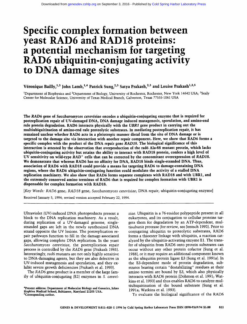
Antibodies specific for RAD6 and RAD18 were isolated from rabbit antisera by affinity chromatography and co- valently coupled to protein A-agarose beads for use in immunoprecipitation experiments (Bailly et al. 1992). After incubation with yeast extract and washing, immu- nobeads were treated with 1% SDS to elute bound pro- teins, which were resolved in denaturing polyacrylamide gels and analyzed by immunoblotting. Anti-RAD6 im- munobeads precipitated the 20-kD RAD6 protein {Sung et al. 1990; Fig. 1A). Immunobeads specific for RAD18 precipitated a 66-kD protein that reacted with anti- RAD18 antibodies in immunoblot analysis (Fig. 1B). This 66-kD band is the product of RAD18/I because (1) it was absent in extract of yeast cells harboring the rad18 mutation (Fig. 1B, lane 2) and (2) its level increased with the RAD18 gene dosage (Fig. 2A, top, cf. lanes 5 and 6). The quantity of immunoprecipitable RAD6 is not af- fected by the rad18A mutation (Fig. 1A, cf. lanes 2 and 3), and likewise, the rad6A mutation does not alter the amount of RAD18 protein (Fig. 1B, cf. lanes 1 and 3). Neither RAD6 nor RAD18 was precipitated by protein A-agarose beads bearing antibodies specific for the nu-
A
k D a
6 6 -
4 5 -
3 1 -
1 2 3 4 5
21 - . , , . . - R A D 6
B
kDa
9 7 -
6 6 -
4 5 -
3 1 -
1 2 3 4 5
- R A D 1 8
Figure 1. (A) Immunoprecipitation of RAD6 protein. Extracts from the RAD + strain LP3041-6D (lane 3) and from its isogenic rad6A derivative EMY1 (lane 1) and rad18A derivative YlJ15 (lane 2) were mixed with protein A-agarose beads bearing anti- RAD6 antibodies. The SDS eluates of immunoprecipitates were analyzed by immunoblotting for their content of RAD6 protein. As shown in lanes 4 and 5, respectively, RAD6 is not precipi- tated from the RAD+extract by anti-RAD1 and anti-RAD10 protein A-agarose immunobeads. {B) Immunoprecipitation of RAD18 protein. The SDS eluates of anti-RAD18 immunopre- cipitates were analyzed by immunoblotting for their content of RAD18. Lanes 1-3 contain samples from rad6A, radl8A, and /LAD + extracts, respectively. RAD18 protein in RAD + extract is not precipitated by anti-RAD1 (lane 4) and anti-RAD10 (lane 5) immunobeads.
812 GENES & DEVELOPMENT
Cold Spring Harbor Laboratory Press on September 3, 2016 - Published by genesdev.cshlp.orgDownloaded from
Complex of RAD6 and RAD18 proteins
I m m u n o b e a d s
Anti- Anti- RAD6 RAD18
i i , |
1 2 3 4 5 6
- R A D 1 8
B
1 2 3 4
~,~,~, ,~ - R A D 1 8
~ : - R A D 6 - R A D 6
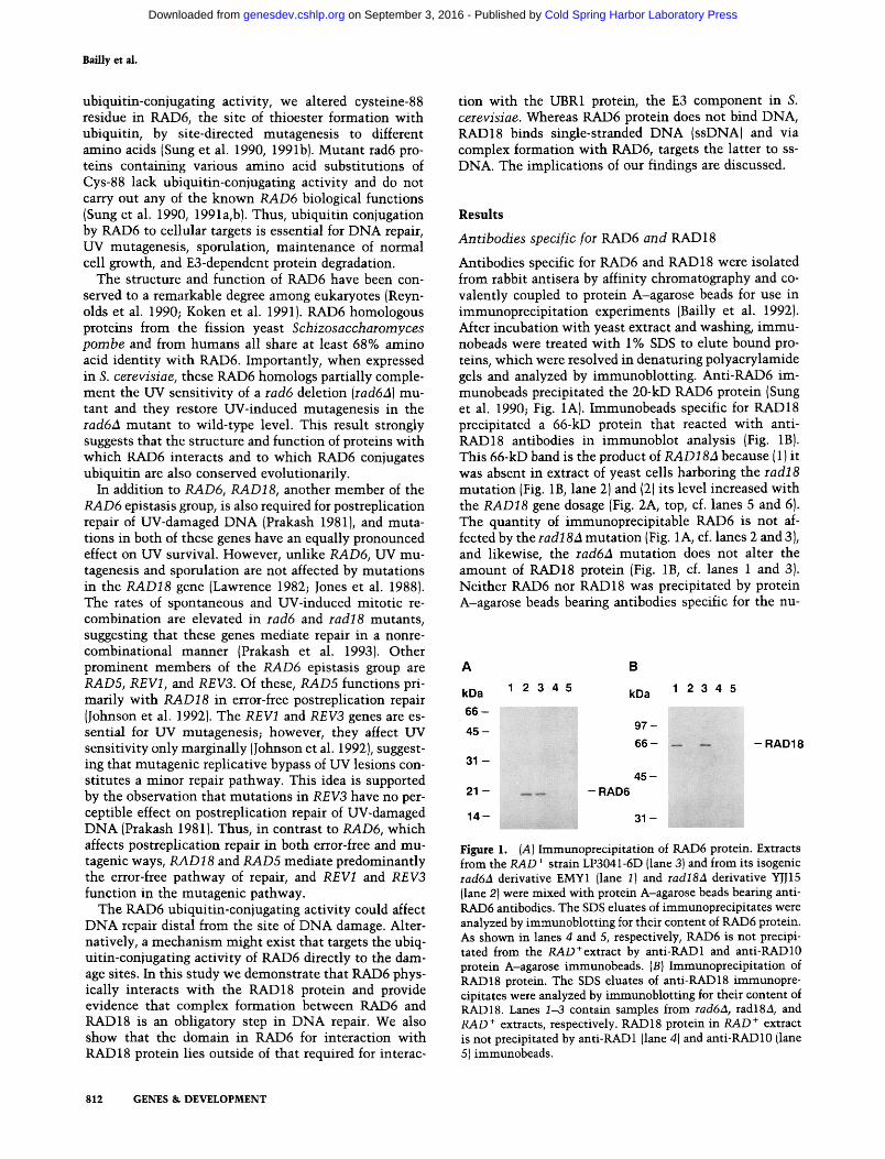
Figure 2. RAD6 and RAD18 proteins are stably associated in vivo. {A) Coimmunoprecipitation of RAD6 and RAD18. Ex- tracts from rad6a cells (lane 1 ), rad18a cells (lane 4), RAD + cells (lanes 2,5), and RAD + cells harboring the 2~L multicopy RAD18- containing plasmid pJJ136 (lanes 3,6) were mixed with anti- RAD6 immunobeads (lanes 1-3) and anti-PAD 18 immunobeads (lanes 4-6). The SDS eluates of immunoprecipitates were ana- lyzed by immunoblotting for their content of RAD6 (bottom) and RAD18 (top). {B) Strength of RAD6/RAD18 interaction. The RAD6/RAD18 complex in 5 ml of extract of PAD + cells harboring pJJ136 was immobilized on 50 VL1 of anti-RAD6 im- munobeads. The immunocomplex was washed at 25°C with 500 wl of 1 M NaC1 (lane 1), followed by the same volume of 0.05% SDS (lane 2), 0.1% SDS (lane 3), and 1% SDS (lane 4). The various washes were treated with cold acetone to precipitate protein and analyzed for their content of RAD6 (bottom) and PAD18 {top).
same extent (Fig. 2A, top, lane 3). These observations indicate that complex formation between RAD6 and RAD 18 is a highly efficient process, with RAD 18 being the limiting component.
R A D 6 / R A D 1 8 complex is highly stable
To examine the strength of interaction, the complex of RAD6 and RAD 18 was isolated on anti-RAD6 immuno- beads and eluted with 1 M NaC1 followed by increasing concentrations of the protein denaturant SDS (0.05%, 0.1%, and 1% ). The eluates were subjected to SDS-PAGE and analyzed by immunoblotting for their content of RAD6 and RAD18. Because RAD6 in the immunocom- plex was bound by antibodies directly, 1% SDS was re- quired for its elution (Fig. 2B, bottom, lane 4). Under the conditions stated, examination of RAD 18 content in the various washes would yield information concerning the avidity of RAD18 for RAD6. As shown in Figure 2B (top, lane 1), the RAD6/RAD 18 complex withstood the chal- lenge of 1 M NaC1 and was partially resistant to low concentration of SDS, as only 16% and 25% of RAD18 was eluted by 0.05% and 0.1% of the protein denaturant, respectively (Fig. 2B, top, lanes 2,3), reflecting a high de- gree of stability of the protein complex. Similar conclu- sions concerning the stability of the RAD6/RAD 18 com- plex were drawn when anti-RAD18 immunobeads were used for isolating the complex (data not shown).
cleotide excision repair proteins RAD1 and RADIO (Bailly et al. 1992, Fig. 1A,B, lanes 4,5).
RAD6 and RAD18 exist as a complex in vivo
To investigate whether the RAD6 and RAD18 proteins interact physically, immunoprecipitation was carried out with anti-RAD6 immunobeads and the SDS eluate examined for the presence of RAD18 protein by immu- noblotting, and conversely, the content of RAD6 protein in anti-RAD 18 immunoprecipitate was determined. Ap- parently, a quantitative amount of RAD18 protein was precipitated by anti-RAD6 immunobeads from extract of wild-type cells (Fig. 2A, top, cf. lanes 2 and 5), but no RAD18 was precipitated by anti-RAD6 immunobeads when extract of rad621 cells was used (Fig. 2A, top, lane 1). Thus, association of RAD18 with anti-RAD6 immu- nobeads requires the RAD6 protein, strongly suggesting the existence of a physical complex of the two proteins in wild-type cells. This conclusion is supported by the observation that a sizable proportion (-10%) of the RAD6 pool in wild type extract coprecipitated with the RAD18 protein on anti-RAD18 immunobeads (Fig. 2A, bottom, cf. lanes 2 and 5). Introduction into a RAD+yeast strain of pJJ136, a multicopy plasmid carry- ing the RAD18 gene in a yeast 2ix vector, not only in- creased the level of immunoprecipitable RAD 18 protein by -10-fold (Fig. 2A, lane 6) but also the amount of RAD18 in the RAD6/RAD18 complex, apparently to the
RAD6 and RAD18 interact directly
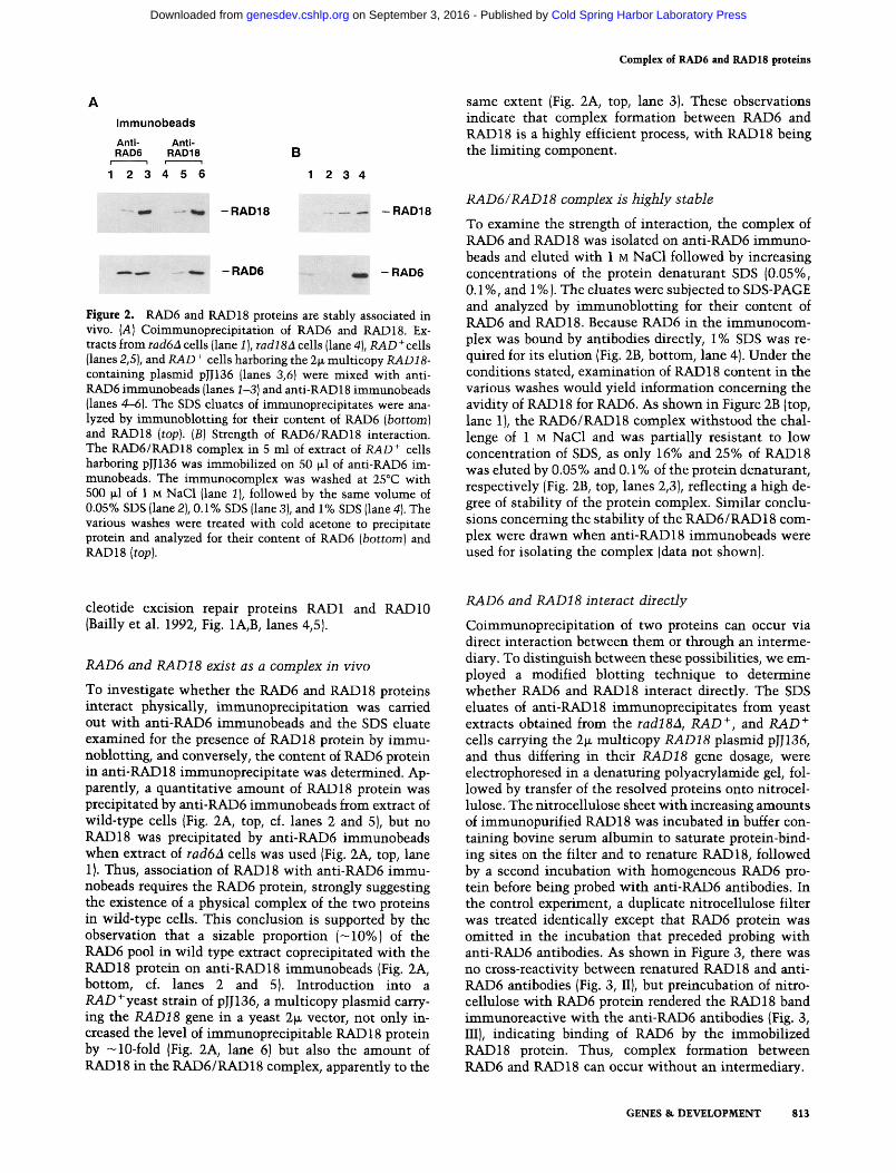
Coimmunoprecipitation of two proteins can occur via direct interaction between them or through an interme- diary. To distinguish between these possibilities, we em- ployed a modified blotting technique to determine whether RAD6 and RAD18 interact directly. The SDS eluates of anti-RAD18 immunoprecipitates from yeast extracts obtained from the rad1821, RAD +, and RAD + cells carrying the 2ix multicopy RAD18 plasmid pJJ136, and thus differing in their RAD18 gene dosage, were electrophoresed in a denaturing polyacrylamide gel, fol- lowed by transfer of the resolved proteins onto nitrocel- lulose. The nitrocellulose sheet with increasing amounts of immunopurified RAD 18 was incubated in buffer con- taining bovine serum albumin to saturate protein-bind- ing sites on the filter and to renature RAD18, followed by a second incubation with homogeneous RAD6 pro- tein before being probed with anti-RAD6 antibodies. In the control experiment, a duplicate nitrocellulose filter was treated identically except that RAD6 protein was omitted in the incubation that preceded probing with anti-RAD6 antibodies. As shown in Figure 3, there was no cross-reactivity between renatured RAD18 and anti- RAD6 antibodies (Fig. 3, II), but preincubation of nitro- cellulose with RAD6 protein rendered the RAD18 band immunoreactive with the anti-RAD6 antibodies (Fig. 3, III), indicating binding of RAD6 by the immobilized RAD18 protein. Thus, complex formation between RAD6 and RAD18 can occur without an intermediary.
GENES & DEVELOPMENT 813
Cold Spring Harbor Laboratory Press on September 3, 2016 - Published by genesdev.cshlp.orgDownloaded from
Bailly et al.
1 2 3
.... I
II
?,g'
III
Figure 3. RAD6 and RAD18 interact directly. Nitrocellulose blot containing SDS eluates of anti-RAD 18 immunoprecipitates from extracts of rad18A cells {lane 11, PAD + cells (lane 2), and PAD + cells harboring the 2ix RAD18 plasmid pJJ136 {lane 3) was probed with anti-RAD 18 antibodies (I}, anti-RAD6 antibod- ies (//), or first incubated with homogeneous RAD6 protein be- fore being probed with anti-RAD6 antibodies (HI). The same region of the nitrocellulose blot is shown in I, II, and III, and these blots were obtained from transfer of three separate sets of SDS eluates of anti-RAD18 immunoprecipitates.
RAD6 associates wi th RAD18 and with UBR1 in separate complexes
The RAD6 ubiquitin-conjugating function mediates deg- radation of proteins bearing destabilizing amino-termi- nal residues (Dohmen et al. 1991; Sung et al. 1991a; Wat- kins et al. 1993)in the amino-end rule-dependent prote- olytic pathway. This mode of protein degradation also requires the UBRl-encoded E3 protein, which provides the binding site for the amino terminus of proteolytic substrates. UBR1 protein physically interacts with RAD6, bringing RAD6 to attach multiple molecules of ubiquitin to a lysine residue in the bound substrates (Dohmen et al. 1991; Sung et al. 1991a; Watkins et al. 1993).
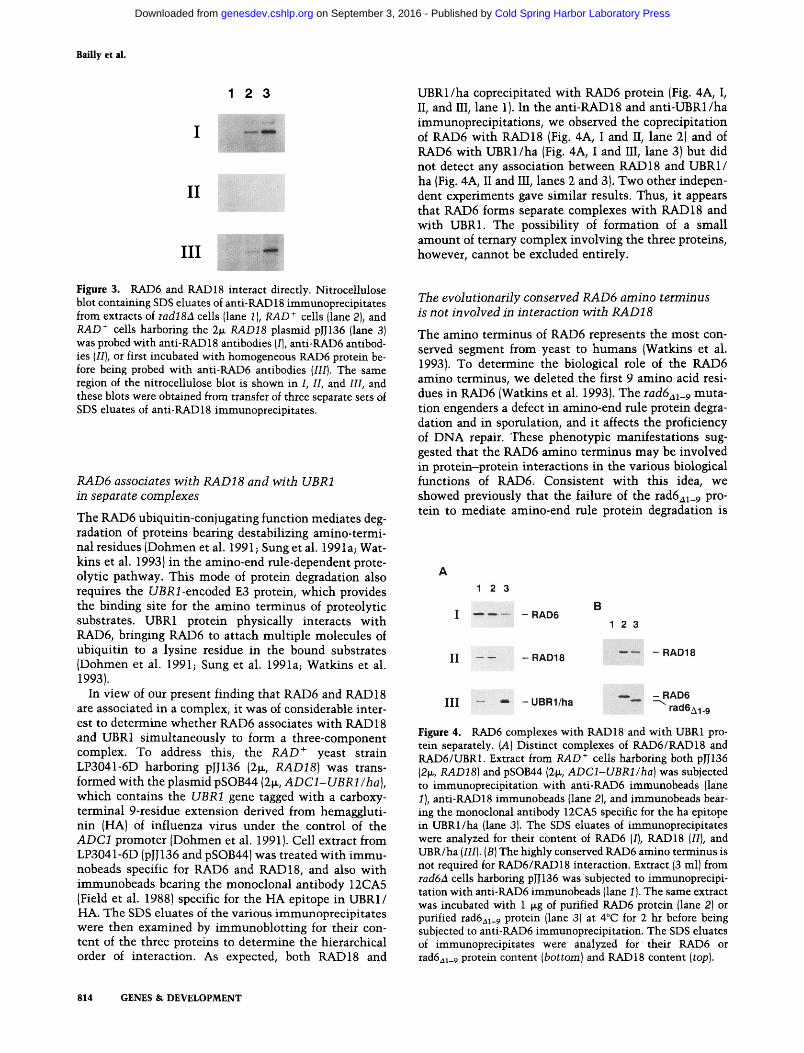
In view of our present finding that RAD6 and RAD18 are associated in a complex, it was of considerable inter- est to determine whether RAD6 associates with RAD 18 and UBR1 simultaneously to form a three-component complex. To address this, the /LAD + yeast strain LP3041-6D harboring pJJ136 (2~, RAD18) was trans- formed with the plasmid pSOB44 (2~, ADC1-UBR1/ha) , which contains the UBR1 gene tagged with a carboxy- terminal 9-residue extension derived from hemaggluti- nin (HA) of influenza virus under the control of the ADC1 promoter (Dohmen et al. 1991). Cell extract from LP3041-6D (pJJ136 and pSOB44) was treated with immu- nobeads specific for RAD6 and RAD18, and also with immunobeads bearing the monoclonal antibody 12CA5 (Field et al. 1988) specific for the HA epitope in UBR1/ HA. The SDS eluates of the various immunoprecipitates were then examined by immunoblotting for their con- tent of the three proteins to determine the hierarchical order of interaction. As expected, both RAD18 and
UBR1/ha coprecipitated with RAD6 protein (Fig. 4A, I, II, and III, lane 1). In the anti-RAD18 and anti-UBR1/ha immunoprecipitations, we observed the coprecipitation of RAD6 with RAD18 (Fig. 4A, I and II, lane 2) and of RAD6 with UBR1/ha {Fig. 4A, I and III, lane 3) but did not detect any association between RAD18 and UBR1/ ha (Fig. 4A, II and III, lanes 2 and 3). Two other indepen- dent experiments gave similar results. Thus, it appears that RAD6 forms separate complexes with RAD18 and with UBR1. The possibility of formation of a small amount of ternary complex involving the three proteins, however, cannot be excluded entirely.
The evolutionarily conserved RAD6 amino terminus is not involved in interaction wi th RAD18
The amino terminus of RAD6 represents the most con- served segment from yeast to humans (Watkins et al. 1993). To determine the biological role of the RAD6 amino terminus, we deleted the first 9 amino acid resi- dues in RAD6 (Watkins et al. 1993). The rad6al_ 9 muta- tion engenders a defect in amino-end rule protein degra- dation and in sporulation, and it affects the proficiency of DNA repair. These phenotypic manifestations sug- gested that the RAD6 amino terminus may be involved in protein-protein interactions in the various biological functions of RAD6. Consistent with this idea, we showed previously that the failure of the rad6al_9 pro- tein to mediate amino-end rule protein degradation is
1 2 3
I . . . . . RAD6
I I - - - - R A D 1 8
1 2 3
, - R A D 1 8
I I I - - , , - U B R 1 / h a ~ R A D 6
J ~
r a u o A 1 . 9
Figure 4. RAD6 complexes with RAD18 and with UBR1 pro- tein separately. (A) Distinct complexes of RAD6/RAD18 and RAD6/UBR1. Extract from/LAD + cells harboring both plJ136 (2~, RAD181 and pSOB44 (2~, ADC1-UBR1/ha)was subjected to immunoprecipitation with anti-RAD6 immunobeads (lane 1 ), anti-RAD18 immunobeads (lane 2), and immunobeads bear- ing the monoclonal antibody 12CA5 specific for the ha epitope in UBRI/ha (lane 3). The SDS eluates of immunoprecipitates were analyzed for their content of RAD6 (I), RAD18 {III, and UBR/ha (III). (BI The highly conserved RAD6 amino terminus is not required for RAD6/RAD18 interaction. Extract (3 mll from rad6A cells harboring pJJ136 was subjected to immunoprecipi- ration with anti-RAD6 immunobeads {lane 1 I. The same extract was incubated with 1 ~g of purified RAD6 protein (lane 2) or purified rad6al_ 9 protein {lane 3) at 4°C for 2 hr before being subjected to anti-RAD6 immunoprecipitation. The SDS eluates of immunoprecipitates were analyzed for their RAD6 or rad6al_9 protein content {bottom) and RAD18 content (top).
814 GENES & DEVELOPMENT
Cold Spring Harbor Laboratory Press on September 3, 2016 - Published by genesdev.cshlp.orgDownloaded from
Complex of RAD6 and RAD18 proteins
attributable to the inability of the mutant protein to in- teract with the UBR1 protein (Watkins et al. 1993).
Here, we examine whether the rad6al_9 mutant pro- tein can interact with RAD18 protein using the same experimental conditions where rad6al_9 protein fails to complex with UBR1 (Watkins et al. 1993). The RAD6 protein or the rad6a~_9 protein, both purified to homo- geneity as described (Sung et al. 1991a; Watkins et al. 1993) were added to extract from rad6a cells harboring the 2~ RAD18 plasmid pJJ136. After incubation on ice, the extract was subjected to immunoprecipitation with anti-RAD6 immunobeads. As shown in Figure 4B, a sim- ilar amount of RAD 18 protein coprecipitated with RAD6 and rad6a~_9 proteins (lanes 2,3), indicating that the amino terminus of RAD6 protein is dispensable for com- plex formation with RAD18.
Semidominance of overproduction of rad6 Ala-88 mu tan t protein suggests biological significance of physical interaction
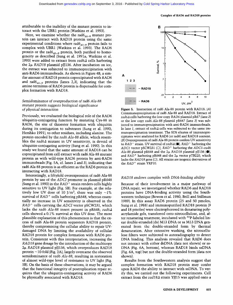
Previously, we evaluated the biological role of the RAD6 ubiquitin-conjugating function by mutating Cys-88 in RAD6, the site of thioester formation with ubiquitin during its conjugation to substrates (Sung et al. 1990; Hershko 1991), to other residues, including alanine. The protein encoded by the rad6 Ala-88 allele, which resem- bles the rad6A mutant in UV sensitivity, is devoid of ubiquitin-conjugating activity (Sung et al. 1990). In this study we found that the same amount of RAD 18 can be coprecipitated from cell extract with rad6 Ala-88 mutant protein as with wild-type RAD6 protein by anti-RAD6 immunobeads (Fig. 5A, cf. lanes 2 and 3), indicating that rad6 Ala-88 protein is as efficient as the RAD6 protein in interacting with RAD 18.
Interestingly, a fiftyfold overexpression of tad6 Ala-88 protein by use of the A D C I promoter in plasmid pR648 (Sung et al. 1990) in the RAD + strain renders cells highly sensitive to UV light (Fig. 5B). For example, at the rela- tively low UV dose of 10 J/m 2, there was only - 1 % survival of RAD + cells harboring pR648, whereas essen- tially no increase in UV sensitivity is observed in the RAD + cells carrying the A D C I vector pSCW231, which lacks the rad6 Ala-88 insert present in pR648; rad6A cells showed a 0.1% survival at this UV dose. The most plausible explanation of this phenomenon is that the ex- cess of tad6 Ala-88 protein sequesters RAD18 protein, thereby compromising the cellular ability to repair UV- damaged DNA by limiting the availability of cellular RAD18 protein for complex formation with RAD6 pro- tein. Consistent with this interpretation, increasing the RAD18 gene dosage by the introduction of the multicopy 2~ RAD18 plasmid pJJ136, which overproduces RAD18 protein -10-fold {Fig. 2A), largely overcame the observed semidominance of rad6 Ala-88, resulting in restoration of almost wild-type level of resistance to UV light (Fig. 5B). On the basis of these observations, it may be argued that the functional integrity of postreplication repair re- quires that the ubiquitin-conjugating activity of RAD6 be physically associated with RAD 18.
1 2 3
- - R A D 6
- R A D 1 8
.1
.01 i I J , I , I
0 2 4 6 8 10
UV, d/m 2
B 1001
.g 1
P
8
Figure 5. Interaction of rad6 Ala-88 protein with RAD18. (A) Coimmunoprecipitation of rad6 Ala-88 and RAD18. Extract of rad6a cells harboring the low-copy RAD6 plasmid pR67 (lane 2) or the low copy racl6 Ala-88 plasmid pR647 (lane 3) was sub- jected to immunoprecipitation with anti-RAD6 immunobeads. In lane I, extract of rad6a cells was subjected to the same im- munoprecipitation treatment. The SDS eluates of immunopre- cipitates were analyzed for RAD6 (or tad6) and RAD18 content. (B) Overexpression of tad6 Ala-88 protein confers UV sensitivity to RAD + strain. UV survival of rad6A (B), RAD + harboring the ADC1 vector pSCW231 (C)), RAD + harboring the ADCl-rad6 Ala-88 plasmid pR648 and the 2~ RAD18 plasmid pJJ136 (0), and RAD + harboring pR648 and the 2~ vector pTB220, which lacks the RAD18 gene ([3). All strains are isogenic derivatives of the RAD + strain YRP 10.
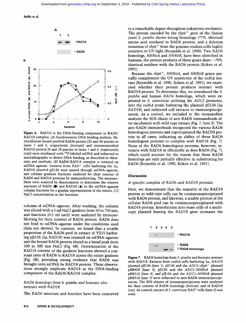
RAD18 endows complex wi th DNA-binding abili ty
Because of their involvement in a major pathway of DNA repair, we investigated whether RAD6 and RAD 18 proteins have DNA-binding activity using the South- western technique (Bowen et al. 1980; Brill and Stillman 1989). In this assay RAD6 protein (25 and 50 pmoles; Sung et al. 1988) and immunopurified RAD18 protein (9 and 18 pmoles) were electrophoresed in denaturing poly- acrylamide gels, transferred onto nitrocellulose, and, af- ter renaturing treatment, incubated with 3~p-labeled lin- ear double-stranded (ds) M13 DNA or ss M13 DNA gen- erated from the double-stranded form by thermal denaturation. After extensive washing, the nitrocellu- lose filters were subjected to autoradiography to detect DNA binding. This analysis revealed that RAD6 does not interact with either dsDNA (data not shown) or ss- DNA (Fig. 6A, bottom), whereas RAD18 binds ssDNA (Fig. 6A, top) but not the double-stranded form (data not shown).
Results from the Southwestern analysis suggest that complex formation with RAD18 protein may confer upon RAD6 the ability to interact with ssDNA. To ver- ify this, we carried out the following experiments. Cell extract from the rad18A strain YJJ 15 was applied onto a
GENES & D E V E L O P M E N T 815
Cold Spring Harbor Laboratory Press on September 3, 2016 - Published by genesdev.cshlp.orgDownloaded from
Bai l ly et al.
A
1 2
~m, o - R A D 1 8
- RAD6
3o
4 , , , . .
~ 2o
~ lO
~ o
- 0.6
• ~-,~. ° ~ [] \ ' m 0.4 - - /
/ / o" \O~- o / ~X~,, 0.2 ~ 3
/ / ;_ j i/jo / ! ; v 1 ~ I , 1 , I 0
2 4 6 8 10
fractions
Figure 6. RAD18 is the DNA-binding component in RAD6/ RAD18 complex. (A) Southwestern DNA binding analysis. Ni- trocellulose bound purified RAD6 protein [25 and 50 pmoles in lanes 1 and 2, respectively (bottom)] and immunopurified RAD18 protein [9 and 18 pmoles in lanes 1 and 2, respectively (top)] were incubated with 32p-labeled ssDNA and subjected to autoradiography to detect DNA binding, as described in Mate- rials and methods. (B) RAD6/RAD18 complex is retained on ssDNA agarose. Extracts from RAD + cells harboring the 2~, RAD18 plasmid pJJ136 were passed through ssDNA-agarose, and column gradient fractions analyzed for their content of RAD6 and RAD18 proteins by immunoblotting. The immuno- blots were scanned by densitometry to determine the relative amounts of RAD6 (O) and RAD18 (&) in the ssDNA-agarose column fractions for a graphic representation of the results. (O) NaG1 concentration in the fractions.
column of ssDNA-agarose. After washing, the column was eluted with a 5 ml NaC1 gradient from 50 to 750 mM, and fractions (0.5 ml each) were analyzed by immuno- blotting for their content of RAD6 protein. RAD6 does not bind to ssDNA-agarose under the conditions used (data not shown). In contrast, we found that a sizable proportion of the RAD6 pool in extract of YJJ15 harbor- ing pJJ136 (2~ RAD18) was retained on ssDNA-agarose and the bound RAD6 protein eluted as a broad peak from 100 to 300 mM NaC1 (Fig. 6B). Determination of the RAD 18 content of the gradient fractions showed a con- stant ratio of RAD6 to RAD 18 across the entire gradient (Fig. 6B), providing strong evidence that RAD6 was brought onto ssDNA by RAD 18 protein. These observa- tions strongly implicate RAD18 as the DNA-binding component of the RAD6/RAD18 complex.
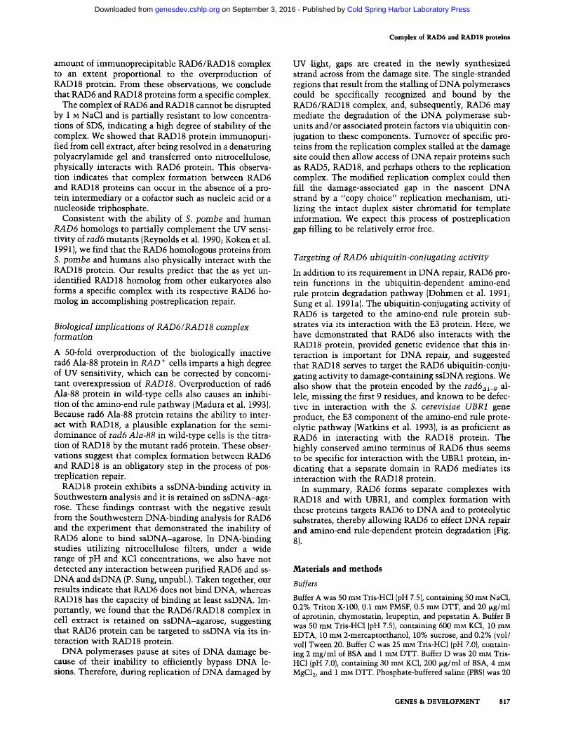
RAD6 homologs from S. pombe and humans also interact with RAD18
The RAD6 structure and function have been conserved
to a remarkable degree throughout eukaryotic evolution. The protein encoded by the rhp6 ÷ gene of the fission yeast S. pombe shows strong homology [77% identical amino acid residues) to RAD6 protein, and a deletion mutation of rhp6 ÷ from the genome renders cells highly sensitive to UV light (Reynolds et al. 1990). Two RAD6 homologs, HHR6A and HHR6B, have been identified in humans; the protein products of these genes share - 7 0 % identical residues with the RAD6 protein (Koken et al. 1991).
Because the rhp6 +, HHR6A, and HHR6B genes par- tially complement the UV sensitivity of the rad6a mu- tant (Reynolds et al. 1990; Koken et al. 1991), we exam- ined whether their protein products interact with RAD 18 protein. To determine this, we introduced the S. pombe and human RAD6 homologs, which were ex- pressed in S. cerevisiae utilizing the A D C i promoter, into the rad6a strain harboring the plasmid pJJ136 (2tr RAD18), and subjected cell extracts to immunoprecipi- tation. As a control, we included in the immunoblot analysis the SDS eluate of anti-RAD6 immunobeads af- ter incubation with wild-type extract (Fig. 7, lane 2). The anti-RAD6 immunobeads recognized the various RAD6 homologous proteins and coprecipitated the RAD 18 pro- tein in all cases, indicating an ability of these RAD6 homologous proteins to complex with RAD18 (Fig. 7). None of the RAD6 homologous proteins, however, in- teracts with RAD18 as efficiently as does RAD6 (Fig. 7), which could account for the reason that these RAD6 homologs are only partially effective in substituting for RAD6 (Reynolds et al. 1990; Koken et al. 1991).
D i s c u s s i o n
A specific complex of RAD6 and RAD18 proteins
Here, we demonstrate that the majority of the RAD18 protein in wild-type cells can be coimmunoprecipitated with RAD6 protein, and likewise, a sizable portion of the cellular RAD6 pool can be coimmunoprecipitated with RAD 18 protein. Introduction into yeast cells of a multi- copy plasmid bearing the RAD18 gene increases the
1 2 3 4 5
- R A D 1 8
• - R A D 6 . : . : ~ !, . :
~ . = R A D 6 h o m o l o g s
Figure 7. RAD6 homologs from S. pornbe and humans interact with RAD18. Extracts from rad6A cells harboring 2tx, RAD18 plasmid pIJ136 (lane 1), pJJ136 and the ADCl-rhp6 + plasmid pRR429 (lane 3), pJJ136 and the ADC1-HHR6A plasmid pRR510 (lane 4), and pJJ136 and the ADC1-HHR6B plasmid pRR518 (lane 5)were subjected to anti-RAD6 immunoprecipi- tation. The SDS eluates of immunoprecipitates were analyzed for their content of RAD6 homologs (bottom) and of RAD18 (top). As control, extract of S. cerevisiae RAD+cells (lane 2) was used.
816 GENES & D E V E L O P M E N T
Cold Spring Harbor Laboratory Press on September 3, 2016 - Published by genesdev.cshlp.orgDownloaded from
Complex of RAD6 and RAD18 proteins
amount of immunoprecipitable RAD6 / RAD 18 complex to an extent proportional to the overproduction of RAD18 protein. From these observations, we conclude that RAD6 and RAD18 proteins form a specific complex.
The complex of RAD6 and RAD 18 cannot be disrupted by 1 M NaC1 and is partially resistant to low concentra- tions of SDS, indicating a high degree of stability of the complex. We showed that RAD18 protein immunopuri- fled from cell extract, after being resolved in a denaturing polyacrylamide gel and transferred onto nitrocellulose, physically interacts with RAD6 protein. This observa- tion indicates that complex formation between RAD6 and RAD 18 proteins can occur in the absence of a pro- tein intermediary or a cofactor such as nucleic acid or a nucleoside triphosphate.
Consistent with the ability of S. pombe and human RAD6 homologs to partially complement the UV sensi- tivity of rad6 mutants (Reynolds et al. 1990; Koken et al. 1991), we find that the RAD6 homologous proteins from S. pombe and humans also physically interact with the RAD18 protein. Our results predict that the as yet un- identified RAD18 homolog from other eukaryotes also forms a specific complex with its respective RAD6 ho- molog in accomplishing postreplication repair.
Biological implications of RAD6/RAD18 complex formation
A 50-fold overproduction of the biologically inactive rad6 Ala-88 protein in RAD + cells imparts a high degree of UV sensitivity, which can be corrected by concomi- tant overexpression of RAD18. Overproduction of rad6 Ala-88 protein in wild-type cells also causes an inhibi- tion of the amino-end rule pathway (Madura et al. 1993). Because tad6 Ala-88 protein retains the ability to inter- act with RAD18, a plausible explanation for the semi- dominance of rad6 Ala-88 in wild-type cells is the titra- tion of RAD 18 by the mutant rad6 protein. These obser- vations suggest that complex formation between RAD6 and RAD18 is an obligatory step in the process of pos- treplication repair.
RAD18 protein exhibits a ssDNA-binding activity in Southwestern analysis and it is retained on ssDNA-aga- rose. These findings contrast with the negative result from the Southwestem DNA-binding analysis for RAD6 and the experiment that demonstrated the inability of RAD6 alone to bind ssDNA-agarose. In DNA-binding studies utilizing nitrocellulose filters, under a wide range of pH and KC1 concentrations, we also have not detected any interaction between purified RAD6 and ss- DNA and dsDNA (P. Sung, unpubl.). Taken together, our results indicate that RAD6 does not bind DNA, whereas RAD18 has the capacity of binding at least ssDNA. Im- portantly, we found that the RAD6/RAD18 complex in cell extract is retained on ssDNA-agarose, suggesting that RAD6 protein can be targeted to ssDNA via its in- teraction with RAD 18 protein.
DNA polymerases pause at sites of DNA damage be- cause of their inability to efficiently bypass DNA le- sions. Therefore, during replication of DNA damaged by
UV light, gaps are created in the newly synthesized strand across from the damage site. The single-stranded regions that result from the stalling of DNA polymerases could be specifically recognized and bound by the RAD6/RAD18 complex, and, subsequently, RAD6 may mediate the degradation of the DNA polymerase sub- units and/or associated protein factors via ubiquitin con- jugation to these components. Tumover of specific pro- teins from the replication complex stalled at the damage site could then allow access of DNA repair proteins such as RAD5, RAD 18, and perhaps others to the replication complex. The modified replication complex could then fill the damage-associated gap in the nascent DNA strand by a "copy choice" replication mechanism, uti- lizing the intact duplex sister chromatid for template information. We expect this process of postreplication gap filling to be relatively error free.
Targeting of RAD6 ubiquitin-conjugating activity
In addition to its requirement in DNA repair, RAD6 pro- tein functions in the ubiquitin-dependent amino-end rule protein degradation pathway (Dohmen et al. 1991; Sung et al. 1991a). The ubiquitin-conjugating activity of RAD6 is targeted to the amino-end rule protein sub- strates via its interaction with the E3 protein. Here, we have demonstrated that RAD6 also interacts with the RAD18 protein, provided genetic evidence that this in- teraction is important for DNA repair, and suggested that RAD18 serves to target the RAD6 ubiquitin-conju- gating activity to damage-containing ssDNA regions. We also show that the protein encoded by the rad6a~_9 al- lele, missing the first 9 residues, and known to be defec- tive in interaction with the S. cerevisiae UBR1 gene product, the E3 component of the amino-end rule prote- olytic pathway (Watkins et al. 1993), is as proficient as RAD6 in interacting with the RAD18 protein. The highly conserved amino terminus of RAD6 thus seems to be specific for interaction with the UBR1 protein, in- dicating that a separate domain in RAD6 mediates its interaction with the RAD18 protein.
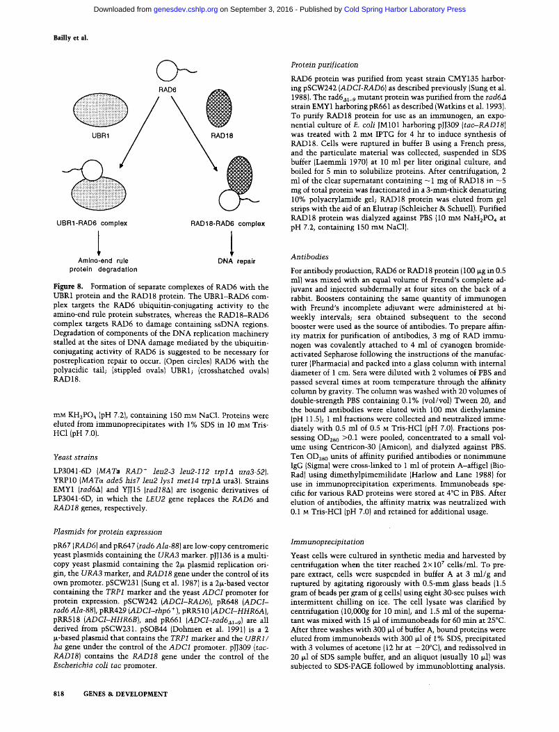
In summary, RAD6 forms separate complexes with RAD18 and with UBR1, and complex formation with these proteins targets RAD6 to DNA and to proteolytic substrates, thereby allowing RAD6 to effect DNA repair and amino-end rule-dependent protein degradation (Fig. 8).
Materials and methods
Buffers
Buffer A was 50 mM Tris-HC1 (pH 7.5), containing 50 mM NaCl, 0.2% Triton X-100, 0.1 mM PMSF, 0.5 mM DTT, and 20 ~g/ml of aprotinin, ¢hymostatin, leupeptin, and pepstatin A. Buffer B was 50 mM Tris-HC1 (pH 7.5), containing 600 mM KC1, 10 mM EDTA, 10 rnM 2-mercaptoethanol, 10% sucrose, and 0.2% (vol/ vol) Tween 20. Buffer C was 25 rr~ Tris-HC1 (pH 7.0), contain- ing 2 mg/ml of BSA and 1 mM DTT. Buffer D was 20 mM Tris- HC1 (pH 7.0), containing 30 mM KC1, 200 ~g/ml of BSA, 4 mM MgC12, and 1 mM DTT. Phosphate-buffered saline (PBS) was 20
GENES & DEVELOPMENT 817
Cold Spring Harbor Laboratory Press on September 3, 2016 - Published by genesdev.cshlp.orgDownloaded from
Bailly et al.
RAD6
UBR1-RAD6 complex RAD18-RAD6 complex
Amino-end rule DNA repair protein degradation
Figure 8. Formation of separate complexes of RAD6 with the UBR1 protein and the RAD18 protein. The UBR1-RAD6 com- plex targets the RAD6 ubiquitin-conjugating activity to the amino-end rule protein substrates, whereas the RAD18-RAD6 complex targets RAD6 to damage containing ssDNA regions. Degradation of components of the DNA replication machinery stalled at the sites of DNA damage mediated by the ubiquitin- conjugating activity of RAD6 is suggested to be necessary for postreplication repair to occur. (Open circles) RAD6 with the polyacidic tail; {stippled ovals) UBR1; {crosshatched ovals} RAD18.
mM KH~PO4 (pH 7.2}, containing 150 mM NaC1. Proteins were eluted from immunoprecipitates with 1% SDS in 10 mM Tris- HC1 (pH 7.0).
Yeast strains
LP3041-6D (MATa RAD + leu2-3 leu2-112 trplA ura3-52). YRP10 (MATa ade5 his7 leu2 lysl metl4 trpla ura3). Strains EMY1 (rad6A) and YJJ15 (radlSA) are isogenic derivatives of LP3041-6D, in which the LEU2 gene replaces the RAD6 and RAD18 genes, respectively.
Plasmids for protein expression
pR67 (RAD6) and pR647 (rad6 Ala-88) are low-copy centromeric yeast plasmids containing the URA3 marker, pJJ136 is a multi- copy yeast plasmid containing the 2la plasmid replication ori- gin, the URA3 marker, and RAD18 gene under the control of its own promoter, pSCW231 (Sung et al. 1987) is a 2la-based vector containing the TRP1 marker and the yeast ADCI promoter for protein expression, pSCW242 (ADCI-RAD6), pR648 (ADCI- rad6 Ala-88), pRR429 (ADCI-rhp6 + ), pRRS10 (ADCI-HHR6A ), pRR518 (ADCI-HHR6B), and pR661 {ADCI-rad6al_9) are all derived from pSCW231, pSOB44 (Dohmen et al. 1991) is a 2 la-based plasmid that contains the TRP1 marker and the UBRI/ ha gene under the control of the ADC1 promoter, pJJ309 (tac- RAD18) contains the RAD18 gene under the control of the Escherichia coli tac promoter.
818 GENES & DEVELOPMENT
Protein purification
RAD6 protein was purified from yeast strain CMY135 harbor- ing pSCW242 (ADCI-RAD6) as described previously (Sung et al. 1988). The rad6al_ 9 mutant protein was purified from the rad6zl strain EMY1 harboring pR661 as described (Watkins et al. 1993). To purify RAD18 protein for use as an immunogen, an expo- nential culture of E. coli JM101 harboring pJJ309 (tac,-RAD18) was treated with 2 mM IPTG for 4 hr to induce synthesis of RAD18. Cells were ruptured in buffer B using a French press, and the particulate material was collected, suspended in SDS buffer (Laemmli 1970) at 10 ml per liter original culture, and boiled for 5 rain to solubilize proteins. After centrifugation, 2 ml of the clear supematant containing -1 mg of RAD18 in - 5 mg of total protein was fractionated in a 3-ram-thick denaturing 10% polyacrylamide gel; RAD18 protein was eluted from gel strips with the aid of an Elutrap (Schleicher & Schuell). Purified RAD18 protein was dialyzed against PBS {10 mM NaH2PO 4 at pH 7.2, containing 150 mM NaC1).
Antibodies
For antibody production, RAD6 or RAD 18 protein (100 tag in 0.5 ml) was mixed with an equal volume of Freund's complete ad- juvant and injected subdermally at four sites on the back of a rabbit. Boosters containing the same quantity of immunogen with Freund's incomplete adjuvant were administered at bi- weekly intervals; sera obtained subsequent to the second booster were used as the source of antibodies. To prepare affin- ity matrix for purification of antibodies, 3 mg of RAD immu- nogen was covalently attached to 4 ml of cyanogen bromide- activated Sepharose following the instructions of the manufac- turer (Pharmacia) and packed into a glass column with internal diameter of 1 cm. Sera were diluted with 2 volumes of PBS and passed several times at room temperature through the affinity column by gravity. The column was washed with 20 volumes of double-strength PBS containing 0.1% [vol/vol) Tween 20, and the bound antibodies were eluted with 100 mM diethylamine (pH 11.5); 1 ml fractions were collected and neutralized imme- diately with 0.5 ml of 0.5 M Tris-HC1 (pH 7.0). Fractions pos- sessing OD2so >0.1 were pooled, concentrated to a small vol- ume using Centricon-30 (Amicon), and dialyzed against PBS. Ten OD~so units of affinity purified antibodies or nonimmune IgG (Sigma) were cross-linked to 1 ml of protein A-affigel {Bio- Rad) using dimethylpimemilidate (Harlow and Lane 1988) for use in immunoprecipitation experiments. Immunobeads spe- cific for various RAD proteins were stored at 4°C in PBS. After elution of antibodies, the affinity matrix was neutralized with 0.1 M Tris-HC1 (pH 7.0) and retained for additional usage.
Immunoprecipitation
Yeast cells were cultured in synthetic media and harvested by centrifugation when the titer reached 2x 10⁷ cells/ml. To pre- pare extract, cells were suspended in buffer A at 3 ml/g and ruptured by agitating rigorously with 0.5-ram glass beads (1.5 gram of beads per gram of g cellst using eight 30-sec pulses with intermittent chilling on ice. The cell lysate was clarified by centrifugation {10,O00g for 10 rain), and 1.5 ml of the supema- tant was mixed with 15 lal of immunobeads for 60 rain at 25°C. After three washes with 300 lal of buffer A, bound proteins were eluted from immunobeads with 300 lal of 1% SDS, precipitated with 3 volumes of acetone (12 hr at -20°C), and redissolved in 20 lal of SDS sample buffer, and an aliquot (usually 10 ixl) was subjected to SDS-PAGE followed by immunoblotting analysis.
Cold Spring Harbor Laboratory Press on September 3, 2016 - Published by genesdev.cshlp.orgDownloaded from
Complex of RAD6 and RAD18 proteins
Gel electrophoresis and immunoblotting
SDS-PAGE was carried out according to Laemmli (1970); 9% and 12% gels were used for RAD18 and RAD6, respectively. After electrophoresis, proteins were electroblotted onto nitro- cellulose sheets (Schleicher & Schuell), which were probed with '/looo dilution of affinity purified antibodies (OD28 o = 3) and de- veloped using the indirect peroxidase procedure of Towbin et al. (1979).
RAD6/RAD18 complex formation on nitrocellulose blot
Nitrocellulose blot containing immunopurified RAD18 protein was soaked at 25°C for i hr in 10 ml of PBS containing 15 mg/ml of BSA, 2 mM 2-mercaptoethanol, and 0.05% (vol/vol) Tween 20 before being incubated in the same buffer with 5 ~g/ml of RAD6 for 2 hr. After washing with 3 x 10 ml aliquots of buffer, the blot was subjected to probing with anti-RAD6 antibodies.
Southwestern D NA-binding analysis
To obtain RAD18 protein for the Southwestern analysis, extract was prepared in buffer A from 10 g of strain LP3041-6D harbor- ing plJ136 (2~ RAD18) using a French press. The clear superna- tant, obtained after high speed centrifugation (100,000g for 60 rain), was applied at 25°C onto a 1-ml column of protein A-aga- rose beads containing cross-linked anti-RAD18 antibodies. The column was washed extensively with buffer A before being eluted with 1% SDS; the protein pool (containing 5 ~g of 25% pure RAD18) was concentrated to 100 ~1 using Centricon-30. BgllI-linearized DNA was treated with calf thymus alkaline phosphatase to remove preexisting 5' phosphate and labeled with the T4 polynucleotide kinase and [~/-3~P]ATP to a specific radioactivity of 6 x 10Scpm/~g, and purified by gel filtration us- ing Sephadex G50. To generate ssDNA, the labeled double- stranded form was boiled in 10 mM Tris-HC1 at pH 7.5 for 3 min and chilled immediately on ice.
Purified RAD6 and RAD18 proteins (0.5 and 1 ~g) were elec- trophoresed in denaturing polyacrylamide gels and transferred onto nitrocellulose, which was soaked at 4°C for 36 hr in an excess of buffer C to block binding sites on nitrocellulose and to renature RAD6 and RAD18 proteins. The filters were rinsed twice with buffer D and incubated at 25°C for 60 rain in 10 ml of buffer D containing 1.5 ~g of a2P-labeled DNA. After being washed with 6x 10 ml buffer D for a total of 20 min, the filters were blot-dried with Whatman 3M paper and subjected to au- toradiography (2--4 hr exposure) using Kodak XRP films.
ssDNA-agarose chromatography
Cell extract was prepared using a French Press from 3 grams of the rad18A yeast strain YJJ15 or the RAD ÷ strain LP3041-6D harboring pJJ136 (21~ RAD18) in 5 ml of buffer A. After centrif- ugation (100,000g for 2 hr), the clear supematant was applied onto a 1-ml column of ssDNA-agarose (BRL). After washing with 5 ml of buffer A, bound proteins were eluted with a 10-ml NaC1 gradient from 50 to 750 mM, and 0.5-ml fractions were collected. For SDS-PAGE and immunoblotting, 10 tzl of the gra- dient fractions was analyzed in 9% gels for RAD18 and in 12% gels for RAD6.
A c k n o w l e d g m e n t s
V.B. and J.L. contributed equally to this work, which was sup- ported by grants from the National Institutes of Health
(GM19261) and from the Department of Energy (DE-FG03- 93ER61706).
The publication costs of this article were defrayed in part by payment of page charges. This article must therefore be hereby marked "advertisement" in accordance with 18 USC section 1734 solely to indicate this fact.
References
Bailly, V., C.H. Sommers, P. Sung, L. Prakash, and S. Prakash. 1992. Specific complex formation between proteins encoded by the yeast DNA repair and recombination genes RAD1 and RADIO. Proc. Nat. Acad. Sci. 89: 8273-8277.
Bowen, B., J. Steinberg, U.K. Laemmli, and H. Weintraub. 1980. The detection of DNA-binding proteins by protein blotting. Nucleic Acids Res. 8: 1-20.
Brill, S.J. and B. Stillman. 1989. Yeast replication factor-A func- tions in the unwinding of the SV40 origin of DNA replica- tion. Nature 342: 92-95.
Dohmen, R.J., K. Madura, B. Bartel, and A. Varshavsky. 1991. The N-end rule is mediated by the UBC2(RAD6) ubiquitin- conjugating enzyme. Proc. Natl. Acad. Sci. 88: 7351-7355.
Field, J., J.-I. Nikawa, D. Broek, B. MacDonald, L. Rodgers, I.A. Wilson, R.A. Lemer, and M. Wigler. 1988. Purification of a RAS-responsive adenyl cyclase complex from Saccharomy- ces cerevisiae by use of an epitope addition method. Mol. Cell. Biol. 8: 2159-2165.
Harlow, E. and D. Lane. eds. 1988. Antibodies: A laboratory manual. Cold Spring Harbor Laboratory, Cold Spring Har- bor, NY.
Hershko, A. 1991. The ubiquitin pathway for protein degrada- tion. Trends Biochem. Sci. 16: 265-268.
lentsch, S. 1992. The ubiquitin-conjugation system. Annu. Rev. Genet. 26: 179-207.
Johnson, R.E., S.T. Henderson, T.D. Petes, S. Prakash, M. Bank- mann, and L. Prakash. 1992. Saccharomyces cerevisiae RAD5-encoded DNA repair protein contains DNA helicase and zinc-binding sequence motifs and affects the stability of simple repetitive sequences in the genome. Mol. Cell. Biol. 12: 3807--3818.
Jones, I.S., S. Weber, and L. Prakash. 1988. The Saccharomyces cerevisiae RAD18 gene encodes a protein that contains po- tential zinc finger domains for nucleic acid binding and a putative nucleotide binding sequence. Nucleic Acids Res. 16: 7119-7131.
Koken, M.H., P. Reynolds, I. laspers-Dekker, L. Prakash, S. Prakash, D. Bootsma, and J.H. Hoeijmakers. 1991. Structural and functional conservation of two human homologs of the yeast DNA repair gene RAD6. Proc. Natl. Acad. Sci. 88: 8865-8869.
Laemmli, U.K. 1970. Cleavage of structural proteins during the assembly of the head of bacteriophage T4. Nature 227: 680- 685.
Lawrence, C.W. 1982. Mutagenesis in Saccharomyces cerevi- siae. Adv. Genet. 21: 173-254.
Madura, K., R.J. Dohmen, and S. Varshavsky. 1993. N-recognin/ Ubc2 interactions in the N-end.rule pathway. ]. Biol. Chem. 268: 12046-12054.
Prakash, L. 1981. Characterization of postreplication repair in Saccharomyces cerevisiae and effects of rad6, tad18, rev3 and tad52 mutations. Mol. Gen. Genet. 184: 471--478.
Prakash, S., P. Sung, and L. Prakash. 1993. DNA repair genes and proteins of Saccharomyces cerevisiae. Annu. Rev. Genet. 27: 33-70.
Reynolds, P., M.H. Koken, ].H. Hoeijmakers, S. Prakash, and L.
GENES & DEVELOPMENT 819
Cold Spring Harbor Laboratory Press on September 3, 2016 - Published by genesdev.cshlp.orgDownloaded from
Bailly et al.
Prakash. 1990. The rhp6 + gene of Schizosaccharomyces pombe: A structural and functional homolog of the RAD6 gene from the distantly related yeast Saccharomyces cerevi- siae. EMBO J. 9: 1423-1430.
Sung, P., S. Prakash, and L. Prakash. 1988. The RAD6 protein of Saccharomyces cerevisiae polyubiquitinates histones, and its acidic domain mediates this activity. Genes & Dev. 2: 1476-1485.
1990. Mutation of cysteine-88 in the Saccharomyces cerevisiae RAD6 protein abolishes its ubiquitin-conjugating activity and its various biological functions. Proc. Natl. Acad. Sci. 87: 2695-2699.
Sung, P., E. Berleth, C. Pickart, S. Prakash, and L. Prakash. 1991a. Yeast RAD6 encoded ubiquitin conjugating enzyme mediates protein degradation dependent on the N-end-rec- ognizing E3 enzyme. EMBO J. 10: 2187-2193.
Sung, P., S. Prakash, and L. Prakash. 1991b. Stable ester conju- gate between the Saccharomyces cerevisiae RAD6 protein and ubiquitin has no biological activity. J. Mol. Biol. 221: 745-749.
Towbin, H., T. Staehelin, and J. Gordon. 1979. Electrophoretic transfer of proteins from polyacrylamide gels to nitrocellu- lose sheets: Procedure and some applications. Proc. Natl. Acad. Sci. 76: 4350-4354.
Watkins, J.F., P. Sung, S. Prakash, and L. Prakash. 1993. The extremely conserved amino terminus of RAD6 ubiquitin- conjugating enzyme is essential for amino-end rule-depen- dent protein degradation. Genes & Dev. 7: 250-261.
820 GENES & DEVELOPMENT
Cold Spring Harbor Laboratory Press on September 3, 2016 - Published by genesdev.cshlp.orgDownloaded from
10.1101/gad.8.7.811Access the most recent version at doi: 1994 8: 811-820 Genes Dev.
V Bailly, J Lamb, P Sung, et al. ubiquitin-conjugating activity to DNA damage sites.proteins: a potential mechanism for targeting RAD6 Specific complex formation between yeast RAD6 and RAD18
References
http://genesdev.cshlp.org/content/8/7/811.full.html#ref-list-1
This article cites 22 articles, 12 of which can be accessed free at:
ServiceEmail Alerting
click here.top right corner of the article orReceive free email alerts when new articles cite this article - sign up in the box at the
http://genesdev.cshlp.org/subscriptionsgo to: Genes & Development To subscribe to
Copyright © Cold Spring Harbor Laboratory Press
Cold Spring Harbor Laboratory Press on September 3, 2016 - Published by genesdev.cshlp.orgDownloaded from


