Embed Size (px)
Citation preview

Commentary 549
IntroductionActivation of the transcription factor nuclear factor kappa B (NF-B) is a crucial step in cellular responses to a variety ofinternal and external stimuli, such as bacterial or viral infections,physical or oxidative stress and hyperosmotic shock (Hoffmannand Baltimore, 2006; Oeckinghaus and Ghosh, 2009). Consistentwith its role downstream of a multitude of stimuli, the target genesinduced by NF-B are numerous and comprise functionally diverseproteins, including immunoregulatory factors, cytokines, cyclinsand regulators of apoptosis as well as of the NF-B pathway itself(Hoffmann and Baltimore, 2006; Oeckinghaus and Ghosh, 2009).
Owing to its role as a central mediator of processes that areessential to homeostasis and the integrity of the organism, aberrantactivation of NF-B can have deleterious consequences (Ben-Neriah and Karin, 2011; Courtois and Gilmore, 2006; Hayden andGhosh, 2008; Wullaert et al., 2011). To avoid pathologicaloutcomes, multiple regulatory mechanisms are in place to preventuntimely or excessive NF-B activation. Many aspects of thisregulation are based on post-translational modifications (Liu andChen, 2011; Perkins, 2006; Wertz and Dixit, 2010). The two mainregulatory modifications are phosphorylation (reviewed by Karinand Ben-Neriah, 2000; Schmitz et al., 2001; Sebban et al., 2006;Viatour et al., 2005) and ubiquitylation.
The term ubiquitylation refers to the covalent attachment of thesmall protein ubiquitin to target proteins. This reaction involves
the concerted action of a ubiquitin-activating enzyme (E1) and aubiquitin-conjugating enzyme (E2), which, in the presence of aubiquitin ligase (E3), mediates the formation of an isopeptide bondbetween the C-terminus of ubiquitin and the -amino group of alysine residue within a target protein. Because all seven lysineresidues (K6, K11, K27, K29, K33, K48 and K63) present inubiquitin, as well as the N-terminal methionine (M1) can be usedto create inter-ubiquitin linkages, eight different ubiquitin chaintypes exist in total. Depending on the linkage type, ubiquitin chainsdiffer both structurally and functionally. The mechanism ofubiquitylation, the structural differences between ubiquitin chains,their recognition by specific ubiquitin-binding domains (UBDs)and the resulting functional differences have been reviewedelsewhere (Behrends and Harper, 2011; Dikic and Dotsch, 2009;Fushman and Walker, 2010; Hochstrasser, 2006; Komander, 2009;Passmore and Barford, 2004), and these aspects will therefore notbe covered in detail here. Instead we will focus on ubiquitylationas a regulatory mechanism that is employed at multiple stages ofNF-B activation: depending on the linkage type, ubiquitylationmediates the processing of NF-B precursors, the degradation ofthe inhibitor of kappa B (IB) proteins (Kanarek et al., 2010) andfavours the assembly of protein complexes that are required forNF-B activation. Recent studies have revealed that, in achievingregulation of NF-B through these mechanisms, the classicallinkage types (i.e. K48- and K63-linked chains) are assisted by
No one can whistle a symphony alone – how differentubiquitin linkages cooperate to orchestrate NF-BactivityAnna C. Schmukle and Henning Walczak*Tumour Immunology Unit, Division of Immunology and Inflammation, Department of Medicine, Imperial College London, 10th floor, Commonwealth Building, Du Cane Road, London W12 0NN, UK*Author for correspondence ([email protected])
Journal of Cell Science 125, 549–559 © 2012. Published by The Company of Biologists Ltddoi:10.1242/jcs.091793
SummaryAlthough it has been known for a long time that ubiquitylation has a major role in the activation and regulation of the nuclear factorkappa B (NF-B) pathway, recent studies have revealed that the picture is a lot more complex than originally thought. NF-B andubiquitylation initially became linked when it was recognised that lysine (K)48-linked ubiquitin chains are involved in the processingof NF-B precursors and the degradation of inhibitor of kappa B (IB) proteins. Soon thereafter, it was reported that K63-linked chainswere involved in the assembly of IB kinase (IKK)-activating complexes and required for activation of the NF-B signalling pathway.Recently, the discovery that atypical ubiquitin linkages, including linear and K11 linkages, are also involved in the activation of NF-B has led to the need to re-evaluate existing models of how activation of this transcription factor is initiated and regulated. It isnow becoming apparent that not only the canonical types of ubiquitin chains but possibly all linkage types have to be investigated inorder to fully comprehend NF-B activation. This can be considered a turning point in our view of the regulation of one of the mostimportant pathways of gene induction. Hence, in this Commentary, we summarise the information that is currently available andincorporate it into a new model of NF-B activation, thereby highlighting the emerging new challenges in understanding the role ofubiquitylation in NF-B activation.This article is part of a Minifocus on Ubiquitin. For further reading, please see related articles: ʻUbiquitin and SUMO in DNA repair at a glanceʼ by Helle D. Ulrich (J.Cell Sci. 125, 249-254). ʻEmerging regulatory mechanisms in ubiquitin-dependent cell cycle controlʼ by Annamaria Mocciaro and Michael Rape (J. Cell Sci. 125, 255-263). ʻThe role of ubiquitylation in receptor endocytosis and endosomal sortingʼ by Kaisa Haglund and Ivan Dikic (J. Cell Sci. 125, 265-275). ʻCellular functions of theDUBsʼ by Michael J. Clague et al. (J. Cell Sci. 125, 277-286). ʻHECT and RING finger families of E3 ubiquitin ligases at a glanceʼ by Meredith B. Metzger et al. (J.Cell Sci. 125, 531-537). ʻNon-canonical ubiquitin-based signals for proteasomal degradationʼ by Yelena Kravtsova-Ivantsiv and Aaron Ciechanover (J. Cell Sci. 125,539-548).
Key words: NF-kappaB, Atypical linkage types, Ubiquitin
Jour
nal o
f Cel
l Sci
ence

atypical ubiquitin chains, such as linear and K11-linked chains(Dynek et al., 2010; Gerlach et al., 2011; Haas et al., 2009; Ikedaet al., 2011; Tokunaga et al., 2011; Tokunaga et al., 2009; Emmerichet al., 2011). In this Commentary, we discuss the effects of boththe well-established, as well as these more recently discovered,ubiquitin chains on NF-B activation.
The NF-B family of transcription factorsThe term NF-B refers not to a single protein but to a family ofdimeric transcription factors. The dimers are formed bycombinations of members of the Rel protein family that arecharacterised by the presence of a Rel homology domain (RHD),which enables the formation of homo- and hetero-dimers (Haydenand Ghosh, 2008) and is important for DNA-binding. The Relprotein family comprises RelA (also known as p65), RelB, Rel,p52 and p50 (Fig. 1A); p50 and p52 are generated by the processingof their precursors, p100 (encoded by NFKB1) and p105 (encodedby NFKB2), respectively.
The combinatorial association of the five NF-B monomersresults in the formation of 15 different dimers, of which nine arepotential transcription factors. The others either cannot bind DNAor, owing to the lack of transactivation domains, do not exhibittranscriptional activity (Hoffmann and Baltimore, 2006; O’Deaand Hoffmann, 2009). A common feature of all transcription factorsof the NF-B family is that their activity is inducible. In theabsence of a stimulus, all NF-B subunits are expressed and pre-formed dimers exist within the cell (Baltimore, 2011; Hoffmannand Baltimore, 2006; Oeckinghaus and Ghosh, 2009). In this latentstate, DNA binding of the NF-B dimers is prevented through theirassociation with members of the IB family. This family ischaracterised by the presence of between five and seven ankyrinrepeat motifs (Oeckinghaus and Ghosh, 2009; Zheng et al., 2011)and comprises the typical members IB, IB and IB, theatypical IBs B-cell CLL/lymphoma 3 (BCL3) and IB, and theNF-B precursors p105 and p100, which can also act as inhibitorsof NF-B activity (Fig. 1B) (Kanarek et al., 2010).
When bound to a p65–p50 dimer, IB, which is the best-studied member of the IB family, masks the p65 nuclearlocalisation sequence (NLS), thereby leading to a predominantlycytoplasmic localisation of the IB-bound NF-B dimer. FreeingNF-B dimers from IBs drastically shifts the balance towardslocalisation in the nucleus, where NF-B dimers can bind Bsequences in regulatory elements of NF-B target genes and initiatetranscription (Baltimore, 2011; Kanarek et al., 2010; Oeckinghausand Ghosh, 2009). The removal of IBs involves bothphosphorylation and ubiquitylation events and can be achievedthrough two distinct and evolutionarily conserved pathways thatare referred to as the canonical (or classical) and the non-canonicalpathways (Hoffmann and Baltimore, 2006; Sun, 2011).
The canonical pathway can be activated by a variety of stimuliand consists of a fast-acting cascade of events that relies on IBkinase (IKK) and NF-B essential modulator (NEMO, alsoreferred to as IKK) as essential mediators in freeing NF-Bdimers from canonical IBs (Box 1, Fig. 2). The canonical pathwayis independent of de novo protein synthesis and is regulated bymultiple feedback mechanisms (Hoffmann and Baltimore, 2006;Shih et al., 2011; Sun, 2011). By contrast, the non-canonicalpathway acts more slowly and uses the kinase activities of the NF-B-inducing kinase (NIK, also known as MAP3K14) and an IKKhomodimer to remove p100 inhibition from NF-B dimers and toprovide long-lasting NF-B activity (Box 2) (Hoffmann and
Baltimore, 2006; Shih et al., 2011; Sun, 2011). Consistent withthese differences, the canonical pathway is predominantly involvedin regulating proliferation and death of lymphoid cells during theimmune response, whereas the non-canonical pathway is essentialfor the development of lymphoid organs (Sun, 2011).
The role of K48-linked ubiquitin in NF-BsignallingK48-linked ubiquitin in the removal of non-functionalproteinsK48-linked ubiquitin chains were the first linkage type to befunctionally characterised (Chau et al., 1989) and the attachmentof ubiquitin molecules linked in this fashion is an important stepin targeting proteins for proteasomal degradation (Pickart, 1997;van Nocker and Vierstra, 1993). However, K48-linked chains arenot only components of a cellular waste-removal system but alsohave an important role in signal transduction pathways by mediatingsignal-induced degradation of both agonists and antagonists ofthese signalling cascades.
Positive regulation of the canonical pathway through theremoval of IBsThe stimulation-induced phosphorylation of the canonical IBs bythe IKK complex generates a degron motif that is recognised bythe SCFTrCP E3 complex (TrCP is also known as FBW1A)(Kanarek et al., 2010), which initiates the attachment of K48-linked ubiquitin chains to and proteasomal degradation of IBproteins (Kanarek et al., 2010). Phosphorylation of p105 by IKKgenerates a similar degron, which results in ubiquitylation of p105at multiple lysine residues and complete degradation of the protein
550 Journal of Cell Science 125 (3)
RHD TADRelA (p65)
RHD TADLZRelB
RHD TADRel
RHD GR A A A A A A DDp100 and p52
RHD GR A A A A A A DDp105 and p50
RHD GR A A A A A A DDp100 and p52
RHD GR A A A A A A DDp105 and p50
A A A A A A PESTIκBα
A A A A A A PESTIκBβ
A A A A A A AIκBε
A A A A A A ABCL3
A A A A AIκBζ
Typical
Atypical
Precursor
A
B
Fig. 1. Members of the NF-B and IB protein families. (A)Schematicrepresentation of the five NF-B (Rel) family members. The proteins p50 andp52 are generated from the precursor proteins p105 and p100, respectively, andlack a transactivation domain. The characteristic Rel homology domains, aswell as other typical features, are indicated schematically. (B)Members of theIB protein family are characterised by the presence of ankyrin repeats andtheir ability to bind and sequester NF-B dimers. The NF-B precursors p100and p105 fulfil both of these criteria and can therefore be assigned to the IBfamily of proteins. According to mechanistic and structural differences,members of this family can be subdivided into typical, atypical and precursorIBs. RHD, Rel homology domain; TAD, transactivation domain; LZ, leucinezipper; GR, glycine-rich region; A, ankyrin repeats; DD, death domain; PEST,proline-, glutamic acid-, serine- and threonine-rich sequence.
Jour
nal o
f Cel
l Sci
ence

(Perkins, 2006). This process is favoured when p105 is bound toother NF-B subunits and differs from both constitutive andstimulation-induced partial processing of p105 to p50, which occurindependently of K48-linked ubiquitylation (Oeckinghaus andGhosh, 2009).
K48-chain-mediated regulation of the non-canonicalpathwayProcessing of p100, when induced by the non-canonical pathway,depends on K48-linked ubiquitylation. The E3 involved in attachingthese chains to p100 is SCFTrCP, which recognises its substrateonce p100 has been phosphorylated by the upstream kinases NIKand IKK (Kanarek et al., 2010; Perkins, 2006). Processing ofp100 at the proteasome is terminated at the glycine-rich domainthat is present N-terminally of the ankyrin repeats and results inthe generation of p52 (Hoffmann and Baltimore, 2006; Oeckinghausand Ghosh, 2009). K48-linked ubiquitylation also positivelyregulates activation of the non-canonical pathway by causingdegradation of components of the NIK-destruction complex. Forexample it has been shown that in CD40 signalling, TNF-receptor-associated factor (TRAF) 2 and 3 are degraded following theirubiquitylation by cIAP1 and/or cIAP2 (cellular inhibitor ofapoptosis, also known as BIRC2 and BIRC3) (Sun, 2011).
In contrast with the above examples, K48-linked ubiquitylationcan also inhibit NF-B activation: in the absence of a stimulus, thesame destructive activity of cIAP1 and/or cIAP2 that is requiredfor TRAF degradation in CD40 signalling is directed towards NIK,thereby restricting constitutive activation of the non-canonicalpathway (Matsuzawa et al., 2008; Vallabhapurapu et al., 2008;Zarnegar et al., 2008).
Destabilisation of agonists of the canonical pathwayOther inhibitory effects of K48-linked ubiquitin on NF-Bactivation are conveyed by the degradation of individual NF-Bsubunits, such as Rel and RelA, following ubiquitylation (Perkins,2006). Apart from the NF-B subunits that are induced by differentstimuli, stimulus-specific agonists of NF-B activation can also bedegraded in a ubiquitin-dependent manner. One example is thedestabilisation of receptor-interacting protein 1 (RIP1, also knownas RIPK1) in the context of the TNF receptor 1 (TNFR1)-associatedsignalling complex. Here, the non-proteolytic K63-linked chainsattached to this protein are replaced with degradative K48-linkedubiquitylation by the A20 ubiquitin-editing complex [for moredetail, refer to Harhaj and Dixit (Harhaj and Dixit, 2011)]. OtherE3s suggested to be involved in RIP1 ubiquitylation anddegradation are RING finger protein 216 (RNF216, also known as
551Ubiquitylation regulates NF-B
Box 1. The canonical NF-B pathwayThe canonical NF-B pathway can be activated by a variety of stimuli, including inflammatory cytokines, bacterial or viral products andoxidative or genotoxic stress conditions (Hadian and Krappmann, 2011; Miyamoto, 2011; Oeckinghaus and Ghosh, 2009). By activating theirrespective receptors, these stimuli induce the formation of large protein complexes that serve as platforms for the recruitment and activationof a preassembled protein complex that contains the kinases IKK, IKK and the regulatory subunit NEMO. Activation of this IKK complex isessential for the induction of IB degradation. Depending on the stimulus, these IKK-activating complexes can either be receptor-associatedor secondary complexes that are dissociated from the originally stimulated receptor. Furthermore, they can differ greatly in their localisationand composition (see Fig. 2) (Harhaj and Dixit, 2011; Liu and Chen, 2011; Ruland, 2011; Wertz and Dixit, 2010).
Despite their distinct compositions, IKK-activating complexes share certain common features: in all of these protein assemblies, kinaserecruitment and activation is achieved in a ubiquitin-dependent manner (Chen and Sun, 2009; Harhaj and Dixit, 2011; Liu and Chen, 2011;Wertz and Dixit, 2010). Polyubiquitin modifications on complex components are specifically recognised by NEMO and the TAK1-bindingproteins 2 and 3 (TAB2 and TAB3) – the regulatory subunits of the IKK and TAB–TAK complexes, respectively (Cheung et al., 2004; Rothwarfet al., 1998; Shibuya et al., 1996) – and hence serve as recruitment platforms for these kinase complexes (Ea et al., 2006; Kanayama et al.,2004; Wu and Ashwell, 2008; Wu et al., 2006). It is not clear whether the phosphorylation events that are required for IKK activation occur bytrans-autophosphorylation or are carried out by an upstream kinase, possibly TAK1 (Hayden and Ghosh, 2008; Oeckinghaus and Ghosh,2009; Wang et al., 2001; Yang et al., 2001). In addition, binding of NEMO to ubiquitin chains results in conformational changes in the proteinitself and possibly also in the associated kinases IKK and IKK. These changes are likely to favour IKK activation. Furthermore, therecruitment of IKK to clusters of signalling proteins induces sufficient proximity between kinases for phosphorylation to occur through either ofthe two mechanisms (Hayden and Ghosh, 2008). In the context of the canonical pathway, activation of IKK is both necessary and sufficientto phosphorylate IBs. Phosphorylated, canonical IBs are recognised by the E3 complex SCFTrCP (Skp, cullin, F-box containing complextogether with -TrCP) (Shirane et al., 1999; Wu and Ghosh, 1999; Yaron et al., 1998). This leads to IB ubiquitylation and proteasomaldegradation, thereby exposing the nuclear localization sequence (NLS) of NF-B subunits and allowing NF-B dimers to translocate to thenucleus and to initiate transcription (Kanarek et al., 2010). The resulting response is limited by different mechanisms, including the inductionof the expression of IBs (Hayden and Ghosh, 2008; Hoffmann and Baltimore, 2006). NES, nuclear export sequence.
IκB
YNLSIκB
PSCFβTrCP
NES
X YNLS
X YNLS
IκBNES
IKK-activatingcomplex
X
IKKαIKKβ
PIKKα
IKKβP
X YNLSIκB
PNES
NLS N
LS
NLS
NEMO
NEMONLS
PKey Non-proteolytic ubiquitin chains Phosphorylated residue
No stimulus After stimulus
K48-linked chainsX YNLS
NLS NF-κB dimer
Jour
nal o
f Cel
l Sci
ence

TRIAD3 and ZIN) (Fearns et al., 2006) and CARP2 (for caspases8 and 10-associated RING finger protein 2) (Liao et al., 2008).Their importance for TNF-induced NF-B activation is, however,controversial (Ahmed et al., 2009).
Destabilisation of agonists is a concept that is also employed inthe regulation of other NF-B signalling cascades. For example, thephosphorylation- and ubiquitylation-dependent degradation ofBCL10 following TCR activation has been reported. The positiveregulators of NF-B signalling that have been described to be subjectto ubiquitylation and degradation in the pathways activated byinterleukin 1 (IL1) or Toll-like receptor (TLR)-stimulation are IL1receptor-associated kinase 1 (IRAK1) and members of the pellinoE3 family. Once activated by phosphorylation, pellino proteins actas agonists of NF-B activation by mediating the signal-promotingK63-linked ubiquitylation of IRAK1 (Moynagh, 2009). RIG-I(retinoic acid inducible gene-I, also known as DDX58) signalling isalso subject to degradation of agonistic components: RIG-I itself ismodified with K48-linked chains by RNF125. The same E3 alsomediates degradation of the downstream protein mitochondrialantiviral signalling protein (MAVS) (Arimoto et al., 2007).
Overall, K48-linked chains are implicated in both positive andnegative regulation of the canonical and non-canonical NF-Bpathway at multiple stages. They act through a number of differentmechanisms, some of which are common to all NF-B-inducingstimuli, whereas others are specific for only some of these stimuli.
The role of K63-linked polyubiquitylation in NF-B signallingIn contrast to K48-linked chains, K63-linked chains do not targetproteins for proteolytic degradation and act mainly in favour ofNF-B activation. Although this type of ubiquitin chain exerts itregulatory effects at different stages of the pathway, its keyregulatory functions are in influencing the assembly and stabilityof IKK-activating complexes and in kinase activation.
K63-linked ubiquitin chains as recruitment platforms inTNF signallingIn the TNFR1-associated signalling complex (TNF-RSC),components that become modified with K63-linked chains includeRIP1, TRAF2, cIAP1 and cIAP2 as well as TGF--activated kinase 1 (TAK1, also known as MAP3K7) and NEMO (Wajantand Scheurich, 2011) (Table 1, Fig. 3). TRAF2 (Lee et al., 2004;Wertz et al., 2004) and cIAP1 and cIAP2 (Bertrand et al., 2008;Park et al., 2004) have been suggested to be the relevant E3 inRIP1 modification. One model proposes that TRAF2, in thepresence of its cofactor sphingosine 1-phosphate (S1P), directlymediates this modification (Alvarez et al., 2010). Another modelsuggests that TRAF2 is responsible for the recruitment of cIAP1and cIAP2 to the receptor complex, and the E3 activity of cIAP1and/or cIAP2, but not of TRAF2, is required for attaching K63-linked chains to RIP1 (Bertrand et al., 2008; Park et al., 2004;
552 Journal of Cell Science 125 (3)
Box 2. The non-canonical NF-B pathwayThe non-canonical NF-B pathway differs from the classical signalling cascade in terms of its slower and more persistent kinetics, itsphysiological functions and its components (Sun, 2011). One of the first steps required for activation of the non-canonical pathway is thestabilisation of the kinase NIK. In the resting state a ʻdestruction complexʼ comprising TRAF2, cIAP1 and cIAP2 as well as TRAF3, which linksNIK to this complex, mediates the proteasomal degradation of NIK, thereby keeping its levels very low (Liao et al., 2004; Vallabhapurapu etal., 2008; Zarnegar et al., 2008). Following recruitment to a receptor, TRAF2 modifies cIAP1 and cIAP2 with non-proteolytic ubiquitin chains,thereby changing their substrate specificity from NIK to TRAF3. This results in degradation of TRAF3, dissociation of the destruction complexfrom NIK and, consequently, in NIK accumulation (Sun, 2011).
Consistent with this mechanism, receptors that can induce the non-canonical NF-B pathway commonly contain a TRAF-binding motif intheir intracellular domain (Sun, 2011). This group of receptors comprises a subset of TNFR superfamily members, including BAFFR, CD40,LTBR, RANK, TNFR2 and FN14 (also known as TNFRSF12A) (Sun, 2011). FN14 employs a mechanistic variant of NIK stabilisation, wherebyremoval of TRAF2 and cIAPs involves lysosomal rather than proteasomal degradation (Vince et al., 2008).
Once sufficient levels of NIK have accumulated, the kinase mediates the phosphorylation of p100 and activates IKKhomodimers, whichallows activated IKK to phosphorylate the p100 subunit at several additional sites (Shih et al., 2011; Xiao et al., 2004; Xiao et al., 2001).Phosphorylated p100 is then recognised by the SCFTrCP complex and the subsequent ubiquitylation of p100 results in partial processing bythe 26S proteasome and the formation of the p52 subunit. This favours the activation of certain, mainly RelB-containing, NF-B dimers.Considering the selectivity of the non-canonical pathway in activating only certain NF-B dimers, it is not surprising that the targets inducedand the functional roles served by this signalling cascade differ from those of the canonical pathway. In fact, this specificity also contributes tothe different kinetic properties because the main mediators of the non-canonical pathway, the RelB-containing dimers, exhibit low affinities forbinding to canonical IBs and are therefore less responsive to the highly dynamic feedback regulation exerted by these inhibitors (Derudderet al., 2003; Shih et al., 2011). Instead, other mechanisms, such as the NF-B-inducible expression of TRAF3 and the inhibitoryphosphorylation of NIK by the downstream kinase IKK (Razani et al., 2010; Shih et al., 2011) are in place to limit activation of the non-canonical pathway.
TRAF2TRAF3
NIKcIAP
NIK
NIK IKKα
IKKα
P
p100P
RelB
P p100P
RelB
P
RelB p52TRAF3TRAF2
TRAF3 TRAF2
cIAPNIK
NIK
NIK
No stimulus After stimulus
SCFβTrCP
PKey Non-proteolytic ubiquitin chains Phosphorylated residueK48-linked chains
Jour
nal o
f Cel
l Sci
ence

Vince et al., 2009). K63-linked ubiquitin is clearly present on RIP1(Gerlach et al., 2011; Newton et al., 2008) and it has been reportedthat it is essential for the recruitment of the IKK- and TAK1-binding protein (TAB)–TAK1-complexes (Ea et al., 2006;Kanayama et al., 2004; Lee et al., 2004; Wu et al., 2006). Especiallythe TAB–TAK1 complex depends on K63-linked ubiquitin for its
recruitment, as its regulatory subunit, TAB2, has been shown topreferentially bind this linkage type (Kulathu et al., 2009).Recruitment of the TAB–TAK1 complex leads to activation of thekinase subunit TAK1. This could be achieved by conformationalchanges induced by ubiquitin binding or by positioning TAK1 inproximity of an E3 that subsequently mediates K63-linked
553Ubiquitylation regulates NF-B
IKKαIKKβ
TRADDTRAF2
cIAP
RIP1
TAK1P
P
TNFTNFR1
IKKα
IKKβ
TRAF2
cIAP
TAK1PP
CD40LCD40
TRAF3
IKKαIKKβ
TAK1P
IL1 or LPSIL1R or TLR
MyD88IRAK1
P
P
TRAF6
Pellino P
TRAF6TRAF2
TCR
PKC
BCL10MALT1
PIKKα
IKKβ
P
P
TRAF6TRAF2RIP2 NOD1 or 2
cIAP
XIAP
TAK1P
IKKα
IKKβP
Bacterialpeptidoglycans
TRAF2
TRIM25
MAVS
Viral RNA
TRAF6
TAB2TAB1
P
IKKα
IKKβP
P
ATM PARP1P
PARP1
PATM P
PIASγ
S Chemotherapeuticdrug
PIKKα
IKKβP
Nucleus
Cytoplasm
XIAP
ELKS
ATM PARP1P
PARP1
ATM P
PIASγ
SIR
PATM TRAF6
cIAPP
IKKαIKKβ
P
Nucleus
Cytoplasm
A B C D
E F G H
NEMO TAB1TA
B2
Sha
rpin HO
IPH
OIL
1
NEM
O
TAB
1TA
B2
Sha
rpin HO
IPH
OIL
1 TAB1
TAB
2 NEMO
NEMO
NEMO
TAB1
TAB2
TAK1
TAB1TA
B2
TAK1
TAB1
TAB
2
TAK1
NEMO
NEMO
NEM
O
IRAK4
IRAK1
TAB1
TAB2
TAK1
CARM
A1
RIG
-I
NEMO NEMORIG
-I
K63-linked ubiquitinLinear ubiquitin chains K48-linked ubiquitinKey
P Phosphorylated residue ADP-ribosylation
Fig. 2. Composition of IKK-activating complexes. In response to ligand stimulation, receptors that activate NF-B recruit intracellular proteins to initiate signaltransduction. (A)TNFR1 recruits TRADD, RIP1, TRAF2, cIAP1 and 2, and LUBAC (which comprises SHARPIN, HOIP and HOIL1). Formation of ubiquitinchains allows the TAB–TAK and IKK complexes to be recruited. (B)The CD40-associated IKK-activating complex consists of TRAF3, TRAF2, cIAPs, LUBACand the TAB–TAK and IKK complexes. (C)Stimulation of IL1R or TLRs induces recruitment of MYD88 and the kinases IRAK1 and IRAK4. IRAK4-mediatedphosphorylation of IRAK1 causes dissociation of IRAK1 from the receptor and formation of a secondary complex that contains IRAK1, TRAF6 and pellino andthe TAB–TAK and IKK complexes. (D)Following TCR engagement, activated protein kinase C (PKC) phosphorylates CARMA1. A complex composed ofCARMA1, BCL10, MALT1, TRAF2 and TRAF6 is assembled, which recruits the TAB–TAK and IKK complexes. (E)Binding of bacterial peptidoglycans toNOD1 or NOD2 results in the assembly of a complex consisting of RIP2, TRAF6, TRAF2, cIAP, XIAP and the TAB–TAK and IKK complexes. (F)TRIM25-mediated ubiquitylation of RIG-I allows association with MAVS and recruitment of TRAF6, TRAF2 and the TAB–TAK and IKK complexes. (G)DNA double-strand breaks (DSBs) induced by ionizing radiation are sensed by poly(ADP-ribose) polymerase 1 (PARP1) and ATM. This triggers formation of a nuclearcomplex, phosphorylation and sumoylation of NEMO and the export of ATM to the cytoplasm. Here, a complex containing TRAF6, cIAP and the TAB–TAK andIKK complexes is assembled. (H)DSBs induced by chemotherapeutic agents are sensed as in G, but lead to coupled nuclear export of ATM and NEMO and to theformation of an XIAP- and ELKS-containing complex which mediates IKK activation. K63-linked ubiquitylation is shown in blue, with linear chains in red. Greencircles marked ‘P’ represent phosphorylation, sumoylation is indicated by purple circles marked ‘S’ and yellow circles in G and H indicate ADP-ribosylation.PIAS, protein inhibitor of activated STAT protein gamma; IR, ionizing irradiation.
Table 1. Ubiquitin linkages present on components of the TNF-RSC
Linkage type
Modified
component Function References
K11 RIP1 Recruitment platform; degradation and possibly signal termination (Dynek et al., 2010; Gerlach et al., 2011)
K48 RIP1 Degradation and signal termination (Gerlach et al., 2011; Newton et al., 2008)
RIP1 Recruitment platform (Ea et al., 2006; Gerlach et al., 2011; Li et al., 2006;
Newton et al., 2008; Wu et al., 2006)TRAF2 Recruitment platform (Li et al., 2009; Shi and Kehrl; 2003)
K63
TAK1 Kinase activation (Fan et al., 2010)
RIP1 Recruitment platform; complex stabilisation (Gerlach et al., 2011)M1
NEMO Recruitment platform; kinase activation; complex stabilisation (Gerlach et al., 2011; Ikeda et al., 2011;
Tokunaga et al., 2011; Tokunaga et al., 2009)
Jour
nal o
f Cel
l Sci
ence
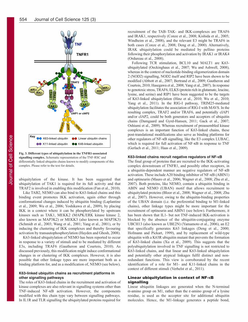
ubiquitylation of the kinase. It has been suggested thatubiquitylation of TAK1 is required for its full activity and thatTRAF2 is involved in enabling this modification (Fan et al., 2010).
Like TAB2, NEMO can also bind to K63-linked chains and thisbinding event promotes IKK activation, again either throughconformational changes induced by ubiquitin binding (Laplantineet al., 2009; Wu et al., 2006; Yoshikawa et al., 2009), by placingIKK in a context where it can be phosphorylated by upstreamkinases such as TAK1, MEKK2 (MAPK/ERK kinase kinase 2,also known as MAP3K2) or MEKK3 (also known as MAP3K3)(Schmidt et al., 2003; Wang et al., 2001; Yang et al., 2001), or byinducing the clustering of IKK complexes and thereby favouringactivation by transautophosphorylation (Hayden and Ghosh, 2008).
K63-linked ubiquitylation of NEMO has been reported to occurin response to a variety of stimuli and to be mediated by differentE3s, including TRAF6 (Gautheron and Courtois, 2010). Asdiscussed previously, this modification might induce conformationalchanges in or clustering of IKK complexes. However, it is alsopossible that other linkage types are more important both as abinding platform for, and as a modification of, NEMO (see below).
K63-linked ubiquitin chains as recruitment platforms inother signalling pathwaysThe roles of K63-linked chains in the recruitment and activation ofkinase complexes are also relevant in signalling systems other thanTNF-induced NF-B activation. However, the componentsmodified with this chain type vary between signalling pathways.In IL1R and TLR signalling the ubiquitylated proteins required for
recruitment of the TAB–TAK- and IKK-complexes are TRAF6and IRAK1, respectively (Conze et al., 2008; Kishida et al., 2005;Windheim et al., 2008), and the relevant E3 might be TRAF6 inboth cases (Conze et al., 2008; Deng et al., 2000). Alternatively,IRAK ubiquitylation could be mediated by pellino proteinsfollowing their phosphorylation and activation by IRAK1 or IRAK4(Ordureau et al., 2008).
Following TCR stimulation, BCL10 and MALT1 are K63-ubiquitylated (Oeckinghaus et al., 2007; Wu and Ashwell, 2008),whereas in the context of nucleotide-binding oligomerization domain2 (NOD2) signalling, NOD2 itself and RIP2 have been shown to bemodified (Abbott et al., 2007; Bertrand et al., 2009; Gautheron andCourtois, 2010; Hasegawa et al., 2008; Yang et al., 2007). In responseto genotoxic stress, TRAF6, ELKS (protein rich in glutamate, leucine,lysine, and serine) and RIP1 have been suggested to be the targetsof K63-linked ubiquitylation (Hinz et al., 2010; Wu et al., 2010;Yang et al., 2011). In the RIG-I pathway, TRIM25-mediatedubiquitylation facilitates the association of RIG-I with MAVS. In theresulting complex, TRAF2 and/or TRAF6, and potentially cIAP1and/or cIAP2, could be both generators and acceptors of ubiquitinchains (Damgaard and Gyrd-Hansen, 2011; Gack et al., 2007;Oshiumi et al., 2009). Whereas recruitment of preassembled kinasecomplexes is an important function of K63-linked chains, thesepost-translational modifications also serve as binding platforms forother regulators of NF-B signalling, like the E3 complex LUBAC,which is required for full activation of NF-B in response to TNF(Gerlach et al., 2011; Haas et al., 2009).
K63-linked chains recruit negative regulators of NF-BThe final group of proteins that are recruited to the IKK-activatingcomplex downstream of TNFR1, and possibly other receptors, ina ubiquitin-dependent manner are negative regulators of NF-Bactivation. These include A20 binding inhibitor of NF-B (ABIN1)and optineurin (Mauro et al., 2006; Wagner et al., 2008; Zhu et al.,2007). Both proteins, like NEMO, contain a ubiquitin binding inABIN and NEMO (UBAN) motif that allows recruitment toubiquitylated proteins (Bloor et al., 2008; Wagner et al., 2008; Zhuet al., 2007). However, owing to the ubiquitin-binding propertiesof the UBAN domain (i.e. the preferential binding to M1-linkedchains), other linkage types might be more important for therecruitment of optineurin and ABIN1 (see below). Furthermore, ithas been shown that IL1- but not TNF-induced IKK-activation isblocked by the absence of the ubiquitin-conjugating enzymeUBCH13 (also known as UBE2N) (Yamamoto et al., 2006), an E2that specifically generates K63 linkages (Deng et al., 2000;Hofmann and Pickart, 1999), and by replacement of wild-typeubiquitin with a K63R ubiquitin mutant that prevents the formationof K63-linked chains (Xu et al., 2009). This suggests that thepolyubiquitylation involved in TNF signalling is not restricted toK63-linked chains, and that linear and K63-linked ubiquitylationand potentially other atypical linkages fulfil distinct and non-redundant functions. This view is corroborated by the recentidentification of a role for M1- and K11-linked chains in thecontext of different stimuli (Verhelst et al., 2011).
Linear ubiquitylation in context of NF-BsignallingLinear ubiquitin linkages are generated when the N-terminal -amino group on M1, rather than the -amino group of a lysineresidue, is used as the acceptor site for additional ubiquitinmolecules. Hence, the M1-linkage generates a peptide bond,
554 Journal of Cell Science 125 (3)
IKKαIKKβ
HOIL1
RIP1
TAK1
TAB
2TA
B1
TAK1
TAB
2TA
B1
IKKβ
RIP1
TNF
TNFR1
TRA
DD
TRAF22
cIAP1 or cIAP2
K63-linked ubiquitin Linear ubiquitin chains
K11-linked ubiquitin K48-linked ubiquitin
2222222
HO
IP
Shar
pin
βIKKα
αααααIIKK
Key
Fig. 3. Different types of ubiquitylation in the TNFR1-associatedsignalling complex. Schematic representation of the TNF-RSC anddifferentially linked ubiquitin chains known to modify components of thiscomplex. Please refer to the text for details.
Jour
nal o
f Cel
l Sci
ence

whereas all other linkage types result in the formation of isopeptidebonds. Nevertheless, M1-linked chains resemble K63-linked chainsin the overall structure, as both chain types adopt an apparentlysimilar extended conformation (Komander et al., 2009b). Althoughthese structural similarities suggest a certain degree of redundancybetween M1- and K63-linked chains, the conformation of M1-linked chains is more restrained, and it has been shown that someUBDs bind exclusively, or at least with much higher affinity, toone or the other linkage type (Kulathu et al., 2009; Lo et al., 2009;Rahighi et al., 2009).
Currently, the only E3 known to promote generation of M1linkages is LUBAC (Kirisako et al., 2006; Verhelst et al., 2011).LUBAC is an E3 complex that consists of HOIL1 (for heme-oxidized IRP2 ubiquitin ligase-1), HOIP (for HOIL1-interactingprotein) and SHARPIN (for shank-associated RH domaininteractor) (Gerlach et al., 2011; Ikeda et al., 2011; Kirisako et al.,2006; Tokunaga et al., 2011). When in complex with SHARPIN,HOIL1 or both in vitro, HOIP exclusively generates linear ubiquitinchains independently of the E2 supplied. M1-linkages were initiallyshown to mediate proteasomal degradation of a model substrate(Kirisako et al., 2006). This observation was supported by studiesthat showed that fusion of a non-cleavable linear tetra-ubiquitinchain to a target protein can lead to the degradation of this protein(Prakash et al., 2009) and that linear chains can bind the sameproteasomal receptors as K48-linked chains (Thrower et al., 2000).In the context of the RIG-I pathway, LUBAC activity has beenlinked to the degradation of TRIM25 and hence to suppression oftype I interferon induction (Inn et al., 2011).
Linear chains positively regulate NF-B signallingA positive regulatory role for linear chains in signalling firstbecame apparent when it was shown that TNF-induced NF-Bactivation is impaired when the expression of one or two of theLUBAC components are suppressed by RNA interference (Haas etal., 2009; Tokunaga et al., 2009) and when LUBAC was identifiedas a functional component of the native TNF-RSC (Haas et al.,2009; Walczak, 2011). Recruitment of LUBAC to the TNF-RSCdepends on HOIP (Gerlach et al., 2011) and the E3 ligase activityof cIAP1 and/or cIAP2, which suggests that LUBAC, throughHOIP, binds to cIAP-generated ubiquitin chains on a componentof the TNF-RSC (Haas et al., 2009). It is probable that furtherUBDs present in SHARPIN and HOIL1 enable more stableinteraction of LUBAC with the TNF-RSC once it has beenrecruited. Since the initial discovery of a role for LUBAC insignalling pathways, additional studies have revealed that itsfunction is not restricted to signalling downstream of TNFR1 andIL1R but that it also regulates CD40-, lipopolysaccharide (LPS)-and lymphotoxin- receptor (LTR)-induced signalling (Gerlachet al., 2011; Ikeda et al., 2011; Tokunaga et al., 2011) and theresponse to genotoxic stress (Niu et al., 2011).
Linear chains in signal activationIn most, if not all, of the pathways involving LUBAC, this E3ligase seems to act by a di- or possibly a tripartite mechanism.Similar to K63-linked chains, linear polyubiquitin serves as arecruitment platform for NEMO, thereby promoting IKK activation.However, binding of NEMO to K63- and linear chains differs bothquantitatively and qualitatively (Lo et al., 2009; Rahighi et al.,2009). Mutations in NEMO that affect its ability to bind linearubiquitin have been shown to lead to decreased NF-B activation(Hadian et al., 2011; Rahighi et al., 2009). Mutations in the UBAN
domain were also identified in patients suffering from X-linkedectodermal dysplasia and immunodeficiency, indicating thatalterations that interfere with the ability of NEMO to bind linearubiquitin not only inhibit the canonical pathway of NF-Bactivation on a molecular level but can also have detrimentaleffects for the whole organism (Rahighi et al., 2009).
The modification of the regulatory subunit of the IKK complexat K285 and K309 with linear ubiquitin chains (Tokunaga et al.,2009) represents the second leg of the mechanism by whichLUBAC could enhance NF-B signalling. In vitro assays haveindicated that NEMO is a direct target of LUBAC-mediatedubiquitylation (Gerlach et al., 2011; Tokunaga et al., 2009).Furthermore, the reconstitution of NEMO-deficient cells with aK285R and K309R double-mutant, in which the ubiquitylationsites are mutated, is not able to rescue the induction of NF-Bsignalling by LUBAC or IL1 (Tokunaga et al., 2009). Although theprecise role of the attachment of M1-linked chains to NEMOremains unclear, this modification might cause conformationalchanges or clustering of IKK units, thereby favouring theiractivation.
In the context of the TNF-RSC, a third aspect of positiveregulation by linear chains has been identified. The activity ofLUBAC, probably at least partially by modification of RIP1 andNEMO (and by providing a binding site for the latter), leads to anoverall stabilisation of the receptor complex (Haas et al., 2009). Byretaining RIP1, TRAF2, cIAP and TAK1 in the complex, LUBACextends the half-life of the TNF-RSC, thereby allowing enhancedIKK activation. This effect of linear ubiquitylation is probably alsoa result of linear chains being more refractory to cleavage by mostof the deubiquitylating enzymes (DUBs) that are present in thereceptor complex (Komander et al., 2009b).
Linear chains in signal terminationIn addition to positively regulating NF-B signalling, linear chainsmight also be required for efficient termination of the NF-Bresponse, as they serve as recruitment platforms for the UBAN-containing proteins ABIN1 and optineurin. It has been reportedthat optineurin interferes with TNF-induced NF-B activation bycompeting with NEMO for binding to ubiquitylated RIP1 (Zhu etal., 2007) and by assisting the DUB protein cylindromatosis(CYLD) in its negative regulatory role in TNF signalling (seeHarhaj and Dixit, 2011). A similar mode of action has beensuggested for ABIN1 which mediates the interaction of the DUBA20 with ubiquitylated NEMO, thereby negatively affectingactivation of IKKs (Harhaj and Dixit, 2011).
Further studies will be required to clarify which aspects of themechanism involving linear ubiquitin chains – namely, (1) provisionof binding platforms by ubiquitylation of RIP1, NEMO and possiblyother factors, (2) kinase activation by conformational changesinduced by NEMO ubiquitylation, and (3) stabilisation of IKK-activating complexes – are relevant in the context of a particularstimulus. In addition, it will be interesting to investigate whetherother E3s are capable of forming M1-linkages in the context ofnative signalling complexes.
Atypical polyubiquitin chains andmonoubiquitylationThere is accumulating evidence that other polyubiquitylation events,and monoubiquitylation, are also important for efficient andcontrolled NF-B activation in different contexts. It was reportedrecently, that, following TNF stimulation, cIAP1, together with
555Ubiquitylation regulates NF-B
Jour
nal o
f Cel
l Sci
ence

UBCH5 (also known as UBE2D1), generates K11-linked chains onRIP1 and that NEMO can bind to K11 and K63 linkages withsimilar affinities, which suggests a role for K11 linkages in promotingNF-B activation (Dynek et al., 2010). However, considering thatK11-linked ubiquitin chains were previously characterised as adegradative signal with roles in cell cycle regulation (Matsumoto et al., 2010), a similar effect on RIP1 and, hence, a negative regulatoryrole in TNF-signalling could also be envisioned.
Another linkage type that has been reported to influence proteincomplex assembly in NF-B signalling is K33-linked ubiquitinchain formation, which has been suggested to negatively regulateTCR signalling by targeting TCR and thereby preventing itsassociation with, and phosphorylation by, the downstream kinasezeta-chain associated protein kinase 70 kDa (ZAP70) (Huang et al., 2010). Furthermore, there are reports on the modification ofNEMO with different ubiquitin linkage types. This includes K27-linked polyubiquitylation following TLR stimulation (Arimoto et al., 2010), in which TRIM23 is the relevant E3, and the cIAP-mediated attachment of K6 linkages, which was described in thecontext of TNFR1 signalling (Tang et al., 2003). It is suspectedthat the role of at least some of the atypical ubiquitin linkages inactivating and regulating signalling pathways has so far beenunderestimated. However, further studies are required to validatethis hypothesis and to identify which linkage types are importantin which context. Similarly, enzymes involved in generating theselinkages and their targets will need to be characterised in moredetail.
The assembly of polyubiquitin is not always necessary to mediatesignalling effects, as modification with single ubiquitin moieties atone (monoubiquitylation) or several sites (multi-monoubiquitylation) can be sufficient as a signal (Hoeller et al.,2006). Indeed, it has been reported that constitutive processing ofp105 to p50 involves monoubiquitylation of p105 (Perkins, 2006).In addition, monoubiquitylation of phosphorylated andSUMOylated NEMO in the nucleus, potentially by cIAP1, leads toits export to the cytoplasm, which represents a crucial step ingenotoxic-stress-induced NF-B activation (Hadian andKrappmann, 2011).
The role of DUBs in NF-B signallingIn addition to the enzymes that mediate post-translationalmodifications, proteins that catalyse the reversal of these processesare equally important for the system to work in a stimulus-dependent and regulated manner. In the case of ubiquitylation, thisis carried out by a set of cysteine- or metallo-proteases (Harhaj andDixit, 2011; Komander et al., 2009a), the so-called DUBs. LikeE3s, DUBs can exhibit specificity for certain linkage types, whichcan be mediated by the presence of UBDs in the DUB itself, byubiquitin-binding adaptor proteins or by selectivity of the catalyticcore (Harhaj and Dixit, 2011; Komander, 2010; Komander andBarford, 2008; Komander et al., 2008).
Several DUBs have been implicated in NF-B signalling. Thisincludes the A20 ubiquitin-editing complex [components of thiscomplex as well as their specific activities are given elsewhere(Harhaj and Dixit, 2011)], CYLD, cezanne (also known asOTUD7B), ubiquitin-specific peptidase 11 (USP11), USP15 andUSP21, which all serve as negative regulators of the canonical NF-B pathway (Bremm et al., 2010; Brummelkamp et al., 2003;Heyninck et al., 1999; Jaattela et al., 1996; Kovalenko et al., 2003;Schweitzer et al., 2007; Sun et al., 2010; Trompouki et al., 2003;Xu et al., 2010).
The DUBs A20, CYLD, cezanne and USP21 work on upstreamcomplexes by removing non-proteolytic ubiquitylation fromcomponents such as RIP1, TRAF6, RIP2, NOD2 and MALT1(Harhaj and Dixit, 2011). These DUBs differ in their linkagespecificities (Wertz et al., 2004) and act in a temporally distinctmanner, but show overlapping target specificities. For instance, allfour DUBs have been shown to remove ubiquitin chains fromRIP1 in the context of TNF signalling. The reason for this overlapin target specificity between the different DUBs has not beenclarified. However, considering that mass spectrometric analysisof the TNF-RSC revealed the presence of K48-, K63-, K11- andM1-linked ubiquitin on TNF-RSC-associated RIP1 (Gerlach et al.,2011), it is an intriguing possibility that different ubiquitin linkages,attached to specific sites on RIP1, fulfil distinct functions and thattheir individual removal is mediated by specialised DUBs.
It has been reported that the two other DUBs, USP11 and USP15negatively regulate NF-B activation by removing K48-linkedchains from IB, which might serve as a fine-tuning mechanismin NF-B activation (Harhaj and Dixit, 2011). Whereas DUBs areas important for the regulation of NF-B signalling as the enzymesthat generate ubiquitin chains, our understanding of the specificitiesand mechanisms of actions of these enzymes is far from complete.Further studies are required to elucidate the complex interplaybetween different ubiquitin linkages, and to clarify the mechanismsleading to their stimulus-dependent generation on and removalfrom individual signalling complex components by specificenzymes.
Conclusions and perspectivesUbiquitylation is a major principle in the regulation of NF-Bactivation. The functional outcome of modification with ubiquitinchains does not only depend on the target protein but also on theresidues involved in forming the inter-ubiquitin linkage. K48-linked ubiquitin chains mediate both positive and negativeregulation of NF-B signalling by targeting both agonists andantagonists of this signalling cascade for proteasomal degradation.Non-proteolytic chain types, such as K63- and M1-linked chains,exert their effects by providing binding platforms for mediators ofNF-B activation, such as the IKK- and TAK–TAB-complexes,and for protein complexes involved in the termination of the signal,such as DUBs and their adaptor proteins. The roles of atypicalchain types are less well understood and further studies are requiredto characterise the generation of these chains in the context of NF-B signalling, to identify target proteins and to analysefunctional outcomes of modification with certain linkage types.
NF-B activation is crucial for raising and maintaining animmune response, but also for the appropriate termination of sucha response (Ben-Neriah and Karin, 2011). Owing to this role as acentral regulator of the immune system, aberrant activation of NF-B is associated with many acute and chronic inflammatorydiseases (Wullaert et al., 2011), and other disorders includingcancer have also been shown to be caused or aggravated bydysregulated NF-B signalling (Ben-Neriah and Karin, 2011;Kirkin and Dikic, 2011). Mutations in components of the NF-Bsignal transduction machinery (e.g. NEMO) or in proteins mediatingthe ubiquitin-dependent regulation of this pathway (e.g. CYLD)(Courtois and Gilmore, 2006; Courtois and Israel, 2011; Shifera,2010) have been associated with a variety of diseases. This makesthe NF-B pathway an attractive target for therapeutic interventionin a number of diseases. However, because of its role in a multitudeof processes required for homeostasis and survival, serious side
556 Journal of Cell Science 125 (3)
Jour
nal o
f Cel
l Sci
ence

effects have to be reckoned with. Therapeutic approaches thattarget more specific regulatory events within the pathway mighttherefore be a promising alternative. Disrupting the attachment ofa particular type of ubiquitin chain to a specific site in a definedtarget protein might be sufficient to interfere with certain outcomesof NF-B signalling, while not affecting others. However, thedesign of this kind of specific therapy requires a completeknowledge of the events taking place during activation andtermination of this pathway and of their direct and indirect effects.Further studies on the specific roles of the different types ofubiquitin linkages, as well as their interplay with other post-translational modifications, are required to truly understand thefascinating mechanisms that regulate the activity of NF-B.
FundingResearch in the Walczak laboratory is supported by grants from CancerResearch UK, the Association for International Cancer Research, theBiotechnology and Biological Sciences Research Council (ERASysBioPLUS), Ovarian Cancer Action and the European Research Council.
ReferencesAbbott, D. W., Yang, Y., Hutti, J. E., Madhavarapu, S., Kelliher, M. A. and Cantley,
L. C. (2007). Coordinated regulation of Toll-like receptor and NOD2 signaling by K63-linked polyubiquitin chains. Mol. Cell. Biol. 27, 6012-6025.
Ahmed, A. U., Moulin, M., Coumailleau, F., Wong, W. W., Miasari, M., Carter, H.,Silke, J., Cohen-Tannoudji, M., Vince, J. E. and Vaux, D. L. (2009). CARP2 deficiencydoes not alter induction of NF-kappaB by TNFalpha. Curr. Biol. 19, R15-R19.
Alvarez, S. E., Harikumar, K. B., Hait, N. C., Allegood, J., Strub, G. M., Kim, E. Y.,Maceyka, M., Jiang, H., Luo, C., Kordula, T. et al. (2010). Sphingosine-1-phosphateis a missing cofactor for the E3 ubiquitin ligase TRAF2. Nature 465, 1084-1088.
Arimoto, K., Takahashi, H., Hishiki, T., Konishi, H., Fujita, T. and Shimotohno, K.(2007). Negative regulation of the RIG-I signaling by the ubiquitin ligase RNF125.Proc. Natl. Acad. Sci. USA 104, 7500-7505.
Arimoto, K., Funami, K., Saeki, Y., Tanaka, K., Okawa, K., Takeuchi, O., Akira, S.,Murakami, Y. and Shimotohno, K. (2010). Polyubiquitin conjugation to NEMO bytriparite motif protein 23 (TRIM23) is critical in antiviral defense. Proc. Natl. Acad. Sci.USA 107, 15856-15861.
Baltimore, D. (2011). NF-kappaB is 25. Nat. Immunol. 12, 683-685.Behrends, C. and Harper, J. W. (2011). Constructing and decoding unconventional
ubiquitin chains. Nat. Struct. Mol. Biol. 18, 520-528.Ben-Neriah, Y. and Karin, M. (2011). Inflammation meets cancer, with NF-kappaB as
the matchmaker. Nat. Immunol. 12, 715-723.Bertrand, M. J., Milutinovic, S., Dickson, K. M., Ho, W. C., Boudreault, A., Durkin,
J., Gillard, J. W., Jaquith, J. B., Morris, S. J. and Barker, P. A. (2008). cIAP1 andcIAP2 facilitate cancer cell survival by functioning as E3 ligases that promote RIP1ubiquitination. Mol. Cell 30, 689-700.
Bertrand, M. J., Doiron, K., Labbe, K., Korneluk, R. G., Barker, P. A. and Saleh, M.(2009). Cellular inhibitors of apoptosis cIAP1 and cIAP2 are required for innateimmunity signaling by the pattern recognition receptors NOD1 and NOD2. Immunity30, 789-801.
Bloor, S., Ryzhakov, G., Wagner, S., Butler, P. J., Smith, D. L., Krumbach, R., Dikic,I. and Randow, F. (2008). Signal processing by its coil zipper domain activates IKKgamma. Proc. Natl. Acad. Sci. USA 105, 1279-1284.
Bremm, A., Freund, S. M. and Komander, D. (2010). Lys11-linked ubiquitin chainsadopt compact conformations and are preferentially hydrolyzed by the deubiquitinaseCezanne. Nat. Struct. Mol. Biol. 17, 939-947.
Brummelkamp, T. R., Nijman, S. M., Dirac, A. M. and Bernards, R. (2003). Loss ofthe cylindromatosis tumour suppressor inhibits apoptosis by activating NF-kappaB.Nature 424, 797-801.
Chau, V., Tobias, J. W., Bachmair, A., Marriott, D., Ecker, D. J., Gonda, D. K. andVarshavsky, A. (1989). A multiubiquitin chain is confined to specific lysine in atargeted short-lived protein. Science 243, 1576-1583.
Chen, Z. J. and Sun, L. J. (2009). Nonproteolytic functions of ubiquitin in cell signaling.Mol. Cell 33, 275-286.
Cheung, P. C., Nebreda, A. R. and Cohen, P. (2004). TAB3, a new binding partner ofthe protein kinase TAK1. Biochem. J. 378, 27-34.
Conze, D. B., Wu, C. J., Thomas, J. A., Landstrom, A. and Ashwell, J. D. (2008).Lys63-linked polyubiquitination of IRAK-1 is required for interleukin-1 receptor- andtoll-like receptor-mediated NF-kappaB activation. Mol. Cell. Biol. 28, 3538-3547.
Courtois, G. and Gilmore, T. D. (2006). Mutations in the NF-kappaB signaling pathway:implications for human disease. Oncogene 25, 6831-6843.
Courtois, G. and Israel, A. (2011). IKK regulation and human genetics. Curr. Top.Microbiol. Immunol. 349, 73-95.
Damgaard, R. B. and Gyrd-Hansen, M. (2011). Inhibitor of apoptosis (IAP) proteins inregulation of inflammation and innate immunity. Discov. Med. 11, 221-231.
Deng, L., Wang, C., Spencer, E., Yang, L., Braun, A., You, J., Slaughter, C., Pickart,C. and Chen, Z. J. (2000). Activation of the IkappaB kinase complex by TRAF6
requires a dimeric ubiquitin-conjugating enzyme complex and a unique polyubiquitinchain. Cell 103, 351-361.
Derudder, E., Dejardin, E., Pritchard, L. L., Green, D. R., Korner, M. and Baud, V.(2003). RelB/p50 dimers are differentially regulated by tumor necrosis factor-alpha andlymphotoxin-beta receptor activation: critical roles for p100. J. Biol. Chem. 278, 23278-23284.
Dikic, I. and Dotsch, V. (2009). Ubiquitin linkages make a difference. Nat. Struct. Mol.Biol. 16, 1209-10.
Dynek, J. N., Goncharov, T., Dueber, E. C., Fedorova, A. V., Izrael-Tomasevic, A.,Phu, L., Helgason, E., Fairbrother, W. J., Deshayes, K., Kirkpatrick, D. S. et al.(2010). c-IAP1 and UbcH5 promote K11-linked polyubiquitination of RIP1 in TNFsignalling. EMBO J. 29, 4198-4209.
Ea, C. K., Deng, L., Xia, Z. P., Pineda, G. and Chen, Z. J. (2006). Activation of IKKby TNFalpha requires site-specific ubiquitination of RIP1 and polyubiquitin binding byNEMO. Mol. Cell 22, 245-257.
Emmerich, C. H., Schmukle, A. C. and Walczak, H. (2011). The emerging role of linearubiquitination in cell signaling. Sci. Signal. 4, re5.
Fan, Y., Yu, Y., Shi, Y., Sun, W., Xie, M., Ge, N., Mao, R., Chang, A., Xu, G.,Schneider, M. D. et al. (2010). Lysine 63-linked polyubiquitination of TAK1 at lysine158 is required for tumor necrosis factor alpha- and interleukin-1beta-induced IKK/NF-kappaB and JNK/AP-1 activation. J. Biol. Chem. 285, 5347-5360.
Fearns, C., Pan, Q., Mathison, J. C. and Chuang, T. H. (2006). Triad3A regulatesubiquitination and proteasomal degradation of RIP1 following disruption of Hsp90binding. J. Biol. Chem. 281, 34592-34600.
Fushman, D. and Walker, O. (2010). Exploring the linkage dependence of polyubiquitinconformations using molecular modeling. J. Mol. Biol. 395, 803-814.
Gack, M. U., Shin, Y. C., Joo, C. H., Urano, T., Liang, C., Sun, L., Takeuchi, O.,Akira, S., Chen, Z., Inoue, S. et al. (2007). TRIM25 RING-finger E3 ubiquitin ligaseis essential for RIG-I-mediated antiviral activity. Nature 446, 916-920.
Gautheron, J. and Courtois, G. (2010). “Without Ub I am nothing”: NEMO as amultifunctional player in ubiquitin-mediated control of NF-kappaB activation. CellMol. Life Sci. 67, 3101-3113.
Gerlach, B., Cordier, S. M., Schmukle, A. C., Emmerich, C. H., Rieser, E., Haas, T.L., Webb, A. I., Rickard, J. A., Anderton, H., Wong, W. W. et al. (2011). Linearubiquitination prevents inflammation and regulates immune signalling. Nature 471,591-596.
Haas, T. L., Emmerich, C. H., Gerlach, B., Schmukle, A. C., Cordier, S. M., Rieser,E., Feltham, R., Vince, J., Warnken, U., Wenger, T. et al. (2009). Recruitment of thelinear ubiquitin chain assembly complex stabilizes the TNF-R1 signaling complex andis required for TNF-mediated gene induction. Mol. Cell 36, 831-844.
Hadian, K. and Krappmann, D. (2011). Signals from the nucleus: activation of NF-kappaB by cytosolic ATM in the DNA damage response. Sci. Signal. 4, pe2.
Hadian, K., Griesbach, R. A., Dornauer, S., Wanger, T. M., Nagel, D., Metlitzky, M.,Beisker, W., Schmidt-Supprian, M. and Krappmann, D. (2011). NF-{kappa}BEssential Modulator (NEMO) Interaction with Linear and Lys-63 Ubiquitin ChainsContributes to NF-{kappa}B Activation. J. Biol. Chem. 286, 26107-26117.
Harhaj, E. W. and Dixit, V. M. (2011). Deubiquitinases in the regulation of NF-kappaBsignaling. Cell Res. 21, 22-39.
Hasegawa, M., Fujimoto, Y., Lucas, P. C., Nakano, H., Fukase, K., Nunez, G. andInohara, N. (2008). A critical role of RICK/RIP2 polyubiquitination in Nod-inducedNF-kappaB activation. EMBO J. 27, 373-383.
Hayden, M. S. and Ghosh, S. (2008). Shared principles in NF-kappaB signaling. Cell132, 344-362.
Heyninck, K., De Valck, D., Vanden Berghe, W., Van Criekinge, W., Contreras, R.,Fiers, W., Haegeman, G. and Beyaert, R. (1999). The zinc finger protein A20 inhibitsTNF-induced NF-kappaB-dependent gene expression by interfering with an RIP- orTRAF2-mediated transactivation signal and directly binds to a novel NF-kappaB-inhibiting protein ABIN. J. Cell Biol. 145, 1471-1482.
Hinz, M., Stilmann, M., Arslan, S. C., Khanna, K. K., Dittmar, G. and Scheidereit,C. (2010). A cytoplasmic ATM-TRAF6-cIAP1 module links nuclear DNA damagesignaling to ubiquitin-mediated NF-kappaB activation. Mol. Cell 40, 63-74.
Hochstrasser, M. (2006). Lingering mysteries of ubiquitin-chain assembly. Cell 124, 27-34.
Hoeller, D., Crosetto, N., Blagoev, B., Raiborg, C., Tikkanen, R., Wagner, S., Kowanetz,K., Breitling, R., Mann, M., Stenmark, H. et al. (2006). Regulation of ubiquitin-binding proteins by monoubiquitination. Nat. Cell Biol. 8, 163-169.
Hofmann, R. M. and Pickart, C. M. (1999). Noncanonical MMS2-encoded ubiquitin-conjugating enzyme functions in assembly of novel polyubiquitin chains for DNArepair. Cell 96, 645-653.
Hoffmann, A. and Baltimore, D. (2006). Circuitry of nuclear factor kappaB signaling.Immunol. Rev. 210, 171-186.
Huang, H., Jeon, M. S., Liao, L., Yang, C., Elly, C., Yates, J. R., 3rd. and Liu, Y. C.(2010). K33-linked polyubiquitination of T cell receptor-zeta regulates proteolysis-independent T cell signaling. Immunity 33, 60-70.
Ikeda, F., Deribe, Y. L., Skanland, S. S., Stieglitz, B., Grabbe, C., Franz-Wachtel, M.,van Wijk, S. J., Goswami, P., Nagy, V., Terzic, J. et al. (2011). SHARPIN forms alinear ubiquitin ligase complex regulating NF-kappaB activity and apoptosis. Nature471, 637-641.
Inn, K. S., Gack, M. U., Tokunaga, F., Shi, M., Wong, L. Y., Iwai, K. and Jung, J. U.(2011). Linear ubiquitin assembly complex negatively regulates RIG-I- and TRIM25-mediated type I interferon induction. Mol. Cell 41, 354-365.
Jaattela, M., Mouritzen, H., Elling, F. and Bastholm, L. (1996). A20 zinc finger proteininhibits TNF and IL-1 signaling. J. Immunol. 156, 1166-1173.
557Ubiquitylation regulates NF-B
Jour
nal o
f Cel
l Sci
ence

Kanarek, N., London, N., Schueler-Furman, O. and Ben-Neriah, Y. (2010).Ubiquitination and degradation of the inhibitors of NF-kappaB. Cold Spring Harb.Perspect Biol. 2, a000166.
Kanayama, A., Seth, R. B., Sun, L., Ea, C. K., Hong, M., Shaito, A., Chiu, Y. H., Deng,L. and Chen, Z. J. (2004). TAB2 and TAB3 activate the NF-kappaB pathway throughbinding to polyubiquitin chains. Mol. Cell 15, 535-548.
Karin, M. and Ben-Neriah, Y. (2000). Phosphorylation meets ubiquitination: the controlof NF-[kappa]B activity. Annu. Rev. Immunol. 18, 621-663.
Kirisako, T., Kamei, K., Murata, S., Kato, M., Fukumoto, H., Kanie, M., Sano, S.,Tokunaga, F., Tanaka, K. and Iwai, K. (2006). A ubiquitin ligase complex assembleslinear polyubiquitin chains. EMBO J. 25, 4877-4887.
Kirkin, V. and Dikic, I. (2011). Ubiquitin networks in cancer. Curr. Opin. Genet. Dev. 21,21-28.
Kishida, S., Sanjo, H., Akira, S., Matsumoto, K. and Ninomiya-Tsuji, J. (2005). TAK1-binding protein 2 facilitates ubiquitination of TRAF6 and assembly of TRAF6 withIKK in the IL-1 signaling pathway. Genes Cells 10, 447-454.
Komander, D. (2009). The emerging complexity of protein ubiquitination. Biochem. Soc.Trans. 37, 937-953.
Komander, D. (2010). Mechanism, specificity and structure of the deubiquitinases. SubcellBiochem. 54, 69-87.
Komander, D. and Barford, D. (2008). Structure of the A20 OTU domain and mechanisticinsights into deubiquitination. Biochem. J. 409, 77-85.
Komander, D., Lord, C. J., Scheel, H., Swift, S., Hofmann, K., Ashworth, A. andBarford, D. (2008). The structure of the CYLD USP domain explains its specificity forLys63-linked polyubiquitin and reveals a B box module. Mol. Cell 29, 451-464.
Komander, D., Clague, M. J. and Urbe, S. (2009a). Breaking the chains: structure andfunction of the deubiquitinases. Nat. Rev. Mol. Cell. Biol. 10, 550-563.
Komander, D., Reyes-Turcu, F., Licchesi, J. D., Odenwaelder, P., Wilkinson, K. D.and Barford, D. (2009b). Molecular discrimination of structurally equivalent Lys 63-linked and linear polyubiquitin chains. EMBO Rep. 10, 466-473.
Kovalenko, A., Chable-Bessia, C., Cantarella, G., Israel, A., Wallach, D. and Courtois,G. (2003). The tumour suppressor CYLD negatively regulates NF-kappaB signallingby deubiquitination. Nature 424, 801-805.
Kulathu, Y., Akutsu, M., Bremm, A., Hofmann, K. and Komander, D. (2009). Two-sided ubiquitin binding explains specificity of the TAB2 NZF domain. Nat. Struct. Mol.Biol. 16, 1328-1330.
Laplantine, E., Fontan, E., Chiaravalli, J., Lopez, T., Lakisic, G., Veron, M., Agou, F.and Israel, A. (2009). NEMO specifically recognizes K63-linked poly-ubiquitin chainsthrough a new bipartite ubiquitin-binding domain. EMBO J. 28, 2885-2895.
Lee, T. H., Shank, J., Cusson, N. and Kelliher, M. A. (2004). The kinase activity of Rip1is not required for tumor necrosis factor-alpha-induced IkappaB kinase or p38 MAPkinase activation or for the ubiquitination of Rip1 by Traf2. J. Biol. Chem. 279, 33185-33191.
Li, H., Kobayashi, M., Blonska, M., You, Y. and Lin, X. (2006). Ubiquitination of RIPis required for tumor necrosis factor alpha-induced NF-kappaB activation. J. Biol.Chem. 281, 13636-13643.
Li, S., Wang, L. and Dorf, M. E. (2009). PKC phosphorylation of TRAF2 mediatesIKKalpha/beta recruitment and K63-linked polyubiquitination. Mol. Cell 33, 30-42.
Liao, G., Zhang, M., Harhaj, E. W. and Sun, S. C. (2004). Regulation of the NF-kappaB-inducing kinase by tumor necrosis factor receptor-associated factor 3-induceddegradation. J. Biol. Chem. 279, 26243-26250.
Liao, W., Xiao, Q., Tchikov, V., Fujita, K., Yang, W., Wincovitch, S., Garfield, S.,Conze, D., El-Deiry, W. S., Schutze, S. et al. (2008). CARP-2 is an endosome-associated ubiquitin ligase for RIP and regulates TNF-induced NF-kappaB activation.Curr. Biol. 18, 641-649.
Liu, S. and Chen, Z. J. (2011). Expanding role of ubiquitination in NF-kappaB signaling.Cell Res. 21, 6-21.
Lo, Y. C., Lin, S. C., Rospigliosi, C. C., Conze, D. B., Wu, C. J., Ashwell, J. D., Eliezer,D. and Wu, H. (2009). Structural basis for recognition of diubiquitins by NEMO. Mol.Cell 33, 602-615.
Matsumoto, M. L., Wickliffe, K. E., Dong, K. C., Yu, C., Bosanac, I., Bustos, D., Phu,L., Kirkpatrick, D. S., Hymowitz, S. G., Rape, M. et al. (2010). K11-linkedpolyubiquitination in cell cycle control revealed by a K11 linkage-specific antibody.Mol. Cell 39, 477-484.
Matsuzawa, A., Tseng, P. H., Vallabhapurapu, S., Luo, J. L., Zhang, W., Wang, H.,Vignali, D. A., Gallagher, E. and Karin, M. (2008). Essential cytoplasmic translocationof a cytokine receptor-assembled signaling complex. Science 321, 663-668.
Mauro, C., Pacifico, F., Lavorgna, A., Mellone, S., Iannetti, A., Acquaviva, R.,Formisano, S., Vito, P. and Leonardi, A. (2006). ABIN-1 binds to NEMO/IKKgammaand co-operates with A20 in inhibiting NF-kappaB. J. Biol. Chem. 281, 18482-18488.
Miyamoto, S. (2011). Nuclear initiated NF-kappaB signaling: NEMO and ATM takecenter stage. Cell Res. 21, 116-130.
Moynagh, P. N. (2009). The Pellino family: IRAK E3 ligases with emerging roles ininnate immune signalling. Trends Immunol. 30, 33-42.
Newton, K., Matsumoto, M. L., Wertz, I. E., Kirkpatrick, D. S., Lill, J. R., Tan, J.,Dugger, D., Gordon, N., Sidhu, S. S., Fellouse, F. A. et al. (2008). Ubiquitin chainediting revealed by polyubiquitin linkage-specific antibodies. Cell 134, 668-678.
Niu, J., Shi, Y., Iwai, K. and Wu, Z. H. (2011). LUBAC regulates NF-kappaB activationupon genotoxic stress by promoting linear ubiquitination of NEMO. EMBO J. 30, 3741-3753.
O’Dea, E. and Hoffmann, A. (2009). NF-kappaB signaling. Wiley Interdiscip. Rev. Syst.Biol. Med. 1, 107-115.
Oeckinghaus, A. and Ghosh, S. (2009). The NF-kappaB family of transcription factorsand its regulation. Cold Spring Harb. Perspect. Biol. 1, a000034.
Oeckinghaus, A., Wegener, E., Welteke, V., Ferch, U., Arslan, S. C., Ruland, J.,Scheidereit, C. and Krappmann, D. (2007). Malt1 ubiquitination triggers NF-kappaBsignaling upon T-cell activation. EMBO J. 26, 4634-4645.
Ordureau, A., Smith, H., Windheim, M., Peggie, M., Carrick, E., Morrice, N. andCohen, P. (2008). The IRAK-catalysed activation of the E3 ligase function of Pellinoisoforms induces the Lys63-linked polyubiquitination of IRAK1. Biochem. J. 409, 43-52.
Oshiumi, H., Matsumoto, M., Hatakeyama, S. and Seya, T. (2009). Riplet/RNF135, aRING finger protein, ubiquitinates RIG-I to promote interferon-beta induction duringthe early phase of viral infection. J. Biol. Chem. 284, 807-817.
Park, S. M., Yoon, J. B. and Lee, T. H. (2004). Receptor interacting protein is ubiquitinatedby cellular inhibitor of apoptosis proteins (c-IAP1 and c-IAP2) in vitro. FEBS Lett. 566,151-156.
Passmore, L. A. and Barford, D. (2004). Getting into position: the catalytic mechanismsof protein ubiquitylation. Biochem. J. 379, 513-525.
Perkins, N. D. (2006). Post-translational modifications regulating the activity and functionof the nuclear factor kappa B pathway. Oncogene 25, 6717-6730.
Pickart, C. M. (1997). Targeting of substrates to the 26S proteasome. FASEB J. 11, 1055-1066.
Prakash, S., Inobe, T., Hatch, A. J. and Matouschek, A. (2009). Substrate selection bythe proteasome during degradation of protein complexes. Nat. Chem. Biol. 5, 29-36.
Rahighi, S., Ikeda, F., Kawasaki, M., Akutsu, M., Suzuki, N., Kato, R., Kensche, T.,Uejima, T., Bloor, S., Komander, D. et al. (2009). Specific recognition of linearubiquitin chains by NEMO is important for NF-kappaB activation. Cell 136, 1098-1109.
Razani, B., Zarnegar, B., Ytterberg, A. J., Shiba, T., Dempsey, P. W., Ware, C. F., Loo,J. A. and Cheng, G. (2010). Negative feedback in noncanonical NF-kappaB signalingmodulates NIK stability through IKKalpha-mediated phosphorylation. Sci. Signal. 3,ra41.
Rothwarf, D. M., Zandi, E., Natoli, G. and Karin, M. (1998). IKK-gamma is anessential regulatory subunit of the IkappaB kinase complex. Nature 395, 297-300.
Ruland, J. (2011). Return to homeostasis: downregulation of NF-kappaB responses. Nat.Immunol. 12, 709-714.
Schmidt, C., Peng, B., Li, Z., Sclabas, G. M., Fujioka, S., Niu, J., Schmidt-Supprian,M., Evans, D. B., Abbruzzese, J. L. and Chiao, P. J. (2003). Mechanisms ofproinflammatory cytokine-induced biphasic NF-kappaB activation. Mol. Cell 12, 1287-1300.
Schmitz, M. L., Bacher, S. and Kracht, M. (2001). I kappa B-independent control ofNF-kappa B activity by modulatory phosphorylations. Trends Biochem. Sci. 26, 186-190.
Schweitzer, K., Bozko, P. M., Dubiel, W. and Naumann, M. (2007). CSN controls NF-kappaB by deubiquitinylation of IkappaBalpha. EMBO J. 26, 1532-1541.
Sebban, H., Yamaoka, S. and Courtois, G. (2006). Posttranslational modifications ofNEMO and its partners in NF-kappaB signaling. Trends Cell. Biol. 16, 569-577.
Shi, C. S. and Kehrl, J. H. (2003). Tumor necrosis factor (TNF)-induced germinal centerkinase-related (GCKR) and stress-activated protein kinase (SAPK) activation dependsupon the E2/E3 complex Ubc13-Uev1A/TNF receptor-associated factor 2 (TRAF2). J.Biol. Chem. 278, 15429-15434.
Shibuya, H., Yamaguchi, K., Shirakabe, K., Tonegawa, A., Gotoh, Y., Ueno, N., Irie,K., Nishida, E. and Matsumoto, K. (1996). TAB1: an activator of the TAK1 MAPKKKin TGF-beta signal transduction. Science 272, 1179-1182.
Shifera, A. S. (2010). The zinc finger domain of IKKgamma (NEMO) protein in healthand disease. J. Cell. Mol. Med. 14, 2404-2414.
Shih, V. F., Tsui, R., Caldwell, A. and Hoffmann, A. (2011). A single NFkappaB systemfor both canonical and non-canonical signaling. Cell Res. 21, 86-102.
Shirane, M., Hatakeyama, S., Hattori, K. and Nakayama, K. (1999). Common pathwayfor the ubiquitination of IkappaBalpha, IkappaBbeta, and IkappaBepsilon mediated bythe F-box protein FWD1. J. Biol. Chem. 274, 28169-28174.
Sun, S. C. (2011). Non-canonical NF-kappaB signaling pathway. Cell Res. 21, 71-85.Sun, W., Tan, X., Shi, Y., Xu, G., Mao, R., Gu, X., Fan, Y., Yu, Y., Burlingame, S.,
Zhang, H. et al. (2010). USP11 negatively regulates TNFalpha-induced NF-kappaBactivation by targeting on IkappaBalpha. Cell Signal. 22, 386-394.
Tang, E. D., Wang, C. Y., Xiong, Y. and Guan, K. L. (2003). A role for NF-kappaBessential modifier/IkappaB kinase-gamma (NEMO/IKKgamma) ubiquitination in theactivation of the IkappaB kinase complex by tumor necrosis factor-alpha. J. Biol. Chem.278, 37297-37305.
Thrower, J. S., Hoffman, L., Rechsteiner, M. and Pickart, C. M. (2000). Recognitionof the polyubiquitin proteolytic signal. EMBO J. 19, 94-102.
Tokunaga, F., Sakata, S., Saeki, Y., Satomi, Y., Kirisako, T., Kamei, K., Nakagawa,T., Kato, M., Murata, S., Yamaoka, S. et al. (2009). Involvement of linearpolyubiquitylation of NEMO in NF-kappaB activation. Nat. Cell Biol. 11, 123-132.
Tokunaga, F., Nakagawa, T., Nakahara, M., Saeki, Y., Taniguchi, M., Sakata, S.,Tanaka, K., Nakano, H. and Iwai, K. (2011). SHARPIN is a component of the NF-kappaB-activating linear ubiquitin chain assembly complex. Nature 471, 633-636.
Trompouki, E., Hatzivassiliou, E., Tsichritzis, T., Farmer, H., Ashworth, A. andMosialos, G. (2003). CYLD is a deubiquitinating enzyme that negatively regulates NF-kappaB activation by TNFR family members. Nature 424, 793-796.
Vallabhapurapu, S., Matsuzawa, A., Zhang, W., Tseng, P. H., Keats, J. J., Wang, H.,Vignali, D. A., Bergsagel, P. L. and Karin, M. (2008). Nonredundant andcomplementary functions of TRAF2 and TRAF3 in a ubiquitination cascade thatactivates NIK-dependent alternative NF-kappaB signaling. Nat. Immunol. 9, 1364-1370.
558 Journal of Cell Science 125 (3)
Jour
nal o
f Cel
l Sci
ence
van Nocker, S. and Vierstra, R. D. (1993). Multiubiquitin chains linked through lysine48 are abundant in vivo and are competent intermediates in the ubiquitin proteolyticpathway. J. Biol. Chem. 268, 24766-24773.
Verhelst, K., Verstrepen, L., Carpentier, I. and Beyaert, R. (2011). Linear ubiquitinationin NF-kappaB signaling and inflammation: what we do understand and what we do not.Biochem. Pharmacol. 82, 1057-1065.
Viatour, P., Merville, M. P., Bours, V. and Chariot, A. (2005). Phosphorylation of NF-kappaB and IkappaB proteins: implications in cancer and inflammation. Trends Biochem.Sci. 30, 43-52.
Vince, J. E., Chau, D., Callus, B., Wong, W. W., Hawkins, C. J., Schneider, P.,McKinlay, M., Benetatos, C. A., Condon, S. M., Chunduru, S. K. et al. (2008).TWEAK-FN14 signaling induces lysosomal degradation of a cIAP1-TRAF2 complexto sensitize tumor cells to TNFalpha. J. Cell Biol. 182, 171-184.
Vince, J. E., Pantaki, D., Feltham, R., Mace, P. D., Cordier, S. M., Schmukle, A. C.,Davidson, A. J., Callus, B. A., Wong, W. W., Gentle, I. E. et al. (2009). TRAF2 mustbind to cellular inhibitors of apoptosis for tumor necrosis factor (tnf) to efficientlyactivate nf-{kappa}b and to prevent tnf-induced apoptosis. J. Biol. Chem. 284, 35906-35915.
Wagner, S., Carpentier, I., Rogov, V., Kreike, M., Ikeda, F., Lohr, F., Wu, C. J.,Ashwell, J. D., Dotsch, V., Dikic, I. et al. (2008). Ubiquitin binding mediates the NF-kappaB inhibitory potential of ABIN proteins. Oncogene 27, 3739-3745.
Wajant, H. and Scheurich, P. (2011). TNFR1-induced activation of the classical NF-kappaB pathway. FEBS J. 278, 862-876.
Walczak, H. (2011). TNF and ubiquitin at the crossroads of gene activation, cell death,inflammation, and cancer. Immunol. Rev. 244, 9-28.
Wang, C., Deng, L., Hong, M., Akkaraju, G. R., Inoue, J. and Chen, Z. J. (2001).TAK1 is a ubiquitin-dependent kinase of MKK and IKK. Nature 412, 346-351.
Wertz, I. E. and Dixit, V. M. (2010). Signaling to NF-kappaB: regulation by ubiquitination.Cold Spring Harb. Perspect. Biol. 2, a003350.
Wertz, I. E., O’Rourke, K. M., Zhou, H., Eby, M., Aravind, L., Seshagiri, S., Wu, P.,Wiesmann, C., Baker, R., Boone, D. L. et al. (2004). De-ubiquitination and ubiquitinligase domains of A20 downregulate NF-kappaB signalling. Nature 430, 694-699.
Windheim, M., Stafford, M., Peggie, M. and Cohen, P. (2008). Interleukin-1 (IL-1)induces the Lys63-linked polyubiquitination of IL-1 receptor-associated kinase 1 tofacilitate NEMO binding and the activation of IkappaBalpha kinase. Mol. Cell. Biol. 28,1783-1791.
Wu, C. and Ghosh, S. (1999). beta-TrCP mediates the signal-induced ubiquitination ofIkappaBbeta. J. Biol. Chem. 274, 29591-29594.
Wu, C. J. and Ashwell, J. D. (2008). NEMO recognition of ubiquitinated Bcl10 isrequired for T cell receptor-mediated NF-kappaB activation. Proc. Natl. Acad. Sci. USA105, 3023-3028.
Wu, C. J., Conze, D. B., Li, T., Srinivasula, S. M. and Ashwell, J. D. (2006). Sensingof Lys 63-linked polyubiquitination by NEMO is a key event in NF-kappaB activation[corrected]. Nat. Cell. Biol. 8, 398-406.
Wu, Z. H., Wong, E. T., Shi, Y., Niu, J., Chen, Z., Miyamoto, S. and Tergaonkar, V.(2010). ATM- and NEMO-dependent ELKS ubiquitination coordinates TAK1-mediatedIKK activation in response to genotoxic stress. Mol. Cell 40, 75-86.
Wullaert, A., Bonnet, M. C. and Pasparakis, M. (2011). NF-kappaB in the regulationof epithelial homeostasis and inflammation. Cell Res. 21, 146-158.
Xiao, G., Harhaj, E. W. and Sun, S. C. (2001). NF-kappaB-inducing kinase regulates theprocessing of NF-kappaB2 p100. Mol. Cell 7, 401-409.
Xiao, G., Fong, A. and Sun, S. C. (2004). Induction of p100 processing by NF-kappaB-inducing kinase involves docking IkappaB kinase alpha (IKKalpha) to p100 andIKKalpha-mediated phosphorylation. J. Biol. Chem. 279, 30099-30105.
Xu, G., Tan, X., Wang, H., Sun, W., Shi, Y., Burlingame, S., Gu, X., Cao, G., Zhang,T., Qin, J. et al. (2010). Ubiquitin-specific peptidase 21 inhibits tumor necrosis factoralpha-induced nuclear factor kappaB activation via binding to and deubiquitinatingreceptor-interacting protein 1. J. Biol. Chem. 285, 969-978.
Xu, M., Skaug, B., Zeng, W. and Chen, Z. J. (2009). A ubiquitin replacement strategyin human cells reveals distinct mechanisms of IKK activation by TNFalpha and IL-1beta. Mol. Cell 36, 302-314.
Yamamoto, M., Okamoto, T., Takeda, K., Sato, S., Sanjo, H., Uematsu, S., Saitoh, T.,Yamamoto, N., Sakurai, H., Ishii, K. J. et al. (2006). Key function for the Ubc13 E2ubiquitin-conjugating enzyme in immune receptor signaling. Nat. Immunol. 7, 962-970.
Yang, J., Lin, Y., Guo, Z., Cheng, J., Huang, J., Deng, L., Liao, W., Chen, Z., Liu, Z.and Su, B. (2001). The essential role of MEKK3 in TNF-induced NF-kappaB activation.Nat. Immunol. 2, 620-624.
Yang, Y., Yin, C., Pandey, A., Abbott, D., Sassetti, C. and Kelliher, M. A. (2007). NOD2pathway activation by MDP or Mycobacterium tuberculosis infection involves thestable polyubiquitination of Rip2. J. Biol. Chem. 282, 36223-36229.
Yang, Y., Xia, F., Hermance, N., Mabb, A., Simonson, S., Morrissey, S., Gandhi, P.,Munson, M., Miyamoto, S. and Kelliher, M. A. (2011). A cytosolic ATM/NEMO/RIP1complex recruits TAK1 to mediate the NF-kappaB and p38 mitogen-activated proteinkinase (MAPK)/MAPK-activated protein 2 responses to DNA damage. Mol. Cell. Biol.31, 2774-2786.
Yaron, A., Hatzubai, A., Davis, M., Lavon, I., Amit, S., Manning, A. M., Andersen, J.S., Mann, M., Mercurio, F. and Ben-Neriah, Y. (1998). Identification of the receptorcomponent of the IkappaBalpha-ubiquitin ligase. Nature 396, 590-594.
Yoshikawa, A., Sato, Y., Yamashita, M., Mimura, H., Yamagata, A. and Fukai, S.(2009). Crystal structure of the NEMO ubiquitin-binding domain in complex with Lys63-linked di-ubiquitin. FEBS Lett. 583, 3317-3322.
Zarnegar, B., Yamazaki, S., He, J. Q. and Cheng, G. (2008). Control of canonical NF-kappaB activation through the NIK-IKK complex pathway. Proc. Natl. Acad. Sci. USA105, 3503-3508.
Zheng, C., Yin, Q. and Wu, H. (2011). Structural studies of NF-kappaB signaling. CellRes. 21, 183-195.
Zhu, G., Wu, C. J., Zhao, Y. and Ashwell, J. D. (2007). Optineurin negatively regulatesTNFalpha- induced NF-kappaB activation by competing with NEMO for ubiquitinatedRIP. Curr. Biol. 17, 1438-1443.
559Ubiquitylation regulates NF-B
Jour
nal o
f Cel
l Sci
ence


