Embed Size (px)
Citation preview

Journal of Magnetic Resonance154,28–45 (2002)doi:10.1006/jmre.2001.2454, available onlineat http://www.idealibrary.com on
Specification and Visualization of Anisotropic InteractionTensors in Polypeptides and Numerical Simulations
in Biological Solid-State NMR
Mads Bak, Robert Schultz, Thomas Vosegaard, and Niels Chr. Nielsen1
Laboratory for Biomolecular NMR Spectroscopy, Department of Molecular and Structural Biology, University of Aarhus, DK-8000 Aarhus C, Denmark
Received May 12, 2001; revised October 3, 2001; published online November 29, 2001
Software facilitating numerical simulation of solid-state NMRexperiments on polypeptides is presented. The Tcl-controlledSIMMOL program reads in atomic coordinates in the PDB for-mat from which it generates typical or user-defined parameters forthe chemical shift, J coupling, quadrupolar coupling, and dipolarcoupling tensors. The output is a spin system file for numericalsimulations, e.g., using SIMPSON (Bak, Rasmussen, and Nielsen,J. Magn. Reson. 147, 296 (2000)), as well as a 3D visualizationof the molecular structure, or selected parts of this, with user-controlled representation of relevant tensors, bonds, atoms, peptideplanes, and coordinate systems. The combination of SIMPSON andSIMMOL allows straightforward simulation of the response of ad-vanced solid-state NMR experiments on typical nuclear spin inter-actions present in polypeptides. Thus, SIMMOL may be considereda “sample changer” to the SIMPSON “computer spectrometer” andproves to be very useful for the design and optimization of pulse se-quences for application on uniformly or extensively isotope-labeledpeptides where multiple-spin interactions need to be considered.These aspects are demonstrated by optimization and simulation ofnovel DCP and C7 based 2D N(CO)CA, N(CA)CB, and N(CA)CXMAS correlation experiments for multiple-spin clusters in ubiquitinand by simulation of PISA wheels from PISEMA spectra of uniaxi-ally oriented bacteriorhodopsin and rhodopsin under conditions offinite RF pulses and multiple spin interactions. C© 2002 Elsevier Science
oc
o
lh,
o
me-sis ofver-
beenodelherhus,ar-
en
ngon of
elythatspinim-igner-nifi-le-
opicsly,di-ulsediesinin-uild-ntti-mesin
ex-uresof
INTRODUCTION
Numerical simulations play an increasingly important rolethe development and application of solid-state NMR methfor determination of the structure and dynamics of biologimacromolecules immobilized by size, aggregation, or mebrane association. This is ascribed to the fact that most sstate NMR experiments used for this purpose strongly depon manipulation of anisotropic interactions to obtain evotion under well-defined isotropic or anisotropic parts of tHamiltonian. Based on advanced RF irradiation schemesten in concert with sample spinning, a large number of pusequences have been devised to accomplish specific c
1 To whom correspondence should be addressed. E-mail: [email protected] .
281090-7807/02 $35.00C© 2002 Elsevier ScienceAll rights reserved.
indsalm-lid-endu-eof-
lseher-
.dk.
ence transfers or measurement of specific structural paraters. Typically, these elements have been developed on baone- or two-spin systems using analytical tools such as aage Hamiltonian theory (1, 2) to tailor the effective Hamiltonianto the appropriate form. Subsequently, the elements havetested by numerical simulations and by experiments on msystems and finally verified in real peptide applications eitdirectly or as elements in more advanced pulse schemes. Tin the development process numerical simulations have primily been used for verification while they so far have only beused sparsely for direct design of pulse sequences (3, 4). In con-trast, numerical simulations in combination with iterative fittiare regarded as being almost indispensable for the extractistructural parameters from experimental spectra (5–15).
Considering the increasing use of uniformly or extensiv13C,15N-labeled samples, it becomes exceedingly importantthe pulse sequence elements work appropriately in multiple-systems with characteristics potentially being far from the sple one- or two-spin cases typically considered in the desof these elements. Under multiple-spin conditions vital cohences may leak to undesired spins which may cause a sigcant reduction in the sensitivity, wrong assignment of multipdimensional spectra, and wrong interpretations of anisotrinteractions in terms of structure and dynamics. Obviouthis problem may scale dramatically with the number ofmensions and coherence transfer steps involved in the psequence. This is an important issue since current remeto the resolution problem of biological solid-state NMR,addition to uniformly labeled samples, appear to involvecreasingly sophisticated combinations of pulse sequence bing blocks in multiple-dimensional experiments. An importaingredient in the solution of this problem may be to invesgate in detail the performance of the available pulse scheon multiple-spin systems closely reflecting the conditionsrelevant peptide structures. This may provide optimizedperimental procedures, motivate the design of new procedon a multiple-spin basis, or result in the recommendationalternative isotope labeling procedures (16) being optimallycompatible with state-of-the-art solid-state NMR technology

i
a
s
o
t
x
hiseom-hileiled
arsattheomthe
edr-icsm-ble
ateinthehed
ck-
om
lec-
tond-om
NUMERICAL SIMULATION OF BIOLOGIC
Facing the facts that (i) analytical pulse sequence evaluatbecome exceedingly difficult in multiple-spin cases, (ii) itnecessary to consider the combined action of the variousments forming the full pulse sequence, and (iii) experimenevaluations require large amounts of spectrometer time anvariety of samples with different structure/labeling, it appeareasonable to conduct such evaluations numerically. Preferthis should be accomplished using software which allowseasy interchange of pulse sequencesandmultiple-spin systemscorresponding to different structures. The recently introducSIMPSON “computer spectrometer” (17) serves most of theneeds for efficient evaluation of advanced multiple-pulsequences on multiple-spin systems. In this paper we preseprogram, SIMMOL, which enables straightforward establisment of the anisotropic interaction tensors required for the eluation of pulse sequences on relevant peptide structures uSIMPSON. Furthermore, SIMMOL provides 3D visualizatioof the molecular structure with user-defined representationrelevant atoms, bonds, peptide planes, coordinate systemsanisotropic interaction tensors. In this regard, we should nthat SIMMOL is a greatly enhanced successor for the prelimnar Octave (18) implemented PDB2SIMPSON program recentintroduced for optimization of biological solid-state NMR experiments (19).
THE SIMMOL PROGRAM
The main idea behind the SIMMOL program is sketchedFig. 1. Based on the atomic coordinates for a molecular struc(Fig. 1a), SIMMOL assigns elements such as peptide planestypical anisotropic interaction tensors to user-specified partthe structure leading to a specific visual representation alwith a file containing spin system parameters in the Tcl fmat of a typical SIMPSON input file (Fig. 1b). Using this ana given pulse sequence (visualized using SIMDPS), SIMPSenables straightforward calculation of the corresponding NMspectrum or relevant coherence transfer efficiencies as incluin a SIMPLOT representation in Fig. 1c. Provided that taim is structure determination, rather than pulse sequencesign/optimization, the simulated spectrum may be comparea corresponding experimental spectrum using nonlinear mmization to obtain structural parameters (Fig. 1d). We note tthis paper focuses primarily on the SIMMOL steps in the uper row of Fig. 1 and we restrict to give examples on the utilof this tool for optimization and simulation of 2D solid-staNMR experiments for rotating powders as well as uniaxiaoriented samples. The comparison to experimental spectrabe conducted using tools available in the SIMPSON softwpackage (17).
In full analogy to the SIMPSON software, SIMMOL containa Tcl interpreter (20) which gives the user a high degree of fle
ibility to control all steps from the input of atomic coordinateto the delivery of SIMPSON spin system input files and 3D iteractive graphics for optimum visualization of the moleculAL SOLID-STATE NMR EXPERIMENTS 29
onsisele-tald arsbly
the
ed
se-nt ah-va-singnof
, andotei-
ly-
intureandof
ongr-dONR
dedhede-
d toini-hatp-ityellymayare
s-
geometry along with the NMR specific interaction tensors. Tis accomplished via a relatively simple Tcl input file, with thcommands listed in Table 1. The basic features of these cmands and examples on typical operations are given below, wwe refer to the internet release (see below) for more a detadescription of all commands.
Availability, Portability, and Requirements
Prior to any description of the various features, it apperelevant to mention that the SIMMOL program is releasedhttp://nmr.imsb.au.dk as open-source software which givesuser full access to the algorithms in the program and freedto compile, correct, modify, and extend the program underterms of the GNU General Public License (23). The source codecan be compiled on any platform with a C++ compiler and aTcl language interpreter (open-source software (20)). This en-sures full operation with respect to establishing thespinsyspartof the SIMPSON input file, while the open-source Unix-basGeomview (24) program is required for interpretation and inteactive 3D visualization of the OOGL (object oriented graphlanguage) output files. Finally, we should mention that precopiled and self-contained binary executables are freely availafor various Unix platforms (including Linux/i386).
Initialization
A first step in numerical evaluation of advanced solid-stNMR experiments, for applications on multiple-spin systemspolypeptides, is to read in atomic coordinates representingstructure elements to be investigated. This may be accomplisusing the Tcl command
set m [mload filename]
loading a PDB file from the disk (the command in square braets) into a descriptorm (using theset Tcl command). The PDBfile may represent a real polypeptide structure obtained frthe PDB database (25) (e.g., XRD, NMR, or cryoelectron mi-croscopy data) or synthetic structures obtained by, e.g., moular modeling (26). Alternatively, it is possible to use
set m [mmake nres phi psi omega]
to create an ideal poly-L-alanine peptide withnres residues andthe torsion angles specified.
Prior to further operation, it may be relevant (optional)specify the output files for the Geomview 3D visualization athespinsys part of the SIMPSON input file. If proton coordinates are not available (as is typically the case for PDB files fr
α
sn-arXRD studies), it may be relevant to add amide and Hprotons tothe molecular structure. Furthermore, it may be relevant to orientthe molecule with the long axis alongz potentially followed by

riments
30 BAK ET AL.
FIG. 1. Schematic representation of the combination of SIMMOL and SIMPSON for numerical simulation of multiple-pulse solid-state NMR expeon polypeptide structures. (a) Atomic coordinates for a molecular structure in the PDB format, (b) typical SIMMOL interpretation of the structure and parameter
output to thespinsys part of the SIMPSON input file, (c) SIMDPS and SIMPLOT visualization of a typical NMR pulse sequence, a coherence transfer function,and a spectrum simulated using SIMPSON, and (d) an experimental spectrum which through SIMPSON iterative fitting may provide NMR information about themolecular structure.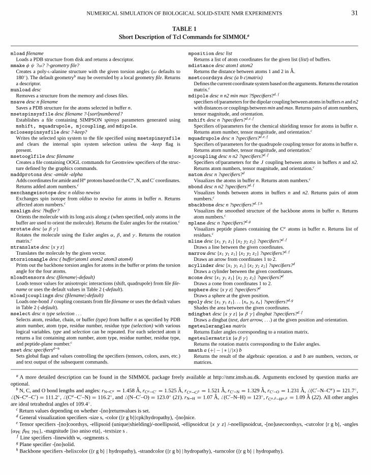
arks are
tation
,
r
r
NUMERICAL SIMULATION OF BIOLOGICAL SOLID-STATE NMR EXPERIMENTS 31
TABLE 1Short Description of Tcl Commands for SIMMOLa
mload filenameLoads a PDB structure from disk and returns a descriptor.
mmake φ ψ ?ω? ?-geometry file?Creates a poly-L-alanine structure with the given torsion angles (ω defaults to180◦). The default geometryb may be overruled by a local geometryfile. Returnsa descriptor.
munload descRemoves a structure from the memory and closes files.
msave desc n filenameSaves a PDB structure for the atoms selected in buffern.
msetspinsysfile desc filename ?-[sort]numbered?Establishes a file containing SIMPSONspinsysparameters generated usingmshift, mquadrupole, mjcoupling, andmdipole.
mclosespinsysfile desc ?-keep?Writes the selected spin system to the file specified usingmsetspinsysfileand clears the internal spin system selection unless the -keep flag ispresent.
msetooglfile desc filenameCreates a file containing OOGL commands for Geomview specifiers of the struc-ture defined by the graphics commands.
maddprotons desc -amide -alphaAdds coordinates for amide and Hα protons based on the Cα , N, and C′ coordinates.Returns added atom numbers.c
mexchangeisotope desc n oldiso newisoExchanges spin isotope fromoldiso to newisofor atoms in buffern. Returnsaffected atom numbers.c
mzalign desc ?buffer?Orients the molecule with its long axis alongz (when specified, only atoms in thebuffer are used to orient the molecule). Returns the Euler angles for the rotation.c
mrotate desc{α β γ }Rotates the molecule using the Euler anglesα, β, andγ . Returns the rotationmatrix.c
mtranslate desc{x y z}Translates the molecule by the given vector.
mtorsionangle desc ( buffer|atom1 atom2 atom3 atom4)Prints out the backbone torsion angles for atoms in the buffer or prints the torsionangle for the four atoms.
mloadtensors desc (filename|-default)Loads tensor values for anisotropic interactions (shift, quadrupole) from filefile-nameor uses the default values in Table 2 (-default).
mloadjcouplings desc (filename|-default)Loads one-bondJ coupling constants from filefilenameor uses the default valuesin Table 2 (-default).
mselect desc n type selection. . .Selects atom, residue, chain, or buffer(type)from buffern as specified by PDBatom number, atom type, residue number, residue type(selection)with variouslogical variables.typeandselectioncan be repeated. For each selected atom itreturns a list containing atom number, atom type, residue number, residue type,and peptide-plane number.c
mset desc specifiersd–h
Sets global flags and values controlling the specifiers (tensors, colors, axes, etc.)and text output of the subsequent commands.
a A more detailed description can be found in the SIMMOL package freely available at http://nmr.imsb.au.dk. Arguments enclosed by question moptional.
b N, C, and O bond lengths and angles:rN–Cα = 1.458 Aa, rCα–C′ = 1.525 A
a, rCα–Cβ = 1.521 A
a, rC′–N = 1.329 Aa, rC′–O = 1.231 Aa, 6 (C′–N–Cα) = 121.7◦,
6 (N–Cα–C′) = 111.2◦, 6 (Cα–C′–N) = 116.2◦, and 6 (N–C′–O)= 123.0◦ (21). rN–H = 1.07 Aa, 6 (C′–N–H)= 123◦, rCα,β–Hα,β = 1.09 A
a(22). All other angles
are ideal tetrahedral angles of 109.4◦.c Return values depending on whether -[no]returnvalues is set.d General visualization specifiers -size s, -color ({r g b}|cpk|hydropathy), -[no]nice.e Tensor specifiers -[no]coordsys, -ellipsoid (unique|shielding)/-noellipsoid, -ellipsoidcut{x y z} /-noellipsoidcut, -[no]usecoordsys, -cutcolor{r g b}, -angles{αPE βPE γPE}, -magnitude{iso aniso eta}, -textsize s .
mposition desc listReturns a list of atom coordinates for the given list (list) of buffers.
mdistance desc atom1 atom2Returns the distance between atoms 1 and 2 inA
a.
msetcoordsys desc (a b c|matrix)Defines the current coordinate system based on the arguments. Returns the romatrix.c
mdipole desc n n2 min max ?Specifiers?d, f
specifiers of/parameters for the dipolar coupling between atoms in buffersn andn2with distances or couplings betweenminandmax. Returns pairs of atom numberstensor magnitude, and orientation.
mshift desc n ?specifiers?d,e, f
Specifiers of/parameters for the chemical shielding tensor for atoms in buffen.Returns atom number, tensor magnitude, and orientation.c
mquadrupole desc n ?specifiers?d,e, f
Specifiers of/parameters for the quadrupole coupling tensor for atoms in buffen.Returns atom number, tensor magnitude, and orientation.c
mjcoupling desc n n2 ?specifiers?d, f
Specifiers of/parameters for theJ coupling between atoms in buffersn andn2.Returns atom numbers, tensor magnitude, and orientation.c
matom desc n ?specifiers?d
Visualizes the atoms in buffern. Returns atom numbers.c
mbond desc n n2 ?specifiers?d, f
Visualizes bonds between atoms in buffersn and n2. Returns pairs of atomnumbers.c
mbackbone desc n ?specifiers?d, f,h
Visualizes the smoothed structure of the backbone atoms in buffern. Returnsatom numbers.c
mplane desc n ?specifiers?d,g
Visualizes peptide planes containing the Cα atoms in buffern. Returns list ofresidues.c
mline desc{x1 y1 z1} {x2 y2 z2} ?specifiers?d, f
Draws a line between the given coordinates.marrow desc{x1 y1 z1} {x2 y2 z2} ?specifiers?d, f
Draws an arrow from coordinates 1 to 2.mcylinder desc{x1 y1 z1} {x2 y2 z2} ?specifiers?d
Draws a cylinder between the given coordinates.mcone desc{x1 y1 z1} {x2 y2 z2} ?specifiers?d
Draws a cone from coordinates 1 to 2.msphere desc{x y z} ?specifiers?d
Draws a sphere at the given position.mpoly desc{x1 y1 z1}. . . {xn yn zn} ?specifiers?d,g
Shades the area between the given coordinates.mdingbat desc{x y z} {α β γ } dingbat ?specifiers?d, f
Draws a dingbat (text, dart arrow,. . .) at the given position and orientation.mgeteulerangles matrix
Returns Euler angles corresponding to a rotation matrix.mgeteulermatrix {α β γ }
Returns the rotation matrix corresponding to the Euler angles.mmath a (+| − | ∗ |/|x) b
Returns the result of the algebraic operation.a andb are numbers, vectors, ormatrices.
f Line specifiers -linewidth w, -segments s.g Plane specifier -[no]solid.h Backbone specifiers -helixcolor ({r g b} | hydropathy), -strandcolor ({r g b} |hydropathy), -turncolor ({r g b} | hydropathy).

tt
l
t
cs
we
-
idl:
yc
ion,
tterera-llly,
pe-inspe,e.g.,
the
y be-ededmple,rel-
atomatri-bse-um-
sors
sub-
ndault
tion
r
gu-
32 BAK E
a user-defined rotation of the molecule relative to the “laborafixed” coordinate system. All this may be accomplished bycommands
msetooglfile $m "test.oogl"msetspinsystem $m "test.spinsys" -numberedmaddprotons $m -amide -alphamzalign $mmrotate $m {90 90 0},
where the atoms are requested to be numbered successivelytest.spinsys file (optional) and the rotation of the molecuis conducted using the given Euler angles. We note thatmolecule may be translated in space using themtranslatecommand and that spin isotopes may be exchanged umexchangeisotope.
Selections and Molecular Representation
The next step in a typical SIMMOL evaluation of the struture file is to select the relevant part of the molecular strucincluding the atoms, bonds, internuclear vectors, and pepplanes of interest. These elements form the framework forsubsequent attribution of anisotropic interaction tensors tostructure. The relevant parts of the structure may be seleusing the commandmselect typically used in constructionsuch as
mselect $m 1 residue 15..25 atom &!N,
specifying that all but the amide N atoms in residues 15 to 25be inserted into buffer 1 (&! represents “and not” among seral logical variables operating on previously selected atomIt should be noted that themselect command offers the flexibility to include a repeated series of type (residue, atom, bohelix, strand, turn, chain, plane, or buffer) and selection (resnumbers, atom type, residue type, helix number, peptide pnumber, etc.) potentially preceded by logical modifiers (and&,or: |, not:!, and all possible combinations of these) to specany part of the molecule in one or several commands usingdescriptors and buffers. For example, specific residues maselected using the typical three-letter codes for the amino aThe flexibility and straightforward combination with standaTcl procedures may be exemplified by
mselect $m 1 atom C |CA |Nforeach residues {29 31..34 65} {mselect $m 2 residue $residues buffer &1...},
which in three distinct operations select the backbone13C and
T AL.
oryhe
in theethe
sing
c-uretidethetheted
illv-s).
nd,ue
ane
ifythebe
ids.rd
15N atoms in residues 29, 31–34, and 65 for further operatand by
mselect $m 1 atom CA |C |O residue &4mselect $m 2 atom CA |N |H residue &5 buffer |1,
which select the atoms defining the peptide plane. The latwo-step operation could also be accomplished using the option mselect $m 2 plane 4. We should note that all logicaoperators are evaluated sequentially from left to right. Finait is relevant to mention thatmselect (in full analogy to manyof the other Tcl commands) returns information about the scific selections in the form of a list that for each atom containformation about the PDB file atom number, the atom tyresidue number, residue type, and peptide-plane number,{450 CA 29 LYS 28} {471 N 31 ILE 30} . . . , which maybe retrieved into a local variable using Tcl constructs oftypeset loclist [mselect . . .].
Details on the output (graphics and return parameters) macontrolled globally using themset command with a large number of optional arguments being specified as flags (precby -) and associated adjustables (e.g., numbers). For exathe general visualization specifiers allow definition of theative size (-size), linewidth (-linewidth), color (-color),and quality of the graphical appearance-[no]nice for all typesof graphical objects. The specifier-[no]returnvalues con-trols the return of parameters such as atom numbers,types, peptide plane numbers, Euler angles, rotation mces, or anisotropic interaction parameters activated by suquent commands. In addition to these comes a large nber of specifiers for the graphical representation of ten(e.g., -[no]ellipsoid, -ellipsoidcut, -[no]coordsys,-magnitude, -angles), peptide planes (-[no]solid), andspecial visualization of the backbone structure (-helixcolor,-strandcolor, -turncolor). A typical command would be
mset $m -noreturnvalues -color {0 0 1} -size 1.2,
where the arguments deactivate return of values fromsequent commands, specify the color blue (argument:{redgreen blue}, all three being in the range from 0 to 1), ascale geometric objects by a factor of 1.2 relative to defsizes. The arguments could also include, e.g.,-nice to drawsphere and tensor surfaces with a high polygon resolu(also changeable within Geomview) and the argumentscpk orhydropathy to the-color specifier to activate the cpk coloscheme or color labeling according to residue hydropathy (27).We note that all settings may be overruled locally by ar
ments to the commands activating the geometrical objects. Alisting of available options can be found in the footnotes toTable 1.
C
a
a
h
a
buc
o
o
p
p
real
r-nd
ing
al-
a-
tofi-
toeem
NUMERICAL SIMULATION OF BIOLOGI
With appropriate settings, atoms, bonds, and peptide plmay be visualized and parameterized using the commands
matom $m 1mplane $m 1 -color {0 1 0}mbond $m 1 2,
where atoms (balls) and peptide planes (spanned by Cα and HN
in the selected residue along with the carbonyl oxygen andα
in the preceding residue) are attributed to the atoms in bu1. Likewise, covalent bonds are established between atombuffers 1 and 2 (defined usingmselect).
In addition to these fundamental commands, SIMMOL hnumerous other commands for custommade visualization,in the form of lines, arrows, cylinders, spheres, polygons,various dingbats as described briefly in Table 1. For examplepolypeptide backbone in buffer 1 may be given in a smootrepresentation using
mbackbone $m 1 -color hydropathy -linewidth 5
with specification of hydropathy color labeling and a certlinewidth.
An important feature of SIMMOL is the possibility for estalishing local coordinate systems that may subsequently beto define the orientation of an NMR interaction tensor. Theordinate system is defined from the atomic coordinates of thatoms
set pos [mposition $m {1 2 3}]msetcoordsys $m [lindex $pos 0][lindex $pos 1] \
[lindex $pos 2],
where the buffers 1, 2, and 3 contain the three involved atA1, A2, and A3 selected a priori using commands suchmselect $m 1 atom N residue &6. The newly establishedcoordinate system has itsx axis oriented alongA1−A2, zalongx×(A3−A2), andy perpendicular tox andz, whereAi denotesthe vector from origin to the position of atomAi . In combina-tion with algebraic operations using themmath command, thisfeature proves useful for the establishment of, e.g., anisotrinteraction tensors in the peptide side chains for whichfault parameters may not be available (vide infra). Finally, themdingbat command allows the association of text and scial markers to emphasize important elements in the molecstructure.
Tensor Representation
While the atoms, bonds, and peptide planes may be controexclusively on basis of the atomic coordinates, the most im
tant part of the SIMMOL operation, namely the anisotropic nclear spin interaction tensors, requires further definition in terof the nuclear spin Hamiltonian. In general, simulations are cAL SOLID-STATE NMR EXPERIMENTS 33
nes
Cffers in
ase.g.,nd
, theed
in
-sedo-ree
msas
picde-
e-ular
lledor-
ducted on basis of a high-field truncated Hamiltonian, whethe internal part of a first-order interaction may take the typicform
Hλ =2∑
m=−2
ω(m)λ,0eimωrtOλ, [1]
with ωr /2π denoting the spinning frequency andOλ the spinoperator. The Fourier coefficients may be written as
ω(m)λ,0 = ωλisoδm,0+ ωλaniso
{D(2)
0,−m
(ÄλPR
)− ηλ√6
[D(2)−2,−m
(ÄλPR
)+ D(2)
2,−m
(ÄλPR
)]}d(2)−m,0(βRL), [2]
using the angular frequenciesωCSiso=ω0δiso, ω
CSaniso=ω0δaniso,
ωJiso = −2π
√3Jiso, ω
Diso = 0, ωD
aniso =√
6bIS, ωQiso = 0, and
ωQaniso = 2π
√6CQ/(4I (2I − 1)) for the chemical shift, scalar
J coupling, dipolar coupling, and quadrupolar coupling inteactions, respectively. A more detailed description can be fouin Ref. (17).
The orientation of an anisotropic tensor is expressed ussecond-rank Wigner (D(2)) and reduced Wigner (d(2)) matri-ces describing coordinate transformations from the principaxis frame (Pλ) to the laboratory-fixed frame (L). For thepresent purpose it proves convenient to let the transformtions further involve a peptide plane frame (E), a molecule(or crystal) fixed frame (C), and a rotor-fixed frame (R). WithÄλXY = {αλXY, β
λXY, γ
λXY} denoting the Euler angles relating two
framesX andY, the transformations relatingP and R may bewritten
D(2)m′,m
(ÄλPR
) = 2∑m′′,m′′′=−2
D(2)m′,m′′
(ÄλPE
)× D(2)
m′′,m′′′(ÄλEC
)D(2)
m′′′,m(ÄCR), [3]
while R is related toL by a Wigner rotation usingαRL = ωrt ,βRL = tan−1
√2, andγRL = 0 for a sample spinning at the
magic angle whileÄRL= (0, 0, 0) for a static sample. WithÄCR
describing the orientation of the individual crystallite relativeR (the “powder angles”), we are left with the need for specication of the most relevant transformations,
PλÄλPE−→ E
ÄEC−→ C, [4]
relating the principal axis frame of the anisotropic tensorsthe crystal/molecular frameC. It should be emphasized that throtations in Eq. [3] represent rotations of the coordinate syst
u-mson-of reference, while commands such asmrotate andmzalignoperate in the opposite manner as they rotate the object (i.e., themolecule) relative to the reference frame.

).
ized
ers
34 BAK ET AL.
TABLE 2Typical Magnitudes and Orientations of Chemical Shift, Scalar J Coupling, Dipole–Dipole Coupling, and Quadrupolar Coupling
Tensors within the Amino Acid Residue or Peptide Plane of Polypeptidesa
Chemical shift Spin δCSiso δCS
aniso ηCS αCSPE βCS
PE γCSPE Ref.
1HN 9.3 7.7 0.65 90 −90 90 3113Cαb 50 −20 0.43 90 90 0 32, 3313C′ 170 −76 0.90 0 0 94 34–3615N 119 99 0.19 −90 −90 −17 31, 35–37
J and dipolar coupling Spins J ISciso bIS/2πd r e
IS βDPE γD
PE
1Hα–13Cα 140 −23328 1.090 —f — f
1HN–15N −92 11341g 1.024g 90 013Cα–13C′ 55 −2142 1.525 90 120.813Cα–13Cβ 35 −2159 1.521 —f — f
13Cα–15N −11 988 1.458 90 115.313C′–15N −15 1305 1.329 90 57
Quadrupolar coupling Spin CQ ηQ αQPE β
QPE γ
QPE Ref.
2HNh 0.210 0.15 −90 90 0 382Hα 0.168 0.10 —f — f — f 39, 4014N 3.21 0.32 0 0 0 35, 4117Oh,i 8.3 0.28 −90 90 0 42
a The Euler angles relate the principal axes frames (Pλ) to the peptide plane (E) havingxE along N–H andzE being the normal to the plane (cf. Fig. 2aChemical shifts (δCS
iso, δCSaniso; defined in Ref.17) are in ppm relative to TMS (1H, 13C) and liq. NH3 (15N). ScalarJ and dipolar couplings (JIS
iso andbIS/2π ) aregiven in Hz, the internuclear distancerIS in A
a, and the quadrupolar coupling in MHz. Due to axial symmetry the dipolar couplingαPE (andαPC) angle can be
chosen arbitrarily.b The orientation of the13Cα chemical shielding tensor depends on the secondary structure and may vary significantly from the given angles (43).c Scalar coupling constants taken from Ref. (44).d Dipole couplings calculated fromrIS.e Typical N–H and C–H bond lengths are taken from Ref. (22), while N–C and C–C bond lengths are taken from Ref. (21). We note that SIMMOL automatically
calculates the bond length; dipolar coupling constants, and dipolar couplingÄDPC Euler angles directly from the PDB structure without reference to the tabel
values.f The Euler angles depend on the secondary structure.g A somewhat lower value ofbIS/2π = 9.9 kHz (corresponding torIS = 1.07 A
a) is typically used for peptides oriented in uniaxially aligned phospholipid bilay
(45).
u e
-r
-
r
t
o
o
r aivenlerate
heseon ofing
hilelt
n
h We note that the magnitude and in particular the orientation of these tei We assume that the17O quadrupolar coupling tensor is oriented with its
plane.
For polypeptides it is possible to establish quite reasonablerameters for the chemical shift, scalarJ coupling, and quadrupolar coupling tensors using typical (or average) values repofor a large number of amino acids and small peptides. In mcases the parameters describing both the magnitude and thentation of the anisotropic parts of these tensors relative topeptide plane (i.e.,ÄPE) exhibit only relatively small dependency on the residue type and the local structure (28–30). Whilethese minor variations may be very important probes for stture determination, the variation is sufficiently small that ttypical values in almost any case will be sufficiently precisesimulation and optimization of pulse sequences for applicaon real structures. The dipolar coupling tensors are even simto establish in the sense that their magnitudes are related tinternuclear distanceri j asbi j = −γi γ jµ0 h-/(r 3
i j 4π ), the tensoris axially symmetric, and the unique element is oriented al
the internuclear axis. Thus, for the dipolar coupling SIMMOestablishes thePλ→C Euler anglesÄλPC without the interme-diatePλ→ E coordinate transformation.ruc-
nsors may be influenzed by hydrogen bonding.nique elementQzz along the C′–O bond axis andQyy perpendicular to the peptid
pa-
tedoste ori-the
uc-heforionplerthe
ngL
Overall, the various tensors within the peptide plane (osingle residue) may be described by the typical parameters gin Table 2 with the principal axis frame to peptide plane EuanglesÄλPE relating the tensors to a peptide plane coordinsystemE with thex axis along the N–H bonding and thez axisperpendicular to the peptide plane as visualized in Fig. 2a. Tor user-defined parameters for the magnitude and orientatirelevant interaction tensors may be loaded into SIMMOL us
mloadtensors "tensor.in"mloadjcouplings "jcouplings.in",
which loads user-specific values from the specified files, wan argument-default would cause loading of the defauchemical shift, quadrupolar, andJ coupling parameters iTable 2.
Equipped with the atomic coordinates for the molecular st
ture and the parameters for the peptide plane interaction ten-sors, SIMMOL allows straightforward association of the NMR
NUMERICAL SIMULATION OF BIOLOGICAL SOLID-STATE NMR EXPERIMENTS 35
FIG. 2. Geomview interpreted 3D graphical output from the SIMMOL program. (a)1H, 13C, and15N chemical shift and17O quadrupolar coupling tensors
within a peptide plane (thex , y , z axes define the peptide plane coordinate systemE). (b) Visualization of a pair of peptide planes in ubiquitin (PDB ID:te
u
n
u
o
to
en-ing
hs
nareidt
-dedr
l-a
e
E E E
1D3Z) with different chemical shift and dipolar coupling (i.e., internuclear ambackbone command, for magainin (PDB ID: 2MAG).
relevant tensor information to the molecular graphics andport of the corresponding parameters to thespinsys part of theSIMPSON input file. This may be accomplished using a sesimple commands giving a high degree of flexibility. Considfor example, the commands
mselect $m 1 atom Nmshift $m 1 -coordsys -ellipsoid unique \
-ellipsoidcut {1 0 0} \-color {1 0 1} -nice -noreturnvalues,
which select all amide nitrogens from the molecular structinto buffer 1 and associate anisotropic chemical shieldtensors to these using themshift command. For the purpose oillustration we included a few specifiers in the latter commaamong which the first three introduce theN-spin principalaxis coordinate system and a tensor ellipsoid in a “uniqelement representation (default). This gives a good visdiscrimination of the three individual principal axis compnents with weighting according to uniqueness, obtained usrelative axis lengths of 1 : 0.5 : 0.25 for theδzz, δxx, δyy elements
(ordered as|δzz− δiso | ≥ |δxx− δiso| ≥ |δyy− δiso| with δiso=13(δxx+ δyy+ δzz), which emphasizes the unique tensor elemeδzz. We note that all coodinate transformations from the prinxes) tensors specified. (c) Demonstration of various graphical features, including the
ex-
ofr,
reingfd,
e”ual-ing
pal axis frame initially rotate around the axis correspondingthe the unique element in accord with common practice (46).Alternatively, the tensor may be visualized in the more convtional shielding representation with the longest axis denotthe most shieldedδ33 element usingshielding as argument tothe-ellipsoid specifier. In this case the relative axis lengtare obtained asl i i = δ11− δi i + (δ11− δ33)/3 using the standardordering δ33≤ δ22≤ δ11 (corresponding toσ33≥ σ22≥ σ11
on the shielding scale) (47). The magnitude and orientatioof the tensor relative to the peptide plane and moleculehighlighted by a missing octant cut out of the tensor ellipsousing theellipsoidcut specifier with the vector argumenspecifying which octant.
In the abovemshift example, it is assumed that the parameters for the tensor take default or user-specific values loausing mloadtensors. Alternatively, they may be entered ooverruled locally by attaching the-magnitude and-anglesspecifiers to themshift commandline. In addition to providingoutput to the OOGL file for interactive Geomview 3D visuaization, themshift command writes the tensor parameters tofile containing thespinsys part of the SIMPSON input file. Wenote that the Tclset command may be employed to retrieve th
ntci-
spin system parameters for internal use, e.g., using
set spar [mshift $m 1 -returnvalues],

n
o
r
ltsor
tededely,
-ara-itylar
Rn-ndsolorully
u-onsre-semiclectonds,
cl-tion
SONR
m-lsechar-udeintoer-antays onbedredter”d thetalncee-
36 BAK E
which for the involved atoms will attribute a list containing thatom number, the tensor magnitude (δCS
iso, δCSaniso, andηCS), and the
tensor orientation (ÄPC) to the local variablespar. The individ-ual values may be extracted using the Tcl[lindex $spar i]construct which returns thei th element from the listspar (start-ing with i = 0).
Spin system parameters and graphical representationdipole–dipole coupling tensors involving, e.g., Cα and C′
carbons separated by 2.5–4A, may be established using
mselect $m 1 atom CA |Cmdipole $m 1 1 2.5AA 4.0AA,
which select all Cα and C′ carbons in buffer 1 and add ainternuclear vector between those satisfying the indicadistance selections in the molecular structure. Furthermmdipoleprovides all relevant information about the dipolar copling constants andÄPC orientational angles required for numeical simulations. Likewise, quadrupolar coupling and scalaJcoupling interactions may be accessed using themquadrupoleandmjcoupling commands, respectively.
Execution
The SIMMOL program may be executed on a standard U(or Linux) environment using the command
>simmol struct.mol,
where> is the Unix prompt andstruct.mol is the user-specific input file. The output (vide infra) will typ-ically be the files struct.oogl and struct.spinsyscontaining commands for Geomview 3D interactive graphand a spin system file which may be inserted as part of a tical SIMPSON input file (or used in combination with othesimulation software), respectively.
Cooperation between SIMMOL and SIMPSON can be pformed using the Tclexec command executing an external program from within the Tcl code. This may be accomplishedthe commands
msetspinsysfile $m "struct.spinsys" -numbered...mclosespinsysfile $mexec simpson psg.in,
wherepsg.in is a normal SIMPSON input file (17) which in-stead of the normalspinsys section contains the line
source "struct.spinsys".
Graphical and Parameter Output
By appropriate combination of the molecular structure a
NMR interaction tensor commands described above, SIMMOoffers the flexibility to establish reliable values for essentiaall relevant tensor parameters and visualization of relevant pT AL.
e
of
tedre,
u-r-
nix
icsyp-r
er--
by
nd
of the molecular structure for optimum description of the resufrom numerical simulations aimed at structure determinationoptimization of experimental procedures. This is demonstrain Figs. 2 and 3 illustrating typical graphical output (visualizusing Geomview) and parameter input/output files, respectivfrom the SIMMOL program.
Figure 2a gives a SIMMOL representation of the various1H,13C, and15N chemical shift and17O quadrupolar coupling tensors relative to the peptide plane corresponding to the pmeters in Table 2. To give a first demonstration of the flexibilof SIMMOL as a tool to visualize relevant parts of a molecustructure, Figs. 2b and 2c show the Val6-Lys7 residues of theubiquitin 1D3Z liquid-state NMR structure (49, 50) and a fullrepresentation of the magainin 2MAG micelle liquid-state NMstructure (51), respectively, with different peptide planes, tesors, and bondings highlighted using various of the commadescribed above. It should be noted that the commands for clabeling of the structure and tensor elements may not be fappreciated in the grayscale representation in Fig. 2.
The straightforward and highly flexible control of the moleclar graphics and the parameter output for numerical simulatiis demonstrated in Fig. 3 showing the Tcl input file and thesulting SIMPSONspinsys file corresponding to the graphicin Fig. 2c. The SIMMOL Tcl input file in Fig. 3a uses a largnumber of the commands described above to load the atocoordinates, specify output files, orient the molecule, and sethe relevant residues, backbone, atoms, peptide planes, band interaction tensors with specific attention to the15N chem-ical shift tensors for the histidine residue (48). Using this file asinput to SIMMOL leads to the 3D graphics in Fig. 2c and the TSIMPSONspinsys file in Fig. 3b. The latter file, which specifies the relevant RF channels, nuclei, and nuclear spin interacparameters, can be used directly as part of a standard SIMPinput file which additionally defines the actual solid-state NMexperiment.
The details of the anisotropic interactions may be very iportant for the setup of reliable numerical simulations of pusequences operating on specific structures associated withacteristic nuclear spin interaction tensors. While the magnitof the various interaction tensors may readily be insertedthe SIMPSON input file, it is often much more tedious androrprone to manually establish the orientation of the relevanisotropic tensors. These difficulties, which in practice mseverely hamper appropriate evaluation of pulse sequencerelevant structures, may be solved using SIMMOL as descriabove. Seen in this perspective, SIMMOL may be considea “sample changer” to the SIMPSON “computer spectromeand may as so be useful for pulse sequence evaluations ansetup of initial parameters for iterative fitting of experimenspectra. In this regard, we should emphasize the importaof having SIMMOL combining molecular graphics and sp
Lllyarts
cific information about the anisotropic tensors for numericalsimulations. This enables direct verification of the tensor ori-entations in the frame of the molecular structure otherwise

er-tionsingy be) ofre.se-es
toeri-n-
adeen-
ally
of
tes-gn-iss as
-so onpeedthe
ughall
ostThistion,ign-thees-
ithnce
he
NUMERICAL SIMULATION OF BIOLOGIC
FIG. 3. Tcl (a) input file corresponding to the SIMMOL graphics omagainin in Fig. 2c. Labels: (1) Initialization, load PDB file, set output files, aload tensors. (2) Draw backbone and bonds emphasizing the His residue. Dpeptide plane and15N amide shift tensor. (3) Tcl routine to set coordinate systeon histidine side chain nitrogens (using msetcoordsys) with itsx-axis along theC–N–C interception andz perpendicular to the ring. (4) Insert the shift tensoaccording to Weiet al.(48). (5) Find all hydrogens coupled to the three nitrogenwith couplings larger than 1200 Hz and find their mutual dipole coupling
(b) Output file (for thespinsys part of a SIMPSON input file) generated bythe above input file and corresponding to the SIMMOL graphics of magaiin Fig. 2c.AL SOLID-STATE NMR EXPERIMENTS 37
fndraw
m
rsss.
at best being limited to the individual peptide plane. Furthmore, the visual representation proves very useful for selecof the appropriate spin systems from peptide structures udifferent isotope labeling schemes. Finally, the graphics mavery useful for precise documentation (e.g., in publicationsanisotropic tensor information in relation to molecular structuThis applies obviously to solid-state NMR, but may also be uful in relation to liquid-state NMR using residual anisotropifor structure refinement (52, 53).
NUMERICAL SIMULATIONS USING SIMMOLIN COMBINATION WITH SIMPSON
With the SIMMOL software established, it is relevantdemonstrate its use in combination with SIMPSON for numcal simulation of biological solid-state NMR experiments. Cosidering that biological solid-state NMR during the past decto a large extent has been based on two different experimtal strategies with one relying on rotating powder samples (54)and the other relying on peptides incorporated into uniaxioriented phospholipid bilayers (55), we will demonstrate typ-ical applications for numerical simulation and optimizationsolid-state NMR spectra within these two directions.
2D MAS N(CO)CA, N(CA)CB, and N(CA)CXExperiments for Spectral Assignment
In order to extract structural information from solid-staNMR spectra of uniformly13C,15N-labeled peptides, it is necesary to establish connectivity information which allows assiment of all13C and15N resonances to their amino acids. Thmay be accomplished using a variety of different experimentrecently described by Tycko (56), Strauset al. (57), Hong andColleagues (58, 59), McDermottet al.(60), Baket al.(19), Pauliet al. (61), and Rienstraet al. (62). The method of choice obviously depends on the desired resonance correlations but althe actual experimental conditions (e.g., sample spinning sand available RF power). Furthermore, it may depend onsecondary structure influencing the dipolar couplings throwhich through-space correlations must be established. Incases, it is fundamental to use the method which in the mefficient and selective manner establishes the correlations.ensures the maximum sensitivity, the best spectral resoluand reduces the risk for ambiguities in the resonance assments. With these aspects in mind, we recently comparedperformance of various pulse sequence building blocks totablish efficient pulse sequences for interresidue Ni –Cαi−1 and
intraresidue Ni –Cβi correlations (19). This led to the conclusionthat N(CO)CA and N(CA)CB correlations can be obtained wvery high sensitivity and selectivity using the pulse sequein Fig. 4a based on DCP (63) for 15N to 13C transfer and C7(64) for 13C to 13C transfer with appropriate parameters for t
ninspinning speed, RF field strength, and carrier offsets. It wasdemonstrated that more than 50% efficiency can be obtained for

38 BAK ET AL.
sce tran
al
FIG. 4. Numerical simulations of DCP- and C7-based N(CO)CA, N(CA)CB, and N(CA)CX experiments on uniformly15N,13C-labeled peptides ob-tained using SIMMOL in combination with SIMPSON. The pulse sequence in (a) employsωr /2π = 5 kHz, ωDCP,C
RF /2π = 35 kHz,ωDCP,NRF /2π = 30 kHz,
andωC7RF/2π = 35 kHz. (b) SIMMOL representation of the Gln40-Gln41 fragment of ubiquitin (PDB ID: 1D3Z) with indication of peptide planes and tensor
used for the numerical simulations. (c) Same fragment as in (b) but with each atom labeled by a symbol used in the following graphs. (d–h) Coherens-fer efficiencies for (d, e) heteronuclear DCP for optimum transfer to C′ (d) and Cα (e) based on the{13C′33–
15N34–13Cα34} three-spin system and for (f–h)
homonuclear C7 transfer from (f )13C′33 in the {13C′33–13Cα33–
13Cβ33} spin system (i.e., N(CO)CA), (g)13Cα32 in the {13C′32–13Cα32–
13Cβ32–13Cγ32} spin system
(i.e., N(CA)CB), and (h)13Cα34 in the{13C′34–13Cα34–
13Cβ34–13Cγ34} spin system (i.e., N(CA)CX). For each graph the optimum condition is indicated by a vertic
arrow. (i–k) Transfer efficiencies for seven of the eight spins of the type in (b) obtained using the pulse sequence in (a) with (i) N(CO)CA:τDCP= τC7 = 1.2 ms,ωDCP
off /2π = 6.0 kHz andωC7off /2π = 1.5 kHz, ( j) N(CA)CB, and (k) N(CA)CX:τDCP = 1.8 ms, τC7= 1.2 ms,ωDCP
off /2π = −7.5 kHz andωC7off /2π =
−8.5 kHz (see text).

C
oi
o
i
S
pu
n
fnl
jorg-
arorXnlyge
p tobe-nce
eC
ealgn-ent
la,ulse, the-
es
d
m
theIle,
ormper-re-ing
tiontialtiple-thencero-
Xndi-ncescar-ed
la-
NUMERICAL SIMULATION OF BIOLOGI
each of the relevant transfers and less than 10% efficiencyundesired transfer to the third spin in typical13C′–15N–13Cα and13C′–13Cα–13Cβ three-spin systems. The high transfer efficiecies, which compare (19) very favorably with those providedby many commonly used pulse sequence elements, are ascto the fact that both sequence elements areγ -encoded (65) andrelatively selective in their coherence transfers.
Using different physical conditions and interactions tensfor various three-, four-, and seven-spin systems involv13Cβi−1, 13Cαi−1, 13C′i−1, 15Ni , 13Cαi , 13Cβi , 13Cγi , and13C′i from theGln31, Asp32, Lys33, and Glu34 residues of ubiquitin as exemplified graphically by the Gln40-Gln41 (a β-sheet area in ubiq-uitin that is better visualized) residues in Fig. 4b, we demstrate here typical elements of a pulse sequence optimizausing SIMPSON in combination with SIMMOL. We take thDCP-C7 N(CO)CA, N(CA)CB, and N(CA)CX 2D pulse sequence in Fig. 4a as a specific example and refer to prevwork for comparison with pulse sequences using different buing blocks (19). The present physical conditions involve MAwith a spinning frequencyωr/2π = 5 kHz and a maximum of35-kHz RF field strength on the13C and15N RF channels. Thisshould ensure sufficient room for efficient1H decoupling (notshown) with an RF field strength exceeding that on the lowγchannels by the recommended factor of at least 3 (66) usingcommonly available solid-state NMR equipment.
On basis of the central13C′33–15N34–13Cα34 three-spin system
(see symbol labeling in Fig. 4c), we optimize the heteronuclDCP transfers to 58 and 72% efficiency for the Ni to C′i−1and Ni to Cαi transfers, respectively, by variation of the13CRF carrier frequency (ωDCP
off /2π ) and the mixing timeτDCP toachieve optimum efficiency and selectivity as illustrated graically in Figs. 4d and 4e. These curves reveal that a DCP psequence withωDCP,C
RF = ωDCP,NRF + ωr/2π = 35 kHz with on-
resonance irradiation at the desired carbon and appropriateing time (Ni –C′i−1: ωDCP
off /2π = 6 kHz andτDCP= 6τr ; Ni –Cαi :ωDCP
off /2π = −7.5 kHz andτDCP = 9τr with τr = 2π/ωr) pro-vides transfer efficiencies close to the theoretical maximumγ -encoded recoupling sequences (65) and that the transfer cabe made highly selective using DCP (less than 1% loss toundesired carbon for the two transfers). Both features aredamental for the overall performance of the 2D dimensioexperiment additionally relying on a subsequent homonuctransfer step.
For the homonuclear transfers we addressed the Lys3313Cβ33–
13Cα33–13C′33 three-spin system and the13Cαi –13Cβi –13Cγi –13C′i
four-spin systems of Asp32 and Glu34 to optimize the C7 trans-fers for the N(CO)CA (Fig. 4f ), N(CA)CB (Fig. 4g), andN(CA)CX (Fig. 4h) correlations. The13C′ to13Cα transfer for theN(CO)CA experiment is readily optimized for selective tranfer into the desired13Cα spin (49% efficiency;τC7= 6τr andωC7
off/2π = 1.5 kHz) without noticeable leak further out in th
side-chain because of the need for C7 to be close to the misotropic chemical shift of the two involved spins (64, 67). Thiscondition cannot be fulfilled for the one-bond13C′–13Cα andAL SOLID-STATE NMR EXPERIMENTS 39
for
n-
ribed
rsng
-
n-tione-ousild-
-
ear
h-lse
mix-
of
theun-al
ear
s-
e
13Cα–13Cβ transfers simultaneously implying that the masource to13Cβ coherence would be through the weak (lonrange)13C′–13Cβ dipole–dipole interaction. In contrast, a similkind of chemical shift truncation is more difficult to achieve fthe 13C–13C transfer in the N(CA)CX-type of experiments (aliphatic). In this case the homonuclear transfer will involve oaliphatic carbons, implying that all one-bond and long-rancouplings will contribute to transfers between the carbons uany terminating carbonyl (or similar) end group. This featurecomes clearly evident from Figs. 4g and 4h showing coheretransfers originating from the13Cα spin in Asp32 and Glu34 andusing the same C7 pulse sequence (optimallyωC
RF/2π = 35 kHz,τC7 = 1.2 ms, andωC7
off/2π = −8.5 kHz) element for coherenctransfer. In the former case the transfer is truncated by theγ
carboxyl of Asp32 leading to transfer of−67% of the coherenceto the desired13Cβ spin in a N(CA)CB-type experiment, whil25% remains on the13Cα spin (Fig. 4g). We note that the residu13Cα coherence typically will not cause problems in the assiments due to the opposite sign and the significantly differisotropic chemical shift regimes for13Cα and13Cβ . The sametype of chemical shift truncation behavior is expected for ASer, Cys, and Asn and, depending on the selectivity of the psequence, for Phe, Tyr, His, and Trp residues. In contrastsame C7 building block will lead to N(CA)CX type of correlations for Val, Pro, Met, Ile, Leu, Glu, Lys, Arg, and Gln residuas demonstrated for the13CδOO−-terminated Glu34 residue inFig. 4h. In this case, the original13Cα coherence is distributewith efficiencies of 25,−33, and 27% on the13Cα, 13Cβ , and13Cγ spins leading to an N(CA)CX type of correlation spectruwhich through13Cβ versus13Cα, 13Cγ sign discrimination mayprove very useful for residue identification. It is noted thatcoherence will be distributed further out in the side chain forLeu, Lys, Arg, and Pro residues having aliphatic13Cδ carbons.
Considering that the two pulse sequence elements will fconsecutive elements in a 2D pulse sequence (Fig. 4a) oating on uniformly labeled peptides, it appears relevant tofine the optimization using a larger spin system and focuson the overall efficiency of transfer to the desired destinaspin(s). In this manner it will be possible to detect poteninterplay between the pulse-sequence elements and mulspin effects. Thus, we conducted a 4D grid search over13C carrier frequencies for the DCP and C7 pulse sequeelements and their durations (restricted to integrals of thetor period) using the13Cβ33–
13Cα33–13C′33–
15N33–13Cα34,15N32–
13Cα32–13Cβ32–
13Cγ32–13C′32, and15N34–14Cα34–
13Cβ34–13Cγ34–
13C′34five-spin systems for the N(CO)CA, N(CA)CB, and N(CA)Cexperiments, respectively, and the same RF field strength cotions as above. This optimization led to the same pulse sequeas obtained for the smaller spin systems above in terms ofrier offset frequencies and mixing times but slightly reducoverall efficiencies. For the N(CO)CA and N(CA)CB corre
eantions the transfer efficiencies for the desired spin are 25.8 and38.6%, respectively, to be compared with the values of 29.1 and47.9% obtained by multiplication of the DCP and C7 transfer
lh
o
s
-tohrsll
3
ac
sio
d
h
eHu
s
onipidthe-
esit,
thealns-rnserre
tab-ed.or-lse
mbi-r
sh
hen of
niteowsob-
-allsens
rstoursz
n.eentith
Hz
acts,
in-tedHz
of
40 BAK E
efficiencies obtained above. For the Lys34 N(CA)CXcorrelation we obtained{13Cα,13Cβ ,13Cγ } efficiencies of{−14.8, 23.3,−11.4}% compared with the multiplicated vaues of{−18.8, 24.5,−18.2}%. In all cases we observe that tpulse sequences optimized on three- and four-spin systemmain optimum while larger, and more realistic, spin systecause reduced transfer efficiencies as may be attributed tfusion of coherences to undesired spins or nondetectablegitudinal or multiple-quantum coherences. To further demstrate this aspect we calculated the effect of the optimpulse sequences on various heteronuclear seven-spin sy(cf., Fig. 4c) originating from the Lys33-Glu34 (N(CO)CA,N(CA)CX) and Gln31-Asp32 (N(CA)CB) residues of ubiquitin and leading to the overall transfer efficiencies illustrain Figs. 4i–4k. These results clearly reveal that it is psible to (i) obtain a very high degree of selectivity in tcoherence transfer processes forming the basis for heteclear correlation experiments and (ii) about double thesitivity of the experiment relative to commonly used pusequences based on broadband and non-γ -encoded transfer eements which in an optimistic view give an overall transefficiency of 10–12% assuming a transfer efficiency of35% for each element under consideration of multiple-seffects (19).
Obviously, the SIMMOL and SIMPSON combination malso be used for simulation of spectra obtained for spepolypeptide structures using advanced solid-state NMRperiments. To demonstrate this aspect and visualize theeronuclear correlations that may be obtained using the 2Dperiments described above, Fig. 5 shows simulated DCPbased N(CO)CA and N(CA)CX 2D spectra obtained for firesidues of ubiquitin as highlighted by the peptide planethe associated SIMMOL molecular graphics representatUsing the isotropic chemical shift values of Wandet al. (50)and under appropriate consideration of the transfer efficcies in Figs. 4i–4k, the 2D spectra were obtained by coad2D spectra from five five-spin simulations involving13Cβi−1–13Cαi−1–13C′i−1–15Ni –13Cαi and15Ni –13Cαi –13Cβi –13Cγi –13C′i forthe N(CO)CA and N(CA)CX experiment, respectively. Tpulse sequences used 256t1 increments,1t1= τr, 1t2 = τr/6(512 points), and the States–Haberkorn–Ruben (68) approachto achieve phase sensitive spectra. The resulting 2D speach requiring 90 h of CPU time on a cluster of five 450-MPentium PCs using SIMPSON’s distributed clustering featclearly demonstrate the predicted selectively and relative insity of the various resonances and reinforce the attractiveencoding of the various resonances in the N(CA)CX experimas a means to discriminate potentially overlapping13Cβ and13Cγ
resonances.
Simulation of 2D PISA Wheel Spectra
For membrane proteins detailed information about the strture and membrane associated conformation may alternat
T AL.
-es re-msdif-lon-
on-umtems
eds-eonu-en-se-fer0–pin
yificex-het-ex--C7ve
inns.
ien-ing
e
ctra,zre,
ten-ign-ent
be obtained by static-sample solid-state NMR experimentsisotope-labeled peptides incorporated into laminar phospholbilayers oriented with the membrane normal parallel toexternal magnetic field (55). In particular, it has proven useful to determine orientation-dependent15N chemical shifts and1H–15N dipolar couplings for15N-labeled peptides using thPISEMA experiment (45). For uniformly labeled samples, thiexperiment gives rise to so-called PISA wheels from whichunder ideal conditions, is possible to assign the15N resonancedirectly to the structure and independently of this determineorientation ofα-helical structures relative to the bilayer norm(69, 70). Furthermore, it has recently been shown that tramembraneβ-strands cause similar types of systematic pattein the PISEMA spectra (71). These attractive features rendthe PISEMA type of experiments very interesting for structudetermination simply for the reason that time-consuming eslishment of accurate assignments potentially may be avoidBefore this conclusion can be taken, however, it is very imptant to investigate how ideal the helical structure and the pusequence must be to prevent wrong interpretations. The conation of SIMMOL and SIMPSON forms an ideal vehicle foinvestigating such effects by exploiting SIMMOL to establitensor information for all relevant1H–15N containing spin clus-ters and SIMPSON for numerically exact calculations of tresponse from the PISEMA experiments under consideratiomultiple-spin and finite RF pulse effects.
To illustrate the influence of the secondary structure and fiRF pulses on the appearance of the PISA wheels, Fig. 6 sha series of molecular structures and associate PISA wheelstained using SIMMOL in combination with SIMPSON for numerically exact calculations and SIMMOL alone for analyticcalculation of PISEMA experiments assuming ideal RF puconditions (70). Figures 6b and 6c show SIMPSON simulatiofor an idealα-helix (φ,ψ = −65◦,−40◦) tilted 20◦ relativeto the magnetic field direction as shown in Fig. 6a. A “wocase” numerical exact simulation represented by the contin Fig. 6b employs relatively low RF field strengths of 40 kH(32.7 kHz) for15N (1H) during theω1/2π evolution period and adecoupling RF field strength of 80 kHz in the direct dimensioThe (Ni , Hi , Hα
i , Hi−1) spin systems in this simulation havbeen chosen as to allow evaluation of the effect of inefficihomonuclear dipole coupling of the protons. Comparison wan ideal SIMMOL-calculated PISA wheel (70) as representedby a solid line in this figure reveals differences of up to 0.5 kin theω1/2π and 2.5 ppm in theω2/2π dimension. However,the overall wheel shape is preserved and the spectral artifespecially around the center of theω1/2π dimension arisingfrom imperfect spin locking and off-resonance effects, dimish as the RF field strengths are increased. This is illustrain Fig. 6c where the RF field strengths are doubled to 80 k(65.3 kHz) for15N (1H). The full-structure SIMMOL simula-tions in Figs. 6e, 6f, and 6h required approximately 2–12 s
uc-ively
CPU time on a 450-MHz Pentium PC while the SIMPSON four-spin simulations (Figs. 6b, 6c, and 6i) of the two-dimensional

tensor.
NUMERICAL SIMULATION OF BIOLOGICAL SOLID-STATE NMR EXPERIMENTS 41
FIG. 5. Simulated DCP-C7 (a) N(CO)CA and (b) N(CA)CX 2D correlation spectra for selected residues in ubiquitin obtained using SIMPSON withparameters for the involved spin systems established from the PDB file using SIMMOL along with the default chemical shift andJ coupling parameters in Table 2The simulations usedωr/2π = 5 kHz,ωC7
RF/2π = ωDCP,CRF /2π = 35 kHz,ωDCP,N
RF /2π = 30 kHz, along with (a)τDCP = τC7 = 1.2 ms and (b)τDCP = 1.8 ms,
n 4ic oundi
u
the
nsi-gate
τC7 = 1.2 ms. The15N carrier was on-resonance with respect to the amideand 4k for N(CO)CA and N(CA)CX, respectively. The chemical shift, scalaJin the SIMPSON input files available at http://nmr.imsb.au.dk.
PISEMA spectra with 256× 128 data points required 3–4 mper residue.
An impression of the known influence of secondary strture on the shape of the PISA wheel (70) is provided by the
ideal wheel employing differentα-helical torsion angles ofφ,ψ = −57◦,−57◦ as depicted in Fig. 6c by a dashed linitrogens (120 ppm) while the13C carrier was set to the optimum values from Figs.roupling, and dipolar coupling parameters used for the simulations can be f
n
c-
The apparent difference in the shape of this wheel relative towheel employing torsion angles ofφ,ψ = −65◦,−40◦ (solidline in Fig. 6c) indicates that PISA wheels are indeed very setive to changes in the secondary structure. To further investi
e.this aspect Figs. 6d–6i address attention to local variations in thetorsion angles in the recent XRD bacteriorhodopsin (PDB ID:

eel for anopsine hel
42 BAK ET AL.
FIG. 6. Numerical simulations of PISEMA experiments (15N shift horizontal;1H–15N dipolar coupling vertical) on uniformly15N-labeled proteins usingSIMMOL in combination with SIMPSON. (a) SIMMOL representation of an ideal 18-residue peptide withφ,ψ = −65◦,−40◦. (b, c) PISEMA spectra for thepeptide in (a) employing numerically exact simulations to account for finite RF pulse, off-resonance, and multiple-spin effects with15N SEMA RF field amplitudes(ωN
RF/2π ) of (b) 40 and (c) 80 kHz (see text). The solid lines correspond to ideal PISA wheels. The dashed line in (c) corresponds to an ideal PISA whα helix with backbone torsion angles ofφ,ψ = −57◦,−57◦. (e, f ) PISEMA spectra of helix 1 (e) and all seven transmembrane helices (f ) of bacteriorhodas visualized in (d) with helix 1 highlighted in black and marked by arrow. (h, i) Simulated PISEMA spectra of helix 4 (h) and all seven transmembranices
ru ti
tn
h
umf)
e-ctra
(i) of rhodopsin as visualized in (g) with helix 4 highlighted in black and mataking finite RF pulses, off-resonance, and multiple-spin effects into accoavailable at http://nmr.imsb.au.dk.
1C3W) (72) and rhodopsin (PDB ID: 1F88) (73) structures. Themost ideal of the seven transmembrane (TM) helices of bacorhodopsin is helix 1 as judged from the standard deviatiothe torsion angles. In Fig. 6d this helix is highlighted in blain the SIMMOL representation of all seven TM helices. T
ideal PISEMA spectrum for helix 1 is shown in Fig. 6e and sreveals a wheel shape that fits fairly well with the wheel forideal helix tilted 20◦ relative to the magnetic field direction (repen
ked by an arrow. The latter spectrum (i) corresponds to a numerically exact simulationnt (see text). The SIMMOL output and SIMPSON input files for the calculaons are
eri-of
cke
resented by the solid line). However, an ideal PISEMA spectrfor all seven TM helices of bacteriorhodopsin (shown in Fig. 6hardly reflects any wheel shapes at all.
The difficulty for the topology-based interpretation bcomes even more apparent by inspecting the PISEMA specalculated for the most perfect helix (Fig. 6h) and all sev
tillan-TM helices (Fig. 6i) of rhodopsin visualized by SIMMOL inFig. 6g. The PISEMA spectrum in Fig. 6i is calculated under

o
c
b
t
uncilIO4-
netic
le-semi-
ul-g in
learion,
MR,
tran-
ity-ag-
cou-
and
te
o-otor
ion
alngle-
o-nt of
r-ion
ina-
NUMERICAL SIMULATION OF BIOLOGIC
consideration of effects from finite RF pulses, off-resonancefects, and imperfect homonuclear decoupling of the protoThat is, the spectrum from each N–H spin pair results fromsimulation of a three- to four-spin system using the same ssystem as for the ideal helix in Fig. 6b (except for the absencamide protons for PRO residues) but with15N (1H) RF ampli-tudes of 50 kHz (40.8 kHz) for the SEMA block. The deviatiofrom the ideal PISA wheel shape in the spectrum for the sgle helical fragment (Fig. 6h) indicates that it may be difficuto obtain precise structure information without assistance frcomplementary methods even for a single helical fragmentmembrane protein. Obviously, this situation becomes even mevident when several differently oriented helices contributethe spectra as revealed by the “powder-like” PISEMA spectrfor all seven TM helices of rhodopsin in Fig. 6i. In this discussion, however, it is important to realize that there mighttwo origins to the distortions of the simulated PISA wheels,pointed out by Crosset al. (74). One is local variations in thetorsion angles of the true molecule structure; another is untainties in the measured molecular structure (i.e., in the Pfile) from which the present PISA wheels are calculated. Obously, the latter problem should be particularly pronouncedstructures refined to low resolution. Thus, this effect may cotribute to the difference in helix ideality for bacteriorhodopsand rhodopsin which are refined to 1.55 and 2.8A resolution,respectively.
CONCLUSION
In conclusion, we have introduced the SIMMOL program ffast and efficient establishment of anisotropic nuclear spin inaction parameters for numerical simulation of solid-state NMexperiments on polypeptides. In addition, the software ena3D visualization of anisotropic tensors, internuclear axes, ptide planes, etc., in the molecular structure being useful insetup of relevant spin systems and for presentation of sostate (or partially aligned liquid-state) NMR results relying oanisotropic interactions. The software is designed to exportformation about the structure-dependent nuclear spin intetions directly into a format readable by the SIMPSON softwapackage, a powerful and widely used tool for numerical simlations. With these features, it is foreseen that SIMMOL wform an indispensable supplementary tool to the SIMPSOsoftware package for evaluation and application of solid-stNMR experiments for assignment and structure determinatiopolypeptides in the solid phase. Through its independent natSIMMOL can obviously also be used for visualization purposalone or for the establishment of nuclear spin tensor informafor use with other software than SIMPSON.
ACKNOWLEDGMENTS
This research was supported by grants from The Danish Research Agin relation to the Danish Biotechnology Instrument Centre (J. No. 000221
AL SOLID-STATE NMR EXPERIMENTS 43
ef-ns.
apine of
nin-lt
omf aoreto
um-beas
er-DBvi-forn-
in
orter-Rles
ep-thelid-nin-
rac-reu-illN
aten ofure,esion
Carlsbergfondet (J. No. 990258/20-863), the Danish Natural Science Co(J. No. 9901954), Novo Nordisk Fonden, and the European Commission (BCT97-2101).
REFERENCES
1. U. Haeberlen and J. S. Waugh, Coherent averaging effects in magresonance,Phys. Rev.175,453–467 (1968).
2. M. Hohwy and N. C. Nielsen, Systematic design and evaluation of multippulse experiments in nuclear magnetic resonance spectroscopy using acontinuous Baker–Campbell–Hausdorff expansion,J. Chem. Phys.109,3780–3791 (1998).
3. H. Liu, S. J. Glaser, and G. Drobny, Development and optimization of mtiple pulse propagators. Applications to homonuclear spin decouplinsolids,J. Chem. Phys.93,7543–7560 (1990).
4. D. Sakellariou, A. Lesage, P. Hodgkinson, and L. Emsley, Homonucdipolar decoupling in solid-state NMR using continuous phase modulatChem. Phys. Lett.319,253–260 (2000).
5. T. Gullion and J. Schaefer, Rotational-echo double resonance NJ. Magn. Reson.81,196–200 (1989).
6. J. Skibsted, N. C. Nielsen, H. Bildsøe, and H. J. Jakobsen, Satellitesitions in MAS NMR spectra of quadrupolar nuclei,J. Magn. Reson.95,88–117 (1991).
7. F. H. Larsen, H. J. Jakobsen, P. D. Ellis, and N. C. Nielsen, Sensitivenhanced quadrupolar-echo NMR of half-integer quadrupolar nuclei. Mnitudes and relative orientation of chemical shielding and quadrupolarpling tensors,J. Phys. Chem. A101,8597–8606 (1997).
8. S. Dusold, E. Klaus, A. Sebald, M. Bak, and N. C. Nielsen, Magnitudesrelative orientations of chemical shielding, dipolar andJ coupling tensorsfor isolated31P–31P spin pairs determined by iterative fitting of31P MASNMR spectra,J. Am. Chem. Soc.119,7121–7129 (1997).
9. X. Feng, Y. K. Lee, D. Sandstr¨om, M. Eden, H. Maisel, A. Sebald, andM. H. Levitt, Direct determination of molecular torsion angle by solid-staNMR, Chem. Phys. Lett.257,314–320 (1996).
10. D. P. Weliky and R. Tycko, Determination of peptide conformations by twdimensional magic-angle spinning NMR exchange spectroscopy with rsynchronization,J. Am. Chem. Soc.118,8487–8488 (1996).
11. M. Hong, J. D. Gross, and R. G. Griffin, Site-resolved determinatof peptide torsion angleφ from relative orientations of backbone N–Hand C–H bonds by solid-state NMR,J. Phys. Chem. A101, 5869–5874(1997).
12. T. Fujiwara, T. Shimomura, Y. Ohigashi, and H. Akutsu, Multidimensionsolid-state nuclear magnetic resonance for determining the dihedral afrom correlation of13C–1H and13C–13C dipolar interactions under magicangle spinning conditions,J. Chem. Phys.109,2380–2393 (1998).
13. M. Hohwy, C. P. Jaroniec, B. Reif, C. M. Rienstra, and R. G. Griffin, Lcal structure and relaxation in solid-state NMR: Accurate measuremeamide N–H bond lengths and H–N–H bond angles,J. Am. Chem. Soc.122,3218–3219 (2000).
14. M. Eden, A. Brinkmann, H. Luthman, L. Erikson, and M. H. Levitt, Detemination of molecular geometry by high-order multiple-quantum evolutin solid-state NMR,J. Magn. Reson.144,266–279 (2000).
15. L. Odgaard, M. Bak, H. J. Jakobsen, and N. C. Nielsen,13C chemical shiftand13C–14N dipolar coupling tensors determined by13C rotary resonancesolid-state NMR,J. Magn. Reson.148,298–308 (2001).
16. M. Kainosho, Isotope labeling of macromolecules for structural determtions,Nat. Struct. Biol.4, 858–861 (1997).
ency4),
17. M. Bak, J. T. Rasmussen, and N. C. Nielsen, SIMPSON: A general simu-lation program for solid-state NMR spectroscopy,J. Magn. Reson.147,296–330 (2000). Internet address and download site: http://nmr.imsb.au.dk.

T
e
o
ively
ino-rium
um,
yer.
a
borssons
en-in
lear
w
ical.
ofuid
dy-y
and
ard,tion
s in
44 BAK E
18. J. W. Eaton and others, GNU Octave, A high-level interactive languafor numerical computations, 3rd Ed., 1997. Open source softwarehttp://www.octave.org.
19. M. Bak, R. Schultz, and N. C. Nielsen, Numerical simulations for expement design and extraction of structural parameters in biological sostate NMR spectroscopy,in “Perspectives on Solid-State NMR in Biology”(S. Kiihne and H. J. M. de Groot, Eds.), pp. 95–109, Kluwer AcademDordrecht, 2001.
20. B. B. Welch, “Practical Programming in Tcl and Tk,” Prentice HalEnglewood cliffs, NJ 1995. The open source Tcl/Tk software candownloaded via, e.g., the Tcl Developers Xhange homepage httdev.scriptics.com.
21. R. A. Engh and R. Huber, Accurate bond and angle parameters for X-protein structure refinement.Acta Crystallogr.A47, 392–400 (1991).
22. G. E. Schulz and R. H. Schirmer, “Principles of Protein Structure,” SpringVerlag, New York, 1988.
23. The GNU General Public License is available from the Free Software Fodation, Boston, MA, at http://www.gnu.org.
24. S. Levy, T. Munzner, M. Philips, and others, Geomview 1.6.1, Unversity of Minnesota, Minneapolis. 1996. Open source softwarehttp://www.geomview.org.
25. F. C. Bernstein, T. F. Koetzle, G. J. Williams, E. E. Meier, Jr., M. D. BricJ. R. Rodgers, O. Kennard, T. Shimanouchi, and M. Tasumi, The ProData Bank: a computer-based archival file for macromolecular structuJ. Mol. Biol. 112, 535–542 (1977). Internet address: http://wwwrcsb.org/pdb.
26. G. Vriend, WHAT IF: A molecular modelling and drug designprogram.J. Mol. Graph.8, 52–56 (1990). Internet address: http://wwwcmbi.kun.nl/whatif.
27. J. Kyte and R. F. Doolittle, A simple method for displaying the hydropathcharacter of a protein.J. Mol. Biol.157,105–132 (1982).
28. S. J. Opella, P. L. Stewart, and K. G. Valentine, Protein structure by sostate NMR spectroscopy.Q. Rev. Biophys.19,7–49 (1987).
29. D. J. Siminovitch, Solid-state NMR studies of proteins: The view from sta2H NMR experiments.Biochem. Cell Biol.76,411–422 (1998).
30. T. A. Cross and J. R. Quine, Protein structure in anisotropic environmeDevelopment of orientational constraints.Concepts Magn. Reson.12,55–70(2000).
31. C. H. Wu, A. Ramamoorthy, L. M. Gierasch, and S. J. Opella, Simultanecharacterization of the amide1H chemical shift and1H–15N dipolar and15Nchemical shift interaction tensors for a peptide bond by three-dimensiosolid-state NMR spectroscopy.J. Am. Chem. Soc.117,6148–6149 (1995).
32. A. Naito, S. Ganapathy, K. Akasaka, and C. A. McDowell, Chemical shieing tensor and13C–14N dipolar splitting in single crystals ofL-alanine,J. Chem. Phys.74,3190–3197 (1981).
33. Y. Wei, D. Lee, and A. Ramamoorthy, Solid-State13C NMR chemical shiftanisotropy tensors of polypeptides,J. Am. Chem. Soc.123, 6118–6126(2001).
34. C. J. Hartzell, M. Whitfield, T. G. Oas, and G. P. Drobny, Determinatioof the 15N and13C chemical shift tensors ofL-[13C]–L-[15N]alanine fromthe dipole-coupled powder patterns,J. Am. Chem. Soc.109, 5966–5969(1987).
35. Q. Teng, M. Iqbal, and T. A. Cross, Determination of the13C chemicalshift and 14N electric field gradient tensor orientations with respectthe molecular frame in a polypeptide.J. Am. Chem. Soc.114,5312–5321(1992).
36. W. M. Tan, Z. Gu, A. C. Zeri, and S. J. Opella, Solid-state NMR tripleresonance backbone assignments in a protein,J. Biomol. NMR13,337–342
(1999).37. D. Lee, Y. Wei, and A. Ramamoorthy, A two-dimensional magic-angdecoupling and magic-angle turning solid-state NMR method: An applic
AL.
geat
ri-lid-
ic,
l,bep://
ray
er-
un-
i-at
,teinres,.
.
ic
lid-
tic
nts:
us
nal
ld-
n
to
-
tion to study chemical shift tensors from peptides that are nonselectlabeled with15N isotope,J. Phys. Chem. B105,4752–4762 (2001).
38. S. Schramm and E. Oldfield, Nuclear magnetic resonance studies of amacids and proteins. Rotational correlation times of proteins by deutenuclear magnetic resonance spectroscopy,Biochemistry22, 2908–2913(1983).
39. A. W. Hing, S. P. Adams, D. F. Silbert, and R. E. Norberg, DeuteriNMR of Val1 · · · (2-2H)Ala3 · · · gramicidin A in oriented DMPC bilayersBiochemistry29,4144–4156 (1990).
40. R. S. Prosser, J. H. Davies, F. W. Dahlquist, and M. A. Lindorfer,2H nuclearmagnetic resonance of the gramicidin A backbone in a phospholipid bilaBiochemistry30,4687–4696 (1991).
41. R. E. Stark, R. A. Haberkorn, and R. G. Griffin,14N NMR determination ofNH bond lengths in solids,J. Chem. Phys.68,1996–1997 (1978).
42. K. Yamauchi, S. Kuroki, I. Ando, T. Ozaki, and A. Shoji,17O chemical shiftsand quadrupole coupling constants in poly(L-alanine)s determined usinghigh-speed MAS technique.Chem. Phys. Lett.302,331–336 (1999).
43. R. H. Havlin, H. Le, D. D. Laws, A. C. de Dios, and E. Oldfield, An ainitio quantum chemical investigation of carbon-13 NMR shielding tensin glycine, alanine, valine, isoleucine, serine, and threonine: Comparibetween helical and sheet tensors, and the effect ofχ1 on shieldingJ. Am.Chem. Soc.119,11951–11958 (1997).
44. M. Sattler, J. Schleucher, and C. Griesinger, Heteronuclear multidimsional NMR experiments for the structure determination of proteinssolution employing pulsed field gradients,Progr. NMR Spectrosc.34,93–158 (1999).
45. C. H. Wu, A. Ramamoorthy, and S. J. Opella, High-resolution heteronucdipolar solid-state NMR spectroscopy,J. Magn. Reson. A109, 270–272(1994).
46. H. W. Spiess, Rotation of molecules and nuclear spin relaxation,NMRBasic Prin. Progr.15,55–214 (1978).
47. M. Mehring, “High-Resolution NMR in Solids,” Springer-Verlag, NeYork, 1983.
48. Y. Wei, A. C. de Dios, and A. E. McDermott, Solid-state15N NMRchemical shift anisotropy of histidines: Experimental and theoretstudies of hydrogen bonding,J. Am. Chem. Soc.121,10389–10394 (1999)
49. G. Cornilescu, J. S. Marquardt, M. Ottiger, and A. Bax, Validationprotein structure from anisotropic carbonyl chemical shifts in dilute liqcrystalline phase,J. Am. Chem. Soc.120,6836–6837 (1998).
50. A. J. Wand, J. L. Urbauer, R. P. McEvoy, and R. J. Bieber, Internalnamics of human ubiquitin revealed by13C-relaxation studies of randomlfractionally labeled protein,Biochemistry35,6116–6125 (1996).
51. J. Gesell, M. Zasloff, and S. J. Opella, Two-dimensional1H NMR exper-iments show that the 23-residue magainin antibiotic peptide is anα-helixin dodecylphosphocholine micelles, sodium dodecylsulfate micelles,trifluoroethanol/water solution.J. Biomol. NMR9, 127–135 (1997).
52. J. R. Tolman, J. M. Flanagan, M. A. Kennedy, and J. H. PrestegaNuclear magnetic dipole interactions in field-oriented proteins: Informafor structure determination in solution,Proc. Natl. Acad. Sci. USA92,9279–9283 (1995).
53. M. Tjandra and A. Bax, Direct measurement of distances and anglebiomolecules by NMR in a dilute liquid crystalline medium,Science278,1111–1114 (1997).
54. R. G. Griffin, Dipolar recoupling in MAS spectra of biological solids,Nat.Struct. Biol.5, 508–512 (1998).
55. S. J. Opella, NMR of membrane proteins,Nat. Struct. Biol.4, 845–848(1997).
lea-
56. R. Tycko, Prospects for resonance assignments in multidimensionalsolid-state NMR spectra of uniformly labeled proteins,J. Biomol. NMR8,239–251 (1996).

r
no
a
ndngf
ting
learants,
m-
K.ane
cture
yi,
A.to,
igh
NUMERICAL SIMULATION OF BIOLOGIC
57. S. K. Straus, T. Bremi, and R. R. Ernst, Experiments and strategies foassignment of fully13C/15N-labelled polypeptides by solid-state NMRJ. Biomol. NMR12,39–50 (1998).
58. M. Hong and R. G. Griffin, Resonance assignments for solid peptidesdipolar-mediated13C/15N correlation solid-state NMR,J. Am. Chem. Soc.120,7113–7114 (1998).
59. M. Hong, Resonance assignment of13C/15N labeled solid proteins by two-and three-dimensional magic-angle-spinning NMR,J. Biomol. NMR15,1–14 (1999).
60. A. McDermott, T. Polenova, A. Bockmann, K. W. Zilm, E. K. PaulseR. W. Martin, and G. T. Montelione, Partial NMR assignments funiformly (13C, 15N)-enriched BPTI in the solid state,J. Biomol. NMR16,209–219 (2000).
61. J. Pauli, M. Baldus, H. J. M. de Groot, and H. Oschinat, Backboneside chain13C and15N signal assignments of theα-spectrin SH3 domainby magic angle spinning solid-state NMR at 17.6 Tesla,Chembiochem2,272–281 (2001).
62. C. M. Rienstra, M. Hohwy, M. Hong, and R. G. Griffin, 2D and 3D15N–13C–13C NMR chemical shift correlation spectroscopy of solids: Assignmeof MAS spectra of peptides,J. Am. Chem. Soc.122,10979–10990 (2000).
63. J. Schaefer, E. O. Stejskal, J. R. Garbow, and R. A. McKay, Quantitadetermination of the concentrations of13C–15N chemical bonds by doublecross-polarization NMR,J. Magn. Reson.59,150–156 (1984).
64. Y. K. Lee, N. D. Kurur, M. Helmle, O. G. Johannessen, N. C. Nielsen, aM. H. Levitt, Efficient dipolar recoupling in the NMR of rotating solidsA sevenfold symmetric radiofrequency pulse sequence,Chem. Phys. Lett.242,304–309 (1995).
65. N. C. Nielsen, H. Bildsøe, H. J. Jakobsen, and M. H. Levitt, Doub
quantum homonuclear rotary resonance: Efficient dipolar recoverymagic-angle spinning nuclear magnetic resonance,J. Chem. Phys.101,1805–1812 (1994).AL SOLID-STATE NMR EXPERIMENTS 45
the,
by
,r
nd
nt
tive
nd.
le-
66. C. M. Rienstra, M. E. Hatcher, L. J. Mueller, B.-Q. Sun, S. W. Fesik, aR. G. Griffin, Efficient multispin homonuclear double-quantum recouplifor magic-angle spinning NMR:13C–13C correlation spectroscopy oU–13C-erythromycin A,J. Am. Chem. Soc.120,10602–10612 (1998).
67. M. Hohwy, H. J. Jakobsen, M. Ed´en, M. H. Levitt, and N. C. Nielsen,Broadband dipolar recoupling in the nuclear magnetic resonance of rotasolids: A compensated C7 pulse sequence,J. Chem. Phys.108,2686–2694(1998).
68. D. J. States, R. A. Haberkorn, and D. J. Ruben, A two-dimensional nucOverhauser experiment with pure absorption phase in four quadrJ. Magn. Reson.48,286–292 (1982).
69. F. Marassi and S. J. Opella, A solid-state NMR index of helical mebrane protein structure and topology,J. Magn. Reson.144, 150–155(2000).
70. J. Wang, J. Denny, C. Tian, S. Kim, Y. Mo, F. Kovacs, Z. Song,Nishimura, Z. Gan, R. Fu, J. R. Quine, and T. A. Cross, Imaging membrprotein helical wheels,J. Magn. Reson.144,162–167 (2000).
71. F. M. Marassi, A simple approach to membrane protein secondary struand topology based on NMR spectroscopy,Biophys. J.80, 994–1003(2001).
72. H. Luecke, B. Schobert, H.-T. Richter, J.-P. Cartailler, and J. K. LanStructure of bacteriorhodopsin at 1.55 Angstrom resolution,J. Mol. Biol.291,899–911 (1999).
73. K. Palczewski, T. Kumasaka, T. Hori, C. A. Behnke, H. Motoshima, B.Fox, L. Le Trong, D. C. Teller, T. Okada, R. E. Stenkamp, M. Yamamoand M. Miyano, Crystal structure of rhodopsin: A G protein-coupledreceptor,Science289,739–745 (2000).
74. T. A. Cross, S. Kim, J. Wang, R. Fu, and J. Quine, From topology to h
in resolution membrane protein structures,in “Perspectives on Solid-StateNMR in Biology” (S. Kiihne and H. J. M. de Groot, Eds.), pp. 55–69,Kluwer Academic, Dordrecht, 2001.


