Embed Size (px)
Citation preview

STRUCTURE AND FUNCTION OF H+-ATPase: WHAT WE HAVE LEARNED FROM Escherichia coli H+-ATPase*
Hiroshi Kanazawa and Masamitsu Futait Department of Microbiology
Faculty of Pharmaceutical Sciences Okayama University Okayama 700, lapan
INTRODUCTION
The proton-translocating ATPase (H+-ATPase) in membranes of chloroplasts, mitochondria, and bacteria synthesizes ATP utilizing energy from the electron- transfer chain (for recent reviews, see References 1-3). The catalytic portion of the complex, F,, consists of five subunits (a, B, y. 6, and t ) and is extrinsic to membranes. The integral membrane part, Fo, functions as a proton channel in energy transduction. The subunit structure of the Fo portion of the complex is less thoroughly characterized than that of F,.
The basic structure and function of the ATPase have been well established, and now the main question is the molecular mechanism of synthesis of ATP, and especially how ATP is synthesized, at the molecular level, by the enzyme utilizing the electrochemical gradient of protons. For understanding the structure and function of FIFO in their true sense, at a molecular level, information on the structure (including primary structure) and function of each subunit is essential. Recent studies on F,Fo from Escherichia coli have contributed significantly to our knowledge of its structure, function, and assembly. When. bacterial F,Fo of sufficient purity was obtained, it was possible to show that Fo has three different subunits (subunits a, b, and c ] . ~ Each subunit of F, from E. coli was purified, and active F, was reconstituted from these s u b ~ n i t s . ~ ~ ~ These studies on the dissocia- tion and reconstitution of F, provided general information on the role of each subunit in the synthesis and hydrolysis of ATP. The techniques of dissociation and reconstitution have also been useful in identifying defective subunits in mutants.
Studies on FIFO of E. coli based on its genetic background have provided further essential information on the complex. Mutants with defects in the complex have been isolated, and structural genes for the complex have been ider~tified.~ Biochemical identification of defective subunits in different strains has also provided valuable information on the role of each subunit. Furthermore, the DNA segment of the structural gene cluster was cloned recently in transduc- ing phages and plasmids of high copy We determined the nucleotide sequence of the entire gene cluster carried by the DNA segment and obtained the primary structures of the ~ubunits. '~~" The secondary structure of the subunits deduced by the method of Chou and Fassman" indicated interesting features of the subunit structures. This article summarizes mainly our contributions to these
*This investigation was supported by grants from the Ministry of Education, Science, and Culture of japan, Toray Science Foundation, Yamada Science Foundation, and Naito Science Foundation.
+To whom correspondence should be addressed. 45
0077-8923/82/0402-0045 $01.75/0 0 1982, NYAS

46 Annals New York Academy of Sciences
exciting studies on the bacterial ATPase and discusses functional aspects of the enzyme deduced from structural studies.
CLONING OF THE GENES FOR H'-ATPase AND PRIMARY SEQUENCE OF ALL THE SUBUNITS
Cloning and Physical Mapping of the Gene Coding for H'-ATPase
In their pioneering work, Gibson and coworkers isolated the first strain with a mutation of ATPase and pointed out that use of E. coli, with its amenability to genetic manipulation, was a promising approach in studies on oxidative phos- phorylation.I6 They mapped the mutations in the mutants they isolated at about 82.5 minutes on the current linkage map of E. ~ o l i . ~ Mutants of F,Fo are unable to grow on respiratory substrates because they are unable to synthesize ATP through oxidative phosphorylation, but they can grow on glucose utilizing glycolytic ATP. These properties have been used in identifying mutants and also in the comple- mentation assays described below.
The first question we raised was whether all the genes for the ATPase are located in one cluster in this region. To answer this question we used a set of transducing phages (Xasn5, Xuncl, and XblgC2)17 carrying segments of the bacte- rial chromosome around this region* (FIGURE 11. When lysogenic cells carrying Xasn5 were induced at high temperature, synthesis of the F, portion of the complex increased to severalfold that of the noninduced cells. The Fo portion increased concomitantly as detected by increase in F, binding sites and proton pathways sensitive to dicyclohexylcarbodiimide [ DCCD]. Furthermore, we iso- lated a series of new independent mutants for genetic study. The transducing phage complemented all the mutants isolated by us and others. In collaboration with Fillingame and coworkers, we demonstrated overproduction of FIFO after its induction." Induced lysogen had a higher amount of the complex than did noninduced lysogen, as shown by purification of the complex in comparable yield from the two types of cell. This experiment also provided the first evidence that three polypeptides, a, b, and c (24K. 19K. and 8K, respectively, by the old nomenclature], are the authentic subunits of the Fo portion. These results indicated that has115 carries a segment of bacterial DNA coding for all the polypeptides of FIFO. This conclusion was confirmed by translation-transcription experiments in ~ i t r 0 . I ~ By comparison of the EcoRI endonuclease cleavage map of Xasn5 with those of two other phages, the genes for FIFO were located within three DNA segments of 1.7, 3.0, and 5.7 megadaltons." However, the segment of DNA encompassing the three EcoRI fragments is much larger than the DNA required to code for the entire complex.
Our further studies were focused on defining the DNA segment carrying the gene cluster for FIFO. We isolated several new transducing phages and plasmids carrying various segments of the above DNA (FIGURE I), and tested their genetic complementation with mutants in our stock and representative strains isolated by other workers.' Analysis of the DNA from these transducing phages and plasmids with various endonucleases suggested that all the structural genes for FIFO are located within a segment of DNA of 4.5 megadaltons (about 7 kilobase pairsj.'This DNA segment was defined physically with endonucleases, and so far 43 indepen- dent mutations of FIFO have been mapped in this segment. In this way, the loci of the genes for a, @, and c (DCCD-binding] subunits were located on the physical map.8.2D

Aa
m-5
Auc
A-1
2.22
Auc
A-l
S.#
An
eA-9
rl#l
rS-O
(
ds1r
S-7
3.10
2
Ab
lC-2
A
d&-6
E. c
di
DN
A
1 E
aR
I H
i8d
PSI
1
EL
UI
oriC
I 1
uc
-1 I
I
cj
d)
0
0,
C.
FIG
UR
E
1. P
hysi
cal o
rgan
izat
ion
of t
he p
ap (u
nc) g
ene
clus
ter.
Segm
ents
of E
. col
i DN
A c
arri
ed b
y pl
asm
ids (
a), a
nd tr
ansd
ucin
g X
phag
es (b
) are
sh
own
sche
mat
ical
ly: s
olid
bar
s, E
. col
i DN
A; h
atch
ed b
ars,
unc
erta
in r
egio
n. T
he p
hysi
cal m
ap of
the
bac
teri
al D
NA
is sh
own
in c
. BgI
I site
s wer
e de
term
ined
onl
y fo
r 1.7
- and
3.0
-meg
adal
ton
frag
men
ts. L
ocat
ions
of m
utat
ions
(d)
wer
e de
term
ined
by
cros
s-st
reak
and
tran
sfor
mat
ion
assa
ys.
The
loca
tion
of th
e pa
p (u
nc) o
pero
n is
sho
wn
by a
n ar
row
(e)
. Siz
es o
f D
NA
seg
men
ts a
re s
how
n in
meg
adal
tons
at t
he to
p of
the
figu
re. [
See
Kan
azaw
a et
01.
for
deta
il^.)^
'
~ v

48 Annals New York Academy of Sciences
DNA Sequence of the Genes Coding for H + -ATPase
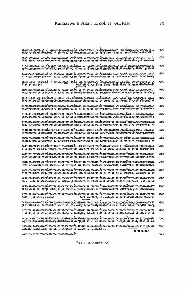
After we had defined the DNA physically as described above, we determined the entire nucleotide sequence of the genes coding for FIFO (FIGURE 2).'0-'4 This sequence was shown to code for each subunit of the complex from the amino terminal sequence obtained by Edman degradation (subunits a, & y,6, e , and b] or from the entire amino acid sequence (subunit c)." The amino acid compositions of subunits a, 0, y. and 6 were consistent with protein chemical data. Walker and coworkers independently determined the sequence using DNA from Xa~n5,2'-'~ which we have shown to carry genes for FIFO. The nucleotide sequence coding for F, was also determined independently by Nielsen et dZ5 Their results are consistent with ours except for some minor differences noted in the legend of FIGURE 2. The organization of the gene cluster defined from the DNA sequence is shown in FIGURE 3, using the new nomenclature for the genes (pap, or proton- translocating ATPase protein].
We found a typical promoter sequence (Pribnow box and -35 region] as shown in FIGURE 2. We concluded that this sequence functions as a promoter from results of DNase-footprint analysis and in vitro transcription.26 The initiation site of transcription for mRNA was suggested to be 74 base pairs upstream of the first letter of the initiation codon of the reading frame. We could not find other promoter sequences in flanking regions, suggesting that the entire gene cluster is transcribed as one mRNA. A typical terminator region was found after gene coding for the t-subunit. It is interesting that a complementary structure is observed between the 5'-end moiety of the putative mRNA of the operon and the terminator region. As discussed previou~ly, '~ the complementary structure in the mRNA could have biological significance, such as in stabilizing mRNA or regulating termination. We found a similar complementary sequence in mRNA of the tryptophan operon of E. cohz7
The open reading frame before the c-subunit was shown genetically to code for the a-subunit. Before the gene for the a-subunit, we found an open reading frame capable of coding for a protein of 130 amino acid residues. This gene is probably involved in the pap operon. because the real promoter for the operon was found in the preceding region of the gene. However, it is too early to conclude that this frame codes for a component of F,F, because we have no evidence for the presence of this protein in purified active F,Fo. It is also possible that this protein regulates biogenesis and assembly of the complex but is not itself incorporated into the assembled complex. One group reported a protein of similar molecular weight in their purified F1F0,28 but there is no evidence that this protein is a real component of the complex or that it is actually coded by the open reading frame found in this study. Genetic and biochemical studies on the product of the open reading frame are extremely important for understanding the gene cluster and the F,F, complex. Downstream from the gene cluster for Fo subunits, we found reading frames for F, subunits.
Possible Regulation of the Biogenesis of the Complex
I t is very likely that the stoichiometry of the subunits of F, is type ~ ~ ~ ~ ~ y , 6 , t , , although this is still controversial.'.' Foster and Fillingame very recently reported an a,b,c,,-type stoichiometry for F, of E. ~ o l i . ' ~ What is the mechanism controlling the biogenesis and assembly of FIFO with such stoichiometry? One possibility is regulation at the transcriptional level: genes for abundant subunits (a-. f i - , and

Kanazawa & Futai: E. coli H+-ATPase
MTIGTATGCAC I b W J U d l A l T T A M C A T T T A l l C A C ~ T T T T G G C T K T T A T T G T ~ T C A C W l G G G C G e A C e G f A T M I T T G A C ~romotrr -
T G C T T W C T C T M G C C T T A ~ A W L M G T T T T A T A C G A C A C ~ C G G C A T A C C T C O M O O G A O E A O E A G T W - ~ ~ a A a n V a l I * t k r V a l k r L ~ u V a l S ~ r A r g
~ G T T G C T C G G M G C T T C ~ G C T C G T T C A G T T A C T G G T O E T G A T A G C M G T O T T G C T ~ T T C A G C C T C A M O AsnVaI AI rArgLysLwLwLauValG1 nLeuLwVa1 Val I 1 aA1 aS~rGlyLauLauPk~SarLwLysAapProPh~Tr~lyVal~rA1 a l l @S G C G G ~ ~ G ~ C T ~ G ~ A G T C T T ~ C T G C C T M C G T T T T G T T T A ~ A W L T A T T T G ~ C T ~ G T C A C ~ ~ G C A T A C ~ C ~ G A ~ ~ C ~ T ~ C T ~ A T T crG1 yGl yLauAl aVal PheLwProAsnVal LauPh-t I 1 rPhrAl rTrpArgHi a61 nAl aH1 aThrProA1 aLysGlyArpVa1Al aTrpThrPh
C W A T T T G G C W G C T T I C ~ G T T C T G W ~ T G T T f f i T ~ T T A C T G G T f f i T G G C G T T ~ G G T T T T ~ ~ G t T A T T C ; T G C C G t T G A T C G T T Y G T G G eAl aPheGlyG1 uAlaPheLysVaILeuA1 ~RtLauVt lLauLeuY~ lVa l~ l aLwAl tVIlLIuLy8Al ~V~lPh~LwProLwllrValThrTrp
G T T T T G ~ T G C T G G T W I T T C X A W L T A C T ~ G ~ A C C G ~ C T G T ~ T T M C M C M A G G G T M M E ~ C A T C A T ~ T T C ~ ? A T G A C G C C G C ~ T T K A T ~I1LcUVa1LeuYalValGlnl IrLauAl aProAl aVaI IlaAsnAsnLyrGlyend - MtAlaSerGl uAsnllrtThrProG1 nAapTyrI 1
A G G A C A C C A C C T ~ M T M C E T T C A K T G G A C C T W G T K ~ T T C T C G ~ T G ~ T G G A T C C A C ~ ~ A M C C C C C A G C C A C C T T C ~ G G A T C C eGlyHi sHi SLeuAsnAsnLauGl nL~uAspLeuArgThrPhaSarLauValAspProG1 nAsnProProA1 rThrPhrTrpThrl IaAanl IoAapSw
ATGTTCTTCTCCGTGGTGC~GCGTCTGTTGTTCCTWITT~TATTCCGTAWGTAGCCA~AGG MtPhePheScrValV~lLcuGlyLeuL~uPheLauValL~uPhaArgS~rV~lAl aLysLysA1 aThrScrGlyValProGlyLysPheGl nThrAl a1
T T G A G C T ~ ~ ~ T W I T C ~ G ~ T T ~ G T T A A T W ~ T I G C G T W M ~ C A T G T A C C A T ~ G ~ ~ G C ~ G C T ~ T T G C T C C W T ~ G ~ ~ C T G A C W T C T T C G T C T G G G T IeGl ULeuVal I IcGlyPhcVal ArnGlyStrValLysAspMetTyrHi sGlyLysSerLysLru1 lcAl aProLeuAlaLeuThrllePheValTrpVa
ATTCCTGAT~AACCT~AT~TTTACTGCCTATC~ACCT~CTGCCGTACATTWTGMC~\TGTACTGGGTCTWCTW~TWGTGTG~TTCCGTCTWG 1 PhtLeuktAsnLeuMtAspLeuLeuPro I 1 eAspLeuLeuProTyrI 1 eAl a G l uHi sVal LeuGl yLtuProAl aLruArgValVa1 ProScrAl a
~ A C G T ~ M C G T M C G ~ T G T E T A T G G C A C T G G G C G T A T T T ~ \ T C C T ~ A T T C T ~ T T C T A C A C ~ A T C A M A T ~ A A A ~ G ~ A T C ~ ~ G ~ T T C ~ ~ T T C ~ AspVa1AsnVa1ThrLeuSerMetAl aLeuGl yVal PheI 1eLculleLcuPheTyrSerI IeLyshtLyrGl y l IcGlyGlyPheThrLysGluLeuA
G ~ T ~ C A G ~ A C G T T C A A T C A E T G G G C G T T C A T T C C T G T C A G C C T G C T G T C C A A A C C A G T ~ T C A C T C ~ ~ ~ T T T W W I C T G T T rgCysSerThrPheAsnHi sTrpAl aPheI1 eProVal AsnLeul IeLeuGl uG1 yValSerLeuLcuSerLysProValSerLeuGlyLcuArgLeuPh
CGGTMCATGTATGCCff iT~GCTW\TTTTCATTCTMTicETGGTCTGTTGCCGTGGTEGTCACAGTGGATCCT~AT~TGCCGlGtGtCATTTTCCAC eClyAsnMetTyrAlaGlyGluLeullePhelleLeulleAlaGlyLeuLeuProTrpTrpSerGlnTrplleLeuAsnValProTrpAl~llePheHis
I IeLeul l e l 1 eThrLeuGl nAl aPhel IePheMetValLeuThrl leva1 TyrLeuSerMetAl aSerGl uGluHi send
8 -
ATCCTCATCATTAC~ETGC~UGCCTTCATCTTCATGGTT~TGAC~ATCGTCTATCTGTCEATGGCGTCTGAAGAACATT~TTTACCMCACTACTACGT
TTTMC ~GAAACAMCT~ACTGTCATGWU\MCCT~ATATGGATCTGCTGTACATEGCTGCCGCTGTGATWITG~ETCTGGCGGAATCCGTGCTG HettluAsnLeuAsnHetAspLeuLeuTyrMetAl aAl aAl aValhtMetGlyLeuAl aAl a l IeGlyAl aA
CCATCffiTATCGGCATCCT~GGG~TAAATTCCTGGAAG~CGCGGCGCGTCMCCTGAT~TW\TTCCTCTGCTGCGTAC~CAGTTCTTTATCGTTATGGG 111 l c t l y I IeGlyl IeLeuGl yGlyLysPheLeuGl uGlyA1 a A l aArgGl nProAspLeu1 IeProLeuLeuArgThrGl nPhePhe1 IeValMetGl
TCTCCTGGATWTATCCCGi \TGATCGCTGTAW;TCTGGGiCTGTACGTtATGTTCGCTGiCGCGTAGTM~GTTGCTT~TATTTAMtA~MTATCAG yLeuValAspA1aI IeProMctl leA1aValGlyLeuGlyLeuTyrValMetPheAl a V a l Alaendend
C-
AMGTTMCTA~UTA~~ATTGTGCTGT~AATCTTMEGCAACAATCCTCGG~CAGG~CATCGCGTTTGTCCTGTTCETTCTGTTCTGCATGMGTAC MetAsnLeuAsnAl aThrl IeLeuGlyGl nAl a l leAl aPheVa1LcuPheValLeuPhdysnetLysTyr
GTATGGCCGCCATTU\TGG~AGCCATCW\AAACGTCA~MtAMTTGCTGACGGCCTiGCTTCCGCAGMCGAGcAC~TMGtACCTT~CCTTGCM ValTrpProProLeuktAlaAIal IeGluLysArgGlnLysGluI leAl aAspGlyLeuAl aSerAl at1 uArgAlaHi sLysAspLeuAspLeuAlaL
yrAl aSerAl aThrAspGl nLeuLysLysAl aLysAl rG1 uAl at1 nVal I l e l IcGluGl nAl aAsnLyrArgArgSerG1 nl IeLcuAspGluAl aLy
sAlaGluAlaGluGlnGl uArgThrLyslleVa1 A l aGlnAl aGlnAlaGl u I IeGluAl aGluArgLysArgAl aArgGluGluLeuArgLysG1 nVal
WTATCCTGtCTGTTGCTG~CGCCGAGAAGATCATCGAA~GTTCCGTGW\TGMGCTGCiAACAGCtACATCGTGWTU~CTTGTCGCTGMCTGTMG AIrI leLcuAl aVaIA1aGlyAlaGluLysI lelleGluArgSerValAspGluAl~AlaAsnSerAsplleValAspLysLeuValAlaGluLeuend
W I G ~ G G C T t A T G T C T ~ A T T l A T T A C G t T A G C T C ~ C C C T A C G C C A A A G C A G C T T i T C A C T T T G C C G T C W \ A C A C ~ A A A G T G T A t A A C G C T G G C A G MctSerGluPhel IeThrValAl aArgProTyrAlaLysAl a A l aPheAsDPheAl aVtlGluHisGlnScrValGluArgTr~Gln
- A~~~CCAGC~C~ACC~ACCA~TW\AAAAAGCW\AAGCGG~A~~CCAGGTAATCATCGAG~AGGCGMCMACGCCGCTC~CAW\TTCTGGACGMGCGM
AGCTGAGC~ AGMCAGGM~GTACTAMATCGTGGCCC A~GCGCAGGCG~AAATTGAAG~CGAGCGTAMCGTGCCCGT~AA~AGCTGCGTMGCMGTT
G A C A T ~ E : G G C G T T T G C ~ G ~ C ~ A G G - A A C : ~ A A A A C G A A ~ A ~ A ~ G G C A G A G C - ~ C - C ~ C ~ G G C G C G C TTGCGCCAUAAEGCTCGCC~GTCGTTTATCG AspMet.euAI aPneAl a A l aGluial l P r L , s A s q 2 1 ,G'nPetAl aGlu.eu.eJSerGlyAl a.cuAI aProGl u1hrLe.A.l aGIuSerPhc1 leA
49
I00
200
$00
400
500
600
700
800
900
1000
1100
I200
1300
1400
1500
1600
1700
1800
I900
2000
2100
2200
2300
2400
FIGURE 2. DNA sequence in the antisense strand is shown with deduced amino acid r e s i d u e ~ . ' ~ ~ ' ~ The sequence shown is from the promoter to the terminator region. Shine- Dalgarno sequences are underlined.67 We have revised the part of the sequence in References 12 and 13 including typing errors. Our sequence is essentially consistent with those of others except in position 1099-1106, 1544,2627. and the carboxyl terminal portion of the t-subunit. We concluded that the e-subunit has 138 residues." rather than the 132 residues reported by Saraste et a].*'

Annals New York Academy of Sciences
CAGTTTGTGGTGAGCAACT~CI \CW\AAACGGTCAGAACCiGATTCGGGTTATGCCT~Ai \ATGGTCGTCTTLACGCGCT~CCf f iATGTTClGGAGCAGlT 2500 I a V ~ I C y r t l y t l uGI nLcuAspGl JAsnGlyGI nAsp.eu I IcArgValMetAl aG1 JAsptlyArqLeuAsnAl aLeuProAspVa ILeuGl uG1 nPh
TAllCACCTGCtTGCCGT~CTGAtGCTACCGCTGAGGT~GACCTCATTTCCGCTtCCG~ACTGAGTGAACAACAGCTC~CCAAAATTTCTGCTCC~ATC 2600 e l let41 sLcuArgAlavalSerGluAlaThrAl aGluVal Arpval ! IeSerAl aAlaAlaLe~SerGluGlnGlnLeuAl dLysI IeSerAl dl l k t
W M C G T C TGTCACGCAhGT T M G C TGAAI I GCAAAi\T C G A T AAGTC TGTAATGGCiGGCGT TATCATCCGAGCGGETCATAI GGTCATTGATGGCA t l u L y s A r g . e ~ S e ~ A r g L y s V ~ l L y s L e u A s n C ~ s L y s l IcAsp.ysSerValHetAl aGlyVal I let IeArqAl aClyAspwetVal lleAspGlYS
GCGTACGCGGTCGTC IlGAtCGCC T TGC AGACGTC TTGCBG TC T T A A G G E C T G G A G C ~ T G C A A C T G A A T T C C A C C ~ A T C A G C ~ C T ~ T C A A G C A MetGlnLeuAsnSerrhrGl~! IeSerGl uLeu! I e L y s t l erva l ArgGI yArqLcvGl JArgLcJAI aAspVaI LeuGl nSerena
G C t C A T T G C T t A G T T C A A T ~ T T G T ~ G T G A A G C l C A C A A ~ C A A G C ~ A C T A T T G T T T C T t i A A G T G A C G G T G T T A T C C G C i \ T l C A C G G C C ~ G G C C ~ ~ ~ ~ ~ 2900 nArg l leAlatlnPheAsnvdlYalSerGluAld~isAsnGluGlyTnr1 IcYalSerValSerAspGlyVallleArgl IeWlsGlyLeuAlaASPtYS
AIGCACGCTGAAAIGAlCT~CCTGCCGGGTAACCGTTAC~TATCGCACTGAACCTCGA~CGCGACTClGTAffiTGCGG~TGTTATGGTCCG~AC~~G 3000 HetCl n t l y t l J M c t I IcScrLcuProGIyAsnArgTyrAI a l IeAIaLeuA~nLeuGIuArgAspSerValGl yAl a v a l V s l l l c t G l y P r o T y r A 1 ~
ACCTTGCCGLAGGCATGA~GTTAAtTGCATTGGCCCTAiCCTCCAAGlTCCGCITGGC~GTffiCCTCCTGU;CCClCT~TTAICACTCT~ffiT~A~~ 3100 IpLeuAldtluGlyMetLysVal.ysCysThrGlyArg1 le~~uCluValProVslGlyArgGlyLcuLeuGlyArgYalArnThrLeuGlyAl aPr
AATCGACGGTAAAGGlCCG~TGGATCACGACGGCTTCTC~CCTCTAGAA~AATCCCTC~GCCCGTTATCGMCGTCAC~CCGTAGI\TCAGCCGGTACAG 3200 o! leArpGlyLyrtlyProLeuArpfl~rAspGlyPheSerAl avalGluAla! leAlaProClyVal I leCluArgGlnSerYalAspGlnProValGln
A C C G G T I A T A ~ G C C C T T $ C T C C A T G A T C C C A I T C G G T E G T G G T C A G C G T G A A T T ~ A T ~ A T C ~ ~ ~ T G A C C G ~ C A W I C A ~ ~ T M ~ C C G C A C T ~ C C T A T C C 3300 I n r C l y TyrLysAl aVal AspScrMec! IcPro I I rGI yArgGl yG1 nArgGl uLeul let leCl y AspArgGlnThrGl yLys ThrAl aLeuAlal leA
A T G C C A I C A T C M C C A G C G ~ G A T T C C G G T A T C M A T G T A ~ C T A T G T C ~ T A T C G G C C A ~ G C G T C C A C C A T T T C T M ~ G T G G T A C G T A M C T G ~ ~ 3400 spAl a l let IeAsnGlnArgAspSerClyI IcLysCysI IeTyrVr l A l a l IcGlyClnLyaAl aSerThrlleScrAsnVaIvIlArgLysLeuGluGl
uW1 IGI yAl aLcuAl aASnThr1 IeYal Val Val Al aThrAl rScrGIuScrAl aAl cLeuC1 nTyrLeuAl t A r g k t P r o V a l Al rLeUktGlyGl u
TACTICCGTWlCCtCGCT~GATtCGCTGATCATTTAC~ATGACCTGTCTAMCAGGCiGTTGCTTACCGTCAGITCT~CCTGCTGCTCCGTCGTCCGC 3600 TyrPheArgAspArgGlyGl uAspAl aLeu I IeI 1 ~TyrAspAspLeuSerLysGl nAl aVrIA1 aTyrArqGl nl1eStrLtuLeuLtuArgArgProP
CAGWICGTCAA~CATTCCCEGGCGACG~TTTCTAC~TC~~CTCTCGTCTGCTCGAGCGTECTGCACGTC~TMCGCC~TACGTTCUCCCTTCACCM 3700 roGlyArgGl uAl aPheProGlyAspVaIPhtTyrLtuH1 sSerArgLeuteuGl uArgAl aAl J r q V a l A s n A l rG1 uTyrVr lGluAl IPhcThrLy
AGGTGMGTGAAAG~CUJCCGG~TCTCTCACCGCACTECCGATTATCGAAACTCACG~GGGTO.CGTTTCTG~GTT~ETTCCGICCMCGTAATCTCC 3860 rGlyCl~~~lLysGlyLysTnrClySer~~ulhrAla~euPro1 1r1 I~GluThrGlnAlrGlyAspVaIS~rAlrPhrYtlProThrASnVrI I I * S r r
A T T A C C W \ T G G T C A G A T C T ~ C C T G G I M C C M C C T G T T C ~ C G C C G G T A T T C G T C C T G C ~ G T T A A C C C G G G T A 7 T T C C G ~ A T C C C G T G T T ~ T G G T W ~ 3900 I leThrAspGlyGlnl lCPhCLCuGluThrAtnLtuPheA~nAI~Gly1 IeArqProAl ~ V r l ArnProGlyllrSirVrlSerArqV~lGlyGlyAl J
I IGI nThrLyi I IaMetLyr~ys.euSerClyGlyl IeArgThrAl c.ruAl ,GI nTyrArgG1 uLeuA1 rAl rPhrSerGl nPhaAl rSerAspLeuAs
PAspAl aThrArgLysGl nLwAspH1 sGlyG1 n.yrVI1 ThrGluLeuLeuLysGl nLysGl nTyrAl sProMetSerVa I A l ~ G l n G l nSerLeuVaI
LcuPheAl aAl rGluArgClyTyrLeuAl J s p V t l C I uLeuSarLyr l IeGlySarPheGI uAl Jl ~ L w L w A I tTyrVa l AtpArgAtprll tAl I P
roLeuMetGl n t l d l I rArnGl nThrGlyGlyTyrAsnArpG1 u f l 6 l uGlyLysL*uLyrGly! IeL iuArpSerPh~LytA l t T h r t l nSerTrprn
M t A l ~GlyAlrLysGlull~ArgkrLysl IrAl iSerVa1GlnAsnT
hrCl nLyr ! IeThrLySAl cMttG1 u k t Y t l A l rAl r S ~ r L y s M t A r g L y s S ~ r G 1 nAspArgMecAI Jl r k r A r g P r o T y r A l aG1 u T h r M t A r
gLysVi l I IrCIyH1 sLwAlaH1 rGlyAsnLruGl uTyrLysHl sProTyrLruG1 uAspArgAspVrlLysArgVI1GlyTyrLeuV~lVi~S~rThr
As~lrgGlyLiuCysGlyGIyL.ulln1 I~AtnLeuPhtLysLyrLruLruAl rG1 d(.tLysThrTrpThrAspLysGly~rlGl nCysAtpLeuAl aH
2 700
2800
0 -
GCAC~GC~~ACTCG~TMCXCCATCGTTGT~~~TAGCAAC~CCGTCTWIATCCGCTCCAC?CC~TACCT~CACCTATCECGCTTGCCTMTGGGCGM 3500
CACAGACCAAGATCAT~AA~UMCTGTCCGGTGGTATCC~TACC~~~CTGGCACAGTATEGTCUCT~~~CAWGTTCTC~CAGTTTGCATCCGACCTTO. 4000
C ~ A T G ~ M C A C C T M ~ ~ A ~ T T G A ~ C A C G G T ~ A W A A C T ~ C C G A A C T G C T G A M C A ~ A C A G T A T G C G C C G I T G T C C ~ T T G C G C U I C A G T C T C T G G T T 4100
CTGTTCGCA~~ACUCGTGETTACCTCCCGWTGTTGM~TGTCWUMTTG~~ACCTTE~AIGCCWTCTGCTGGCTTXCGTCGICCGTCITCACGCTC ~ Z W
C G T T G A T G C M W G A ~ C M E C A W C C G G T G ~ ~ T A C M C ~ C ~ A T C C U G G C M G C T ~ G G E A T C C T C ~ T T C C T T E A M G E ~ C C M T C C T G G T A 43w
A C G T C T G ~ ~ C G C T T C C C T T ~ G G G C A G G C C G C M G ~ ~ A T T ~ ~ ~ ~ ~ T C A T G G C C G G ~ G C ~ G I ~ T A C G T A G T ~ ~ T C W M W G T C C A ~ C A 4400
CGCMAAWTCACTAM~~~TCWGAT~~~TCGCCCCTTEG 4500
C A A A ~ T C P ~ T G G I C A C C T T E C A C A C G G T A A T C T ~ T A ~ M C C A C C C T T A C C T ~ ~ C C W ~ C G T T A ~ C C G T G ~ ~ A C C T ~ T ~ T G T C G I C C 4600
G A C C G T G G T T I G T G ~ G G T G ~ T T T G M C A T T M C C T G T T C ~ M C I C C T ~ C C C A T ~ ~ C C T ~ C G I C ~ G ~ G T T C M T W O . C C T C ~ ~ M 47w
1-
~GATCGCCTCGAAA~~~GTETCGTTCTTCMCTCCGTGG~EGWAATGTTGTTCC~CAG~TCA~CGGCATG~CWTMEC~TT~CCTGTCCGMCTGAT Ct I l eGlySerLysGlyV~ lSr rPhePheArnSerV~ lGlyGlyAsn~~ lV~ lA l~GlnVr lThrGlyMeCGlyAspAsnProSerL~uSerGluL~ul I
4800
FIGURE 2 (continued)
Kanazawa & Futai: E. coli H+-ATPase 5 1
C G G i C C t C i A A A A C T W \ T G ~ I G C A G G C C T A C G A C ~ A A ~ ~ ~ C G l C T G W \ C A A G C l T l A C A i l C T C A G C A A C A A A l l l A l l ~ C A C C A l G l C T C A W ; l T C C G 4900 eClyProvsliysValnetLeuGlnAlalyrAspGluGlyArg~ruAsp.ysLeuTyrl IeYalSerAsnLysPhel IeAsnThrnetScrGlnValPro
lhrl IcSevGI nLcuLeJPrOLeuProAl aSerAspAspAspAspLluLysH1 sLysSerTrpAsplyrLeulyrGl uProArpProLysAI aLeJLeUA
ATACCCIGClGCGlCGlTA~GTCGAATClCAttlTTATC~tGGCGlGGlTGAAAACCTG~CCACCtAtCAGGCCtCCCGiAlGGTGCCO\TGAAAGCCCC 5100 SpThrLeuLeuArgArgTyrVa IGI cScrGl nVal TyrGl nGl yVaI Valtl uAsn.euAl aScrGldGl nAl aAl aArgMetVaIA1 aMetLysAl aAl
alhrAspAsnGlyGlySerLeulleLysGl uLeuGlnieuVal TyrAsnLysAl aArgClnAl aSerl IelhrGlntlcLeuThrGluI I e V a l S W G l Y
AlaAlaAleValeno FetAlaThrG'yLysI IeValGInVal IleGlyAlaVtlYalAspVilGluPheProGl
G t A T t C C ~ T A C C G C G ~ G T G ~ A C t A T G C T C l l W G C T G C A ~ A T G G l A A T W G C G l C l G C ~ G C T G t A A G l T C A G C A G C A ~ T C W ; C G C C G G T A l C G T A C G l 5400 nASpAldVa1 ProArgVaI IyrAspAI aLruCl ~vdlGlnAsnG1yAsnCluArqLeuValLeutluV~lGlnGlnGlnLeuGlyGlyGlyl IeValArg
A C C A T C G C A A l G G G l T C C T ~ C C A C G C l C l G C t T C C C t C ~ ~ T G W \ I G l A W W \ C C T C G A i \ C A C C C G A l T W U C T C C C G C i A G G T A I A C C W C T C T ~ G C C 5500 lhrl IcAlanetGlySerSerArpClyLeuArgArqGlyLeuAspVal LysAspLeuCluHi sProl leGluValProValGlyLysAl bThrLeUGlyA
rglle~tAsnYalLeuG1yGluPro~alAspnet.ysGlyG1ulleClyGluGluGluArglrp~lalleHi~~rgAlaAl~ProScrTyrCIuCluLe
uSerAsnSerCI ntl u l eu.euG1 uThrtl yL Ie-ysVal I leAsp,cUMetCysProPheAl aLy~Gl y C 1 yiysVtlGIyLeuPheGlyGI y A I aG\Y
G T A G G l U A A C C G T A M C A ~ C A T G W \ ~ l C A l T C t l A A C ~ l C G C t A l C W \ C C A C T C C t G ~ T A C T C T G T G T T T t t G C C C G ~ A G C T C C G l A C l C G l t A f f i 5800 VtlGlyLyslhrJdlAsnMetMetGluLeu I IeArgAsnlleAlal IeCluHi sSertlylyrSerVelPheA1 aClyVslGlyGlulrgTnrArgGIUG
G T A A C t A C l T C l A C C A C ~ A l W C C G A C T C C A A C G l T A ~ C G A C A A I G l A T C C C T G G l G ~ A l G G C C A C A T C A A C t A G C C ~ C C ~ U I C C G T C l ~ ~ G ~ 5900 lyAsnAspPnelyrd{ stl uMetlhrAspScrAsnVal : 1 cAspLysValSerLeuWaI TyrGlyGI nWt A s n G 1 nProProtlyAsnArgLcuArgVa
lGCTClGACCCGTClWCC~TCGClGAWlAAllCCGTtA~GlUGGTCGltACGIlCTGCiGTTCGTTGACMCAlCTAT~GTTACACCClttCCGGlAC~ 6000 IAIaLe~lhrCly~ecTnr~etAlaGIuLysPheArgAspGI dGlyArgArpYalLeu.euPheValAspAsnl IelyrArgTyrlhrLeuAlaGlylhr
GI uJal SetAlr.euLeuGlyArgMetPro5erAl aValtl ylyrtl nProThr.euA1 aG1 *GI uMcfGlyVal LeuGl nGI uArgl IeTnrSerThrL
A M C l t t l l C T A l C A C C l C ~ G T A C A G C C A G T A l A C G l A C ~ l C C G G A T W \ C T i G A C l t A C ~ C G T C i C C G G C M C C A C C T T i G C G C A C C l T t A C C C M C C ~ T 6200 yrTnrtlySerl IeThrSerVa IGI nAl aval Tyrval ProAl1AspAspLcuThrArpProSerProAl aThrThrPhcAl aHi sLcuAspAl alhrYa
G t l A C T t A G C C G T C A ~ ~ C ~ C G ~ C ~ C T G G G l A l C T A C C C . ~ G C C C T T S R C C C X T S G ~ C ~ ~ C A ~ C A G C C G ~ C A C C l ~ O X ~ C G C ~ G G T I ~ T G G i C A G G A A 6300 1 Val LeuScrArgGl nl l r l l aScrLe&ly I IeTyrProAl avrlAspProLeuAspSerTnrSerArgG1 nLeuAspProLeuVslVa1ClyGl nG1 u C A C l A C G A C A C C G C G C i r T t ~ G T l C A G T C C A T C C l t C A I ~ G T T A T C A G G A A C l G A A t A ~ A T C A T C G C C A l C C T t C G T A ~ ~ T C A A C T G T C l G A A C A A G 6400 H i STyrAsplnrAl aArqGlyValGl nSerI IcLeutl nArgTyrtl nG1 uLeuLysAspl It1 leAl a1 IeLeuGl yHetArpC1 uLeuScrGl utl uA
hCAAACTGGTGGlACCGCt~GCTCGTUGATCCAtCGCTiCCTGlCCCAGCCGTlCTlC~TtGCAtAAGlAlTCACCGG~lCTCCGGGTAAAlACGTCTC 6 5 0 0 sp~ys.cuVa1ValAl aArgAl aArgLysl leG1 nArgPneLedSertl nProPhePhcValAl at1 ~ValPheThrGly5srProGlyLysTyrVal5e
rLeuLysAsplhr1 IeArqGIyPheLysGlyI Ic~ctGluGlyGlulyrAsp~is~euProGluGlnAlaPheTyrMetValGlySerlleGluGluAla
G l W ; A A A U I C C C M A I A I C ~ l l A A C G C C T T A A l C ~ t ~ T G A T A T G G C A A l t A C T i A C ~ A C C T C W \ C G T C G T C A G C ~ i \ t A G C A I C A A A T G T l C l C T G G 6 7 0 0 Valtl ~ L y ~ A I a L y s ~ y s ~ e u e n d MtAl aMetTnrTyrni sLeuAspVal ValSerAl aG1 uG1 ntl nFetPheSerGl
yLeuVaIGluLysl IeGlnVal ThrGlySerCluGlyCl~LcuGlyl IrTyrProtlyHi sAI aProLeuLtulnrAltI IeLysProGlyMet I IeArg
AlCGTtAAACAGCACGGTCi\CGAAGAGllTAlClAlCTG~CTGGCGCCAlTCllGAAGT~CAGCClGCCAICGlW\CCG~lCTGGCCW\CACCGCAAlTC 6900 I IrVaILystl ndisGlytiisGldGluPhrl IcTyrLeuSerClyGlyl IrLeuGluYalCl nProGlyAsnVelTnrvrlLeuAl aAspThrA1aI leA
rqGI yGI nAspLeuAspGluAl rArgA1 aMe ttl uAl aLysArgLysnl aGluGl uni rl IeSerSrrSerHi stl yArpY8I LspTyrAl aGlnAl 8%
A C C A T C A G ~ C A G C ~ G C T C C ~ G ~ ~ A C C ~ Q A ~ C A ~ A T S A T G A T ~ A T C ~ W \ A A C A ~ ~ A T C ~ ~ G G ~ A T ~ A C C T G : A C ~ A A C E C W \ : C C G A A G C C ~ : T ~ C ~ G G 5000
C A C C ~ A C A A T G ~ ~ G G C A G C ~ T W \ T T A A A O \ G C T G C A G ; ~ ~ G T A T A C A A C ~ A ~ C T C ~ T C ~ ~ ~ C C A C C A T T A C T C A G O \ A E T C A C C W \ ~ A T C G I C T C G G G G 5200
G C C ~ C C G C G G T T T A A A C A ~ ~ ~ T T A T T I C ~ T A W ~ T T T A ~ ~ A T G G C T A C T ( ; G A A A G A T T ~ ~ T C C A G G T A A T C G G C G C C ~ T ~ G T T ~ A C C T C W V \ T T C C C I C A 5300
I$-
G T A T C A T G A A C G ~ A C T ~ G G ~ G A A C C G C T C ~ A C A T C A A ~ ~ ~ C ~ A ~ A T C G ~ ~ ~ ~ W U \ ~ ~ E ~ T T G G ~ ~ W \ T T C A C C W ~ ~ ~ A C C T T C C T A C G M W \ G C T 5 6 0 0
C T C A M C T C T C A G C A A C T G E I G W \ A I C C ~ ~ T A T C A A A G T ~ A T C C A C C T G A T G T ~ T C C G T ~ C ~ C T A I G G W G C T A A A G T T E ~ T C T ~ T T C G ~ T ~ G T G C ~ C ~ 5700
~AAGTATCCGCACTGCTGG~~CCGTAT~CCTTCAGCGCTAEGTTATCAGCCWCCCT~GCEGAAWIW\TC~~CGTTCT~XG~AACGIATCACCTCCACCA 6 1 0 0
C C T W \ A I ~ C A C C A T C C G T ~ G C T T T A A A ~ ~ C A T C A T C G A ~ \ G G ~ G A I T A C ~ A T C A C C T ~ C ~ ~ ~ A G C A G G C C T T C T A C A T ~ ~ ~ C G ~ T T C C A T C ~ M G A I G C T 6600
1 -
~ C T G G T C ~ A ~ A ~ A T C C A ~ ~ I U C G G G T A G C ~ A A G G T C ~ C T ~ C ~ C A T C T A C C C T G ~ C C ~ C ~ C A C C C C T G C T C A C C G C C ~ T T A A G C C T ~ T A T ~ A ~ T C G C moo
t c ~ ~ c c i t G I \ i c ICCACW~\~CCCW\GCCATGWUGC~~CGTAA~~~TGGC TC T C A C G G C ~ G T A G A T ~ACGCTCAGGCG IC 7000
T G C G t A A C T G G C C A A A G C C i \ l C W W A G C l t C t C G T T A l ~ G A G T T G A C C A A I ~ C C t A ~ G l U I C A C C G W T T ~ A A ~ A C A A M t t C A G T C T G G A M ~ rAl aG1 uLeuAl aLysAl aL leAl at1 nLeuArgVal1 leGl ULeuThrLyrLyrAl cuetend
Term i n ( t o r AGGCTGGCTTTT~TTTGCG~GTGTGACCCGTCCT~AA~A~C
FIGURE 2 (continued]
7100
7141

52 Annals New York Academy of Sciences
DNA I 1 1 I I I I I
0 1 2 3 4 5 4 7 x WOO bp
FIGURE 3. Organization of the pap (unc] gene cluster determined from the DNA sequence. The coding frame of each gene with its old (unc) and new (pap) nomenclatures is shown. Promoter (P) and possible terminator (T) are shown. The new nomenclature was proposed by us: pap is an abbreviation for proton-translocating ATPase protein." 14K is an open reading frame of unknown function. Other reading frames correspond to subunits of FIFO. The cleavage sites for endonucleases are shown as follows: H, HindIII; E, EcoRI; R, BarnHI. The scale shown at the bottom corresponds to numbers of base pairs in FIGURE 2. (See text and References 10-14 for details.]
c-subunits) are transcribed more efficiently than those for less abundant subunits (7-, 6 - , c-, b-, and a-subunits). Intercistronic sequences (FIGURE 2) were analyzed to study this possibility. No usual promoterlike sequence was found in the noncod- ing region between genes for 6 and a (15 base pairs] and y and @ (25 base pairs) (FIGURE 2). Thus there is little possibility that the gene for a or @ is transcribed independently as a single transcriptional unit. However, sequences between the genes for a and y (49 base pairs], for a and c (46 base pairs), and for c and b (59 base pairs) are longer than flanking regions between 6 and a, and y and @. Although the structures of these sequences are not as complete as reported promoter sequence^.^' a promoterlike (Pribnow box) sequence was observed between the genes for the a- and c-subunits and the c- and b-subunits. An inverted repeat sequence was found between the genes for the a- and y-subunits. Thus there may be some transcriptional regulation in these flanking regions."
As pointed out above, transcriptional regulation in biogenesis of the a-, p-, and y-subunits seems unlikely, although F, has an a,@,y, stoichiometry. We analyzed usage of codons to find evidence for possible regulation at the translational level. Recently, Grantham et a). classified bacterial mRNAs into two groups, those highly expressed and those weakly expressed in protein synthesis, and found that the two groups showed differences in codon usage.,' We compared codon usage in each subunit with those classified previo~sly.~' We found that the usage of c, a, and @ is similar to that of the highly expressed type, whereas the usage of the other subunits is similar to that of the weakly expressed type.
We also analyzed the relation between the frequency of codon usage in these genes and the abundance of tRNA by the method of I k e m ~ r a . ~ ~ The correlation could be expressed by the equation y = a + bx (x is the amount of tRNA; y is the frequency of codon usage). Results indicated that the a-, @-, and c-subunits have similar patterns to the highly expressed type, while the y-, 6 - , and a-subunits have
Kanazawa & Futai: E. coli H+-ATPase 53
similar patterns to the weakly expressed type. These results suggest that the a-, (3-, and c-subunits are translated more frequently than the other subunits. Recently, Ikemura reported that the frequency of the optimal codons in E. coli genes is closely related to the amount of the protein synthesized in v ~ v o . ~ ~ We found that the frequency of usage of optimal codons was clearly higher in abundant subunits than in less abundant subunits. All these analyses suggested that the amount of subunits of FIFO synthesized is determined, at least to some extent, by the frequency of codon usage in each gene. It should be noted that the stoichiometry of the a- and b-subunits expected from the results of this analysis agreed well with that determined biochemi~ally.2~ It would be interesting to know how much this regulation contributes to determination of the exact stoichiometry of the sub- units.
Primary Structure of Subunits of H'-ATPase
The primary structure of each subunit was obtained from the DNA sequence (FIGURE 2). Information on each subunit deduced from the structure is summa- rized in TABLE 1. Subunits of Fo had the characteristic properties of membrane proteins. The amino acid sequence of the hydrophobic c-subunit obtained from DNA was in complete agreement with results of protein chemical analysis by Sebald et aLzl The b-subunit had a high content of polar amino acid residues (48.1%) and about the same overall polarity as do average soluble proteins. However, it had relatively long domains of hydrophobic residues near amino terminal (11-33 residues) and carboxyl terminal (124-132 residues] portions. Thus the b-subunit may be integrated into the membrane through these portions. The primary structure of the a-subunit deduced from the nucleotide sequence indicated that it is a hydrophobic protein with a polar amino acid content of 32.1%. The primary structure of this subunit had significant homology with that af subunit 6 of yeast mitochondria1 ATpa~e ,~ ' suggesting that subunit 6 and the a-subunit have the same function. However, there is less homology between the a-subunit and subunit 6 of human35 or mouse36 mitochondria. It should also be
TABLE 1 COMPONENTS OF FIFO OF Escherichia coli
Number of Amino Acid Molecular
Secondary Structure?
Subunit Residues Weight Polality* a P 513 55,264 0.43 46.4% 19.1% 460 50,157 0.44 27.2 13.7 287 31,387 0.46 35.0 15.0 177 19,310 0.44 61.0 19.2 138 14,914 0.44 43.4 12.3
271 30,275 0.32 23.6 21.7 156 17,244 0.48 76.9 8.3 79 8,246 0.23 50.6 31.6
Fo {L 14K 130 14,087 0.25
*By the formulation of Capaldi and Vanderkooi?o ?Estimated by the procedure of Chou and Fasman."

54 Annals New York Academy of Sciences
noted that only the primary structures of subunits c and 6 of mitochondrial F,Fo are known. The molecular weight of the a-subunit (30,276 daltons), calculated from the primary structure, is higher than values estimated by gel electrophoresis by previous w0rkers.S.4.~~
The amino terminal sequences of all the subunits except the a-subunit agree with protein chemical data, and values for the molecular weights of the subunits are essentially the same as those determined by gel electrophoresis. Thus these subunits are not like mitochondria1 F, subunits, which are syrithesized in precursor forms and processed during a~sembly.~' In this connection it will be of interest to determine the amino terminal sequence of subunit a, because this protein may be processed.
MATGKIV~VIGAVVOVEFPQ0AVPRVYOALEVQNGNERLVLEVQQQLGGGlVRTIAMGSSDGLRRGLOVKOLEHPl~~~~ 80
* ** k A T i 8 * R I M N V L G E P V D M K G E I GEEERWAIHRAAPSYEELSNSQELLETGI K V I DLMCPFAKGGKVGLF$A$VGJVNM 160
A s !! * * *** M E L l R N l AIEHSGYSVFAGVGERTREGNDFYHEMTDSNVl OLVSLVY GQMNQPPGNRLRVALTGLTMEKFROEGRE
F V b N I Y RY TLAGTEVSALLGRMPSAVGYQPTLAEEMGViGh TSTKTGSI TSVEAVY *VPADDLTOPSPATTFAHLDATV
E
VLSRQIASLGI Y PAVDPLDSTSRQLDPLVVGQEHY OTARGVQSI LQRYQELKOI I A l LGMDELSEEDKLVVARARKIQRF
240 - I.. I
B * * * * *
320
400 2 #..
F
LSQPFFVAEVFTGSPGKY VSLKOTI RGFKGIMEGEY DHLPEQAFY MVGS 1 EEAVEKAKKL 460
FIGURE 4. Primary structure of the &subunit. The amino acid sequence homologous with part of the primary structure of the a-subunit is indicated with asterisks. Portions of the primary structure of the &subunit homologous with those of enzymes capable of nucleotide binding are shown as follows: A, adenylate kinase from porcine skeletal muscle; B, recA protein of E. coli. The portion of the subunit forminga Rossman fold is underlined (E). C and D: DCCD-binding sites in the &subunits of thermophilic bacterium PS3 F, and E. coli or beef heart F,, respe~tively.'~~'' Amino acid residues that are homologous with those reported for beef heart F,@' were observed around the Tyr residue (F) (binding site of p-fluorosul- fonylbenzoyl-5'-adenosine).
The a- and &subunits both have nucleotide-binding sites as discussed below. The amino acid sequence of the @-subunit is partially homologous with that of the a-subunit, as indicated in FIGURE 2. We looked for a homologous sequence in enzymes that bind adenine nucleotide, because such enzymes may have a similar structure for nucleotide binding. We found that one portion (residues 150-1561 in the sequence of the @-subunit is similar to that of adenylate kinase from porcine skeletal muscle39 and another (residues 186-2021 is similar to that of recA protein of E. C O I P O [FIGURE 4). Similarly, we found that a portion of the sequence of the a-subunit is similar to that of alanyl and tyrosyl tRNA synthetases, nitrogenase reductase. and p-subunit of RNA polymera~e.~' A striking feature is that the five proteins have a common sequence of GluArgGlyLeuAla. This homologous sequence may be related to a common function of the proteins, possibly ATP binding. However, it is not clear at present whether these similar sequences in

Kanazawa & Futai: E. coli H’-ATPase 55
different enzymes indicate convergence of actual nucleotide-binding site. It would be interesting to study these proteins by chemical or immunological methods and compare their precise sites for nucleotide binding. It is reported that DCCD binds to a specific Glu residue in the p-subunit of F, of beef heart, thermophilic bacteria, and E. ~oli .P’-~~ The regions around this Glu residue have a high degree of homology. This sequence homology in subunits of different origins suggests that the structure of this subunit is evolutionarily conserved.
Possible Secondary Structure of Subunits of H+-ATPose
The secondary structures of subunits were estimated from the amino acid sequence by the method of Chou and Fasman” (FIGURE 5). TABLE 1 summarizes their contents of a-helices and B-sheets. The helical contents of the a-, @-, T-, t-, and a-subunits are similar to the average values for soluble globular proteins. An alternating structure of a-helix and @-sheet domains in a protein, a so-called Rossman fold, is known to bind nucleotides in kinases and dehydr~genases.~’ A similar structure was observed in the @-subunit at residues 240-330, but not in the a-subunit (FIGURES 3 and 4). This folding structure in the p-subunit may be related to the ATP-binding site. However, it must be pointed out that the folding structure does not include the DCCD-binding site or sequences homologous to those of other proteins that bind ATP.
The structures of the 6- and b-subunits deduced showed interesting features. The a-helical content of the &subunit was 61.0% of the total residues, the longest helical domain being 50 residues. This high helical content is in good agreement with previous results obtained by circular dichroism measurement (55-7070).~’ This finding is also consistent with the long Stoke’s radius of the purified
This molecule has been suggested, from studies on dissociation and reconstitution of F,, to be located in F,Fo in the connecting portion of the major subunit assembly of F, and Fo.1,5,6 Thus this subunit may form a stalk connecting the knob and membrane portion observed by electron microscopy. The long helical structure of the subunit may be suitable for forming this structure on assembly. The deduced helical contents of the other subunits of F, are essentially consistent with data of circular dichroism measurements of preparations from E. c01j46.47 and thermophilic bacteriaS4*
The b-subunit also has a high helical content (76.9701 and two long helical domains (51 and 60 residues). Because the helical portion consists of hydrophilic amino acid residues, this subunit may be extruded from the membrane to the cytoplasm, possibly forming a stalk with the &subunit. However, it must be noted that the secondary structures deduced for highly hydrophobic proteins such as the a- and c-subunits may not represent actual structures, because they were obtained by a method developed using data from soluble proteins.
ROLE OF SUBUNITS OF H’-ATPase
lsolation of a-, @-, and y-Subunits and Reconstitution of ATPase
The three major subunits [a, p. and y) of F, from E. coli were obtained in active Individual subunits did not have ATPase activity, but reconstitution by
mixing the three subunits and dialyzing the mixture against buffer containing M$+ and ATP indicated that these three subunits are required for formation of a

,
rub
rnit
E u
bmm
it
c4 b Illu
ii
FIG
UR
E
5. P
ossi
ble
seco
ndar
y st
ruct
ure o
f FI
FO. T
he s
econ
dary
str
uctu
res o
f al
l the
sub
units
of F
IFO
wer
e es
timat
ed b
y th
e pr
oced
ure
of C
hou
and
Fasm
an."
The
a-h
elix
(A
Ql]
. @-s
heet
(w
).
and
@-t
urn [ - 3)
are
indi
cate
d sc
hem
atic
ally
. (Se
e tex
t and
ref
eren
ces
for d
etai
l~).
'l-'
~
9
a
c)
D)
'c

Kanazawa & Futai: E. coli H+-ATPase 57
complex with ATPase activity. The 6 - and t - s u b u n i t ~ ~ " ~ ~ are required for binding of the reconstituted catalytic portion to F0.6
Subunits of F, were also obtained from a thermophilic bacterium, and ATPase was reconstituted from a mixture of a-, @-, and y - s u b ~ n i t s . ~ ~ ~ ~ " Thus it became of interest to know whether hybrid enzymes could be formed by mixing combina- tions of subunits from the two entirely different bacteria. In collaboration with Kagawa, we formed three hybrid enzyme^.^',^^ The @-subunit from E. coli could be reconstituted with the a- and y-subunits from the thermophilic bacterium, and the y from either source could reconstitute with a and @ from other sources. These results are consistent with the fact that FIFO is ubiquitously distributed. In contrast, the a-subunit could not form a hybrid with the @ and y from other sources. Thus the a may be species specific.
Conformational Changes of the a- and @-Subunits upon Nucleotide Binding
The isolated a-subunit had a tight nucleotide-binding site for ATP or ADP [one site per mole of a ) with dissociation constants (Kd) of 0.1 and 0.9 wM, respectively.6 Detailed physical studies were made on the con formational change of this subunit upon nucleotide bindings3-" In this study, we examined the conformational change using t r y p ~ i n ~ ~ and a fluorescent maleimide derivatives7 as an initial step to correlate the conformational changes to the primary and secondary structure. The subunit was cleaved with a small amount of trypsin to peptides of less than 8,000 daltons in the absence of ATP, but to two main polypeptides (30,000 and 25.000 daltons) in the presence of sufficient ATP to saturate the tight binding site. These results suggest that the subunit changed its conformation to form two trypsin-resistant domains upon binding ATP. The reactivity of Cys residues (residues 47, 90, and 193) in the a-subunit to N- (l-anilinonaphthyl-4)-maleimide also changed on addition of ATP: three moles of this reagent bound to one mole of a in the absence of ATP, but only one mole bound in the presence of ATP.57 Thus the binding of ATP to a caused significant change in the conformation around the Cys residues.
The binding affinity of ATP to the @-subunit was low and could not be detected by equilibrium dialysk6 However, low-affinity binding was suggested from the conformational change of subunits detected using l-anilinonaphthyl- 8-sulfonate The fluorescence of ANS increased upon addition of @, possibly as a result of binding to the hydrophobic domain of the subunit. This fluorescence was quenched by addition of ATP (10-3-10-4 M) with shift of the peak of the fluorescence emission spectrum to a longer wavelength. These changes suggest that the conformation of the hydrophobic domain of the @-sub- unit changed on binding of nucleotide to the site of low affinity. The fluorescence of ANS also increased on addition of the a-subunit, but this fluorescence did not change on addition of ATP. No evidence that y binds adenine nucleotides has been obtained. The @-subunit had a site for binding of aurovertin,' as found in the mitochondria1 subunit.' The fluorescence intensity of aurovertin was increased markedly upon addition to the P-subunit but not to the a-subunit.
Dunn suggested that the nucleotide-binding site in the @-subunit may be a regulatory site." The high affinity of the a for ATP, the large conformational change when ATP was bound, and the slow rate of release of ATP from the ATP-a complex support his proposal. However, Matsuoka et al. recently suggested that an ATP analogue [2'-(5-dimethyl-aminonaphthalene-l-sulfo- ny1)amino-2' deoxy ATP] bound to the high-affinity site of the a was hydrolyzed,

58 Annals New York Academy of Sciences
from kinetic comparison of the binding of the analogue to a and F,." They suggested the presence of a high-affinity catalytic site and a low-affinity regula- tory site. Although their model did not exclude other models, this view is opposite to the widely accepted hypothesis. Further studies are required to clarify the role of the sites in a- and @-subunits involved in the synthesis and hydrolysis of ATP.
A Mutation in the a-Subunit Gave F, with No ATPase Activity and Altered Subunit Interaction
The roles of the subunits in catalysis and assembly can also be studied using ATPases with mutational alterations of subunits. We used this approach in studies on the F, portion. The defective subunits of this portion can easily be identified by determining whether active F, is formed by mixing dissociated mutant F, with one of the subunits isolated from the wild type. Kanazawa et al. and Dunn indepen- dently showed by this procedure that a mutant AN120 (uncA40l) was defective in the a - ~ u b u n i t . ~ ~ ~ ~ The purified F, from the mutant had no ATPase activity, but active ATPase was reconstituted by mixing dissociated F, from the mutant and isolated a from the wild type. This finding suggests that the a-subunit is essential for ATPase activity.
Transmission of conformational change between the a- and @-subunits was altered in this mutant." In wild-type F,, fluorescence of aurovertin was decreased by addition of sufficient ATP to saturate the high-affinity binding site of the a-subunit. On the other hand, the aurovertin fluorescence of F, of the mutant AN120 did not decrease even with 0.1 mM ATP [FIGURE 6) . As described above, aurovertin binds only to the @-subunit. Thus these results suggest that fluores- cence of aurovertin bound to the @ was decreased by binding of ATP to the high-affinity site of the a. Thus conformational change of the a due to ATP binding was transmitted to the @-subunit in wild-type F, and the @ changed in conformation, as shown by the change in aurovertin fluorescence. The transmis- sion of conformation change must be altered in F, of AN120 because this response of aurovertin fluorescence was not observed in the mutant. This confirms the similar findings of Wise et al. that binding of ADP to the high-affinity site increased the fluorescence of aurovertin and that this response was not observed in the mutant." Thus transmission of the con formational change between sub- units may be essential in the ATPase reaction.
The aurovertin fluorescence of wild-type F, was also decreased by addition of M$+, while that of the mutant was increased. This difference may also be related to differences in conformation transmission between the two subunits. Allison and coworkers concluded that the Glu residue binding DCCD in the @-subunit is the M$+-binding ~ i t e . ~ ' . ~ ~ Results on our mutant (uncD11) support this assump- t i ~ n , ' ~ but Senior found more than one site for M$+ bindingeB4 Therefore, more detailed study is necessary to interpret results of the effect of M$+ binding on fluorescence.
A Mutation in the @-Subunit Gave F, with Altered Specificity for Cations
We identified a mutation in the @-subunit (KF11, uncD11) by the procedure described ab0ve.6~ Tryptic peptide analysis of the @-subunit of F, showed a difference in a single tryptic peptide from that of the subunit of the wild type, confirming that the mutation is in this subunit. The F, from the mutant had

A. I-
-
B.
-
L
J +
t YLld T
y# F
1 2
t m
tmtt-
) FI
.;, 3
0.-
ar
mti
n
0.W
oum
vert
ln
FIG
UR
E
6. A
urov
ertin
flu
ores
cenc
e of
F, f
rom
wild
type
[A] a
nd m
utan
t (A
N12
0) (B
). A
t the
tim
es in
dica
ted
by a
rrow
s, a
urov
ertin
, F,,
M$+
, or
A
TP w
as a
dded
. Flu
ores
cenc
e of
auro
vert
in w
as m
easu
red
at a
n ex
cita
tion
wav
elen
gth
of 365
nm a
nd a
n em
issi
on w
avel
engt
h of
480
nm. T
he
nv
nto
in r
nn
ro
ntr
itin
n n
f F
ti
co
d i
.wsr I f
i m
o/m
l

60 Annals New York Academy of Sciences
ATPase activity with altered divalent cation specificity: in the mutant, the ATPase activity dependent on Mg2+ was 10-15 times lower than that of the wild type, and the F, had higher Ca2+-dependent than Mg’+-dependent ATPase activity-the ratio in mutant F, was about 3.5 and that in the wild type was about 0.8 [FIGURE 7). The highly Ca2+-dependent ATPase was reconstituted from mutant 0- and wild-type a- and y-subunits. These results suggest that the subunit is essential in determining the divalent cation specificity of F,.
Mutations of Subunits Affect Assembly of the Entire Complex
As discussed above, transmission between CY and @ of conformational change was altered by a mutation in the a-subunit. More drastic change resulting in
FIG~JRE 7. Effect of Mg2’ and Ca” on the ATPase activity of F, from an E. coli mutant (KF11, uncDll) with a defect in the P-subunit (A] and the wild type (B). The ATPase activities of F, from the mutant and wild type were assayed with 4.0 mM ATP in the presence of various concentrations of MgCl, and CaCI,.” Other properties of the mutant are described in Reference 63.
altered assembly was observed by mutations. The mutant F, from uncDll was more unstable than the wild type and dissociated when we tried to purify it by the procedure developed for wild-type F,. Thus special precaution was required during chromatography for its purification. In the case of strain DL54, only the a-subunit was released from membranes of DL54 by washing them with ethylene- diaminetetraacetic acid [ EDTA), which releases the entire complex from wild- type membranes, and other subunits of F, (@, y. and c) were found immunochemi- cally in the washed membrane^.^' Furthermore, strains with mutations in the y-subunit recently isolated did not have F, complex in their membrane [in preparation). Gibson and coworkers also isolated mutants with defective assem-

Kanazawa & Futai: E. coli H+-ATPase 61
Hies7 These results suggest that even change of a single amino acid residue causes alteration of the assembly of the subunits. So far we have isolated 43 independent mutants. Most of them had some extent of defect in assembly. Thus it is difficult to determine mutations of specific genes from apparent defects of the F,Fo complex in isolated membranes.
Intracistronic Mapping of Mutations Showed Defective Domains of Subunits
It is of interest to know the functions of specific domains of the subunits. Because the entire DNA sequence of the gene cluster is known, we can isolate plasmids carrying the defined portion of DNA. Mutations can be mapped in different domains of subunits using such plasmids by analyzing recombinants of the plasmids and mutants. We have started studies along these lines and have found differences in mutants mapped in the amino terminal half and carboxyl terminal half of the y-subunit." We have also mapped the mutation of AN120 in the central part of the a-subunit.BB
SUMMARY
We have identified a transducing phage Xasn5 carrying a set of structure genes coding for F,Fo. New transducing phages and plasmids carrying a part of the DNA fragment in Xasn5 were isolated and assayed by genetic complementation with mutants of FIFO. After analysis of DNA from these phages and plasmids, we mapped the genes for F,F, within a physically defined segment of DNA of 4.5 megadaltons. The nucleotide sequence of the DNA segment was determined, and the primary amino acid sequences of all the subunits were determined. We discuss the homology of the sequence with those of other proteins capable of nucleotide binding. The secondary structures of the subunits were deduced, and a Rossman fold was found in the &subunit. The b- and &subunits had unique secondary structures.
The roles of the subunits of F, were studied by analysis of isolated subunits and mutationally altered subunits. Conformational changes of the a- and p- subunits and transmission of conformational change between the two subunits were observed. Intracistronic mapping of mutations was achieved.
ACKNOWLEDGMENTS
We thank Dr. Y. Kagawa of Jichi Medical School for encouragement and discussion during the course of this work. We are also grateful to F. Tamura, K. Takeda, K. Mabuchi, T. Kayano, M. Senda, T. Noumi, M. Hirano, and T. Kiyasu, students of this department, for collaboration in parts of the work reported here.
REFERENCES
1. FUTAI. M. & H. KANAZAWA. 1980. Curr. Top. Bioenerg. 10: 181-215. 2. FILLINGAME, R. H. 1980. Curr. Top. Bioenerg. 11: 35-106. 3. DUNN, S. D. & L. A. HEPPEL. 1981. Arch. Biochem. Biophys. 210 421-436. 4. FOSTER, D. L. & R. H. FILLINGAME. 1979. J. Biol. Chem. 2 5 4 8230-8236.

62 Annals New York Academy of Sciences
5. 6. 7. 8.
9.
10.
11.
12.
13.
14.
15. 16. 17.
18.
19.
20. 21.
22. 23. 24.
25.
26.
27.
28. 29. 30. 31. 32.
33. 34. 35.
36.
37. 38. 39.
40.
FUTAI, M. 1977. Biochem. Biophys. Res. Commun. 7 9 1231-1237. DUNN, S. D. & M. FUTAI. 1980. J , Biol. Chem. 255: 113-118. DOWNIE. J. A,. F. GIBSON & G. B. COX. 1979. Annu. Rev. Biochem. 4 8 103-131. KANAZAWA, H., T. MIKI, F. TAMURA, T. YURA & M. FUTAI. 1979. Proc. Nat. Acad. Sci.
KANAZAWA. H.. F. TAMURA. K. MABUCHI. T. MIKI & M. FUTAI. 1980. Proc. Nat. Acad. Sci.
KANAZAWA. H., K. MABUCHI, T. KAYANO. F. TAMURA & M. FUTAI. 1981. Biochem.
MABUCHI, K., H. KANAZAWA, T. KAYANO & M. FUTAI. 1981. Biochem. Biophys. Res.
KANAZAWA, H., T. KAYANO, K. MABUCHI & M. FUTAI. 1981. Biochem. Biophys. Res.
KANAZAWA, H., K. MABUCHI. T. KAYANO, T. NOUMI, T. SEKIYA & M. FUTAI. 1981.
KANAZAWA, H.. T. KAYANO, T. KIYASU & M. FUTAI. 1982. Biochem. Biophys. Res.
CHOU. P. Y. & G. D. FASMAN. 1978. Adv. Enzymol. 47: 45-148. BUTLIN, J. D., G. B. COX & F. GIBSON. 1971. Biochem. 1.124: 74-81. MIKI, T.. S. HIRAGA, T. NAGATA & T. YURA. 1978. Proc. Nat. Acad. Sci. USA
FOSTER, D. L.. M. E. MOSHER, M. FUTAI & R. H. FILLINGAME. 1980. 1. Biol. Chem.
BRUSILOW. W. S. A., R. P. GUNSALUS. E. C. HARDEMAN, K. P. DECKER & R. D. SIMONI.
TAMURA. F., H. KANAZAWA, T. TSUCHIYA & M. FUTAI. 1981. FEBS Lett. 127: 48-52. SEBALD. W.. M. SEBALD-ALTHAUS & E. WACHTER. 1977. In Energy Conservation in
Biological Membranes. 29th Mosbacher Colloquium: 228-236. Springer-Verlag. Berlin, Federal Republic of Germany.
USA 76: 1126-1130.
USA 7 7 7005-7009.
Biophys. Res. Commun. 1 0 0 219-225.
Commun. 102: 172-179.
Commun. 103: 604-612.
Biochem. Biophys. Res. Commun. 103: 613-620.
Commun. 105: 1257-1264.
7 4 5099-5103.
255: 12037-12041.
1981. J. Biol. Chem. 2 5 6 3141-3144.
GAY, N. J. & J. E. WALKER. 1981. Nucleic Acid Res. 9: 2187-2194. GAY, N. J . & J. E. WALKER. 1981. Nucleic Acid Res. 9: 3919-3926. SARASTE, M., N. 1. GAY, A. EBERLE, M. 1. RUNSWICK & J. E. WALKER. 1981. Nucleic Acid
NIELSEN, J., F. G. HANSEN, J . HOPPE, P. FRIEDL & K. VON MEYENBURG. 1981. Mol. Gen.
KANAZAWA. H., K. MABUCHI & M. FUTAI. 1982. Biochem. Biophys. Res. Commun.
YANOFSKY. C., T. PLATT, I. P. CRAWFORD, B. S. NICHOLS, M. CHRISIE, G. E. HOROWITZ, M. VAN CLEEMPUT & A. M. WU. 1981. Nucleic Acid Res. 9: 6647-6668.
SCHNEIDER. E. & K. ALTENDORF. 1980. FEBS Lett. 116: 173-176. FOSTER, D. & R. H. FILLINGAME. 1982. J. Biol. Chem. 257: 2009-2015. MACINO. G. & A. TZAGOLOFF. 1980. Cell 2 0 507-517. ROSENBERG, M. & D. COURT. 1979. Annu. Rev. Biochem. 1 3 319-353. GRANTHAM, R., D. GAUTIER, M. GOUY, M. JACOBZONE & R. MERCIER. 1981. Nucleic Acid
IKEMURA. T. 1981.1. Mol. Biol. 1 4 6 1-21. IKEMURA, T. 1981. J. Mol. Biol. 151: 389-409. ANDERSON, S., A. T. BANKIER, B. G. BARRELL. M. H. L. DE BRUIIIN, A. R. COULSON, J .
BROUDIN, I. C. EPERON, D. 0. NIELICH. B. A. ROE, F. SANGER, P. H. SCHREIER. A. 1. H. SMITH, R. STADEN & T. G. YOUNG. 1981. Nature 290: 457-465.
MAUREEN, J. B., R. A. V. ETTEN, C. T. WRIGHT, M. W. WALBERG & D. A. CLAYTON. 1981. Cell 26: 167-180.
FRIEDL, P., C. FRIEDL & H. U. SCHAIRER. 1979. Eur. J. Biochem. 100: 175-180. NELSON. N. & G. SCHATZ. 1979. Proc. Nat. Acad. Sci. USA 7 6 4365-4369. HEIL, A., G. MULLER. L. NODA. T. PINDER, H. SHIRMER, I. SHIRMER & I. VON ZABERN. 1974.
HORII, T., T. OGAWA & H. OGAWA. 1980. Proc. Nat. Acad. Sci. USA 77: 313-317.
Res. 9: 5287-5296.
Genet. 184: 33-39.
107: 568-575.
Res. 9: 43-74.
Eur. J . Biochem. 43: 131-144.

Kanazawa & Futai: E. coli H+-ATPase 63
41. KANAZAWA. H. & T. KAYANO. (In press.) 42. YOSHIDA, M., J. W. POSTER, W. S. ALLISON & F. S. ESCH. 1981. J. Biol. Chem.
43. ESCH, F. S.. P. BOHLEN, A. S. OTSUKA, M. YOSHIDA & W. S. ALLISON. 1981.1. Biol. Chem.
44. YOSHIDA, M., W. S. ALLISON, F. S. ESCH & M. FUTAI. 1982.1. Biol. Chem. (In press.] 45. ROSSMAN. M. G. & P. ARGOS. 1981. Annu. Rev. Biochem. 50 497-532. 46. STERNWEIS. P. C. & J. B. SMITH. 1977. Biochemistry 16: 4020-4025. 47. STERNWEIS, P. C. & 1.8. SMITH. 1980. Biochemistrv 19: 526-531.
256: 148-153.
256: 9084-9089.
48.
49. 50. 51.
52.
53. 54. 55. 56.
57. 58. 59. 60. 61. 62. 63.
64. 65. 66. 67. 68. 69.
70.
YOSHIDA, M.. N. SONE, H. HIRATA, Y. K A G ~ W A & N. UI. 1979. 1. Biol. Chem.
YOSHIDA, M., N. SONE, H. HIRATA & Y. KAGAWA. 1977.1. Biol. Chem. 252: 3480-3485. KAGAWA, Y. & N. NUKIWA. 1981. Biochem. Biophys. Res. Commun. loo: 1370-1376. FUTAI, M., H. KANAZAWA, T. TAKEDA & Y. KAGAWA. 1980. Biochem. Biophys. Res.
TAKEDA, K., M. HIRANO, H. KANAZAWA. N. NUKIWA, Y. KAGAWA & M. FUTAI. 1982. I.
PARADIES. H. H. 1980. FEBS Lett. 120: 289-292. PARADIES, H. H. 1981. Eur. 1. Biochem. 118: 187-194. DUNN. S. D. 1980. J. Biol. Chem. 255 11857-11860. SENDA. M., H. KANAZAWA. T. TSUCHIYA & M. FUTAI. 1982. Arch. Biochem. Biophys. (In
HIRANO, M., M. SENDA. H. KANAZAWA, T. TSucHrYA & M. FUTAI. (In preparation.) MATSUOKA. I., K. TAKEDA. M. FUTAI & Y. TONOMURA. 1982.1. Biochem. (In press.) KANAZAWA. H.. S. SAITO & M. FUTAI. 1978.1. Biochem. 8 4 1513-1517. DUNN. S. D. 1978. Biochem. Biophys. Res. Commun. 8 2 596-602. KANAZAWA, H., T. HIRATA & M. FUTAI. (Unpublished.) WISE. 1. G., L. R. LATCHNEY & A. L. SENIOR. 1981.1. Biol. Chem. 256: 10383-10389. KANAZAWA. H., Y. HORIUCHI, M. TAKAGI, I. ISHINO & M. FUTAI. 1980. 1. Biochem.
SENIOR, A. E. 1981. J. Biol. Chem. 256: 4763-4767. KANAZAWA, H. & M. FUTAI. 1980. FEBS Lett. 109: 104-106. KANAZAWA, H., T. NOUMI & M. FUTAI. 1982. (In preparation.) SHINE, 1. & L. DAGLARNO. 1974. Proc. Nat. Acad. Sci. USA 71: 1342-1346. ESCH, F. S. & W. S. ALLISON. 1978. J. Biol. Chem. 253: 6100-6106. FUTAI, M., P. C. STERNWEIS & C. A. HEPPEL. 1974. Proc. Nat. Acad. Sci. USA
CAPALDI. R. A. & G. VANDERKOOI. 1972. Proc. Nat. Acad. Sci. USA 6 9 930-932.
254 9525-9533.
Commun. 96: 227-234.
Biochem. 91: 695-701.
press.]
88: 695-703.
71: 2725-2729.
DISCUSSION
L. M. AMZEL ( /ohm Hopkins University, Baltimore, Md.): You gave data on a proteolytic cleavage of the a-subunit. Can you get the same cleavage on the intact F,-ATPase? Does the @-subunit have a similar cleavage site? Also, in your reconstitution data, what is the minimum combination of subunits that will produce oligomers of molecular weights higher than those of the individual subunits?
M. FUTAI: Stanley Dunn showed that only the amino terminal portion (15 residues) of a cleaved when intact F, was incubated with the same amount of trypsin. We could not get similar cleavage on the @-subunit. As to your second

64 Annals New York Academy of Sciences
question, the minimum combination to obtain a complex with ATPase activity is a + @ + y. Some combinations of two different subunits give oligomers, but it is not easy to conclude that such oligomers have any biological significance.
B. MCKEEVER (State University of New York, Stony Brook, N.Y.): Is there a leader sequence present to facilitate insertion of those polypeptides that are integrated into the membrane?
M. FUTAI: Subunits except a have no leader sequence because their amino terminal sequences from DNA sequence are the same as those from protein chemistry. We do not know the amino terminal of the subunit a from protein chemistry. Thus, the possibility of the leader sequence still remains for this subunit.
B. MCKEEVER: Is it acceptable to use the Chou-Fasman program to predict the secondary structure of membrane proteins when it is based on data obtained from soluble proteins?
M. FUTAI: The procedure is for the approximation. So far we do not have many examples of membrane proteins. We tried to deduce the structure of bacteriorho- dopsin by the method of Chou and Fasman. We found the turn at the same place as with crystallographic data, hut the positions of a-helix and @-sheet were different from those data.
B. MCKEEVER: Is the sequence of the a-subunit of the Fo assembly in any way homologous to the sequence of bacteriorhodopsin?
M. FUTAI: We have not found homology so far. A. S. LEWIN [Indiana University, Bloomington, Ind.): You mentioned that
codon usage may determine the subunit stoichiometry of F,F,; could you please elaborate.
M. FUTAI: We have analyzed codon usage by three methods: the method of Grantham et al. [See Reference 321 and those of Ikemura [see References 33 and 341. We found that the frequency of usage of those optimal codons was clearly higher in abundant subunits than in less abundant ones.
P. L. PEDERSEN (lohns Hopkins University, Baltimore, Md.): How does the genetic data on E. coli F, favor an a,@,ySc stoichiometry?
M. FUTAI: All analyses of codon usage suggested that the amounts of subunits of F,Fo synthesized are determined, at least to some extent, by the frequency of codon usage. However, we are not sure whether this is the mechanism for an exact stoichiometry like a,@,ySt.
D. H. MACLENNAN (University of Toronto, Toronto, Ontario, Canada): Can you speculate on the role of the 14K subunit in the bacterial ATPase?
M. FUTAI: We do not have evidence of this protein in F,Fo. One group reported a protein of similar molecular weight in F,F,, but there is no evidence that it is actually coded by the open reading frame found in this study. It may he possible that this protein 14K regulates biogenesis and assembly of F,Fo, hut it is not incorporated into F,Fo.
W. SEBALD [Institute for Biotechnological Research, Braunschweig- Stockheim, Federal Republic of Germany): In confirmation of Dr. Futai’s answer, I would like to add that, using four different prediction methods, @-turns were correctly assigned to bacterial rhodopsin. which is the only membrane protein at this time where conformation has been determined experimentally.


