Embed Size (px)
Citation preview

Biochimica et Biophysica Acta 1827 (2013) 1048–1085
Contents lists available at SciVerse ScienceDirect
Biochimica et Biophysica Acta
j ourna l homepage: www.e lsev ie r .com/ locate /bbabio
Review
The prokaryotic Mo/W-bisPGD enzymes family: A catalytic workhorsein bioenergetic☆
Stéphane Grimaldi a, Barbara Schoepp-Cothenet a, Pierre Ceccaldi a,b,Bruno Guigliarelli a,⁎, Axel Magalon b,⁎⁎a CNRS, Aix Marseille Université, IMM FR3479, Unité de Bioénergétique et Ingénierie des Protéines (BIP) UMR 7281, F-13402 Marseille Cedex 20, Franceb CNRS, Aix Marseille Université, IMM FR3479, Laboratoire de Chimie Bactérienne (LCB) UMR 7283, F-13402 Marseille Cedex 20, France
☆ This article is part of a Special Issue entitled:Metals inSystems.⁎ Correspondence to: B. Guigliarelli, Unité de Bioénergét
UMR7281, CNRS/AMU, FR3479, F-13402 Marseille Cedex567; fax: +33 491 164 097.⁎⁎ Correspondence to: A. Magalon, Laboratoire de ChimiAMU, FR3479, F-13402 Marseille Cedex 20, France. Tel.: +167 194.
E-mail addresses: [email protected] (B. Guigliare(A. Magalon).
0005-2728/$ – see front matter © 2013 Elsevier B.V. Alhttp://dx.doi.org/10.1016/j.bbabio.2013.01.011
a b s t r a c t
a r t i c l e i n f oArticle history:Received 23 November 2012Received in revised form 21 January 2013Accepted 23 January 2013Available online 31 January 2013
Keywords:MolybdenumProkaryoteBioenergeticIron–sulfurQuinoneElectron transfer
Over the past twodecades, prominent importance ofmolybdenum-containing enzymes in prokaryotes has beenputforward by studies originating from different fields. Proteomic or bioinformatic studies underpinned that the list ofmolybdenum-containing enzymes is far from being complete with to date, more than fifty different enzymesinvolved in the biogeochemical nitrogen, carbon and sulfur cycles. In particular, the vast majority of prokaryoticmolybdenum-containing enzymes belong to the so-called dimethylsulfoxide reductase family. Despite its extraordi-nary diversity, this family is characterized by the presence of a Mo/W-bis(pyranopterin guanosine dinucleotide)cofactor at the active site. This review highlights what has been learned about the properties of the catalytic site,the modular variation of the structural organization of these enzymes, and their interplay with the isoprenoid qui-nones. In the last part, this reviewprovides an integrated viewof how these enzymes contribute to the bioenergeticsof prokaryotes. This article is part of a Special Issue entitled: Metals in Bioenergetics and Biomimetics Systems.
© 2013 Elsevier B.V. All rights reserved.
1. Introduction
Approximately one-third of all proteins contain metal ions ormetal-containing cofactors [1]. Molybdenum (Mo) and tungsten(W) are the only known second- and third-row transition metals tooccur in biomolecules.Mo/Ware found in nearly all organisms (Saccharo-myces is a prominent exception [2]), in all kingdoms of life. More than 50different Mo/W enzymes have been described in nature so far (see[2] for the most exhaustive census). This list is far to be completesince the strict anaerobic bacterium isolated from contaminatedsoil with tetrachloroethene in Japan, Desulfitobacterium hafnienseY51 [3], for example, encodes more than 50 identified membersof the Mo/W-enzyme family [4]. The molybdoproteome moreovermight be even more diverse than previously recognized in prokaryotesas suggested by the proteomic approach conducted by Cvetkovic et al.
Bioenergetics and Biomimetics
ique et Ingénierie des Protéines20, France. Tel.: +33 491 164
e Bactérienne UMR7283, CNRS/33 491 164 668; fax: +33 491
lli), [email protected]
l rights reserved.
[5] revealing the existence of several Mo-binding proteins with unrelatedsequence homology to any known molybdoenzymes. Although presentonly at 1 ppm in the Earth's crust, Mo is broadly distributed in biologicalsystems due to the high solubility, and therefore bioavailability, of themolybdate anion (MoO4
2−). Despite the high similarity between thechemical properties of Mo and W, W-containing enzymes are rare com-pared toMo-containing enzymes. In particular, thermophilic prokaryotesare known to contain W-enzymes [6,7]. This fact has often beenconnected to the observed higher thermostability of the W-enzymescompared to the Mo-enzymes [8]. Differential bioavailability in anoxicenvironment [9] let some authors to propose that W prior to Mo hasbeen used as early as in the Archaean.
With the exceptions of the multinuclear MoFe7 cluster found innitrogenase [10] and the binuclear MoCu center found in CO dehy-drogenase [11], Mo/W is found in active sites in a mononuclearform coordinated by the organic cofactor pyranopterin (referred inthe literature as Moco/Wco; see for example the review of Hille[12]). This pterin consists of a tricyclic pyranopterin moiety synthe-sized through a conserved multiple steps pathway [13]. In eukary-otes, the pyranopterin is found in the simple monophosphate formwhereas in prokaryotes it is conjugated to nucleosides, usually eithercytosine (pterin cytosine dinucleotide, PCD) or guanosine (pteringuanosine dinucleotide, PGD). A large proportion of Mo/W-enzymesencountered in prokaryotes and furthermore present in more than90% of Mo/W-utilizing prokaryotic organisms [2,14] harbor in theiractive site a Mo/W atom coordinated by two molecules of PGD

1049S. Grimaldi et al. / Biochimica et Biophysica Acta 1827 (2013) 1048–1085
(Fig. 1). These latter enzymes are diverse in terms of structure and/orsubunit composition (see below) but were named, referred to the firstrepresentative that has been crystallized [15], as members of the“DMSO reductase family”. The latter enzyme represents an exceptionrather than the rule among this family (see Section 3.1) and this de-nomination furthermore referred to a precise enzymatic activity. Thename Complex Iron–Sulfur Molybdoenzyme (CISM) superfamily hasbeen recently introduced for molybdoenzymes harboring such aMo-or W-bisPGD cofactor [16]. Rothery et al. in fact describe the mem-bers of this family, although with some exceptions, as heterotrimericenzymes containing both a FeS subunit harboring four FeS clusters (de-noted FCP) and amembrane anchor protein (denotedMAP). As it can beseen in Table 1, exceptions are today more frequent than the rule! Wetherefore preferred the denomination “Mo/W-bisPGD enzymes”.
Many reviews have been published in the last decades even a recentspecial issue dedicated exclusively to Mo/W enzymes (CoordinationChemistry Reviews 2011, vol 255, issues 9–10). The Mo/W-enzymesuperfamily is so large that it is indeed impossible to exhaustivelyaddress all together the structural, functional and biosynthetic aspects(see [17–19] for recent reviews on separate aspects). The biosynthesisof Moco and its assembly in Mo/W enzymes invokes a rather complexpathway. A dedicated overview article on this question is presented inthis special issue. In the present review, we will focus on structuraland functional aspects and highlight salient features of these enzymes.Since the Mo/W-bisPGD enzymes family illustrates in itself the
Fig. 1. Schematic description of the catalytic subunit of Mo/W-bisPGD enzymes family.A. Structure of the molybdenum cofactor showing the bis-pyranopterindithiolene coor-dination. The group R is guanosine (PGD) or cytosine (PCD). The Mo ligands X and Ycan be: X=O or S; Y=Ser, Cys, SeCys, Asp. B. Schematic representation of thefour-domain organization found in Mo/W-bisPGD enzymes. It is to note that thesefour domains do not correspond to consecutives sequences in the protein. Allmembers of this family, except Ah, have an active site funnel along the red conesituated between domains II and III. Ah is the only currently known case where thefunnel is situated at the intersection of domains I, II and III.
extraordinary variety of Mo/W-enzyme superfamily (Table 1), analyz-ing their catalysis should allow us to set of principles common to Mo/W catalysis. In an ultimate step, applying them to the design ofbio-mimetic constructs should contribute to improve the technologyas practiced today. In particular, the Mo/W-bisPGD enzymes areinvolved in redox reactions, either with oxygen or sulfur atomtransfer reactions or oxidative hydroxylations. In these reactions,the Mo or W undergoes two-electron oxidation/reduction betweenthe states Mo(IV)/W(IV) and Mo(VI)/W(VI) (see for recent overview[20]). An exception to this rule is the tungsten–iron–sulfur enzymeacetylene hydratase (Ah) from Pelobacter acetylenicus catalyzing anon-redox reaction [21]. Although the pyrogallol:phloroglucinolhydroxyltransferase (Th) of Pelobacter acidigallici [22] catalyzesa net non-redox reaction as well, it involves a reductivedehydroxylation and an oxidative hydroxylation as separate butconcomitant events. In contrast to the diversity (in term of chemicalnature and redoxpotential; Table 2) of the substrates and the chemical re-actions they support, the Mo/W-bisPGD enzymes could appear as rela-tively homogenous in term of catalytic site. The detailed examination ofthe Mo/W ion coordination mode reveals however that here is probablythe determinant of the specific catalysis of each enzyme. Consequently,we will present, in a first part, a detailed description of the catalytic siterelevant to its functioning (Section 2).
The Mo/W-bisPGD enzymes catalyze redox reactions that are keysteps in carbon, nitrogen and sulfur cycles. The Mo/W-bisPGD structuralmodule thus appears to have served multiple purposes for life, especiallyin energy harvesting. To this aim, the large catalytic Mo/W containing-subunit, present in all members of the family, is supplemented by avariable number of additional subunits to generate the multitude of indi-vidual subfamilies. This family is therefore a textbook example of theconstruction kit model introduced by Baymann et al. [23] and actualizedby Schoepp-Cothenet et al. [24]. Recently, a detailed phylogenetic studyrevealed that several members of the Mo/W-bisPGD enzymes familyhave been present in LUCA [25]. The high solubility of MoS42− saltssupplied by alkaline hydrothermal vents [26,27] and their abundance inthe oceans likely contributed to the early adoption in evolution of Mo asa redox cofactor [28]. The Mo/W-bisPGD enzyme structural module thusseems to have been recruited right from the life origins. Whereas theMo/W coordination modulation likely represents the catalytic specificitydeterminant of the enzyme, the variation in subunit and cofactorcomposition together with the sub-cellular localization of theenzyme rationalizes its bioenergetic integration. The Mo/W-bisPGDenzymes have indeed been shown to contribute to the generation ofchemiosmotic potential by coupling electron transfers they support toproton translocation. The published reviews focusing on this latteraspect are mainly focusing on nitrogen cycle (see for example [29,30]).The bioenergetic impact of the Mo/W-bisPGD enzymes are however ofgreat importance for life in all major geochemical cycles, letting one ofthe author involved in the present review call the Mo/W-bisPGDenzyme catalytic subunit “the catalytic workhorse of bioenergetics”[24]. Consequently, we will have, in a second part, a description of theenzymes in terms of subunit composition and sub-cellular localizationfinally addressing the question of chemiosmotic potential genera-tion by the enzymes (Section 3). We will especially discuss the con-nection of the involved enzymes with the liposoluble hydrogen carriers,i.e. isoprenoid quinones. Because the bioenergetic balance of a specificenzyme cannot be dissociated from the chain in which this enzyme isintegrated, we will present, in a final part, an exhaustive overview ofmetabolic chains where the Mo/W-bisPGD enzymes are known to beinvolved (Section 4).
2. Structure and reactivity of the molybdenum/tungsten cofactorin Mo/W-bisPGD enzymes
Molybdenum is a well known metal having a high affinity foroxygen and sulfur, and its oxides and sulfides compounds are largely

Table 1Actualizedmembers of theMo/W-bisPGD family. Catalytic subunits are denotedα, withα containing bothMo/W-bisPGDcofactor and [4Fe–4S] cluster,α′ containingMo/W-bisPGD cofactor butno [4Fe–4S] cluster, andα″ containingMo/W-bisPGD cofactor and [4Fe–4S] and [2Fe–2S] clusters. Electron transfer subunits are denoted β, withβ folded as two ferredoxins containing three tofour [Fe–S] clusters,β′ folded as globular di-heme c cytochrome,β″ folded as a Rieske proteinwith a [2Fe–2S] cluster, andβ‴ containing FAD cofactor and zero or two [2Fe–2S] clusters. Electronentry/exit subunits are denotedγ,withγ the 4 TMdihemic cytbI,γ′ the 5 TMdihemic cytbII,γ″ themonohemichydrophilic cytbIII, andγ‴ themembranousNrfD-typeunit devoid of any cofactor.In addition to these subunits associatedwithmultiple complexes, three units have been proposed to be part of specific complexes such as the tetrahemic cytochromes c3 of Fdh inDesulfovibriodesulfuricans or c554 of Pcr inDechloromonas species and, dihemic cytochrome cNarC of cNar in Thermus thermophilus. These elements are denoted δ. Formylmethanofuran dehydrogenases havebeen barely characterized and will be no further structurally described in this review. “nd” in the fourth column denotes undetermined in isolated protein.
Enzymes Abb. Example organism Metal in active site Subunit composition
Nitrogen metabolismPeriplasmic nitrate reductase Nap Rhodobacter sphaeroides Mo αβ′
Desulfovibrio desulfuricans Mo αRespiratory nitrate reductase nNar Escherichia coli Mo αβγ′
cNar Thermus thermophilus Mo αβγ′δpNar Haloferax mediterranei Mo αβγ″pNar Pyrobaculum aerophilum Mo/W αβγ″
Nitrite oxidoreductase nNxr Nitrobacter hamburgensis Mo αβγ′pNxr Candidatus Nitrospira defluvii nd αβ
Assimilatory nitrate reductase NarB Synechococcus Mo αNasA Klebsiella pneumonia Mo α″βNasC Bacillus subtillis Mo αβ‴
TMAO reductase Tor Shewanella massilia Mo α′
Sulfur metabolismPolysulfide reductase Psr Thermus thermophilus Mo αβγ‴
Wolinella succinogenes Mo αβγ‴Sulfur reductase Sre Aquifex aeolicus Mo αβγ‴Tetrathionate reductase Ttr Salmonella typhimurium nd αβγ‴Thiosulfate reductase Phs Salmonella typhimurium nd αβγDMSO reductase Dms Escherichia coli Mo αβγ‴
Dms Halobacterium sp. NRC-1 Mo αβγ‴DMSO reductase Dor Rhodobacter sphaeroides Mo α′
Dimethylsulfide dehydrogenase Ddh Rhodovulum sulfidophilum Mo αβγ″
Carbon metabolismFormate dehydrogenase Fdn Escherichia coli Mo αβγ
W-Fdh Desulfovibrio gigas W αβFdh Desulfovibrio desulfuricans Mo αβδ
Formate dehydrogenase FdhF Escherichia coli Mo αFormylmethanofuran dehydrogenase Fmd Methanobacterium thermoautotrophicum Mo αβ
Fwd Methanobacterium thermoautotrophicum W αβεγ
Diverse metabolismsBiotin-d-sulfoxide reductase BisC Escherichia coli Mo α′
Selenate reductase Ser Thauera selenatis Mo αβγ″Srd Bacillus selenatarsenatis nd αβγ‴
Perchlorate reductase Pcr Dechloromonas aromatica Mo αβδChlorate reductase Clr Ideonella dechloratans Mo αβγ″Arsenite oxidase Aio Alcaligenes faecalis Mo αβ″Arsenate reductase Arr Shewanella ANA-3 Mo αβ
Wolinella succinogenes nd αβγ‴Alternative arsenite oxidase Arx Alkalilimnicola ehrlichii nd αβ
Halorhodospira halophila nd αβγ‴Ethylbenzene dehydrogenase Ebdh Azoarcus str. EbN1 Mo αβγ″Acetylene hydratase Ah Pelobacter acetylenicus W αPyrogallol–phloroglucinol transhydroxylase Th Pelobacter acidigallici Mo αβResorcinol hydroxylase Rh Azoarcus anaerobius nd αβC25dehydrogenase C25dh Sterolibacterium denitrificans Mo αβγ″
1050 S. Grimaldi et al. / Biochimica et Biophysica Acta 1827 (2013) 1048–1085
used as heterogeneous catalysts in important industrial processes likeoxidative production of formaldehyde from methanol or methane,hydrodesulfurization of petroleum fractions, and olefin metathesis[31]. When metal oxides are deposited on porous support like silica,molybdenum is progressively dispersed [32] and it has been shownthat the active sites where oxidative formaldehyde formation occursare monomeric species with pentahedral Mo(O)(OSi)4 or tetrahedralMo(O)2(OSi)2 structure. The Mo_O sites located in the lateral planeare the most active and selective [33], in a way that is reminiscent ofthe situation encountered for the Mo cofactor of enzymes of sulfiteoxidase family [12]. The redox states of Mo-oxides that are stable inaqueous solutions are Mo(III), Mo(IV) and Mo(VI), but at pH>3Mo(III) disproportionate and only Mo(IV) and Mo(VI) are thermody-namically stable [34]. In contrast, when the metal is coordinated bythe dithiolene moiety of the pyranopterin within a protein matrix, theMo(V) state can be stabilized and the Moco is usually considered to
cycle between Mo(IV), (V) and (VI) redox states during enzyme turn-over [12]. It is worth noting that the chemical environment providedby the pterin cofactor and by the polypeptide chain strongly modulatesthe redox properties of the Mo ion and then appears as a key factor tofinely tune the catalytic properties for a specific substrate. As a conse-quence, a salient property of Mo/W-bisPGD enzymes is their ability toensure either two consecutive one electron transitions with a stableMo(V) state (see below) or a direct two electrons transition betweenthe Mo(IV) and Mo(VI) states. Such versatility of the redox processesat the active site has broadened up the range of redox reactions cata-lyzed by members of the Mo/W-bisPGD family as exemplified belowwith the one electron reduction of nitrite to nitric oxide or their abilityto achieve uphill redox reactions.
Among the three main families of molybdoenzymes identified sofar [12], the Mo/W-bisPGD enzymes family is the most diverse interms of structural organization and used substrates. In this family,

Table 2Principal redox couples considered in this review. Non-common substrates are indicatedboth with chemical formula and names.
Redox couples Redox potential at pH7 E0′ (mV)
References
QuinonesMMK/MMKH2 −220a [227]MK/MKH2 −74a [463]DMK/DMKH2 +36;−9a [464,465]UQ/UQH2 +113a [466]
Environmental compoundsMethanofuran/formylmethanofuranC34H37N4O14+CO2/C35H39N4O16
−497;−530 [12,427]
Carbon dioxide/formate CO2/HCOOH −430 [466]2H+/H2 −410 [466]Thiosulfate/sulfide+sulfiteS2O3
2−/HS−+HSO3−
−400 [466]
Sulfur/sulfide S0/HS− −270 [466]Pyrogallol/1,2,3,5-tetrahydroxybenzene −84 [431]Resorcinol/hydroxyhydroquinone −33 [437]Tetrathionate/thiosulfate S4O6
2−/S2O32− +24;+170 [466,467]
1-Phenylethanol/ethylbenzene +30 [440]C25-hydroxycholesterol/cholesterol +30 [167]Arsenate/arsenite HAsO4
2−/H3AsO3 +60;+139 [466,468]TMAO/TMA +130 [65]DMSO/DMS +160 [466]Uranium trioxide/uranium dioxide UO3/UO2 +260;+410 [301]Nitrate/nitrite NO3
−/NO2− +433 [466]
Selenate/selenite SeO42−/HSeO3
− +475 [466]Chlorate/chlorite ClO3
−/ClO2− +708 [469]
Fe(III)-citrate/Fe(II)-citrate +385 [307]Perchlorate/chlorate ClO4
−/ClO3− +788 [469]
O2/H2O +820 [466]
a For quinones given redox potentials are Em′, rather than E0′, at pH 7. These valuesrepresent average values between those of the Q/SQ and SQ/QH2 transitions.
1051S. Grimaldi et al. / Biochimica et Biophysica Acta 1827 (2013) 1048–1085
the metal ion bound to the bis(pyranopterin guanosine dinucleotide)cofactor is coordinated by the four sulfur atoms of two dithiolenegroups (Fig. 1A). Depending on the enzyme and its redox state, thecoordination sphere of the Mo atom is completed by other ligands:an amino acid side chain in fifth position, and oxygen from an oxogroup or a solvent molecule, or sulfur from a sulfido in sixth position.The nature of the coordinating amino acid can be used to subdividethis enzyme family in three groups: a first one where Mo is coordinat-ed by Cys or SeCys, a second one where Mo is coordinated by Asp,a third one where Mo is coordinated by Ser. It is to note that noamino acid side chain coordinates the Mo ion in arsenite oxidase (Aio).This subdivision corresponds, with several exceptions, to the I, II and IIIclasses introduced byWeiner and co-workers [35] based on coordination
Table 3Metal coordination of crystallized members of the Mo/W-bisPGD enzymes family. “dth” in
Metal coordination Metal in active site Function
M(dth)2(O)(OSer) Mo DMSO reductaseDMSO reductaseTMAO reductasePyrogallol–phloroglucinol transhydr
M(dth)2(S)(SCys) Mo Nitrate reductase
Mo Polysulfide reductaseM(dth)2(S)(SeCys) Mo Formate dehydrogenase
W Formate dehydrogenaseW Acetylene hydratase
M(dth)2(O2CAsp) Mo Nitrate reductaseM(dth)2(O)(OAsp) Mo Nitrate reductase
Mo Ethylbenzene dehydrogenaseM(dth)2(O) Mo Arsenite oxidase
of FS0 iron–sulfur center (see below Section 3.1). Although Mo and Wshare similar chemical characteristics, due to their different bioavailabilityin oxic andmesophilic environments,Mo-enzymes are largely distributedwhile W-enzymes are found in a limited range of mainly anaerobic andthermophilic microorganisms. Also, the differences between their redoxproperties make the reactions catalyzed byW-enzymes generally havinglower redox potentials than those catalyzed by Mo-enzymes [36].
Members of the Mo/W-bisPGD enzymes family are exclusivelyfound in prokaryotes and use a broad diversity of substrates includ-ing oxides of nitrogen, sulfur, carbon, halogens, and metalloids inredox reactions which can be considered for most of them as simpleoxygen atom transfer (OAT) between the substrate and the solvent.This is the case for DMSO reductase (Dor, Dms), DMS dehydrogenase(Ddh), biotin-d-sulfoxide reductase (BisC), nitrate reductases (Nars,Nap, Nas), nitrite oxidoreductases (Nxrs), TMAO reductase (Tor),alternative arsenite oxidase (Arx), selenate reductases (Ser andSrd), perchlorate reductase (Pcr), chlorate reductase (Clr), arsenatereductase (Arr), and formylmethanofuran dehydrogenase (Fmd, Fwd)which obey the general scheme: R−O+2e−+2H+=R+H2O. Inthese reactions, the electrons are transferred by other redox cofactorsin the enzyme. Similarly, sulfur atom transfer (SAT) reactions are cata-lyzed by polysulfide reductase (Psr), sulfur reductase (Sre), thiosulfatereductase (Phs), and tetrathionate reductase (Ttr) leading to the forma-tion of H2S, except for Ttr. Other important catalyzed reactions are theconversion of formate into CO2 by formate dehydrogenases (Fdh), andthe hydroxylation processes identifiedwith substrates such as ethylben-zene for ethylbenzene dehydrogenase (Ebdh), resorcinol for resorcinolhydroxylase (Rh) and very recently with cholesterol side chain forC25dehydrogenase (C25dh), according to the scheme: R−H+H2O=R−OH+2e−+2H+. In a less common way, some members of theMo/W-bisPGD enzymes family are able to catalyze overall non-redoxreactions such as the hydration of acetylene by acetylene hydratase(Ah) and the transfer of OH group between phenols by pyrogallol–phloroglucinol transhydroxylase (Th).
Thus, in spite of a limited range of coordination structure,members oftheMo/W-bisPGD enzymes family catalyze a verywide diversity of reac-tions. Additionally, no correlation appears between the nature of the Mocoordination and the catalyzed reactions (Table 3). This emphasizes thecrucial role played by the surrounding protein to modulate the Moco re-activity and to date the structural factors that trigger substrate specificity,enzyme directionality and catalytic efficiency remain to decipher. Thisquestion was largely addressed during the last three decades and ahuge amount of spectroscopic data, mainly based on EPR and EXAFS,was gained on various Mo-enzymes to investigate their catalytic mech-anisms (see for instance [37]). Moreover, the increasing number ofX-ray structure determinations for about 15 years brought invaluable
the first column denotes the dithiolene moiety of the PGD molecules.
Abb. Organism Structure Ref.
Dor Rhodobacter sphaeroides 1EU1 [42]Dor Rhodobacter capsulatus 3DMR [40]Tor Shewanella massilia 1TMO [41]
oxylase Th Pelobacter acidigallici 1TI2 [22]Nap Rhodobacter sphaeroides 1OGY [68]Nap Desulfovibrio desulfuricans 2JIP [70]Nap Escherichia coli 2NYA [69]Nap Cupriavidus necator 3ML1 [71]Psr Thermus thermophilus 2VPZ [88]FdhF Escherichia coli 1FDO [91]FdhF Escherichia coli 2IV2 [94]Fdn Escherichia coli 1KQG [92]W-Fdh Desulfovibrio gigas 1H0H [93]Ah Pelobacter acetylenicus 2E7Z [104]nNar Escherichia coli 1Q16 [114]nNar Escherichia coli 1R27 [115]Ebdh Aromateum aromaticum 2IVF [131]Aio Alcaligenes faecalis 1G8K [169]
1052 S. Grimaldi et al. / Biochimica et Biophysica Acta 1827 (2013) 1048–1085
information on the Moco environment (see for instance [17]) which isnow currently used in combination with theoretical studies to modelMo-enzyme reactivity (see for instance [38]). A conspicuous intrinsicproperty of the Mo ion is however, the great versatility of its coordina-tion sphere and its affinity to various anions that made very complicat-ed the analysis of this wealth of data. In many cases, spectroscopy hasrevealed that the Moco is present as a mixture of several species in agiven enzyme preparation, the relevance of these species for the cata-lytic process being still largely debated. Moreover, such heterogeneitieswere also observed in the three-dimensional structures of the enzymesand due to the limitation of resolution, they led to controversial analysisof the Mo ion environment in a number of cases (see below).
In this section, we attempt to give an overview of the current knowl-edge on relationships between structural properties of theMo/Wcofactorenvironment and its reactivity. We focus on systems for which availableexperimental data and theoretical studies are sufficient to provide arational understanding of this challenging question. For sake of clarity,enzyme systems are classified according to the amino acid ligand presentin the first coordination sphere of the metal ion.
2.1. Mo/W-bisPGD enzymes with serine ligand
2.1.1. The first coordination sphere of the Mo ionThe difficulties related to the heterogeneity of the Moco are well
exemplified by crystallographic studies of DMSO reductases (Dor)from Rhodobacter species. Crystal structure determinations of Dorfrom Rhodobacter species indicated an overall identical polypeptidefold of the catalytic subunit core arranged in four domains (I–IV)surrounding the Mo-bisPGD cofactor, with a substrate access funneldefined by the two domain II, III interface (Fig. 1). However, significantvariations in the Moco coordination were observed in these studies. Inthe oxidized Mo(VI) enzyme, a pentacoordinate Mo-ion was initiallyobserved in R. sphaeroides [39] with three sulfur atoms from the twodithiolenes, one Mo_O and the Oγ of the serine ligand (Ser147). Con-versely, the crystallographic studies of the R. capsulatus enzymereported a pentacoordination of the Mo(VI) ion with two oxo-ligands,two sulfur atoms from one dithiolene and the Ser147 Oγ ligand [15].Subsequently, a seven-coordinate Mo-ion was proposed in this latterenzyme by including two additional sulfur atoms from the seconddithiolene group [40] and a similar structure was determined in theclosely related TMAO reductase (Tor) enzyme from Shewanellamassilia [41]. These crystallographic structures were largely debatedtogether with EXAFS data (see for details [37]), and the situation wasclarifiedwhen a high resolution structure of the R. sphaeroidesDor dem-onstrated that the enzyme is present as a mixture of variable amountsof pentacoordinate and hexacoordinate Mo-ion in crystals [42]. Thehexacoordinate structure of the Mo(VI) ion with four sulfur atomsfrom the two dithiolenes, a single Mo_O group and the Ser Oγ ligandwas in agreement with EXAFS [43,44] and resonance Raman [45] dataand is now considered to correspond to the active oxidized enzyme(Table 3). In contrast, the pentacoordinate one likely results from anenzyme inactivation with the dissociation of one PGD from the Moand its replacement by a second oxo group, a phenomenon that seemsto be common among members of the Mo/W-bisPGD enzymes family[46,47]. As the first crystal structures of reduced Dor have indicated
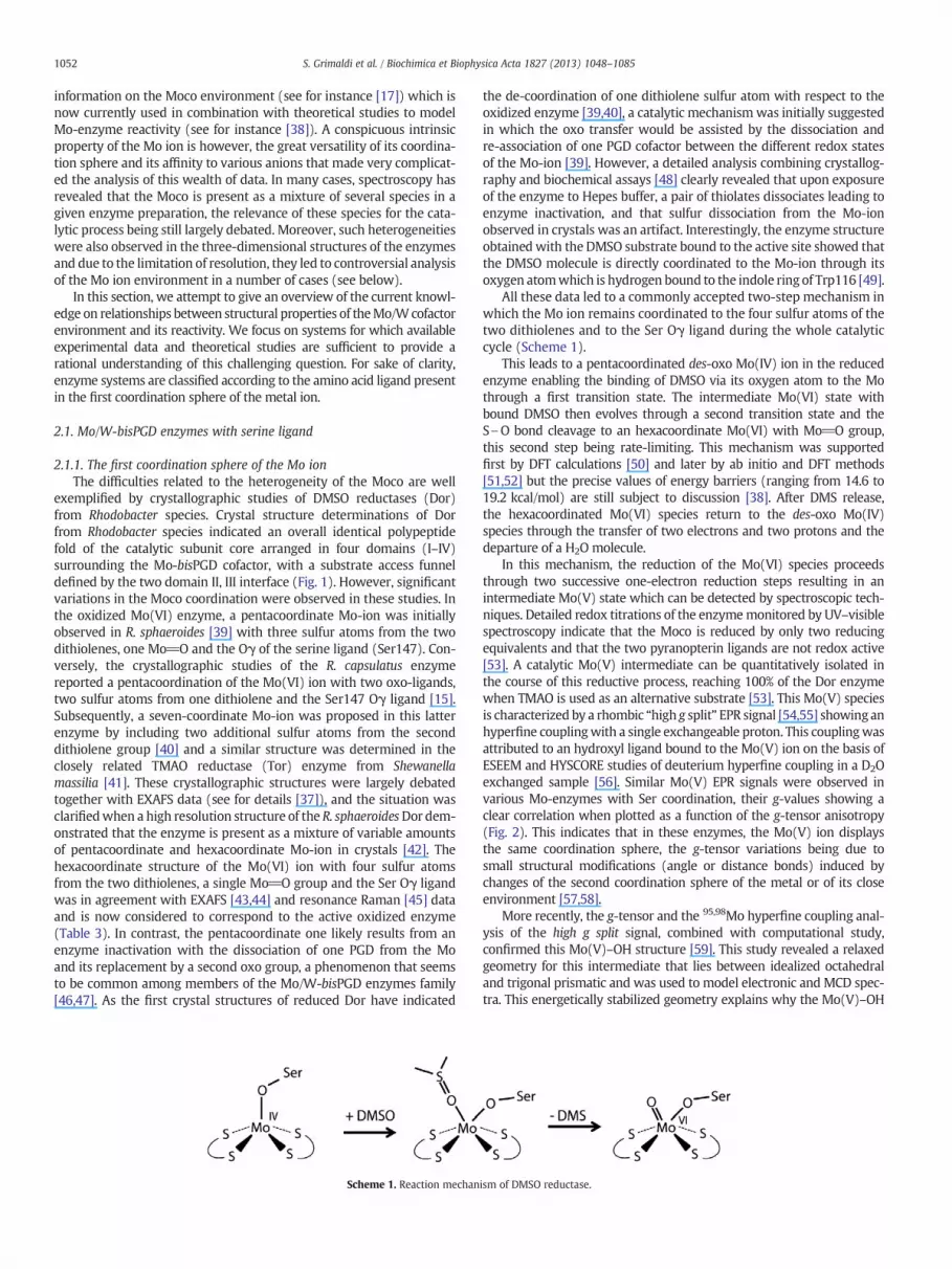
Scheme 1. Reaction mechan
the de-coordination of one dithiolene sulfur atom with respect to theoxidized enzyme [39,40], a catalytic mechanismwas initially suggestedin which the oxo transfer would be assisted by the dissociation andre-association of one PGD cofactor between the different redox statesof the Mo-ion [39]. However, a detailed analysis combining crystallog-raphy and biochemical assays [48] clearly revealed that upon exposureof the enzyme to Hepes buffer, a pair of thiolates dissociates leading toenzyme inactivation, and that sulfur dissociation from the Mo-ionobserved in crystals was an artifact. Interestingly, the enzyme structureobtained with the DMSO substrate bound to the active site showed thatthe DMSO molecule is directly coordinated to the Mo-ion through itsoxygen atomwhich is hydrogen bound to the indole ring of Trp116 [49].
All these data led to a commonly accepted two-step mechanism inwhich the Mo ion remains coordinated to the four sulfur atoms of thetwo dithiolenes and to the Ser Oγ ligand during the whole catalyticcycle (Scheme 1).
This leads to a pentacoordinated des-oxo Mo(IV) ion in the reducedenzyme enabling the binding of DMSO via its oxygen atom to the Mothrough a first transition state. The intermediate Mo(VI) state withbound DMSO then evolves through a second transition state and theS\O bond cleavage to an hexacoordinate Mo(VI) with Mo_O group,this second step being rate-limiting. This mechanism was supportedfirst by DFT calculations [50] and later by ab initio and DFT methods[51,52] but the precise values of energy barriers (ranging from 14.6 to19.2 kcal/mol) are still subject to discussion [38]. After DMS release,the hexacoordinated Mo(VI) species return to the des-oxo Mo(IV)species through the transfer of two electrons and two protons and thedeparture of a H2O molecule.
In this mechanism, the reduction of the Mo(VI) species proceedsthrough two successive one-electron reduction steps resulting in anintermediate Mo(V) state which can be detected by spectroscopic tech-niques. Detailed redox titrations of the enzymemonitored by UV–visiblespectroscopy indicate that the Moco is reduced by only two reducingequivalents and that the two pyranopterin ligands are not redox active[53]. A catalytic Mo(V) intermediate can be quantitatively isolated inthe course of this reductive process, reaching 100% of the Dor enzymewhen TMAO is used as an alternative substrate [53]. This Mo(V) speciesis characterized by a rhombic “high g split” EPR signal [54,55] showinganhyperfine couplingwith a single exchangeable proton. This couplingwasattributed to an hydroxyl ligand bound to the Mo(V) ion on the basis ofESEEM and HYSCORE studies of deuterium hyperfine coupling in a D2Oexchanged sample [56]. Similar Mo(V) EPR signals were observed invarious Mo-enzymes with Ser coordination, their g-values showing aclear correlation when plotted as a function of the g-tensor anisotropy(Fig. 2). This indicates that in these enzymes, the Mo(V) ion displaysthe same coordination sphere, the g-tensor variations being due tosmall structural modifications (angle or distance bonds) induced bychanges of the second coordination sphere of the metal or of its closeenvironment [57,58].
More recently, the g-tensor and the 95,98Mo hyperfine coupling anal-ysis of the high g split signal, combined with computational study,confirmed this Mo(V)–OH structure [59]. This study revealed a relaxedgeometry for this intermediate that lies between idealized octahedraland trigonal prismatic and was used to model electronic and MCD spec-tra. This energetically stabilized geometry explains why the Mo(V)–OH
ism of DMSO reductase.

Fig. 2. Plots of the g-values of Mo(V) species against anisotropy for members of theMo/W-bisPGD enzymes family. A. Moco with Ser ligand. High g split signals fromR. sphaeroides Dor (1, 2) [44,56], BisC (3) [63], S. massilia Tor (4) [453]. B. Mocowith Cys or SeCys ligand. Close symbols, high g type signals: E. coli NapA high g resting(5) [69], R. sphaeroides NapAB high g resting (6) [68] and NapA high g resting (7) [84],P. pantotropha NapAB high g resting (8) [55], and high g nitrate (9) [75], S. gelidimarinaNapAB high g resting (10) [454], D. desulfuricans NapA high g nitrate (11) [74] and high gturnover (12) [70],D. desulfuricans Fdh (13) [98], andW. succinogenes Psr treatedwith poly-sulfide (14) [89]. Open symbols, very high g type signals:Methanobacterium formicicum Fdh(15) [455], R. sphaeroidesNapAB (16) [68], P. pantotrophus NapAB (17) [75], Synechococcussp. NarB (18) [87], A. vinelandiiNas (19) [456], and very high g split fromW. succinogenes Psr(20) [89]. C. Moco with Asp ligand. High-pH signals from E. coli NarGHI (21) [122],(22) [120], (23) [119], high-pH (24) and high-pD (25) from M. hydrocarbonoclasticusNarGH [127]. low-pH signals (26), nitrate (27), nitrite (28) [119], nitrate 1 (29), nitrate 2(30), nitrite 1 (31), nitrite 2 (32), chloride (33), fluoride (34) from E. coli NarGHI [120].low-pH signal (35), low-pD (36), nitrate (37), and nitrite (38) fromM. hydrocarbonoclasticusNarGH [127]. Linear correlations between the available data sets within each family can beextrapolated and are schematically indicated by straight or dotted lines to help the reader.
1053S. Grimaldi et al. / Biochimica et Biophysica Acta 1827 (2013) 1048–1085
to Mo(IV)+H2O reduction step is rate limiting when TMAO is used assubstrate. Indeed, the calculations indicate that, with this substrate, theOAT event is activationless.
2.1.2. Role of the environment on the Moco reactivityThis oxygen atom transfer mechanism appears to be commonwithin
Ser coordinated Mo/W-bisPGD enzymes, which generally have a fairlyopen substrate binding funnel and exhibit broad substrate specificitytowards S- and N-oxides [16]. Several mutagenesis studies were under-taken to address the structural factors influencing this specificity. Severalresidues (Thr148, Ala178, Arg217, Gly167, Gln179) predicted to form thewall of the funnel leading to the Mo active-site in E. coli Dms weremutated and found to affect substrate binding and/or specificity, whilenot spatially clustered [60]. Two highly conserved aromatic residuesTyr114 and Trp116 arranged around the Mo ion have attracted muchattention as potential candidates involved in catalysis and/or in substratespecificity. In the structures of Dor from R. capsulatus, Trp116 hydrogen-bonds to the labile oxo group of oxidized enzyme [15,40] and to theoxygen of the DMSO in the structure with substrate bound at thereduced active site [49]. Trp116 is conserved in Tor and in BisC.Whereassubstitution of this residue to phenylalanine in Dor caused a significantreduction in kcat towardsDMSOandTMAO, theKm for DMSO is notmark-edly affected and the Km for TMAO is slightly reduced [61]. This suggeststhat Trp116 does not greatly influence substrate bindingwhile having animportant role in the catalytic cycle. Spectroscopic studies of theas-isolated Trp116Phemutant indicate that it harbors a pentacoordinatedioxoMo center inwhich one dithiolene group is outside theMo coordi-nation sphere and replaced by a second Mo_O group [61,62], similarlyto the “Hepes-modified” inactive form of the enzyme [42,48]. As such,Trp116 would prevent movement of the Mo-oxo unit away from thecoordinating dithiolene in the resting Mo(VI) enzyme. Redox cyclingthe Trp116Phe mutant, however, regenerates transiently a wild-typelike enzymeDMSO reducing activity [62]. Similarmutations of the equiv-alent residue (Trp90) in BisC exhibited alsomarkedly decreased catalyticactivities and drastic changes on theMo(V) EPR spectral properties, withno significant alterations in either substrate affinity or specificity [63].Thus, in agreement with mechanistic scheme presented above, by stabi-lizing the mono-oxo Mo(VI) species by hydrogen bond, this Trp residuecould have a direct influence on the rate-limiting step of the catalysis andwould play an important role in controlling the catalytic efficiency in Dorand BisC.
Tyr114 was found hydrogen-bound to the oxo ligand of the mono-oxoMo(VI) in thefirst structure ofDor fromR. sphaeroides [39]. The pres-ence of this oxo-group as Mo ligand was however reassessed and it wasproposed to be an artifact following an erroneous interpretation of theelectron density maps caused by discrete heterogeneity of the Mo posi-tion in the crystals [42,48]. On the other hand, the tyrosine OH oxygenis located at 3.3 Å from the sulfur of the DMSO in the structure of Dorfrom R. capsulatus complexed with its substrate [49]. Interestingly, thisresidue is conserved in BisC [63,64]. The unique but striking differencein the arrangement of the residues directly surrounding the active centerat theMo ion in theDor and Tor structures is the absence of Tyr114 in thelatter [41]. While it is replaced by a threonine (Thr116) or a serinedepending on the organism, no other residue occupies the space leftempty by the missing aromatic side chain. This led to the hypothesisthat Tyr114 is critical for the control of substrate specificity in theS-oxide/N-oxide reductases [41,65]. Substitution of this residue for aphenylalanine (Tyr114Phe) in recombinant Dor from R. sphaeroidesexpressed in E. coli [64], or in R. capsulatus [62,66] leads to a significantalteration of the catalytic properties toward DMSO and TMAO, resultingin an ~10 fold decreased efficiency of the enzyme toward DMSO and aslightly less efficiency with TMAO. Noteworthy, the substitution doesnot affect the Mo(V) EPR spectrum, indicating no interaction of theresidue with the Mo center in this redox state [62]. In addition, proteinfilm voltammetry measurements indicated that this residue is criticalfor protonation steps at the active site [66]. Insertion of a Tyr into Tor

1054 S. Grimaldi et al. / Biochimica et Biophysica Acta 1827 (2013) 1048–1085
in an equivalent position results in a decreased preference for TMAOrelative to DMSO [64]. Altogether, these studies indicate that Tyr114would function in substrate recognition: it would be important forcorrect positioning of the substrate in the Dor active site, most likely byproviding a hydrogen bond with DMSO.
2.2. Mo/W-bisPGD enzymes with cysteine or selenocysteine ligand
Investigations of the structure and mechanism of Mo/W-bisPGDenzymeswith Cys or Se-Cys coordinationwere subjected to long contro-versy due to uncertainties in the determination of the Mo ion ligands byX-ray crystallography and to the plasticity of this coordination sphere asshown by spectroscopy experiments. Moreover, by comparison withseveral enzymes carrying a Ser coordinated Moco (Dor, Tor, BisC), allthose harboring a Cys or SeCys coordinated Moco contain at least oneadditional FeS cluster in the catalytic subunit (see Section 3.1). Thismakes spectroscopic approaches more complicated to address theircatalytic mechanism. In this section, the structure and mechanism ofseveral representative enzymes of this group, Nap, Psr, Fdh, Ah, whichcatalyze a large diversity of reactions, are reported.
2.2.1. Periplasmic nitrate reductases
2.2.1.1. The first coordination sphere of the Mo ion. In the case ofperiplasmic dissimilatory nitrate reductases (Nap), the first structureobtained for the oxidized enzyme NapA from Desulfovibrio desulfuricanshas shown a Mo ion coordinated by the four sulfur atoms of the twoPGDdithiolenes and by afifth sulfur fromaCys residue [67]. An addition-al sixth ligand being OH or OH2was determined, and the same coordina-tion was proposed from crystallographic studies at lower resolution ofthe enzymes NapAB from R. sphaeroides [68] and NapA from E. coli[69]. Detailed investigations of subsequent crystallographic dataobtained with D. desulfuricans NapA, together with B factor analysis,assigned however this sixth ligand to a sulfur atom partially bondwith the thiolate of the Cys ligand in the “as prepared” state of theenzyme [70]. This six sulfur coordination of the Mo ion was alsoconfirmed by the recent high resolution structure (1.5 Å) of the NapABenzyme from Cupriavidus necator (formerly Ralstonia eutropha) [71].
According to the initial NapA structure, the first theoretical investiga-tions of enzyme reaction proposed amechanism closely related to that ofDor, the serinate ligand being replaced by a cysteinate one. In the firststep of this mechanism, nitrate binds through one oxygen atom to theMo(IV) ion. This is followed by the N\O bond breaking step, leadingto the release of nitrite and to the formation of aMo(VI)–O intermediate[72,73]. However, by contrast with Ser coordinated Mo/W-bisPGDenzymes, EPR studies of Naps revealed a number of substoichiometricdistinctMo(V) species whose catalytic relevancewas strongly discussed.These species were classified in three main groups according to their gvalues as high g, very high g, and low g species (Fig. 2B) [55,68,69,74].Depending on enzyme preparations and treatments, the proportions ofthese species vary greatly, the high g species being usually the mostabundant while the two others are considered as resulting from partialdegradation of theMoco [75]. Mo(V) high g-type signals show character-istic hyperfine splittings attributed to the β-CH2 protons of the cysteineligand [76]. Depending on the enzyme, this signal can be observed indifferent conditions: in “as prepared” enzymes (high g resting), in enzymeunder turnover with nitrate (high g nitrate or high g turnover), or inreduced enzyme. Moreover, kinetic studies of Nap by protein filmvoltammetry revealed that the enzyme activity decreases at very lowpotential suggesting that nitrate can bind more efficiently to Mo(V)than to Mo(IV) state [77,78]. The potential role of the Mo(V) state innitrate binding was also emphasized in the catalytic cycle model pro-posed in line with the E. coli NapA structure determination [69]. Basedon the redox potential of Mo(V) high g species deduced from EPR studyof R. sphaeroides NapAB, a quantitative kinetic model was developed tak-ing into account nitrate binding to both Mo(IV) and Mo(V) redox states
[79]. This model was further improved by considering the kinetics ofsubstrate binding and release, and protonation steps [80]. It enabled theinterpretation of catalytic voltammograms obtained with R. sphaeroidesNapAB in a broad range of substrate concentrations and pH. This led tothe conclusion that in this enzyme, substrate binding is irreversible onthe time scale of turnover, and that combined with protonation, it raisesthe redox potential of the Mo(V/VI) couple, triggering the energetics ofintramolecular electron transfer with FeS within NapA [80].
The discovery of the sixth sulfur atom in the coordination sphere ofthe Mo ion in Nap [70] has strongly stimulated the reinterpretation ofthe structural models of the spectroscopically detected Mo species andenzyme mechanisms. Thus, a recent DFT investigation of the magneticparameters (g and A(1H) tensors) of possible models for Mo(V) EPRdetected species concluded that the high g Mo(V) species correspondsto a six sulfur coordination sphere in a pseudotrigonal prismatic geome-try, with a partial disulfide bond between the sulfur ligand and thethiolate of the Cys ligand [58]. The very high gMo(V) specieswould resultfrom the two electrons reduction of this disulfide bond, leading to sepa-rated sulfido and thiolato ligands, but maintaining the six sulfur coordi-nation of the Mo(V) ion. Moreover, the attribution of the low g speciesto a Moco with only one coordinated pterin moiety was confirmed inthis study [58]. Interestingly, it is clear that independently of the bacterialspecies or of the conditions used to produced the Mo(V) high g species,all reported g values exhibit a strong correlation (Fig. 2B). This showsthat in these species the Mo ion has the same coordination sphere, theg-tensor variations being due to more distant structural modifications.The same trend is also observed for the very high g species (Fig. 2B). Inparticular, this indicates that in the so-called high g nitrate or high gturnover species, nitrate or nitrite ions if present are not directly boundto Mo ion. These results emphasize the heterogeneity of the Moco inthe “as prepared” enzyme. The structure proposed for high g Mo(V)from DFT calculations [58] is then very similar to that found in themore recent crystallographic studies of oxidized D. desulfuricans NapA[70] and C. necator NapAB [71]. However, the physiological relevance ofthis species was recently questioned. By combining EPR spectroscopyand protein film voltammetry of R. sphaeroides NapAB, it was shownthat the high gMo(V) species detected in “as prepared” enzyme cor-responds to an inactivated form which needs to be reduced forreactivation. Thus, the disappearance of the high g Mo(V) EPR signalupon reduction is not a simple redox process but is accompanied by astill unknown chemical transformation making this reduction notreversible [47]. As such, this observation questions the significance ofthe Moco redox potentials determined by EPR and their use in catalyticmodels.
Taking into account the six sulfur coordination of the Moco, severaltheoretical and computational investigations of the Nap mechanismwere performed [81–83]. As the disulfide bond between the sulfidoand Cys-thiolato ligands blocks the direct access of nitrate to the Moatom, various kinds of inner and outer coordination sphere mechanismswere investigated, the Mo ion being in the Mo(VI) or Mo(V) state(Scheme 2).
In outer sphere mechanisms, nitrate is guided through a narrowfunnel to the active site and interacts with the sulfido Moco ligand lead-ing to a O2NO\S bond [81,83]. After release of nitrite, the remainingO_S\ ligand of Mo must be reduced to restore the initial state of theenzyme. In inner sphere mechanisms, the direct binding of nitrate tothe Mo ion requires a rearrangement of the active site in which theCys-thiolato disconnects from Mo but remains attached to Mo by theterminal sulfur atom giving a Cys-S\S\ ligand. This rearrangementwould be stabilized by conserved methionines, Met141 adjacent tothe Cys140 ligand and Met308, and seems consistent with Moco struc-tural changes observed in the crystal structure of dithionite reducedC. necator NapAB [71]. In addition to the redox chemistry of the Mo ion,both kinds of mechanisms involve also a sulfur-based redox chemistry.Although the high energy barrier of the O2NO\S bond formation likelyexcludes outer sphere mechanisms, the redox states of the Mo ion in

Scheme 2. One of the inner sphere reaction mechanisms proposed for Nap. The valence states of the Mo ion were not indicated since it was suggested that redox chemistry involvessulfur ligands. Adapted from [81,83].
1055S. Grimaldi et al. / Biochimica et Biophysica Acta 1827 (2013) 1048–1085
themechanism and the subsequent reduction steps of the formed oxo li-gand into water molecule remains discussed [38].
2.2.1.2. Role of the environment on the Moco reactivity. The analysis ofthe D. desulfuricans NapA structure suggests that several conservedand charged residues (Asp155, Glu156, Asp355, and Arg354) enable toguide the nitrate ion from the solvent to the Moco [81]. In R. sphaeroidesNapAB, the role of Arg392 (homologous to Arg354 of D. desulfuricansNapA) and of Met153, adjacent to the coordinating Cys152, have beenaddressed by site-directed mutagenesis. These conserved residueswhich lie at ~9 and 6 Å from the Mo ion, respectively, were substitutedby Ala, and the purified mutants where characterized in detail [84,85].For both mutants, Met153Ala and Arg392Ala, the substitution affectedonly the Km for nitrate (increased by 10 and 200-fold, respectively), butnot the kcat, indicating that these residues are not involved in the catalyticreduction of nitrate but promote substrate binding [84]. These resultssubstantiate a previous study of mutants of the equivalent residues(Arg421 and Met182) in NapAB from C. necator that revealed a drastic(Arg421Lys) or a complete (Arg421Glu, Met182His) loss of enzymaticactivity monitored on periplasmic extracts [86]. In addition, a zinc inhib-itory effect that seems specific to Nap was revealed [84]. Zn2+ bindinginside the substrate channel in the presence of nitrate was found to befacilitated by the absence of the guanidiumgroup of Arg392 and requiresthe Met153 side chain that likely coordinates this metal ion. It was thenproposed that Arg392 act as a filter in protecting the active site againstinhibition and direct reduction by small molecules [84].
Clearly, in spite of the numerous works done on the Nap enzymes,further investigations are required to rationalize the data gained bycrystallography, spectroscopy, kinetics, and theoretical calculations.Noteworthy, the relationships between the various Mo(V) speciesidentified by spectroscopy [58], those involved in the catalytic schemededuced from kinetic studies [80], the crystallographic structure of theMoco [71], the potential catalytic intermediates [38] and inhibitedforms [47] are still to establish. Such investigations would be alsorelevant for prokaryotic assimilatory nitrate reductases (Nas), whichhave a coordinating Cys residue of Moco and exhibit Mo(V) EPR signalssimilar to those given by Nap [87] but remain poorly characterized.
2.2.2. Polysulfide reductasePolysulfide reductase (Psr) is another enzyme with Cys coordinated
Moco, but instead of the commonOAT, it catalyzes a sulfur atom transfer(SAT) reaction, giving HS− as reaction product according to the equa-tion: Sn2−+H++2e−=Sn−1
2− +HS− above pH 7. The structure of theoxidized Thermus thermophilus enzyme has been determined at 2.4 Åresolution and shows that, in addition to the four sulfur atoms fromthe bis-PGD cofactor, the Mo ion is coordinated by the thiolate group ofCys173 [88]. Moreover, a sixth ligand interpreted as an oxo group wasproposed. This coordination was considered to be consistent with thefirst EPR analysis of Psr from Wolinella succinogenes which revealedMo(V) signals resembling the very high g signal of Nap [89]. However,it is worth noting that the Mo(V) Psr signals obtained upon dithionitereduction or with polysulfide substrate are rather related to high gsignals, while that produced after borohydride reduction correlateswith very high g signatures (Fig. 2). According to the structural interpre-tation of these signals in Nap [58], they should correspond in both casesto Moco species with six sulfur coordination, indicating that very likely,
the sixth ligand is sulfur as in Nap. These spectroscopic results havealso suggested that polysulfide substrate binds directly to Mo [89]which was supported by a recent HYSCORE study of 33S isotope-labeled polysulfide treated enzyme strongly indicating that the 33S isindeed the sixth ligand of the Mo(V) center [90]. However, thesespectroscopic studies require further analysis for these structural conclu-sions to be confirmed.
According to the Psr crystal structure, substrate selectivity of Psrwould be related to the narrow funnel connecting the active site toenzyme surface, with basic residues limiting its entrance. As for Nap,an Arg residue (Arg332) is located in close vicinity ofMoco and is hydro-gen bound to two water molecules. This residue was proposed tostabilize the polysulfide substrate [88], but at this time, details on thecatalytic mechanism remain to decipher.
2.2.3. Formate dehydrogenaseFormate dehydrogenases (Fdh) possess a Moco which shares com-
mon structural propertieswithNap but catalyze a very different reaction,namely the conversion of formate into CO2 that can be considered as aformal hydrogen atom transfer: HCOO−=CO2+2e−+H+.
Three crystal structures of Fdh have been reported, the two Mo-enzymes FdhF [91] and FdnGHI [92] from E. coli, and the W-containingFdhAB enzyme from Desulfovibrio gigas [93]. In the oxidized enzyme,all of them show a metal ion coordinated by the four sulfur atoms of abis-PGD cofactor, and by the selenium atom of a SeCys. As for Nap, thenature of a sixth ligand was largely debated. First modeled as a longMo\O bond in E. coli FdhF [91], this sixth ligand was attributed to asulfur atom in D. gigas FdhAB [93]. Moreover, the initial crystal structuredetermination of formate reduced E. coli FdhF has suggested that thesixth ligandwas lost upon reduction, but the re-analysis of data indicatedthat SeCys is no longer bound to Mo which becomes pentacoordinatedwith a sulfur atom as axial ligand in the reduced state [94]. This refinedstructure is in agreement with previous EXAFS studies of the enzyme[95] and with recent biochemical results showing that the sulfurationof the E. coli FdhF enzyme is essential for its catalytic activity [96].
Surprisingly, EPR studies of formate reduced E. coli FdhF revealed anearly axial highly anisotropic Mo(V) signal (g=2.094, 2.001, 1.990),which exhibits a strong hyperfine coupling with 77Se in 77Se-enrichedenzyme [97]. This coupling reflects a direct coordination of SeCys totheMo ion in the EPR active species, in contradiction with crystal struc-ture of formate reduced enzyme, suggesting that theMo(V) species hasno relation with the crystallographically detected state, or that theenzyme was altered by X-ray in crystallographic studies [37]. Alterna-tively, EPR studies performed on D. desulfuricans Fdh suggest that thesevariations could be due to the influence of some enzyme inhibitors(azide, cyanide) [98]. In the absence of these inhibitors, the Mo(V) EPRsignal obtained becomes much less anisotropic. Its g-values are thenreminiscent of the high g Mo(V) signal found in Nap enzymes (Fig. 2),but with a stronger anisotropy which is consistent with replacement ofa sulfur ligand from Cys by a selenium as observed for other enzymessuch as NiFe hydrogenases [99] and can be explained by the increasedcovalence effects of selenium.
Based on structural homology between Fdhs, the conserved SeCys140ligand of Mo and the proximal His141 and Arg333 (numbering of E. coliFdhF) are considered as essential residues for catalysis. The replacementof SeCys140 for Cys by site-directedmutagenesis in E. coli FdhF decreases

1056 S. Grimaldi et al. / Biochimica et Biophysica Acta 1827 (2013) 1048–1085
the enzyme activity by two orders of magnitude [100]. Moreover, Arg333is homologous of Arg392 of R. sphaeroides NapA which was shown to beinvolved in nitrate binding [84]. In the X-ray structure of the oxidizedE. coli FdhF, the side chain of Arg333 points towards the active site in aposition to interact and orient the formate molecule, whereas in theformate-reduced enzyme, Arg333 is in contact with the free selenolfrom Se-Cys [94].
According to the initial E. coli FdhF structure, thefirst proposedmech-anism resembles that of DMSO reductasewith the replacement of theOHligand by a formate ion coordinated through an oxygen atom to theMo(VI) ion. In a second step, the formate C\H bond is cleaved withconcomitant binding of hydrogen to Se, transfer of two electrons to theMo, and release of CO2 [97]. This leads to a pentacoordinated Mo(IV)ion with a protonated SeCys ligand which is subsequently oxidized toMo(V) with the transfer of the proton from SeCys to the proximalHis141. The catalytic cycle is then completed with the oxidation ofMo(V) to Mo(VI), the transfer of the proton to the solvent and the bind-ing of a hydroxide to theMo(VI) ion. After thefinding of a sulfur ligand inthe coordination sphere of the reduced E. coli FdhF Moco, this mecha-nism was adapted by replacing the OH ligand by SH and was addressedby theoretical calculations [101]. An alternative mechanism was alsoproposed in which the SH ligand remains bound to Mo in the wholecatalytic cycle, while the Mo(VI) SeCys ligand is released to enableformate binding [94]. In thismechanism, the unbound SeCys is stabilizedby interaction with proximal Arg333, and enables hydrogen atomabstraction from bound formate. Calculations of activation energy forC\H bond cleavage showed that the proton abstraction is much morefavored with unbound SeCys (19 kcal/mol compared to 36 kcal/mol)[101]. This study did not consider, however, the role of the proximalHis141 and Arg333 residues, and in a more recent DFT study, Mota etal. [102] have investigated the entire catalytic cycle by taking intoaccount the influence of these residues. Their proposed mechanism isreminiscent of the inner sphere mechanism determined by Cerqueiraet al. [81] for Nap (Scheme 3).
Starting from a hexacoordinated Mo(VI) structure with SeCys andsulfido ligands partly boundby a seleno-sulfide bond, the catalyticmech-anism involves decoordination of SeCyswith the transient formation of aCys-Se\S Mo ligand to enable formate binding. The Se\S bond is thenbroken, allowing the formed selenide to abstract the hydrogen atomfrom formate. This leads to a cyclic intermediate with the CO2 moleculebound toMo(IV) ion by one oxygen and to the sulfur ligand by its carbonatom. After CO2 release, Mo(IV) oxidation and proton transfer, the
Scheme 3. Reaction mechanism proposed for FdhF witAdapted from [102].
catalytic site is restored. As no Mo(V) state is involved in this mecha-nism, this questions the relevance of the EPR-detected Mo(V) speciesas for Nap. These DFT calculations indicate that Arg333 has indeed akey role in driving the formate ion toward the sulfur ligand throughtwo strong hydrogen bonds, and promoting its binding to the metalion by a single bond. A role of the positively charged side chain ofArg333 in driving the product release was also proposed. In addition,it was found that His141 lowers the activation energy for the dissocia-tion of SeCys from the molybdenum by forming a hydrogen bondwith the selenide anion, then allowing proton abstraction from thesubstrate to the SeCys. In this model, His141 is not directly involved inthe removal of the formate α-proton. However, the role of His141 incatalysis, together with that of possible H acceptors in the active site,has beenquestioned by recent theoretical calculations [103]. The resultsfavor a mechanism in which the metal center mediates H transfer fromsubstrate to the SeCys by forming a transient Mo\H intermediate.Considering the existence of thiolate-coordinated Mo hydride com-pounds, these authors then conclude that a hydride transfer couldbe operating in FdhF catalysis. Thus, the exact role of His141 in thepeculiar hydrogen atom transfer that is specific of FdhF still needsto be established.
2.2.4. Acetylene hydrataseBy contrast with other enzymes with Cys or Secys Mo ligand, acety-
lene hydratase (Ah) catalyzes a non-redox reaction, the hydration ofacetylene to acetaldehyde. This tungsto-enzyme from P. acetylenicuswas crystallized in anaerobic conditions in the reduced most activestate. The high resolution structure (1.26 Å) revealed that the W atomis coordinated by a Cys residue and by a water molecule hydrogen-bound to a close aspartate residue (Asp13) [104]. One striking differencewith other Mo/W-bisPGD enzymes is a major rearrangement of theregion between domain II and III leading to a completely different funnelaccessing the active site in AH. This substrate access funnel is located atthe intersection of domains I, II and III, giving access to a new face of theprotein surface (Fig. 1). It leads to a different portion of the metal coordi-nation sphere and was considered as a key factor for substrate specificityand reactivity. In the proposed mechanism, the W(IV)-bound watermolecule would be activated by the Asp13 residue thanks to a pKa shiftinduced by the proximal FeS cluster [104]. Although acetylene hydrationreaction is not a redox process, it is striking that the lysine residue that isconsidered as essential for electron transfer between the FeS cluster andthe Moco in Mo/W-bisPGD enzymes is also conserved in Ah (Lys48). It
h the activation step based on sulfur-shift process.

1057S. Grimaldi et al. / Biochimica et Biophysica Acta 1827 (2013) 1048–1085
is proposed that in Ah, the rearrangement of a loop region between thetwo metal cofactors in Ah might be responsible for a strong increase inAsp13 pKa induced by the proximal FeS cluster [104]. In the followingstep, the activated water would attack the triple bond of acetylenethrough an electrophilic addition mechanism. The critical role played bythe carboxylic group of Asp13was confirmed by site-directedmutagene-sis experiments: enzyme activity is almost suppressed by Asp13Alamutation while it is nearly unchanged in Asp13Glu variant [105]. More-over, the presence of a hydrophobic cavity enabling substrate accommo-dation at the active site is supported by the strong decrease of activitywhen Ile142, which belongs to this cavity, is mutated into Ala.
Alternative mechanism involving a nucleophilic attack of wateron a W-bound acetylene was also suggested [106], but DFT calcula-tions indicated that these two mechanisms have very high energy bar-riers (~40 kcal/mol) and proposed that reaction proceeds through aW_C_CH2 vinylidene intermediate [107]. Several other mechanisticschemes have been investigated by DFT methods on large active sitemodels [108]. These authors concluded that themost energetically favor-able mechanism involves the η2 binding of acetylene on the W(IV) ionwith the displacement of the water molecule ligand. This molecule issubsequently deprotonated by the carboxylate group of Asp13 andperforms a nucleophilic attack on the bound acetylene, giving a vinylanion stabilized by binding to W ion. Shuttled by Asp13, a proton isthen transferred to this anion leading to a vinyl alcohol intermediate,which gives acetaldehyde after tautomerization. This first shell mecha-nism was recently supported by theoretical calculations performed ona biomimetic W(IV)O(S4) tungsten complex but with the participationof the W(IV)–OH ligand to deprotonate the water molecule [109].
2.3. Mo/W-bisPGD enzymes with Aspartate ligand
Enzymes where Moco is coordinated by Asp generally present a tri-meric organization (Table 1) and contain several other redox cofactors,FeS clusters and hemes, giving a large diversity of spectroscopic signa-tures. The coordination of the Moco by Asp was demonstrated by crys-tallography in only two enzymes, nNar and Ebdh whose properties aredescribed in this section.
2.3.1. Respiratory nitrate reductaseThemost representative enzymeof this class is themembrane-bound
respiratory nitrate reductase from E. coli NarGHI (i.e. nNar) that wasspectroscopically investigated by Bray and coworkers more than thirtyfive years ago [110]. The ability to produce the enzyme either in amembrane-bound form NarGHI or in a soluble form NarGH was a greatadvantage to address the thermodynamic and structural properties ofthe various metal centers [111–113]. This was also used to determinethe crystal structure of the oxidized enzyme in the two forms NarGHI[114] and NarGH [115] and to demonstrate the presence of an aspartateligand to the Mo ion (Asp222). By comparison with Ser coordinatedMoco enzymes, the catalytic subunit NarG has important sequenceinsertions that define an additional fifth domain in the structure. Thisdomain V delineates a much narrow substrate binding funnel in NarG,and could play a role in modulating its substrate specificity towardsnitrate and the small number of oxyanions identified as substrates[116,117]. In both structures, theMo ion is coordinated by the four sulfuratoms of the two PGD cofactors, but surprisingly, the aspartate coordina-tion is different in the two systems. InNarGHI, the Asp ligand is bidentatewith the two oxygen atoms from the carboxylate group bound to Mo. Inaddition, one PGD cofactor was shown to be bicyclic with an open pyranring [114]. In contrast, in NarGH structure, the two PGD cofactors arein the usual tricyclic pyranopterin state, and the aspartate ligand ismonodentate, the sixth coordination position of the Mo being occupiedby an oxo group at 1.8 Å [115]. However, these structures show highlydistortedMo coordinationswith short distances between the oxo ligand,one of the dithiolene sulfur and the coordinating aspartate oxygenwhichcould results fromaveraged structural heterogeneities arising fromX-ray
photoreduction [37]. These structural variations between enzymes led tosuggest that the flexibility of the aspartate coordination and the pyranring opening and closing could play a role in catalysis, especially for theproton transfer [115,118].
Early spectroscopic studies performed on E. coli nNar have reportedtwo characteristic pH-dependent Mo(V) EPR signals, the low-pH andhigh-pH signals [119], both signals showing hyperfine coupling with asolvent exchangeable proton. This coupling is stronger for the low-pHsignal which was shown to be sensitive to the presence of anions likenitrate, nitrite, chloride and fluoride [120]. The hyperfine couplingobserved with 19F showed that this anion was located close to theMo(V) ion, but no such coupling was found with 17O enriched nitrateindicating that these anions were not directly bound to the metal[121]. By comparing the pKa of interconversion between high and lowpH species (pKa~8.3) with enzyme activity profile, it was proposedthat only the low pH species is catalytically active [119]. However, thispKa value was determined in the presence of chloride and it was subse-quently shown that anion binding modifies the high pH/low pH signaltransition pKa [120,121]. Later, a pH dependence study of Mo(V) speciesredox potentials performed in absence of contaminating anions hassuggested that both species could be involved in the catalytic cycle[122]. Moreover, this study showed that the replacement of the His49ligand of the proximal FS0 cluster by Cys also affects the pKa of the tran-sition, while the structural organization of the Moco and the FS0 centersis not modified [123], indicating that this pKa value is dependent on theH-bond network around the Moco. These Mo(V) EPR signals weredetected in several other respiratory nNars from Paracoccus denitrificans[124] and Marinobacter hydrocarbonoclasticus [125]. In all cases theseMo(V) EPR active species were substoichiometric, revealing structuralheterogeneities of the Moco in enzyme preparations and questioningtheir relationships with the structure determined by crystallography orEXAFS [121]. Interestingly, the binding of nitrate, nitrite or other anionschanges only weakly theMo(V) EPR signatures which remain correlatedto the low- and high-pH signals (Fig. 2). This shows that the binding ofthese anions does not occur in the first coordination sphere of Mo(V)ion which remains likely unchanged in these different species. Themore realistic interpretation of thisMo(V) coordination is that the aspar-tate ligand is monodentate and that the sixth position is occupied by anOH group. In the NarGH crystal structure, the monodendate molybde-num ligand Asp222 is hydrogen-bound to the Nε of the conservedHis546 residue [115], whereas this H-bond is lost in the bidendateNarGHI structure [114]. This led to the hypothesis that this His may bethe ionisable residue with pKa~8 that defines the transition betweenthehighand the low-pHMo(V) forms evidencedbyEPR. The interconver-sionbetweenhigh- and low-pH species could bedue to theprotonationofthis nearby residue, leading to a reorientation of the Mo(V)–OH ligandand to a change of the proton hyperfine coupling [125,126].
In spite of the long standing studies on nNar, it is striking that itsreaction mechanism and the catalytic relevance of the Mo(V) species isstill a matter of controversy. Notably, the ability of these enzymes toreduce various oxides of nitrogen, halogens [127] and metalloids [117],and to be a potential NO production enzyme [128–130] will be requiredto understand more deeply the interactions between the Moco, itssurroundings and these substrates.
2.3.2. Ethylbenzene dehydrogenaseAmonodentate aspartate ligand of Moco, Asp223, was also observed
in the crystal structure of the ethylbenzene dehydrogenase (Ebdh) fromAromateum aromaticum [131], an enzyme closely related to Nar. In thiscase, one of the PGD has an open pyran ring. The sixth ligand of Mo isan oxygen atom from an acetate ion, the metal ion being considered tobe in the reducedMo(IV) state due to crystallization conditions. Interest-ingly, the second oxygen from the Asp223 carboxylate side chain ishydrogen bound to a Lys450-Nζ, and the same Asp–Lys pair is conservedin otherMo-enzymes of this group, selenate reductase (Ser) and chloratereductase (Clr) [131].

1058 S. Grimaldi et al. / Biochimica et Biophysica Acta 1827 (2013) 1048–1085
By contrast with most enzymes with Mo/W-bisPGD cofactor whichperform an OAT, Ebdh catalyzes the oxygen-independent hydroxylationof hydrocarbons, a process known to require the activation of C\Hbond.Because of the high diversity of this enzyme in hydroxylating variousalkylaromatic and heterocyclic compounds [132], the determination ofits catalytic mechanism has attracted much attention. Different mecha-nisms considering that the C\H activation proceeds through homolyticor heterolytic cleavage were investigated by DFT computations [133].In these calculations, theMo(VI) ion of the oxidized enzymewas consid-ered to be coordinated by a monodentate Asp223 ligand and by an oxogroup, and the role of His192 lying near the Moco in protonationprocesses was also investigated. In this model, one of the pyran rings istaken open but the influence of the ring closure on the catalysis wasnot addressed. The results of calculations indicate that the lower energybarrier mechanism involves a radical formation with two one electrontransfer steps: i) the homolytic activation of the C\H bond leading to aradical intermediate and the formation of Mo(V)–OH; and ii) a secondelectron transfer coupled to OH-rebound on the carbon atom. Modelswith His192 protonated or not give plausible reaction pathways andthis mechanism led to calculated kinetic isotope effect in qualitativeagreement with those measured experimentally [133]. More recently,this two step mechanism involving a substrate-derived radical andcarbocation intermediateswas shown to be consistentwith kinetic prop-erties of a broad range of substrate analogs [132].
2.4. Conclusion
At the present time, it is clear that it is not possible to reveal a uniqueprinciple which triggers the reactivity of Mo/W-bisPGD enzymes. Exam-ination of the wealth of structural, spectroscopic, kinetic and computa-tional data emphasizes the extraordinary high catalytic plasticity ofthese enzymes that is responsible for their evolutionary success andspreading in prokaryotes. Theoretical calculations have shown that cata-lytic pathways are extremely sensitive to proton and electron transferprocesses. H-bond networks around the Moco and their variations inthe catalytic cycle are strongly involved in the fine tuning of the cofactorreactivity and substrate selectivity. In spite of the spectacular progressesmade by crystallographic approaches, the knowledge on these networksremains hardly accessible. Going further in this understanding will needto establish relationships between the Moco structures identified bycrystallography and the spectroscopically detected species, and to deci-pher the structural basis of Moco heterogeneities. Going beyond thevery close vicinity of Moco in these analyses will be also required asillustrated by inhibition processes of Nap which likely occur in amore distant surroundings [47,84]. In this respect, using Mo(V) speciesas magnetic probe to monitor long range structural changes of theenzyme or intercenter medium variations will be a powerful approachas shown in other Mo-enzyme families [134–136]. Moreover, it willalso be essential to progress in trapping and characterizing catalyticintermediates and to make assignments to hypothetic species involvedin kinetics models. This is specially required for enzymes with Cys(SeCys) or Asp Moco ligand that catalyze the most diverse reactionsand for which mechanisms at play are still uncertain. Finally, beyondthe Mo ion, the role of pyranopterins of Moco remains poorlyaddressed. Although it is well established that pterins can have a redoxactivity [118], as demonstrated for tetrahydrobiopterin cofactor in NOsynthases [137,138], no evidence for the involvement of pyranopterinsas redox cofactors was given. Instead, a recent extensive structural anal-ysis of pyranopterins in Mo-enzymes suggests that changing the redoxstate of these cofactors could be a way to modify their conformationand to modulate the tuning of the Moco redox chemistry [139].
3. Molecular variation of the Mo/W-bisPGD enzymes
TheMo/W-bisPGD enzymes family shows an extraordinary variety ofmolecular organization and the unique common subunit is the catalytic
one. The enzymes of this family can be monomeric, dimeric (with anadditional subunit playing the role of electron transfermodule, containinghemes, or FeS clusters and/or FAD cofactor), and trimeric (withan additional subunit playing — often but not always — the role of con-nection to the membranous quinone pool) (Fig. 3). We will briefly pres-ent all types of elementwith the aim of highlighting structural variabilitybut first of all common functional features. An important pitfall whengiving a Mo/W-bisPGD enzyme denomination from the first descriptionis that it takes into account, inmost cases, the enzymatic activity and notthe phylogenetic affiliation. Actual viewof thediversity and relationshipswithin the Mo/W-bisPGD enzymes family should lead to reconsiderearlier enzyme denomination thus facilitating a better description oftheir function. Readers are encouraged to read the review from Rotheryet al. for an evolutionary point of view [16].
3.1. The catalytic subunit α
In the vast majority of enzymes from the Mo/W-bisPGD enzymesfamily the catalytic subunit, denoted α in Table 1, is named A referringto the genes' order in the operons where the gene encoding the catalyticsubunit is located, in the vast majority, before all other genes encodingfor structural proteins. In the case of the aio (formerly aox) operon,homogenization to the global Mo/W-bisPGD enzymes nomenclaturehas been recently obtained [140] and the gene coding for the catalyticsubunit (formerly aoxB [141]) is now denoted aioA, whereas the geneencoding for the electron transfer subunit (formerly aoxA) is nowdenot-ed aioB. Some recalcitrant cases to the homogenization do still exist sincein the nar operons (there are two Nar complexes in E. coli), the genesencoding for the catalytic subunits are denoted narG and narZ [142]and the genes encoding for the catalytic subunits of assimilatory nitratereductase are denoted narB in Cyanobacteria or nasC in Bacillus subtilis(see [143] for recent review).
Although of highly variable size (from 710 residues for NasC to 1200residues for NarG), all the available structures show a similar scaffolddefining four different conserveddomains (I–IV) in the catalytic subunit(Fig. 1B). The remarkable feature is the non-contiguous locationof thesedomains within the primary protein sequence. This is the result of along evolutionary history from the origins of life that provided a greatdivergence of sequences despite a high folding conservation. Whereasthe catalytic subunit shows great divergence in term of amino acidsequences (less than 8% identity between catalytic subunits from Aioand Nar for themost divergent representatives of the family) structurescan be easily superimposed (see the work of Schoepp-Cothenet et al.[25]). The four domains (I–IV) (related by an internal pseudo-two-fold axis; see Fig. 1B) bury the cofactors deep inside the protein. Accessto this site is consequently provided via a funnel-like entrance whoseposition (but not the detailed architecture, that defines the enzymaticspecificity), between Domains II and III, is almost conserved in thefamily. The Ah (ID: 2E7Z) [104] is the only presently known exceptionin the family showing a completely different access, at the intersectionof Domains I, II, and III (see Fig. 1B). Much of the sequence that doesnot align structurally in between all the catalytic subunits is locatedwithin a fifth domain denoted Domain V. This domain, variable in size(eventually even absent) is proposed to play an essential role in defin-ing the substrate binding cavity [116,117,144].
In a largemajority, Mo/W-bisPGD enzymes catalytic subunit containsnot only the Mo/W cofactor but also a [4Fe–4S] cluster known as FS0.In these Mo/W-bisPGD enzymes, with the exception of Ah, this FS0represents the electron transfer relay between the catalytic site and theelectron transfer subunit. Referring to this FS0, Trieber et al. have intro-duced a classification among the Mo/W-bisPGD enzymes family [35].Dor (ID: 1EU1) [15,39], Tor (ID: 1TMO) [41,145], BisC [146] and Th (ID:1TI2) [22] being the only currently known Mo/W-bisPGD enzymes notcontaining any FeS cluster in the catalytic subunit are grouped in aClass III of this classification. Sequence homologies suggest the Resorcin-ol hydroxylase (Rh) to belong to this group. Phylogenetic analyses
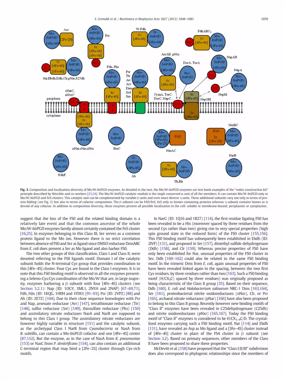
Fig. 3. Composition and localization diversity of Mo/W-bisPGD enzymes. As detailed in the text, the Mo/W-bisPGD enzymes are text book examples of the “redox construction kit”principle described by Nitschke and co-workers [23,24]. The Mo/W-bisPGD catalytic module is the single conserved α unit of all the members. It can contain Mo/W-bisPGD only orMo/W-bisPGD and FeS clusters. This catalytic unit can be complemented by variable β units and even more diverse γ units. These additional subunits vary not only in terms of pro-tein folding (see Fig. 3) but also in terms of cofactor composition. The β subunit can be FAD/FeS, FeS only or hemes containing proteins whereas γ subunit contains hemes or isdevoid of any cofactor. In addition to composition diversity, these enzymes present all possible localization in the cell: soluble or membrane-bound; periplasmic or cytoplasmic.
1059S. Grimaldi et al. / Biochimica et Biophysica Acta 1827 (2013) 1048–1085
suggest that the loss of the FS0 and the related binding domain is arelatively late event and that the common ancestor of the wholeMo/W-bisPGDenzymes family almost certainly contained the FeS cluster[16,25]. In enzymes belonging to this Class III, Ser serves as a commonprotein ligand to the Mo ion. However there is no strict correlationbetween absence of FS0 and Ser as ligand sinceDMSO reductaseDmsABCfrom E. coli does present a Ser as Mo ligand and also harbor FS0.
The two other groups of this classification, Class I and Class II, weredenoted referring to the FS0 ligands motif. Domain I of the catalyticsubunit holds the N-terminal Cys group that provides coordination tothis [4Fe–4S] cluster. Four Cys are found in the Class I enzymes. It is tonote that this FS0 binding motif is observed in all the enzymes present-ing a Seleno-Cys/Cys coordination of theMo/W that are, in largemajor-ity, enzymes harboring a β subunit with four [4Fe–4S] clusters (seeSection 3.2.1): Nap (ID: 1OGY, 3ML1, 2NYA and 2NAP) [67–69,71],Fdh, Fdn (ID: 1KQG, 1H0H and 1FDO) [91–93], Psr (ID: 2VPZ) [88] andAh (ID: 2E7Z) [104]. Due to their close sequence homologies with Psrand Nap, arsenate reductase (Arr) [147], tetrathionate reductase (Ttr)[148], sulfur reductase (Sre) [149], thiosulfate reductase (Phs) [150]and assimilatory nitrate reductases NasA and NarB are supposed tobelong to this Class I group. The assimilatory nitrate reductases arehowever highly variable in structure [151] and the catalytic subunit,as the archetypal Class I NarB from Cyanobacteria or NasA fromB. subtillis, can contain a Mo-bisPGD cofactor and one [4Fe–4S] center[87,152]. But the enzyme, as in the case of NasA from K. pneumoniae[153] or NasC from P. denitrificans [154], can also contain an additionalC-terminal region that may bind a [2Fe–2S] cluster through Cys-richmotifs.
In NarG (ID: 1Q16 and 1R27) [114], the first residue ligating FS0 hasbeen revealed to be a His (moreover spaced by three residues from thesecond Cys rather than two) giving rise to very special properties (highspin ground state in the reduced form) of the FS0 cluster [155,156].This FS0 binding motif has subsequently been established in Ebdh (ID:2IVF) [131], and proposed in Ser [157], dimethyl sulfide dehydrogenase(Ddh) [158], and Clr [159]. Whereas precise properties of FS0 haveonly been established for Nar, unusual properties of the FS0 cluster inSer, Ddh [160–162] could also be related to the same FS0 bindingmotif. In the trimeric Dms from E. coli, again unusual properties of FS0have been revealed linked again to the spacing, between the two firstCys residues, by three residues rather than two [163]. Such a FS0 bindingmotif (H/CX3C) spaced by three residues) was originally proposed asbeing characteristic of the Class II group [35]. Based on their sequence,Ddh [160], E. coli and Halobacterium salinarum NRC-1 Dms [163,164],Ser [161], proteobacterial nitrite oxidoreductases (nNxr), Clr, or Pcr[165], archaeal nitrate reductases (pNar) [166] have also been proposedto belong to this Class II group. Recently however new binding motifs of“Class II” enzymes have been revealed in C25dehydrogenase (C25dh)and nitrite oxidoreductases (pNxr) [165,167]. Today the FS0 bindingmotif of “Class II” enzymes is considered to be H/CX3–4C/D. The crystal-lized enzymes carrying such a FS0 binding motif, Nar [114] and Ebdh[131], have revealed an Asp as Mo ligand and a [3Fe–4S] cluster insteadof [4Fe–4S] cluster in place of the FS4 cluster in β subunit (seeSection 3.2). Based on primary sequences, other members of the ClassII have been proposed to share these properties.
McDevitt et al. [158] have proposed that the “Class I/II/III” subdivisiondoes also correspond to phylogenic relationships since the members of

Fig. 4. Structural modularity cascade of the Mo/W-bisPGD enzymes. Several, but not alltypes of Mo/W-bisPGD enzymes have been crystallized (see Table 3). The available struc-tures give a picture of a modularity in cascade among this family from the monomericcases to the heterotrimeric cases. Indeed whereas several monomeric cases with only theα subunit have been resolved in structure, only three types of β subunits potentially asso-ciated with an α subunit are available in data bases: the Rieske-type from Aio, theferredoxin-type from Nar and the cytochrome from NapAB. Among these β subunits, onlyone, i.e. the bacterial ferredoxin-type, have been observed associated with a γ subunit.This γ subunit is highly variable in structure as exemplied by the cytbI from Fdn, cytbII
from Nar, the cytbIII from Ebdh and the PsrC from Psr.
1060 S. Grimaldi et al. / Biochimica et Biophysica Acta 1827 (2013) 1048–1085
these different classes group together into separate monophyletic cladesin the phylogenetic tree of the Mo/W-bisPGD enzymes family. Recentpublications are however against this idea [25,165,168]. The “Class III”enzymes all cluster together in a branch but with Dms enzymes, consid-ered to be “Class II” enzymes. “Class II” enzymes also cluster togetherwith the exception of the latter enzyme. The “Class I” enzymes representtoday the vast majority of knownMo/W-bisPGD enzymes and clearly donot cluster together on a single branch. It appears to us more rigorous toclassify the enzymes based on the Mo/W ligand: Ser-group, Asp-groupand Cys-group, replacing III, II and I classes, respectively. This is probablymore functionally relevant. The Aio [169] is atypical in theMo/W-bisPGDenzymes family and does not belong to any of these Classes or ligand-group: its FS0 is a high redox potential [3Fe–4S] cluster coordinated byonly three Cys andMo does not have any protein ligand. The new classi-fication would therefore allow to create a new group: None-group.
One important feature of the catalytic subunit is the relationshipbetween the presence of a twin arginine translocation peptide signal(tat leader sequence) at the N-terminus of the immature protein andthe final sub-cellular location of the mature complex. This sequencecan be the archetypal RRXFLK but more often is slightly modifiedto RRXFIK, RRXFLR, RRXLLK, RRXWLK, RRXFLQ, RRXFLS, or RRXLMQ.When present in the catalytic subunit of αβγ enzymes, the tat leadersequence targets the heterodimer αβ to the periplasmic compartmentwhere it associates with its eventual γ subunit. The Ttr is a special casesince both the catalytic and the electron transfer subunits can, in someorganisms, harbor a tat leader sequence [170,171]. In the case of dimericNapAB enzymes, the tat leader sequence targets theα subunit to theperiplasmic compartment where it associates with the cytochromeβ subunit exported via the sec system [172]. In the case of Nars,whereas bacterial enzymes are found attached to the cytoplasmicmembrane facing the cytoplasm, archaeal enzymes have beenshown to be facing the periplasm. This localization seems to wellcorrelate with the presence of a predicted tat leader sequence atthe N-terminus of the immature pNarG (see [166] for review).Fine examination of all the available bacterial narG genes revealeda vestigial leader sequence uncleaved in the mature NarG protein[173,174]. A similar vestigial non-functional and uncleaved leadersequence also exists in the mature cytoplasmic and soluble BisC enzyme[174]. Aio represents an exception in the Mo/W-bisPGD enzymesfamily since the tat leader sequence (Aio is indeed periplasmic) isharbored by the electron transfer subunit rather than the catalyticone [175,176].
3.2. The electron transfer subunit β
3.2.1. Archetypal β subunitMost of theMo/W-bisPGD enzymes contain amultiple FeS-harboring
protein as β subunit (Fig. 4), from which several structures have beenresolved (ID: 1TI2, 2IVF, 1H0H, 1KQG, 2VPZ, 1Q16 and 1R27). Despitethis common feature, the size, ranging from 191 residues in the case oftheWolinella succinogenes Psr to 512 residues for nNar, is highly variable.The harbored FeS are four [4Fe–4S] clusters in the Fdn (ID: 1KQG) [92] orthe Psr (ID: 2VPZ) [88]. The FS1–FS4 (with increasing distance from theFS0) clusters are coordinated by typical CAXXCBX2–11CCX3CD motifs.From sequence analysis, the β subunits encountered in Ttr [148],Sre [149], Srd [177] and Arr [178] are supposed to harbor also four[4Fe–4S] clusters. Three [4Fe–4S] and one [3Fe–4S] can also be carriedby the β subunit as in nNar (ID: 1Q16,1Y4Z) [114] and Ebdh (ID: 2IVF)[131], or only three [4Fe–4S] in Th (ID: 1TI2) [22] and the W-containing Fdh (ID: 1H0H) [93]. Ddh [160], Dms [163], Ser [161],Clr, or Pcr have also been proposed to carry three [4Fe–4S] andone [3Fe–4S]. Based on sequence analysis, Phs has been proposedto carry three [4Fe–4S] and one [3Fe–4S] on its β subunit [150].The sequence of PhsB however carries the same four FeS binding motifsthan PsrB demonstrated to harbor four [4Fe–4S]. This is in line with itscloser phylogenetic relationship with Psr [147] than to Nar. Together
with the FS0 when exists, these FS1–FS4 clusters are arranged in a Wshape. As in the case of the catalytic subunit, domains can be delimitedin this type of β subunit. Domains I and II are ferredoxin-like domainsharboring two FeS clusters each sandwiched between two helices onone side and a β-sheet on the other, with the exception of Th in whichDomain II only holds one cluster [22]. Although not superimposable,structural Domains I and II are observed in nNar, Fdh, Ebdh, and also inTh where an additional third domain is folded in β-barrels [22]. In addi-tion to these relatively well characterized systems, publications reportedthat the pNxr [165] and C25dh [167] would possess such FeS subunit.
3.2.2. “Exotic” β subunitsMo/W-bisPGD enzymes carrying a β subunit distinct from the
multiple-FeS protein are rare. We presently know four other types of βsubunits present each in one specific system: a di-heme cytochrome(c-type) in the NapAB heterodimers [68,69], a Rieske type protein inthe AioAB heterodimer [169], and two different FAD/[2Fe–2S] proteinsin the dimeric Nas [152,179]. Only the first two proteins have beenresolved in structure.
NapAB are all soluble and periplasmic enzymes. Whereas NapAB ofα-proteobacteria, such as R. sphaeroides purifies as a tight heterodimericcomplex [68], the γ-proteobacterial E. coliNapA and NapB proteins puri-fy independently due to weaker interactions but form together the func-tional complex. In R. sphaeroides NapB (ID: 1OGY) two extended armsfold away from the core structure to embrace the NapA subunit [68]. InNapAB, heme II is located into the vicinity of FS0 whereas heme I isexposed to the solvent, arguing for this latter heme to be the electron

1061S. Grimaldi et al. / Biochimica et Biophysica Acta 1827 (2013) 1048–1085
acceptor from the physiological partner NapC. Thismembranous partneris responsible for the connection of the enzyme to the membranousquinol pool (see below Section 3.3.3).
The AioB Rieske protein (ID: 1G8K), by its [2Fe–2S] cluster and itsprotein fold, is homologous to the well characterized subunit of theRieske/cytb complex (also known as complex III in mitochondria, bc1complex in proteobacteria and b6f complex in plants and cyanobacteria)[169,180]. A characteristic Cys-X-His-Xn-Cys-X2-His sequence motif,observed in other Rieske proteins, binds the [2Fe–2S] cluster. In theA. faecalis Aio structure, two loops containing each a Cys/His couple areheld together by a disulfide bridge which is strictly conserved in theRieske/cytb complexes and considered to be crucial for properties ofthe cluster in this latter complex. This bridge is not fully conserved inthe Aio, where the properties of the enzyme do not seem to depend onit [181,182]. The [2Fe–2S] cluster, exposed to the solvent, is the electrondonor to the partner, which in proteobacterial systems (the only charac-terized) are soluble c-type cytochromes (see [183] for a review). All Aioare periplasmic but only some of them have been observed attached tothe cytoplasmic membrane. It is not clear at present whether the tatsignal peptide, harbored by AioB and which would then remainuncleaved, could be responsible for the membrane association ina same manner as observed in the Rieske/cytb complex [183].
Data on the assimilatory nitrate reductases other than genetic arevery scarce. Among them, the NasBC from B. subtilis [152] and theNasAC from K. pneumonia [179] or from R. capsulatus [184] are the onlyheterodimeric enzymes characterized in some extent. NasB in B. subtilisand NasC in K. pneumonia or R. capsulatus are both responsible for theelectron transfer from NAD(P)H to the catalytic subunit in cytoplasm.The two proteins are however highly divergent, not only at a globalsequence level, but also at a structural and cofactor composition levels.The NasB-type subunit is 771 residues long and therefore larger thanthe catalytic one whereas the NasC-type protein is 396 residues long.Sequence and biochemical analyses suggest that NasB and NasC bothharbor a FAD cofactor but NasB presents in addition clear motifs for thecoordination of two [2Fe–2S] clusters.
3.3. The electron entry/exit γ subunit
This component is the most variable element of the Mo/W-bisPGDenzymes but each type of γ subunit is encountered in several representa-tives of the Mo/W-bisPGD enzymes family. The cytc3 characterized withDesulfovibrio species Fdh [185,186], the tetrahemic c554-type cytochromecharacterized in the Dechloromonas Pcr [187] and the dihemic cyto-chrome characterized in the T. thermophilus cNar [188–190], have beenobserved each in a single system. With or without cofactors, integratedor not into the membrane, this γ subunit is of primordial importancefor the mode of enzyme connection to the whole bioenergetic chain inwhich it is involved. In the greatmajority of cases, this third subunit is re-sponsible of the reaction with the membranous isoprenoid quinones(see Section 3.4).
3.3.1. CytbI, bII and bIII γ subunitsTwo types of transmembrane diheme cytochrome b and one type
of hydrophilic monoheme cytochrome b, which have all been re-solved in structure (Fig. 4), are present as γ subunit in members ofthe Mo/W-bisPGD enzymes family. All represent typical “redox kitunits” as already discussed and form different enzymatic systemseven out of the Mo/W-bisPGD enzymes family [23,24]. The firstcytochrome cytbI (ID: 1KQG) usually contains 4 trans-membranehelices (TM) and the two hemes are ligated by histidine residueson TM1 (1 His), TM2 (1 His) and TM4 (2 His). CytbII (ID: 1Q16)features 5 TM and the heme ligands are located on TM2 (2 His)and TM5 (2 His). CytbI, takes part to the FdhN [92]. Due to its globalsequence homology (although containing a fifth TM) to the FdhN γsubunit, PhsC is proposed to be also a cytbI cytochrome [16]. CytbII isexemplified by the γ subunit of the bacterial nNar enzyme [114] and
has been proposed as third subunit in nNxr and cNar [29]. The functionof such units is to connect the related enzymes to the membranousquinol pool. Why there are two structurally distinct membrane-integralb-type cytochromes for this purpose remains unknown. The idea thatcytbI could serve as quinone-reducing, and cytbII as quinol-oxidizingelement, does not match with the function of PhsC in oxidizing MK toreduce thiosulfate.
A third cytochrome b is a hydrophilic antiparallel β-sheetmonohemicone: cytbIII. This subunit is encountered as γ subunit of Ebdh fromwhich3D-structure is available (ID: 2IVF) [131]. Based on sequence homologiesand/or spectroscopy, archaeal pNar [166,191,192], Ser [157,161,193], Ddh[158,162], Clr [194] and C25dh [167] have furthermore been proposed tocontain such a subunit. This unit doesn't connect the enzymes directly tothe quinol pool but one of them (named Orf7 or NarM) could howeverbind its related complex, archaeal pNar, to the cytoplasmic mem-brane by two α helices in both N- and C-termini [166,192]. Althoughobserved associated with the cytoplasmic membrane, the C25dh doesnot present a C25dhC unit predicted to harbor a TM. In this case theassociation could be mediated by a currently unknown protein[167]. All these cytochromes b have in common unusual Met andLys residues as axial heme ligands. The wire function of this proteinis achieved by exposing two distinct heme edges at the proteinsurface.
3.3.2. The NrfD-type γ subunitThe “NrfD-type” unit (named from the first representative of the
group, characterized in periplasmic nitrite reductase [195]) is amembrane-integrated protein devoid of any cofactor but still responsibleof injecting or extracting electrons to or from quinones/quinols. Se-quence homologies indicate that this protein takes part in several diverseenzymes in the Mo/W-bisPGD enzymes family, such as Psr [196], Ttr[148], Sre [149] and Dms [197]. Arsenate reductase-type enzymes(Arr/Arx) are special cases where this subunit exists only in someof them [147]. Although clearly related together, these units havea variable number of TM: 8 in the case of PsrC, Arr and DmsC, and9 in the case of TtrC. The crystal structure of T. thermophilus PsrCprovided the single 3D picture of this element (Fig. 4). Crystal containingMK (ID: 2VPW) allowed to precisely visualize the quinone binding site inthis complex (Fig. 7). Scarce if not no information does exist however onthe other NrfD-containing systems of theMo/W-bisPGD enzymes family,and it is too early to try listing conserved features on these sites (seeSection 3.4).
3.3.3. c-Type cytochromes as additional subunitsIn D. vulgaris [186] and D. desulfuricans [185], very special Fdh,
FdhABC3, have been purified and characterized which contain, in addi-tion to the catalytic subunit and the ferredoxin β subunit, a 16 kDacytochrome. This last subunit is homologous to the typical soluble cyto-chrome c3 (TpIc3) that plays the role of redox partner for periplasmichydrogenases from which 3D-structure is available (ID: 2BQ4) [198].TpIc3 are small (13–15 kDa) globular cytochromes containing fourcovalently bound hemes all with bis-histidinyl axial coordination (see[199] for review) having negative redox potentials. The close proximityof the hemes results in redox cooperativity giving rise to special redoxproperties of these cytochromes. All these characteristics are alsoencountered in the C3 subunit of the FdhABC3 [185,186]. Similarly tothe cytbIII of Ebdh this c3 does not bind FdhABC3 to the membrane.
The pcr operon of two Dechloromonas species [187], revealed a pcrCgene coding for a protein product similar to the soluble tetrahemiccytochrome c554 from Nitrosomonas europaea (ID: 1FT5) [200]. As itshomolog, PcrC would not be membranous and therefore would notlink the Pcr enzyme to the membrane and would play the role of addi-tional electron wire for the enzyme, similar to the c3 subunit depictedabove. Recently, Nilsson et al. [194] proposed that this element couldbe a partner rather than a true subunit of the enzyme. Since no furthercharacterization has been performed, this question remains open.
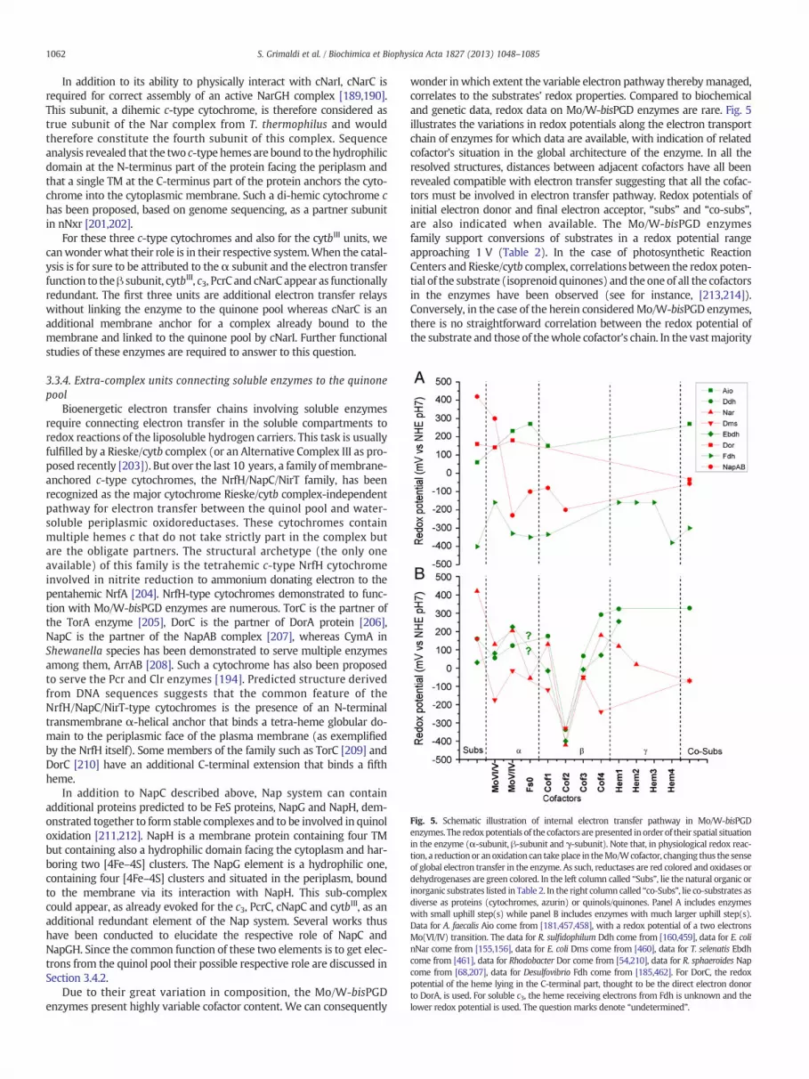
Fig. 5. Schematic illustration of internal electron transfer pathway in Mo/W-bisPGDenzymes. The redoxpotentials of the cofactors are presented in order of their spatial situationin the enzyme (α-subunit, β-subunit and γ-subunit). Note that, in physiological redox reac-tion, a reductionor anoxidation can takeplace in theMo/Wcofactor, changing thus the senseof global electron transfer in the enzyme. As such, reductases are red colored and oxidases ordehydrogenases are green colored. In the left column called “Subs”, lie the natural organic orinorganic substrates listed in Table 2. In the right column called “co-Subs”, lie co-substrates asdiverse as proteins (cytochromes, azurin) or quinols/quinones. Panel A includes enzymeswith small uphill step(s) while panel B includes enzymes with much larger uphill step(s).Data for A. faecalis Aio come from [181,457,458], with a redox potential of a two electronsMo(VI/IV) transition. The data for R. sulfidophilum Ddh come from [160,459], data for E. colinNar come from [155,156], data for E. coli Dms come from [460], data for T. selenatis Ebdhcome from [461], data for Rhodobacter Dor come from [54,210], data for R. sphaeroides Napcome from [68,207], data for Desulfovibrio Fdh come from [185,462]. For DorC, the redoxpotential of the heme lying in the C-terminal part, thought to be the direct electron donorto DorA, is used. For soluble c3, the heme receiving electrons from Fdh is unknown and thelower redox potential is used. The question marks denote “undetermined”.
1062 S. Grimaldi et al. / Biochimica et Biophysica Acta 1827 (2013) 1048–1085
In addition to its ability to physically interact with cNarI, cNarC isrequired for correct assembly of an active NarGH complex [189,190].This subunit, a dihemic c-type cytochrome, is therefore considered astrue subunit of the Nar complex from T. thermophilus and wouldtherefore constitute the fourth subunit of this complex. Sequenceanalysis revealed that the two c-type hemes are bound to the hydrophilicdomain at the N-terminus part of the protein facing the periplasm andthat a single TM at the C-terminus part of the protein anchors the cyto-chrome into the cytoplasmic membrane. Such a di-hemic cytochrome chas been proposed, based on genome sequencing, as a partner subunitin nNxr [201,202].
For these three c-type cytochromes and also for the cytbIII units, wecanwonderwhat their role is in their respective system.When the catal-ysis is for sure to be attributed to theα subunit and the electron transferfunction to theβ subunit, cytbIII, c3, PcrC and cNarC appear as functionallyredundant. The first three units are additional electron transfer relayswithout linking the enzyme to the quinone pool whereas cNarC is anadditional membrane anchor for a complex already bound to themembrane and linked to the quinone pool by cNarI. Further functionalstudies of these enzymes are required to answer to this question.
3.3.4. Extra-complex units connecting soluble enzymes to the quinonepool
Bioenergetic electron transfer chains involving soluble enzymesrequire connecting electron transfer in the soluble compartments toredox reactions of the liposoluble hydrogen carriers. This task is usuallyfulfilled by a Rieske/cytb complex (or an Alternative Complex III as pro-posed recently [203]). But over the last 10 years, a family ofmembrane-anchored c-type cytochromes, the NrfH/NapC/NirT family, has beenrecognized as the major cytochrome Rieske/cytb complex-independentpathway for electron transfer between the quinol pool and water-soluble periplasmic oxidoreductases. These cytochromes containmultiple hemes c that do not take strictly part in the complex butare the obligate partners. The structural archetype (the only oneavailable) of this family is the tetrahemic c-type NrfH cytochromeinvolved in nitrite reduction to ammonium donating electron to thepentahemic NrfA [204]. NrfH-type cytochromes demonstrated to func-tion with Mo/W-bisPGD enzymes are numerous. TorC is the partner ofthe TorA enzyme [205], DorC is the partner of DorA protein [206],NapC is the partner of the NapAB complex [207], whereas CymA inShewanella species has been demonstrated to serve multiple enzymesamong them, ArrAB [208]. Such a cytochrome has also been proposedto serve the Pcr and Clr enzymes [194]. Predicted structure derivedfrom DNA sequences suggests that the common feature of theNrfH/NapC/NirT-type cytochromes is the presence of an N-terminaltransmembrane α-helical anchor that binds a tetra-heme globular do-main to the periplasmic face of the plasma membrane (as exemplifiedby the NrfH itself). Some members of the family such as TorC [209] andDorC [210] have an additional C-terminal extension that binds a fifthheme.
In addition to NapC described above, Nap system can containadditional proteins predicted to be FeS proteins, NapG and NapH, dem-onstrated together to form stable complexes and to be involved in quinoloxidation [211,212]. NapH is a membrane protein containing four TMbut containing also a hydrophilic domain facing the cytoplasm and har-boring two [4Fe–4S] clusters. The NapG element is a hydrophilic one,containing four [4Fe–4S] clusters and situated in the periplasm, boundto the membrane via its interaction with NapH. This sub-complexcould appear, as already evoked for the c3, PcrC, cNapC and cytbIII, as anadditional redundant element of the Nap system. Several works thushave been conducted to elucidate the respective role of NapC andNapGH. Since the common function of these two elements is to get elec-trons from the quinol pool their possible respective role are discussed inSection 3.4.2.
Due to their great variation in composition, the Mo/W-bisPGDenzymes present highly variable cofactor content. We can consequently
wonder inwhich extent the variable electron pathway therebymanaged,correlates to the substrates' redox properties. Compared to biochemicaland genetic data, redox data on Mo/W-bisPGD enzymes are rare. Fig. 5illustrates the variations in redox potentials along the electron transportchain of enzymes for which data are available, with indication of relatedcofactor's situation in the global architecture of the enzyme. In all theresolved structures, distances between adjacent cofactors have all beenrevealed compatible with electron transfer suggesting that all the cofac-tors must be involved in electron transfer pathway. Redox potentials ofinitial electron donor and final electron acceptor, “subs” and “co-subs”,are also indicated when available. The Mo/W-bisPGD enzymesfamily support conversions of substrates in a redox potential rangeapproaching 1 V (Table 2). In the case of photosynthetic ReactionCenters and Rieske/cytb complex, correlations between the redox poten-tial of the substrate (isoprenoid quinones) and the one of all the cofactorsin the enzymes have been observed (see for instance, [213,214]).Conversely, in the case of the herein consideredMo/W-bisPGD enzymes,there is no straightforward correlation between the redox potential ofthe substrate and those of thewhole cofactor's chain. In the vastmajority

Fig. 6. Structure of isoprenoid quinones and of some quinone analogs discussed in this re-view. NQ, naphthoquinone; menaquinone (MK-n) corresponds to [R2=CH3, R5=R8=H],demethylmenaquinone (DMK-n) to [R2=R5=R8=H], and methylmenaquinone (MMK-n)is found with [R2=CH3, R5=CH3, R8=H] or [R2=CH3, R5=H, R8=CH3]; UQ-n, ubiqui-none (UQ-n); HQNO, 2-heptyl-4-hydroxyquinolineN-oxide; and PCP, pentachlorophenol.n is the number of isoprenoid units.
1063S. Grimaldi et al. / Biochimica et Biophysica Acta 1827 (2013) 1048–1085
of cases, the thermodynamics of the overall enzyme reaction appears toonly be driven by the physiological substrate's and redox partner's(co-substrate) redox potentials. Hence, in the cases of reductases, theredox potential of the co-substrate is lower than that of the substrate(e.g. Dms, with DMSO and MK) thermodynamically favoring the globalreaction. The reciprocal is true with dehydrogenases and oxidases forwhich the redox potential of the co-substrate is higher than that of thesubstrate (e.g. Aio, with As(III) and cytochrome c552). Noteworthy, oneor several steps of the global exergonic reaction can be uphill definingtwogroups of enzymes. In thefirst group, shown in panel A and exempli-fiedbyAio, Dor, Napor Fdh, the uphill step(s) do(es) not exceed 200 mV.In the second group, shown in panel B and exemplified by Ddh, nNar,Dms and Ebdh, the uphill step(s) can largely exceed 300 mV such asthe electron transfer steps to or from the FS2 cluster in the archetypalβ subunits. However, this shared property does not necessarily have afunctional signification since it does not represent any convergence.All the enzymes of this latter group are indeed of the Class II (seeSection 3.1) and are phylogenetically related. Published data addressingthe question of the necessity of such a step are scarce. For instance,shifting the low potential of the nNar's FS2 to higher redox potential bysite-directed mutagenesis has no dramatic functional impact suggestingthat this uphill step is not absolutely required [113].
3.4. Role of quinones in Mo/W-bisPGD enzymes bioenergetics
Numerous Mo/W-bisPGD enzymes are known to directly interactwith isoprenoid quinones/quinols either through their membranesubunit that contains two b-type hemes (cytbI and bII subunits) or thatdo not bind any cofactor (NrfD-type γ subunit), or via extra-complexcomponents that transiently bind to the enzyme and contain multiplec-type hemes or FeS clusters. Thus, understanding the reactivity of suchquinone-utilizingmolybdoenzymes towards thesemolecules constitutesan essential step for delineating their individual mechanism and eluci-dating their bioenergetic role. In this context, the work on respiratoryMo/W-bisPGD enzymes reviewed hereafter greatly contributed to theadvancement of our knowledge on transverse questioning, namely(i) the determination at atomic resolution of quinone binding modewithin protein complexes, (ii) the specificity of quinone utilizationby a given complex in those organisms containing several types ofquinones differing in their structure and properties, in line withenergy conservation, catalytic directionality and defining the role of theprotein environment, and finally (iii) the impact of the orientation (peri-plasmic or cytoplasmic) of a quinone binding site within the membranesubunit on its bioenergetic efficiency.
3.4.1. Structures and properties of relevant quinonesIsoprenoid quinones are amphiphilic molecules found in nearly all
living organisms [24,215]. They are composed of a polar head groupand a hydrophobic side chain. The apolar isoprenoid side chain givesthemolecules a lipid-soluble character and allows them to freely diffusein biologicalmembranes. The quinone head group has a ringmoietywithtwo oxygen atoms at positions 1 and 4 and is directly involved in redoxreactions (Fig. 6). Indeed, quinones can oscillate between three differentoxidation states. The oxidized quinone (Q) state has two keto groups.The addition of one electron results in the formation of an anionicsemiquinone intermediate (SQ•−), and with the uptake of one proton,a neutral semiquinone intermediate (SQH•) is formed. The addition ofa second electron and proton generates the fully reduced quinol (QH2).Therefore, quinones couple electron transfer reactions to proton bindingand release, a property that is crucial to transmembrane proton trans-port. In prokaryotic bioenergetic processes involving molybdoenzymes,quinones serve mainly as redox mediators between dehydrogenasesand terminal reductases, donating or accepting two electrons to orfrom theMo active site through a one-electron transfer chain containingFeS clusters and eventually hemes. Thus, quinones act as “gates”between one and two-electron processes. This involves the obligatory
transient formation of highly reactive semiquinone catalytic intermedi-ates during enzyme turnover. E. coli NarGHI is the sole molybdoenzymein which such an intermediate has been specifically studied (seeSection 3.4.3) [216–221].
Although quinones are considered to be highly mobile in the lipidbilayer of the membrane, reactivity of enzymatic complexes towardsquinones occurs at specific well-structured protein sites named Q sites.Their accurate description is currently a major challenge for a detailedunderstanding of the function of Mo/W-bisPGD enzymes. For instance,previous studies on photosynthetic or alternative respiratory proteinsto Mo/W-bisPGD enzymes demonstrated that the protein environmentcan dramatically modulate the quinone redox chemistry to an extentthat a quinone can have very different functional properties when asso-ciated with different sites. Indeed, while the quinone redox potential ina bacterialmembrane is expected to be close to thatmeasured in organ-ic solution [222], the midpoint redox potentials of the two one-electronprocesses Q/SQ and SQ/QH2 can drastically change upon binding to aprotein Q site, considering in particular the associated proton transferchemistry. In addition, conformational flexibility during quinone turn-over may take place.
The great majority of biological isoprenoid quinones belong tonaphthoquinones (NQ) or benzoquinones. Their hydrophobic side chaincan vary not only in the length but also in the degree of saturation, aswell as in the presence of additional groups. Menaquinone (MK-n) isthe most widespread microbial respiratory quinone (Fig. 6). It contains2-methyl-1,4-naphthoquinone ring as a head group. Its side chain ismost frequently composed of n=6–10 prenyl units. In someprokaryotes,compounds with different modifications of the head group are found.Among them, demethylmenaquinones (DMK) andmethylmenaquinones(MMK) are often used by proteobacteria and gram-positive bacteria[215,223–226]. In contrast, ubiquinones (UQ-n, also named coenzymes

1064 S. Grimaldi et al. / Biochimica et Biophysica Acta 1827 (2013) 1048–1085
Q) are found only in α-, β- and γ-proteobacteria and consequently inmitochondria. They are 2,3-dimethoxy-5-methyl-1,4-benzoquinonewith an isoprenyl tail containing a variable number n of isoprenoidunits, ranging mostly between n=8–10 in bacteria.
Another important parameter distinguishing the different kinds ofquinones is their midpoint redox potential Em′ (Q/QH2) (Table 2). MKand DMK (and MMK as proposed in [227]) are low-potential electroncarriers with negative redox potential values whereas the high-potential UQ has an Em′-value of about +100 mV. The distribution ofMK and UQ on the phylogenetic tree of species strongly indicates thatMK are the most ancient type of isoprenoid quinones [24,213]. In addi-tion, high-potential quinones such as UQ have only appeared in speciespermanently or periodically exposed to high concentration of oxygen,suggesting that highO2 tensions and/or concomitant increase in ambientredox potential are evolutionary unfavorable for organisms using MK-based energy-converting systems. The reduced menaquinones (MKH2,menaquinols) indeed becomes rapidly and non-catalytically oxidized inthe presence of oxygen, therefore these compounds cannot efficientlyoperate in atmosphere containing oxygen [213].
3.4.2. Specificity of quinone utilization by Mo/W-bisPGD enzymesMany prokaryotes can synthesize more than one type of quinones
(see for reviews, [215,228]).Moreover, the quinone contents are regulat-ed by a yet unknown mechanism in response to the type of electronacceptor present in the medium, the growth phase, and the availablecarbon source [229–232]. Altogether, this contributes to the high meta-bolic flexibility and complexity of these organisms that can rapidlyadapt to environmental changes [233]. Thus, the question of the specific-ity of quinone utilization by Mo/W-bisPGD enzymes was raised. This isdifficult to study in classical enzyme assays either due to the insolubilityof the native substrates or due to the inability of native substrate to beremoved from the enzymewithout altering enzyme structure. Therefore,this issue has been addressed using mutant strains lacking genesinvolved at specific stages of the quinone biosynthetic pathways inwhich the quinone content has been chemically analyzed. This approachhas been combined with growth assays using different electron donorsor acceptors or with in vitro assays, both eventually complementedwith the use of different quinones or analogs (for reviews, see[234–236]).
The facultatively anaerobic proteobacterium E. coli is the most stud-ied organism in this context. UQ-8, MK-8 and DMK-8 are the majorquinones but smaller amounts of UQ-1 to UQ-7, MK-6 to MK-9 andDMK-7 have also been reported [215]. Using E. coli strains deficient inthe synthesis of UQ, (MK+DMK) or of (MK+UQ), it has been shownthat not all molybdoenzymes are able to use all three types of quinones.Our current knowledge of the specificity of quinone utilization bymolybdoenzymes in prokaryotes allows distinguishing two groups ofenzymes:
(i) in group 1 enzymes, the specificity observed can be explainedsimply on the basis of the redox potentials of the quinones andof the acceptors. Typical examples include E. coli DmsABC andTorAC. In anaerobic respiration with DMSO (E0′ (DMSO/DMS)=+160 mV) and TMAO (E0′ (TMAO/TMA)=+130 mV), thenaphthoquinones (MK+DMK) are required and cannot bereplaced by UQ (Table 2) [235,237–240]. Further, whereasE. coli TorAC and DmsABC cannot use UQ, both are able tooxidize MK and DMK as well [239]. In contrast, mutantsof Shewanella oneidensis (formerly S. putrefaciens MR-1 orAlteromonas putrefaciens) deficient in MK and MMK, butwhich have wild-type levels of UQ, retain the ability to useTMAO as an electron acceptor [241]. Whereas the specificity ofTtr has been much less investigated, available data suggestthat UQ are not involved in electron transport to tetrathionate(E0′ (S4O6
2−/S2O32−)=+24 mV to +170 mV, Table 2) in
S. typhimurium or in Citrobacter freundii [242,243].
Some Arr and Arx are predicted to contain a membraneintegral subunit Arr/ArxC that might serve as anchor to theArr/ArxAB complex and which is proposed to promoteelectron transfer between the quinone pool and the enzyme.The structural homology between PsrC and ArrC suggests asimilar function for both enzymes. In the case of Arr, the elec-trons would be transferred from liposoluble MKH2 to the cat-alytic molybdenum subunit and then onto As(V) [147]. Thiswould be consistent with the substantial driving force providedby the low-potential MK given the midpoint potential of thearsenate/arsenite couple (E0′ (HAsO4
2−/H3AsO3)~+60 to+139 mV, Table 2). In line with this reasoning, a recent ge-nomic survey searching for quinone biosynthetic pathwaygenes indicates that only the group of Arr-containing bacteriapresents species synthesizing MK alone whereas only thegroup of bacteria containing Arx presents a species synthe-sizing only UQ [182]. This led the authors to stipulate that allArr-harboring strains oxidize MKH2 via an As(V) reductionprocess whereas the Arx-harboring strains reduce UQ via anAs(III) oxidation process. While this hypothesis still needsto be tested by detailed kinetic studies on both enzymes, itis similar to the behavior of quinol-fumarate reductase inE. coli, which uses MK for reduction of fumarate and UQ foroxidation of succinate [244]. Overall, this example illustratesthe possible role of the electrochemical potential of the quinonein determining the enzymes' directionality.Whereas the ε-proteobacterium W. succinogenes containsMK-6 and 2-,[5,8]-dimethyl-3-hexaprenyl-1,4-naphthoquinone(MMK-6) in its cytoplasmicmembrane [224], activities of polysul-fide respiration measured in nearly-inactive proteoliposomescontaining purified Psr and either hydrogenase or formate asprimary dehydrogenases was restored upon addition of MMK-6but was maintained at a basal level upon addition of MK6 [196].This indicates that MMK-6 is specifically involved in polysulfiderespiration and cannot be substituted by MK-6. While the redoxpotential of the substrate is not known, it is assumed to be closeto that of the S0/HS− couple i.e. ~−270 mV (Table 2). On theother hand, MMK-6 might have a much lower redox potential(~−220 mV) [227] than that initially predicted [196]. However,to our knowledge, no detailed study has been published yet thatsupports this value. If it is correct, the reduction of polysulfideby MMKH2 is likely to be slightly endergonic and the specificityof W. succinogenes Psr towards MMK-6 is probably due to differ-ences in the redox potential of the two naphthoquinone species.A common feature of the above mentioned enzymes is that thesubstrate redox potential lies in the range of that covered by thedifferent quinones, or at least close to it. This is not the case forS. enterica PhsABC that belongs to group 1 enzymes. Whereasthis bacterium can synthesize UQ,MK andDMK, thiosulfate respi-ration in this organism is blocked in strains that cannot synthesizeNQ [237,245]. Further, the very low rates of thiosulfate reductionexhibited in the ubiE mutant deficient in UQ and MK synthesisimply that DMKH2 is a poor substrate for PhsABC and that conse-quently MKH2 is virtually the sole electron donor to the enzyme[246]. A remarkable aspect of thiosulfate reduction in S. entericais that under standard conditions, the overall reaction of thiosul-fate reduction (E0′(S2O3
2−/HS−+HSO3−)=−400 mV) by MKH2
is highly endergonic, unless less than with the other quinols[235]. This results in an unfavorable ΔE0′ of ~−330 mV for thereaction. Based on the sequence similarity between the FdnGHIsubunits of E. coli formate dehydrogenase and the PhsABCsubunits of S. enterica thiosulfate reductase, a structural modelof PhsABC has been proposed, suggesting that this enzyme usesa reverse redox-loop mechanism that consumes the pmf (Fig. 8),in consistency with its strong inhibition by agents that degradethe pmf [246]. In addition, this would become thermodynamically

1065S. Grimaldi et al. / Biochimica et Biophysica Acta 1827 (2013) 1048–1085
possible as the value ΔE0′ of ~−330 mV would be attenuated bythemore physiologically reasonable concentrations of thiosulfate,sulfite and sulfide in the cell, and possibly by the redox state of theMK pool. Interestingly, the reaction is reversible under physiolog-ical conditions, and is then pmf-independent. It can function withDMK as electron acceptor, while the physiological forward reac-tion cannot utilize it. It was thus suggested that the inability ofDMKH2 to support thiosulfate reduction is due to the unfavorableredox potential of DMKH2 relative to MKH2 rather than discrimi-nation between the two types of naphthoquinols by the quinoloxidizing site of thiosulfate reductase [246]. Thus, the quinonespecificity of the group 1 enzymes listed above does not appearto be based on quinone discrimination by the Q site, but ratherto be related to the quinone redox potentials that drive enzymereactivity.
(ii) in group 2 enzymes, the observed quinone specificity cannot beexplained on the basis of the redox potentials of quinones andacceptors. Prominent examples are given by bacterial nitratereductases. Considering the nitrate/nitrite redox potential of+420 mV, basically, low and high-redox potential quinones canserve as electron donors. Using E. colimutants of quinone biosyn-thetic pathways, it has been shown that eitherMKH2 or UQH2 cantransfer electrons to Nap in E. coli, but not DMKH2 which is how-ever intermediate in redox potential [247]. However, Nap couplesmore effectively toMKH2 oxidation than to UQH2 oxidation [248].NapC was shown to be essential for electron transfer from bothUQH2 and MKH2 to NapAB. In contrast, NapG and NapH werefound to be essential for periplasmic nitrate respiration in cellsthat only contained UQH2 [248,249]. Thus, it was concluded thatNapGandNapH forma specializedUQH2dehydrogenase requiredto transfer electrons via NapC to the NapAB complex [211,248]. Aworking model for electron transfer within the NapABCGHcomplex was proposed, that mainly includes two, partiallyindependent, electron transfer pathways to the Nap complexdepending on the nature of the quinol used. 2-Methyl-1,4-naphthoquinone (Menadiol, E0′ (menadiol/menadione ~−1 mV),a closer structural analog of MKH2 than UQH2, was used as exog-enous electron donor and was shown to be able to supportNapGH-dependent electron transfer via NapC to the NapABcomplex, in contrast to the apparently poor electron donorUQ-0 (Em′ (UQH2-0/UQ-0~+162 mV) [211]. This illustrates thedifficulty to extrapolate results obtained from experiments thatrely on quinone analogs to the function of the system withits natural endogenous quinone substrates. Interestingly, inthe ε-proteobacterium W. succinogenes, nitrate respirationis independent of a NapC-type protein [250]. Rather, theNapGH module mediates electron transport from MKH2 tothe periplasmic NapAB complex, thereby substituting thefunction conventionally carried out by NapC [251]. The qui-none specificity of this enzyme towards the two endogenousredox carriers MK-6 and MMK-6, the role and the number of Qsites are still to be evaluated [212].Synthesis of the membrane-bound nitrate reductase nNarGHI isoptimally induced under anaerobic conditions and high nitrateconcentrations. Under these conditions, whereas all threequinones are found in high concentrations with DMK as themajor species, nNarGHI functions with UQ and MK, but forunknown reasons, not with DMK, similar to the Nap system[238,239,248,252,253]. In contrast to Nap, bacterial growthassays of mutant strains synthesizing only UQ or NQ usingnitrate as electron acceptor indicate that NarGHI oxidizes UQH2
and MKH2 with similar efficiency [238].
The selectivity of group 2 enzymes is likely to be due to the structur-al properties of the quinones (Fig. 6). It should also be mentioned thatthe redox potential of the bound quinone may differ from that of the
freely diffusing molecule. In both cases, the protein environmentcould drive this specificity by tuning the quinone binding and/orredox properties. Thus, describing the atomic details of the quinonebinding mode to Mo-bisPGD enzymes is an essential step for under-standing the reactivity and specificity of these systems.
3.4.3. Resolving quinone binding in Mo/W-bisPGD enzymesDeep understanding of the reactivity of a quinol-utilizing enzyme
requires detailed structural information on the quinone bindingmode in different redox states of the enzyme. This is challenginggiven the location within the membrane and the high molecularweight of the complexes involved, the quinone lability, and the tran-sient character of quinone/protein interactions.
To date, there is no evidence for the presence of quinones thatfunction as tightly bound (i.e. non-exchangeable) cofactors in quinoneprocessing Mo/W-bisPGD enzymes, as found in the QA site of the photo-synthetic reaction center [254] or in the QH site of cytochrome bo3[255–257]. Therefore, whereas the presence of multiple Q sites hasbeen discussed in the past for some cases, the more recent experimentalevidence indicates that Mo/W-bisPGD enzymes harbor a single Q site.Using protein-film electrochemistry, it has been recently proposed thatMK-7 may function not just as a substrate but also as a tightly boundcofactor in CymA from Shewanella [258,259]. Indeed, catalytic signals incyclic voltammetry are observed when MK-0 or UQ-10 is supplied toCymA films for which the tightly bound MK-7 has not been removedwhereas these signals are absent in n-dodecyl β-D-maltoside-washedCymA films devoid of MK-7 [259].
Only a limited number of high-resolution three dimensional struc-tures of quinone-interacting Mo-bisPGD enzymes are available. Athree-dimensional structure of PsrABC from T. thermophilus, an obligateaerobe thermophilic bacterium that containsMK-8 andMK-7 as its soleisoprenoid quinones [215], has been solved at 1.9 Å resolution in com-plex with MK-7 [88] (Fig. 7A). MK-7 binds in a mainly hydrophobicpocket situated in the heme-lacking PsrC subunit, on the periplasmicside of the membrane and in close proximity to the proximal [4Fe–4S]of the electron-transfer subunit PsrB. In addition, the resolution of thePsrABC crystal structures co-crystallized with either UQ-1 or with theinhibitor pentachlorophenol (PCP) (Fig. 7B) indicated that all threecompounds bind in the same pocket, suggesting that this site is thephysiological Q site. Slight variations with respect to the hydrogen-bonding of the substrate are resolved, that may be relevant forunderstanding enzyme reactivity and specificity towards quinols. Theavailable structural data cannot be further discussed with regard tothe specificity of quinone utilization by this enzyme. In particular, invitro measurements of UQH2 or MKH2:polysulfide oxidoreductaseactivity of T. thermophilus PsrABC have never been reported. In contrast,no high-resolution structural information on the quinone bindingmodeinW. succinogenes PsrABC is available that could provide useful molec-ular details to understand its high specificity towards MMK-6.
High-resolution crystal structures of two other quinone pro-cessing molybdoenzymes have been obtained in the presence ofbound inhibitors, namely E. coli FdnGHI in complex with 2-heptyl4-hydroxyquinoline N oxide (HQNO) [92] and nitrate reductaseNarGHI from the same organism with PCP bound to it [260], at 2.8and 1.9 Å resolution, respectively. A histidine ligand (FdnI His169,NarI His66) of the heme (bC in FdnI, bD in NarI) that forms part ofthe inhibitor binding pocket is hydrogen bonded to the N-oxidegroup of HQNO or to the hydroxyl group of PCP. An additional hydro-gen bond is formed between the PCP and one of the heme bD propio-nate group. The ligand for the OH group of HQNO (equivalent to theMK O1) is less clear and the involvement of a water molecule(w1990) or of the Nε atom of Asn110 has been discussed. Moreover,it has been recently shown that the presence of an anionic cardiolipintightly associated to NarGHI is required for quinol binding to theenzyme [218] and a similar situation has been proposed to occur inFdnGHI [261].
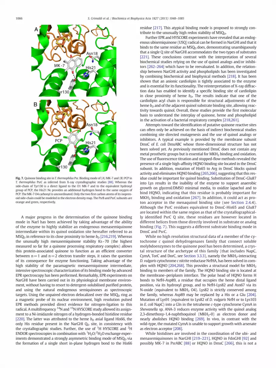
Fig. 7. Quinone binding site in T. thermophilus Psr. Bindingmode of (A)MK-7 and (B) PCP inT. thermophilus PsrC as inferred from X-ray crystallographic studies [88]. Whereas theside-chain of Tyr130 is a direct ligand to the O1 MK-7 and to the equivalent hydroxylgroup of PCP, the His21 Nε provides an additional hydrogen bond to the same oxygen ofPCP. TheMK-7O4 carbonyl is uncoordinated. Only the two first carbon atoms of its isopren-oid side-chain could bemodeled in the electrondensitymap. The PsrB and PsrC subunits areorange and green, respectively.
1066 S. Grimaldi et al. / Biochimica et Biophysica Acta 1827 (2013) 1048–1085
A major progress in the determination of the quinone bindingmode in NarI has been achieved by taking advantage of the abilityof the enzyme to highly stabilize an endogenous menasemiquinoneintermediate within its quinol oxidation site hereafter referred to asMSQD in reference to its close proximity to heme bD [216,219]. Whereasthe unusually high menasemiquinone stability Ks~70 (the highestmeasured so far for a quinone processing respiratory complex) allowsthis protein-associated quinone to function as an efficient converterbetween n=1 and n=2 electron transfer steps, it raises the questionof its consequence for enzyme functioning. Taking advantage of thehigh stability of the paramagnetic menasemiquinone intermediate,intensive spectroscopic characterization of its bindingmodeby advancedEPR spectroscopy has been performed. Remarkably, EPR experiments onNarGHI have been carried out in its physiological membrane environ-ment, without having to resort to detergent-solubilised purified protein,and using the natural endogenous semiquinones as spectroscopictargets. Using the unpaired electron delocalized over the MSQD ring asa magnetic probe of its nuclear environment, high resolution pulsedEPR methods provided direct evidence for nitrogen-ligation to thisradical. Amultifrequency 14N and 15NHYSCORE study allowed its assign-ment to a Nδ imidazole nitrogen of a hydrogen-bonded histidine residue[220]. The latter was attributed to the heme bD axial ligand His66, theonly His residue present in the NarGHI QD site, in consistency withthe crystallographic studies. Further, the use of 1H HYSCORE and 2HENDOR spectroscopies in combinationwith 1H2O/2H2O exchange exper-iments demonstrated a strongly asymmetric binding mode of MSQD viathe formation of a single short in-plane hydrogen bond to the His66
residue [217]. This atypical binding mode is proposed to strongly con-tribute to the unusually high redox stability of MSQD.
Further EPR andHYSCORE experiments have revealed that an endog-enous ubisemiquinone (USQ) radical can be formed inNarGHI and that itbinds to the same residue as MSQD does, demonstrating unambiguouslythat a single Q site of NarGHI accommodates the two types of substrates[221]. These conclusions contrast with the interpretation of severalbiochemical studies relying on the use of quinol analogs and/or inhibi-tors [262–264] which have to be reevaluated. In addition, the relation-ship between NarGHI activity and phospholipids has been investigatedby combining biochemical and biophysical methods [218]. It has beenshown that an anionic cardiolipin is tightly associated to the enzymeand is essential for its functionality. The reinterpretation of X-ray diffrac-tion data has enabled to identify a specific binding site of cardiolipinin close proximity of heme bD. The results indicate that one of thecardiolipin acyl chain is responsible for structural adjustments of theheme bD and of the adjacent quinol substrate binding site, allowing reac-tivity towards quinol. Overall, these studies provide the first molecularbasis to understand the interplay of quinone, heme and phospholipidin the activation of a bacterial respiratory complex [218,261].
Attempts toward the identification of putative quinone reactive sitescan often only be achieved on the basis of indirect biochemical studiescombining site directed mutagenesis and the use of quinol analogs orinhibitors. A typical example is provided by the membrane subunitDmsC of E. coli DmsABC whose three-dimensional structure has notbeen solved yet. As previously mentioned DmsC does not contain anymetal prosthetic groups but is essential for MKH2 binding and oxidation.The use of fluorescence titration and stopped-flowmethods revealed thepresence of a single high-affinity HQNO binding site located in the DmsCsubunit. In addition, mutation of His65 to Arg in DmsC blocks quinolactivity and eliminatesHQNObinding [265,266], suggesting that this res-idue could be important for quinol binding. Substitution of DmsC-Glu87into Lys results in the inability of the enzyme to support anaerobicgrowth on glycerol/DMSO minimal media, to oxidize lapachol and tobind HQNO, indicating that this residue is probably important forMKH2 binding and oxidation [267]. In addition, it could act as pro-ton acceptor in the menaquinol binding site (see Section 2.4.4).Although the PsrC residues equivalent to DmsC-His65 and Glu87are located within the same region as that of the crystallographical-ly identified PsrC Q site, these residues are however located indifferent helices from those directly involved in substrate or analogbinding (Fig. 7). This suggests a different substrate binding mode inDmsC and PsrC.
While no high resolution structural data of a member of the cy-tochrome c quinol dehydrogenases family that connect solublemolybdoenzymes to the quinone pool has been determined, a crys-tal structure of the archetype of this family (that includes NapC,CymA, TorC and DorC, see Section 3.3.3), namely the MKH2-interactingD. vulgaris cytochrome cnitrite reductaseNrfHA, has been solved in com-plex with HQNO [204,268]. This provides a structural model for MKH2
binding to members of the family. The HQNO binding site is located atthe membrane–periplasm interface. The polar head of HQNO forms Hbonds to NrfH-Asp89, a residue that occupies the heme distal ligandposition, via its hydroxyl group, and to NrfH-Lys82 and Asn67 via itsN-oxide (equivalent to MKH2 O4). Lys82 is strictly conserved amongthe family, whereas Asp89 may be replaced by a His or a Glu [204].Mutation of Lys91 (equivalent to Lys82 of D. vulgaris NrfH or to Lys103in E. coli NapC) into a Gln in the tetraheme c-type cytochrome CymA inShewanella sp. ANA-3 reduces enzyme activity with the quinol analog2,3-dimethoxy-1,4-naphthoquinol (MKH2-0) as electron donor andnearly abolishes HQNO binding [269]. In vivo, in contrast with thewild-type, themutated CymA is unable to support growthwith arsenateas electron acceptor [208].
While histidines are involved in the coordination of the ubi- andmenasemiquinones in NarGHI [219–221], HQNO in FdnGHI [92] andpossibly MK-7 in PsrABC [88] or HQNO in DmsC [266], this is not a

1067S. Grimaldi et al. / Biochimica et Biophysica Acta 1827 (2013) 1048–1085
general trend for quinone binding and no common pattern for qui-none binding has yet emerged, in spite of a previous tentative [270].
3.4.4. Quinones as hydrogen carriersIn addition to their role in electron transfer processes, quinones have
the ability to exchange protons by being either mono- or biprotonated.During catalytic turnover, two protons can thus be either released atquinol oxidation sites, or can be picked up at quinone reducing sites.Therefore, given that biological membranes are not permeable toprotons, the orientation of a Q site on either the cytoplasmic or theperiplasmic side of the membrane is an essential factor in determiningthe coupling efficiency of a given enzyme. This can be illustrated by theNarGHI complex. The overall topology of the complex ensures that thefree energy in theQH2/NO3
− redox couple is conserved as pmf via an elec-trogenic redox half loop mechanism (Fig. 8). During quinol oxidation atthe periplasmically oriented QD site within the b-cytochrome NarI, elec-trons are transferred one at a time to the hemes giving rise to thetransient formation of a semiquinone intermediate [216,217,220,221].Electrons are subsequently transferred through an ~9 nm electron wireof eight metal centers and ultimately reduce nitrate at the N side of thecytoplasmic membrane. The two protons issued from quinol oxidationare released on the P side of the membrane while nitrate reduction onthe N side consumes two protons (Fig. 8). This charge separationmakes the nNar enzyme electrogenic (or proton motive) in that a net
Fig. 8. Illustration of topological variations at the quinone reacting sites of selected Mo/W-(H+/e−=0.5 or 1), electroneutral (H+/e−=0) or pmf-dissipating (H+/e−=−1) overall re
of two positive charges is translocated across the cytoplasmicmembraneduring transfer of two electrons to nitrate (H+/e−=1).
When no high resolution structural data are available, the Q-siteorientation may be predicted from the knowledge of the ratio (H+/e−)of translocated protons per electron transferred to or from the substrateduring enzyme functioning which can be experimentally measured, orfrom structural models based on sequence similarities with enzymes ofknown structures. Combining site-directed mutagenesis, enzymaticassays using quinol analogs and/or inhibitors, potentiometric redox titra-tion monitored by EPR spectroscopy and HQNO fluorescence quenchtitrations, the Q site of DmsC was predicted to be located close to theperiplasmic side of the cell membrane [265–267,271] (Fig. 8). Thissuggests that there is a direct transfer of electrons from the Q site tothe proximal FeS cluster located in the DmsB subunit. DmsB Tyr104was discovered to play a major role for communication between bothsites [271].
The H+/e− ratio accounts for (i) proton translocation by quinone/quinol turnover, (ii) pmf generation by susbstrate turnover and(iii) possibly direct proton pumping. Prokaryotic Mo-bisPGD enzymescan be differentiated on the basis of (i) the location of the active sitesfor the electron donor or electron acceptor on the positive (P) or negative(N) sides of the membrane, (ii) the location of the quinone/quinolbinding site on the P or the N sides and the direction of electron transfer(P to N or N to P), and (iii) the number of protons involved in substrate
bisPGD enzymes. Quinone-dependent substrate turnover results in either electrogenicactions. For simplicity, only monomeric enzyme forms are shown.

1068 S. Grimaldi et al. / Biochimica et Biophysica Acta 1827 (2013) 1048–1085
conversion. The different localizations of the electron donor/acceptorand quinone/quinol centers allows for electrogenic i.e. proton motive(e.g. Fdn, nNar, Psr and Ttr), electroneutral i.e. non-proton motive(e.g. Dms) or even energy-dissipating reactions (e.g. Phs and Arr)(Fig. 8). Finally, it has been proposed that a protonmotive Q-cyclecould operate in the NapGH complex of W. succinogenes or in someperiplasmically-oriented “Archaeal”membrane-bound nitrate reductases[166,212].
The relative order of (de)protonation and electron transfer events isgenerally very speculative. Proton transfer from or to the Q site out ofthe membrane must be facilitated by a combination of either (de)protonable/polar residues, water molecules and eventually heme propio-nate groups that are present in the Q site. In the cytoplasmically-orientedHQNO binding site of FdnI, the water molecule (w1990) that seems to bea direct ligand of the inhibitor appears connected to the cytoplasmthrough a water chain maintained by hydrogen bonds to polar residuesand a heme bC propionate. This chain appears to be a proton pathwayto the MK binding site allowing quinone protonation during enzymeturnover [92]. Quite similarly, a chain ofwatermolecules located betweenthe propionate of heme bD and the bulk solvent has been resolved in thePCP binding site of E. colinNarGHI. This “protonwire” could be involved inproton shuttling from the quinol toward the periplasm [260]. Molecularmodeling of the physiological substrates UQH2 and MKH2 based on thePCP position suggested the involvement of the NarI-Lys86 residue, locat-ed at one end of the PCP binding pocket at the protein surface, in quinolbinding anddeprotonation. Interestingly, substitution of this residue intoAla drastically reduces or even abolishes quinol oxidase activity of theenzyme towards various quinol analogs and prevents semiquinoneformation and HQNO binding [219,260]. In addition, HYSCORE experi-ments exclude a direct H-bond between the semiquinone and Lys86[217,220]. Rather, a through-space interaction has been detectedbetween MSQD and the Nξ of Lys86 using 15N selective labeling [272].A redox-dependent binding mode of the quinone at the QD site hasbeen recently proposed that involves Lys86 as a direct hydrogen bonddonor to the reduced quinol [217].
To conclude,more biochemical and structural information is requiredto draw a more conclusive picture of the structure and function of thequinone-reactive molybdoenzymes, including those discussed here. Inthose cases where the quinone/quinol-reactive modules do not tightlyassociate with the terminal oxidoreductase (Fig. 3), purification andcharacterization is often more difficult owing to lack of convenientassays. These systems represent particular challenges for the future.
4. Metabolic chains involving Mo/W-bisPGD enzymes
As outlined above, members of the Mo/W-bisPGD family are exclu-sively found in bacteria and archaea, and often sustain catalytic reactionswhich involve oscillation of the redox state of the Mo or W atom at theactive site. Electrons are transferred to or from the active site by a num-ber of variousmetal centers (FeS clusters, heme b or c) present in associ-ated subunits or by interacting partners. These enzymes with a complexoverall architecture are also diverse in terms of cellular localization(Fig. 3). Although this review focuses on bioenergetic contribution ofthe Mo/W-bisPGD family members, it appeared interesting to us topresent those members which do not contribute to the pmf generation.These rare enzymes highlight by themselves the primordial role of thisfamily for the cellular energy conservation in prokaryotes.
4.1. Nitrogen cycle
Molybdoenzymes of the Mo/W-bisPGD family are well-known toparticipate to the nitrogen cycle through the conversion of nitrate tonitrite by the so-called nitrate reductases (see for review [29]). Incontrast to the eukaryotic nitrate reductase which contains a Mo-PPTcofactor, the prokaryotic ones share in common a Mo/W-bisPGD-containing catalytic subunit where the reduction of nitrate to nitrite
occurs. Nitrate reductases have been classified by taking into consider-ation, (i) localization of the enzyme in the cell, (ii) molecular propertiesof the catalytic center, (iii) structural organization and (iv) func-tion. Thereof, at least three different types of nitrate reductases,each of which providing a different physiological role, can be found inprokaryotes (Fig. 3). The three types are the soluble, assimilatorynitrate reductases (Nas) located in the cytoplasm; membrane-associated “respiratory” nitrate reductases (Nar); and the soluble,“respiratory” nitrate reductases (Nap) located in the periplasm. Ithas to be mentioned that Nar and Nas can be found in both archaeaand bacteria whereas Nap are only found in a sub-group of bacteria.Basically, each nitrate reductase falls clearly into the assimilatory orrespiratory class. On the basis of their cellular location and structur-al organization, the respiratory Nar enzymes again fall into threesubgroups. The nNar and cNar enzymes are firmly attached to thecytoplasmic side of the inner membrane while it remains unclearwhether periplasmic pNar enzymes are connected to the membrane(see Section 4.1.2). A fourth distinct category can be illustrated byMo-bisPGD enzymes catalyzing the nitrate reduction in the reversedirection i.e. nitrite oxidation and named Nxr. A prominent partici-pation of Nxr in nitrite oxidation can be taken with the nitrificationprocess and the anammox process producing N2 from nitrite andammonium. All categories will be developed in the following witha specific emphasis on the participation of nNars to the bioenerget-ics of the cells (Fig. 9).
4.1.1. Nas and nitrogen assimilationThe first reduction step in nitrate assimilation, the two-electron
reduction of nitrate to nitrite catalyzed by Nas, is followed by thesix-electron reduction of nitrite to ammonium. The ammonium soformed is then predominantly incorporated into either glutamine orglutamate. The assimilatory reduction of nitrate to ammonium is anenergetically expensive process since it requires eight electrons andcomplex prosthetic groups for the nitrate and nitrite reductase enzymes,in addition to the high affinity active nitrate transport system. As such,the nitrate assimilation pathway functions as an electron sink. In orderto avoid this energetic cost under unnecessary environmental condi-tions, archaea and bacteria have evolved a similar strict control of theexpression of the assimilatory pathway. Thus expression of the nasgenes is subjected to dual control based on ammonium repression by ageneral nitrogen-regulatory system, and specific nitrate or nitrite induc-tion [273–276].
Genetic and biochemical characterization of assimilatory nitrate-reducing systems has been focused mainly on cyanobacteria [277–281].For instance, the overwhelming majority of Synechococcus sp. can utilizenitrate or nitrite as sole nitrogen sources in oceanic surface waters evenin presence of ammonium which indicates that a different regulation isat play and exploited under those nitrogen-limited environments [282].In comparison, only few physiological, genetic and biochemicalstudies have been conducted on heterotrophic bacteria such as Klebsiellaoxytoca [179,283], Azotobacter vinelandii [284], P. denitrificans [154],the photoheterotroph R. capsulatus [285], the Gram-positive B. subtilis[152] ormore recently on Actinomycetes [286,287]. Bioinformatic analy-ses have revealed that nas genes are present in many archaea [288] but,to date, physiological, genetic and biochemical characterization of nitrateassimilation have only been performed on Haloferax mediterranei[276,289–292].
Although nitrate assimilation pathways are dependent on cytoplas-mic nitrate and nitrite reductases, a high degree of flexibility is observedregarding the nature of the physiological electron donor to these en-zymes which has immediate consequences in terms of integration with-in the cell metabolism. In cyanobacteria, a [2Fe–2S] ferredoxin reducedby Photosystem I constitutes the electron donor to both the nitrate andnitrite reductases NarB and NirA, respectively, thus linking functionallynitrate assimilation to photosynthetic processes [275]. On the contrary,a Rieske-type [2Fe–2S] ferredoxin, NasG, was shown to be essential for

Fig. 9. Schematic representation of the bioenergetic chains involving nNar in prokaryotes. A. nNar linked to the denitrification pathway in P. denitrificans allowing sequential con-version of nitrate to dinitrogen. Electrons are provided by complex I (colored in gray) ensuring quinone reduction while both nNar and the bc1 complex catalyze quinol oxidation.The Nir, Nor and Nos enzymes are reduced thanks to various soluble c-type cytochromes reduced by the bc1 complex. B. Respiratory nitrate ammonification involving twoMo-bisPGD enzymes, formate dehydrogenase FdnGHI as quinone reductase and nitrate reductase NarGHI as quinol oxidase. The third complex colored in gray is the nitrite reduc-tase NrfABCD (for example in E. coli). C. nNar linked to sulfur oxidation involving a periplasmic sulfide:quinone oxidoreductase (colored in gray) attached peripherically to the innermembrane and nNar (for example in T. denitrificans). D. nNar linked to arsenite oxidation involving two Mo-bisPGD enzymes, a periplasmic facing arsenite oxidase Arx ensuringquinone reduction at a yet undefined site within the membrane anchor subunit and the nNar (for example in A. ehrlichii). Owing to the various postulated mechanisms used byneutrophilic anaerobic nitrate-dependent iron-oxidizing bacteria, the reader has to refer to the following reference for their extensive description [310].
1069S. Grimaldi et al. / Biochimica et Biophysica Acta 1827 (2013) 1048–1085

1070 S. Grimaldi et al. / Biochimica et Biophysica Acta 1827 (2013) 1048–1085
coupling of the physiological electron donor, NADH, to both the nitrateand nitrite reductases NasC and NasB, respectively in P. denitrificans[154]. NasG, encoded by the nasG gene (also named nirD, nasD ornasE), is conserved in several bacterial nas gene clusters. Nitrate assimi-lation is thus linked to the redox status of the NADH pool. In haloarchaeasuch as H. volcanii or H. mediterranei, the physiological electron donor tothe assimilatory nitrate is proposed to be [2Fe–2S] ferredoxin and notNADH [289].
4.1.2. Nar and denitrification or nitrate respirationA prominent example of the participation of molybdoenzymes of the
Mo/W-bisPGD family to the nitrogen cycle can be taken with denitrifica-tion, a key process that contributes to the nitrogen cycle on Earth throughthe biological production of NO, N2O and N2 gasses from nitrate underanoxic conditions (Fig. 9A). The ability to use nitrate as a terminal electronacceptor in energy metabolism is found in most prokaryotes, manyof them being able to perform the entire denitrification process. Forinstance, Enterobacteriaceae are not able to denitrify nitrate or nitrite toN2. Instead, reduction of nitrate via nitrite to ammonia occurs duringanaerobic growth. In recent years, the denitrification pathway has beenimplicated in the virulence of several bacterial species, including Brucella,Pseudomonas or the obligate human pathogensNeisseria gonorrhoeae andNeisseria meningitidis [293–295]. Conversely this process has only beenpartially described genetically or biochemically in some Haloarchaeaspecies.
The first step of the bacterial denitrification pathway, catalyzed bythe cytoplasmically-oriented membrane-bound NarGHI-type nitratereductase (i.e. nNar), is directly coupled to the generation of a pmf tosustain cell growth. The nNar is largely conserved among all nitraterespiring bacteria. The enzyme has been extensively studied inmesophilic nitrate reducing bacteria such as the ammonifier E. coliand the denitrifiers Paracoccus and Pseudomonas species. The E. coliNarGHI enzyme has been crystallized and serves as the prototype inour understanding of the structure and function of respiratory nitratereductases in other bacteria [114,115]. The overall topology of theNarGHI complex ensures that the free energy in the QH2/NO3
− redoxcouple is conserved as pmf via an electrogenic redox half loop mecha-nism. Considering the nitrate/nitrite redox potential of +420 mV,quinones of low and high-potential can serve as electron donors (seeSection 3.4). Accordingly, the reduction of nitrate can be coupled to awide range of electron donors, including NADH, formate, lactate,glycerol-3-phosphate, hydrogen or succinate yielding electron transferchains with various H+/e− ratio. In E. coli, formate is the preferredelectron donor to NarGHI thanks to a similar transcriptional pattern.By contrast to NarGHI, the formate dehydrogenase FdnGHI is inverselyoriented with the Mo catalytic site in the periplasm [92]. These twomembrane-bound enzyme complexes from E. coli form together theparadigmatic Fdn-Nar full redox loop [296] (Fig. 9B). Both enzymes con-tribute to the pmf and cycle quinone/quinol between them with anoverall coupling stoichiometry of 4H+/2e− and a ΔE0′ approaching0.8 V. In the denitrifier Pseudomonas species containing only UQ,NADH or formate can also serve as electron donor to nNar througheither one of the three different NADH:quinone oxidoreductases orthe typical FdnGHI, respectively [297]. In the chemolithoautotrophicfacultative anaerobe proteobacterium Thiobacillus denitrificans, it haslong been reported that sulfur oxidation to elemental sulfur can beperformed using nitrate as terminal electron acceptor [298]. In additionto aerobic metabolism capability, T. denitrificans can perform the wholedenitrification pathway but also sulfur-compound oxidation coupledwith energy conservation. The recent availability of the completegenome sequence together with transcriptional analyses revealed thatgenes encoding the nNar complex are part of a transcriptional regulonup-regulated under denitrifying conditions which also includes genesencoding sulfide:quinone oxidoreductases (Sqr) [299,300]. Underanaerobic conditions, sulfur-compounds oxidation ensured by Sqr on
the P side of the membrane may be electrogenically linked to thenNar, catalyzing the first denitrification step (Fig. 9C). More recently,anaerobic nitrate-dependent U(IV) oxidation, a novel and potentiallycounteracting metabolic process involving nNar has been described inT. denitrificans. Considering the redox potential of the UO3/UO2(s)couple of +410 mV or +260 mV for crystalline or amorphous UO2,respectively [301] (Table 2), strong thermodynamic constraints areimposed for nitrate-dependent UO2 oxidation. Such a situation wouldrequire participation of the whole denitrification process involvingreactions with much higher E0′ than with nNar only. Beller et al.reported that two putativelymembrane-associated c-type cytochromesplay a major role in this metabolic process in T. denitrificans [302]. Suchobservation satisfies the condition that electron carriers with sufficient-ly high redox potentials are required to accept electrons from UO2.However, the exact delineation of the electron transfer chain cou-pling UO2 oxidation with nitrate reduction remains enigmatic. InT. denitrificans, nNar can also participate to anaerobic oxidation ofa number of electron donors with high redox potentials such asFe(II) [303] or pyrite (FeS2) with dinitrogen being the final endproduct [304]. Another example of hownNar can be electronically linkedwith various electron donors in bacteria can be takenwith the facultativechemoautotrophic γ-proteobacterium Alkalilimnicola ehrlichii whichcouples arsenite oxidation to nitrate reduction (Fig. 9D) [305]. Finally,it has long been reported that a phylogenetically diverse group ofbacteria sustains anaerobic iron oxidation coupledwith nitrate reductionby a nNar enzyme [303,306]. However, iron oxidation only proceeds inpresence of an additional electron donor such as acetate. While in acidicenvironments, iron oxidation is systematically coupledwith O2 as termi-nal electron acceptor due to the high E0′ of the Fe(II)/Fe(III) couple at lowpH as exemplified in the best-studied case Acidithiobacillus ferrooxidans,in neutrophilic environments, Fe(II) can be used as electron donor inanoxic environments to awider range of substrates (O2, nitrate, chlorate)owing to its lower redox couple (Table 2) [307,308]. Only recently,experimental proof of an energy benefit from this metabolism hasbeen provided in the neutrophilic β-proteobacterium Strain 2002[306] or in the bacterium Acidovorax sp. Strain 2AN [309]. To date,Fe(II) takes place likely outside the cell but the nature of the compo-nents connecting iron oxidation to quinol oxidation by nNar remainselusive. Accordingly, several alternative mechanisms have been pro-posed (refer to [310] for their extensive description) and remain to beexperimentally tested.
While the nNar enzyme is associated with anaerobic metabo-lism, it appears that this enzyme is also present in a number ofbacteria considered as obligate aerobes such as B. subtilis [311,312] orin the pathogen Mycobacteria sp. [313]. In fact, anaerobic nitrate reduc-tion was shown to be coupled to energy generation in B. subtilis sincecells grow anaerobically in minimal medium using glycerol as anonfermentable carbon source only if nitrate is present [314]. Genomicanalysis of B. subtilis strikingly revealed that this organismonly possessesa limited set of primary electron-donating dehydrogenases amongwhich several putative non-proton pumping NADH dehydroge-nases (Ndh, YumB and YutJ), a glycerol-3-phosphate dehydrogenase(GlpD) and a succinate:quinone oxidoreductase (SdhABC) [315]. If theexact nature of the electron-donating enzyme to the MK pool remainsunclear under anaerobic conditions, B. subtilis transfers the electronsexclusively to the nNar enzyme. Moreover, the corresponding electrontransfer chains will only be energy-coupled at the nitrate reductaselevel which can explain the low growth yield attained under these con-ditions. If the significance of nitrate reduction in the physiology of theabove-mentioned organisms is partly understood to date, expression ofan active nNar complex in Mycobacteria is essential in protecting thehypoxic cells from environmental stresses [316] and in contributingto their virulence under hypoxic conditions by allowing cell prolif-eration [317,318]. Under hypoxic conditions, mild acidic conditionspromote the dissipation of the pmf, rapid ATP depletion and celldeath. It was shown that the presence of nitrate was associated

1071S. Grimaldi et al. / Biochimica et Biophysica Acta 1827 (2013) 1048–1085
with ATP synthesis, maintenance of a membrane potential and thuswas sufficient to restore cell survival under hypoxic acidicconditions [316]. On the other hand, produced nitrite is activelysecreted to avoid toxicity.
Several archaeal denitrifiers possess a Nar complexwhich shows twomain differences as compared to the cytoplasmic version found in mostbacteria: a periplasmic location thanks to the presence of an intact Tatsignal sequence in NarG (i.e. pNar) and the presence of a monohemiccytbIII subunit named Orf7. Such situation has been reported in severalHaloarchaea such as H. mediterranei, Haloferax denitrificans, H. volcaniior Haloarcula marismortui [319–322]. As mentioned before, presence oftwo putative transmembrane helices at both N and C termini of themonohemic cytbIII subunit may serve as membrane anchor of theNarGH complex. At present, it is not known how electrons move fromthe Q-pool to pNar and whether energy conservation is ensured duringnitrate reduction in archaea due to its periplasmic location. Analysis ofthe genes of the pnar cluster has allowed many authors to speculate ontheoverall structural organization of this enzyme [166,321,323]. Proteinsencoded within the same operon as narGH genes include a transmem-brane diheme cytochrome b (NarC) sharing similarities with thecytochrome b of cytochrome bc1 complexes [323], a Rieske-type FeSsubunit (NarB), two other potentialmembrane proteins and the dedicat-ed chaperone NarJ. Interestingly enough, one may notice the absencewithin the operon of a gene encoding for a homolog of cytochromes cor f supporting at least the existence of a Rieske/cytb complex [324].Accordingly, a supercomplex made up of NarGH and Rieske/cytb com-plex has been hypothesizedwhichwould display quinol:nitrate oxidore-ductase activity coupled with an effective generation of pmf throughan electrogenic Q-cycle-type mechanism [166], instead of the typicalredox-loop mechanism. The absence of a c-type cytochrome ensuringelectron transferwould be compensating by the hydrophilicmonohemiccytbIII subunit. Apart the Haloarchaea, pNar with either Mo or W ion isalso encountered in the hyperthermophilic denitrifier and facultativeanaerobe crenarchaeon Pyrobaculum aerophilum able to perform dissim-ilatory nitrate reduction using H2, thiosulfate or organic compounds assubstrates [325]. Indeed, NarGH has been copurified with a monohemecytbIII subunit (NarM) from the membrane fraction [191], that corre-sponds to the above-mentioned Orf7. In fact, pNar from all Pyrobaculumspecies contains a hydrophilic monohemic cytbIII [192]. Interestingly, avery different genetic context for the pnar cluster is observed inPyrobaculum sp. as compared with Haloarchaea with the absenceof additional genes encoding a Rieske/cytb complex. However,anaerobically grown cells with nitrate produced two membranousc-type cytochromes proposed to be part of nitrite reductase and ofa Rieske/cytb complex retrieved elsewhere in the genome [326]. Thischain, although with a pNar, would therefore resemble that of aParacoccus-type denitrification chain, with complex I, Rieske/cytb com-plex, cd1-nitrite reductase and qNor (Fig. 9A). Recently, transcriptomicanalysis of P. aerophilum revealed that the pNar enzyme isexpressed independent of the terminal acceptor (oxygen, nitrate,arsenate or ferrous iron) [327]. At this stage, energy-coupling en-sured by pNar in archaea remains an open question and only scarceinformation are available concerning how pNar are integrated in thecell metabolism.
Variations of the nNar organization have been observed in the hyper-thermophilic bacterium T. thermophilus HB8 which encodes an atypicalNarCGHI complex (i.e. cNar) and a NADH dehydrogenase (Nrc) by aself-transposable element allowing anaerobic growth with nitrate asterminal electron acceptor [328,329]. Both enzymes are considered toform a supercomplex ensuring electron transfer from NADH to nitrateusing MK-8 as electron carrier based on the observed interactionbetween their respective transmembrane subunits NrcE and NarI [188].The unique difference with nNar is the participation of a fourth subunit,NarC, a periplasmic di-heme c-type cytochrome. Furthermore, integrityof the NarCGHI complex is required for electron transport towards theNir, the Nor or the Nos reductases in denitrifying T. thermophilus strains
[330]. Together with the diheme cytochrome b NarI, NarC cytochromec component of the cNar enzyme is used as a branching point to deviateelectrons towards enzymes of the denitrification pathway. Finally, cNarfrom the obligatory anaerobic Geobacter metallireducens has recentlybeen proposed to be involved in degradation of aromatic compounds[331].When grownwith benzoate, cells showed higher nitrate reductaseactivity and upregulation at the transcriptional level of the correspond-ing genes. Similar to the nNar from T. denitrificans, G. metallireducenswas reported to perform anaerobic nitrate-dependent U(IV) oxidation[332]. However, clear understanding of the participation of cNar toboth metabolic processes in G. metallireducens awaits further studies.
Aside from the nitrate reductase activity displayed by nNar enzymes,several studies have reported the ability of nNar from E. coli [128] orS. typhimurium [129] to catalyze nitrite reduction, a one-electron stepending to nitric oxide (NO), a highly reactive and toxic gas that can freelydiffuse into cells and attack DNA molecules or the redox centers ofproteins [333,334]. Recently, it has been reported that endogenousS-nitrosylation of proteins is a prominent feature of anaerobic respirationon nitrate in E. coli, thanks to the nitrite reductase activity of nNar [335].While the importance of such activity in bioenergetic terms remainselusive, Rowley et al. reported that up to 20% of catabolized nitrate isconverted into NO in S. typhimurium [130]. Such NO-producing activityhas now to be reconciled with the participation of nNar to the virulenceof a number of microbial pathogens or to the reported inflammation-induced nitrate respiration [336–338].
4.1.3. Nap and anaerobic respiration or dissipation of excess reductantAn alternative to the membrane-bound nitrate reductases are
the periplasmic ones (also called Nap) found almost exclusivelyin Proteobacteria and which fulfill different physiological rolesdepending on the organisms [29,339–341]. As described above,membrane-bound nitrate reductases but also Nap enzymes catalyzethe first step of the denitrification processwhich is regarded as an anoxicor micro-oxic process [342,343]. To date, no strictly aerobic denitrifierhas ever been isolated which indicate that organisms have to choosebetween oxygen and nitrate if both are available. Themost often oxygenpreference is due to the fact that more energy is gained from oxygenrespiration than from nitrate respiration. However, nitrate reduction byNap enzymes can alternatively be used for other purposes than respira-tion, i.e. redox homeostasis under oxic conditions. This is typically thecase during chemoheterotrophic or photoheterotrophic growth in thepresence of highly-reduced carbon substrates [344–349]. The generatedexcess of reducing equivalents can then be dissipated by Nap enzymeswhich are directly uncoupled from energy conservation. Indeed, inbioenergetic terms, quinols which serve as electron donors for Nap areoxidized on the periplasmic side of the cytoplasmic membrane by twodistinct membrane-bound quinol-oxidizing systems (NapC or NapGH)depending on the organism and the electrons flow towards the peri-plasmwhere they ultimately reduce nitrate to nitrite. Thus, as comparedto nNar, the free energy of theQH2/NO3
− redox couple is not conserved aspmf. However, how Nap can contribute to the anaerobic respirationmetabolism? Studies performed on E. coli cells have clearly shown thatNap can support anaerobic respiratory growth under nitrate-limitingconditions using glycerol as non-fermentable carbon source owing to ahigher nitrate affinity as compared to nNar [350,351]. Based on suchobservation, it has been considered that Nap would be required to scav-enge nitrate present in low levels in themicro-oxic environment of somepathogenic bacteria as Haemophilus influenzae [340] or Campylobacterjejuni [352,353]. Owing to the periplasmic location of the Nap enzyme,the non-utilization of the nitrate uptake system (NarK/NarU) whichalso functions as nitrate/H+ symport [354–356] and as such beingenergetically disadvantageous may provide an explanation. Inter-estingly, during anaerobic growth by glucose fermentation, E. coliNap fulfils a redox-balancing role rather than an energy-conservationrole reinforcing the idea that Nap may serve different functionswithin the same organism depending on growth conditions [211]. In

1072 S. Grimaldi et al. / Biochimica et Biophysica Acta 1827 (2013) 1048–1085
ε-proteobacteria, respiratory nitrate reduction sustained by Nap exclu-sively results in the production of either ammonium (ammonifica-tion) or dinitrogen gas (denitrification) (see for review [353]). Forinstance, the ε-proteobacterium W. succinogenes grows by respiratorynitrate ammonification using formate or H2 as electron donor, energyconservation being made at the electron input level only [357]. Todate, there is some controversy whether the food-borne pathogenε-proteobacterium C. jejuni is able to grow anaerobically on nitratethrough a Nap enzyme despite the use of formate or H2 as electrondonors [358,359]. If respiratory nitrate ammonification sustained inC. jejuni by Nap and Nrf enzymes plays an important role in hostinvasion and colonization in micro-oxic environments such as theintestinal tract [352,359], the exact role of Nap either in redox-balancing or in anaerobic respiration remains to be defined. Respira-tory nitrate ammonification was also demonstrated in S. oneidensisMR-1 using Nap instead of nNar in Enterobacteriaceae [360]. In thisparticular case, CymA, an analog of NapC, serves as electron donorto Nap. Noteworthy, CymA is also the electron donor to the solubleand periplasmic cytochrome c nitrite reductase Nrf catalyzing thesubsequent step in the ammonification process [361]. An atypicalsituation is encountered in Nap of the δ-proteobacteriumD. desulfuricanswith two main differences: the absence of the NapB subunit and the ad-ditional presence of NapM, a soluble and periplasmic tetrahemic c-typecytochrome [362]. How electrons flow towards NapA thus remainsunclear with several putative routes including quinol-dependent onesinvolving some of the NapC, G, and H proteins and quinol-independentone involving NapM (Fig. 3).
However, asmentioned above, Nap is also associatedwith other func-tions such as dissipating excess reductant, a process that is not systemat-ically linked to anaerobic respiration metabolism [340,341]. A typicalexample can be taken with the denitrifier Paracoccus pantotrophus inwhich aerobic nitrate reduction by Nap only occurs using butyrate orcaproate (highly reduced carbon sources) and not with succinate ormalate (more oxidized substrates) [345,348]. Moreover, under anaerobicdenitrifying conditions, Nap is not expressed in P. pantotrophus [348] thusconfining Nap to aerobic growth conditions, a situation also encounteredin C. necator [363]. Recently, it has been shown that magnetite biominer-alization in Magnetospirillum gryphiswaldense MSR-1 is closely linked tonitrate reduction catalyzed byNap [364]. As observed in other organisms,the authors postulate thatNap is involved in redoxbalancing using nitrateas oxidant to dissipate excess reductant thus facilitatingmagnetite forma-tion. Finally, Nap enzymes can also function as electron sinks, e.g. duringphotosynthesis in R. sphaeroides [172,365].
4.1.4. Nxr and nitrification or anammoxNitrification is the microbially catalyzed sequential oxidation of
ammonia to nitrate via nitrite as reaction intermediate and constitutesper se a key process in the biogeochemical nitrogen cycle in both aquaticand terrestrial ecosystems. While the first step i.e. ammonia oxidation isperformed by both aerobic ammonia-oxidizing bacteria (AOB) orarchaea (AOA), the second step of the nitrification process is exclusivelysustained by chemolithoautotrophic nitrite-oxidizing bacteria (NOB)which gain energy from the oxidation of nitrite to nitrate [366,367].Under oxic conditions, nitrite oxidation by NOB is the principal sourceof nitrate, which constitutes the major fraction of the fixed nitrogen inthe oceans but also the predominant inorganic nitrogen form in aeratedsoils. Nitrite oxidation is catalyzed by a Mo-bisPGD enzyme, nitriteoxidoreductase (nNxr), amembrane-bound enzyme found inNitrobacterspecies which shows high sequence similarity with nNar [368] whileshowing a similar structural organization to cNar (Section 3.3.3). Theenzyme can catalyze in vivo reaction in both directions, nitrate reductionbeing part of the dissimilatory nitrate reduction pathway using organiccompounds as electron donors [369–371]. In this latter case, nNxr aresupposed to have quinol oxidizing activity through the membraneanchor subunit nNxrI. Whether quinone reduction is associated withnitrite oxidation remains to be defined. Interestingly enough, the nNxr
of Nitrobacter and Nitrococcus is oriented towards the cytoplasm whilegenomic analyses revealed that Nxr in Nitrospira is phylogenetically dis-tant and oriented on the periplasmic side of the membrane (i.e. pNxr)[165,168]. As such, NOB are phylogenetically diverse (α, γ, βproteobacteria, nitrospirae or chloroflexi) and occur in a wide varietyof aquatic and terrestrial environments [165,168,201,202,366,372].Biochemical studies performed on nNxr from Nitrobacter winogradskyihave shown that electrons issued from the strongly endergonic nitriteoxidation reaction by cytoplasmically-oriented Nxr are transferred to aterminal cytochrome c oxidase via a cytochrome c550 [373]. Genomicanalyses of the chloroflexi Nitrolancetus hollandicus let some authors topropose a similar electron transport chain [168]. However, physiologicaland biochemical information are scarce which limits our understandingof how nNxr are connected to the electron transfer chains.
An alternative pathway involving pNxr is the anammox processi.e. anaerobic ammonium oxidation that produces N2 by reducing nitriteand oxidizing ammonium, a unique bacterial trait performed by autotro-phic anoxic ammonia-oxidizing bacteria (AnAOB) (see for review [374]).During this anoxic process, nitrite can also be oxidized to nitratewith thereleased electrons being used for carbon dioxide reduction by theacetyl-coenzyme A pathway [375,376]. More interestingly, anammoxbacteria such as Kuenenia stuttgartiensis and Scalindua sp. have beenshown to perform dissimilatory nitrate reduction using organic electrondonors such as formate, propionate or acetate [377,378]. As such, thereduction of nitrate to nitrite and to ammonia serves as a source ofsubstrates for N2 formation by the anammox process. Such metabolicconnection may explain the presence of anammox bacteria in marineenvironment and their ability to out-compete other organisms inammonium-limited conditions. Complete genome sequence of Kueneniastuttgartiensis revealed the presence of a single operon encoding NarGHsubunits of an Nxr-type [375]. In the exergonic nitrate reduction reac-tion, electrons are issued from oxidation of organic compounds usingMK as electron carrier. However it has to be pointed out that, in thereverse strongly endergonic nitrite oxidation reaction, no biochemicalinformation is available. The presence of a canonical Rieske/cytb complexand of genes encoding cytochromes and clustered with narGH in K.stuttgartiensis constitute elements of a hitherto uncharacterized electrontransport chain which may function in a similar manner to NOB[375,379]. Finally, the anammox pathway is spatially confined to a com-partment named anammoxosome bound by a single bilayer membrane[374]. It is thus considered that Nxr in K. stuttgartiensis is orientedtowards the anammoxosome thus participating in either nitrite oxida-tion or nitrate reduction [376]. The question of the exact location of theelectron donor enzymes such as formate dehydrogenase in the dissimila-tory nitrate reduction pathway is thus open.
4.1.5. Trimethylamine N-oxide, a source of organic nitrogenMe3NO (TMAO) is an abundant nitrogen storage compound in fish
and other marine organisms but detectable levels of TMAO are alsopresent in seawater. It constitutes an alternative substrate for theDMSO reductases (both Dms and Dor, see below) but also the specificsubstrate of the TMAO reductases TorA [205]. Closely homologous toDorA, TorA studied only in E. coli and Shewanella species, is a periplas-mic soluble enzyme reducing its substrate with electrons delivered bya membrane-bound pentahemic cytochrome [205,380] from MK andDMK [239], or from UQ pool [241], respectively. This system thereforepresents a net balance that does not contribute to the proton gradientformation by itself. The pmf generated by this chain [381] thus prob-ably comes from an involved NADH dehydrogenase [382].
4.2. Sulfur cycle
Molybdoenzymes of theMo-bisPGD family have long been reported toparticipate to the sulfur cycle in prokaryotes which can utilize amongelectron acceptors, sulfur compounds (elemental sulfur, thiosulfate,

1073S. Grimaldi et al. / Biochimica et Biophysica Acta 1827 (2013) 1048–1085
tetrathionate and polysulfide). It is worth mentioning that elementalsulfur is probably the most widespread sulfur species in sediments andgeochemical deposits. In fact, the ability to reduce sulfur compoundsusing H2 or organic substrates as electron donors is widespread amongbacteria and archaea, the latter being mostly hyperthermophilic (see forreviews, [383–386]). Such anaerobic chemolithoautotrophic H2 oxidationwith sulfur reduction can be exemplified with the best-characterizedε-proteobacterium W. succinogenes and involves a short electron trans-port chain composed of two periplasmically-facing membrane-boundenzymes, a [NiFe] hydrogenase and a sulfur reductase also named PsrABC[387–389]. More importantly, dissimilatory reduction of elemental sulfurto hydrogen sulfide is linked with energy conservation as evidenced bygrowth on H2 and S0. Formate can also serve as electron donor to sulfurreduction in W. succinogenes yielding similar growth rates [390,391]. Asimilar situation has been reported in the deep-sea ε-proteobacteriumSulforovum sp. NBC37-1 with chemolithoautrophic H2 oxidation with S0
reduction catalyzed by an enzymatic complex denoted PsrABC [392]. Anidentical electron transport chain has been reported in the acidophiliccrenarchaeon Acidianus ambivalens grown under anaerobic conditionswhere both enzymes copurified strongly suggesting the existence of asupercomplex [393,394]. The observed decrease of the electron transportactivity of a chain using formate orH2 as electron donor and S0 as electronacceptor in proteoliposomes ofW. succinogenes as a function of the degreeof dilution by adding phospholipids [395] can also be interpreted as theconsequence of a loss of supercomplex stability [396]. Physical associationof the hydrogenase and the sulfur reductase denoted SreABCmay consti-tute a common trait as such a supercomplex has been purified andcharacterized in the hyperthermophilic and microaerophilic bacteriumA. aeolicus [149]. Most interestingly, the sulfur reductase faces the cyto-plasmic side of the membrane owing to the absence of a TAT signalpeptide either on SreA and SreB giving rise to opposite locations of thecatalytic sites within the supercomplex. However, it remains open whatis the exact substrate in vivo (despite that tetrathionate is the best sub-strate in vitro and thiosulfate is not) andwhether such electron transportpathway is energy-conservative [149,386]. To date, the overall yield of theH2:S0 oxidoreductase pathway is far from being efficient according to theoverall topology of the components. Therefore, generation of an electro-chemical gradientwould require opposite location of the quinone bindingsite on each membrane-bound complex as observed in the paradigmaticformate:nitrate respiratory chain in E. coli [30]. Quinone reduction at thecytoplasmic side of the innermembrane in the hydrogenase unit coupledwith a quinol oxidation step at a periplasmically oriented site in the Psrwould allow net translocation of protons across the membrane.Indirect measurement of the H+/e− ratio using polysulfide as electronacceptor in W. succinogenes indicates a value of 0.5 (Fig. 8) [196,383]which is half that obtained using fumarate respiration. In 2008, Jormakkaet al. reported the crystal structure of a putative (based on sequence sim-ilarity with PsrABC from W. succinogenes) Psr from T. thermophilus [88].Owing to the aerobic growth conditions employed to purify the enzymecomplex [397] and the absence of report on polysulfide reduction inT. thermophilus, the actual activity of the purified enzyme remains to bedetermined. However, the crystal structure of this putative PsrABCcomplex revealed the existence of a quinone binding site at the periplas-mic side of the membrane anchor PsrC subunit avoiding charge separa-tion during enzymatic reaction (Fig. 7) [88]. Owing to the low basic pHat which the sulfur reduction occurs in the periplasmically-orientedcatalytic dimer PsrAB, such topological organization is in accordancewith the H+/e− ratio of 0.5 without the need to consider a putativeproton-translocation machinery as proposed by Jormakka et al. [88].
The facultative anaerobe γ-proteobacterium S. oneidensis MR-1 isable to respire anaerobically inorganic sulfur compounds as terminalelectron acceptors such as S0, SO3
2−, S2O32− and S4O6
2− using lactate orformate as electron donor [398]. A thiosulfate reductase encoded by anoperon denoted psrABC but highly homologous to the phsABC operonof the enteric γ-proteobacterium S. enterica, is responsible for anaerobicrespiration of S0 and S2O3
2− but not S4O62− [399]. However, while the
molecular basis of thiosulfate respiration has been long reported anddescribed in S. enterica [150], the exact functioning of the Phs complexremains enigmatic considering the very low redox potential of the accep-tor couple [S2O3
2−/HS−+SO32−] of −400 mV (Table 2). As expected,
only low potential quinones such as MK were shown to be required forthiosulfate reduction in S. typhimurium [242]. To reconcile the observedthiosulfate respiratory capacity of a number of bacteria (mostlysulfate-reducing bacteria) and reported structural organization ofPhsABC with the highly endergonic reaction, Stoffels et al. recentlyshowed that the reaction requires the pmf suggesting the existence of areverse redox-loopwithMKH2 oxidation at the cytoplasmic side coupledwith thiosulfate reduction in the periplasm of S. enterica (Fig. 8) [246]. Asimilar observation was made with the Psr of W. succinogenes showingpmf-dependent activitywithMMKH2 as electron donor [196] supportinga similar functional trait of these sulfur reductases (PsrABC or PhsABC).Finally, growth on S0 or S2O3
2− is only permitted with fermentablecarbon sources in S. enterica [400] withminor contribution to the overallyield of ATP production [150] suggesting a role for sulfur reduction inredox-balancing of the quinone pool as in the case of Nap (seeSection 4.1.3). Accordingly, Stoffels et al. reported that formate oxidationwith thiosulfate reduction occurs mainly via the fermentative pathwayin S. enterica and the participation of hydrogenases [246].While evidencefor chemolithoautotrophic H2 oxidation with sulfur reduction by hyper-thermophilic archaea has long been reported [401], evidence for theexistence of a thiosulfate/polysulfide reductase in the hyperthermophiliccrenarchaeon Pyrobaculum islandicum supporting anaerobic respirationof S0 or S2O3
2− has only been provided recently [402]. Transcriptionalanalyses revealed, under the absence of terminal electron acceptor, theup-regulation of an operon encoding a putative thiosulfate reductase inP. aerophilum [327], in accordance with the reported growth on thiosul-fate [325]. In this context, the availability of complete genome sequencesof several hyperthemophilic crenarchaeon will allow in the near futuregetting a better understanding on their respiratory capabilities and iden-tification of the involved enzymes [403,404].
The third sulfur reductase having respiratory function is the Ttr,another Mo-bisPGD enzyme extensively studied in S. typhimurium[148]. Here, formate, H2 or non-fermentable carbon substrates are usedas electron donors for tetrathionate reduction into thiosulfate in accor-dancewith themore positive redox potential of the S4O6
2−/S2O32− couple
between+24 and+170 mV [148] (Table 2). If tetrathionate respirationwas reported until now in bacteria taking benefit from this abundantsubstrate in marine or soil sediments [242], anaerobic tetrathionaterespiration of S. typhimurium was shown to provide a significantgrowth advantage over bacteria using fermentation [405]. Moreover,tetrathionatewas shown to be produced in the lumen during gut inflam-mation. Interestingly, Ttr, Phs and Psr from S. enterica display strongspecificities towards the sulfur compound substrates. The Ttr is able toalternatively use S3O6
2− but not S2O32− whereas the Phs can use S0 and
S2O32− but not S4O6
2−using eitherH2 or formate as electron donors [400].In addition to the inorganic sulfur compounds described above,
several organosulfur compounds are metabolized by members of theMo/W-bisPGD family. The first one dimethylsulfide (Me2S; DMS) con-tributes about 50% of the global biogenic sulfur input to the atmospherefrom marine environments and it is recognized that biological activitieshave a major influence on its level in marine systems. DMS is indeed asubstrate of the unique Ddh enzyme characterized so far that catalyzesits oxidation allowing Rhodovulum sulfidophilum to grow photoautotro-phically with DMS as sole electron donor [406]. In this electron transferchain, theDdhoxidizes theDMS in the periplasm, consequently reducinga soluble cytochrome c2, electron donor to the photosynthetic reactioncenter [158]. This chain is therefore complementary to the classical cyclicphotosynthetic electron transfer chain involving the bc1 complex, liber-ating protons in the periplasm and therefore contributing to the genera-tion of the pmf.
The reaction catalyzed by the Ddh is formally the reverse of thereaction catalyzed by the more widespread dimethyl sulfoxide

1074 S. Grimaldi et al. / Biochimica et Biophysica Acta 1827 (2013) 1048–1085
(Me2SO; DMSO) reductases (see [25] for phylogenetic distribution).These enzymes are anaerobic terminal reductases frequently en-countered in bacteria. In the case of heterotrimeric DmsABC, typifiedby the membrane-bound DMSO reductase from E. coli [407], the reduc-tion of DMSO is achieved using electrons delivered by DmsC from theMK and DMK pool [239]. In the case of the monomeric soluble DorADMSO reductase encountered in R. capsulatus and R. sphaeroides, theelectrons reducing the DMSO are delivered by the membrane-boundpentahemic cytochrome DorC [206] from the UQ pool [210] in the ab-sence of light or during photosynthetic growth. In the case of S. oneidensisDor system, the electrons reducing the DMSO are delivered by themultifunctional tetraheme CymA from the MK pool [408]. DMSO reduc-tases were also reported inHaloarchaea such asHalobacterium salinarum(formerly Halobacterium sp. NRC-1 [409]) where a heterotrimericDmsABC complex is responsible for both anaerobic DMSO andTMAO respiration using organic substrates as electron donors[164,410]. All the DMSO reduction systems display reaction balancesthat do not contribute to the proton gradient formation by themselves.The pmf generated by these chains comes from the involvedNADHdehy-drogenase [411].
4.3. Carbon cycle
Two types of Mo/W-bisPGD enzymes are known to participate to thecarbon cycle through oxidoreduction of C1 compounds. Formate dehy-drogenase (Fdh) catalyzes the reversible conversion of formate to CO2
while the much less studied formylmethanofuran dehydrogenase(Fmd) catalyzes the covalent attachment of CO2 to methanofuran, bothbeing either Mo or W enzymes.
Formate can either be the end product of much bacterial fermenta-tion such as themixed-acid fermentation of enterobacteria or the inter-mediate of energy metabolism of acetogenic bacteria during thereduction of CO2 to acetic acid. Thanks to the very low redox potentialof formate, its oxidation is coupled to the reduction of a number ofterminal electron acceptors ranging from sulfur compounds, to nitrate,fumarate, As(V), Fe(III), Mn(IV) or oxygen. In E. coli, three formatedehydrogenases are present, two being involved in respiration (FdnGHIand FdoGHI), the third one (FdhF) being a component of the formatehydrogenlyase (FHL) complex involved in anaerobic fermentation.Basically, FdnGHI or its isoenzyme FdoGHI transfers electrons to thequinone pool which ultimately reduces a number of alternative termi-nal electron acceptors. As an example, the electron transport chain in-volving the FdnGHI and NarGHI complexes forms the paradigmatic fullproton motive redox loop with a H+/e− ratio of 2 based on charge sepa-ration at both complexes [296] (Figs. 8 and 9B). These periplasmically-oriented formatedehydrogenasesmay also be involved in theperiplasmicnitrate ammonification pathway of enteric bacteria or W. succinogenesthat couple quinol oxidation to nitrate reduction by Nap producing nitritefurther reduced to ammonium [357]. In this case, the electron transportchain relies on pmf generation at the level of electron input into thequinone pool by the Fdh enzyme. On the opposite, H2 issued fromformate oxidation by the FHL complex in the cytoplasm is recycledby the periplasmically-oriented hydrogenases 1 and 2 and electronsreintroduced in the quinone pool [412]. A slightly different situationis encountered in respiratory nitrite ammonification, a strictly peri-plasmic process [29,357]. Formate oxidation by a membrane-boundformate dehydrogenase (FdhABC) is coupled to nitrite reduction bya cytochrome c nitrite reductase (NrfAH) in the ε-proteobacteriumW. succinogenes [357] while in the δ-proteobacteria D. desulfuricansand D. vulgaris, formate oxidation is catalyzed by a periplasmic andsoluble formate dehydrogenase (FdhABC3) [185,186]. Recently, bio-chemical studies have allowed a better understanding of how elec-trons issued from formate oxidation by the periplasmic FdhABC3 ofD. desulfuricans are ultimately transferred to alternative pathwaysthrough a cytochrome c network [413]. Formate dehydrogenase ap-pears to be able to reduce several periplasmic cytochrome c feeding
electrons to alternative terminal electron acceptors such as oxygen,nitrate, nitrite or sulfate. How formate oxidation coupled with nitritereduction can yield an overall H+/e− ratio of 1 remains to be explained[414,415]. Similarly, how electrons issued form formate oxidationended up to Nap in D. desulfuricans remains open. As discussed inSection 4.1.1, the soluble and periplasmic tetrahemic c-type cyto-chrome NapM [362] may provide a rationale for transferring electronswithout connection with the quinone pool.
Dissimilatory reduction of Fe(III) or Mn(IV) appears to be majorrespiratory processes in aquatic environments and is found both inbacteria and archaea. Prominent examples of organisms supportingsuch respiratory pathway using formate as electron donor are theδ-proteobacteria Geobacter sp. or the γ-proteobacterium S. putrefaciens(see for review, [416,417]). It was reported that Fe(III) or Mn(IV)reduction involves the multiheme c-type cytochrome CymA connectingthe quinone pool to the final reductases present in the outermembrane [418,419]. Dissimilatory arsenate reduction coupled withformate oxidation, an energy-conserving process, was reported in thefirmicutes Desulfitobacterium sp. where formate is not only the electrondonor but also the sole carbon source [420]. Bioinformatic analyses ofthe D. hafniense Y51 genome revealed the existence of an operonencoding a periplasmically-oriented formate dehydrogenase (FdoGHI)[4]. Despite the uncertainty about how quinols are oxidized at the ArrCmembrane anchor subunit of the Arr complex, energy conservation isensured through formate oxidation coupled with As(V) reduction atboth enzymes level.
More interestingly, formate dehydrogenases have recently beeninvolved in energy conservation during methanogenesis, a path-way allowing reduction of CO2 to methane using H2 as reductant.A multiprotein complex composed of heterodisulfide reductase,F420-nonreducing hydrogenase, formate dehydrogenase, andformylmethanofuran dehydrogenase was isolated and characterized inthe methanogenic euryarchaeon Methanococcus maripaludis [421].Importantly, when H2 is limiting, formate can now be used as electrondonor for methanogenesis, electrons being transferred to theheterodisulfide reductase. Two molybdoenzymes, namely the for-mate dehydrogenase and the formylmethanofuran dehydrogenaseare thus involved. In contrast to Fdh, much less information isavailable concerning the latter. The enzyme has been purifiedfrom a number of methanogens such as Methanothermobactermarburgensis or Methanosarcina barkeri giving rise to the isolationof a multimeric complex of varying number of subunits (rangingfrom 3 to 6) with Mo or W and a high number of [Fe–S] clusters[422–427].
4.4. Diverse or “exotic” metabolisms
In this last section are listed Mo/W-bisPGD enzymes which partic-ipate in so-called “exotic” metabolisms but, in most cases, withoutcontributing by themselves to the pmf.
As a first example, E. coli BisC, that shares significant homology withthe DMSO and TMAO reductases, is a cytoplasmic enzyme which is notinvolved in a respiratory chain [146]. Rather, BisC is involved in recyclingbiotin sulfoxide or methionine sulfoxide [428] for use by cells, acceptingelectrons from NADH [429] and could thus play a role in protecting cellsfrom oxidative damage [430].
On the other hand, several hydrocarbon compounds, amongthem, acetylene, pyrogallol, resorcinol, ethylbenzene and cholester-ol are substrates of Mo/W-bisPGD enzymes, entering thereby intothe carbon cycle. Acetylene is hydrated to acetaldehyde by Ah aspart of a strict anaerobic degradation pathway of unsaturated hydrocar-bon [21]. This reaction, actually known only in P. acetylenicus, does notinvolve any electron transfer and is not bioenergetic at all. All the otherlisted substrates are aromatic hydrocarbons. Aerobic metabolism, viaoxygenases, is often in charge of the degradation of such compoundsusing reactive molecular oxygen as co-substrate. Prokaryotes have

1075S. Grimaldi et al. / Biochimica et Biophysica Acta 1827 (2013) 1048–1085
however recruitedmultipleMo/W-bisPGD enzymes, catalyzing degrada-tion of such compounds under anaerobic conditions. For instance, theδ-proteobacterium P. acidigallici ferments pyrogallol producing itsisomer phloroglucinol in the cytoplasm using the enzyme Th [431].This enzyme is known only in the latter species but BLAST searchesrevealed that Th is probably encoded by the Clostridium sp. SY8519 andHolophaga faetida DSM 6591 genomes (YP_004707640/YP_004707641and ZP_09576407/ZP_09576408, respectively). It is proposed thatthe reaction consists on a hydroxyl group transfer from 1,2,3,5-tetrahydroxybenzene to pyrogallol producing again 1,2,3,5-tetra-hydroxybenzene concomitant to phloroglucinol. This reactionis therefore a net non-redox reaction although the pyrogallolmolecule is oxidized to 1,2,3,5-tetrahydroxybenzene and 1,2,3,5-tetra-hydroxybenzene is reduced to phloroglucinol. This reaction furthermoredoes not contribute to the energy conservation. The Th is closely relatedto another Mo/W-bisPGD enzyme contributing to energy generation:resorcinol hydroxylase (Rh) [432].
Resorcinol (1,3-dihydroxybenzene) is produced and utilized in largeamounts by the timber, adhesives, and oil industries and enters freshwa-ter environments through the release of effluents. Resorcinol is also anintermediate product of several other hydrocarbon compounds. Therehave been several reports of bacteria able to utilize resorcinol as carbonand energy sources in the presence or absence of oxygen by differentbiochemical strategies. Anaerobic metabolisms of resorcinol are foundin sulfate-reducing bacteria [433], fermenting bacteria [434], and denitri-fiers [434–436]. In the nitrate reducing bacterium Azoarcus anaerobius,resorcinol is hydroxylated by Rh at position 4 of the aromatic ring toform hydroxyhydroquinone (HHQ). HHQ is subsequently oxidized withnitrate to 2-hydroxy-1,4-benzoquinone (HBQ). Based on thermody-namic considerations, the electrons from resorcinol and HHQoxidation can enter the denitrification process and may allow energyconservation by proton translocation as observed in A. anaerobius[437]. The question remains howall thewhole chain is organized.Where-as sequence of Rh (αβ-type enzyme, Table 1) suggests this enzyme tobe soluble in cytoplasm, the activity is detected exclusively in cytoplasmicmembrane. The first question is thus the membrane-association modeof the enzyme and if the enzyme is connected to the quinol pool.The second question is the connection way to the nitrate reduction pro-posed in this metabolism. Although only characterized in A. anaerobius,closely homologous enzymes are revealed by BLAST searches inSinorhizobium fredii HH103 (YP_005191078/YP_005191077), Rhizobiummesoamericanum STM3625 (ZP_11265595/ZP_11265594) and numer-ous Burkholderia.
Ethylbenzene, a pollutant resulting from surface spills of gasoline orleaking underground storage tanks, is a third aromatic hydrocarboncompound to be degraded by prokaryotes. The total mineralization ofethylbenzene to CO2 provides not only carbon source but also energy[438]. Three bacteria are known to date capable to use this substrate,all belonging to the genus Azoarcus. In these species, the Ebdh is respon-sible of the anaerobic oxidation of ethylbenzene to (S)-1-phenylethanolin the periplasm [439].Whereas in itself the oxidation of ethylbenzene inperiplasmdoes not generate pmf, its coupling (theway ofwhich remainsunknown) to denitrification [438] or O2 respiration [440] has beenshown energetic. No data is available today on the precise chain involvedin these metabolisms but periplasmic cytochrome c has been proposedas natural electron acceptor [440]. This would suggest that instead ofnitrate reductase, it would be NO-genic nitrite reductase which couldbe directly connected to ethylbenzene oxidation under denitrification.
Finally, a last Mo/W-bisPGD enzyme, closely related to the Ebdh, hasbeen recently revealed to catalyze one of the initial steps of anaerobicmetabolism of cholesterol in the proteobacterial Sterolibacteriumdenitrificans [167]. The ability of prokaryotes to grow aerobically oncholesterol was well documented, but the steroid C25 dehydrogenase(C25dh) is the first enzyme shown to contribute to a prokaryotic anaer-obic growth on cholesterol. In this cholesterol degradation pathway, themembrane-associated C25dh is responsible for the hydroxylation of the
tertiary C25 atom of the side chain of cholesta-1,4-dien-3-one at themembrane bilayer surface. C25dh is capable to perform the same reac-tion in presence of oxygen or nitrate. Commercial cytochrome c hasbeen shown to be able to receive electrons from C25dh [167], theconnection with terminal O2 reductases is therefore straightforwardrationalizing an eventual energy production under oxic conditions. Asdescribed above for Ebdh, the direct link between cholesterol degrada-tion and denitrification is probably the NO-genic nitrite reductase againvia cytochrome c.
Beside compounds containing nitrogen, sulfur or and/or carbon,metabolized also by eukaryotes, numerous compounds consideredas toxic for the latter are source of energy for prokaryotes. There areat least eight Mo/W-bisPGD enzymes known today to be involved intoxic metabolisms such as arsenics, selenium or oxochlorates.
The use of oxochlorates, i.e. chlorate and perchlorate, as respiratorysubstrates by prokaryotes has been revealed already in 1925 but thesereactions have been resolved in molecular level only recently (see [194]for recent review). Chlorate is well known as toxic for plants being thusused as herbicide and defoliant whereas perchlorate, widely used asoxidizer in solid rocket propellant, is well known as a public health'shazard. In prokaryotes, the two Mo/W-bisPGD enzymes responsible fortheir metabolism are Clr and Pcr, only identified in few organisms [194].Whereas Pcr is able to reduce both chlorate and perchlorate, Clr is onlyable to reduce chlorate. Neither of them contributes to the pmf formationby themselves. Studies on Ideonella dechloratans showed, however, thatthe Clr is connected to the membranous quinol pool via a solublecytochrome and a cytochrome bc1 complex. The soluble cytochrome hasbeen moreover shown to be able to serve as electron donor to a cbb3terminal oxidase [441]. Whereas Clr consumes proton in the periplasm,both bc1 complex and cbb3 activities contribute to the proton gradientformation, rending this chain globally bioenergetic. Similarly, it has beenproposed that in Dechloromonas species, whereas the Pcr consumesprotons in the periplasm, a connected multihemic cytochrome(NrfH/NapC-type), by its quinol dehydrogenase activity, contrib-utes to the pmf formation. The involvement of a cbb3 oxidase,connected to the Pcr by the PcrC would moreover enhance theenergetic efficiency of the chain [194].
Whereas selenium is an essential element used for the synthesisof selenocysteine contained in selenoproteins, high concentration of se-lenium oxyanions can have detrimental effects on wildlife [442]. Seleni-um oxyanions have however been shown to be a bioenergetic substratefor numerous bacteria. Several systems have been identified as selenatereductases. The first system, characterized in E. coli [443] is the YgfKMNone, where YgfN is amolybdoprotein proposed to belong to the xanthineoxidase family and therefore not from the Mo/W-bisPGD family. Thesecond one, characterized in Enterobacter cloacae strain SLD1a-1 [444],is a membrane-bound periplasmic heterotrimeric enzyme allowing thelatter strain to reduce selenate aerobically. Protein sequences remainunknown, but biochemical characterization suggests the selenate reduc-tase from E. cloacae to be a Mo-enzyme with a monohemic membrane-bound cytb as γ subunit. It is presently not known if this enzyme is aMo/W-bisPGD enzyme. A YgfKMN cluster has however been revealed(YP_003942324-7) in the E. cloacae strain SCF1 and it is not presentlyknown if these two enzymes represent a single and same entity.Whatev-er the nature of this enzyme, it cannot support anaerobic growth onnon-fermentable carbon substrates and seems to have a role only inselenate detoxification [444]. The third one, Ser, characterized in Thaueraselenatis, is a soluble, periplasmic heterotrimeric Mo/W-bisPGD enzyme,harboring a cytbIII as γ subunit [193]. This system allows T. selenatis toreduce selenate in an anaerobic respiration. Connected to the quinolpool by a soluble c4 cytochrome and a cytochrome bc1 complex, theSer contributes to the generation of the pmf using selenate as thesole terminal electron acceptor. The fourth system, Srd, characterizedin Bacillus selenatarsenatis [177], is a membrane-bound heterotrimericMo/W-bisPGD enzyme facing the periplasm. Whereas Ser belongs tothe Asp-group of the family, Srd, highly homologous to Ttr from

1076 S. Grimaldi et al. / Biochimica et Biophysica Acta 1827 (2013) 1048–1085
S. enterica belongs to the Cys-group of the family. Whereas no study hasbeen performed today, the localization of the Srd and the thermodynam-ics of the MK:selenate oxidoreductase activity allows a possible role ofthe enzyme in the pmf generation if the quinol oxidation site is at theperiplasmic side.
Although at low concentrations, arsenic commonly occurs naturallyas local geological constituent and has been adopted as bioenergeticsubstrate by numerous prokaryotes. To the contrary to the precedingmetabolisms, the arsenic bioenergetic metabolisms appear to be any-thing but exotic considering the recent burst in discovered arseniteoxidizing and arsenate respiring microbes (see [182] for recent review).Prokaryotes have been demonstrated to use both As(V) as electronacceptor for respiration, via Arr, or As(III) as electron donor, via Aio orArx, for energy conservation. Arr funnels reducing equivalents fromorganic matter (pyruvate, lactate, succinate…) to the terminal acceptorAs(V) in an anaerobic respiration involving directly the quinol pool.This metabolism has been discovered as early as 1994 [445] but onlyten years later were the genes responsible for this metabolismsequenced [178]. In these respiratory chains, Arr, a membrane-boundheterotrimeric enzyme facing the periplasm, consumes protons in theperiplasm when reducing arsenate. Only the quinol dehydrogenaseactivity of Arr and the proton translocation activity of Complex I makethe balance of proton positive.
Most of the characterized As(III) oxidizers are proteobacterialmesophilic aerobes. In these cases, Aio transfers electrons arising fromthe oxidation of As(III) towards a periplasmic soluble cytochromeultimately reducing O2 [176]. There are furthermore several bacterialstrains containing aio geneswhich oxidize As(III) under anaerobic condi-tions. A link between Aio activity and chlorate reduction has also beenestablished in Dechloromonas and Azospira strains [446]. Although notestablished it is conceivable that in such a chain, the soluble cytochrome,reduced by Aio, reduces the Clr. Secondly, genomic surveys revealedthree photosynthetic bacteria, among them two strict anaerobic photo-synthetic Chlorobium species, containing aio genes. This finding suggestsa link between Aio and photosynthesis [147]. The reduction in the peri-plasm of the photosynthetic reaction center by the electron carrier,reduced itself by Aio, would be pmf generating. The single bioenergeticanaerobic photosynthetic oxidation of As(III) established today involvesArx in Ectothiorhodospiraceae [447]. Nothing is presently known on thelatter enzyme's functioning but biochemical and genomic characteriza-tion of Arx [182,448] revealed that this enzyme is very close to Arr. Thereversion of Arr's directionality would suggest that Arx transferselectrons arising from the oxidation of As(III) towards the quinonepool. In a similar manner as sulfur compounds, arsenite, from its oxida-tion, would allow the formation of reductant, NADH, by a reverse elec-tron flow, concomitant to the cyclic electron transfer managed bycytochrome bc1 and photosynthetic reaction center [182,447] generatingpmf. A link between arsenite oxidation and denitrification has beenmoreover established. For example, two proteobacteria are known touse Aio to oxidize As(III) coupled to nitrate (NO3
−) reduction [449,450].The fact that Aio reduces periplasmic soluble electron carriers makesthe link with nitrate reduction not straightforward and further studiesare needed to understand such a chain. To the contrary, a chain involvingArx and Nar allowing Alkalilimnicola ehrlichii [305] to produce nitratefrom the oxidation of As(III) is easy to imagine (Fig. 9D). The twoenzymes would form a redox loop similar to thewell known constitutedby Fdh and Nar in E. coli [182]. Finally, it has been suggested that Arxfunctioning could be linked to selenate reduction [451] but such achain remains unknown.
5. Concluding remarks
As presented in Section 4 of the present review, the metabolicdiversity of Mo/W-bisPGD enzyme-dependent bioenergetic chainsin prokaryotes revealed by studies of the last two decades seems tohave no limits. As exemplified by the case of ethylbenzene, a pollutant
from recent petroleum industry, the time taken by microorganisms toadapt to new substrates is very short. In the vast majority of metabo-lisms, the involvement of a Mo/W-bisPGD enzyme is established reveal-ing this structural motif as the workhorse of prokaryotic catalysis. Asexposed in Section 2, prokaryotes can thus be regarded as extraordinaryefficient catalyst engineering laboratories. Nevertheless, the associatedcatalytic mechanisms are presently only partly understood. Notably, itis clear that it is not possible to reveal a unique principle that triggersthe reactivity ofMo/W-bisPGDenzymes. By the same token, the questionof the link between the Mo/W ligand and the reaction specificity is stillopen. Examination of the wealth of structural, spectroscopic, kineticsand computational data emphasizes the extraordinary high catalyticplasticity of these enzymes that is responsible for their evolutionarysuccess and spreading in prokaryotes. A number of research directionshave been proposed in the actual review and will not be listed herein.
Bacteria and Archaea are using all compounds available on earth,even the more toxic of them, e.g. arsenic, for energetic purposes, andhave for this aim recruitedMo/W-bisPGD enzymes. However, as detailedin Sections 3.4 and 4, the special organization and even the compositionof the whole chains giving rise to energy are often still putative. Asdiscussed in Section 3, in the vast majority of cases, the thermodynamicsof the overall enzyme reaction appears to only be driven by the redoxpotential of the physiological substrates. The general rule of energygeneration has been announced as early as 1966 by Peter Mitchellunder the “chemiosmotic theory”: coupling of local chemical reactionsthrough a delocalized, cumulative physicochemical entity, the pmf [452].The molecular details of this theory are however often still un-known and, one of the central question remains the elucidation ofquinols/quinones oxido-reduction mechanisms.
In conclusion, despite the progress made, the current knowledgeon catalysis of Mo/W-bisPGD enzymes and their implications in thebioenergetics in prokaryotes is still very limited, providing ampleopportunities for future research.
Acknowledgements
We thankWolfgang Nitschke, Bénédicte Burlat and Frédéric Biaso forhelpful discussions. We acknowledge all colleagues who have contribut-ed to thiswork directly or through stimulating discussions.We thank theCentre National de la Recherche Scientifique (CNRS), the AgenceNationale de la Recherche (ANR) and the Aix-Marseille University fortheir financial support.
References
[1] J. Frausto da Silva, R.Williams, The Biological Chemistry of the Elements: The Inorgan-ic Chemistry of Life, Oxford Univ Press, Oxford, UK, 2001.
[2] Y. Zhang, V.N. Gladyshev, Molybdoproteomes and evolution of molybdenumutilization, J. Mol. Biol. 379 (2008) 881–899.
[3] A. Suyama, R. Iwakiri, K. Kai, T. Tokunaga, N. Sera, K. Furukawa, Isolation and charac-terization of Desulfitobacterium sp. strain Y51 capable of efficient dehalogenation oftetrachloroethene and polychloroethanes, Biosci. Biotechnol. Biochem. 65 (2001)1474–1481.
[4] H. Nonaka, G. Keresztes, Y. Shinoda, Y. Ikenaga, M. Abe, K. Naito, K. Inatomi, K.Furukawa, M. Inui, H. Yukawa, Complete genome sequence of the dehalorespiringbacterium Desulfitobacterium hafniense Y51 and comparison with Dehalococcoidesethenogenes 195, J. Bacteriol. 188 (2006) 2262–2274.
[5] A. Cvetkovic, A.L. Menon,M.P. Thorgersen, J.W. Scott, F.L. Poole II, F.E. Jenney Jr., W.A.Lancaster, J.L. Praissman, S. Shanmukh, B.J. Vaccaro, S.A. Trauger, E. Kalisiak, J.V. Apon,G. Siuzdak, S.M. Yannone, J.A. Tainer, M.W. Adams, Microbial metalloproteomes arelargely uncharacterized, Nature 466 (2010) 779–782.
[6] M.K. Johnson, D.C. Rees, M.W. Adams, Tungstoenzymes, Chem. Rev. 96 (1996)2817–2840.
[7] A. Kletzin, M.W. Adams, Tungsten in biological systems, FEMS Microbiol. Rev. 18(1996) 5–63.
[8] E.J. Dridge, C.S. Butler, Thermostable properties of the periplasmic selenate reductasefrom Thauera selenatis, Biochimie 92 (2010) 1268–1273.
[9] A.D. Anbar, Oceans. Elements and evolution, Science 322 (2008) 1481–1483.[10] O. Einsle, F.A. Tezcan, S.L. Andrade, B. Schmid, M. Yoshida, J.B. Howard, D.C. Rees, Ni-
trogenase MoFe-protein at 1.16 A resolution: a central ligand in the FeMo-cofactor,Science 297 (2002) 1696–1700.

1077S. Grimaldi et al. / Biochimica et Biophysica Acta 1827 (2013) 1048–1085
[11] J. Wilcoxen, B. Zhang, R. Hille, Reaction of the molybdenum- and copper-containingcarbon monoxide dehydrogenase from Oligotropha carboxydovoranswith quinones,Biochemistry 50 (2011) 1910–1916.
[12] R. Hille, Themononuclearmolybdenumenzymes, Chem. Rev. 96 (1996) 2757–2816.[13] G. Schwarz, R.R. Mendel, M.W. Ribbe, Molybdenum cofactors, enzymes and
pathways, Nature 460 (2009) 839–847.[14] Y. Zhang, S. Rump, V.N. Gladyshev, Comparative genomics and evolution ofmolybde-
num utilization, Coord. Chem. Rev. 255 (2011) 1206–1217.[15] F. Schneider, J. Lowe, R. Huber, H. Schindelin, C. Kisker, J. Knablein, Crystal structure
of dimethyl sulfoxide reductase from Rhodobacter capsulatus at 1.88 A resolution,J. Mol. Biol. 263 (1996) 53–69.
[16] R.A. Rothery, G.J. Workun, J.H. Weiner, The prokaryotic complex iron–sulfurmolybdoenzyme family, Biochim. Biophys. Acta 1778 (2008) 1897–1929.
[17] H. Dobbek, Structural aspects of mononuclear Mo/W enzymes, Coord. Chem.Rev. 255 (2011) 1104–1116.
[18] R.H. Holm, E.I. solomon, A. Majumdar, A. Tenderhold, Comparative molecularchemistry of molybdenum and tungsten and its relation to hydroxylase andoxotransferase enzymes, Coord. Chem. Rev. 255 (2011) 22.
[19] A. Magalon, J.G. Fedor, A.Walburger, J.H. Weiner, Molybdenum enzymes in bacteriaand their maturation, Coord. Chem. Rev. 255 (2011) 19.
[20] M.J. Romao, Molybdenum and tungsten enzymes: a crystallographic and mechanis-tic overview, Dalton Trans. (2009) 4053–4068.
[21] B. Schink, Fermentation of acetylene by an obligate anaerobe, Pelobacter acetylenicussp. nov. Arch. Microbiol. 142 (1985) 295–301.
[22] A. Messerschmidt, H. Niessen, D. Abt, O. Einsle, B. Schink, P.M. Kroneck, Crystal struc-ture of pyrogallol–phloroglucinol transhydroxylase, an Mo enzyme capable ofintermolecular hydroxyl transfer between phenols, Proc. Natl. Acad. Sci. U. S. A. 101(2004) 11571–11576.
[23] F. Baymann, E. Lebrun, M. Brugna, B. Schoepp-Cothenet, M.T. Giudici-Orticoni, W.Nitschke, The redox protein construction kit: pre-last universal common ancestorevolution of energy-conserving enzymes, Philos. Trans. R. Soc. Lond. B Biol. Sci. 358(2003) 267–274.
[24] B. Schoepp-Cothenet, R. van Lis, A. Atteia, F. Baymann, L. Capowiez, A.L. Ducluzeau, S.Duval, F. Ten Brink, M.J. Russell, W. Nitschke, On the universal core of bioenergetics,Biochim. Biophys. Acta 1827 (2013) 79–93.
[25] B. Schoepp-Cothenet, R. van Lis, P. Philippot, A. Magalon, M.J. Russell, W. Nitschke,The ineluctable requirement for the trans-iron elements molybdenum and/or tung-sten in the origin of life, Sci. Rep. 2 (2012) 263.
[26] M.J. Russell, A.J. Hall, The emergence of life from iron monosulphide bubbles at a sub-marine hydrothermal redox and pH front, J. Geol. Soc. London 154 (1997) 377–402.
[27] B.E. Erickson, G.R. Helz, Molybdenum(VI) speciation in sulfidic waters: stability andlability of thiomolybdates, Geochim. Cosmochim. Acta 64 (2000) 1149–1158.
[28] W. Nitschke, M.J. Russell, Hydrothermal focusing of chemical and chemiosmoticenergy, supported by delivery of catalytic Fe, Ni, Mo/W, Co, S and Se, forced lifeto emerge, J. Mol. Evol. 69 (2009) 481–496.
[29] J. Simon, M.G. Klotz, Diversity and evolution of bioenergetic systems involved inmicrobial nitrogen compound transformations, Biochim. Biophys. Acta 1827(2013) 114–135.
[30] J. Simon, R.J. van Spanning, D.J. Richardson, The organisation of proton motiveand non-proton motive redox loops in prokaryotic respiratory systems, Biochim.Biophys. Acta 1777 (2008) 1480–1490.
[31] J.A. Dumesic, G.W. Huber, M. Boudart, Handbook of Heterogeneous Catalysis,2nd ed., 2008, pp. 1–15.
[32] G.A. Somorjai, Y. Li, Introduction to Surface Chemistry and Catalysis, 2nd ed.,John Wiley & Sons, Hoboken, NJ, 2010.
[33] M.R. Smith, U.S. Ozkan, Transient isotopic labeling studies under steady-stateconditions in partial oxidation of methane to formaldehyde over MoO3 catalysts,J. Catal. 142 (1993) 226–236.
[34] M. Pourbaix, Atlas of Electrochemical Equilibria in Aqueous Solutions, 2nd ed.National Association of Corrosion Engineers (NACE International), Houston, 1974.
[35] C.A. Trieber, R.A. Rothery, J.H. Weiner, Engineering a novel iron–sulfur cluster intothe catalytic subunit of Escherichia coli dimethyl-sulfoxide reductase, J. Biol. Chem.271 (1996) 4620–4626.
[36] L.E. Bevers, P.L. Hagedoorn, W.R. Hagen, The bioinorganic chemistry of tungsten,Coord. Chem. Rev. 253 (2009) 269–290.
[37] M.J. Pushie, G.N. George, Spectroscopic studies of molybdenum and tungstenenzymes, Coord. Chem. Rev. 255 (2011) 1055–1084.
[38] S. Metz, W. Thiel, Theoretical studies on the reactivity of molybdenum enzymes,Coord. Chem. Rev. 255 (2011) 1085–1103.
[39] H. Schindelin, C. Kisker, J. Hilton, K.V. Rajagopalan, D.C. Rees, Crystal structure ofDMSO reductase: redox-linked changes in molybdopterin coordination, Science272 (1996) 1615–1621.
[40] A.S. McAlpine, A.G. McEwan, A.L. Shaw, S. Bailey, Molybdenum active centreof DMSO reductase from Rhodobacter capsulatus: crystal structure of theoxidised enzyme at 1.82-A resolution and the ditionite-reduced enzyme at2.8-A resolution, J. Biol. Inorg. Chem. 2 (1997) 690–701.
[41] M. Czjzek, J.P. Dos Santos, J. Pommier, G. Giordano, V. Mejean, R. Haser, Crystal struc-ture of oxidized trimethylamine N-oxide reductase from Shewanella massilia at 2.5 Aresolution, J. Mol. Biol. 284 (1998) 435–447.
[42] H.-K. Li, C. Temple, K.V. Rajagopalan, H. Schindelin, The 1.3 A crystal structureof Rhodobacter sphaeroides dimethyl sulfoxide reductase reveals two distinctmolybdenum coordination environments, J. Am. Chem. Soc. 122 (2000)7673–7680.
[43] G.N. George, J. Hilton, K.V. Rajagoapalan, X-ray absorption spectroscopy of dimethylsulfoxide reductase from Rhodobacter sphaeroides, J. Am. Chem. Soc. 118 (1996)1113–1117.
[44] G.N. George, J. Hilton, C. Temple, R.C. Prince, K.V. Rajagoapalan, Structure of themolybdenum site of dimethyl sulfoxide reductase, J. Am. Chem. Soc. 121 (1999)1256–1266.
[45] S.D. Garton, J. Hilton, H. Oku, B.R. Crouse, K.V. Rajagoapalan, M.K. Johnson, Active sitestructures and catalytic mechanism of Rhodobacter sphaeroides dimethyl sulfoxidereductase as revealed by resonance Raman spectroscopy, J. Am. Chem. Soc. 119(1997) 12906–12916.
[46] L. Zhang, K.J. Nelson, K.V. Rajagopalan, G.N. George, Structure of themolybdenum siteof Escherichia coli trimethylamine N-oxide reductase, Inorg. Chem. 47 (2008)1074–1078.
[47] V. Fourmond, B. Burlat, S. Dementin, P. Arnoux, M. Sabaty, S. Boiry, B. Guigliarelli,P. Bertrand, D. Pignol, C. Leger, Major Mo(V) EPR signature of Rhodobactersphaeroides periplasmic nitrate reductase arising from a dead-end species thatactivates upon reduction. Relation to other molybdoenzymes from the DMSOreductase family, J. Phys. Chem. B 112 (2008) 15478–15486.
[48] R.C. Bray, B. Adams, A.T. Smith, B. Bennett, S. Bailey, Reversible dissociation ofthiolate ligands frommolybdenum inan enzymeof thedimethyl sulfoxide reductasefamily, Biochemistry 39 (2000) 11258–11269.
[49] A.S. McAlpine, A.G. McEwan, S. Bailey, The high resolution crystal structureof DMSO reductase in complex with DMSO, J. Mol. Biol. 275 (1998) 613–623.
[50] C.E. Webster, M.B. Hall, The theoretical transition state structure of a modelcomplex bears a striking resemblance to the active site structure of DMSO reductase,J. Am. Chem. Soc. 123 (2001) 5820–5821.
[51] A. Thapper, R.J. Deeth, E. Nordlander, A density functional study of oxygen atomtransfer reactions between biological oxygen atom donors and molybdenum(IV)bis(dithiolene) complexes, Inorg. Chem. 41 (2002) 6695–6702.
[52] J.P. McNamara, J.A. Joule, I.H. Hillier, C.D. Garner, Promotion of oxygen atom transferin Mo and W enzymes by bicyclic forms of the pterin cofactor, Chem. Commun.(Camb.) (2005) 177–179.
[53] N. Cobb, T. Conrads, R. Hille, Mechanistic studies of Rhodobacter sphaeroides Me2SOreductase, J. Biol. Chem. 280 (2005) 11007–11017.
[54] N.R. Bastian, C.J. Kay, M.J. Barber, K.V. Rajagopalan, Spectroscopic studies of themolybdenum-containing dimethyl sulfoxide reductase from Rhodobacter sphaeroidesf. sp. denitrificans, J. Biol. Chem. 266 (1991) 45–51.
[55] B. Bennett, N. Benson, A.G. McEwan, R.C. Bray, Multiple states of the molybdenumcentre of dimethylsulphoxide reductase from Rhodobacter capsulatus revealed byEPR spectroscopy, Eur. J. Biochem. 225 (1994) 321–331.
[56] A.M. Raitsimring, A.V. Astashkin, C. Feng, J.H. Enemark, K.J. Nelson, K.V. Rajagopalan,Pulsed EPR studies of the exchangeable proton at the molybdenum center ofdimethyl sulfoxide reductase, J. Biol. Inorg. Chem. 8 (2003) 95–104.
[57] P. Bertrand, B. Guigliarelli, J.P. Gayda, P. Beardwood, J.F. Gibson, A ligand-field modelto describe a new class of 2Fe–2S clusters in proteins and their synthetic analogues,Biochim. Biophys. Acta 831 (1985) 261–266.
[58] F. Biaso, B. Burlat, B. Guigliarelli, DFT investigation of the molybdenum cofactorin periplasmic nitrate reductases: structure of the Mo(V) EPR-active species,Inorg. Chem. 51 (2012) 3409–3419.
[59] R.P. Mtei, G. Lyashenko, B. Stein, N. Rubie, R. Hille, M.L. Kirk, Spectroscopic and elec-tronic structure studies of a dimethyl sulfoxide reductase catalytic intermediate: im-plications for electron- and atom-transfer reactivity, J. Am. Chem. Soc. 133 (2011)9762–9774.
[60] J.L. Simala-Grant, J.H. Weiner, Modulation of the substrate specificity of Escherichiacoli dimethylsulfoxide reductase, Eur. J. Biochem. 251 (1998) 510–515.
[61] J.P. Ridge, K.F. Aguey-Zinsou, P.V. Bernhardt, G.R. Hanson, A.G. McEwan, Thecritical role of tryptophan-116 in the catalytic cycle of dimethylsulfoxide re-ductase from Rhodobacter capsulatus, FEBS Lett. 563 (2004) 197–202.
[62] N. Cobb, C. Hemann, G.A. Polsinelli, J.P. Ridge, A.G. McEwan, R. Hille, Spectroscopicand kinetic studies of Y114F and W116F mutants of Me2SO reductase fromRhodobacter capsulatus, J. Biol. Chem. 282 (2007) 35519–35529.
[63] V.V. Pollock, R.C. Conover, M.K. Johnson, M.J. Barber, Biotin sulfoxide reductase:tryptophan 90 is required for efficient substrate utilization, Arch. Biochem.Biophys. 409 (2003) 315–326.
[64] K.E. Johnson, K.V. Rajagopalan, An active site tyrosine influences the ability ofthe dimethyl sulfoxide reductase family of molybdopterin enzymes to reduceS-oxides, J. Biol. Chem. 276 (2001) 13178–13185.
[65] J. Buc, C.L. Santini, R. Giordani, M. Czjzek, L.F. Wu, G. Giordano, Enzymatic andphysiological properties of the tungsten-substituted molybdenum TMAO reduc-tase from Escherichia coli, Mol. Microbiol. 32 (1999) 159–168.
[66] J.P. Ridge, K.F. Aguey-Zinsou, P.V. Bernhardt, I.M. Brereton, G.R. Hanson, A.G.McEwan, Site-directed mutagenesis of dimethyl sulfoxide reductase fromRhodobacter capsulatus: characterization of a Y114→F mutant, Biochemistry 41(2002) 15762–15769.
[67] J.M. Dias,M.E. Than, A. Humm, R. Huber, G.P. Bourenkov, H.D. Bartunik, S. Bursakov, J.Calvete, J. Caldeira, C. Carneiro, J.J. Moura, I. Moura, M.J. Romao, Crystal structure ofthe first dissimilatory nitrate reductase at 1.9 A solved by MAD methods, Struct.Fold. Des. 7 (1999) 65–79.
[68] P. Arnoux, M. Sabaty, J. Alric, B. Frangioni, B. Guigliarelli, J.M. Adriano, D. Pignol,Structural and redox plasticity in the heterodimeric periplasmic nitrate reduc-tase, Nat. Struct. Biol. 10 (2003) 928–934.
[69] B.J. Jepson, S. Mohan, T.A. Clarke, A.J. Gates, J.A. Cole, C.S. Butler, J.N. Butt, A.M.Hemmings, D.J. Richardson, Spectropotentiometric and structural analysis of theperiplasmic nitrate reductase from Escherichia coli, J. Biol. Chem. 282 (2007)6425–6437.
[70] S. Najmudin, P.J. Gonzalez, J. Trincao, C. Coelho, A. Mukhopadhyay, N.M.Cerqueira, C.C. Romao, I. Moura, J.J. Moura, C.D. Brondino, M.J. Romao, Periplas-mic nitrate reductase revisited: a sulfur atom completes the sixth coordinationof the catalytic molybdenum, J. Biol. Inorg. Chem. 13 (2008) 737–753.

1078 S. Grimaldi et al. / Biochimica et Biophysica Acta 1827 (2013) 1048–1085
[71] C. Coelho, P.J. Gonzalez, J.G. Moura, I. Moura, J. Trincao, M. Joao Romao, The crystalstructure of Cupriavidus necator nitrate reductase in oxidized and partially reducedstates, J. Mol. Biol. 408 (2011) 932–948.
[72] M. Leopoldini, N. Russo, M. Toscano, M. Dulak, T.A. Wesolowski, Mechanism ofnitrate reduction by Desulfovibrio desulfuricans nitrate reductase—a theoreticalinvestigation, Chemistry 12 (2006) 2532–2541.
[73] M. Hoffmann, Density functional theory studies of model complexes formolybdenum-dependent nitrate reductase active sites, J. Biol. Inorg. Chem. 12(2007) 989–1001.
[74] P.J. Gonzalez, M.G. Rivas, C.D. Brondino, S.A. Bursakov, I. Moura, J.J. Moura, EPRand redox properties of periplasmic nitrate reductase from Desulfovibriodesulfuricans ATCC 27774, J. Biol. Inorg. Chem. 11 (2006) 609–616.
[75] C.S. Butler, J.M. Charnock, B. Bennett, H.J. Sears, A.J. Reilly, S.J. Ferguson, C.D.Garner, D.J. Lowe, A.J. Thomson, B.C. Berks, D.J. Richardson, Models for molybde-num coordination during the catalytic cycle of periplasmic nitrate reductasefrom Paracoccus denitrificans derived from EPR and EXAFS spectroscopy,Biochemistry 38 (1999) 9000–9012.
[76] C.S. Butler, S.A. Fairhurst, S.J. Ferguson, A.J. Thomson, B.C. Berks, D.J. Richardson, D.J.Lowe, Mo(V) co-ordination in the periplasmic nitrate reductase from Paracoccuspantotrophus probed by electron nuclear double resonance (ENDOR) spectroscopy,Biochem. J. 363 (2002) 817–823.
[77] C. Leger, S.J. Elliott, K.R. Hoke, L.J. Jeuken, A.K. Jones, F.A. Armstrong, Enzymeelectrokinetics: using protein film voltammetry to investigate redox enzymesand their mechanisms, Biochemistry 42 (2003) 8653–8662.
[78] L.J. Anderson, D.J. Richardson, J.N. Butt, Catalytic protein film voltammetry from arespiratory nitrate reductase provides evidence for complex electrochemicalmodula-tion of enzyme activity, Biochemistry 40 (2001) 11294–11307.
[79] B. Frangioni, P. Arnoux, M. Sabaty, D. Pignol, P. Bertrand, B. Guigliarelli, C. Leger, InRhodobacter sphaeroides respiratory nitrate reductase, the kinetics of substratebinding favors intramolecular electron transfer, J. Am. Chem. Soc. 126 (2004)1328–1329.
[80] P. Bertrand, B. Frangioni, S. Dementin,M. Sabaty, P. Arnoux, B. Guigliarelli, D. Pignol, C.Leger, Effects of slow substrate binding and release in redox enzymes: theory and ap-plication to periplasmic nitrate reductase, J. Phys. Chem. B 111 (2007) 10300–10311.
[81] N.M. Cerqueira, P.J. Gonzalez, C.D. Brondino, M.J. Romao, C.C. Romao, I. Moura,J.J. Moura, The effect of the sixth sulfur ligand in the catalytic mechanism ofperiplasmic nitrate reductase, J. Comput. Chem. 30 (2009) 2466–2484.
[82] M. Hofmann, Density functional theory study of model complexes for the revisednitrate reductase active site in Desulfovibrio desulfuricans NapA, J. Biol. Inorg. Chem.14 (2009) 1023–1035.
[83] H. Xie, Z. Cao, Enzymatic reduction of nitrate to nitrite: insight from densityfunctional calculations, Organometallics 29 (2010) 436–441.
[84] S. Dementin, P. Arnoux, B. Frangioni, S. Grosse, C. Leger, B. Burlat, B. Guigliarelli,M. Sabaty, D. Pignol, Access to the active site of periplasmic nitrate reductase:insights from site-directed mutagenesis and zinc inhibition studies, Biochem-istry 46 (2007) 9713–9721.
[85] V. Fourmond, B. Burlat, S. Dementin,M. Sabaty, P. Arnoux, E. Etienne, B. Guigliarelli, P.Bertrand, D. Pignol, C. Leger, Dependence of catalytic activity on driving force in so-lution assays and protein film voltammetry: insights from the comparison of nitratereductase mutants, Biochemistry 49 (2010) 2424–2432.
[86] T. Hettmann, R.A. Siddiqui, C. Frey, T. Santos-Silva, M.J. Romao, S. Diekmann, Muta-genesis study on amino acids around the molybdenum centre of the periplasmicnitrate reductase from Ralstonia eutropha, Biochem. Biophys. Res. Commun. 320(2004) 1211–1219.
[87] B.J. Jepson, L.J. Anderson, L.M. Rubio, C.J. Taylor, C.S. Butler, E. Flores, A. Herrero,J.N. Butt, D.J. Richardson, Tuning a nitrate reductase for function. The firstspectropotentiometric characterization of a bacterial assimilatory nitrate reductasereveals novel redox properties, J. Biol. Chem. 279 (2004) 32212–32218.
[88] M. Jormakka, K. Yokoyama, T. Yano, M. Tamakoshi, S. Akimoto, T. Shimamura, P.Curmi, S. Iwata, Molecular mechanism of energy conservation in polysulfiderespiration, Nat. Struct. Mol. Biol. 15 (2008) 730–737.
[89] T. Prisner, S. Lyubenova, Y. Atabay, F. MacMillan, A. Kroger, O. Klimmek,Multifrequency cw-EPR investigation of the catalytic molybdenum cofactor of poly-sulfide reductase fromWolinella succinogenes, J. Biol. Inorg. Chem. 8 (2003) 419–426.
[90] S. Lyubenova, T. Maly, K. Zwicker, U. Brandt, B. Ludwig, T. Prisner, Multifrequencypulsed electron paramagnetic resonance on metalloproteins, Acc. Chem. Res. 43(2010) 181–189.
[91] J.C. Boyington, V.N. Gladyshev, S.V. Khangulov, T.C. Stadtman, P.D. Sun, Crystalstructure of formate dehydrogenase H: catalysis involving Mo, molybdopterin,selenocysteine, and an Fe4S4 cluster, Science 275 (1997) 1305–1308.
[92] M. Jormakka, S. Tornroth, B. Byrne, S. Iwata, Molecular basis of proton motiveforce generation: structure of formate dehydrogenase-N, Science 295 (2002)1863–1868.
[93] H. Raaijmakers, S. Macieira, J.M. Dias, S. Teixeira, S. Bursakov, R. Huber, J.J.Moura, I. Moura, M.J. Romao, Gene sequence and the 1.8 A crystal structure ofthe tungsten-containing formate dehydrogenase from Desulfovibrio gigas, Struc-ture 10 (2002) 1261–1272.
[94] H.C. Raaijmakers, M.J. Romao, Formate-reduced E. coli formate dehydrogenaseH: the reinterpretation of the crystal structure suggests a new reaction mecha-nism, J. Biol. Inorg. Chem. 11 (2006) 849–854.
[95] G.N. George, C.M. Colangelo, J. Dong, R.A. Scott, S.V. Khangulov, V.N. Gladyshev, T.C.Stadtman, X-ray absorption spectroscopy of the molybdenum site of Escherichiacoli formate dehydrogenase, J. Am. Chem. Soc. 120 (1998) 1267–1273.
[96] R. Thome, A. Gust, R. Toci, R. Mendel, F. Bittner, A. Magalon, A. Walburger, Asulfurtransferase is essential for activity of formate dehydrogenases inEscherichia coli, J. Biol. Chem. 287 (2012) 4671–4678.
[97] S.V. Khangulov, V.N. Gladyshev, G.C. Dismukes, T.C. Stadtman, Selenium-containing formate dehydrogenaseH from Escherichia coli: amolybdopterin enzymethat catalyzes formate oxidation without oxygen transfer, Biochemistry 37 (1998)3518–3528.
[98] M.G. Rivas, P.J. Gonzalez, C.D. Brondino, J.J.Moura, I.Moura, EPR characterizationof themolybdenum(V) forms of formate dehydrogenase from Desulfovibrio desulfuricansATCC 27774 upon formate reduction, J. Inorg. Biochem. 101 (2007) 1617–1622.
[99] L. De Gioia, P. Fantucci, B. Guigliarelli, P. Bertrand, Ab initio investigation of the struc-tural and electronic differences between active-site models of [NiFe] and [NiFeSe]hydrogenases, Int. J. Quantum Chem. 73 (1999) 187–195.
[100] M.J. Axley, A. Bock, T.C. Stadtman, Catalytic properties of an Escherichia coli formate de-hydrogenasemutant inwhich sulfur replaces selenium, Proc. Natl. Acad. Sci. U. S. A. 88(1991) 8450–8454.
[101] M. Leopoldini, S.G. Chiodo, M. Toscano, N. Russo, Reaction mechanism ofmolybdoenzyme formate dehydrogenase, Chemistry 14 (2008) 8674–8681.
[102] C.S. Mota, M.G. Rivas, C.D. Brondino, I. Moura, J.J. Moura, P.J. Gonzalez, N.M.Cerqueira, The mechanism of formate oxidation by metal-dependent formatedehydrogenases, J. Biol. Inorg. Chem. 16 (2011) 1255–1268.
[103] M. Tiberti, E. Papaleo, N. Russo, L. De Gioia, G. Zampella, Evidence for the forma-tion of a Mo–H intermediate in the catalytic cycle of formate dehydrogenase,Inorg. Chem. 51 (2012) 8331–8339.
[104] G.B. Seiffert, G.M. Ullmann, A. Messerschmidt, B. Schink, P.M. Kroneck, O. Einsle,Structure of the non-redox-active tungsten/[4Fe:4S] enzyme acetylenehydratase, Proc. Natl. Acad. Sci. U. S. A. 104 (2007) 3073–3077.
[105] F. Tenbrink, B. Schink, P.M. Kroneck, Exploring the active site of the tungsten,iron–sulfur enzyme acetylene hydratase, J. Bacteriol. 193 (2011) 1229–1236.
[106] S. Antony, C.A. Bayse, Theoretical studies of models of the active site of thetungstoenzyme acetylene hydratase, Organometallics 28 (2009) 4938–4944.
[107] M.A. Vincent, I.H. Hillier, G. Periyasamy, N.A. Burton, A DFT study of the possiblerole of vinylidene and carbene intermediates in the mechanism of the enzymeacetylene hydratase, Dalton Trans. 39 (2010) 3816–3822.
[108] R.Z. Liao, J.G. Yu, F. Himo, Mechanism of tungsten-dependent acetylene hydratasefrom quantum chemical calculations, Proc. Natl. Acad. Sci. U. S. A. 107 (2010)22523–22527.
[109] Y.F. Liu, R.Z. Liao, W.J. Ding, J.G. Yu, R.Z. Liu, Theoretical investigation of thefirst-shell mechanism of acetylene hydration catalyzed by a biomimetic tung-sten complex, J. Biol. Inorg. Chem. 16 (2011) 745–752.
[110] R.C. Bray, S.P. Vincent, D.J. Lowe, R.A. Clegg, P.B. Garland, Electron-paramagnetic-resonance studies on the molybdenum of nitrate reductase from Escherichia coliK12, Biochem. J. 155 (1976) 201–203.
[111] B. Guigliarelli, M. Asso, C. More, V. Augier, F. Blasco, J. Pommier, G. Giordano, P.Bertrand, EPR and redox characterization of iron–sulfur centers in nitrate reductasesA and Z from Escherichia coli. Evidence for a high-potential and a low-potential classand their relevance in the electron-transfer mechanism, Eur. J. Biochem. 207 (1992)61–68.
[112] B. Guigliarelli, A.Magalon,M. Asso, P. Bertrand, C. Frixon, G. Giordano, F. Blasco, Com-plete coordination of the four Fe–S centers of the beta subunit from Escherichia colinitrate reductase. Physiological, biochemical, and EPR characterization of site-directedmutants lacking the highest or lowest potential [4Fe–4S] clusters, Biochem-istry 35 (1996) 4828–4836.
[113] F. Blasco, B. Guigliarelli, A. Magalon, M. Asso, G. Giordano, R.A. Rothery, The coordina-tion and function of the redox centres of the membrane-bound nitrate reductases,Cell. Mol. Life Sci. 58 (2001) 179–193.
[114] M.G. Bertero, R.A. Rothery, M. Palak, C. Hou, D. Lim, F. Blasco, J.H. Weiner, N.C.Strynadka, Insights into the respiratory electron transfer pathway from the structureof nitrate reductase A, Nat. Struct. Biol. 10 (2003) 681–687.
[115] M. Jormakka, D. Richardson, B. Byrne, S. Iwata, Architecture of NarGH reveals astructural classification of Mo-bisMGD enzymes, Structure (Camb) 12 (2004)95–104.
[116] C. Avazeri, R.J. Turner, J. Pommier, J.H. Weiner, G. Giordano, A. Vermeglio, Telluritereductase activity of nitrate reductase is responsible for the basal resistance ofEscherichia coli to tellurite, Microbiology 143 (Pt 4) (1997) 1181–1189.
[117] M. Sabaty, C. Avazeri, D. Pignol, A. Vermeglio, Characterization of the reductionof selenate and tellurite by nitrate reductases, Appl. Environ. Microbiol. 67 (2001)5122–5126.
[118] S.J. Nieter Burgmayer, D.L. Pearsall, S.M. Blaney, E.M.Moore, C. Sauk-Schubert, Redoxreactions of the pyranopterin system of the molybdenum cofactor, J. Biol. Inorg.Chem. 9 (2004) 59–66.
[119] S.P. Vincent, R.C. Bray, Electron-paramagnetic-resonance studies on nitrate reduc-tase from Escherichia coli K12, Biochem. J. 171 (1978) 639–647.
[120] G.N. George, R.C. Bray, F.F. Morpeth, D.H. Boxer, Complexes with halide andother anions of the molybdenum centre of nitrate reductase from Escherichiacoli, Biochem. J. 227 (1985) 925–931.
[121] G.N. George, N.A. Turner, R.C. Bray, F.F. Morpeth, D.H. Boxer, S.P. Cramer,X-ray-absorption and electron-paramagnetic-resonance spectroscopic studiesof the environment of molybdenum in high-pH and low-pH forms of Escherichiacoli nitrate reductase, Biochem. J. 259 (1989) 693–700.
[122] A. Magalon, M. Asso, B. Guigliarelli, R.A. Rothery, P. Bertrand, G. Giordano, F.Blasco, Molybdenum cofactor properties and [Fe–S] cluster coordination inEscherichia coli nitrate reductase A: investigation by site-directed mutagenesisof the conserved his-50 residue in the NarG subunit, Biochemistry 37 (1998)7363–7370.
[123] R.A. Rothery, M.G. Bertero, T. Spreter, N. Bouromand, N.C. Strynadka, J.H. Weiner,Protein crystallography reveals a Role for the FS0 cluster of Escherichia coli ni-trate reductase A (NarGHI) in enzyme maturation, J. Biol. Chem. 285 (2010)8801–8807.

1079S. Grimaldi et al. / Biochimica et Biophysica Acta 1827 (2013) 1048–1085
[124] N. Turner, A.L. Ballard, R.C. Bray, S. Ferguson, Investigation by electron paramag-netic resonance spectroscopy of the molybdenum centre of respiratory nitratereductase from Paracoccus denitrificans, Biochem. J. 252 (1988) 925–926.
[125] C. Correia, S. Besson, C.D. Brondino, P.J. Gonzalez, G. Fauque, J. Lampreia, I. Moura, J.J.Moura, Biochemical and spectroscopic characterization of the membrane-boundnitrate reductase from Marinobacter hydrocarbonoclasticus 617, J. Biol. Inorg. Chem.13 (2008) 1321–1333.
[126] S.J. Field, N.P. Thornton, L.J. Anderson, A.J. Gates, A. Reilly, B.J. Jepson, D.J.Richardson, S.J. George, M.R. Cheesman, J.N. Butt, Reductive activation of nitratereductases, Dalton Trans. (2005) 3580–3586.
[127] J. Marangon, P.M. Paes de Sousa, I. Moura, C.D. Brondino, J.J. Moura, P.J.Gonzalez, Substrate-dependent modulation of the enzymatic catalytic activi-ty: reduction of nitrate, chlorate and perchlorate by respiratory nitrate reduc-tase from Marinobacter hydrocarbonoclasticus 617, Biochim. Biophys. Acta1817 (2012) 1072–1082.
[128] C.E. Vine, S.K. Purewal, J.A. Cole, NsrR-dependent method for detecting nitricoxide accumulation in the Escherichia coli cytoplasm and enzymes involved inNO production, FEMS Microbiol. Lett. 325 (2011) 108–114.
[129] N.J. Gilberthorpe, R.K. Poole, Nitric oxide homeostasis in Salmonella typhimurium:roles of respiratory nitrate reductase and flavohemoglobin, J. Biol. Chem. 283(2008) 11146–11154.
[130] G. Rowley, D. Hensen, H. Felgate, A. Arkenberg, C. Appia-Ayme, K. Prior, C.Harrington, S.J. Field, J.N. Butt, E. Baggs, D.J. Richardson, Resolving the contribu-tions of the membrane-bound and periplasmic nitrate reductase systems tonitric oxide and nitrous oxide production in Salmonella enterica serovarTyphimurium, Biochem. J. 441 (2012) 755–762.
[131] D.P. Kloer, C. Hagel, J. Heider, G.E. Schulz, Crystal structure of ethylbenzene dehydro-genase from Aromatoleum aromaticum, Structure 14 (2006) 1377–1388.
[132] D. Knack, C. Hagel, M. Szaleniec, A. Dudzik, A. Salwinski, J. Heider, Sub-strate and inhibitor spectra of ethylbenzene dehydrogenase: perspectives on ap-plication potential and catalytic mechanism, Appl. Environ. Microbiol. 78 (2012)6475–6482.
[133] M. Szaleniec, T. Borowski, K. Schuhle, M. Witko, J. Heider, Ab initio modeling of eth-ylbenzene dehydrogenase reaction mechanism, J. Am. Chem. Soc. 132 (2010)6014–6024.
[134] J. Caldeira, V. Belle, M. Asso, B. Guigliarelli, I. Moura, J.J.G. Moura, P. Bertrand,Analysis of the electron paramagnetic resonance properties of the [2Fe–2S]1+
centers in molybdenum enzymes of the xanthine oxidase family: assignmentof signals I and II, Biochemistry 39 (2000) 2700–2707.
[135] P. Bertrand, C. More, B. Guigliarelli, A. Fournel, B. Bennett, B. Howes, Biologicalpolynuclear clusters coupled by magnetic interactions: from the point dipoleapproximation to a local spin model, J. Am. Chem. Soc. 116 (1994) 3078–3086.
[136] A.V. Astashkin, A. Rajapakshe, M.J. Cornelison, K. Johnson-Winters, J.H. Enemark,Determination of the distance between the Mo(V) and Fe(III) heme centers ofwild type human sulfite oxidase by pulsed EPR spectroscopy, J. Phys. Chem. B116 (2012) 1942–1950.
[137] S. Stoll, Y. NejatyJahromy, J.J. Woodward, A. Ozarowski, M.A. Marletta, R.D.Britt, Nitric oxide synthase stabilizes the tetrahydrobiopterin cofactor radicalby controlling its protonation state, J. Am. Chem. Soc. 132 (2012) 11812–11823.
[138] A. Brunel, J. Santolini, P. Dorlet, Electron paramagnetic resonance characteriza-tion of tetrahydrobiopterin radical formation in bacterial nitric oxide synthasecompared to mammalian nitric oxide synthase, Biophys. J. 103 (2012) 109–117.
[139] R.A. Rothery, B. Stein,M. Solomonson,M.L. Kirk, J.H.Weiner, Pyranopterin conforma-tion defines the function of molybdenum and tungsten enzymes, Proc. Natl. Acad.Sci. U. S. A. 109 (2012) 14773–14778.
[140] M.C. Lett, D. Muller, D. Lievremont, S. Silver, J. Santini, Unified nomenclature forgenes involved in prokaryotic aerobic arsenite oxidation, J. Bacteriol. 194 (2012)207–208.
[141] D. Muller, D. Lievremont, D.D. Simeonova, J.C. Hubert, M.C. Lett, Arsenite oxidaseaox genes from a metal-resistant beta-proteobacterium, J. Bacteriol. 185 (2003)135–141.
[142] V. Bonnefoy, J. Ratouchniak, F. Blasco, M. Chippaux, Organization of the nargenes at the chlZ locus, FEMS Microbiol. Lett. 147 (1997) 147–149.
[143] V.M. Luque-Almagro, A.J. Gates, C. Moreno-Vivian, S.J. Ferguson, D.J. Richardson,M.D. Roldan, Bacterial nitrate assimilation: gene distribution and regulation,Biochem. Soc. Trans. 39 (2011) 1838–1843.
[144] F.F. Morpeth, D.H. Boxer, Kinetic analysis of respiratory nitrate reductase fromEscherichia coli K12, Biochemistry 24 (1985) 40–46.
[145] S. Gon, J.C. Patte, V. Mejean, C. Iobbi-Nivol, The torYZ (yecK bisZ) operon encodes athird respiratory trimethylamine N-oxide reductase in Escherichia coli, J. Bacteriol.182 (2000) 5779–5786.
[146] V.V. Pollock, M.J. Barber, Biotin sulfoxide reductase. Heterologous expression andcharacterization of a functional molybdopterin guanine dinucleotide-containingenzyme, J. Biol. Chem. 272 (1997) 3355–3362.
[147] S. Duval, A.L. Ducluzeau, W. Nitschke, B. Schoepp-Cothenet, Enzyme phylogeniesas markers for the oxidation state of the environment: the case of respiratoryarsenate reductase and related enzymes, BMC Evol. Biol. 8 (2008) 206.
[148] M. Hensel, A.P. Hinsley, T. Nikolaus, G. Sawers, B.C. Berks, The genetic basis oftetrathionate respiration in Salmonella typhimurium, Mol. Microbiol. 32 (1999)275–287.
[149] M. Guiral, P. Tron, C. Aubert, A. Gloter, C. Iobbi-Nivol, M.T. Giudici-Orticoni, Amembrane-bound multienzyme, hydrogen-oxidizing, and sulfur-reducing com-plex from the hyperthermophilic bacterium Aquifex aeolicus, J. Biol. Chem. 280(2005) 42004–42015.
[150] N.K. Heinzinger, S.Y. Fujimoto, M.A. Clark, M.S. Moreno, E.L. Barrett, Sequenceanalysis of the phs operon in Salmonella typhimurium and the contribution of
thiosulfate reduction to anaerobic energy metabolism, J. Bacteriol. 177 (1995)2813–2820.
[151] B.J. Jepson, A. Marietou, S. Mohan, J.A. Cole, C.S. Butler, D.J. Richardson, Evolution ofthe soluble nitrate reductase: defining themonomeric periplasmic nitrate reductasesubgroup, Biochem. Soc. Trans. 34 (2006) 122–126.
[152] K. Ogawa, E. Akagawa, K. Yamane, Z.W. Sun, M. LaCelle, P. Zuber, M.M. Nakano,The nasB operon and nasA gene are required for nitrate/nitrite assimilation inBacillus subtilis, J. Bacteriol. 177 (1995) 1409–1413.
[153] J.T. Lin, B.S. Goldman, V. Stewart, Structures of genes nasA andnasB, encoding assim-ilatory nitrate and nitrite reductases in Klebsiella pneumoniae M5al, J. Bacteriol. 175(1993) 2370–2378.
[154] A.J. Gates, V.M. Luque-Almagro, A.D. Goddard, S.J. Ferguson, M.D. Roldan, D.J.Richardson, A composite biochemical system for bacterial nitrate and nitrite assimi-lation as exemplified by Paracoccus denitrificans, Biochem. J. 435 (2011) 743–753.
[155] P. Lanciano, A. Savoyant, S. Grimaldi, A. Magalon, B. Guigliarelli, P. Bertrand, Newmethod for the spin quantitation of [4Fe–4S]+ clusters with S=(3)/(2). Appli-cation to the FS0 center of the NarGHI nitrate reductase from Escherichia coli, J.Phys. Chem. B 111 (2007) 13632–13637.
[156] R.A. Rothery,M.G. Bertero, R. Cammack,M. Palak, F. Blasco,N.C. Strynadka, J.H.Weiner,The catalytic subunit of Escherichia coli nitrate reductase A contains a novel [4Fe–4S]cluster with a high-spin ground state, Biochemistry 43 (2004) 5324–5333.
[157] T. Krafft, A. Bowen, F. Theis, J.M. Macy, Cloning and sequencing of the genesencoding the periplasmic-cytochrome B-containing selenate reductase ofThauera selenatis, DNA Seq. 10 (2000) 365–377.
[158] C.A. McDevitt, P. Hugenholtz, G.R. Hanson, A.G. McEwan, Molecular analysis ofdimethyl sulphide dehydrogenase from Rhodovulum sulfidophilum: its place in thedimethyl sulphoxide reductase family of microbial molybdopterin-containing en-zymes, Mol. Microbiol. 44 (2002) 1575–1587.
[159] H.D. Thorell, K. Stenklo, J. Karlsson, T. Nilsson, A gene cluster for chloratemetabolismin Ideonella dechloratans, Appl. Environ. Microbiol. 69 (2003) 5585–5592.
[160] N.L. Creevey, A.G. McEwan, G.R. Hanson, P.V. Bernhardt, Thermodynamic character-ization of the redox centers within dimethylsulfide dehydrogenase, Biochemistry 47(2008) 3770–3776.
[161] E.J. Dridge, C.A. Watts, B.J. Jepson, K. Line, J.M. Santini, D.J. Richardson, C.S. Butler, In-vestigation of the redox centres of periplasmic selenate reductase from Thaueraselenatis by EPR spectroscopy, Biochem. J. 408 (2007) 19–28.
[162] C.A. McDevitt, G.R. Hanson, C.J. Noble, M.R. Cheesman, A.G. McEwan, Characteriza-tion of the redox centers in dimethyl sulfide dehydrogenase from Rhodovulumsulfidophilum, Biochemistry 41 (2002) 15234–15244.
[163] H. Tang, R.A. Rothery, J.E. Voss, J.H. Weiner, Correct assembly of iron–sulfurcluster FS0 into Escherichia coli dimethyl sulfoxide reductase (DmsABC) is aprerequisite for molybdenum cofactor insertion, J. Biol. Chem. 286 (2011)15147–15154.
[164] J.A. Muller, S. DasSarma, Genomic analysis of anaerobic respiration in the archaeonHalobacterium sp. strain NRC-1: dimethyl sulfoxide and trimethylamine N-oxide asterminal electron acceptors, J. Bacteriol. 187 (2005) 1659–1667.
[165] S. Lucker, M. Wagner, F. Maixner, E. Pelletier, H. Koch, B. Vacherie, T. Rattei, J.S.Damste, E. Spieck, D. Le Paslier, H. Daims, A Nitrospira metagenome illuminatesthe physiology and evolution of globally important nitrite-oxidizing bacteria,Proc. Natl. Acad. Sci. U. S. A. 107 (2010) 13479–13484.
[166] R.M. Martinez-Espinosa, E.J. Dridge, M.J. Bonete, J.N. Butt, C.S. Butler, F. Sargent,D.J. Richardson, Look on the positive side! The orientation, identification andbioenergetics of ‘Archaeal’ membrane-bound nitrate reductases, FEMS Microbiol.Lett. 276 (2007) 129–139.
[167] J. Dermer, G. Fuchs, Molybdoenzyme that catalyzes the anaerobic hydroxylationof a tertiary carbon atom in the side chain of cholesterol, J. Biol. Chem. 287(2012) 36905–36916.
[168] D.Y. Sorokin, S. Lucker, D. Vejmelkova, N.A. Kostrikina, R. Kleerebezem, W.I. Rijpstra,J.S. Damste, D. Le Paslier, G. Muyzer, M. Wagner, M.C. van Loosdrecht, H. Daims,Nitrification expanded: discovery, physiology and genomics of a nitrite-oxidizingbacterium from the phylum Chloroflexi, ISME J. 6 (2012) 2245–2256.
[169] P.J. Ellis, T. Conrads, R. Hille, P. Kuhn, Crystal structure of the 100 kDa arseniteoxidase from Alcaligenes faecalis in two crystal forms at 1.64 A and 2.03 A, Struc-ture 9 (2001) 125–132.
[170] S.J. Coulthurst, A. Dawson, W.N. Hunter, F. Sargent, Conserved signal peptiderecognition systems across the prokaryotic domains, Biochemistry 51 (2012)1678–1686.
[171] A.P. Hinsley, N.R. Stanley, T. Palmer, B.C. Berks, A naturally occurring bacterialTat signal peptide lacking one of the ‘invariant’ arginine residues of the consen-sus targeting motif, FEBS Lett. 497 (2001) 45–49.
[172] F. Reyes, M.D. Roldan,W. Klipp, F. Castillo, C. Moreno-Vivian, Isolation of periplasmicnitrate reductase genes from Rhodobacter sphaeroidesDSM158: structural and func-tional differences among prokaryotic nitrate reductases, Mol. Microbiol. 19 (1996)1307–1318.
[173] B. Ize, S.J. Coulthurst, K. Hatzixanthis, I. Caldelari, G. Buchanan, E.C. Barclay, D.J.Richardson, T. Palmer, F. Sargent, Remnant signal peptides on non-exported en-zymes: implications for the evolution of prokaryotic respiratory chains,Microbiology155 (2009) 3992–4004.
[174] R.J. Turner, A.L. Papish, F. Sargent, Sequence analysis of bacterial redox enzymematuration proteins (REMPs), Can. J. Microbiol. 50 (2004) 225–238.
[175] A. Lieutaud, R. van Lis, S. Duval, L. Capowiez, D. Muller, R. Lebrun, S. Lignon, M.L.Fardeau, M.C. Lett, W. Nitschke, B. Schoepp-Cothenet, Arsenite oxidase fromRalstonia sp. 22: characterization of the enzyme and its interaction with solublecytochromes, J. Biol. Chem. 285 (2010) 20433–20441.
[176] J.M. Santini, R.N. vanden Hoven, Molybdenum-containing arsenite oxidase of thechemolithoautotrophic arsenite oxidizer NT-26, J. Bacteriol. 186 (2004) 1614–1619.

1080 S. Grimaldi et al. / Biochimica et Biophysica Acta 1827 (2013) 1048–1085
[177] M. Kuroda, M. Yamashita, E. Miwa, K. Imao, N. Fujimoto, H. Ono, K. Nagano, K. Sei,M.Ike, Molecular cloning and characterization of the srdBCA operon, encoding the re-spiratory selenate reductase complex, from the selenate-reducing bacterium Bacillusselenatarsenatis SF-1, J. Bacteriol. 193 (2011) 2141–2148.
[178] C.W. Saltikov, D.K. Newman, Genetic identification of a respiratory arsenatereductase, Proc. Natl. Acad. Sci. U. S. A. 100 (2003) 10983–10988.
[179] J.T. Lin, B.S. Goldman, V. Stewart, The nasFEDCBA operon for nitrate and nitriteassimilation in Klebsiella pneumoniae M5al, J. Bacteriol. 176 (1994) 2551–2559.
[180] E. Lebrun, J.M. Santini,M. Brugna, A.L. Ducluzeau, S. Ouchane, B. Schoepp-Cothenet, F.Baymann, W. Nitschke, The Rieske protein: a case study on the pitfalls of multiplesequence alignments and phylogenetic reconstruction, Mol. Biol. Evol. 23 (2006)1180–1191.
[181] S. Duval, J.M. Santini, W. Nitschke, R. Hille, B. Schoepp-Cothenet, The smallsubunit AroB of arsenite oxidase: lessons on the [2Fe–2S] Rieske protein super-family, J. Biol. Chem. 285 (2010) 20442–20451.
[182] R. van Lis, W. Nitschke, S. Duval, B. Schoepp-Cothenet, Arsenics as bioenergeticsubstrates, Biochim. Biophys. Acta 1827 (2013) 176–188.
[183] R. van Lis,W. Nitschke, T.P.Warelow, L. Capowiez, J.M. Santini, B. Schoepp-Cothenet,Heterologously expressed arsenite oxidase: a system to study biogenesis andstructure/function relationships of the enzyme family, Biochim. Biophys. Acta 1817(2012) 1701–1708.
[184] R. Blasco, F. Castillo, M. Martinez-Luque, The assimilatory nitrate reductase fromthe phototrophic bacterium, Rhodobacter capsulatus E1F1, is a flavoprotein, FEBSLett. 414 (1997) 45–49.
[185] C. Costa, M. Teixeira, J. LeGall, J.J.G. Moura, I. Moura, Formate dehydrogenase fromDesulfovibrio desulfuricans ATCC 27774: isolation and spectroscopic characterizationof the active sites (heme, iron–sulfur centers andmolybdenum), J. Biol. Inorg. Chem.2 (1997) 198–208.
[186] C. Sebban, L. Blanchard, M. Bruschi, F. Guerlesquin, Purification and characteri-zation of the formate dehydrogenase from Desulfovibrio vulgaris Hildenborough,FEMS Microbiol. Lett. 133 (1995) 143–149.
[187] K.S. Bender, C. Shang, R. Chakraborty, S.M. Belchik, J.D. Coates, L.A. Achenbach,Identification, characterization, and classification of genes encoding perchloratereductase, J. Bacteriol. 187 (2005) 5090–5096.
[188] F. Cava, O. Zafra, J. Berenguer, A cytochrome c containing nitrate reductase playsa role in electron transport for denitrification in Thermus thermophilus withoutinvolvement of the bc respiratory complex, Mol. Microbiol. 70 (2008) 507–518.
[189] O. Zafra, F. Cava, F. Blasco, A. Magalon, J. Berenguer, Membrane-associated matura-tion of the heterotetrameric nitrate reductase of Thermus thermophilus, J. Bacteriol.187 (2005) 3990–3996.
[190] O. Zafra, S. Ramirez, P. Castan, R. Moreno, F. Cava, C. Valles, E. Caro, J.Berenguer, A cytochrome c encoded by the nar operon is required for the syn-thesis of active respiratory nitrate reductase in Thermus thermophilus, FEBSLett. 523 (2002) 99–102.
[191] S. Afshar, E. Johnson, S. de Vries, I. Schroder, Properties of a thermostable nitrate re-ductase from the hyperthermophilic archaeon Pyrobaculum aerophilum, J. Bacteriol.183 (2001) 5491–5495.
[192] S. de Vries, M. Momcilovic, M.J. Strampraad, J.P. Whitelegge, A. Baghai, I. Schroder,Adaptation to a high-tungsten environment: Pyrobaculum aerophilum contains anactive tungsten nitrate reductase, Biochemistry 49 (2010) 9911–9921.
[193] I. Schroder, S. Rech, T. Krafft, J.M. Macy, Purification and characterization of the sele-nate reductase from Thauera selenatis, J. Biol. Chem. 272 (1997) 23765–23768.
[194] T. Nilsson, M. Rova, A. Smedja Backlund, Microbial metabolism of oxochlorates: abioenergetic perspective, Biochim. Biophys. Acta 1827 (2013) 189–197.
[195] H. Hussain, J. Grove, L. Griffiths, S. Busby, J. Cole, A seven-gene operon essentialfor formate-dependent nitrite reduction to ammonia by enteric bacteria, Mol.Microbiol. 12 (1994) 153–163.
[196] W. Dietrich, O. Klimmek, The function of methyl-menaquinone-6 and polysul-fide reductase membrane anchor (PsrC) in polysulfide respiration of Wolinellasuccinogenes, Eur. J. Biochem. 269 (2002) 1086–1095.
[197] J.H. Weiner, G. Shaw, R.J. Turner, C.A. Trieber, The topology of the anchor subunitof dimethyl sulfoxide reductase of Escherichia coli, J. Biol. Chem. 268 (1993)3238–3244.
[198] L. Pieulle, X. Morelli, P. Gallice, E. Lojou, P. Barbier, M. Czjzek, P. Bianco, F.Guerlesquin, E.C. Hatchikian, The type I/type II cytochrome c3 complex: an elec-tron transfer link in the hydrogen-sulfate reduction pathway, J. Mol. Biol. 354(2005) 73–90.
[199] R.O. Louro, Proton thrusters: overview of the structural and functional featuresof soluble tetrahaem cytochromes c3, J. Biol. Inorg. Chem. 12 (2007) 1–10.
[200] T.M. Iverson, D.M. Arciero, A.B. Hooper, D.C. Rees, High-resolution structures ofthe oxidized and reduced states of cytochrome c554 from Nitrosomonaseuropaea, J. Biol. Inorg. Chem. 6 (2001) 390–397.
[201] S.R. Starkenburg, P.S. Chain, L.A. Sayavedra-Soto, L. Hauser, M.L. Land, F.W.Larimer, S.A. Malfatti, M.G. Klotz, P.J. Bottomley, D.J. Arp, W.J. Hickey, Genomesequence of the chemolithoautotrophic nitrite-oxidizing bacterium Nitrobacterwinogradskyi Nb-255, Appl. Environ. Microbiol. 72 (2006) 2050–2063.
[202] S.R. Starkenburg, F.W. Larimer, L.Y. Stein, M.G. Klotz, P.S. Chain, L.A. Sayavedra-Soto,A.T. Poret-Peterson, M.E. Gentry, D.J. Arp, B. Ward, P.J. Bottomley, Complete genomesequence ofNitrobacter hamburgensisX14 and comparative genomic analysis of spe-cies within the genus Nitrobacter, Appl. Environ. Microbiol. 74 (2008) 2852–2863.
[203] P.N. Refojo, M. Teixeira, M.M. Pereira, The alternative complex III: properties andpossible mechanisms for electron transfer and energy conservation, Biochim.Biophys. Acta 1817 (2012) 1852–1859.
[204] M.L. Rodrigues, T.F. Oliveira, I.A. Pereira, M. Archer, X-ray structure of themembrane-bound cytochrome c quinol dehydrogenase NrfH reveals novelhaem coordination, EMBO J. 25 (2006) 5951–5960.
[205] V. Mejean, C. Iobbi-Nivol, M. Lepelletier, G. Giordano, M. Chippaux, M.C. Pascal,TMAO anaerobic respiration in Escherichia coli: involvement of the tor operon,Mol. Microbiol. 11 (1994) 1169–1179.
[206] T. Ujiiye, I. Yamamoto, H. Nakama, A. Okubo, S. Yamazaki, T. Satoh, Nucleotide se-quence of the genes, encoding the pentaheme cytochrome (dmsC) and the trans-membrane protein (dmsB), involved in dimethyl sulfoxide respiration fromRhodobacter sphaeroides f. sp. denitrificans, Biochim. Biophys. Acta 1277 (1996) 1–5.
[207] M.D. Roldan, H.J. Sears, M.R. Cheesman, S.J. Ferguson, A.J. Thomson, B.C. Berks,D.J. Richardson, Spectroscopic characterization of a novel multiheme c-type cy-tochrome widely implicated in bacterial electron transport, J. Biol. Chem. 273(1998) 28785–28790.
[208] J.N. Murphy, C.W. Saltikov, The cymA gene, encoding a tetraheme c-type cyto-chrome, is required for arsenate respiration in Shewanella species, J. Bacteriol.189 (2007) 2283–2290.
[209] S. Gon, M.T. Giudici-Orticoni, V. Mejean, C. Iobbi-Nivol, Electron transfer andbinding of the c-type cytochrome TorC to the trimethylamine N-oxide reductasein Escherichia coli, J. Biol. Chem. 276 (2001) 11545–11551.
[210] A.L. Shaw, A. Hochkoeppler, P. Bonora, D. Zannoni, G.R. Hanson, A.G. McEwan,Characterization of DorC from Rhodobacter capsulatus, a c-type cytochrome in-volved in electron transfer to dimethyl sulfoxide reductase, J. Biol. Chem. 274(1999) 9911–9914.
[211] T.H. Brondijk, A. Nilavongse, N. Filenko, D.J. Richardson, J.A. Cole, NapGH compo-nents of the periplasmic nitrate reductase of Escherichia coli K-12: location, to-pology and physiological roles in quinol oxidation and redox balancing,Biochem. J. 379 (2004) 47–55.
[212] M. Kern, J. Simon, Characterization of the NapGH quinol dehydrogenase com-plex involved in Wolinella succinogenes nitrate respiration, Mol. Microbiol. 69(2008) 1137–1152.
[213] B. Schoepp-Cothenet, C. Lieutaud, F. Baymann, A. Vermeglio, T. Friedrich, D.M.Kramer, W. Nitschke, Menaquinone as pool quinone in a purple bacterium,Proc. Natl. Acad. Sci. U. S. A. 106 (2009) 8549–8554.
[214] W. Nitschke, D.M. Kramer, A. Riedel, U. Liebl, From naphto- to benzoquinones,(r)evolutionary reorganisation of electron transfer chains, in: P. Mathis (Ed.), Pho-tosynthesis: From Light to Biosphere, Kluwer Academic Publishers, Dortrecht, 1995,pp. 945–950.
[215] M.D. Collins, D. Jones, Distribution of isoprenoid quinone structural types inbacteria and their taxonomic implication, Microbiol. Rev. 45 (1981) 316–354.
[216] S. Grimaldi, P. Lanciano, P. Bertrand, F. Blasco, B. Guigliarelli, Evidence for anEPR-detectable semiquinone intermediate stabilized in the membrane-boundsubunit NarI of nitrate reductase A (NarGHI) from Escherichia coli, Biochemistry44 (2005) 1300–1308.
[217] S. Grimaldi, R. Arias-Cartin, P. Lanciano, S. Lyubenova, R. Szenes, B. Endeward,T.F. Prisner, B. Guigliarelli, A. Magalon, Determination of the proton environ-ment of high stability Menasemiquinone intermediate in Escherichia coli nitratereductase A by pulsed EPR, J. Biol. Chem. 287 (2012) 4662–4670.
[218] R. Arias-Cartin, S. Grimaldi, J. Pommier, P. Lanciano, C. Schaefer, P. Arnoux, G.Giordano, B. Guigliarelli, A. Magalon, Cardiolipin-based respiratory complex activa-tion in bacteria, Proc. Natl. Acad. Sci. U. S. A. 108 (2011) 7781–7786.
[219] P. Lanciano, A. Magalon, P. Bertrand, B. Guigliarelli, S. Grimaldi, High-stabilitysemiquinone intermediate in nitrate reductase A (NarGHI) from Escherichiacoli is located in a quinol oxidation site close to heme bD, Biochemistry 46(2007) 5323–5329.
[220] S. Grimaldi, R. Arias-Cartin, P. Lanciano, S. Lyubenova, B. Endeward, T.F. Prisner,A. Magalon, B. Guigliarelli, Direct evidence for nitrogen ligation to the high sta-bility semiquinone intermediate in Escherichia coli nitrate reductase A, J. Biol.Chem. 285 (2010) 179–187.
[221] R. Arias-Cartin, S. Lyubenova, P. Ceccaldi, T. Prisner, A. Magalon, B. Guigliarelli, S.Grimaldi, HYSCORE evidence that endogenous mena- and ubisemiquinone bind atthe same Q site (Q(D)) of Escherichia coli nitrate reductase A, J. Am. Chem. Soc. 132(2010) 5942–5943.
[222] U. Liebl, S. Pezennec, A. Riedel, E. Kellner, W. Nitschke, The Rieske FeS centerfrom the gram-positive bacterium PS3 and its interaction with the menaquinonepool studied by EPR, J. Biol. Chem. 267 (1992) 14068–14072.
[223] G.M. Carlone, F.A. Anet, Detection ofmenaquinone-6 and a novelmethyl-substitutedmenaquinone-6 in Campylobacter jejuni and Campylobacter fetus subsp. fetus, J. Gen.Microbiol. 129 (1983) 3385–3393.
[224] M.D. Collins, F. Fernandez, Menaquinone-6 and thermoplasma-quinone-6 inWolinella succinogenes, FEMS Microbiol. Lett. 22 (1984) 273–276.
[225] T. Itoh, H. Funabashi, Y. Katayama-Fujimura, S. Iwasaki, H. Kuraishi, Structure ofmethylmenaquinone-7 isolated from Alteromonas putrefaciens, Biochim.Biophys. Acta 840 (1985) 51–55.
[226] M. Akagawa-Matsushita, T. Itoh, Y. Katayama, H. Kuraishi, K. Yamasato, Isoprenoidquinone composition of somemarine Alteromonas,Marinomonas,Deleya, Pseudomo-nas and Shewanella species, J. Gen. Microbiol. 138 (1992) 2275–2281.
[227] R. Thummer, O. Klimmek, R.A. Schmitz, Biochemical studies of Klebsiellapneumoniae NifL reduction using reconstituted partial anaerobic respiratorychains of Wolinella succinogenes, J. Biol. Chem. 282 (2007) 12517–12526.
[228] B. Nowicka, J. Kruk, Occurrence, biosynthesis and function of isoprenoid quinones,Biochim. Biophys. Acta 1797 (2010) 1587–1605.
[229] A.I. Shestopalov, A.V. Bogachev, R.A. Murtazina, M.B. Viryasov, V.P. Skulachev,Aeration-dependent changes in composition of the quinone pool in Escherichia coli.Evidence of post-transcriptional regulation of the quinone biosynthesis, FEBS Lett.404 (1997) 272–274.
[230] B.J. Wallace, I.G. Young, Role of quinones in electron transport to oxygen andnitrate in Escherichia coli. Studies with a ubiA–menA– double quinone mutant,Biochim. Biophys. Acta 461 (1977) 84–100.
1081S. Grimaldi et al. / Biochimica et Biophysica Acta 1827 (2013) 1048–1085
[231] M. Bekker, G. Kramer, A.F. Hartog, M.J. Wagner, C.G. de Koster, K.J. Hellingwerf,M.J. de Mattos, Changes in the redox state and composition of the quinonepool of Escherichia coli during aerobic batch-culture growth, Microbiology 153(2007) 1974–1980.
[232] R. Meganathan, O. Kwon, Biosynthesis of menaquinone (vitamin K2) and ubiqui-none (coenzyme Q), in: R. Curtiss III, J.B. Kaper, C.L. Squires, P.D. Karp, F.C.Neidhardt, J.M. Slauch (Eds.), EcoSal, ASM Press, Washington DC, 2009.
[233] D.J. Richardson, Bacterial respiration: a flexible process for a changing environ-ment, Microbiology 146 (Pt 3) (2000) 551–571.
[234] G. Unden, J. Bongaerts, Alternative respiratory pathways of Escherichia coli: energet-ics and transcriptional regulation in response to electron acceptors, Biochim.Biophys. Acta 1320 (1997) 217–234.
[235] G. Unden, P. Dünnwald, The aerobic and anaerobic respiratory chain of Escherichiacoli and Salmonella enterica: enzymes and energetics, in: R. Curtiss III, J.B. Kaper,C.L. Squires, P.D. Karp, F.C. Neidhardt, J.M. Slauch (Eds.), EcoSal, ASM Press,Washing-ton, DC, 2008.
[236] B. Soballe, R.K. Poole, Microbial ubiquinones: multiple roles in respiration, generegulation and oxidative stress management, Microbiology 145 (Pt 8) (1999)1817–1830.
[237] H.S. Kwan, E.L. Barrett, Roles for menaquinone and the two trimethylamine oxide(TMAO) reductases in TMAO respiration in Salmonella typhimurium: Mu d(Apr lac)insertion mutations in men and tor, J. Bacteriol. 155 (1983) 1147–1155.
[238] U. Wissenbach, A. Kroger, G. Unden, The specific functions of menaquinone anddemethylmenaquinone in anaerobic respiration with fumarate, dimethylsulfoxide,trimethylamine N-oxide and nitrate by Escherichia coli, Arch. Microbiol. 154 (1990)60–66.
[239] U. Wissenbach, D. Ternes, G. Unden, An Escherichia coli mutant containingonly demethylmenaquinone, but no menaquinone: effects on fumarate,dimethylsulfoxide, trimethylamine N-oxide and nitrate respiration, Arch.Microbiol. 158 (1992) 68–73.
[240] R. Meganathan, Inability of men mutants of Escherichia coli to use trimethylamine-N-oxide as an electron acceptor, FEMS Microbiol. Lett. 24 (1984) 57–62.
[241] C.R. Myers, J.M. Myers, Role of menaquinone in the reduction of fumarate, nitrate,iron(III) and manganese(IV) by Shewanella putrefaciens MR-1, FEMS Microbiol.Lett. 114 (1993) 215–222.
[242] E.L. Barrett, M.A. Clark, Tetrathionate reduction and production of hydrogen sulfidefrom thiosulfate, Microbiol. Rev. 51 (1987) 192–205.
[243] C. Novotny, F. Kapralek, Participation of quinone and cytochrome b intetrathionate reductase respiratory chain of Citrobacter freundii, Biochem. J.178 (1979) 237–240.
[244] E. Maklashina, G. Cecchini, Comparison of catalytic activity and inhibitors of quinonereactions of succinate dehydrogenase (Succinate-ubiquinone oxidoreductase) andfumarate reductase (Menaquinol-fumarate oxidoreductase) from Escherichia coli,Arch. Biochem. Biophys. 369 (1999) 223–232.
[245] H.S. Kwan, E.L. Barrett, Map locations and functions of Salmonella typhimuriummengenes, J. Bacteriol. 159 (1984) 1090–1092.
[246] L. Stoffels, M. Krehenbrink, B.C. Berks, G. Unden, Thiosulfate reduction in Salmonellaenterica is driven by the proton motive force, J. Bacteriol. 194 (2012) 475–485.
[247] K. Tyson, R. Metheringham, L. Griffiths, J. Cole, Characterisation of Escherichia coliK-12 mutants defective in formate-dependent nitrite reduction: essential roles forhemN and the menFDBCE operon, Arch. Microbiol. 168 (1997) 403–411.
[248] T.H. Brondijk, D. Fiegen, D.J. Richardson, J.A. Cole, Roles of NapF, NapG and NapH,subunits of the Escherichia coli periplasmic nitrate reductase, in ubiquinol oxidation,Mol. Microbiol. 44 (2002) 245–255.
[249] L.C. Potter, J.A. Cole, Essential roles for the products of the napABCD genes, butnot napFGH, in periplasmic nitrate reduction by Escherichia coli K-12, Biochem.J. 344 (Pt 1) (1999) 69–76.
[250] J. Simon, M. Sanger, S.C. Schuster, R. Gross, Electron transport to periplasmic nitratereductase (NapA) of Wolinella succinogenes is independent of a NapC protein, Mol.Microbiol. 49 (2003) 69–79.
[251] M. Kern, A.M. Mager, J. Simon, Role of individual nap gene cluster products inNapC-independent nitrate respiration of Wolinella succinogenes, Microbiology153 (2007) 3739–3747.
[252] G. Unden, Differential roles for menaquinone and demethylmenaquinone inanaerobic electron transport of E. coli and their fnr-independent expression,Arch. Microbiol. 150 (1988) 499–503.
[253] B. Soballe, R.K. Poole, Requirement for ubiquinone downstream of cytochrome(s) bin the oxygen-terminated respiratory chains of Escherichia coli K-12 revealed usinga null mutant allele of ubiCA, Microbiology 144 (Pt 2) (1998) 361–373.
[254] M.Y. Okamura, M.L. Paddock, M.S. Graige, G. Feher, Proton and electron transferin bacterial reaction centers, Biochim. Biophys. Acta 1458 (2000) 148–163.
[255] M. Sato-Watanabe, T. Mogi, H. Miyoshi, Y. Anraku, Characterization and functionalrole of the QH site of bo-type ubiquinol oxidase from Escherichia coli, Biochemistry37 (1998) 5356–5361.
[256] M. Sato-Watanabe, T.Mogi, T. Ogura, T. Kitagawa, H.Miyoshi, H. Iwamura, Y. Anraku,Identification of a novel quinone-binding site in the cytochrome bo complex fromEscherichia coli, J. Biol. Chem. 269 (1994) 28908–28912.
[257] L.L. Yap, M.T. Lin, H. Ouyang, R.I. Samoilova, S.A. Dikanov, R.B. Gennis, Thequinone-binding sites of the cytochrome bo3 ubiquinol oxidase from Escherichiacoli, Biochim. Biophys. Acta 1797 (2010) 1924–1932.
[258] S.J. Marritt, T.G. Lowe, J. Bye, D.G. McMillan, L. Shi, J. Fredrickson, J. Zachara, D.J.Richardson, M.R. Cheesman, L.J. Jeuken, J.N. Butt, A functional description ofCymA, an electron-transfer hub supporting anaerobic respiratory flexibility inShewanella, Biochem. J. 444 (2012) 465–474.
[259] D.G. McMillan, S.J. Marritt, J.N. Butt, L.J. Jeuken, Menaquinone-7 is specific cofactor intetraheme quinol dehydrogenase CymA, J. Biol. Chem. 287 (2012) 14215–14225.
[260] M.G. Bertero, R.A. Rothery, N. Boroumand, M. Palak, F. Blasco, N. Ginet, J.H.Weiner, N.C. Strynadka, Structural and biochemical characterization of a quinolbinding site of Escherichia coli nitrate reductase A, J. Biol. Chem. 280 (2005)14836–14843.
[261] R. Arias-Cartin, S. Grimaldi, P. Arnoux, B. Guigliarelli, A. Magalon, Cardiolipin bindingin bacterial respiratory complexes: structural and functional implications, Biochim.Biophys. Acta 1817 (2012) 1937–1949.
[262] R. Giordani, J. Buc, A. Cornish-Bowden, M.L. Cardenas, Kinetics of membrane-boundnitrate reductase A from Escherichia coli with analogues of physiological electrondonors—different reaction sites for menadiol and duroquinol, Eur. J. Biochem. 250(1997) 567–577.
[263] Z. Zhao, R.A. Rothery, J.H. Weiner, Transient kinetic studies of heme reduction inEscherichia coli nitrate reductase A (NarGHI) by menaquinol, Biochemistry 42(2003) 5403–5413.
[264] R. Giordani, J. Buc, Evidence for two different electron transfer pathways in thesame enzyme, nitrate reductase A from Escherichia coli, Eur. J. Biochem. 271(2004) 2400–2407.
[265] R.A. Rothery, J.H. Weiner, Interaction of an engineered [3Fe–4S] cluster with amenaquinol binding site of Escherichia coli DMSO reductase, Biochemistry 35(1996) 3247–3257.
[266] Z. Zhao, J.H. Weiner, Interaction of 2-n-heptyl-4-hydroxyquinoline-N-oxidewith dimethyl sulfoxide reductase of Escherichia coli, J. Biol. Chem. 273 (1998)20758–20763.
[267] P. Geijer, J.H. Weiner, Glutamate 87 is important for menaquinol binding inDmsC of the DMSO reductase (DmsABC) from Escherichia coli, Biochim. Biophys.Acta 1660 (2004) 66–74.
[268] M.L. Rodrigues, K.A. Scott, M.S. Sansom, I.A. Pereira, M. Archer, Quinol oxidationby c-type cytochromes: structural characterization of the menaquinol bindingsite of NrfHA, J. Mol. Biol. 381 (2008) 341–350.
[269] K. Zargar, C.W. Saltikov, Lysine-91 of the tetraheme c-type cytochrome CymA isessential for quinone interaction and arsenate respiration in Shewanella sp.strain ANA-3, Arch. Microbiol. 191 (2009) 797–806.
[270] N. Fisher, P.R. Rich, Amotif for quinone binding sites in respiratory andphotosyntheticsystems, J. Mol. Biol. 296 (2000) 1153–1162.
[271] V.W. Cheng, R.A. Rothery, M.G. Bertero, N.C. Strynadka, J.H. Weiner, Investiga-tion of the environment surrounding iron–sulfur cluster 4 of Escherichia colidimethylsulfoxide reductase, Biochemistry 44 (2005) 8068–8077.
[272] S. Grimaldi, R. Arias-Cartin, B. Guigliarelli, A. Magalon, unpublished results.[273] A. Hochman, A. Nissany, M. Amizur, Nitrate reduction and assimilation by amod-
erately halophilic, halotolerant bacterium BA-1, Biochim. Biophys. Acta 965(1988) 82–89.
[274] C. Moreno-Vivian, P. Cabello, M. Martinez-Luque, R. Blasco, F. Castillo, Prokaryoticnitrate reduction: molecular properties and functional distinction among bacterialnitrate reductases, J. Bacteriol. 181 (1999) 6573–6584.
[275] E. Flores, J.E. Frias, L.M. Rubio, A. Herrero, Photosynthetic nitrate assimilation incyanobacteria, Photosynth. Res. 83 (2005) 117–133.
[276] M.J. Bonete, R.M. Martinez-Espinosa, C. Pire, B. Zafrilla, D.J. Richardson, Nitrogenmetabolism in haloarchaea, Saline Syst. 4 (2008) 9.
[277] I. Luque, A.Herrero, E. Flores, F.Madueno, Clustering of genes involved innitrate as-similation in the cyanobacterium Synechococcus, Mol. Gen. Genet. 232 (1992) 7–11.
[278] J.E. Frias, E. Flores, A. Herrero, Nitrate assimilation gene cluster from theheterocyst-forming cyanobacterium Anabaena sp. strain PCC 7120, J. Bacteriol. 179(1997) 477–486.
[279] Q. Wang, H. Li, A.F. Post, Nitrate assimilation genes of the marine diazotrophic,filamentous cyanobacterium Trichodesmium sp. strain WH9601, J. Bacteriol.182 (2000) 1764–1767.
[280] A.C.Martiny, S. Kathuria, P.M. Berube,Widespreadmetabolic potential for nitrite andnitrate assimilation among Prochlorococcus ecotypes, Proc. Natl. Acad. Sci. U. S. A. 106(2009) 10787–10792.
[281] J.E. Frias, E. Flores, Negative regulation of expression of the nitrate assimilation nirAoperon in the heterocyst-forming cyanobacterium Anabaena sp. strain PCC 7120, J.Bacteriol. 192 (2010) 2769–2778.
[282] M.Wyman, C. Bird, Lack of control of nitrite assimilation by ammonium in an oceanicpicocyanobacterium, Synechococcus sp. strainWH8103, Appl. Environ. Microbiol. 73(2007) 3028–3033.
[283] Q. Wu, V. Stewart, NasFED proteins mediate assimilatory nitrate and nitrite trans-port in Klebsiella oxytoca (pneumoniae) M5al, J. Bacteriol. 180 (1998) 1311–1322.
[284] J.C. Gutierrez, F. Ramos, L. Ortner, M. Tortolero, nasST, two genes involved in theinduction of the assimilatory nitrite–nitrate reductase operon (nasAB) ofAzotobactervinelandii, Mol. Microbiol. 18 (1995) 579–591.
[285] C. Pino, F. Olmo-Mira, P. Cabello, M. Martinez-Luque, F. Castillo, M.D. Roldan, C.Moreno-Vivian, The assimilatory nitrate reduction systemof thephototrophic bacte-rium Rhodobacter capsulatus E1F1, Biochem. Soc. Trans. 34 (2006) 127–129.
[286] J. Wang, G.P. Zhao, GlnR positively regulates nasA transcription in Streptomycescoelicolor, Biochem. Biophys. Res. Commun. 386 (2009) 77–81.
[287] Z. Shao, J. Gao, X. Ding, J. Wang, J. Chiao, G. Zhao, Identification and functional anal-ysis of a nitrate assimilation operon nasACKBDEF from Amycolatopsis mediterraneiU32, Arch. Microbiol. 193 (2011) 463–477.
[288] P. Cabello,M.D. Roldan, C.Moreno-Vivian, Nitrate reduction and the nitrogen cycle inarchaea, Microbiology 150 (2004) 3527–3546.
[289] R.M. Martinez-Espinosa, F.C. Marhuenda-Egea, M.J. Bonete, Assimilatory nitratereductase from the haloarchaeonHaloferax mediterranei: purification and character-isation, FEMS Microbiol. Lett. 204 (2001) 381–385.
[290] R.M. Martinez-Espinosa, F.C. Marhuenda-Egea, M.J. Bonete, Purification and charac-terisation of a possible assimilatory nitrite reductase from the halophile archaeonHaloferax mediterranei, FEMS Microbiol. Lett. 196 (2001) 113–118.

1082 S. Grimaldi et al. / Biochimica et Biophysica Acta 1827 (2013) 1048–1085
[291] B. Lledo, F.C. Marhuenda-Egea, R.M. Martinez-Espinosa, M.J. Bonete, Identificationand transcriptional analysis of nitrate assimilation genes in the halophilic archaeonHaloferax mediterranei, Gene 361 (2005) 80–88.
[292] R.M. Martinez-Espinosa, B. Lledo, F.C. Marhuenda-Egea, S. Diaz, M.J. Bonete,NO3
−/NO2− assimilation in halophilic archaea: physiological analysis, nasA and
nasD expressions, Extremophiles 13 (2009) 785–792.[293] S.H. Baek, G. Rajashekara, G.A. Splitter, J.P. Shapleigh, Denitrification genes regulate
Brucella virulence in mice, J. Bacteriol. 186 (2004) 6025–6031.[294] N.E. Van Alst, K.F. Picardo, B.H. Iglewski, C.G. Haidaris, Nitrate sensing and metabo-
lismmodulatemotility, biofilm formation, and virulence in Pseudomonas aeruginosa,Infect. Immun. 75 (2007) 3780–3790.
[295] K.R. Barth, V.M. Isabella, V.L. Clark, Biochemical and genomic analysis of the denitri-fication pathway within the genus Neisseria, Microbiology 155 (2009) 4093–4103.
[296] D. Richardson, G. Sawers, Structural biology. PMF through the redox loop, Science295 (2002) 1842–1843.
[297] H.D. Williams, J.E. Zlosnik, B. Ryall, Oxygen, cyanide and energy generation in thecystic fibrosis pathogen Pseudomonas aeruginosa, Adv. Microb. Physiol. 52 (2007)1–71.
[298] M. Aminuddin, D.J. Nicholas, Sulphide oxidation linked to the reduction of nitrateand nitrite in Thiobacillus denitrificans, Biochim. Biophys. Acta 325 (1973) 81–93.
[299] H.R. Beller, P.S. Chain, T.E. Letain, A. Chakicherla, F.W. Larimer, P.M. Richardson, M.A.Coleman, A.P. Wood, D.P. Kelly, The genome sequence of the obligatelychemolithoautotrophic, facultatively anaerobic bacterium Thiobacillus denitrificans,J. Bacteriol. 188 (2006) 1473–1488.
[300] H.R. Beller, T.E. Letain, A. Chakicherla, S.R. Kane, T.C. Legler, M.A. Coleman,Whole-genome transcriptional analysis of chemolithoautotrophic thiosulfateoxidation by Thiobacillus denitrificans under aerobic versus denitrifying condi-tions, J. Bacteriol. 188 (2006) 7005–7015.
[301] H.R. Beller, Anaerobic, nitrate-dependent oxidation of U(IV) oxide minerals bythe chemolithoautotrophic bacterium Thiobacillus denitrificans, Appl. Environ.Microbiol. 71 (2005) 2170–2174.
[302] H.R. Beller, T.C. Legler, F. Bourguet, T.E. Letain, S.R. Kane, M.A. Coleman, Identificationof c-type cytochromes involved in anaerobic, bacterial U(IV) oxidation, Biodegrada-tion 20 (2009) 45–53.
[303] K.L. Straub, M. Benz, B. Schink, F. Widdel, Anaerobic, nitrate-dependent microbialoxidation of ferrous iron, Appl. Environ. Microbiol. 62 (1996) 1458–1460.
[304] J. Bosch, K.Y. Lee, G. Jordan, K.W. Kim, R.U. Meckenstock, Anaerobic, nitrate-dependent oxidation of pyrite nanoparticles by Thiobacillus denitrificans, Environ.Sci. Technol. 46 (2012) 2095–2101.
[305] S.E. Hoeft, J.S. Blum, J.F. Stolz, F.R. Tabita, B. Witte, G.M. King, J.M. Santini, R.S.Oremland, Alkalilimnicola ehrlichii sp. nov., a novel, arsenite-oxidizing haloalkaliphilicgammaproteobacterium capable of chemoautotrophic or heterotrophic growth withnitrate or oxygen as the electron acceptor, Int. J. Syst. Evol. Microbiol. 57 (2007)504–512.
[306] K.A. Weber, J. Pollock, K.A. Cole, S.M. O'Connor, L.A. Achenbach, J.D. Coates,Anaerobic nitrate-dependent iron(II) bio-oxidation by a novel lithoautotrophicbetaproteobacterium, strain 2002, Appl. Environ. Microbiol. 72 (2006) 686–694.
[307] L.J. Bird, V. Bonnefoy,D.K. Newman, Bioenergetic challenges ofmicrobial ironmetab-olisms, Trends Microbiol. 19 (2011) 330–340.
[308] M. Ilbert, V. Bonnefoy, Insight into the evolution of the iron oxidation pathways,Biochim. Biophys. Acta 1827 (2013) 161–175.
[309] A. Chakraborty, E.E. Roden, J. Schieber, F. Picardal, Enhanced growth of Acidovorax sp.strain 2AN during nitrate-dependent Fe(II) oxidation in batch and continuous-flowsystems, Appl. Environ. Microbiol. 77 (2011) 8548–8556.
[310] H.K. Carlson, I.C. Clark, R.A. Melnyk, J.D. Coates, Toward a mechanistic under-standing of anaerobic nitrate-dependent iron oxidation: balancing electronuptake and detoxification, Front. Microbiol. 3 (2012) 57.
[311] T. Hoffmann, B. Troup, A. Szabo, C. Hungerer, D. Jahn, The anaerobic life of Bacillussubtilis: cloning of the genes encoding the respiratory nitrate reductase system,FEMS Microbiol. Lett. 131 (1995) 219–225.
[312] M.M. Nakano, P. Zuber, Anaerobic growth of a “strict aerobe” (Bacillus subtilis),Annu. Rev. Microbiol. 52 (1998) 165–190.
[313] H.I. Boshoff, C.E. Barry III, Tuberculosis—metabolism and respiration in the absenceof growth, Nat. Rev. Microbiol. 3 (2005) 70–80.
[314] M.M. Nakano, Y.P. Dailly, P. Zuber, D.P. Clark, Characterization of anaerobic fermen-tative growth of Bacillus subtilis: identification of fermentation end products andgenes required for growth, J. Bacteriol. 179 (1997) 6749–6755.
[315] E. Härtig, D. Jahn, Regulation of the anaerobic metabolism in Bacillus subtilis,Adv. Microb. Physiol. 61 (2012) 195–216.
[316] M.P. Tan, P. Sequeira, W.W. Lin, W.Y. Phong, P. Cliff, S.H. Ng, B.H. Lee, L. Camacho,D. Schnappinger, S. Ehrt, T. Dick, K. Pethe, S. Alonso, Nitrate respiration protectshypoxic Mycobacterium tuberculosis against acid- and reactive nitrogen speciesstresses, PLoS One 5 (2010) e13356.
[317] I. Weber, C. Fritz, S. Ruttkowski, A. Kreft, F.C. Bange, Anaerobic nitrate reductase(narGHJI) activity ofMycobacterium bovis BCG in vitro and its contribution tovirulence in immunodeficient mice, Mol. Microbiol. 35 (2000) 1017–1025.
[318] C.D. Sohaskey, Nitrate enhances the survival of Mycobacterium tuberculosis duringinhibition of respiration, J. Bacteriol. 190 (2008) 2981–2986.
[319] L.I. Hochstein, F. Lang, Purification and properties of a dissimilatory nitrate reductasefrom Haloferax denitrificans, Arch. Biochem. Biophys. 288 (1991) 380–385.
[320] S. Bickelsandkötter, M. Ufer, Properties of a dissimilatory nitrate reductasefrom the halophilic archaeon Haloferax volcanii, Z. Naturforsch. C Biosci. 50(1995) 365–372.
[321] B. Lledo, R.M. Martinez-Espinosa, F.C. Marhuenda-Egea, M.J. Bonete, Respiratorynitrate reductase from haloarchaeon Haloferax mediterranei: biochemical andgenetic analysis, Biochim. Biophys. Acta 1674 (2004) 50–59.
[322] K. Yoshimatsu, T. Sakurai, T. Fujiwara, Purification and characterization of dissimilato-ry nitrate reductase from a denitrifying halophilic archaeon, Haloarcula marismortui,FEBS Lett. 470 (2000) 216–220.
[323] K. Yoshimatsu, O. Araya, T. Fujiwara, Haloarcula marismortui cytochrome b-561is encoded by the narC gene in the dissimilatory nitrate reductase operon,Extremophiles 11 (2007) 41–47.
[324] M. Schutz,M. Brugna, E. Lebrun, F. Baymann, R. Huber, K.O. Stetter, G. Hauska, R. Toci,D. Lemesle-Meunier, P. Tron, C. Schmidt,W. Nitschke, Early evolution of cytochromebc complexes, J. Mol. Biol. 300 (2000) 663–675.
[325] P. Volkl, R. Huber, E. Drobner, R. Rachel, S. Burggraf, A. Trincone, K.O. Stetter,Pyrobaculum aerophilum sp. nov., a novel nitrate-reducing hyperthermophilicarchaeum, Appl. Environ. Microbiol. 59 (1993) 2918–2926.
[326] L.F. Feinberg, J.F. Holden, Characterization of dissimilatory Fe(III) versus NO3− reduc-
tion in the hyperthermophilic archaeon Pyrobaculum aerophilum, J. Bacteriol. 188(2006) 525–531.
[327] A.E. Cozen, M.T. Weirauch, K.S. Pollard, D.L. Bernick, J.M. Stuart, T.M. Lowe, Tran-scriptional map of respiratory versatility in the hyperthermophilic crenarchaeonPyrobaculum aerophilum, J. Bacteriol. 191 (2009) 782–794.
[328] S. Ramirez-Arcos, L.A. Fernandez-Herrero, J. Berenguer, A thermophilic nitratereductase is responsible for the strain specific anaerobic growth of Thermusthermophilus HB8, Biochim. Biophys. Acta 1396 (1998) 215–227.
[329] F. Cava, O. Zafra, A. Magalon, F. Blasco, J. Berenguer, A new type of NADH dehydroge-nase specific for nitrate respiration in the extreme thermophile Thermusthermophilus, J. Biol. Chem. 279 (2004) 45369–45378.
[330] F. Cava, O. Zafra, M.S. da Costa, J. Berenguer, The role of the nitrate respirationelement of Thermus thermophilus in the control and activity of the denitrificationapparatus, Environ. Microbiol. 10 (2008) 522–533.
[331] D. Heintz, S. Gallien, S. Wischgoll, A.K. Ullmann, C. Schaeffer, A.K. Kretzschmar,A. van Dorsselaer, M. Boll, Differential membrane proteome analysis revealsnovel proteins involved in the degradation of aromatic compounds in Geobactermetallireducens, Mol. Cell. Proteomics 8 (2009) 2159–2169.
[332] K.T. Finneran, M.E. Housewright, D.R. Lovley, Multiple influences of nitrate onuranium solubility during bioremediation of uranium-contaminated subsurfacesediments, Environ. Microbiol. 4 (2002) 510–516.
[333] J.O. Lundberg, E. Weitzberg, J.A. Cole, N. Benjamin, Nitrate, bacteria and humanhealth, Nat. Rev. Microbiol. 2 (2004) 593–602.
[334] C.E. Vine, J.A. Cole, Unresolved sources, sinks, and pathways for the recovery ofenteric bacteria from nitrosative stress, FEMS Microbiol. Lett. 325 (2011)99–107.
[335] D. Seth, A. Hausladen, Y.J. Wang, J.S. Stamler, Endogenous protein S-nitrosylation inE. coli: regulation by OxyR, Science 336 (2012) 470–473.
[336] A. Arkenberg, S. Runkel, D.J. Richardson, G. Rowley, The production and detoxifica-tion of a potent cytotoxin, nitric oxide, by pathogenic enteric bacteria, Biochem.Soc. Trans. 39 (2011) 1876–1879.
[337] A. Trivedi, N. Singh, S.A. Bhat, P. Gupta, A. Kumar, Redox biology of tuberculosispathogenesis, Adv. Microb. Physiol. 60 (2012) 263–324.
[338] C.A. Lopez, S.E. Winter, F. Rivera-Chavez, M.N. Xavier, V. Poon, S.P. Nuccio, R.M.Tsolis, A.J. Baumler, Phage-mediated acquisition of a type III secreted effectorprotein boosts growth of Salmonella by nitrate respiration, MBio 3 (2012).
[339] W.G. Zumft, Cell biology and molecular basis of denitrification, Microbiol. Mol. Biol.Rev. 61 (1997) 533–616.
[340] D.J. Richardson, B.C. Berks, D.A. Russell, S. Spiro, C.J. Taylor, Functional, biochemical andgenetic diversity of prokaryotic nitrate reductases, Cell. Mol. Life Sci. 58 (2001)165–178.
[341] L. Potter, H. Angove, D. Richardson, J. Cole, Nitrate reduction in the periplasm ofGram-negative bacteria, Adv. Microb. Physiol. 45 (2001) 51–112.
[342] H.P. Liu, S. Takio, T. Satoh, I. Yamamoto, Involvement in denitrification of thenapKEFDABC genes encoding the periplasmic nitrate reductase system in thedenitrifying phototrophic bacterium Rhodobacter sphaeroides f. sp. denitrificans,Biosci. Biotechnol. Biochem. 63 (1999) 530–536.
[343] L. Bedzyk, T. Wang, R.W. Ye, The periplasmic nitrate reductase in Pseudomonas sp.strain G-179 catalyzes the first step of denitrification, J. Bacteriol. 181 (1999)2802–2806.
[344] D.J. Richardson, G.F. King, D.J. Kelly, A.G.McEwan, S.J. Ferguson, J.B. Jackson, The role ofauxiliary oxidants in maintaining redox balance during phototrophic growth ofRhodobacter capsulatus on proprionate or butyrate, Arch. Microbiol. 150 (1988)131–137.
[345] D.J. Richardson, S.J. Ferguson, The influence of carbon substrate on the activity of theperiplasmic nitrate reductase in aerobically grown Thiosphaera pantotropha, Arch.Microbiol. 157 (1992) 535–537.
[346] M.D. Roldan, F. Reyes, C. Morenovivian, F. Castillo, Chlorate and nitrate reduction inthe phototrophic bacteria Rhodobacter capsulatus and Rhodobacter sphaeroides,Curr. Microbiol. 29 (1994) 241–245.
[347] H.J. Sears, S. Spiro, D.J. Richardson, Effect of carbon substrate and aeration on nitratereduction and expression of the periplasmic and membrane-bound nitratereductases in carbon-limited continuous cultures of Paracoccus denitrificansPd1222, Microbiology-(UK) 143 (1997) 3767–3774.
[348] H.J. Sears, G. Sawers, B.C. Berks, S.J. Ferguson, D.J. Richardson, Control of periplasmicnitrate reductase gene expression (napEDABC) from Paracoccus pantotrophus inresponse to oxygen and carbon substrates, Microbiology 146 (Pt 11) (2000)2977–2985.
[349] M. Gavira, M.D. Roldan, F. Castillo, C. Moreno-Vivian, Regulation of nap gene expres-sion and periplasmic nitrate reductase activity in the phototrophic bacteriumRhodobacter sphaeroides DSM158, J. Bacteriol. 184 (2002) 1693–1702.
[350] L.C. Potter, P. Millington, L. Griffiths, G.H. Thomas, J.A. Cole, Competition betweenEscherichia coli strains expressing either a periplasmic or amembrane-bound nitrate

1083S. Grimaldi et al. / Biochimica et Biophysica Acta 1827 (2013) 1048–1085
reductase: does Nap confer a selective advantage during nitrate-limited growth?Biochem. J. 344 (Pt 1) (1999) 77–84.
[351] V. Stewart, Y. Lu, A.J. Darwin, Periplasmic nitrate reductase (NapABC enzyme)supports anaerobic respiration by Escherichia coli K-12, J. Bacteriol. 184 (2002)1314–1323.
[352] M.S. Pittman, D.J. Kelly, Electron transport through nitrate and nitrite reductasesin Campylobacter jejuni, Biochem. Soc. Trans. 33 (2005) 190–192.
[353] M. Kern, J. Simon, Electron transport chains and bioenergetics of respiratory nitrogenmetabolism in Wolinella succinogenes and other Epsilonproteobacteria, Biochim.Biophys. Acta 1787 (2009) 646–656.
[354] J.J. Rowe, T. Ubbink-Kok, D. Molenaar, W.N. Konings, A.J. Driessen, NarK is anitrite-extrusion system involved in anaerobic nitrate respiration by Escherichiacoli, Mol. Microbiol. 12 (1994) 579–586.
[355] N.J. Wood, T. Alizadeh, D.J. Richardson, S.J. Ferguson, J.W. Moir, Two domains of adual-function NarK protein are required for nitrate uptake, the first step of deni-trification in Paracoccus pantotrophus, Mol. Microbiol. 44 (2002) 157–170.
[356] W. Jia, N. Tovell, S. Clegg, M. Trimmer, J. Cole, A single channel for nitrate uptake,nitrite export and nitrite uptake by Escherichia coli NarU and a role for NirC innitrite export and uptake, Biochem. J. 417 (2009) 297–304.
[357] J. Simon, Enzymology and bioenergetics of respiratory nitrite ammonification,FEMS Microbiol. Rev. 26 (2002) 285–309.
[358] M.J. Sellars, S.J. Hall, D.J. Kelly, Growth of Campylobacter jejuni supported byrespiration of fumarate, nitrate, nitrite, trimethylamine-N-oxide, or dimethylsulfoxide requires oxygen, J. Bacteriol. 184 (2002) 4187–4196.
[359] R.A. Weingarten, J.L. Grimes, J.W. Olson, Role of Campylobacter jejuni respiratoryoxidases and reductases in host colonization, Appl. Environ. Microbiol. 74 (2008)1367–1375.
[360] C. Cruz-Garcia, A.E. Murray, J.A. Klappenbach, V. Stewart, J.M. Tiedje, Respiratorynitrate ammonification by Shewanella oneidensis MR-1, J. Bacteriol. 189 (2007)656–662.
[361] H. Gao, Z.K. Yang, S. Barua, S.B. Reed, M.F. Romine, K.H. Nealson, J.K. Fredrickson,J.M. Tiedje, J. Zhou, Reduction of nitrate in Shewanella oneidensis depends onatypical NAP and NRF systems with NapB as a preferred electron transport pro-tein from CymA to NapA, ISME J. 3 (2009) 966–976.
[362] A. Marietou, D. Richardson, J. Cole, S. Mohan, Nitrate reduction by Desulfovibriodesulfuricans: a periplasmic nitrate reductase system that lacks NapB, but in-cludes a unique tetraheme c-type cytochrome, NapM, FEMS Microbiol. Lett.248 (2005) 217–225.
[363] R.A. Siddiqui, U. Warnecke-Eberz, A. Hengsberger, B. Schneider, S. Kostka, B.Friedrich, Structure and function of a periplasmic nitrate reductase in Alcaligeneseutrophus H16, J. Bacteriol. 175 (1993) 5867–5876.
[364] Y. Li, E. Katzmann, S. Borg, D. Schuler, The periplasmic nitrate reductase napis required for anaerobic growth and involved in redox control of magnetitebiomineralization in Magnetospirillum gryphiswaldense, J. Bacteriol. 194(2012) 4847–4856.
[365] F. Reyes, M. Gavira, F. Castillo, C. Moreno-Vivian, Periplasmic nitrate-reducingsystem of the phototrophic bacterium Rhodobacter sphaeroides DSM 158: transcrip-tional and mutational analysis of the napKEFDABC gene cluster, Biochem. J.331 (Pt 3) (1998) 897–904.
[366] H.P. Koops, A. Pommerening-Roser, Distribution and ecophysiology of the nitrifyingbacteria emphasizing cultured species, FEMS Microbiol. Ecol. 37 (2001) 1–9.
[367] S.E. Vlaeminck, A.G. Hay, L. Maignien, W. Verstraete, In quest of the nitrogen oxidiz-ing prokaryotes of the early Earth, Environ. Microbiol. 13 (2011) 283–295.
[368] K. Kirstein, E. Bock, Close genetic relationship between Nitrobacter hamburgensisnitrite oxidoreductase and Escherichia coli nitrate reductases, Arch. Microbiol.160 (1993) 447–453.
[369] H. Sundermeyer-klinger, W. Meyer, B. Warninghoff, E. Bock, Membrane-boundnitrite oxidoreductase of nitrobacter — evidence for a nitrate reductase system,Arch. Microbiol. 140 (1984) 153–158.
[370] A. Freitag, M. Rudert, E. Bock, Growth of Nitrobacter by dissimilatoric nitrate reduc-tion, FEMS Microbiol. Lett. 48 (1987) 105–109.
[371] E. Bock, P.A. Wilderer, A. Freitag, Growth of Nitrobacter in the absence of dissolvedoxygen, Water Res. 22 (1988) 245–250.
[372] A. Teske, E. Alm, J.M. Regan, S. Toze, B.E. Rittmann, D.A. Stahl, Evolutionary relation-ships among ammonia- and nitrite-oxidizing bacteria, J. Bacteriol. 176 (1994)6623–6630.
[373] T. Yamanaka, Y. Fukumori, The nitrite oxidizing system of Nitrobacter winogradskyi,FEMS Microbiol. Rev. 4 (1988) 259–270.
[374] L. van Niftrik, M.S. Jetten, Anaerobic ammonium-oxidizing bacteria: uniquemicroorganisms with exceptional properties, Microbiol. Mol. Biol. Rev. 76(2012) 585–596.
[375] M. Strous, E. Pelletier, S. Mangenot, T. Rattei, A. Lehner, M.W. Taylor, M. Horn, H.Daims, D. Bartol-Mavel, P. Wincker, V. Barbe, N. Fonknechten, D. Vallenet, B.Segurens, C. Schenowitz-Truong, C. Medigue, A. Collingro, B. Snel, B.E. Dutilh, H.J.Op den Camp, C. van der Drift, I. Cirpus, K.T. van de Pas-Schoonen, H.R. Harhangi, L.van Niftrik, M. Schmid, J. Keltjens, J. van de Vossenberg, B. Kartal, H. Meier, D.Frishman, M.A. Huynen, H.W. Mewes, J. Weissenbach, M.S. Jetten, M. Wagner, D.Le Paslier, Deciphering the evolution and metabolism of an Anammox bacteriumfrom a community genome, Nature 440 (2006) 790–794.
[376] B. Kartal, W.J. Maalcke, N.M. de Almeida, I. Cirpus, J. Gloerich, W. Geerts, H.J. Op denCamp, H.R. Harhangi, E.M. Janssen-Megens, K.J. Francoijs, H.G. Stunnenberg, J.T.Keltjens, M.S. Jetten, M. Strous, Molecular mechanism of anaerobic ammonium oxi-dation, Nature 479 (2011) 127–130.
[377] B. Kartal, M.M. Kuypers, G. Lavik, J. Schalk, H.J. Op den Camp, M.S. Jetten, M. Strous,Anammox bacteria disguised as denitrifiers: nitrate reduction to dinitrogen gas vianitrite and ammonium, Environ. Microbiol. 9 (2007) 635–642.
[378] J. van de Vossenberg, J.E. Rattray, W. Geerts, B. Kartal, L. van Niftrik, E.G. vanDonselaar, J.S. Sinninghe Damste, M. Strous, M.S. Jetten, Enrichment and characteri-zation of marine anammox bacteria associated with global nitrogen gas production,Environ. Microbiol. 10 (2008) 3120–3129.
[379] N.M. de Almeida, W.J. Maalcke, J.T. Keltjens, M.S. Jetten, B. Kartal, Proteins andprotein complexes involved in the biochemical reactions of anaerobicammonium-oxidizing bacteria, Biochem. Soc. Trans. 39 (2011) 303–308.
[380] J.F. Heidelberg, I.T. Paulsen, K.E. Nelson, E.J. Gaidos, W.C. Nelson, T.D. Read, J.A. Eisen,R. Seshadri, N.Ward, B.Methe, R.A. Clayton, T.Meyer, A. Tsapin, J. Scott, M. Beanan, L.Brinkac, S. Daugherty, R.T. DeBoy, R.J. Dodson, A.S. Durkin, D.H. Haft, J.F. Kolonay, R.Madupu, J.D. Peterson, L.A. Umayam, O. White, A.M. Wolf, J. Vamathevan, J.Weidman, M. Impraim, K. Lee, K. Berry, C. Lee, J. Mueller, H. Khouri, J. Gill, T.R.Utterback, L.A. McDonald, T.V. Feldblyum, H.O. Smith, J.C. Venter, K.H. Nealson,C.M. Fraser, Genome sequence of the dissimilatory metal ion-reducing bacteriumShewanella oneidensis, Nat. Biotechnol. 20 (2002) 1118–1123.
[381] I. Yamamoto, M. Ishimoto, Anaerobic growth of Escherichia coli on formate by reduc-tion of nitrate, fumarate, and trimethylamine N-oxide, Z. Allg. Mikrobiol. 17 (1977)235–242.
[382] A.G. McEwan, S.J. Ferguson, J.B. Jackson, Electron flow to dimethylsulphoxide ortrimethylamine-N-oxide generates a membrane potential in Rhodopseudomonascapsulata, Arch. Microbiol. 136 (1983) 300–305.
[383] R. Hedderich, O. Klimmek, A. Kröger, R. Dirmeier, M. Keller, K.O. Stetter, Anaerobicrespiration with elemental sulfur and with disulfides, FEMS Microbiol. Rev. 22(1999) 353–381.
[384] A. Kletzin, T. Urich, F. Muller, T.M. Bandeiras, C.M. Gomes, Dissimilatory oxidationand reduction of elemental sulfur in thermophilic archaea, J. Bioenerg. Biomembr.36 (2004) 77–91.
[385] G.D. Fauque, L.L. Barton, Hemoproteins in dissimilatory sulfate- and sulfur-reducingprokaryotes, Adv. Microb. Physiol. 60 (2012) 1–90.
[386] M. Guiral, L. Prunetti, C. Aussignargues, A. Ciaccafava, P. Infossi, M. Ilbert, E.Lojou, M.T. Giudici-Orticoni, The hyperthermophilic bacterium Aquifex aeolicus:from respiratory pathways to extremely resistant enzymes and biotechnologicalapplications, Adv. Microb. Physiol. 61 (2012) 125–194.
[387] I. Schroder, A. Kroger, J.M. Macy, Isolation of the sulfur reductase and reconstitutionof the sulfur respiration of Wolinella succinogenes, Arch. Microbiol. 149 (1988)572–579.
[388] T. Krafft, M. Bokranz, O. Klimmek, I. Schroder, F. Fahrenholz, E. Kojro, A. Kroger,Cloning and nucleotide sequence of the psrA gene of Wolinella succinogenespolysulphide reductase, Eur. J. Biochem. 206 (1992) 503–510.
[389] T. Krafft, R. Gross, A. Kroger, The function ofWolinella succinogenes psr genes in elec-tron transport with polysulphide as the terminal electron acceptor, Eur. J. Biochem.230 (1995) 601–606.
[390] J.M. Macy, I. Schroder, R.K. Thauer, A. Kroger, Growth of Wolinella succinogenes onH2S plus fumarate and on formate plus sulfur as energy sources, Arch. Microbiol.144 (1986) 147–150.
[391] O. Klimmek, A. Kroger, R. Steudel, G. Holdt, Growth of Wolinella succinogeneswith polysulfide as terminal acceptor of phosphorylative electron transport,Arch. Microbiol. 155 (1991) 177–182.
[392] M. Yamamoto, S. Nakagawa, S. Shimamura, K. Takai, K. Horikoshi, Molecularcharacterization of inorganic sulfur-compound metabolism in the deep-seaepsilonproteobacterium Sulfurovum sp. NBC37-1, Environ. Microbiol. 12 (2010)1144–1153.
[393] S. Laska, F. Lottspeich, A. Kletzin, Membrane-bound hydrogenase and sulfur reduc-tase of the hyperthermophilic and acidophilic archaeonAcidianus ambivalens, Micro-biology 149 (2003) 2357–2371.
[394] A.Magalon,R. Arias-Cartin,A.Walburger, Supramolecular organization in prokaryot-ic respiratory systems, in: R.K. Poole (Ed.), Adv Microb Physiol, Academic press,2012, pp. 217–266.
[395] A. Jankielewicz, O. Klimmek, A. Kröger, The electron transfer from hydrogenase andformate dehydrogenase to polysulfide reductase in the membrane of Wolinellasuccinogenes, Biochim. Biophys. Acta 1231 (1995) 157–162.
[396] G. Lenaz, M.L. Genova, Structure and organization of mitochondrial respiratorycomplexes: a new understanding of an old subject, Antioxid. Redox Signal. 12(2010) 961–1008.
[397] K. Yokoyama, T. Oshima, M. Yoshida, Thermus thermophilus membrane-associateATPase: indication of a eubacterial V-type ATPase, J. Biol. Chem. 265 (1990)21946–21950.
[398] D.P. Moser, K.H. Nealson, Growth of the facultative anaerobe Shewanella putrefaciensby elemental sulfur reduction, Appl. Environ. Microbiol. 62 (1996) 2100–2105.
[399] J.L. Burns, T.J. DiChristina, Anaerobic respiration of elemental sulfur and thiosul-fate by Shewanella oneidensisMR-1 requires psrA, a homolog of the phsA gene ofSalmonella enterica serovar typhimurium LT2, Appl. Environ. Microbiol. 75 (2009)5209–5217.
[400] A.P. Hinsley, B.C. Berks, Specificity of respiratory pathways involved in the re-duction of sulfur compounds by Salmonella enterica, Microbiology 148 (2002)3631–3638.
[401] F. Fischer, W. Zillig, K.O. Stetter, G. Schreiber, Chemolithoautotrophic metabolism ofanaerobic extremely thermophilic archaebacteria, Nature 301 (1983) 511–513.
[402] L.F. Feinberg, R. Srikanth, R.W. Vachet, J.F. Holden, Constraints on anaerobic respira-tion in the hyperthermophilic Archaea Pyrobaculum islandicum and Pyrobaculumaerophilum, Appl. Environ. Microbiol. 74 (2008) 396–402.
[403] S.T. Fitz-Gibbon, H. Ladner, U.J. Kim, K.O. Stetter, M.I. Simon, J.H. Miller, Genomesequence of the hyperthermophilic crenarchaeon Pyrobaculum aerophilum, Proc.Natl. Acad. Sci. U. S. A. 99 (2002) 984–989.
[404] P.P. Chan, A.D. Holmes, A.M. Smith, D. Tran, T.M. Lowe, The UCSC Archaeal genomebrowser: 2012 update, Nucleic Acids Res. 40 (2012) D646–D652.
1084 S. Grimaldi et al. / Biochimica et Biophysica Acta 1827 (2013) 1048–1085
[405] S.E. Winter, P. Thiennimitr, M.G. Winter, B.P. Butler, D.L. Huseby, R.W. Crawford, J.M.Russell, C.L. Bevins, L.G. Adams, R.M. Tsolis, J.R. Roth, A.J. Baumler, Gut inflammationprovides a respiratory electron acceptor for Salmonella, Nature 467 (2010) 426–429.
[406] S.P. Hanlon, R.A. Holt, G.R. Moore, A.G. McEwan, Isolation and characterization ofa strain of Rhodobacter sulfidophilus: a bacterium which grows autotrophicallywith dimethylsulfide as electron donor, Microbiology 140 (1994) 6.
[407] J.H. Weiner, R.A. Rothery, D. Sambasivarao, C.A. Trieber, Molecular analysis ofdimethylsulfoxide reductase: a complex iron–sulfur molybdoenzyme of Escherichiacoli, Biochim. Biophys. Acta 1102 (1992) 1–18.
[408] D.A. Saffarini, S.L. Blumerman, K.J. Mansoorabadi, Role of menaquinones inFe(III) reduction by membrane fractions of Shewanella putrefaciens, J. Bacteriol. 184(2002) 846–848.
[409] C. Gruber, A. Legat, M. Pfaffenhuemer, C. Radax, G.Weidler, H.J. Busse, H. Stan-Lotter,Halobacterium noricense sp. nov., an archaeal isolate from a bore core of an alpinePermian salt deposit, classification of Halobacterium sp. NRC-1 as a strain of H.salinarum and emended description of H. salinarum, Extremophiles 8 (2004)431–439.
[410] A. Oren, H.G. Trüper, Anaerobic growth of halophilic archaebacteria by reduction ofdimethylsulfoxide and trimethylamine N-oxide, FEMS Microbiol. Lett. 70 (1990)33–36.
[411] S.L. McCrindle, U. Kappler, A.G. McEwan, Microbial dimethylsulfoxide andtrimethylamine-N-oxide respiration, Adv. Microb. Physiol. 50 (2005) 147–198.
[412] G. Sawers, The hydrogenases and formate dehydrogenases of Escherichia coli,Antonie Van Leeuwenhoek 66 (1994) 57–88.
[413] S.M. da Silva, I. Pacheco, I.A. Pereira, Electron transfer between periplasmic for-mate dehydrogenase and cytochromes c in Desulfovibrio desulfuricans ATCC27774, J. Biol. Inorg. Chem. 17 (2012) 831–838.
[414] D.J. Steenkamp, H.D. Peck Jr., Proton translocation associated with nitrite respi-ration in Desulfovibrio desulfuricans, J. Biol. Chem. 256 (1981) 5450–5458.
[415] L.L. Barton, J. LeGall, J.M. Odom, H.D. Peck Jr., Energy coupling to nitrite respira-tion in the sulfate-reducing bacterium Desulfovibrio gigas, J. Bacteriol. 153(1983) 867–871.
[416] D.R. Lovley, D.E. Holmes, K.P. Nevin, Dissimilatory Fe(III) and Mn(IV) reduction,Adv. Microb. Physiol. 49 (2004) 219–286.
[417] D.R. Lovley, T. Ueki, T. Zhang, N.S. Malvankar, P.M. Shrestha, K.A. Flanagan, M.Aklujkar, J.E. Butler, L. Giloteaux, A.E. Rotaru, D.E. Holmes, A.E. Franks, R.Orellana, C. Risso, K.P. Nevin, Geobacter: the microbe electric's physiology, ecol-ogy, and practical applications, Adv. Microb. Physiol. 59 (2011) 1–100.
[418] C.R. Myers, J.M. Myers, Cloning and sequence of cymA, a gene encoding atetraheme cytochrome c required for reduction of iron(III), fumarate, and ni-trate by Shewanella putrefaciens MR-1, J. Bacteriol. 179 (1997) 1143–1152.
[419] L. Shi, T.C. Squier, J.M. Zachara, J.K. Fredrickson, Respiration of metal (hydr)ox-ides by Shewanella and Geobacter: a key role for multihaem c-type cytochromes,Mol. Microbiol. 65 (2007) 12–20.
[420] A. Niggemyer, S. Spring, E. Stackebrandt, R.F. Rosenzweig, Isolation and characteriza-tion of a novel As(V)-reducing bacterium: implications for arsenic mobilization andthe genus Desulfitobacterium, Appl. Environ. Microbiol. 67 (2001) 5568–5580.
[421] K.C. Costa, P.M. Wong, T. Wang, T.J. Lie, J.A. Dodsworth, I. Swanson, J.A. Burn, M.Hackett, J.A. Leigh, Protein complexing in amethanogen suggests electron bifurcationand electron delivery from formate to heterodisulfide reductase, Proc. Natl. Acad. Sci.U. S. A. 107 (2010) 11050–11055.
[422] M. Karrasch, G. Borner, M. Enssle, R.K. Thauer, Formylmethanofuran dehydroge-nase from methanogenic bacteria, a molybdoenzyme, FEBS Lett. 253 (1989)226–230.
[423] M. Karrasch, G. Borner, M. Enssle, R.K. Thauer, The molybdoenzymeformylmethanofuran dehydrogenase from Methanosarcina barkeri contains apterin cofactor, Eur. J. Biochem. 194 (1990) 367–372.
[424] M. Karrasch, G. Borner, R.K. Thauer, The molybdenum cofactor offormylmethanofuran dehydrogenase from Methanosarcina barkeri is amolybdopterin guanine dinucleotide, FEBS Lett. 274 (1990) 48–52.
[425] P.A. Bertram, R.K. Thauer, Thermodynamics of the formylmethanofuran dehy-drogenase reaction in Methanobacterium thermoautotrophicum, Eur. J. Biochem.226 (1994) 811–818.
[426] A. Hochheimer, R.A. Schmitz, R.K. Thauer, R. Hedderich, The tungstenformylmethanofuran dehydrogenase from Methanobacterium thermoautotrophicumcontains sequence motifs characteristic for enzymes containing molybdopterin di-nucleotide, Eur. J. Biochem. 234 (1995) 910–920.
[427] A. Hochheimer, D. Linder, R.K. Thauer, R. Hedderich, The molybdenumformylmethanofuran dehydrogenase operon and the tungsten formylmethanofurandehydrogenase operon from Methanobacterium thermoautotrophicum. Structuresand transcriptional regulation, Eur. J. Biochem. 242 (1996) 156–162.
[428] B. Ezraty, J. Bos, F. Barras, L. Aussel,Methionine sulfoxide reduction and assimilationin Escherichia coli: new role for the biotin sulfoxide reductase BisC, J. Bacteriol. 187(2005) 231–237.
[429] V.V. Pollock, M.J. Barber, Kinetic and mechanistic properties of biotin sulfoxidereductase, Biochemistry 40 (2001) 1430–1440.
[430] D.E. Pierson, A. Campbell, Cloning and nucleotide sequence of bisC, the structur-al gene for biotin sulfoxide reductase in Escherichia coli, J. Bacteriol. 172 (1990)2194–2198.
[431] A. Brune, B. Schink, Pyrogallol-to-phloroglucinol conversion and other hydroxyl-transfer reactions catalyzed by cell extracts of Pelobacter acidigallici, J. Bacteriol. 172(1990) 1070–1076.
[432] P.I. Darley, J.A. Hellstern, J.I. Medina-Bellver, S. Marques, B. Schink, B. Philipp,Heterologous expression and identification of the genes involved in anaerobicdegradation of 1,3-dihydroxybenzene (resorcinol) in Azoarcus anaerobius,J. Bacteriol. 189 (2007) 3824–3833.
[433] S. Schnell, F. Bak, N. Pfennig, Anaerobic degradation of aniline and dihydroxybenzenesbynewly isolated sulfate-reducingbacteria anddescriptionofDesulfobacteriumanilini,Arch. Microbiol. 152 (1989) 556–563.
[434] A. Tschech, B. Schink, Fermentative degradation of resorcinol and resorcylicacids, Arch. Microbiol. 143 (1985) 8.
[435] C. Kluge, A. Tschech, G. Fuchs, Anaerobic metabolism of resorcyclic acids(m-dihydroxybenzoic acids) and resorcinol (1,3-benzenediol) in a fermentingand in a denitrifying bacterium, Arch. Microbiol. 155 (1990) 7.
[436] N. Springer, W. Ludwig, B. Philipp, B. Schink, Azoarcus anaerobius sp. nov., aresorcinol-degrading, strictly anaerobic, denitrifying bacterium, Int. J. Syst. Bacteriol.48 (Pt 3) (1998) 953–956.
[437] B. Philipp, B. Schink, Evidence of two oxidative reaction steps initiating anaero-bic degradation of resorcinol (1,3-dihydroxybenzene) by the denitrifying bacte-rium Azoarcus anaerobius, J. Bacteriol. 180 (1998) 3644–3649.
[438] H.A. Ball, H.A. Johnson, M. Reinhard, A.M. Spormann, Initial reactions in anaero-bic ethylbenzene oxidation by a denitrifying bacterium, strain EB1, J. Bacteriol.178 (1996) 5755–5761.
[439] O. Kniemeyer, J. Heider, (S)-1-phenylethanol dehydrogenase of Azoarcus sp.strain EbN1, an enzyme of anaerobic ethylbenzene catabolism, Arch. Microbiol.176 (2001) 129–135.
[440] O. Kniemeyer, J. Heider, Ethylbenzene dehydrogenase, a novel hydrocarbon-oxidizing molybdenum/iron–sulfur/heme enzyme, J. Biol. Chem. 276 (2001)21381–21386.
[441] A.S. Backlund, J. Bohlin, N. Gustavsson, T. Nilsson, Periplasmic c cytochromes andchlorate reduction in Ideonella dechloratans, Appl. Environ. Microbiol. 75 (2009)2439–2445.
[442] M.K. Saiki, T.P. Lowe, Selenium in aquatic organisms from subsurface agricultur-al drainage water, San Joaquin Valley, California, Arch. Environ. Contam. Toxicol.16 (1987) 657–670.
[443] M. Bebien, J. Kirsch, V. Mejean, A. Vermeglio, Involvement of a putative molyb-denum enzyme in the reduction of selenate by Escherichia coli, Microbiology148 (2002) 3865–3872.
[444] H. Ridley, C.A. Watts, D.J. Richardson, C.S. Butler, Resolution of distinct membrane-bound enzymes from Enterobacter cloacae SLD1a-1 that are responsible for selectivereduction of nitrate and selenate oxyanions, Appl. Environ. Microbiol. 72 (2006)5173–5180.
[445] D. Ahmann, A.L. Roberts, L.R. Krumholz, F.M. Morel, Microbe grows by reducingarsenic, Nature 371 (1994) 750.
[446] W. Sun, R. Sierra-Alvarez, L. Milner, J.A. Field, Anaerobic oxidation of arsenitelinked to chlorate reduction, Appl. Environ. Microbiol. 76 (2010) 6804–6811.
[447] T.R. Kulp, S.E. Hoeft, M. Asao, M.T. Madigan, J.T. Hollibaugh, J.C. Fisher, J.F. Stolz,C.W. Culbertson, L.G. Miller, R.S. Oremland, Arsenic(III) fuels anoxygenic photo-synthesis in hot spring biofilms from Mono Lake, California, Science 321 (2008)967–970.
[448] C. Richey, P. Chovanec, S.E. Hoeft, R.S. Oremland, P. Basu, J.F. Stolz, Respiratoryarsenate reductase as a bidirectional enzyme, Biochem. Biophys. Res. Commun.382 (2009) 298–302.
[449] E.D. Rhine, S.M. Ni Chadhain, G.J. Zylstra, L.Y. Young, The arsenite oxidase genes(aroAB) in novel chemoautotrophic arsenite oxidizers, Biochem. Biophys. Res.Commun. 354 (2007) 662–667.
[450] W. Sun, R. Sierra-Alvarez, L. Milner, R. Oremland, J.A. Field, Arsenite and ferrous ironoxidation linked to chemolithotrophic denitrification for the immobilization of arse-nic in anoxic environments, Environ. Sci. Technol. 43 (2009) 6585–6591.
[451] J.C. Fisher, J.T. Hollibaugh, Selenate-dependent anaerobic arsenite oxidation bya bacterium from Mono Lake, California, Appl. Environ. Microbiol. 74 (2008)2588–2594.
[452] P. Mitchell, Chemiosmotic coupling in oxidative and photosynthetic phosphory-lation, Biol. Rev. Camb. Philos. Soc. 41 (1966) 445–502.
[453] B. Frangioni, B. Guigliarelli, C. Iobbi-Nivol, Unpublished results.[454] P.J. Simpson, A.A. McKinzie, R. Codd, Resolution of two native monomeric 90 kDa
nitrate reductase active proteins from Shewanella gelidimarina and the sequenceof two napA genes, Biochem. Biophys. Res. Commun. 398 (2010) 13–18.
[455] M.J. Barber, H.D. May, J.G. Ferry, Inactivation of formate dehydrogenase fromMethanobacterium-formicicum by cyanide, Biochemistry 25 (1986) 8150–8155.
[456] R. Gangeswaran, D.J. Lowe, R.R. Eady, Purification and characterization of the assim-ilatory nitrate reductase of Azotobacter vinelandii, Biochem. J. 289 (Pt 2) (1993)335–342.
[457] K.R. Hoke, N. Cobb, F.A. Armstrong, R. Hille, Electrochemical studies of arseniteoxidase: an unusual example of a highly cooperative two-electron molybdenumcenter, Biochemistry 43 (2004) 1667–1674.
[458] C.A. Libeu, M. Kukimoto, M. Nishiyama, S. Horinouchi, E.T. Adman, Site-directedmutants of pseudoazurin: explanation of increased redox potentials from X-raystructures and from calculation of redox potential differences, Biochemistry 36(1997) 13160–13179.
[459] N.L. Creevey, A.G. McEwan, P.V. Bernhardt, A mechanistic and electrochemicalstudy of the interaction between dimethyl sulfide dehydrogenase and its elec-tron transfer partner cytochrome c2, J. Biol. Inorg. Chem. 13 (2008) 1231–1238.
[460] R.A. Rothery, C.A. Trieber, J.H. Weiner, Interactions between the molybdenumcofactor and iron–sulfur clusters of Escherichia coli dimethylsulfoxide reductase,J. Biol. Chem. 274 (1999) 13002–13009.
[461] C. Hagel, Struktur und Funktion der Ethylbenzol Dehydrogenase, eineranaeroben Kohlenwasserstoff-Hydroxylase (Dissertation), Technical Universityof Darmstadt, Darmstadt, 2006.
[462] M.A. Picarra-Pereira, D.L. Turner, J. LeGall, A.V. Xavier, Structural studies onDesulfovibrio gigas cytochrome c3 by two-dimensional 1H-nuclear-magnetic-resonance spectroscopy, Biochem. J. 294 (Pt 3) (1993) 909–915.

1085S. Grimaldi et al. / Biochimica et Biophysica Acta 1827 (2013) 1048–1085
[463] R.K. Thauer, K. Jungermann, K. Decker, Energy conservation in anaerobicchemotrophic bacteria, Bacteriol. Rev. 41 (1977) 80.
[464] A. Kröger, G. Unden, The function of menaquinone in bacterial electron trans-port, in: G. Lenaz (Ed.), Coenzyme Q: Biochemistry, Bioenergetics, and ClinicalApplications of Ubiquinone, JohnWiley and Sons Ltd., Chichester [West Sussex];New York, 1985, pp. 285–300.
[465] P. Infossi, E. Lojou, J.-P. Chauvin, G. Herbette, M. Brugna, M.-T. Giudici-Orticoni,Aquifex aeolicus membrane hydrogenase for hydrogen biooxidation: role oflipids and physiological partners in enzyme stability and activity, Int. J. Hydrog.Energy 35 (2010) 10778–10789.
[466] R.K. Thauer, K. Jungermann, K. Decker, Energy conservation in chemotrophic an-aerobic bacteria, Bacteriol. Rev. 41 (1977) 100–180.
[467] F. Kapralek, The physiological role of tetrathionate respiration in growingcitrobacter, J. Gen. Microbiol. 71 (1972) 133–139.
[468] B.W. Vink, Stability relations of antimony and arsenic compounds in the light ofrevised and extended Eh–pH diagrams, Chem. Geol. 130 (1996) 9.
[469] F. Maixner, M. Wagner, S. Lucker, E. Pelletier, S. Schmitz-Esser, K. Hace, E. Spieck,R. Konrat, D. Le Paslier, H. Daims, Environmental genomics reveals a functionalchlorite dismutase in the nitrite-oxidizing bacterium ‘Candidatus Nitrospiradefluvii’, Environ. Microbiol. 10 (2008) 3043–3056.


