Embed Size (px)
Citation preview

Journal of Immunological Methods, 139 (1991) 91-100 © 1991 Elsevier Science Pubfishers B.V. 0022-1759/91/$03.50 ADONIS 002217599100164Y
91
JIM 05918
The specificity of the anti-dsDNA ELISA
A closer look
Kees B r i n k m a n a, R o s e m a r i e T e r m a a t 2, Heleen V a n den Br ink 1, Jo Berden 2
a n d R u u d S m e e n k 1
1 Central Laboratory of the Netherlands Red Cross Blood Transfusion Service, and Laboratory for Experimental and Clinical Immunology, University of Amsterdam, The Netherlands, and 2 Department of Nephrology, University Hospital, Nijmegen,
The Netherlands
(Received 2 February 1990, revised received 21 January 1991; accepted 29 January 1991)
The ant i -dsDNA ELISA is probably one of the most popular techniques for determining antibody reactivity towards dsDNA, since this assay system has proven high sensitivity and is easy to perform. An important difference from other ELISA systems is the use of an intermediate layer (e.g., protamine sulphate or poly-L-lysine) which has been found to be necessary in order to obtain sufficient coating of d sDNA to the plates. When a panel of monoclonal antibodies to D N A (n = 56), all reactive in this ant i -dsDNA ELISA were tested on plates coated only with protamine sulphate (PS) a large number were positive, although with a lower reactivity than with DNA. Binding to protamine sulphate occurred via two mechanisms: (1) D N A / a n t i - D N A immune complexes, present in hybr idoma culture supernatants and adherent to protamine sulphate and (2) some IgM antibodies appear to possess an intrinsic affinity for PS. The latter mechanism gives rise to a false positive reaction in the ant i -dsDNA ELISA. It was found that 18% of the clones that were unreactive in any ant i -dsDNA assay other than the ant i -dsDNA ELISA were labelled ' an t i -dsDNA' incorrectly. We therefore propose that antibody reactivity towards dsDNA in an ELISA system must be confirmed in other ant i -dsDNA assays before such antibodies can be termed ' ant i-dsDNA'.
Key words: Anti-dsDNA antibody; ELISA, anti-dsDNA; Specificity; Natural autoantibody; Protamine sulfate
Correspondence to: R.J.T. Smeenk, Department of Autoim- mune Disease, Central Laboratory of the Netherlands Red Cross Blood Transfusion Service, P.O. Box 9406, 1006 AK Amsterdam, The Netherlands.
Abbreviations: dsDNA, double stranded DNA; SLE, sys- temic lupus erythematosus; PBS, phosphate-buffered saline; PT, PBS-0.02% (v/v) Tween-20; PS, protamine sulphate; IFT, immunofluorescence test.
Introduction
In the development of the immune repertoire and antibody diversity the existence of antibodies to self antigens has posed an intriguing question for many investigators (Sanz and Capra, 1988). Certain autoantibodies are regarded as markers

92
for several autoimmune diseases (Tan, 1982; Tan et al., 1988), but also occur in low titers in the circulation of healthy individuals (Cohen and Cooke, 1986; Dighiero et al., 1986; Ternynck and Avrameas, 1986). Little is known about the mech- anisms by which they are induced; both poly- clonal B cell activation and antigen driven anti- body maturation are probably implicated (Piset- sky, 1987; Dziarski, 1988).
In studies concerned with autoantibody speci- ficity the reactivity of antibodies towards DNA has always been of particular interest. Anti- dsDNA antibodies are considered the hallmark for the autoimmune disease systemic lupus erythe- matosus (SLE) (Tan, 1982). Moreover, in studies of naturally occurring autoantibodies (Karounos et al., 1988; Kaushik et al., 1988; Logtenberg et al., 1988; Poncet et al., 1988; Souroujon et al., 1988), reaction patterns with both ssDNA and dsDNA are always included.
In studies of mAb to DNA, antibody reactivity is generally measured in an enzyme-linked im- munosorbent assay (ELISA). Although in many aspects comparable to other ELISA systems, an important difference of this particular ELISA is the special way in which coating of antigen to the solid phase is achieved. Since negatively charged dsDNA itself does not adhere to ELISA plates spontaneously, a positively charged intermediate agent (for example protamine sulphate or poly-L- lysine) is required (reviewed in Smeenk, 1986a). We describe in this article the problems that arise through the use of such an intermediate layer and discuss the specificity of this ELISA system.
Materials and methods
Monoclonal antibodies Anti-DNA monoclonal antibodies. The pre-
paration and characterization of the mAb to dsDNA employed in this study have recently been described (Smeenk et al., 1988). Briefly, hy- bridomas were isolated after fusion of murine myeloma cells (line SP2/0-Agl4) with spleen cells derived from autoimmune mice that produced high titres of anti-dsDNA (NZB/W, M R L / l p r and mice with graft-vs.-host disease (induced by inject- ing (C57BL/10 x DBA/2)F1, mice with parental
TABLE I
BINDING SPECIFICITIES OF THE NON-ANTI-DNA MONOCLONAL ANTIBODIES USED
Clone Isotype Murine Directed against origin
Anti-C3/3 IgM BALB/c Complement factor C3 Anti-C3/12 IgM BALB/c Complement factor C3 Anti-C3/24 IgM BALB/c Complement factor C3 ANA-64 IgM NZB/W Unidentified nuclear
protein ANA-94 IgM MRL/Ipr Unidentified nuclear
protein ANA-101 IgM MRL/lpr Unidentified nuclear
protein Anti-SS-A/6 IgM BALB/C Ro/SS-A Anti-SS-A/9 lgM BALB/C Ro/SS-A SP-6 IgM BALB/C Protein S IH 9.4 IgM BALB/c Unidentified T cell
membrane protein Anti-C3/24 IgG BALB/c Complement factor C3 Anti-TNP IgG BALB/c TNP Anti-RBC IgG BALB/c Sheep red blood cells Anti-F3 IgG BALB/c Factor XII Anti-Fl IgG BALB/c Factor XI! Anti-TIII/10 IgG BALB/c Thrombin 1 x 1 lgG BALB/c CD3 Anti-AT3/0 lgG BALB/c Alpha-l-antitrypsin Anti-P/3 IgG BALB/c Plasmin Anti-MBP/2 IgG BALB/c Myelin basic protein
D B A / 2 spleen cells (Van Elven et al., 1981)). Cells were cultured in Iscove's modified Dulbec- co's medium (Gibco, Scotland), supplemented with 5% fetal calf serum (Sera-Lab, U.K.), 50 /~M 2- mercaptoethanol, penicillin (100 IU/ml) , strep- tomycin (100 /~g/ml) and rlL-6 (50 U/ml ) (Aar- den et al., 1987). Supernatants were harvested from full-grown cell cultures and stored at - 2 0 ° C .
Control monoclonal antibodies. In a number of experiments, mAb with specificities other than DNA were employed. The binding specificities of these antibodies have been summarized in Table I.
Purification of antibodies Supernatants were concentrated 100-fold with
the use of an Amicon YM10 filtration unit. The concentrate obtained was dialysed against a solu- tion of 3 M NaC1, 1.5 M glycine-NaOH, pH 8.9 (buffer A). This dialysate was then applied to a protein A-Sepharose column (Pharmacia, Sweden),

equilibrated in buffer A. Bound immunoglobulin was eluted with a solution of 0.1 M citric acid of which the pH value varied between 4 and 6, depending on the immunoglobulin isotype to be eluted (according to the manufacturers' instruc- tions). The eluate was dialysed against phosphate buffered saline (PBS, 0.01 M sodium phosphate, 0.14 M NaC1, pH 7.4), 0.02% NaN 3 and stored at 4°C.
DNase treatment of supernatants Supernatants were incubated with DNase-I
(Sigma Chemical Co., U.S.A.; final concentration 40 /xg/ml) and MgC12 (final concentration 10 mM). After stirring gently for 1 h at 37 o C, tetra- sodium ethylene diamine tetraacetic acid (EDTA) (Siegfried Zofingen, Switzerland) was added to a final concentration of 15 raM. As a control, a duplicate volume of supernatant was treated in a comparable fashion, but omitting DNase. Both samples were simultaneously tested in various as- says and the reactivity of the DNase treated sam- ple was expressed as a percentage of the reactivity of the non-DNase treated sample.
Sucrose gradient centrifugation Antibody preparations were analysed on iso-
kinetic sucrose gradients (5-25%), prepared by the addition of 32.4% (w/w) sucrose in PBS to 14.4 ml 5%(w/w) sucrose in PBS in a mixing chamber with a constant volume (Noll, 1967). Antibody preparations (25-500/zl) were applied to an 11 ml gradient for the TST 41.14 rotor (Sorvall) and centrifuged at 34,000 rpm at 4 ° C for 16 h. Frac- tions were collected through a needle inserted to the bottom of the tube and were tested in the ELISAs described below.
ELISA procedures Anti-dsDNA ELISA. Calf- thymus D N A
(Worthington Biochemicals, U.K.; 2 # g / m l in PBS, 150 /~l/well) was coated overnight at room temperature to microtitre plates (Dynatech Laboratories, U.K.) that had been precoated with protamine sulphate (PS) (Organon, The Nether- lands; 0.5 m g /ml in distilled water, 150 #l /well) for 2 h at room temperature (Smeenk, 1986b). Antibody containing solutions were serially di- luted (starting at 1/10) in PTB buffer (PBS con-
93
taining 0.02% Tween 20 and 2% BSA (Povite, The Netherlands)) and were incubated in the plates (100 ~l/well) for 2 h at room temperature. Plates were washed (3 × PT), incubated for 1 h with a dilution of goat anti-mouse Ig antiserum (1/160 in 10% normal goat serum/PBS containing 0.02% Tween 20 (NGS), 100 gl /well ; batch GM17-01- PO3, this institute), washed again and finally in- cubated for 30 min with a dilution of mouse monoclonal horseradish peroxidase/anti-horse- radish peroxidase complexes (1/1000 in NGS; batch ME74-C74-M2, this institute). After a final wash plates were developed with a solution of 100 # g / m l of 3,5,3',5'-tetramethylbenzidine (Merck, F.R.G.) in 0.11 M sodium acetate, pH 5.5, supple- mented with 0.003% H202 (100/d/wel l ) . After 10 min, the reaction was stopped by the addition of an equal volume of 2 M H2SO 4 to the wells. Absorbance values were read at 450 nm in a Titertek Multiskan reader.
Anti-protamine sulphate (PS) ELISA. This as- say was identical to the anti-dsDNA ELISA, ex- cept for the coating of calf-thymus DNA. The mean absorption of nonrelated (IgG) mAb diluted 1 /10 (n = 20) was 0.12 (SD = 0.08) in the anti- dsDNA ELISA, 0.04 (SD = 0.03) on blank plates, and 0.08 (SD = 0.05) on protamine-coated plates (Smeenk et al., 1988). Reactivities in the ELISAs were expressed in uni ts /ml, defined as the re- ciprocal dilution of a sample giving an absorption of 1.0 (maximal absorption 2.0). This was chosen because titration curves of assayed samples were observed to run in parallel at an absorption of 1.0. Samples were considered positive if they con- tained 10 or more U / m l .
To compare this ELISA system with that used by others, protamine sulphate was in some ones substituted by protamine chloride (Kabi, Sweden; 0.5 mg/ml) or poly-L-lysine (Sigma Chemical Co., U.S.A.; 50 #g /ml ) with no difference in the re- sults obtained.
Other anti-dsDNA assays Other anti-dsDNA assays used in this study
included the indirect immunofluorescence test (IFT) on Crithidia luciliae, the PEG assay and the Farr assay.
Crithidia assay. Details of this method have been published elsewhere (Aarden and Smeenk,

94
1981). Briefly, serial dilutions made in PBS were incubated with spots of Crithidia luciliae for 30 rain. After washing three times with PBS, spots were incubated for 30 min with a dilution (1/100 in PBS) of goat anti-mouse Ig antiserum, labelled with fluorescein isothiocyanate (GM17-07-F, pre- pared in this institute). After a final wash (PBS, three changes) slides were mounted and read. The last dilution giving a positive fluorescence was taken as the endpoint titre of the tested sample.
Farr assay. This assay was performed as de- scribed by Wold et al. (1968) and modified by Aarden and Smeenk (1981). Briefly, a total of 50 /xl of a solution of 2 /~g /ml of 3H-PM2 DNA was mixed with 100/~1 of a dilution of the sample to be assayed and 50 #1 of a solution of 16 m g / m l human gamma-globulin (Cohn fraction II). All dilutions were made with PBS. Incubation was allowed to take place for lh at 37°C and was followed by the addition of 5 ml of a cold solution of 50% saturated ammonium sulphate. After in- cubation for 30 min at 4 ° C, the precipitate was spun down at 3000 × g, washed twice with 5 ml 50%-saturated ammonium sulphate and dissolved in 1 ml of Soluene 100 (Packard Instruments, Coversham, U.K.). After the addition of 10 ml of scintillation mixture (Instafluor II; Packard), ra- dioactivity was counted in a Packard liquid scintil- lation counter (model Tri-Carb 2450). The anti-
dsDNA content was expressed in units/ml; 1 U of anti-dsDNA was defined as the amount of anti-dsDNA able to bind 30% of the input DNA under the conditions described above. Samples containing > 10 U / m l were considered positive.
PEG assay. The PEG assay was performed as described previously (Riley et al., 1979; Smeenk and Aarden, 1980). Briefly, 100 /~1 of (a dilution of) the sample to be assayed were mixed with 50 /~1 of a solution of 1.6 m g / m l of human gamma- globulin, 50/~1 of a solution of 2 /~g/ml of 3H-PM2 DNA, and 200 /~1 of a solution of 7% PEG 6000 (Koch-Light). All dilutions were made with PBS and the mixtures incubated for 1 h at 37 °C fol- lowed by for 2 h at 4°C. After centrifugation for 15 rain at 3000 × g, 200 /zl of the supernatant were taken, dissolved in 8 ml NE-260sp (New England Nuclear, Boston, MA). and counted or radioactivity. Units were defined by the same criteria as used for the Farr assay.
Indirect immunofluorescence test to detect anti- nuclear antibodies
The IFT on HEp-2 cell lines was performed as described (Moroi et al., 1980) with some minor alterations. Briefly, antibody preparations were di- luted in PBS and incubated for 30 min at room temperature with HEp-2 cells, which were fixed to slides in 96% ethanol. Bound antibodies were de-
1 0 0 0 0
1000
100
10
unita/ml
0
[] 0 OOOO0 00000
O0 O0
[]
mo
[]0[]• •
RoB 0 0 0
OoO 0
o %°8~
anti-PS anti-DNA
ratio
[ ] 0 o 0 o 0 o
0
0 [] 0
0 Elo m
o ~ ~ oOlm° 0
ratio ant i -DNA/anti -PS
>>>
- 200
150
100
50
1
0.5
Fig. 1. Reactivity of a panel of monoclonal an t i -dsDNA antibodies (n = 56) in the anti-protamine sulphate and the ant i -dsDNA ELISA procedures. The ratios of an t i -DNA/an t i -PS reactivity of the individual monoclonal antibodies are shown in the panel to the
right. The closed squares represent the antibodies mentioned in the text.

95
tected with a fluorescein conjugated goat anti- mouse immunoglobulin antiserum (1/200 in PBS; batch GM17-07-F, this institute). After each in- cubation, the slides were washed for 30 min (three changes) in PBS. After the final wash, counter- staining was performed with a solution of 0.01% (w/v) Evans Blue in PBS (30 s; Merck, F.R.G.) and slides were mounted with a solution of 65% (w/v) sucrose in PBS, before reading in a Leitz Orthoplan microscope with incident illumination.
In order to ascertain whether the presence of DNA was obligatory for a positive nuclear stain- ing of the antibodies under test, the slides were pretreated with DNase-I (40/~g/ml in PBS, supp- lemented with MgC12 (10 mM)) for 1 h at 37°C and washed in PBS before incubation.
Results
Reactivity with protamine sulphate in relation to reactivity with dsDNA
In the anti-dsDNA ELISA, plates are pre- coated with protamine sulphate in order to facili- tate better binding of dsDNA to the plates. In general every ELISA system should be checked for reactivities based on the interaction of anti- bodies with uncoated wells but in the case of the anti-dsDNA ELISA a second check has to be made on possible reactivity associated with pro- tamine sulphate.
Of a panel of monoclonal anti-dsDNA produc- ing hybridomas (n = 56), none of the monoclonal antibodies (mAb) bound to the uncoated plates. However, a substantial number of the clones re- acted with plates coated only with protamine sulphate although to a much lower degree than with dsDNA coated plates. In the example shown in Fig. 1 this amounted to 71% of the clones, but this percentage differed from batch to batch of culture supernatant. The reactivity with protamine sulphate was not merely a function of the anti- DNA level, as can be deduced from the large differences in anti-DNA/anti-PS ratios obtained using individual culture supernatants of anti-DNA mAb (Fig. 1).
Since different substances had been used to precoat the plates before coating with dsDNA, we checked whether the observed binding was only associated with protamine sulphate. Similar or even higher levels of reactivity were observed when protamine chloride or poly-L-lysine were used in- stead of protamine sulphate (data not shown).
Reacitivity with protamine sulphate is not only due to DNA / anti-DNA immune complexes
Recently we were able to demonstrate the pres- ence of DNA/ant i -DNA immune complexes in the culture supernatants of anti-dsDNA produc- ing hybridomas (Brinkman et al., 1989). These complexes were shown to be responsible for the indirect binding of anti-dsDNA to several struc-
TABLE II
REACTIVITIES OF SIX SELECTED MONOCLONAL ANTIBODIES WITH SPECIFICITY FOR dsDNA
Clone Isotype Anti-DNA assays ELISA procedures
number Crith a PEG b Farr b Anti-DNA c Anti-PS c Ratio
DNase DNase DNase DNase a n t i - D N A / anti-PS _ d + d d + d
Hep2 Hep 2 untreated DNase treated
N e C f N C
no. 8 IgM 0 0 0 160 190 no. 21 IgG2b 0 0 0 40 30 no. 35 IgG1 0 20 0 4,800 5,400 no. 38 IgM 0 0 0 210 210 no. 42 IgG2a 160 40 370 290 480 no. 53 IgG1 100 0 0 200 300
40 40 4 80 75 0.5
160 15 30 40 40 5.3
110 3 2.6 30 1 6.7
0 125 0 125 2 2 2 2 0 25 0 5 0 25 0 25
25 0 0 0 5 0 0 0
a Final titration positive in the IFT on Crithidia luciliae. b PEG or Farr U /ml . e ELISA U/ml . d Reactivity of culture supematant with or without DNase treatment. c Final titration displaying nuclear staining. f Final titration displaying cytoplasmic staining.

96
tures such as histones and protamine sulphate and the binding was abolished after removal of the complexes. However, for three monoclonals (clones 8, 21 and 38) the reactivity with protamine sulphate was unaltered after procedures to remove the com- plexes, i.e., treatment of supernatants with DNase- I or purification of antibodies under dissociating conditions.
To evaluate the binding of the latter mono- clonals to protamine sulphate, we subjected them to sucrose gradient centrifugation. As a control, we analyzed three other anti-dsDNA monoclonals (clones 35, 42 and 53), selected for their relatively strong reactivity with PS. The isotypes and indi- vidual reactivities of the monoclonals tested are outlined in Table II.
Three of the four IgG monoclonals (clones 35, 42 and 53) showed anti-PS and anti-dsDNA reac- tivity not only in the 7 S region, but also in fractions closer to the bottom of the gradient, demonstrating the presence of immune complexes. The fourth IgG monoclonal (clone 21) only showed reactivity at the 7 S position. The anti-PS and anti-dsDNA reactivity of the two IgM mono- clonals (clones 8 and 38) sedimentated at the expected 19 S position (Fig. 2).
Removal of D N A / a n t i - D N A immune com- plexes before centrifugation by purification of the IgG antibodies using protein A-Sepharose chro- matography under dissociating conditions com- pletely removed the anti-PS reactivity of clones 35, 42 and 53 in all gradient fractions, while their
19S 7S
/•, -~ - aP$ ELISA
• '*"i % IDNA EL~SA
" • • • l i e
' 9S ?S
,.oom t 1
~ IPS ELLS^
-~ - aDNA ELISA ~ ] 018 " ~ IPS ELISA * DN l | e
. . . . . . . . . . . . . . . . . . . /¢,,{ . , \
0 .6
°.4 ¢/ ; i
0.2 ,~3"
0
1
0 ,8
0.6
0.4
0.2
00
E450nm C E460nm O 2,51
-~ - l up l rn l l l n t
lup • DNI I *
prolA Drea
1.5
1
0.5
0
~ " ~ ' t - x - =uparnel lnt . ,~. , Iup • DN I I I
protA prep
, ~ , ~ , - ~ ~ , , , .~-.-~' , 2 2 4 6 8 10 12 14 16 18 20 22 4 6 8 10 12 14 16 18 20 22
f r ac t ions aPS EL ISA f rac t ions aDNA ELISA
Fig. 2. Sedimentation analysis of antibody preparations in a 25-5% sucrose gradient. The reactivity of the fractions was tested in the anti-PS ELISA and the ant i -DNA ELISA procedures. A: clone 8 (IgM) culture supernatant, with or without DNase treatment (comparable to clone 38). B: clone 21 (IgG2b) culture supernatant, with or without DNase treatment. C: clone 42 (IgG2a) culture supernatant, DNase-treated culture supernatant and protein A purified antibody preparation tested in the anti-PS ELISA (comparable to clones 35 and 53). D: clone 42 culture supernatant, DNase-treated culture supernatant and protein A purified
antibody preparation tested in the ant i -dsDNA ELISA.

anti-dsDNA reactivity only sedimentated at 7 S. Comparable results were obtained following DNase treatment, although this did not lead to a complete removal of the complexes. DNase treat- ment did not influence the sedimentation patterns of clones 8, 21 and 38 (Fig. 2). Further purifica- tion of clone 21 did not alter its anti-PS reactivity or sedimentation profile (Fig. 2). In our hands (and according to the manufacturer's instructions) protein A-Sepharose chromatography was not possible for IgM antibodies (clones 8 and 38).
It appears therefore that the presence of D N A / a n t i - D N A immune complexes explains the reactivity of the supernatants with protamine sulphate in the case of clones 35, 42 and 53, although not in the case of clones 8, 21 and 38.
Immunofluorescence on HEp-2 is still present after removal of DNA
Clones 8, 21, and 38 are believed to produce anti-dsDNA mAb because their supernatants re- act in the anti-dsDNA ELISA despite the fact that in other anti-dsDNA assays they do not show any reactivity (Table II). The six selected DNase treated supernatants were therefore subjected to an indirect IFT using HEp-2 cells as substrate. Although clone 42 and 53 showed a strictly nuclear staining pattern, clone 8, 35 and 38 only stained the cytoplasm whereas clone 21 reacted with both. To find out whether or not DNA was necessary for these reactivity patterns we also treated the HEp-2 slides with DNase. Since the nuclear stain-
anti-PS anti-DNA u n i t s / m l
1 O0
10
O
O
0
O n
o
D 00(3
0 0 0 [] 0 D 0 0 0
0
0
0 0
O<3O
000 0000
o IgM con t ro l s 0 IgG con t ro l s
Fig. 3. Reactivity of a control panel of mAb of IgM (n = 10) and IgG (n =10) isotype in the anti-protamine sulphate and
anti-dsDNA ELISA procedures.
97
ing of clones 42 and 53 (clones also positive in other anti-dsDNA assays) was completely abro- gated after this treatment, we interpreted this as a positive control for the efficient removal of DNA from the slides by DNase. Nevertheless, the stain- ing patterns of clone 8, 21 and 38 remained unal- tered after DNase treatment of the slides, while the staining pattern of clone 35 was partially diminished (Table II). These findings strongly sug- gest that antigens other than DNA are responsible for the staining patterns of these clones.
Protamine sulphate reactivity of controls Since two of the three questionable mono-
clonals (clones 8 and 38) were of the IgM isotype, we also tested culture supernatants of non-related hybridomas (Table I) for reactivity in the anti-PS and the anti-dsDNA ELISA. Of this group, the IgG controls (n = 10) showed scarcely any reactiv- ity with protamine sulphate a n d / o r dsDNA, whereas for some IgM controls (n = 10) relatively high reactivities were found (Fig. 3). Such reactivi- ties were not seen if the (nonrelated) IgM mono- clonals were tested on blank plates (mean OD = 0.12). Therefore, we conclude that IgM has an intrinsic affinity towards protamine. When pro- tamine chloride or poly-L-lysine were used instead of protamine sulphate comparable reactivities were noted (data not shown).
Discussion
Several assays have been developed to de- termine antibody reactivity towards dsDNA (Smeenk, 1986b). Recently, we demonstrated that the reactivity of anti-dsDNA antibodies in these assays correlated with their avidity towards dsDNA (Smeenk et al., 1988); for instance, the anti-dsDNA ELISA is sensitive to rather low avid- ity anti-dsDNA and therefore detects almost all anti-dsDNA antibodies, while the Farr assay only detects highly avid anti-dsDNA, a much smaller subpopulation. For practical reasons (the system requires no special equipment) as well as sensitiv- ity reasons, the ELISA system is probably the most favoured assay for the determination of anti- body reactivity towards dsDNA.
In this study we have subjected the specificity

98
of the anti-dsDNA ELISA to closer scrutiny. Of a panel of 56 monoclonal anti-dsDNA antibodies, all reactive in the anti-dsDNA ELISA, 17 were positive in the anti-dsDNA ELISA only (group I), while 39 monoclonals were also positive in at least one other anti-dsDNA assay (group II). Our aim was to determine whether the antibodies in group I should be called 'anti-dsDNA' antibodies be- cause of their ELISA reactivity.
Culture supernatants of hybridomas of both groups were found to react equally well with pro- tamine sulphate, but this reactivity was not a reflection of the anti-DNA level. During culturing of anti-dsDNA producing hybridomas, D N A / anti-DNA immune complexes are formed in the supernatants and these complexes can bind to protamine sulphate (Brinkman et al., 1989). Re- moval of such complexes (by protein A-Sepharose chromatography under dissociating conditions) eliminated the anti-PS reactivity from super- natants of all but one of the IgG producing clones (clone 21, group I). Two IgM mAb (clones 8 and 38, both from group I) remained reactive with protamine after D N A / a n t i - D N A complexes h a d - for the greater p a r t - been removed by DNase treatment. Additional evidence for the re- activity of these three clones with protamine was obtained by analysis of sucrose gradients of cul- ture supernatants of these mAb. A comparable analysis of three mAb of group II clearly demon- strated the presence of complexes reactive with protamine. Of these, clone 35 appeared to contain anti-DNA of a rather low avidity since neither the Farr assay, the Crithidia test nor HEp-2 IFT were able to detect anti-DNA of this clone.
Clones 8, 21 and 38 are designated anti-dsDNA because of their positive reactivity in the anti- dsDNA ELISA (group I). Using the IFT on HEp-2 cells we were able to check whether these antibod- ies showed any reactivity towards dsDNA at all. The results obtained suggest that DNA is not the antigen responsible for the immunological reactiv- ity of these clones. Since the reactivity found in the anti-dsDNA ELISA can be solely explained by the reactivity with free protamine sulphate binding sites in this system, there is no further justification for referring to these clones as 'anti- dsDNA'. In this respect it is remarkable that whereas 2 out of 16 (13%) IgM monoclonals were
falsely categorized as anti-dsDNA only 1/44 (2%) IgG monoclonal were incorrectly designated in this manner.
The specificity of the antibody interaction with protamine sulphate is unclear. The IgG clone 21 possibly recognizes epitopes present on PS (and/or PS-like structures in the HEp-2 cytoplasm), but in the case of the IgM clones nonspecific mecha- nisms (i.e., binding of Fc portions of IgM to PS) might play a role. For example, an accepted method for purifying IgM antibodies makes use of affinity chromatography with a column of pro- tamine sulphate coupled to Sepharose (Hudson and Hay, 1989). In a group of non-related mAb, we found 6/10 IgM monoclonals to be reactive in the anti-PS ELISA whereas only one of ten differ- ent IgG monoclonals showed any reactivity (Fig. 3). Furthermore, of our panel of 56 'anti-dsDNA' clones 13% of the IgM mAb but only 2% of the IgG mAb were falsely called 'anti-dsDNA'. These findings indicate the pitfalls associated with the detection of IgM anti-DNA by ELISA and strongly argue against measurement of IgM anti- DNA by ELISA.
In studies concerned with natural autoantibod- ies a substantial amount of work is devoted to measuring anti-DNA reactivity (Karounos et al., 1988; Kaushik et al., 1988; Logtenberg et al., 1988; Poncet et al., 1988; Souroujon et al., 1988). These antibodies are restricted to the IgM isotype and tend to be multispecific (Ternynck and Avrameas, 1986). They possess rather low affini- ties and display reactivity not only towards ssDNA and dsDNA but also towards various other au- tologous structures (Rolink et al., 1987; Logten- berg et al., 1988). Since the anti-dsDNA reactivity of these antibodies is usually established by means of ELISA systems, the IgM nature of the antibod- ies can give rise to falsely positive ELISA results. In this respect, it is not surprising that a group of natural autoantibodies (all IgM) designated as re- active in the anti-dsDNA ELISA (Logtenberg et al., 1988) displayed comparable reactivities in the anti:PS ELISA (Logtenberg, personal communica- tion).
In this study we have highlighted the pitfalls of the anti-dsDNA ELISA system. The use of an intermediate layer of protamine sulphate (or pro- tamine chloride, poly-L-lysine) to ensure ad-
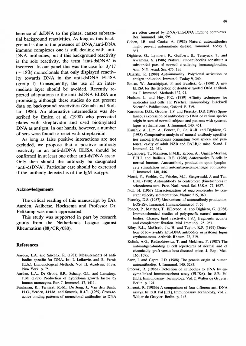
herence of d s D N A to the plates, causes substan-
tial background reactivities. As long as this back- ground is due to the presence of DNA/anti-DNA i m m u n e complexes one is still deal ing with anti- D N A antibodies, bu t if this background reactivity is the sole reactivity, the term ' a n t i - d s D N A ' is incorrect. In our panel this was the case for 3 / 1 7
( = 18%) monoclona ls that only displayed reactiv-
ity towards D N A in the a n t i - d s D N A ELISA (group I). Consequent ly , the use of an inter-
mediate layer should be avoided. Recent ly re-
por ted adapta t ions to the a n t i - d s D N A ELISA are promising, a l though these studies do not present data on background reactivities (Zouali and Stol- lar, 1986). A n al ternat ive in termediate was de- scribed by Emlen et al. (1990) who precoated
plates with s t reptavidin and used b io t inyla ted D N A as antigen. In our hands, however, a n u m b e r
of sera were found to react with streptavidin. As long as false positive in teract ions are no t
excluded, we propose that a positive an t ibody reactivity in an a n t i - d s D N A ELISA should be
conf i rmed in at least one other a n t i - d s D N A assay. Only then should the an t ibody be designated ' a n t i - d s D N A ' . Part icular care should be exercised
if the an t ibody detected is of the IgM isotype.
Acknowledgements
The critical reading of this manuscr ip t by Drs. Aarden, Aalberse, Hoekzema and Professor Dr.
Fe l tkamp was much appreciated. This s tudy was suppor ted in part by research
grants f rom the Nether lands League against Rheumat i sm ( 8 8 / C R / 0 8 0 ) .
References
Aarden, L.A. and Smeenk, R. (1981) Measurements of anti- bodies specific for DNA. In: I. Lefkovits and B. Pemis (Eds.), Immunological Methods, Vol. II. Academic Press, New York, p. 75.
Aarden, L.A., De Groot, E.R., Schaap, O.L. and Lansdorp, P.M. (1987) Production of hybridoma growth factor by human monocytes. Eur. J. Immunol. 17, 1411.
Brinkman, K., Termaat, R.-M., De Jong, J., Van den Brink, H.G., Berden, J.H.M. and Smeenk, R.J.T. (1989) Cross-re- active binding patterns of monoclonal antibodies to DNA
99
are often caused by DNA/anti-DNA immune complexes. Res. Immunol. 140, 595.
Cohen, I.R. and Cooke, A. (1986) Natural autoantibodies might prevent autoimmune disease. Immunol. Today 7, 363.
Dighiero, G., Lymberi, P., Guilbert, B., Ternynck, T. and Avrameas, S. (1986) Natural autoantibodies constitute a substantial part of normal circulating immunoglobulins. Ann. N.Y. Acad. Sci. 475, 135.
Dziarski, R. (1988) Autoimmunity: Polyclonal activation or antigen induction. Immunol. Today 9, 340.
Emlen, W., Jarusiripipat, P. and Burdick, G. (1990) A new ELISA for the detection of double-stranded DNA antibod- ies. J. Immunol. Methods 132, 91.
Hudson, L. and Hay, F.C. (1989) Affinity techniques for molecules and cells. In: Practical Immunology. Blackwell Scientific Publications, Oxford. P. 319.
Karounos, D.G., Grudier, J.P. and Pisetsky, D.S. (1988) Spon- taneous expression of antibodies to DNA of various species origin in sera of normal subjects and patients with systemic lupus erythematosus. J. Immunol. 140, 451.
Kaushik, A., Lira, A., Poncet, P., Ge, X.-R. and Dighiero, G. (1988) Comparative analysis of natural antibody specifici- ties among hybridomas originating from spleen and peri- toneal cavity of adult NZB and BALB/c mice. Scand. J. Immunol. 27, 461.
Logtenberg, T., Melissen, P.M.B., Kroon, A., Gmelig-Meyling, F.H.J. and Ballieux, R.E. (1988) Autoreactive B cells in normal humans. Autoantibody production upon lympho- cyte stimulation with autoantigen-xenoantigen conjugates. J. Immunol. 140, 446.
Moroi, Y., Peebles, C., Fritzler, M.J., Steigerwald, J. and Tan, E.M. (1980) Autoantibody to centromere (kinetochore) in scleroderma sera. Proc. Natl. Acad. Sci. U.S.A. 77, 1627.
Noll, H. (1967) Characterization of macromolecules by con- stant velocity sedimentation. Nature 215, 360.
Pisetsky, D.S. (1987) Mechanisms of autoantibody production. EOS-Riv. Immunol. Immunofarmacol. 7, 53.
Poncet, P., Matthes, T., Billecocq, A. and Dighiero, G. (1988) llmnunochemical studies of polyspecific natural autoanti- bodies: Charge, lipid reactivity, Fab~ fragments activity and complement fixation. Mol. Immunol. 25, 981.
Riley, R.L., McGrath, Jr., H. and Taylor, R.P. (1979) Detec- tion of low avidity anti-DNA antibodies in systemic lupus erythematosus. Arthritis Rheum. 22, 219.
Rolink, A.G., Radaszkiewicz, T. and Melchers, F. (1987) The autoantigen-binding B cell repertoires of normal and of chronically graft-versus-host-diseased mice. J. Exp. Med. 165, 1675.
Sanz, I. and Capra, J.D. (1988) The genetic origin of human autoantibodies. J. Immunol. 140, 3283.
Smeenk, R. (1986a) Detection of antibodies to DNA by en- zyme-hnked immunosorbent assay (ELISA). In: S.B. Pal (Ed.), Immunoassay Technology, Vol. 2. Walter de Gruyter, Berlin, p. 121.
Smeenk, R. (1986b) A comparison of four different anti-DNA assays. In: S.B. Pal (Ed.), Immunoassay Technology, Vol. 2. Walter de Gruyter, Berlin, p. 145.

100
Smeenk, R. and Aarden, I,.A. (1980) The use of polyethylene glycol precipitation to detect low avidity anti-DNA anti- bodies in systemic lupus erythematosus. J. lmmunol. Meth- ods 39, 165.
Smeenk, R.J.T., Brinkman, K., Van den Brink, H.G. and Westgeest, A.A.A. (1988) Reaction patterns of monoclonal antibodies to DNA. J. Immunol. 140, 3786.
Souroujon, M., White-Scharf, M.E., Andr6-Schwartz, J., Gefter, M.L. and Schwartz, R.S. (1988) Preferential autoantibody reactivity of the preimmune B cell repertoire in normal mice. J. Immunol. 140, 4173.
Tan, E.M. (1982) Autoantibodies to nuclear antigens (ANA): their immunobiology and medicine. Adv. Immunol. 33, 167.
Tan, E.M., Chart, E.K.L., Sullivan, K.F. and Rubin, R.L. (1988) Antinuclear antibodies (ANAs): diagnostically specific immune markers and clues toward the understand- ing of systemic autoimmunity)Clin. Immunol. Immuno- pathol. 47, 121.
Ternynck, T. and Avrameas, S. (1986) Murine natural mono- clonal autoantibodies: a study of their polyspecificity and their affinities. Immunol. Rev. 94, 99.
Van Elven, E.H., Rolink, A.G., Van der Veen, F.M., issa, Ph., Duin, Th.M. and Gleichmann, E. (1981) Diseases caused by reactions of T lymphocytes to incompatible structures of the major histocompatibility complex. V. High titers of IgG autoantibodies to double-stranded DNA. J. Immunol. 127, 2435.
Wold, R.T., Young, F.E., Tan, E.M. and Farr, R.S. (1968) Deoxyribonucleic acid antibody: a method to detect its primary interaction with deoxyribonucleic acid. Science 161, 806.
Zouali, M. and Stollar, D.B. (1986) A rapid ELISA for mea- surement of antibodies to nucleic acid antigens using UV- treated polystyrene microplates. J. Immunol. Methods 90, 105.


