Embed Size (px)
Citation preview
Biochemistry 1989, 28, 9949-9956 9949
Banci, L., Bencini, A., Benelli, C., Gatteschi, D., & Zanchini, C. (1982) Struct. Bonding (Berlin) 52, 37-86.
Beisswenger, T. B., Holmquist, B., & Vallee, B. L. (1985) Proc. Natl. Acad. Sci. U.S.A. 82, 8369-8373.
Bicknell, R., Schaffer, A., Waley, S . G., & Auld, D. S . (1 986) Biochemistry 25, 7208-7215.
BrandBn, C.-I., Jornvall, H., Eklund, H., & Furugren, B. (1975) Enzymes (3rd Ed. ) 11, 103-190.
Colonna-Cesari, F., Perahia, D., Karplus, M., Eklund, H., Brandin, C.-I., & Tapia, 0. (1986) J . Biol. Chem. 261,
Dafeldecker, W. P., & Vallee, B. L. (1982) J . Protein Chem.
Davies, R. B., & Abraham, E. P. (1974) Biochem. J . 143,
Eklund, H., & Branden, C.-I. (1987) in Biological Macro- molecules and Assemblies (Jurnak, F. A., & McPherson, A., Eds.) pp 73-142, Wiley, New York.
Eklund, H., Horjales, E., Jornvall, H., Branden, C.-I., & Jeffery, J . (1985) Biochemistry 24, 8005-8012.
Holmquist, B. (1988) Methods Enzymol. 158, 6-12. Hoog, J.-O., von Bahr-Lindstrom, H., Heden, L.-O., Holm-
quist, B., Larsson, K., Hempel, J., Vallee, B. L., & Jornvall, H. (1987) Biochemistry 26, 1926-1932.
Jeffery, J., Cummins, L., Carlquist, M., & Jornvall, H. (1981) Eur. J . Biochem. 140, 17-23.
Jornvall, H., Hempel, J., & Vallee, B. L. (1987a) Enzyme 37,
Jornvall, H., Hoog, J.-O., von Bahr-Lindstrom, H., & Vallee, B. L. (1987b) Proc. Natl. Acad. Sci. U S A . 84,2580-2584.
Jornvall, H., Persson, B., & Jeffery, J. (1987~) Eur. J . Bio- chem. 167, 195-201.
15273-15280.
I, 59-69.
129-1 3 5 .
5-18.
Kaiser, R., Holmquist, B., Hempel, J., Vallee, B. L., &
Karlsson, C., Maret, W., Auld, D. S., Hiiiig, J.-O., & Jornvall,
Maret, W. (1 980) Dissertation, Universitat des Saarlandes,
Maret, W., & Zeppezauer, M. (1986) Biochemistry 25,
Maret, W., & Auld, D. S. (1988) Biochemistry 27,
Maret, W., Anderson, I., Dietrich, H., Schneider-Bernlohr, H., Einarsson, R., & Zeppezauer, M. (1979) Eur. J . Bio- chem. 98, 501-512.
May, S . W., & Kuo, J.-Y. (1978) Biochemistry 17, 3333-3 338.
Peretz, M., & Burstein, Y. (1989) Biochemistry 28, 6549-655 5.
Pettersson, G. (1987) CRC Crit. Rev. Biochem. 21, 349-389. Schneider-Bernlohr, H., Formicka-Kozlowska, G., Biihler, R.,
von Wartburg, J.-P., & Zeppezauer, M. (1988) Eur. J . Biochem. 173, 275-280.
Scopes, R. K. (1982) Protein Purification, p 266, Springer, New York.
Sutton, B. J., Artymiuk, P. J., Cordero-Borboa, A. E., Little, C., Phillips, D. C., & Waley, S. G. (1987) Biochem. J. 248, 181-188.
Sytkowski, A. J., & Vallee, B. L. (1976) Proc. Natl. Acad. Sci. U.S.A. 73, 344-348.
Valkonen, K. H., & Goldman, D. (1988) Electrophoresis (Weinheim, Fed. Repub. Ger.) 9, 132-135.
Wagner, F. W., Pares, X., Holmquist, B., & Vallee, B. L. (1984) Biochemistry 23, 2193-2199.
Jornvall, H. (1988) Biochemistry 27, 1132-1 140.
H. (1989) Eur. J . Biochem. (in press).
Saarbrucken, FRG.
1584-1 588.
1622-1628.
Thermodynamics of Protein-RNA Recognition in a Highly Conserved Region of the Large-Subunit Ribosomal RNA+
Patricia C. Ryan and David E. Draper* Department of Chemistry, Johns Hopkins University, Baltimore, Maryland 21 21 8
Received May IO, 1989; Revised Manuscript Received July 18, 1989
ABSTRACT: Ribosomal protein L11 from Escherichia coli specifically binds to a highly conserved region of 23s ribosomal RNA. The thermodynamics of forming a complex between this protein and several different r R N A fragments have been investigated, by use of a nitrocellulose filter binding assay. A 57-nucleotide region of the R N A (C1052-U1108) contains all the protein recognition features, and an R N A fragment containing this region binds L11 103-104-fold more tightly than tRNA. Binding constants are on the order of 10 pM-' and are only weakly dependent on K+ concentration (a log K / d log [K+] = -1 -4) or temperature. Binding requires multivalent cations; Mg2+ is taken up into the complex with an affinity of -3 mM-'. Other multivalent cations tested, Ca2+ and C O ( N H & ~ + , promote binding nearly as well. The pH dependence of binding is a bell-shaped curve with a maximum near neutral pH, but the entire curve is shifted to higher pH for the smaller of two R N A fragments tested. This result suggests that the smaller fragment favors a conformation stabilizing protonated forms of the R N A recognition site and is potentially relevant to a hypothesis that this rRNA region undergoes an ordered series of conformational changes during the ribosome cycle.
%e discovery of ribozymes (Cech & Bass, 1986) has en- couraged speculation that ribosomal RNAs are major con-
'This research was supported by NIH Grant GM 29048 and by a
tributors to the substrate recognition and catalytic activities of the ribosome. Several kinds of evidence support of this view: ribosomal RNAs are intimately involved in mRNA and tRNA binding and subunit aSSOCiatiOn ( Z i I I l ~ ~ I l l N " et a]., 1979; Burma et al., 1983; Jacob et al., 1987); antibiotics which block Research Career Development Award to D.E.D. (CA 01081).
0006-2960/89/0428-9949$01.50/0 0 1989 American Chemical Society
9950 Biochemistry, Vol. 28, No. 26, 1989
ribosome functions are sensitive to rRNA mutations or mod- ifications (Noller, 1984); many ribosomal proteins are dis- pensable for ribosome function and cell growth (Nomura & Held, 1974; Dabbs, 1986). RNase P, an enzyme with both protein and RNA components, provides a potential parallel with ribosomes. The RNA component alone is catalytically active, but its activity is modified by the binding of a small, basic protein similar to ribosomal proteins (Gardiner et al., 1985; Hansen et al., 1985). Most ribosomal proteins interact with rRNA (Stern et al., 1988); by analogy with RNase P, their role may be to "tune" the rRNA structure and functions.
If the functional importance of rRNA is accepted, then it is important to ask how ribosomal proteins bind to rRNA and what influence they have on rRNA structure and dynamics. We have initiated studies of complexes between ribosomal protein L11 and 23s rRNA fragments for this purpose. The functional equivalent of L11 has been found in eubacteria, archaebacteria, and eukaryotes, and Escherichia coli L11 recognizes a very highly conserved rRNA structure from all of these sources (Stark et al., 1980; Beauclerk et al., 1985; El-Baradi et a]., 1987). Even though the Lll-rRNA inter- action is widely conserved, it is not essential, since bacterial mutants lacking L11 are viable (Stoffler et al., 1980). Ri- bosomes purified from these strains have a low level of un- coupled GTPase activity which is stimulated by addition of purified L11 (Stark et al., 1980). (Ribosomes catalyze GTP hydrolysis in the presence of elongation factors, an activity presumably related to the normal turnover of GTP during the ribosome cycle.) Physical studies of L1 1-RNA complexes may be able to suggest how the ability of L11 to promote a specific ribosome activity is related to its effects on the properties of an rRNA structure. In this paper we present the first ther- modynamic study of L1 1-RNA recognition, defining the minimum RNA structure recognized by L11 and exploring the salt, pH, and temperature requirements for complex for- mation.
EXPERIMENTAL PROCEDURES Buffers and Reagents. Water was distilled and run through
a Millipore MilliQ deionizer and filter, and all buffers were autoclaved or filter sterilized. Buffers used were as follows: TE, 10 mM Tris, pH 7.6, 1 mM Na,EDTA; TK, 30 mM Tris, pH 7.6, 350 mM KCl, 7 mM @-mercaptoethanol; TM20K350, TK buffer plus 20 mM MgS04. Other concentrations of MgS04 or KC1 in TMK buffers are indicated by appropriate subscripts. 32P- and 35S-labeled nucleotides were obtained from Amersham, and enzymes were from BRL or New England Biolabs. [Co(NH,),]Cl, [hexamminecobalt(III) chloride] was Aldrich Gold Label. T7 RNA polymerase was purified by S. Morse in this laboratory from an overproducing strain (Danvaloo et al., 1984).
Protein Purification. Ribosomal proteins were prepared from E . coli MRE600 cells (Grain Processing, Muscatine, IA) and fractionated by a 0-0.4 M KCl gradient on a preparative cation exchange HPLC column in buffer containing 6 M urea, 20 mM Na2P04, and 0.5 mM dithiothreitol, adjusted to pH 5.6 with methylamine (Draper et al., 1988b). Fractions containing L11 were pooled and rechromatographed on a 7.5 X 75 mm Bio-Rad TSK SP-5-PW column with a 30 mL gradient from 0.1 to 0.3 M KC1 in the same urea-phosphate buffer. The protein obtained was homogeneous, as determined by SDS-polyacrylamide electrophoresis. Protein concentration was determined from the absorbance at 230 nm with an ex- tinction coefficient of 85 X lo3 L mol-' cm-', on the basis of an average extinction of 610 L mol-' cm-l per peptide (Jensen et a]., 1976). L11 protein was stored at -20 OC in the chro-
Ryan and Draper
matography buffer. Protein was concentrated by precipitation with five volumes of acetone; controls showed that this pro- cedure does not affect the affinity of L11 for RNA.
Clone Construction and RNA Purification. A series of plasmids containing the phage T7 RNA polymerase promoter followed by fragments of the 23s rRNA gene were prepared; the RNA sequences transcribed from these plasmids are listed in Table I. Plasmid pTRS2 contains an EcoRI-Sal1 fragment (corresponding to 843-1343 in the 23s rRNA sequence) from pKK3535 (Brosius et al., 1978) inserted into pT7-1 ( U S . Biochemicals). A partial A h 1 digest of the same EcoRI-Sal1 DNA was cloned into the SmaI site of pT7/T3-19 (BRL) and a plasmid containing DNA corresponding to the rRNA se- quence 998-1 157 selected; this is pTA19. A DNA fragment containing the T7 promoter and 23s rRNA sequences was obtained from pTA19 by PuuII and EcoRI digestion and cloned into SmaI-EcoRI-digested M13mp19. A 33-base synthetic DNA oligomer was used to create a site-directed deletion (Kunkel et al., 1987) fusing the first two nucleotides of the T7 transcript to A1029 of the rRNA sequence; this is mA19A. The dideoxy sequencing method was used to verify the deletion (Sanger et al., 1977). An EcoRI-Hind111 frag- ment from the phage replicative form was transferred to plasmid pUC18 by L. Laing to give pLL1. Plasmids were propagated in HBlOl and purified for transcription as de- scribed by Draper et al. (1988a).
All RNAs were transcribed in vitro from linear plasmid DNA with phage T7 RNA polymerase; restriction enzymes used to prepare different-length transcripts are listed in Table I. RNAs for binding assays were labeled during transcription with a- [35S] thio-ATP, purified over Nensorb reverse-phase cartridges (New England Nuclear), and stored in TE buffer as described by Draper et al. (1988b). Unlabeled RNA was transcribed in 0.5-mL reactions and purified by gel filtration through a 1.6 X 50 cm Superose 12 (Pharmacia) column (Draper et al., 1988a).
Nitrocellulose Filter Binding Assays. Protein-RNA com- plexes were detected by nitrocellulose filter binding assays. Protein was diluted 20-fold into TK buffer and warmed to 37 OC for 30 min, and RNA was renatured in either TM,,K,,, or TM20K175 buffer for 5 min at 65 OC. A total of 20 pL of protein, ~ 4 0 0 0 cpm of RNA, and buffer to bring the final volume to 50 pL were added together on ice, incubated for a further 10 min, and then filtered through 0.45-pm Schleicher & Schuell BA85 nitrocellulose filters as previously described (Draper et al., 1988b). The filters were not rinsed further, as that reduces the retention of the complex. Superimposable binding curves were obtained with 25-, SO-, or 100-pL reaction volumes, indicating that filter-bound protein is unable to shift the equilibrium during the filtration. The filters were dried at 100 OC and counted for 35S in a liquid scintillation counter. In the assays done at different pH, 30 mM MES, MOPS, or HEPES buffers were substituted for Tris in the TK and TMK buffers used; both renaturation and complex formation were carried out at the altered pH.
Data Analysis. Titration data were fit to a hyperbolic binding isotherm by a nonlinear least-squares routine which varies both the binding constant and the retention efficiency (Draper et al., 1988b). Labeled RNA concentrations in the assays were on the order of 0.01 pM, and thus negligible compared to the protein concentrations. The pH dependences of the binding constant were fit to a logarithmic form of the hyperbolic binding curve by a similar routine, varying the apparent pK and the maximum binding constant.
L1 1-rRNA Fragment Complexes
Table I: LI 1 Bindine: to 23s rRNA Fragments
Biochemistry, Vol. 28, No. 26, 1989 9951
sequence’ binding constant (FM-’)~ plasmid enzyme 5’ 23s rRNA 3’ A B C D E pTRS2 Sal1 I 843-1347 10 4.3 & 2.6 4.0 6.2 f 1.8 nd’ pTRS2 DraI I 843-1083 < 1 < 1 < 1 < 1 nd pTRS2 EstUI I 843-1 122 10 4.5 f 2.5 4.0 7.7 f 2.6 nd pTA19 EcoRI I1 998-1157 111 12 f 2.1 4.5 f 1.5 4.3 6.7 f 1.2 nd pTA19 EstUI I1 998-1 122 14 f 2.2 4.3 f 1 . 1 4.7 f 1.7 8.2 f 2.1 nd pLLl BstUI GG 1029-1122 1 1 f 0.83 4.4 4.3 5.1 f 0.5 4.8 f 0.1 pLLl DraI GG 1029-1083 <1 nd nd nd nd
OTranscripts were made from plasmid DNA linearized with the listed restriction enzyme, as described under Experimental Procesures. The transcripts contained the indicated segment of 23s rRNA sequence, with additional nucleotides at the 5’ or 3’ termini as indicated: (I) GGGA- GACC; (11) GGGAGACCCAAGCUUGCAUGCCUGCAGGUCGACUCUAGAGGAUCCCC; (111) GGGCGAGCUCGAAUU. Binding con- stants were determined from titrations of 3sS-labeled RNA fragments with L11 protein, as described under Experimental Procedures. Errors given are for the averages of two to five titrations. The L1 1-RNA complexes were incubated under different conditions: (A) TM2KlT5 buffer, 0 “C; (B) TM,K3so buffer, 0 O C ; (C) TM20K3s0 buffer, 0 OC; (D) TM2K350 buffer, 37 “C; (E) TM2K350 buffer, 25 OC. CBinding constant not determined.
Binding of labeled RNA to protein in the presence of an excess of competitor RNA is described by
where Ct, and Ptot are the total concentrations of competitor and protein, respectively, Kc and K R are the equilibrium constants for competitor and labeled RNA binding, respec- tively, and v is the fraction of labeled RNA bound to protein. This equation is derived from the relevant mass action and mass conservation laws and was used to estimate the ratio of rRNA fragment to tRNA binding affinities.
“Filter Selection” Experiments. Approximately the pro- cedure described by Carey et al. (1983) was used. 3’-End- labeled RNA was prepared by reaction with cytidine [32P]- bisphosphate and T4 RNA ligase (D’Alessio, 1981). 5’- End-labeled material was prepared from RNA that had been transcribed in the presence of 1 mM GMP; under these con- ditions a 5’-monophosphate begins the transcript (Sampson & Uhlenbeck, 1988). The label was introduced by an ex- change reaction with [ Y - ~ ~ P I A T P and polynucleotide kinase. The RNAs were purified before labeling by electrophoresis on 6% acrylamide-50% urea gels, cutting the RNA band out of the gel, and extracting the RNA by the “freeze and squeeze” method (Kean & Draper, 1985). RNAs were repurified by the same method after labeling. Partial alkaline hydrolysates were made by incubating the RNAs in 50 mM NaHC03, pH 9, at 100 OC for 10 min and then neutralizing with 140 mM Tris-HC1, pH 6. The RNA was then diluted into TM20K350 buffer, renatured for 5 min at 65 “C, and used in a standard filter binding assay with 0.4 pM L1 1. A total of (2-3) X lo4 cpm of RNA was used in a reaction. 32P-Labeled RNA was recovered from the filters by extraction with TE-saturated phenol followed by ethanol precipitation. A total of 10 pg of poly(dA) was included as carrier for the precipitation. The RNA samples were run on 8% acrylamide50% urea gels along with controls, including a partial TI RNase digestion for se- quence reference.
RESULTS L11 Binds Specifically to a Set of 23s rRNA Fragments.
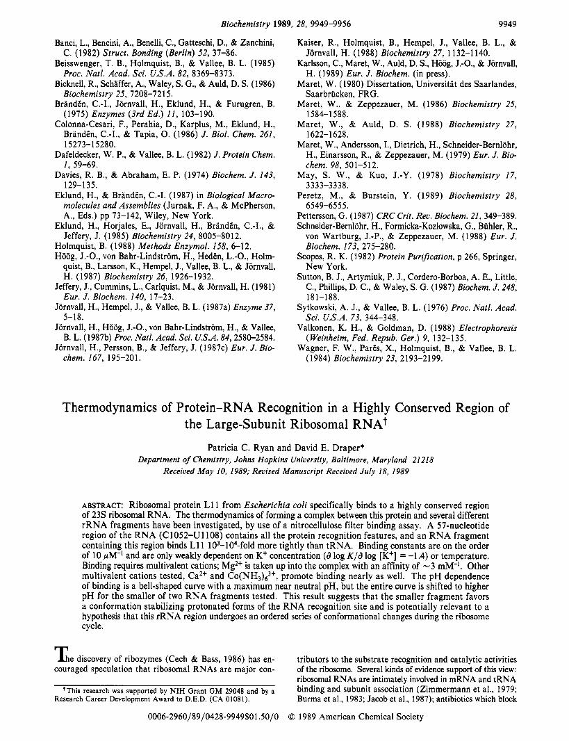
Nuclease protection experiments have suggested that L11 recognizes nucleotides 1052-1 110 of the E . coli 23s rRNA (Schmidt et al., 1981; Beauclerk et al., 1984). The secondary structure of this RNA and the surrounding sequence, as de- duced from phylogenetic comparisons (Noller, 1984; Leffers et al., 1987; Egebjerg et al., 1989), is shown in Figure 1. To measure the L11 affinity for this RNA, we first prepared several 23s rRNA fragments containing the 1052-1 110 se- quence, by in vitro transcription of plasmid DNAs with T7
1 140-E R G G
G R G
\ m C - G R U - R R
1 1 0 0 - C -G R - G - C
R G u u R A
FIGURE 1 : Secondary structure of the 998-1157 RNA fragment binding L11. Base numbering is from the 5’ terminus of the E. coli 2 3 s rRNA. The phylogentically conserved secondary structure is shown. Tick marks are located every 10 bases.
fi 0 0 0 1 0 2 0 3 0 4 0 5 0 0 0 1 0 2 0 3 0 4 0 5 0 6
IL111, ALM [ L l l l , pM
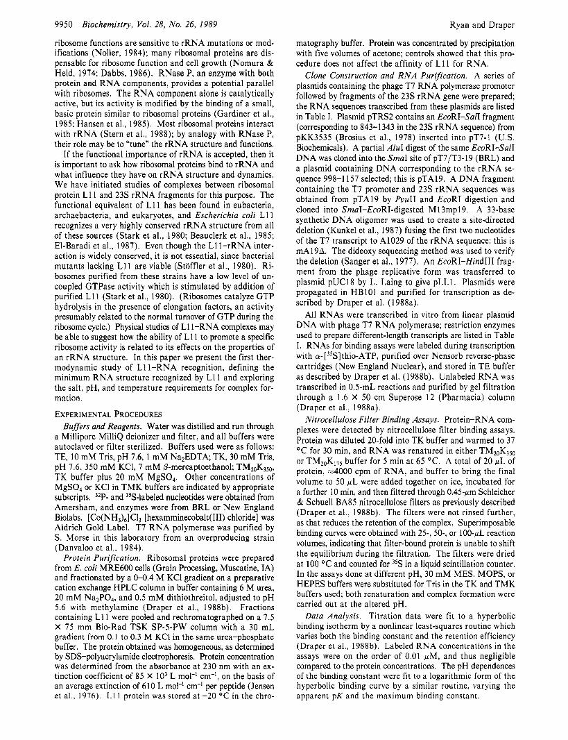
FIGURE 2: Titration of 23.5 rRNA fragments with L1 1 . Titrations were done in TM2K17s buffer at 0 OC. (A) Titration of low ( ~ 0 . 0 1 r M ) concentrations of RNA. Open and closed squares are two independent titrations of the 1029-1 122 RNA. The curve is calculated for a binding constant of 10.5 rM-’ and 65% retention efficiency. Circles are a titration of 1029-1083 RNA. (B) Titration of 0.125 r M 1029-1 122 RNA. The extrapolated break point in the titration is at 0.14 ILM L11.
RNA polymerase. Complexes of these RNAs with L11 pro- tein were detected in a nitrocellulose filter binding assay; an example titration is shown in Figure 2A. The RNA sequences tested and their affinities for L11 under several different conditions are listed in Table I. All five fragments which contain the 1052-1 112 sequence bind L11 with about the same affinity under a given set of conditions, while fragments ter- minating at U1083 do not bind detectably. The binding constants are on the order of 10 pM-l, typical for ribosomal protein-rRNA interactions (Schwarzbauer & Craven, 198 1).
The measurements reported in Table I were made with low concentrations of 35S-labeled RNA and an excess of protein; calculation of the binding constant assumes that a single protein retains one RNA molecule on the filter. To confirm this, the titration was repeated in the presence of 0.125 pM unlabeled RNA (Figure 2B). Under these conditions protein binding is nearly stoichiometric, and we calculate that there are 1.1 mol of protein/mol of RNA in the complex. Unit stoichiometry is expected from the single copy of L11 asso- ciated with ribosomes (Hardy, 1975).
We attempted to measure the specificity of the complex by adding increasing concentrations of unlabeled yeast tRNAPhe
9952 Biochemistry, Vol. 28, No. 26, 1989 Ryan and Draper
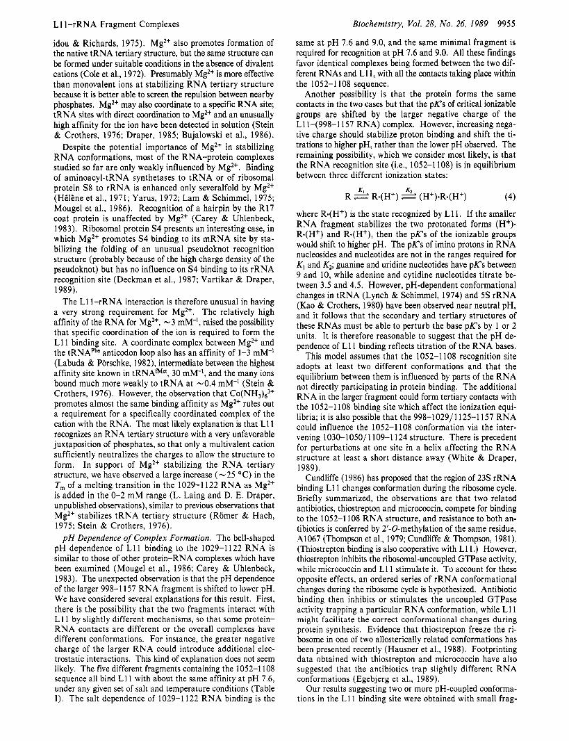
B 1 2 3 4
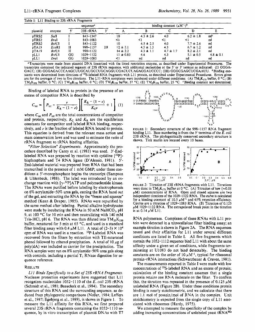
FIGURE 3: LI 1 filter mention of alkalinbhydrolyled 102%1122 RNA fragment. Filter selection experiments were performed as described under Experimental Procedures with either 5’-end-lakled RNA (panel A) or 3’-end-labeled RNA (panel B). The lanes are (panel A) ( I ) unhydrolyzed RNA retained by LI I on a filter, (2) hydrolyzed RNA retained on a filter, (3) the hydrolyzed RNA before filtering, and (4) partial TI RNase digest. For panel B, lanes 1 and 2 are RNAs retained by LI I on filters as in panel A, while lane 3 is the partial TI RNase digestion and lane 4 is the hydrolyzed RNA. Labeled RNA was also filtered in the absence of protein, extracted. and run on the same gels; no exposure was detectable (not shown). Numbers next to bands indicate the 3’ (panel A) or 5‘ (panel B) terminal nucleotide deduced from the partial TI RNase digests. The arrows indicate the approximate positions of the shortest RNAs retained by LI 1. The variations in the intensities of the bands retained by LI 1 duplicate the variations in the alkaline hydrolysis pattern.
to the binding assays. Only weak competition was ever ob- served, reducing the rRNA fragment retention by only - 15% even a t tRNA concentrations more than 1000-fold in excess of LI 1 (data not shown). It was not possible to include much higher tRNA concentrations in the assays, as a high back- ground (>20%) of labeled RNA became bound by the filter in the absence of protein. Since the decreases in labeled RNA retention with added tRNA were comparable to the normal variation in retention between experiments, we can only set a lower limit of -2000 for the ratio of the 1029-1 122 RNA and tRNA affinities and estimate that the ratio is probably larger than 5000. (The calculations assume one nonspecific LI 1 binding site per tRNA; the specificity will be even greater if there is more than one potential binding site per tRNA.)
All the fragments tested show a similar, small increase in binding affinity when the temperature is raised from 0 to 37 ‘C (compare conditions B, D, and E in Table I). Therefore, the AH for complex formation in this temperature range is small and perhaps slightly positive.
Minimum Recognition Site. The smallest RNA able to bind LI 1 specifically was determined by a filter selection experiment with a set of randomly fragmented rRNA fragments. The 1029-1 122 RNA was first labeled with 32P a t either the 5’ or 3’ terminus and then partially hydrolyzed at alkaline pH. After the mixture of RNA fragments had been renatured, they were equilibrated with LI I and filtered through nitrocellulose, as for a binding assay. The retained RNA was then extracted and run on a sequencing gel alongside the original fragment mixture and a partial hydrolysis generated by TI RNase (G specific). The results of the experiment are shown in Figure
04 . I I
PH
4 5 6 7 8 9 1 0
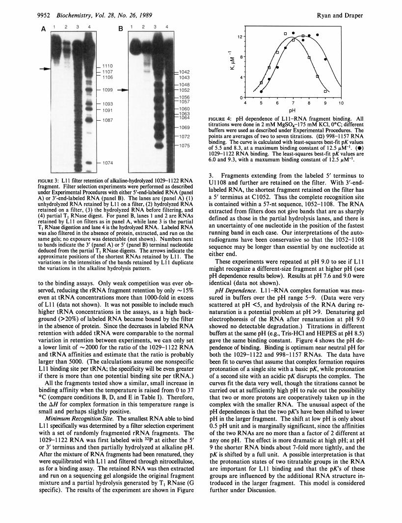
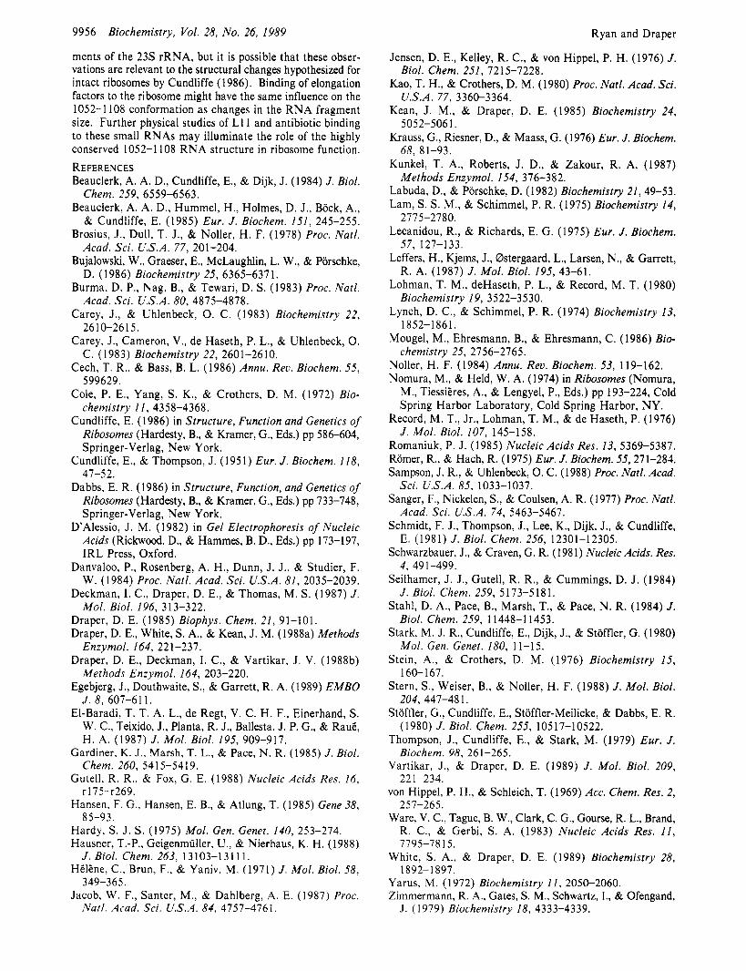
FIGURE 4 pH dependence of LII-RNA fragment binding. All titrations were done in 2 mM MgSO.475 mM KCI, ODC, different buffers were used as described under Experimental Procedures. The points are averages of two to seven titrations. (0 ) 998-1 157 RNA binding. The curve is calculated with least-squares best-fit pK values of 5.5 and 8.3, at a maximum binding constant of 12.5 pM-I. (0) 1029-1 122 RNA binding. The least-squares best-fit pKvalues are 6.0 and 9.3, with a maxumum binding constant of 12.5 pM-I.
3. Fragments extending from the labeled 5’ terminus to U1108 and further are retained on the filter. With 3’-end- labeled RNA, the shortest fragment retained on the filter has a 5’ terminus at C1052. Thus the complete recognition site is contained within a 57-nt sequence, 1052-1 108. The RNA extracted from filters does not give bands that are as sharply defined as those in the partial hydrolysis lanes, and there is an uncertainty of one nucleotide in the position of the fastest running band in each case. Our interpretations of the auto- radiograms have been conservative so that the 1052-1108 sequence may be longer than essential by one nucleotideat either end.
These experiments were repeated at pH 9.0 to see if LI I might recognize a different-size fragment at higher pH (see pH dependence results below). Results at pH 7.6 and 9.0 were identical (data not shown).
pH Dependence. LI I-RNA complex formation was mea- sured in buffers over the pH range 5-9. (Data were very scattered a t pH <5, and hydrolysis of the RNA during re- naturation is a potential problem a t pH >9. Denaturing gel electrophoresis of the RNA after renaturation at pH 9.0 showed no detectable degradation.) Titrations in different buffers at the same pH (e.g., Tris-HCI and HEPES at pH 8.5) gave the same binding constant. Figure 4 shows the pH de- pendence of binding. Binding is optimum near neutral pH for both the 1029-1 122 and 998-1 157 RNAs. The data have been fit to CUN~S that assume that complex formation requires protonation of a single site with a basic pK, while protonation of a second site with an acidic pK disrupts the complex. The curves fit the data very well, though the titrations cannot be carried out a t sufficiently high pH to rule out the possibility that two or more protons are cooperatively taken up in the complex with the smaller RNA. The unusual aspect of the pH dependences is that the two pKs have been shifted to lower pH in the larger fragment. The shift at low pH is only about 0.5 pH unit and is marginally significant, since the affinities of the two RNAs are no more than a factor of 2 different at any one pH. The effect is more dramatic a t high pH; at pH 9 the shorter RNA binds about 7-fold more tightly, and the pK is shifted by a full unit. A possible interpretation is that the protonation states of two titratable groups in the RNA are important for LI 1 binding and that the pKs of these groups are influenced by the additional RNA structure in- troduced in the larger fragment. This model is considered further under Discussion.
L1 1-rRNA Fragment Complexes Biochemistry, Vol. 28, No. 26, 1989 9953
O\ I -1.2 -1.0 -0.8 -0.6 -0.4 -0.2
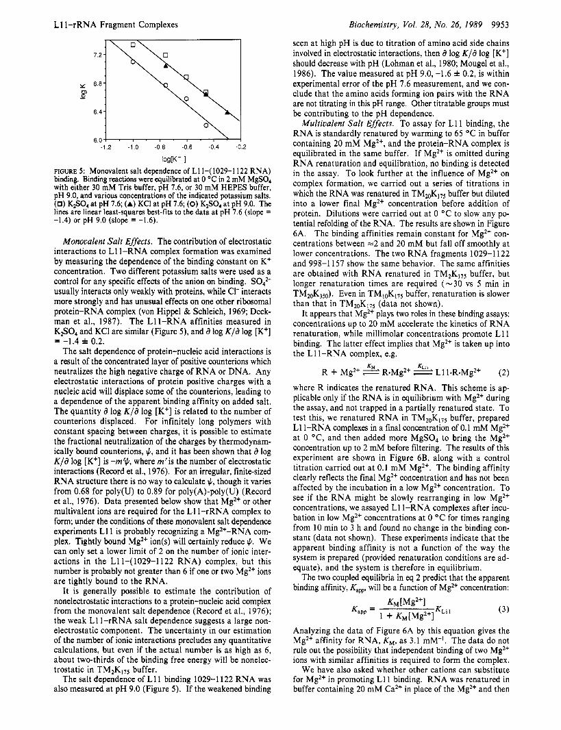
W K + 1 FIGURE 5: Monovalent salt dependence of L11-(1029-1122 RNA) binding. Binding reactions were equilibrated at 0 OC in 2 mM MgS04 with either 30 mM Tris buffer, pH 7.6, or 30 mM HEPES buffer, pH 9.0, and various concentrations of the indicated potassium salts. (0) K2S0, at pH 7.6; (A) KC1 at pH 7.6; (0) K2SO4 at pH 9.0. The lines are linear least-squares best-fits to the data at pH 7.6 (slope = -1.4) or pH 9.0 (slope = -1.6).
Monovalent Salt Effects. The contribution of electrostatic interactions to L1 1-RNA complex formation was examined by measuring the dependence of the binding constant on K+ concentration. Two different potassium salts were used as a control for any specific effects of the anion on binding. S042- usually interacts only weakly with proteins, while C1- interacts more strongly and has unusual effects on one other ribosomal protein-RNA complex (von Hippel & Schleich, 1969; Deck- man et al., 1987). The L11-RNA affinities measured in K 8 0 4 and KCl are similar (Figure 5), and d log K/d log [K+]
The salt dependence of protein-nucleic acid interactions is a result of the concentrated layer of positive counterions which neutralizes the high negative charge of RNA or DNA. Any electrostatic interactions of protein positive charges with a nucleic acid will displace some of the counterions, leading to a dependence of the apparent binding affinity on added salt. The quantity d log K/d log [K+] is related to the number of counterions displaced. For infinitely long polymers with constant spacing between charges, it is possible to estimate the fractional neutralization of the charges by thermodynam- ically bound counterions, +, and it has been shown that d log K/d log [K+] is -mV, where m'is the number of electrostatic interactions (Record et al., 1976). For an irregular, finite-sized RNA structure there is no way to calculate +, though it varies from 0.68 for poly(U) to 0.89 for poly(A)-poly(U) (Record et al., 1976). Data presented below show that Mg2+ or other multivalent ions are required for the L1 1-rRNA complex to form; under the conditions of these monovalent salt dependence experiments L11 is probably recognizing a Mg2+-RNA com- plex. Tightly bound Mg2+ ion(s) will certainly reduce +. We can only set a lower limit of 2 on the number of ionic inter- actions in the L11-(1029-1122 RNA) complex, but this number is probably not greater than 6 if one or two Mg2+ ions are tightly bound to the RNA.
It is generally possible to estimate the contribution of nonelectrostatic interactions to a protein-nucleic acid complex from the monovalent salt dependence (Record et al., 1976); the weak L1 1-rRNA salt dependence suggests a large non- electrostatic component. The uncertainty in our estimation of the number of ionic interactions precludes any quantitative calculations, but even if the actual number is as high as 6, about two-thirds of the binding free energy will be nonelec- trostatic in TM2K17, buffer.
The salt dependence of L11 binding 1029-1 122 RNA was also measured at pH 9.0 (Figure 5). If the weakened binding
= -1.4 f 0.2.
seen at high pH is due to titration of amino acid side chains involved in electrostatic interactions, then d log K / d log [K'] should decrease with pH (Lohman et al., 1980; Mougel et al., 1986). The value measured at pH 9.0, -1.6 f 0.2, is within experimental error of the pH 7.6 measurement, and we con- clude that the amino acids forming ion pairs with the RNA are not titrating in this pH range. Other titratable groups must be contributing to the pH dependence.
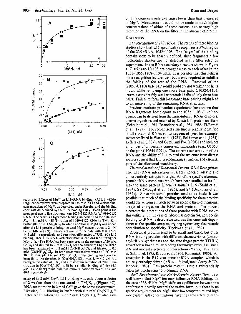
Multivalent Salt Effects. To assay for L11 binding, the RNA is standardly renatured by warming to 65 "C in buffer containing 20 mM Mg2+, and the protein-RNA complex is equilibrated in the same buffer. If Mg2+ is omitted during RNA renaturation and equilibration, no binding is detected in the assay. To look further at the influence of Mg2+ on complex formation, we carried out a series of titrations in which the RNA was renatured in TM2,KI7, buffer but diluted into a lower final Mg2+ concentration before addition of protein. Dilutions were carried out at 0 "C to slow any po- tential refolding of the RNA. The results are shown in Figure 6A. The binding affinities remain constant for Mg2+ con- centrations between =2 and 20 mM but fall off smoothly at lower concentrations. The two RNA fragments 1029-1 122 and 998-1 157 show the same behavior. The same affinities are obtained with RNA renatured in TMzK17, buffer, but longer renaturation times are required (-30 vs 5 min in TM2,K3,,). Even in TMIOKI7, buffer, renaturation is slower than that in TM2,K17, (data not shown).
It appears that Mg2+ plays two roles in these binding assays: concentrations up to 20 mM accelerate the kinetics of RNA renaturation, while millimolar concentrations promote L 1 1 binding. The latter effect implies that Mg2+ is taken up into the Lll-RNA complex, e.g.
R + Mg2+ & R.Mg2+ e L1 l.R.Mg2+ (2) where R indicates the renatured RNA. This scheme is ap- plicable only if the RNA is in equilibrium with Mg2+ during the assay, and not trapped in a partially renatured state. To test this, we renatured RNA in TM2,K,,, buffer, prepared L1 1-RNA complexes in a final concentration of 0.1 mM Mg2+ at 0 "C, and then added more MgS04 to bring the Mg2+ concentration up to 2 mM before filtering. The results of this experiment are shown in Figure 6B, along with a control titration carried out at 0.1 mM Mg2+. The binding affinity clearly reflects the final Mg2+ concentration and has not been affected by the incubation in a low Mg2+ concentration. To see if the RNA might be slowly rearranging in low Mg2+ concentrations, we assayed L 1 1-RNA complexes after incu- bation in low Mg2+ concentrations at 0 "C for times ranging from 10 min to 3 h and found no change in the binding con- stant (data not shown). These experiments indicate that the apparent binding affinity is not a function of the way the system is prepared (provided renaturation conditions are ad- equate), and the system is therefore in equilibrium.
The two coupled equilibria in eq 2 predict that the apparent binding affinity, Kapp, will be a function of Mg2+ concentration:
KLll
(3)
Analyzing the data of Figure 6A by this equation gives the Mg2+ affinity for RNA, KM, as 3.1 mM-I. The data do not rule out the possibility that independent binding of two Mg2+ ions with similar affinities is required to form the complex.
We have also asked whether other cations can substitute for Mg2+ in promoting L11 binding. RNA was renatured in buffer containing 20 mM Ca2+ in place of the Mg2+ and then
9954 Biochemistry, Vol. 28, No. 26, 1989 Ryan and Draper
binding constants only 2-3 times lower than that measured in Mg2+. Measurements could not be made in much higher concentrations of either of these cations, due to very high retention of the RNA on the filter in the absence of protein.
DISCUSSION L l l Recognition of 23s rRNA. The results of these binding
studies show that L11 specifically recognizes a 57-nt region of the 23s rRNA, 1052-1 108. The “edges” of the binding domain seem to be sharply defined, since fragments a few nucleotides shorter are not detected in the filter selection experiment. In the RNA secondary structure shown in Figure 1, C1052 and U1108 are brought close to each other in the 1051-1055/1108-1104 helix. It is possible that this helix is not a recognition feature itself but is only required to stabilize the folding of the rest of the RNA. Removal of the G10514J1108 base pair would probably not weaken the helix much, while removing one more base pair, C1052.Gl107, leaves a considerably weaker potential helix of only three base pairs. Failure to form this long-range base pairing might lead to an unraveling of the remaining RNA structure.
Previous nuclease protection experiments have shown that RNA fragments homologous to the 1052-1108 E . coli se- quence can be derived from the large-subunit rRNAs of several diverse organisms and retained by E. coli L11 protein on filters (Schmidt et al., 1981; Beauclerk et al., 1984, 1985; El-Baradi et al., 1987). The recognized structure is readily identified in all ribosomal RNAs so far sequenced [see, for example, sequences listed in Ware et al. (1983), Seilhamer et al. (1984), Leffers et al. (1987), and Gutell and Fox (1988)l and includes a number of universally conserved nucleotides (e.g., U1060, or the pair C1064.Gl074). The extreme conservation of the RNA and the ability of L11 to bind the structure from diverse sources suggest that L11 is recognizing an ancient and essential part of the ribosomal machinery.
Thermodynamics of Ribosomal Protein-RNA Recognition. The L1 1-RNA interaction is largely nonelectrostatic and almost entirely entropic in origin. All of the specific ribosomal protein-RNA complexes which have been studied so far fall into the same pattern [Bacillus subtilis L16 (Stahl et al., 1984), S8 (Mougel et al., 1986), and S4 (Deckman et al., 1987)l. Since ribosomal proteins tend to be basic, it was possible that much of the binding specificity for these proteins would derive from a match between specific three-dimensional arrays of charges on the RNA and protein. The modest electrostatic interactions of these proteins with RNA makes this unlikely. In the case of ribosomal protein S4, nonspecific binding to tRNA is detectable and has the same salt depen- dence as the specific complex, arguing against any electrostatic contribution to specificity (Deckman et al., 1987).
Ribosomal proteins tend to be small and basic, but other RNA-binding proteins with different characteristics (amino- acyl-tRNA synthetases and the zinc finger protein TFIIIA) nevertheless have similar binding thermodynamics, i.e., small A H and modest electrostatic interactions (Yarus, 1972; Lam & Schimmel, 1975; Krauss et al., 1976; Romaniuk, 1985). An exception is the R17 coat protein-RNA complex, which is entirely enthalpy driven (AH = -19 kcal/mol; Carey & Uh- lenbeck, 1983). This protein may thus use a substantially different mechanism to recognize RNA.
M?+ Requirement for RNA-Protein Recognition. It is well-known that Mg2+ ion may influence RNA folding. In the case of 5s rRNA, Mg2+ shifts an equilibrium between two conformers heavily toward the native form, but there is no specific requirement for Mgz+ since low temperature and high monovalent salt concentrations have the same effect (Lecan-
12- 0 A -
- a - F x- -
4 -
0.00 0.1 0 0.20 0.30 0.40
[LI 1 I , FM
60
F! 50 1 A
10 0 0 0 0 10 0.20 0 3 0 0 4 0
U-11 I , IN FIGURE 6: Effects of Mg2+ on Ll l -RNA binding. (A) Ll l -RNA fragment complexes were prepared in 175 mM KCI and various final concentrations of Mg2+, as described under Results, and the binding constants determined by the filter binding assay. Each point is the average of two to five titrations. (h) 1029-1 122 R N A (0) 998-1 157 RNA. The curve is a hyperbolic binding isotherm fit to the data with KM = 3.1 mM-l. (B) Titration of 1029-1122 RNA in TMO1Kl75 buffer (E) or in TM0,1K175 to which additional MgS04 was added after the LI 1 protein to bring the total Mg2+ concentration to 2 mM before filtering (0). The curves are fit to the data with K = 1.5 or 14.5 wM-I, respectively, and retention efficiencies of 75%. (C) L11 binding 1029-1 122 RNA with other multivalent ions substituting for Mg2+. (0) The RNA has been renatured in the presence of 20 mM CaCI2 and diluted to 2 mM CaC12 for the titration; (A) the RNA has been renatured with 2 mM [CO(NH,)~]C~, and titrated in 0.2 mM [CO(NH~)~]CI, . In both cases incubations were at 0 OC with 30 mM Tris, pH 7.6, and 175 m M KCI. The binding isotherm has been fit to the titration in [ C O ( N H ~ ) ~ ] C ~ , , with K = 4.9 pM-I, a background value of lo%, and a maximum retention of 70%. The titration in [CO(NH&]CI3 is fit by a similar binding affinity (4.1 wM-I) and background and maximum retention values of 17% and 60%, respectively.
assayed in 2 mM Ca2+; L11 binding was only about a factor of 2 weaker than that measured in TM2K17S (Figure 6C). RNA renaturation in 2 mM CaZ+ gave the same measurement. Likewise, L11 binding in buffer with 0.2 mM C O ( N H ~ ) ~ ~ + [after renaturation in 0.2 or 2 mM C O ( N H ~ ) ~ ~ + ] also gave
L1 1-rRNA Fragment Complexes
idou & Richards, 1975). Mg2+ also promotes formation of the native tRNA tertiary structure, but the same structure can be formed under suitable conditions in the absence of divalent cations (Cole et al., 1972). Presumably Mg2+ is more effective than monovalent ions at stabilizing RNA tertiary structure because it is better able to screen the repulsion between nearby phosphates. Mg2+ may also coordinate to a specific RNA site; tRNA sites with direct coordination to Mg2+ and an unusually high affinity for the ion have been detected in solution (Stein & Crothers, 1976; Draper, 1985; Bujalowski et al., 1986).
Despite the potential importance of Mg2+ in stabilizing RNA conformations, most of the RNA-protein complexes studied so far are only weakly influenced by Mg2+. Binding of aminoacyl-tRNA synthetases to tRNA or of ribosomal protein S8 to rRNA is enhanced only severalfold by Mg2+ ( H e h e et al., 1971; Yarus, 1972; Lam & Schimmel, 1975; Mougel et al., 1986). Recognition of a hairpin by the R17 coat protein is unaffected by Mg2+ (Carey & Uhlenbeck, 1983). Ribosomal protein S4 presents an interesting case, in which Mg2+ promotes S4 binding to its mRNA site by sta- bilizing the folding of an unusual pseudoknot recognition structure (probably because of the high charge density of the pseudoknot) but has no influence on S4 binding to its rRNA recognition site (Deckman et al., 1987; Vartikar & Draper, 1989).
The L1 1-rRNA interaction is therefore unusual in having a very strong requirement for Mg2+. The relatively high affinity of the RNA for Mg2+, -3 mM-l, raised the possibility that specific coordination of the ion is required to form the L11 binding site. A coordinate complex between Mg2+ and the tRNAPhe anticodon loop also has an affinity of 1-3 mM-' (Labuda & Porschke, 1982), intermediate between the highest affinity site known in tRNAmet, 30 mM-', and the many ions bound much more weakly to tRNA at -0.4 mM-' (Stein & Crothers, 1976). However, the observation that Co(NH3):+ promotes almost the same binding affinity as Mg2+ rules out a requirement for a specifically coordinated complex of the cation with the RNA. The most likely explanation is that L11 recognizes an RNA tertiary structure with a very unfavorable juxtaposition of phosphates, so that only a multivalent cation sufficiently neutralizes the charges to allow the structure to form. In support of Mg2+ stabilizing the RNA tertiary structure, we have observed a large increase (-25 "C) in the T,,, of a melting transition in the 1029-1 122 RNA as Mg2+ is added in the 0-2 mM range (L. Laing and D. E. Draper, unpublished observations), similar to previous observations that Mg2+ stabilizes tRNA tertiary structure (Romer & Hach, 1975; Stein & Crothers, 1976).
p H Dependence of Complex Formation. The bell-shaped pH dependence of L11 binding to the 1029-1 122 RNA is similar to those of other protein-RNA complexes which have been examined (Mougel et al., 1986; Carey & Uhlenbeck, 1983). The unexpected observation is that the pH dependence of the larger 998-1 157 RNA fragment is shifted to lower pH. We have considered several explanations for this result. First, there is the possibility that the two fragments interact with L11 by slightly different mechanisms, so that some protein- RNA contacts are different or the overall complexes have different conformations. For instance, the greater negative charge of the larger RNA could introduce additional elec- trostatic interactions. This kind of explanation does not seem likely. The five different fragments containing the 1052-1 108 sequence all bind L11 with about the same affinity at pH 7.6, under any given set of salt and temperature conditions (Table I ) . The salt dependence of 1029-1 122 RNA binding is the
Biochemistry, Vol. 28, No. 26, 1989 9955
same at pH 7.6 and 9.0, and the same minimal fragment is required for recognition at pH 7.6 and 9.0. All these findings favor identical complexes being formed between the two dif- ferent RNAs and L11, with all the contacts taking place within the 1052-1 108 sequence.
Another possibility is that the protein forms the same contacts in the two cases but that the pK's of critical ionizable groups are shifted by the larger negative charge of the L11-(998-1157 RNA) complex. However, increasing nega- tive charge should stabilize proton binding and shift the ti- trations to higher pH, rather than the lower pH observed. The remaining possibility, which we consider most likely, is that the RNA recognition site (Le., 1052-1 108) is in equilibrium between three different ionization states:
K R 2 R.(H+) & (H+)-R.(H+) (4)
where R.(H+) is the state recognized by L1 1. If the smaller RNA fragment stabilizes the two protonated forms (H'). R.(H+) and R.(H+), then the px"s of the ionizable groups would shift to higher pH. The pK's of imino protons in RNA nucleosides and nucleotides are not in the ranges required for K1 and K2; guanine and uridine nucleotides have pK's between 9 and 10, while adenine and cytidine nucleotides titrate be- tween 3.5 and 4.5. However, pH-dependent conformational changes in tRNA (Lynch & Schimmel, 1974) and 5 s rRNA (Kao & Crothers, 1980) have been observed near neutral pH, and it follows that the secondary and tertiary structures of these RNAs must be able to perturb the base pK's by 1 or 2 units. It is therefore reasonable to suggest that the pH de- pendence of L1 l binding reflects titration of the RNA bases.
This model assumes that the 1052-1 108 recognition site adopts at least two different conformations and that the equilibrium between them is influenced by parts of the RNA not directly participating in protein binding. The additional RNA in the larger fragment could form tertiary contacts with the 1052-1 108 binding site which affect the ionization equi- libria; it is also possible that the 998-1029/1125-1157 RNA could influence the 1052-1 108 conformation via the inter- vening 1030-1050/1109-1124 structure. There is precedent for perturbations at one site in a helix affecting the RNA structure at least a short distance away (White & Draper, 1989).
Cundliffe (1 986) has proposed that the region of 23s rRNA binding L11 changes conformation during the ribosome cycle. Briefly summarized, the observations are that two related antibiotics, thiostrepton and micrococcin, compete for binding to the 1052-1108 RNA structure, and resistance to both an- tibiotics is conferred by 2'-O-methylation of the same residue, A1067 (Thompson et al., 1979; Cundliffe & Thompson, 1981). (Thiostrepton binding is also cooperative with L1 1.) However, thiostrepton inhibits the ribosomal-uncoupled GTPase activity, while micrococcin and L11 stimulate it. To account for these opposite effects, an ordered series of rRNA conformational changes during the ribosome cycle is hypothesized. Antibiotic binding then inhibits or stimulates the uncoupled GTPase activity trapping a particular RNA conformation, while L11 might facilitate the correct conformational changes during protein synthesis. Evidence that thiostrepton freeze the ri- bosome in one of two allosterically related conformations has been presented recently (Hausner et al., 1988). Footprinting data obtained with thiostrepton and micrococcin have also suggested that the antibiotics trap slightly different RNA conformations (Egebjerg et al., 1989).
Our results suggesting two or more pH-coupled conforma- tions in the L1 l binding site were obtained with small frag-
9956
ments of the 23s rRNA, but it is possible that these obser- vations are relevant to the structural changes hypothesized for intact ribosomes by Cundliffe (1986). Binding of elongation factors to the ribosome might have the same influence on the 1052-1 108 conformation as changes in the RNA fragment size. Further physical studies of L11 and antibiotic binding to these small RNAs may illuminate the role of the highly conserved 1052-1 108 RNA structure in ribosome function. REFERENCES Beauclerk, A. A. D., Cundliffe, E., & Dijk, J. (1 984) J . Biol.
Chem. 259, 6559-6563. Beauclerk, A. A. D., Hummel, H., Holmes, D. J., Bock, A.,
& Cundliffe, E. (1985) Eur. J . Biochem. 151, 245-255. Brosius, J., Dull, T. J., & Noller, H. F. (1978) Proc. Natl.
Acad. Sci. U.S.A. 77, 201-204, Bujalowski, W., Graeser, E., McLaughlin, L. W., & Porschke,
D. (1986) Biochemistry 25, 6365-6371. Burma, D. P., Nag, B., & Tewari, D. S. (1983) Proc. Natl.
Acad. Sci. U.S.A. 80, 4875-4878. Carey, J., & Uhlenbeck, 0. C. (1983) Biochemistry 22,
Carey, J., Cameron, V., de Haseth, P. L., & Uhlenbeck, 0. C. (1983) Biochemistry 22, 2601-2610.
Cech, T. R., & Bass, B. L. (1986) Annu. Reu. Biochem. 55, 599629.
Cole, P. E., Yang, S. K., & Crothers, D. M. (1972) Bio- chemistry 1 1 , 4358-4368.
Cundliffe, E. (1986) in Structure, Function and Genetics of Ribosomes (Hardesty, B., & Kramer, G., Eds.) pp 586-604, Springer-Verlag, New York.
Cundliffe, E., & Thompson, J. (1951) Eur. J . Biochem. 118,
Dabbs, E. R. (1986) in Structure, Function, and Genetics of Ribosomes (Hardesty, B., & Kramer, G., a s . ) pp 733-748, Springer-Verlag, New York.
D’Alessio, J. M. ( 1 982) in Gel Electrophoresis of Nucleic Acids (Rickwood, D., & Hammes, B. D., Eds.) pp 173-197, IRL Press, Oxford.
Danvaloo, P., Rosenberg, A. H., Dum, J. J., & Studier, F. W. (1984) Proc. Natl. Acad. Sci. U.S.A. 81, 2035-2039.
Deckman, I . C., Draper, D. E., & Thomas, M. S. (1987) J. Mol. Biol. 196, 3 13-322.
Draper, D. E. (1985) Biophys. Chem. 21, 91-101. Draper, D. E., White, S. A., & Kean, J. M. (1988a) Methods
Draper, D. E., Deckman, I . C., & Vartikar, J . V. (1988b)
Egebjerg, J., Douthwaite, S., & Garrett, R. A. (1989) EMBO
El-Baradi, T. T. A. L., de Regt, V. C. H. F., Einerhand, S. W. C., Teixido, J., Planta, R. J., Ballesta, J. P. G., & Raut, H. A. (1987) J . Mol. Biol. 195, 909-917.
Gardiner, K . J., Marsh, T. L., & Pace, N. R. (1985) J . Biol. Chem. 260, 5415-5419.
Gutell, R. R., & Fox, G . E. (1988) Nucleic Acids Res. 16, r 1 7 5-r269.
Hansen, F. G., Hansen, E. B., & Atlung, T. (1985) Gene 38,
Hardy, S. J. S. (1975) Mol. Gen. Genet. 140, 253-274. Hausner, T.-P., Geigenmuller, U., & Nierhaus, K. H. (1988)
Htlcne, C., Brun, F., & Yaniv, M. (1971) J . Mol. Biol. 58,
Jacob, W. F., Santer, M., & Dahlberg, A. E. (1987) Proc.
Biochemistry, Vol. 28, No. 26, 1989
26 10-26 15.
47-52.
Enzymol. 164, 221-237.
Methods Enzymol. 164, 203-220.
J . 8 , 607-6 1 1.
85-93.
J . Biol. Chem. 263, 13103-13111.
349-365.
Natl. Acad. Sci. U.S.A. 84, 4757-4761.
Ryan and Draper
Jensen, D. E., Kelley, R. C., & von Hippel, P. H. (1976) J.
Kao, T. H., & Crothers, D. M. (1980) Proc. Natl. Acad. Sci.
Kean, J. M., & Draper, D. E. (1985) Biochemistry 24,
Krauss, G., Riesner, D., & Maass, G. (1976) Eur. J. Biochem.
Kunkel, T. A., Roberts, J. D., & Zakour, R. A. (1987)
Labuda, D., & Porschke, D. (1982) Biochemistry 21,49-53. Lam, S. S. M., & Schimmel, P. R. (1975) Biochemistry 14,
Lecanidou, R., & Richards, E. G. (1975) Eur. J . Biochem.
Leffers, H., Kjems, J., 0stergaard, L., Larsen, N., & Garrett,
Lohman, T. M., deHaseth, P. L., & Record, M. T. (1980)
Lynch, D. C., & Schimmel, P. R. (1974) Biochemistry 13,
Mougel, M., Ehresmann, B., & Ehresmann, C. (1986) Bio-
Noller, H. F. (1984) Annu. Reu. Biochem. 53, 119-162. Nomura, M., & Held, W. A. (1974) in Ribosomes (Nomura,
M., TiessiEres, A,, & Lengyel, P., Eds.) pp 193-224, Cold Spring Harbor Laboratory, Cold Spring Harbor, NY.
Record, M. T., Jr., Lohman, T. M., & de Haseth, P. (1976) J . Mol. Biol. 107, 145-158.
Romaniuk, P. J. (1985) Nucleic Acids Res. 13, 5369-5387. Romer, R., & Hach, R. (1975) Eur. J . Biochem. 55,271-281. Sampson, J. R., & Uhlenbeck, 0. C. (1988) Proc. Natl. Acad.
Sanger, F., Nickelen, S., & Coulsen, A. R. (1977) Proc. Natl.
Schmidt, F. J., Thompson, J., Lee, K., Dijk, J., & Cundliffe,
Schwarzbauer, J., & Craven, G. R. (1981) Nucleic Acids. Res.
Seilhamer, J. J., Gutell, R. R., & Cummings, D. J. (1984)
Stahl, D. A., Pace, B., Marsh, T., & Pace, N. R. (1984) J.
Stark, M. J. R., Cundliffe, E., Dijk, J., & Stoffler, G. (1980)
Stein, A., & Crothers, D. M. (1976) Biochemistry 15,
Stern, S., Weiser, B., & Noller, H. F. (1988) J . Mol. Biol.
Stoffler, G., Cundliffe, E., Stoffler-Meilicke, & Dabbs, E. R.
Thompson, J., Cundliffe, E., & Stark, M. (1979) Eur. J.
Vartikar, J., & Draper, D. E. (1989) J . Mol. Biol. 209,
von Hippel, P. H., & Schleich, T. (1969) Acc. Chem. Res. 2,
Ware, V. C., Tague, B. W., Clark, C. G., Gourse, R. L., Brand, R. C., & Gerbi, S. A. (1983) Nucleic Acids Res. 1 1 ,
White, S. A., & Draper, D. E. (1989) Biochemistry 28,
Yarus, M. (1972) Biochemistry 1 1 , 2050-2060. Zimmermann, R. A., Gates, S. M., Schwartz, I., & Ofengand,
Biol. Chem. 251, 7215-7228.
U.S.A. 77, 3360-3364.
5052-5061.
68, 81-93.
Methods Enzymol. 154, 376-382.
2775-2780.
57, 127-133.
R. A. (1987) J . Mol. Biol. 195, 43-61.
Biochemistry 19, 3522-3530.
1852-1 86 1.
chemistry 25, 2756-2765.
Sci. U.S.A. 85, 1033-1037.
Acad. Sci. U.S.A. 74, 5463-5467.
E. (1981) J . Biol. Chem. 256, 12301-12305.
4, 491-499.
J . Biol. Chem. 259, 5173-5181.
Biol. Chem. 259, 11448-1 1453.
Mol. Gen. Genet. 180, 11-15.
160-1 67.
204, 447-48 1.
(1980) J . Biol. Chem. 255, 10517-10522.
Biochem. 98, 261-265.
221-234.
257-265.
7795-781 5.
1 892- 1 897.
J. (1979) Biochemistry 18, 4333-4339.