Embed Size (px)
Citation preview
challenging analytical prob-lem in the field of heritagec o n s e rvation is the need for
in-situ visual analysis and under-standing of time-dependent pro-cesses such as the weathering ofstone and other building materi-als. This type of information iscritical, but rare, since the tools forsuch a task and the ability to inte-grate them economically haveonly recently become available.Nevertheless, dynamic informa-tion acquired on the nanometer tocentimeter scale is critical to ourunderstanding the evolution ofthese processes and how the dam-age caused by weathering varieswith key environmental parame-ters. This, in turn, constrains ourability to conserve monuments:from the Sphinx in Egypt to theendangered Buddhist caves alongthe Silk Road. Ideally, this infor-mation should be collected in situon the site; however, laboratorystudies using the appropriate tech-nology have proven to be useful instudying model systems.
Past studies have generated agreat deal of data on the initial andfinal states of a process (for exam-ple, salt weathering). However, lit-tle could be inferred concerningthe in-between states and kinetics.Thus, for a complete understand-ing of a time-dependent process(e.g., weathering), it is necessary todevelop and use analytical tech-niques that allow in situ observ a-tion and quantification of the dy-namic, intermediate states of a
particular process. In addition, therange of motion that the humanvisual system is capable of compre-hending is limited: too slow andno motion in observed; too fastand everything is blurred. In orderto document and better visualizedynamic experiments, a flexiblesystem based on digital image cap-ture and time-lapse analysis is re-q u i r e d .1 This paper presents anddiscusses the fundamentals and ap-plication in conservation scienceof the coupled use of time-lapsevideo microscopy (macroscaleanalysis) and environmental scan-ning electron microscopy (ESEM)for the in situ dynamic study ofweathering processes (salt weather-ing) in porous materials. As theybecome more refined, these meth-ods can be considered useful newtools in the inventory of analyticalmethods for studying a wide rangeof dynamic phenomena.
Time-lapse video and ESEM
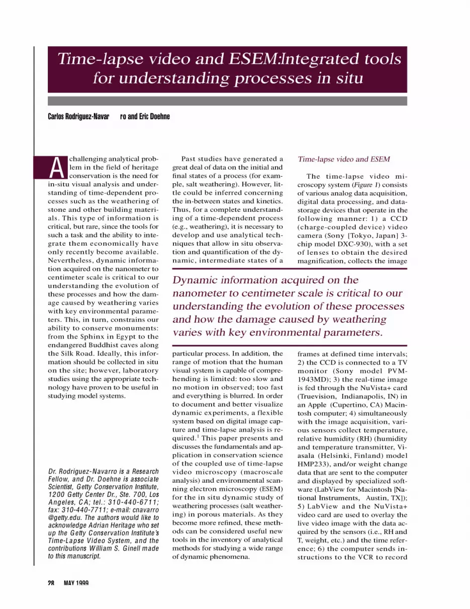
The time-lapse video mi-croscopy system (Figure 1) consistsof various analog data acquisition,digital data processing, and data-storage devices that operate in thefollowing manner: 1) a CCD(charge-coupled device) videocamera (Sony [ Tokyo, Japan] 3-chip model DXC-930), with a setof lenses to obtain the desiredmagnification, collects the image
frames at defined time interv a l s ;2) the CCD is connected to a TVmonitor (S o n y model PVM-1943MD); 3) the real-time imageis fed through the NuVista+ card(Truevision, Indianapolis, IN) inan A p p l e (Cupertino, CA) M a c i n-tosh computer; 4) simultaneouslywith the image acquisition, vari-ous sensors collect temperature,relative humidity (RH) (humidityand temperature transmitter, Vi-asala (Helsinki, Finland) modelHMP233), and/or weight changedata that are sent to the computerand displayed by specialized soft-ware (LabView for Macintosh [Na-tional Instruments, Austin, TX]);5 ) L a b View and the NuVi s t a +video card are used to overlay thelive video image with the data ac-quired by the sensors (i.e., RH andT, weight, etc.) and the time refer-ence; 6) the computer sends in-structions to the VCR to record
D r. Rodriguez-Navarro is a Researc hF e l l o w, and Dr. Doehne is associa teScientist, Getty Conservation Institute ,1200 Getty Center Dr. , Ste. 700, LosAngel es, C A; t el . : 310-440-6711;fax: 310-440-7711; e-ma il: c n a v a rro@ g e t t y.edu. The authors would like toacknowledge Adrian Heritage who setup the G e tty Conserva t ion Institute ’sT ime-L apse V ideo Syst em , and thecontributions W illiam S. G inell madeto this manuscript.
Time-lapse video and ESEM:Integrated toolsfor understanding processes in situ
Carlos Rodriguez-Navar ro and Eric Doehne
28 MAY 1999
A
Dynamic information acquired on thenanometer to centimeter scale is critical to ourunderstanding the evolution of these processesand how the damage caused by weatheringvaries with key environmental parameters.

image frames at fixed time inter-vals (i.e., every 30 sec or everyminute, for instance), while therest of the data are collected con-tinuously; and 7) the computer-processed composite image is fi-nally stored either on thecomputer hard drive or in a VCRtape (Bectacam, S o n y m o d e lU V W-1800). Details on the differ-ent steps of the process (i.e., imagecapture, data acquisition, com-puter processing, and controlledrecording) can be found else-where. With appropriate lenses,
the video camera can offer valu-able information on a millimeterto decimeter scale. Optimum reso-lution is achieved for 50× l e n s e swith a depth of field of 4 mm at aworking distance of 30 mm. Thisallows a complete recording of themacroscale evolution of a decayprocess. An environmental cham-ber (M C T , Microclimate T e c h -nology [Burlington, Canada]model 92MCG-TC) is used to pro-vide the fixed or cyclic tempera-ture and relative humidity condi-tions under which a particular
ESEM continued
30 MAY 1999
a
c
b
Figure 1 Time-lapse video setup and components: a) environmental chamber andmicro climate generator (MCC); b) data collection unit (CCD camera , TV monitor,RH and T transceiver) plus VCR; and c) data processing and storage unit (com -puter).

experiment will be carried out.H o w e v e r, the system has limitedresolution. Thus, the developmentof the ESEM over the past decadefor the study of micron-scale dy-namic phenomena complementsthe macroscale system.1
The ESEM (F i g u re 2) is essen-tially a scanning electron micro-scope with a set of differentiallypumped vacuum regions separatedby small apertures and a gaseouss e c o n d a ry electron detector(GSED) that make possible the ob-s e rvation of uncoated samples atv e ry low vacuum (typically 2.5–10torr water vapor) and at high mag-nification (2-nm resolution at 7torr and 30 kV on more recentfield emission instruments). Theuse of a cooling stage, located inthe ESEM sample chamber, per-mits the heating or cooling of thesample, thus allowing water con-densation/evaporation cycles to bep e r f o r m e d .2 Another mode of op-eration is the possibility of inject-ing a solution onto the sample sur-face using a syringe. The ESEM hasbeen used to perform a wide rangeof in situ and in tempore high-magnification studies of dynamicprocesses, such as those involvingdissolution and crystallization ofs a l t s .3 The ESEM model used inthis experiment (E l e c t r o s c a n[ Wilmington, MA] model E-3) out-puts a high-quality National Te l e-vision System Committee (NTSC)video signal in real time. The ana-log video signal is recorded using abroadcast-quality Betacam (UVW-1800) or DV (S o n y D H R - 1 0 0 0 )videotape recorder. After recordingthe experiments in real time, seg-ments of the tapes are then digi-tized or downloaded onto a hard-disk array in a compressed format(Media 100 [Datatrans, Inc., M a r l-boro, MA] or DV format). AdobePremier (Adobe Systems Inc.,Mountain Vi e w, CA) digital videoediting software is then used toedit each dynamic sequence,changing the time-lapse speed to alevel that reveals the most infor-mation, and adding titles, arrows,environmental data, and transi-tions. The final edited segment canthen be transferred back to Beta-
AMERICANLABORATORY 31

Although many researchers haveinvestigated this subject,6 the ex-act processes and dynamics of saltdamage are still poorly under-s t o o d .3 The poor understanding ofthe dynamics and factors control-ling salt decay has resulted in theuse of inadequate and sometimesharmful conservation treatments.7
Considering the importance ofthis decay process from a culturaland economical standpoint, it isessential to reveal the ultimatemechanisms leading to salt dam-age, identify the key parameterscontrolling this process, and usethis new understanding of saltweathering to advance the devel-opment of new methods for miti-gating this problem in the fields ofengineering and heritage conser-vation. Time-lapse video andESEM seem to be appropriate tech-niques for this complex task.
To evaluate the potential of thecombined use of both techniques,and to gain a better understandingof the process of salt damage to or-namental stone, crystallization ofsodium sulfate decahydrate wasstudied in a porous oolitic lime-stone (Monks Park limestone) thathas been used for centuries as aconstruction material in the U.K.F i g u re 3 shows the macroscale ex-perimental setup (for details, seeRefs. 5 and 8). The stone was par-tially immersed in a saturated solu-tion of Na2S O4. The surface of thesolution was covered with meltedp a r a f fin wax, and crystallization ofmirabilite (Na2S O4· 1 0 H2O) tookplace in the stone following capil-l a ry rise and evaporation. The pro-cess of salt crystallization anddamage to the stone (at a macro-scale level) was then recorded viatime-lapse video at the rate of oneimage frame every minute withthe simultaneous recording of thetemperature, RH, and lapsed time.Sequences of the evolution of thesalt crystallization and growthwith time are shown in F i g u re 4 f o ra ) low RH (35%) and b) medium-high RH (65%) conditions. Thec rystal habit evolution, the loca-tion of precipitates, and the differ-ential damage are clearly illus-trated in the recorded images.
ESEM continued
32 MAY 1999
Figure 2 The environmental scanning electron microscope (ESEM).
Figure 3 Macroscale salt damage test experimental setup.
cam, DV, S-VHS, or VHS videotape.Details of this process can befound elsewhere.1
Salt damage to ornamentalporous stones
Porous ornamental and struc-tural stones used in buildings and
sculptures are vulnerable to decaydue to physical–chemical pro-cesses when exposed to both out-door and indoor environments.4
Among these decay processes, saltc rystallization and growth withina porous material is an importantdamage mechanism that affects ar-chitectural and sculptural stones.5

Massive whiskers (from ~2 cm upto ~4 cm long) were observ e dgrowing at the high RH condition(~3 mm/day growth rate), andmassive loss of stone material(scales and loose grains plus saltc rystals) at low RH conditions (~2mm/day surface recession rate).
Sodium sulfate-laden lime-stone samples (1 × 2 × 2 mm insize) were submitted to condensa-tion/evaporation cycles in theESEM. After placing the sample onthe cooling stage, the temperaturewas reduced to 5 °C. Moisturecondensation was observed whenthe pressure reached 5.5 torr.C rystallization and growth ofmirabilite within the stone poresoccurred following a pressure re-duction to 4.0 torr, and damage tothe stone, due to cry s t a l l i z a t i o npressure buildup, was observed atthis stage. Upon reduction of thepressure down to 2.5 torr, dehy-dration of mirabilite took place,and thenardite (Na2S O4) wasformed. A few images of this dy-namic ESEM study of the processof deliquescence, cry s t a l l i z a t i o n(damage to the stone), and dehy-dration of mirabilite are shown inF i g u re 5. Whisker-like or rhombo-hedric, well-developed mirabilitec rystals, indicative of low super-saturation ratios,9 precipitate atmedium-high RH (5 °C and 4torr), while anhedral mirabilitecrystals precipitate at low RH con-ditions in the ESEM chamber(15 °C and 3.5 torr).10
Conclusion
The results of both time-lapsevideo and ESEM dynamic saltc rystallization studies show thatdamage due to crystallization of asoluble salt (mirabilite) in aporous stone is strongly depen-dent on the environmental condi-tions (in particular, RH). Environ-mental conditions control cry s t a lhabit and morphology, as well asc rystal growth rate, and result invariable stone surface recessionrates. High supersaturation ratios,due to more rapid evaporation atlow RH, result in anhedral cry s t a lmorphologies that cause signifi-
ESEM continued
34 MAY 1999
Figure 4 Macroscale time-lapse video snapshots of the decay of a porous lime -stone through salt crystallization at: a) high relative humidity (60%) and b) lowrelative humidity (35%) conditions. Elapsed time is shown at the bottom of eachframe.
Figure 5 Microscale, ESEM time-lapse video snapshots of the stone pores: a) be -fore and b) after water condensation (deliquescence), c) after crystallization andgrowth of mirabilite (arrows), and d) after mirabilite dehydration to formthenardite (Na2S O 4). W ell-developed, euhedral mirabilite crystals (arrows in [c])grew at 65% RH conditions (5 °C and 4 torr), and dehydrated (d) following RHreduction to 35% (2.5 torr and 5 °C). Elapsed time is shown at the bottom of eachframe.
b
dc
a

cant damage, but at the low super-saturation ratios that are achievedat high RH, whisker or well-devel-oped rhombohedr ic mirabilitec rystals grow, which results inmuch less damage.
The combined use of these twotechniques allows us to under-stand, quantify, and follow in situ,in a dynamic way, the relationshipbetween damage due to salt cry s-tallization in porous materials andthe environmental conditions,and its evolution with time. Thisinformation suggests that it mightbe possible to design preventivec o n s e rvation measures for materi-als affected by this particular dam-age mechanism, for example, bymodifying the RH in the sur-roundings.
This analytical methodology canbe useful not only to conserv a t i o nscientists, but to chemists, physi-cists, geologists, engineers, and biol-ogists, since it can allow the visualtracking and analysis, and aid in theunderstanding of many phenom-ena that are morphologically timedependent (i.e., crystallization, dis-solution, growth, corrosion, struc-tural damage, erosion, evaporation,swelling, and crack propagation).
References
1. Doehne E. ESEM development andapplication in cultural heritagec o n s e rvation. In: Gai PL, ed. In-situ microscopy in materials re-s e a rch. Boston: Kluber AcademicPublishers, 1997:45–62.
2. Doehne E, Stulik D. Applicationsof the environmental scanningelectron microscope to conserv a-tion science. Scan Microsc 1990;4(2):275–86.
3 . Doehne E. In situ dynamics ofsodium sulfate hydration and de-hydration in stone pores: o b s e rv a-tions at high magnification usingthe environmental scanning elec-tron microscope. In: Fassina V, OttH, Zezza F, eds. The conservation ofmonuments in the Mediterraneanbasin. Ve n i c e : CMU, 1994:143–5 0 .
4. Price CA. Stone conserv a t i o n : a no v e rview of current research. LosAngeles, CA: Getty Conserv a t i o nInstitute, 1996:73.
5 . Rodriguez-Navarro C, Doehne E,Ginell W, Sebastian E. Salt growth
in capillary and porous media. In:Sebastian E, Valverde I, Zezza U,eds. 3rd International Congress ofRestoration of Building and Arc h i-tectural Heritage. Granada: C E D E X -MOTMA and Universidad deGranada, 1996:509–14.
6. Zehnder K, Arnold A. Cry s t a lgrowth in salt efflorescence. JCryst Growth 1989; 97(2):513–21.
7. Rodriguez-Navarro C, Hansen E, Se-bastian E, Ginell W. The role ofclays in the decay of ancient Egyp-tian limestone sculptures. J Am InstC o n s e rvat 1997; 36(2):151–63.
8. Rodriguez-Navarro C, Doehne E.Salt weathering: influence of evap-oration rate, supersaturation andc rystallization pattern. Earth SurfProc and Landforms, accepted forpublication.
9. Sunagawa I. Character istics ofc rystal growth in nature as seenfrom the morphology of mineralc rystals. Bull Mineralogie 1981;104:81–7.
10. Messier P, Vitale T. Cracking in al-bumen photographs—an ESEMinvestigation. Microsc Res Te c h1993; 25:374–83.
AMERICANLABORATORY 35


