Embed Size (px)
Citation preview

181Development 122, 181-193 (1996)Printed in Great Britain © The Company of Biologists Limited 1996DEV3283
Two distinct phases of apoptosis in mammary gland involution: proteinase-
independent and -dependent pathways
Leif R. Lund1,*,, John Rømer2, Nicole Thomasset3, Helene Solberg2, Charles Pyke2, Mina J. Bissell3, Keld Danø2 and Zena Werb1,†
1Laboratory of Radiobiology and Environmental Health, University of California, San Francisco, California 94143-0750, USA2Finsen Laboratory, Rigshospitalet, Strandboulevarden 49, DK-2100 Copenhagen, Denmark3Life Sciences Division, Lawrence Berkeley Laboratory, Berkeley, California 94720, USA
*Present address2
†Author for correspondence; e-mail: [email protected]
Postlactational involution of the mammary gland is char-acterized by two distinct physiological events: apoptosis ofthe secretory, epithelial cells undergoing programmed celldeath, and proteolytic degradation of the mammary glandbasement membrane. We examined the spatial andtemporal patterns of apoptotic cells in relation to those ofproteinases during involution of the BALB/c mousemammary gland. Apoptosis was almost absent duringlactation but became evident at day 2 of involution, whenβ-casein gene expression was still high. Apoptotic cells werethen seen at least up to day 8 of involution, when β-caseingene expression was being extinguished. Expression ofsulfated glycoprotein-2 (SGP-2), interleukin-1β convertingenzyme (ICE) and tissue inhibitor of metalloproteinases-1was upregulated at day 2, when apoptotic cells were seeninitially. Expression of the matrix metalloproteinasesgelatinase A and stromelysin-1 and the serine proteinaseurokinase-type plasminogen activator, which was lowduring lactation, was strongly upregulated in parallelstarting at day 4 after weaning, coinciding with start of thecollapse of the lobulo-alveolar structures and the intensivetissue remodeling in involution. The major sites of mRNAsynthesis for these proteinases were fibroblast-like cells inthe periductal stroma and stromal cells surrounding thecollapsed alveoli, suggesting that the degradative phase ofinvolution is due to a specialized mesenchymal-epithelialinteraction. To elucidate the functional role of these pro-
SUMMARY
teinases during involution, at the onset of weaning wetreated mice systemically with the glucocorticoid hydro-cortisone, which is known to inhibit mammary gland invo-lution. Although the initial wave of apoptotic cells appearedin the lumina of the gland, the dramatic regression andtissue remodeling usually evident by day 5 was substan-tially inhibited by systemic treatment with hydrocortisone.mRNA and protein for gelatinase A, stromelysin-1 anduPA were weakly induced, if at all, in hydrocortisone-treated mice. Furthermore, mRNA for membrane-typematrix metalloproteinase decreased after hydrocortisonetreatment and paralleled the almost complete inhibition ofactivation of latent gelatinase A. Concomitantly, the glandfilled with an overabundance of milk. Our data support thehypothesis that there are at least two distinct phases ofinvolution: an initial phase, characterized by induction ofthe apoptosis-associated genes SGP-2 and ICE andapoptosis of fully differentiated mammary epithelial cellswithout visible degradation of the extracellular matrix, anda second phase, characterized by extracellular matrixremodeling and altered mesenchymal-epithelial interac-tions, followed by apoptosis of cells that are losing differ-entiated functions.
Key words: apoptosis, matrix metalloproteinases, urokinase,extracellular matrix, involution, mammary gland
INTRODUCTION
When sexually mature mice become pregnant, the mammarygland begins a fascinating cycle of lobulo-alveolar develop-ment and maturation that finally results in full functional differ-entiation and production of milk by the secretory epitheliumduring lactation. The extracellular matrix (ECM) maintainsthese structures in a functional conformation (Barcellos-Hoffet al., 1989; Streuli and Bissell, 1990; Talhouk et al., 1992;Walker et al. 1989). Equally remarkable are the morphologi-cal, ultrastructural and biochemical changes in the mammary
gland after cessation of lactation. The involution that followsweaning results in the quenching of milk protein geneexpression, collapse of the alveolar structures, removal of thesecretory luminal epithelial cells and endothelial and myoep-ithelial cells by programmed cell death, phagocytosis bymacrophages, proteolytic degradation of the basementmembranes, and replacement of most of the epithelial cells byadipose tissue (Helminen and Eriksson, 1968; Lascelles andLee, 1978; Marti et al., 1994; Strange et al., 1992; Talhouk etal., 1992; Walker et al., 1989; Williams, 1942). The structureof the involuting gland becomes almost identical to that of the

182 L. R. Lund and others
resting virgin gland after 10-15 days, depending on the mousestrain studied (Lascelles and Lee, 1978).
Although the molecular mechanisms underlying the gain offunction during mammary epithelium differentiation have beeninvestigated intensively in recent years, the events during theloss of function have received much less scrutiny. During thematuration of the mammary gland, interactions between themesenchyme and epithelium are important for normal devel-opment (Sakakura, 1991). The matrix metalloproteinases(MMPs) stromelysin-1, stromelysin-3 and gelatinase A and theserine proteinase urokinase-type plasminogen activator (uPA)increase during involution (Busso et al., 1989; Dickson andWarburton, 1992; Lefebvre et al., 1992; Li et al., 1994; Strangeet al., 1992; Talhouk et al., 1992) and are probably responsi-ble for the degradation of the ECM and basement membranes.The localization of stromelysin-3 in fibroblastic cells duringinvolution (Lefebvre et al., 1992) suggests an active role forthe mesenchyme, although no known function of stromelysin-3 has been described; similarly, stromelysin-1 and gelatinaseA have been localized to the stromal or myoepithelial cells byimmunohistochemical analysis (Dickson and Warburton,1992; Talhouk et al., 1992; Li et al., 1994). In transgenic miceoverexpressing stromelysin-1 during pregnancy or inmammary epithelial cells in culture, the degradation of ECMinitiates apoptosis in part by inducing expression of inter-leukin-1β converting enzyme (ICE) (Boudreau et al., 1995).The increase in ICE and stromelysin-1 during involution isbelieved to be related to the apoptotic process. The aim of thepresent study was to explore the relationship of apoptosis ofmammary epithelium to tissue remodeling during involutionand to investigate mesenchymal-epithelial interactions duringinvolution. We have examined the temporal and spatialexpression and function of stromelysin-1, gelatinase A anduPA in relation to the other physiological events that take placeduring involution of the mammary gland.
MATERIALS AND METHODS
Materials T7, T3 and SP6 RNA polymerases, RNase inhibitor, restrictionendonucleases, a random DNA labeling kit and monoclonal anti-bodies specific for macrophages (Mac-1 and Mac-2) were obtainedfrom Boehringer Mannheim, Germany. A monoclonal antibodydirected against mouse smooth muscle α-actin (clone no. 1A4),amiloride, β-estradiol 17-acetate, progesterone, testosterone acetateand hydrocortisone 21-acetate were obtained from Sigma ChemicalCompany (St Louis, MO). The Apoptag kit for detection of apoptoticcells was obtained from Oncor Inc. (Gaithersburg, MD) and Super-frost+ slides were obtained from Fisher Scientific (Pittsburgh, PA).All other materials used were described earlier (Rømer et al., 1991;Kristensen et al., 1991a,b; Pyke et al. 1994; Talhouk et al., 1992;Sympson et al., 1994).
Animal and tissue treatment procedures Tissue from mammary glands was isolated from BALB/c mice in theirfirst pregnancy. The mice were obtained either from the FibigerInstitute (Copenhagen, Denmark) or from Charles River (Wilmington,MA). The tissue was collected from mice at various stages of lactationor during involution of the mammary gland after cessation of lactation.The number of pups was adjusted to 7 for each mouse; they wereallowed to lactate for 7 days in order to reach a state of full lactation,before the offspring were removed. The intervals for involution are
indicated for each experiment. Tissue samples from at least 3 micewere collected and analyzed separately for each stage in the experi-ment. The mice were anesthetized by intraperitoneal injection ofavertin and perfused intracardially with 20 ml ice-cold phosphate-buffered saline (PBS). The inguinal and abdominal mammary glandswere removed for RNA isolation or protein extraction as describedbelow. The mice used for in situ hybridization and immunohisto-chemistry were treated similarly, except that perfusion with cold PBSwas followed by intracardial perfusion-fixation with 4% (w/v)paraformaldehyde in PBS. The mammary glands were removed andpostfixed for 16 hours in 4% paraformaldehyde in PBS. The tissue wasthen rinsed in PBS, dehydrated and embedded in paraffin. For all themorphological analyses, only the left and right inguinal (number 4)mammary glands were used. For histological analyses, sections fromall tissue samples were routinely stained with hematoxylin and eosin,Gomori’s one-step trichrome stain or toluidine blue. The percentageof collapsed alveoli versus intact secretory alveoli at each time pointpostweaning was determined by analyzing 200 alveoli and assessingtheir physiological state in sections from two different animals. Insome cases, either at the start of involution or at various times afterweaning, the mice were treated daily with subcutaneous injection of0.5 mg of hydrocortisone 21-acetate in PBS per g body weight. Allexperiments were performed under protocols approved by the DanishAnimal Research Committee, the Animal Welfare and ResearchCommittee (Lawrence Berkeley Laboratory) or the Committee onAnimal Research (University of California, San Francisco).
Preparation of RNA probes The following fragments of mouse uPA (Belin et al., 1985), mousegelatinase A (Reponen et al., 1992), mouse gelatinase B (Reponen etal., 1994) and mouse stromelysin-1 (Hammani et al., 1992) weresubcloned: for uPA, pMUPA07 fragment (608-1642) in pGEM5z, orpMUPA09 fragment (37-428) in pBluescript KS(+); for gelatinase A,(604-1165) fragment in pSP64 and pSP65 or the HaeIII fragment(1924-2259) in pGEM-3; for gelatinase B, (805-1099) fragment inpSP64 and pSP65; for stromelysin-1, pmTRM11 fragment (3115-4051) and pmTRM12 fragment (2205-2918) in pBluescript KS(+).Pure plasmid preparations were made by banding through a CsClgradient and, before transcription, the plasmids were linearized byusing the following restriction endonucleases: pMUPA07, PstI orEcoRI; pMUPA09, BamHI; pSP64, EcoRI; pSP65, HindIII;pmTRM11, SacI or KpnI; pmTRM12, SacI or XhoI. The in vitro RNAtranscriptions were performed as described previously (Rømer et al.,1991) and the labeled probes were stored at −20°C until use.
In situ hybridization analysis of mRNA In situ hybridization was performed essentially as described by Kris-tensen et al. (1991a). Briefly, 5 µm paraffin sections on Superfrost+
slides were treated as described by Pyke et al. (1994). After hybrid-ization overnight at 47°C with radiolabeled RNA probes, the sectionswere washed, treated with RNase A (20 µg/ml) and RNase T1 (20U/ml), dehydrated and air-dried. Autoradiographic emulsion wasapplied, and sections were developed after 10 days of exposure,lightly counterstained with either hematoxylin and eosin or Gomori’strichrome stain and mounted with Permount. The presence ofapoptotic cells was detected by means of the Apoptag kit (Oncor).
Immunohistochemical analysis The sections were deparaffinized in xylene, rehydrated with ethanol,rinsed in water and incubated for 10 minutes at 37°C with 0.1% (w/v)trypsin in 0.05 M Tris/HCl and 0.1% (w/v) CaCl2, pH 7.4.Macrophages were stained with antibodies against Mac-1 and Mac-2antigens, as described previously (Feiken et al., 1995), by using thealkaline phosphatase anti-alkaline phosphatase method (Cordell et al.,1984). Smooth muscle α-actin was stained with a biotin-conjugatedmonoclonal primary antibody, followed by streptavidin-AP complex(D396; Dako, Copenhagen, Denmark). For all the immunohisto-

183Two pathways of mammary involution
chemical analyses, control experiments were performed in whichprimary or secondary antibodies were omitted.
RNA isolation and analysis Total RNA was isolated from the inguinal and abdominal mammaryglands from at least three mice at each stage of involution or treatment,by the acid guanidine-thiocyanate-phenol-chloroform method (Chom-czynski and Sacchi, 1987). The concentration oftotal RNA in the samples was determined spec-trophotometrically, and either RNA was useddirectly for RNA blotting analysis (Talhouk et al.,1992) or poly(A)+ RNA was isolated (Rømer et al.,1991) and used for reverse transcription-poly-merase chain reaction (RT-PCR) analysis (Ploug etal., 1992). Total RNA (20 µg) was denatured andseparated in 1.5% agarose/formaldehyde gels. TheRNA blots were hybridized with the antisenseRNA probes used for the in situ hybridizations forgelatinase A, stromelysin-1 or uPA, or with thefollowing random-primed labeled cDNA probes:mouse ICE (Boudreau et al. 1995), mousestromelysin-2 (Gack et al., 1994), β-casein (pBCL-1), mouse tissue inhibitor of metalloproteinases(TIMP)-1 (Gewert et al., 1987) and TIMP-2(Alexander and Werb, 1992), mouse sulfated gly-coprotein-2 (SGP-2) (Strange et al., 1992), mouseplasminogen activator inhibitor (PAI)-1 (Prender-gast and Cole, 1989) and mouse PAI-2 (Belin etal., 1989), or a cDNA for human membrane-type(MT)-MMP (Sato et al., 1994). For the latter, lessstringent conditions were used in the washingprocedure: 1 hour with 2× SSC (1× SSC=0.15 Msodium chloride, 0.015 M sodium citrate), 0.1%sodium dodecyl sulfate (SDS) at 42°C, 1 hour with2× SSC, 0.1% SDS at 68°C, and finally for 1 hourwith 0.5× SSC, 0.1% SDS at 68°C. The blots wereexposed to Kodak X-AR 5 film for various times,as indicated in the figure legends. As a control forequal loading, the blots were stripped and rehy-bridized with a cDNA probe for 28S RNA. Forquantification of the signals, the RNA blots wereanalyzed in a Molecular Dynamics PhosphorIm-ager SF. Detection of uPA receptor (uPAR), PAI-1 and PAI-2 mRNA by means of RT-PCR
Fig. 1. Hematoxylin-eosin and trichrome stainingof lactating and involuting mouse mammarygland. (A,B) Mammary gland after 7 days oflactation. Open arrow indicates fibroblasts; solidarrow indicates myoepithelial cells.(C,D) Mammary gland after 2 days of involution.(E) Mammary gland after 3 days of involution.Arrow indicates apoptotic cells in the lumen ofan alveolus shown in higher magnification in F.(G,H) Mammary gland after 4 days of involution.Straight arrow indicates a group of collapsedalveoli; curved arrow indicates adipocytes.(I,J) Mammary gland after 8 days of involution.(K,L) Staining for apoptotic cells after 3 days ofinvolution. (K) control without and (L) withterminal deoxynucleotidyl transferase added.A,C,D,E,G,H,I, bar, 100 µm; B,F,J,K,L, bar, 25µm. A,B,C,E,G,I,J were stained withhematoxylin and eosin; K,L were counterstainedwith hematoxylin; D,H were stained withtrichrome.
generally followed the scheme described previously (Ploug et al.,1992).
Mammary gland protein extract The extracts used for gelatinolytic assays were prepared as describedpreviously (Talhouk et al., 1991) from the inguinal and abdominalmammary glands from at least three mice for each stage. The extracts

184 L. R. Lund and others
lated from mouse mammary gland involuting for various times.E, SGP-2, TIMP-1 and TIMP-2 in extracts of lactating or involutingithin each stage is indicated below in arabic numerals. 20 µg of total. The blots were exposed for the following times: ICE, 8 days; SGP-2, 22, 8 days. (B) Quantification of the mRNA signals obtained for theIMP-1 and the apoptosis marker SGP-2 in A. Probed blots were. The values obtained at day 7 lactation (L7), at the start of theal to 1, and the subsequent time points are indicated as -fold induction or against the value obtained for the 28S RNA hybridization, in order toe in loading and transfer of the various RNA samples. (C) RNA blot, stromelysin-1 (SL-1), MT-MMP, β-casein mRNA and 28S RNA. The
llowing times: uPA, 3 days; gelatinase A, 2 days; stromelysin-1, 1 day;, 2 hours; 28S, 12 hours. (D) Quantification of the mRNA signalsnd β-casein in C, as described above, except that the value obtained for. Numbers on the left indicate -fold induction of the proteinases, and onsein expression.
for uPA fibrin zymography and enzyme-linked immunosorbent assay(ELISA) were isolated as described (Andreasen et al., 1990a). Theprotein concentrations of the extracts were determined by the Folin-Ciocalteu phenol reagent, with bovine serum albumin as a standard.
Zymography The gelatinolytic assays were carried out as described in detail pre-viously (Talhouk et al., 1991), and plasminogen activator activity wasdetected in casein gels with plasminogen added (Talhouk et al., 1991).As control for specificity, plasminogen was omitted from the substrategel, and mouse bladder urine was used as a positive control for uPAactivity. Alternatively, plasminogen activator activity in the SDS-polyacrylamide gels was detected by fibrin zymography as describedby Andreasen et al. (1990a).
Enzyme-linked immunosorbentassay for uPA A Nunc 96-well immunoplate(Maxisorp) was coated overnight at4°C with 2 µg/ml rabbit anti-mouseuPA IgG in 0.1 M Na2CO3, pH 9.8.Remaining protein binding sites weresaturated with 1% (w/v) bovine serumalbumin in PBS for 3 minutes at 37°C.Extract samples with unknown con-centrations of mouse uPA wereanalyzed either alone or together witha mouse uPA standard (concentrationdetermined by amino acid analysis)diluted in 1% (w/v) bovine serumalbumin in PBS, 0.1% (v/v) Tween 20(buffer A) and incubated for 1 hour atambient temperature with shaking.The bound uPA was measured byadding biotinylated rabbit anti-mouseuPA IgG (2 µg/ml in buffer A) for 1hour at ambient temperature withshaking, followed by peroxidase-con-jugated avidin (P347; Dako) diluted inbuffer A and incubated for 1 hour atambient temperature with shaking.The ELISA was developed asdescribed previously (Andreasen etal., 1990a).
RESULTS
Relationship ofmorphological changes toexpression of mRNA andprotein for ECM remodelingproteinases in the mammarygland during involutionAt the time of weaning, thelactating mammary gland ofBALB/c mice consisted mainly ofclusters of alveoli lined by acuboidal epithelium secreting milkproteins, carbohydrates and lipidsinto the lumina of the alveoli (Fig.1A,B). There was a well-branchedductal system, with the largerducts surrounded by a basementmembrane and thicker layers of
Fig. 2. Analysis of mRNA iso(A) RNA blot analysis for ICmammary glands. The day wRNA was loaded in each lanedays; TIMP-1, 2 days; TIMP-specific proteinase inhibitor Tscanned in a PhosphorImagerinvolution, have been set equreduction after normalizationcorrect for the slight differencanalysis for uPA, gelatinase Ablots were exposed for the foMT-MMP, 10 days; β-caseinobtained for the proteinases aβ-casein at L7 was set to 100the right the reduction of β-ca
connective tissues. The lobules were enclosed by fibrousseptae. In addition, there was an extensive vascular andlymphatic network. The well-organized, secretory lobulo-alveolar structures characteristic of lactating animals remainedintact for the first 3 days after weaning (Fig. 1C-F). Up to day3 postweaning, more than 95% of all alveoli remained intact,followed by collapse of 60% of the alveoli at day 4, 70% atday 5, 85% at day 7, and ≥95% at day 10. By trichromestaining, we found connective tissue collagens around majorducts and vessels, whereas only very faint staining or none wasobserved around the intact lobules and the interstitial septae(Fig. 1D). Severe distension was observed in all alveoli at days

185Two pathways of mammary involution
Fig. 3. The uPA content of involuting mammary gland as determinedby ELISA and fibrin zymography. Extracts were prepared from theinguinal and abdominal glands from mice that had been lactating for7 days, followed by weaning for the indicated number of days. Theconcentration of uPA was determined by ELISA, and the proteinconcentration with the Folin-Ciocalteu reagent, as described inMaterials and Methods. (Inset) Fibrin overlay zymography analysisof uPA and tPA activity in extracts of mammary glands. Tissueextracts (100 µg protein) or 5 µl rat bladder urine were subjected toSDS-polyacrylamide gel electrophoresis and zymography asdescribed in Materials and Methods.
1 and 2 after weaning, owing to the continuous synthesis andsecretion of milk for up to 2 days after weaning; partial dis-ruption of some of the alveolar basal membranes and flow ofliquid into the interstitium was seen after 2 or 3 days of invo-lution. Infiltration of adipocytes began at day 2 after weaning.
The number of epithelial cells undergoing apoptosis andshedding into the alveolar lumina increased dramaticallybetween 2 and 3 days after weaning, as detected both by con-ventional morphological criteria (Fig. 1C-F) and by specificstaining for apoptopic cells (Fig. 1K,L). This occurred withoutvisible changes in the relationship between mesenchymal andepithelial compartments. There followed a distinct secondphase of involution involving very intensive tissue remodel-ing that started at 4 days after weaning, when the lobulo-alveolar structure collapsed (Fig. 1G-J). At day 4, theparenchyma consisted mainly of ducts, vessels and clusters ofepithelial cords, some with small lumina, all surrounded bycondensed connective tissue and increasing numbers ofadipocytes (Fig. 1H). Further degradation of the secretorystructures and a dramatic increase in the number of adipocytescontinued up to day 8 (Fig. 1I,J). A few, isolated secretoryalveoli and ducts containing secretory material surrounded byadipocytes and interspersed by thickened stromal elementsremained present for up to 16 days after weaning. Themammary gland was fully regressed after 21 days and reor-ganized with ducts, ready for another cycle of pregnancy andlactation (data not shown).
Molecular analysis of gene expression also revealed twodistinct phases of involution. Differentiated function, asmeasured by β-casein mRNA expression, decreased from thevery high levels during lactation to one fourth by day 3 afterweaning, a level similar to that at the end of pregnancy, and thento even lower levels over the next 2 days (Fig. 2C,D). WhereasICE, SGP-2 and TIMP-1 mRNA increased during the first 2 daysafter lactation and then decreased (Fig. 2A,B), stromelysin-1 anduPA mRNAs were low in lactating mammary gland and duringthe first 3 days after weaning, then increased 30-fold on day 4,followed by a gradual decrease up to day 10 (Fig. 2C,D). Gelati-nase A mRNA was expressed at much lower levels, althoughwith an induction pattern similar to that of stromelysin-1 anduPA (Fig. 2C,D). In contrast, the single 3.5 kb MT-MMP mRNAband, which we detected in mammary gland after 7 days oflactation and throughout involution, remained unchanged (Fig.2C). TIMP-2 mRNA was not regulated and was present at lowlevels throughout the 16 days after weaning (Fig. 2A). No signalfor stromelysin-2 was detected (data not shown). As measuredby RT-PCR analysis on poly(A)+ RNA samples, uPAR mRNAincreased after the start of involution, whereas little PAI-1 andPAI-2 mRNA was detected during lactation and involution (datanot shown). These data suggest that the initial involution eventsat 1 to 3 days after weaning have a pattern of gene expressionthat is distinct from that of the major remodeling phase fromdays 4-10.
Protein extracts prepared from the same mice used for RNAisolation were analyzed by zymography. Although no activegelatinase A was present in extracts isolated from lactatingmice, active gelatinase A began to be observed at day 2 of invo-lution (data not shown). The amount of both latent and activeforms of gelatinase A increased between days 2 and 3 afterweaning and increased further with involution time. Thehighest activity was present at day 10 after weaning, consis-
tent with the RNA data. A sandwich ELISA was used to obtaina quantitative measurement of the amount of uPA in themammary gland. As shown in Fig. 3, the concentrations ofuPA, normalized against the total protein concentration in theextracts, increased during the first 7 days after weaning, thendeclined. Analysis of the same extracts by fibrin overlayzymography showed a similar expression pattern for uPA, withthe highest activity in extracts from day 7 involuting mammarygland (Fig. 3, inset). A single band of uPA activity, withmolecular mobility corresponding to 48×103 Mr, and the68×103 Mr band corresponding to tissue-type plasminogenactivator (tPA) were detected (Talhouk et al., 1992).
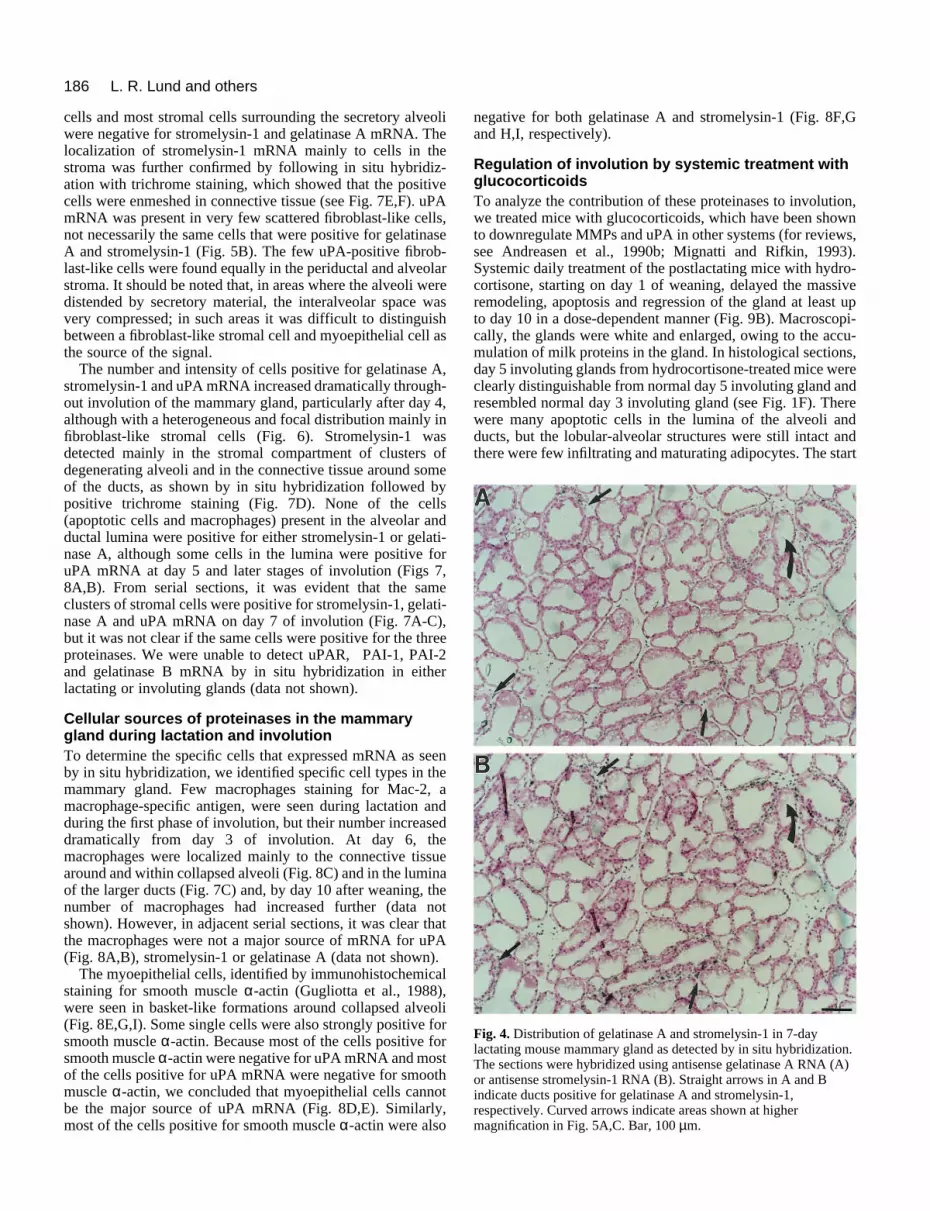
Expression of stromelysin-1, gelatinase A and uPAmRNA in the mesenchymal compartment duringlactation and involutionBecause the initial apoptotic events and the rapid remodelingphases of involution appeared to be distinct, we next used insitu hybridization to determine which cell types expressedstromelysin-1, gelatinase A and uPA mRNA. First, weexamined the distribution of proteinase mRNA duringlactation. Using serial sections of 7-day lactating glands, wedetected gelatinase A and stromelysin-1 mRNA in the samefibroblast-like periductal stromal cells that surrounded mostbut not all intact ducts (Fig. 4A,B), as well as around someblood vessels (data not shown). Higher magnification showedno signal was detected in epithelial or myoepithelial cells (Fig.5A-D), and no specific signal above background was seenwhen sense probes for stromelysin-1 (Fig. 5E), gelatinase A oruPA (data not shown) were used. Notably, the myoepithelial
186 L. R. Lund and others
Fig. 4. Distribution of gelatinase A and stromelysin-1 in 7-daylactating mouse mammary gland as detected by in situ hybridization.The sections were hybridized using antisense gelatinase A RNA (A)or antisense stromelysin-1 RNA (B). Straight arrows in A and Bindicate ducts positive for gelatinase A and stromelysin-1,respectively. Curved arrows indicate areas shown at highermagnification in Fig. 5A,C. Bar, 100 µm.
cells and most stromal cells surrounding the secretory alveoliwere negative for stromelysin-1 and gelatinase A mRNA. Thelocalization of stromelysin-1 mRNA mainly to cells in thestroma was further confirmed by following in situ hybridiz-ation with trichrome staining, which showed that the positivecells were enmeshed in connective tissue (see Fig. 7E,F). uPAmRNA was present in very few scattered fibroblast-like cells,not necessarily the same cells that were positive for gelatinaseA and stromelysin-1 (Fig. 5B). The few uPA-positive fibrob-last-like cells were found equally in the periductal and alveolarstroma. It should be noted that, in areas where the alveoli weredistended by secretory material, the interalveolar space wasvery compressed; in such areas it was difficult to distinguishbetween a fibroblast-like stromal cell and myoepithelial cell asthe source of the signal.
The number and intensity of cells positive for gelatinase A,stromelysin-1 and uPA mRNA increased dramatically through-out involution of the mammary gland, particularly after day 4,although with a heterogeneous and focal distribution mainly infibroblast-like stromal cells (Fig. 6). Stromelysin-1 wasdetected mainly in the stromal compartment of clusters ofdegenerating alveoli and in the connective tissue around someof the ducts, as shown by in situ hybridization followed bypositive trichrome staining (Fig. 7D). None of the cells(apoptotic cells and macrophages) present in the alveolar andductal lumina were positive for either stromelysin-1 or gelati-nase A, although some cells in the lumina were positive foruPA mRNA at day 5 and later stages of involution (Figs 7,8A,B). From serial sections, it was evident that the sameclusters of stromal cells were positive for stromelysin-1, gelati-nase A and uPA mRNA on day 7 of involution (Fig. 7A-C),but it was not clear if the same cells were positive for the threeproteinases. We were unable to detect uPAR, PAI-1, PAI-2and gelatinase B mRNA by in situ hybridization in eitherlactating or involuting glands (data not shown).
Cellular sources of proteinases in the mammarygland during lactation and involutionTo determine the specific cells that expressed mRNA as seenby in situ hybridization, we identified specific cell types in themammary gland. Few macrophages staining for Mac-2, amacrophage-specific antigen, were seen during lactation andduring the first phase of involution, but their number increaseddramatically from day 3 of involution. At day 6, themacrophages were localized mainly to the connective tissuearound and within collapsed alveoli (Fig. 8C) and in the luminaof the larger ducts (Fig. 7C) and, by day 10 after weaning, thenumber of macrophages had increased further (data notshown). However, in adjacent serial sections, it was clear thatthe macrophages were not a major source of mRNA for uPA(Fig. 8A,B), stromelysin-1 or gelatinase A (data not shown).
The myoepithelial cells, identified by immunohistochemicalstaining for smooth muscle α-actin (Gugliotta et al., 1988),were seen in basket-like formations around collapsed alveoli(Fig. 8E,G,I). Some single cells were also strongly positive forsmooth muscle α-actin. Because most of the cells positive forsmooth muscle α-actin were negative for uPA mRNA and mostof the cells positive for uPA mRNA were negative for smoothmuscle α-actin, we concluded that myoepithelial cells cannotbe the major source of uPA mRNA (Fig. 8D,E). Similarly,most of the cells positive for smooth muscle α-actin were also
negative for both gelatinase A and stromelysin-1 (Fig. 8F,Gand H,I, respectively).
Regulation of involution by systemic treatment withglucocorticoids To analyze the contribution of these proteinases to involution,we treated mice with glucocorticoids, which have been shownto downregulate MMPs and uPA in other systems (for reviews,see Andreasen et al., 1990b; Mignatti and Rifkin, 1993).Systemic daily treatment of the postlactating mice with hydro-cortisone, starting on day 1 of weaning, delayed the massiveremodeling, apoptosis and regression of the gland at least upto day 10 in a dose-dependent manner (Fig. 9B). Macroscopi-cally, the glands were white and enlarged, owing to the accu-mulation of milk proteins in the gland. In histological sections,day 5 involuting glands from hydrocortisone-treated mice wereclearly distinguishable from normal day 5 involuting gland andresembled normal day 3 involuting gland (see Fig. 1F). Therewere many apoptotic cells in the lumina of the alveoli andducts, but the lobular-alveolar structures were still intact andthere were few infiltrating and maturating adipocytes. The start

187Two pathways of mammary involution
gelatinase A, stromelysin-1 and uPA in 7-day lactating mouseected by in situ hybridization. Higher magnification of the in situ Fig. 4. The sections were hybridized by using antisense gelatinase A uPA (B,B*), antisense stromelysin-1 RNA (C,C*), or sense
) as described in Materials and Methods. Tissue sections were viewedr dark-field (A*,B*,C*,E) microscopy. Arrow in C indicates areafication in D. Positive control experiments were performed byrent antisense probes, covering nonoverlapping parts of each of thebed in Materials and Methods. These probes were adjusted to the sameplied to adjacent sections of the mammary gland. In all cases, the twol hybridization patterns for each specific mRNA (data not shown). As a RNA probes transcribed from each of the three cDNAs were applied to specimens, and in these sections no signal was obtained aboves treated with RNase A before hybridization, no signal was detected
of hydrocortisone treatment could be delayed up to 3 days afterweaning and still delay the regression of the gland consider-ably, as judged by morphological and biochemical criteria(data not shown). The hydrocortisone-treated mice, ascompared with the saline-treated normal involuting mice,showed little or no induction of uPA,gelatinase A and stromelysin-1 mRNAsat day 5 after weaning. Moreover, in thehydrocortisone-treated gland, β-caseindecreased only to approximately 35% ofits lactating level, similar to that of anormal day 2 involuting gland, whereasin the saline-treated involuting mice itdecreased to 5% (Figs 2C,D, 9D). Inter-estingly, the signal for MT-MMP mRNAwas totally abolished by the hydrocorti-sone treatment (Fig. 9D). This contrastswith untreated and saline-treated mice,which expressed this gene duringlactation, when no tissue remodelingtakes place (Fig. 2C). By zymography, noactive gelatinase A was detected inextracts prepared from the hydrocorti-sone-treated mice, whereas latent gelati-nase A was still detectable, but at lowerlevels than in the saline-treated controls.Identical results were obtained in all micetested (n=5). Similarly, uPA and, to alesser extent, tPA activity was reduced(Fig. 9C). Thus, hydrocortisone main-tained an early involuting phenotypewhile preventing the main remodelingphase. Hydrocortisone had no visibleeffect on the morphology of themammary gland or gelatinolytic activityof age-adjusted virgin mice for up to 5days (data not shown). No effect wasdetected after treatment of involutingmice with β-estradiol, progesterone ortestosterone for 5 days (data not shown).
DISCUSSION
Involution of mammary gland ischaracterized by two distinctphasesBy a combination of biochemical and his-tological analysis and pharmacologicalinterventions, we have demonstrated thatthe postlactational involution of themammary gland can be divided into twodistinct phases, each characterized byspecific gene expression: an early phasecharacterized by engorgement of the glandwith milk and initial apoptosis of epithe-lial cells, which accumulate in the lumina,and a second phase characterized bybiosynthesis of proteinases and intensivetissue remodeling. During the first phase,mammary gland-specific differentiated
Fig. 5. Distribution of mammary gland as dethybridization shown inRNA (A,A*), antisensestromelysin-1 RNA (Eby bright-field (A-D) oshown at higher magniapplication of two diffethree cDNAs, as descrispecific activity and approbes showed identicanegative control, senseadjacent sections of allbackground. In section
functions, characterized by β-casein with the antisense probe
mRNA expression, remained relatively high, declining to aboutone fourth of the lactating level at day 3 after weaning, compa-rable to the levels in late pregnancy. At this time in the involu-tion process, very low levels of MMPs, uPA and tPA werepresent in the gland, whereas TIMP-1 was upregulated, shifting
s. A,A*,B,B*,C,C*,E, bar, 25 µm; D, bar, 5 µm.

188 L. R. Lund and others
dization for gelatinase A, stromelysin-1 and uPA mRNA in mousering involution. In situ hybridization analysis was performed on tissue-C, A*-C*), day 5 (D-F, D*-F*) or day 7 (G-I, G*-I*) after weaning.of (A,D,G) gelatinase A expression; (B,E,H) stromelysin-1 expression;sion. Bar, 100 µm. Arrows in G-I indicate areas shown at higherg. 7A-C. (A-I) Bright-field microscopy; (A*-I*) dark-field microscopy.
the proteolytic balance in favor of inhibition. During this initialphase of involution there was a strong, transient induction ofSGP-2 and ICE mRNA, two genes participating in the apoptoticprocess (Fig. 2A,B); a third gene, p53, shows a more prolongedinduction (Boudreau et al., 1995; Guenette et al., 1994; Strangeet al., 1992). The second phase began at day 4 of involution inBALB/c mice and was characterized by upregulation of mRNAand activity for proteolytic enzymes, including gelatinase A,stromelysin-1 and uPA, and downregulation of the inhibitorTIMP-1, resulting in active tissue remodeling, including destruc-tion of basement membranes and alveolarstructures and irreversible loss of the differen-tiated function of the mammary gland(Martinez-Hernandez et al., 1976, Warburtonet al., 1982).
Regulation of proteolytic activityduring involutionBoth MMPs and uPA and their specificinhibitors have been implicated in a numberof degradative processes, such as woundhealing, ovulation, trophoblast invasion,involution of the prostate and cancer invasion(for reviews see Alexander and Werb, 1991;Andreasen et al., 1990b; Birkedal-Hansen etal., 1993; Danø et al., 1985, 1994; Matrisian,1990; Vassalli et al., 1991; Woessner, 1991).The results reported here also stronglysupport a physiological role of the proteinasesin active tissue remodeling. The overall pro-teolytic activity is regulated at different, inde-pendent levels. Both MMPs and uPA aresecreted as latent proenzymes with very littleor no enzymatic activity. Activation of theproenzymes is an important regulatory step inthe control of proteolytic activity. Althoughthe mechanism by which the latent proen-zymes are activated in vivo is unknown, invitro experiments have indicated that plasminand other proteolytic enzymes are able toactivate prourokinase and procollagenase(Petersen et al., 1988; Werb et al., 1977).Recently, MT-MMP has been shown toactivate progelatinase A in vitro (Sato et al.,1994). Furthermore, the enzymatic activity ofMMPs and uPA is controlled by specificinhibitors. Members of the MMP family areinhibited by TIMP-1, TIMP-2 or TIMP-3,whereas uPA is inhibited by PAI-1 or PAI-2.Expression of gelatinase A, stromelysin-1 anduPA and of their specific inhibitors isregulated by a number of growth factors andcytokines in vitro (for reviews, see Andreasenet al., 1990b; Laiho and Keski-Oja, 1989;Mignatti and Rifkin, 1993), but which factorsare responsible for the concerted increase inmRNA and protein levels during involutionof the mammary gland are unknown. It hasbeen shown that protein kinase A and c-Fos/c-JunD are induced during involution ofthe mammary gland (Marti et al., 1994), sug-
Fig. 6. In situ hybrimammary gland duisolated at day 3 (APhotomicrographs (C,F,I) uPA expresmagnification in Fi
gesting that these transduction signal pathways are involved inthe regulation of specific gene expression. During developmentof the mammary gland, a number of growth factors are impli-cated in the mesenchymal-epithelial interactions, includingmembers of the transforming growth factor-β and fibroblastgrowth factor families (Coleman-Krnacik and Rosen, 1994;Jhappan et al., 1993; Maier et al., 1991; Robinson et al., 1991;for a review see Topper and Freeman, 1980). Similar studiesare required to elucidate the molecular mechanisms involvedduring involution of the mammary gland.

189Two pathways of mammary involution
gelatinase A, stromelysin-1 and uPA mRNA in the mammary glandn. A-C are higher magnifications of the in situ hybridizations shown in
r gelatinase A (A), stromelysin-1 (B) and uPA (C). (D) In situelysin-1 mRNA in transversal sections of day 7 involuting mammaryrome staining. (E,F) In situ hybridizations for stromelysin-1 mRNA in day 7 lactating mammary gland, followed by trichrome staining infield (F) microscopy. A-D, bar, 10 µm; E,F, bar, 25 µm.
Interestingly, we were able to detect uPA only in its highmolecular weight form during mammary gland involution.Most of the uPA detected in the involuting ventral prostateoccurs in the low molecular weight form (Andreasen et al.,1990a), which, although enzymatically active, cannot bind touPAR (Stoppelli et al., 1985). A recent study has shown thatmatrilysin, a metalloproteinase of the stromelysin subclass,which is able to cleave uPA in vitro into low molecular weightuPA and its amino terminal fragment (Marcotte et al., 1992),is also induced during involution of the ventral prostate afterhormonal ablation (Bowden et al., 1995). The level of uPA inthe involuting mammary gland in the present study wasapproximately one tenth of that in the involuting prostate(Andreasen et al., 1990a). These data suggest that uPA mayhave different physiological roles in these two involuting organsystems.
Mesenchymal-epithelial interaction in involutionWe have identified the cells responsible for the synthesis ofgelatinase A, stromelysin-1 and uPA. In contrast to earlierstudies (Busso et al., 1989; Dickson andWarburton, 1992; Li et al., 1994;Ossowski et al., 1979; Strange et al.,l992), we found that, for gelatinase A,stromelysin-1 and uPA, both mRNA andprotein were expressed during lactation,although at a low level, as detected byzymograms and RNA blots. By in situhybridization, we found that the mRNAfor gelatinase A, stromelysin-1 and uPAin lactating mammary glands was syn-thesized by some but not all fibroblast-like periductal stromal cells. Similarly,during involution, the signals for thethree proteinases were found in bothperiductal and perialveolar fibroblast-like stromal cells, although with a veryfocal and heterogeneous distribution.The data described here are in apparentdisagreement with findings described byDickson and Warburton (1992) and Li etal. (1994). By immunohistochemicalanalysis, these investigators found thatmyoepithelial cells were the source ofpositive immunoreactivity for bothstromelysin-1 and gelatinase A duringinvolution of the rat and mousemammary gland, respectively. We alsoobserved that immunoreactive gelati-nase A and stromelysin-1 are enriched inmyoepithelial cells (data not shown).Similarly, uPA immunoreactivity waslocalized to the epithelial cells (Larssonet al., 1984). A possible explanation forthe apparent discrepancy between theimmunocytochemistry and in situhybridization could be that, althoughfibroblasts synthesize and secrete theproteinases, after secretion theseenzymes are bound to other cell types orto ECM components in the mammary
Fig. 7. Localization of after 7 days of involutioFig. 6G-I for mRNA fohybridizations for stromgland, followed by trichlongitudinal sections ofbright-field (E) or dark-
gland. It is interesting to note that similar discrepancies existfor mRNA and protein localization for MMPs in tumor tissue(Garbisa et al., 1990; Pyke et al., 1993; Stetler-Stevenson etal., 1993). Our data directly confirm the indirect observationby Ossowski et al. (1979) that macrophages cannot be themajor source of uPA production. In agreement with earlierobservations (Mayberry, 1964), we also demonstrated that theinflux of macrophages into the mammary gland is a late eventduring involution. Furthermore, we have shown thatmacrophages are not the source of either stromelysin-1 orgelatinase A, supporting the role of macrophages as scav-engers.
A similar, but not identical, temporal and spatial expressionpattern has been described for stromelysin-3 during develop-ment, differentiation and involution of the mammary gland.Although no mRNA for stromelysin-3 was detected duringdevelopment and pregnancy or lactation, during involution itwas detected at day 3, with maximal expression by day 6 infibroblasts around the disorganized clusters of epithelial cells(Lefebvre et al., 1992). As we observed for stromelysin-1 and

190 L. R. Lund and others
Fig. 8. Immunohistochemicalidentification of cell types ininvoluting mammary gland.Immunohistochemicalstaining for macrophage-specific antigens and smoothmuscle α-actin and in situhybridization for uPA,stromelysin-1 and gelatinaseA were performed asdescribed in Materials andMethods. (A-C) In situhybridization for uPAmRNA andimmunohistochemicalstaining for Mac-2 antigen onday 6 of involution. Adjacentserial sections of involutingmammary gland werehybridized with an antisenseuPA RNA probe (A,B) orwere stained with Mac-2antibody (C). Curved arrowsin A,B show areas positivefor uPA mRNA, but with nomacrophage-positive cells.Straight arrows in (C) pointto macrophages present, butno uPA mRNA. (D-I) In situhybridization for uPA,gelatinase A andstromelysin-1 andimmunohistochemicallystaining for smooth muscleα-actin antigen on day 6 ofinvolution. Sections ofinvoluting gland werehybridized with antisenseRNA probes for uPA (D),gelatinase A (F) orstromelysin-1 (H), andadjacent sections wereimmunohistochemicallystained for the presence ofsmooth muscle α-actin(E,G,I). A-E,H,I, bar, 25 µm;F,G, bar, 10 µm.
gelatinase A, epithelial cells were negative for stromelysin-3.Taken together, these data suggest a major contribution of boththe epithelium and the neighboring fibroblasts in the prote-olytic degradation of the ECM during involution. Thus, likedevelopment (Sympson et al., 1994) and cancer invasion (Danøet al., 1994), involution of epithelia requires the collaborationof mesenchymal and epithelial cells. In the cell culture modelsystem for apoptosis and involution that we have developed(Boudreau et al. 1995), the CID-9 mammary cell strain
contains populations of both epithelial and mesenchymal-likecells. It will be interesting to see if such an interaction can bereconstituted in culture models of involution, using definedpopulations of mammary and epithelial cells and fibroblasticcells such as those described by Desprez et al. (1993).
Regulatory mechanisms involved in glucocorticoidinhibition of tissue remodeling during involution We used hydrocortisone treatment to separate the two phases

191Two pathways of mammary involution
Fig. 9. Effect of systemic treatmentwith hydrocortisone on theinvolution of the mammary gland.(A,B) Histological analysis of themammary gland after involution-inhibitory treatment. Hematoxylinand eosin staining of mammarygland involuting for 5 days afterdaily subcutaneous injections ofsaline (A) or after dailysubcutaneous injections ofhydrocortisone (B). Bar, 100 µm.(C) Effect of hydrocortisonetreatment on gelatinase andplasminogen activator activity.Substrate zymograms for gelatinasesand plasminogen activators. (Lane 1)Involuting day 5, (lane 2) involutingday 5 + hydrocortisone. +/−indicates in vitro 4-aminophenylmercuric acetateactivation of the extract for 1 hour(+) or no activation (−). Samplesfrom two different mice are shownfor the plasminogen activatorzymogram. The mobility of bothuPA and tPA are slightly higher insubstrate gels than in SDS-PAGEgels. (D) Effect of hydrocortisonetreatment on specific geneexpression. RNA blot analysis forgelatinase A, stromelysin-1, uPA,
MT-MMP, β-casein and 28 S RNA. (Lane 1) Involuting day 5, (lane 2) involuting day 5 + hydrocortisone. After hybridization of the RNAblots with the various probes, the blots were analyzed in a PhosphorImager analyzer. After normalization to the level obtained for the 28SRNA, the value obtained at day 5 of involution without treatment was set equal to 1 for each probe, and the values after hydrocortisonetreatment are indicated as -fold induction or reduction. The results shown are from a typical single experiment, which included pooled tissueextracts from at least 3 mice to reduce the effect of variability between individual mice.
of involution. Hydrocortisone had no effect on the initialapoptosis, even though it decreased the already low basallevels of proteinases. However, it completely blocked thesecond, remodeling phase, even when added as late as day 3of involution. Two observations support the hypothesis thathydrocortisone has a specific action in inhibiting the prote-olytic phase, rather than just maintaining the lactational state:(1) the fact that MT-MMP mRNA was abolished by hydro-cortisone, and (2) the observation that the early involutionalphase, characterized by the release of apoptotic cells into thealveolar and ductal lumina, was not completely inhibited byhydrocortisone. Hydrocortisone treatment had multiple physi-ological effects, although they all worked in a concertedmanner: the treatment downregulated gelatinase A,stromelysin-1 and uPA mRNAs and protein. These datasuggest that gelatinase A, stromelysin-1 and uPA geneexpression is under hormonal control in vivo.
A new observation with important implications was thealmost complete inhibition of activation of latent gelatinase Aby hydrocortisone treatment. With the downregulation of allthe MMPs, the relative tissue concentrations of TIMP-1 and-2would increase, possibly affecting activation. In this context,it is an important finding that MT-MMP, a potential activatorof progelatinase A (Sato et al., 1994; Cao et al., 1995), is alsodownregulated by hydrocortisone treatment. In a recent studyMT-MMP and gelatinase A, but not stromelysin-1 and inter-
stitial collagenase, were shown to be co-expressed by stromalcells in human colon, breast and head and neck cancer (Okadaet al., 1995). These data suggest that this newly discoveredmember of the MMP family could play a significant role in theregulation of overall proteolytic activity. Future studies arerequired to determine the cells responsible for the synthesis ofMT-MMP in the mammary gland and the biochemical effectsof this potential activator of progelatinase A in vivo. Gluco-corticoid treatment has earlier been shown to inhibit the invo-lution of the ventral prostate gland after castration and to affectapoptosis-related gene expression (Rennie et al., 1989;Freeman et al., 1990). Glucocorticoid treatment reduces theplasminogen activity in the ventral prostate by decreasing thelevel of uPA mRNA and protein, whereas no effect on PAI-1protein level can be detected (Freeman et al., 1990). Similaranalyses are required for a detailed molecular understanding ofthe inhibitory effect of glucocorticoids on the involution of themammary gland.
We thank Dr K. Tryggvason and Dr Y. Eeckhout for the gift ofplasmids for mouse gelatinase A and B and stromelysin-1, respec-tively, Dr D. Belin for the mouse uPA and PAI-2 cDNA, Dr H. Satofor the human MT-MMP cDNA and Dr R. Strange for the SGP-2cDNA. The excellent technical assistance of Ole Behrendtsen, DorteHolm, Kirsten Lund Jakobsen, Jette Mandelbaum and AnneMargrethe Poulsen is gratefully acknowledged. We thank MaryMcKenney for editing the manuscript. This work was supported by

192 L. R. Lund and others
the Danish Cancer Society, the Danish Biotechnology Program, FruAstrid Thaysens Legat for Lœge-videnskabelig Grundforskning, theDanish Medical Research Council, the National Cancer Institute(grant CA 57621), the Women’s Health Initiative, Office of theDirector NIH (CA 5762151), the US Department of Energy, Officeof Health and Environmental Research (contracts DE-AC03-76-SF01012 and DE-AC03-76-SF00098), Institut Nationale de la Santéet de la Recherche Medicale (INSERM) and a fellowship fromNATO.
REFERENCES
Alexander, C. M. and Werb, Z. (1991). Extracellular matrix degradation. InCell Biology of Extracellular Matrix, 2nd ed. (ed. E. D. Hay), pp. 255-302.New York: Plenum.
Alexander, C. M. and Werb, Z. (1992). Targeted disruption of the tissueinhibitor of metalloproteinases gene increases the invasive behavior ofprimitive mesenchymal cells derived from embryonic stem cells in vitro. J.Cell Biol. 118, 727-739.
Andreasen, P. A., Kristensen, P., Lund, L. R. and Danø, K. (1990a).Urokinase-type plasminogen activator is increased in the involuting ventralprostate of castrated rats. Endocrinology 126, 2567-2576.
Andreasen, P. A., Georg, B., Lund, L. R., Riccio, A. and Stacey, S. N.(1990b). Plasminogen activator inhibitors: Hormonally regulated serpins.Mol. Cell. Endocrinol. 68, 1-19.
Barcellos-Hoff, M. H., Aggeler, J., Ram, T. G. and Bissell, M. J. (1989).Functional differentiation and alveolar morphogenesis of primary mammarycultures on reconstituted basement membrane. Development 105, 223-235.
Belin, D., Vassalli, J.-D., Combèpine, C., Godeau, F., Nagamine, Y., Reich,E., Kocher, H. P. and Duvoisin, R. M. (1985). Cloning, nucleotidesequencing and expression of cDNAs encoding mouse urokinase-typeplasminogen activator. Eur. J. Biochem. 148, 225-232.
Belin, D., Wohlwend, A., Schleuning, W.-D., Kruithof, E. K. O. andVassalli, J.-D. (1989). Facultative polypeptide translocation allows a singlemRNA to encode the secreted and cytosolic forms of plasminogen activatorsinhibitor 2. EMBO J. 8, 3287-3294.
Birkedal-Hansen, H., Moore, W. G. I., Bodden, M. K., Windsor, L. J.,Birkedal-Hansen, B., DeCarlo, A. and Engler, J. A. (1993). Matrixmetalloproteinases: a review. Crit. Rev. Oral Biol. Med. 4, 197-250.
Boudreau, N., Sympson, C. J., Werb Z. and Bissell, M. J. (1995).Suppression of ICE and apoptosis in mammary epithelial cells byextracellular matrix. Science 267, 891-893.
Bowden, G. T., Knox, J. D., Powell, W. C., von Bredow, D. C.,Sundareshan, P., Klein, R. D., Boyd, J. L., Cress, A. E. and Nagle, R. B.(1995). The role of matrilysin in human prostate tumor cell invasion. J. Cell.Biochem. Suppl. 19B, 3.
Busso, N., Huarte, J., Vassalli, J.-D., Sappino, A.-P. and Belin, D. (1989).Plasminogen activators in the mouse mammary gland. Decreased expressionduring lactation. J. Biol. Chem. 264, 7455-7457.
Cao, J., Sato, H., Takino, T. and Seiki, M. (1995). The C-terminal region ofmembrane type matrix metalloproteinase is a functional transmembranedomain required for pro-gelatinase A activation. J. Biol. Chem. 270, 801-805.
Chomczynski, P. and Sacchi, N. (1987). Single-step method of RNA isolationby acid guanidinium thiocyanate-phenol-chloroform extraction. Anal.Biochem. 162, 156-159.
Coleman-Krnacik, S. and Rosen, J. M. (1994). Differential temporal andspatial gene expression of fibroblast growth factor family members duringmouse mammary gland development. Mol. Endocrinol. 8, 218-229.
Cordell, J. L., Falini, B., Erber, W. N., Ghosh, A. K., Abdulaziz, Z.,MacDonald, S., Pulford, K. A. F, Stein, H. and Mason, D. Y. (1984).Immunoenzymatic labeling of monoclonal antibodies using immunecomplexes of alkaline phosphatase and monoclonal anti-alkalinephosphatase (APAAP complexes). J. Histochem. Cytochem. 32, 219-229.
Danø, K., Andreasen, P. A., Grøndahl-Hansen, J., Kristensen, P., Nielsen,L. S. and Skriver, L. (1985). Plasminogen activators, tissue degradation,and cancer. Adv. Cancer Res. 44, 139-266.
Danø, K., Behrendt, N., Brünner, N., Ellis, V., Ploug, M. and Pyke, C.(1994). The urokinase receptor. Protein structure and role in plasminogenactivation and cancer invasion. Fibrinolysis 8, 189-203.
Desprez, P. Y., Roskelley, C., Campisi, J. and Bissell, M. J. (1993). Isolation
of functional cell lines from a mouse mammary epithelial cell strain: theimportance of basement membrane and cell-cell interaction. Mol. Cell.Differ. 1, 99-110.
Dickson, S. R. and Warburton, M. J. (1992). Enhanced synthesis ofgelatinase and stromelysin by myoepithelial cells during involution of the ratmammary gland. J. Histochem. Cytochem. 40, 697-703.
Feiken, E., Rømer, J., Eriksen, J. and Lund, L. R. (1995). Neutrophilsexpress tumor necrosis factor-α during mouse skin wound healing. J. Invest.Dermatol., 105, 120-123.
Freeman, S. N., Rennie, P. S., Chao, J., Lund, L. R and Andreasen, P. A.(1990). Urokinase- and tissue- type plasminogen activators are suppressedby cortisol in the involuting prostate of castrated rats. Biochem. J. 269, 189-193.
Gack, S., Vallon, R., Schaper, J., Rüther, U. and Angel, P. (1994).Phenotypic alterations in fos- transgenic mice correlate with changes inFos/Jun-dependent collagenase type I expression. Regulation of mousemetalloproteinases by carcinogens, tumor promoters, cAMP, and Fosoncoprotein. J. Biol. Chem. 269, 10363-10369.
Garbisa, S., Èrico, A-D., Grigioni, W. F., Biagnini, G., Caenzzo, C.,Fastelli, G., Stetler-Stevenson, W. and Liotta, L. A. (1990). Type IVcollagenase augmentation associated with colorectal and gastric cancerprogression. In Genetic Mechanisms in Carcinogenesis and TumorProgression (Ed. C. C. Harris and L. A. Liotta), pp. 203-212. NewYork:Wiley-Liss.
Gewert, D. R., Coulombe, B., Castelino, M., Skup, D. and Williams, B. R.G. (1987). Characterization and expression of a murine gene homologous tohuman EPA/TIMP: a virus-induced gene in the mouse. EMBO J. 6, 651-657.
Guenette, R. S., Corbeil, H. B., Léger, J., Wong, K., Mézl, V., Mooibroek, M.and Tenniswood, M. (1994). Induction of gene expression during involutionof the lactating mammary gland of the rat. J. Mol. Endocrinol. 12, 47-60.
Gugliotta, P., Sappino, A., Macri, L., Skalli, O., Gabbiani, G. andBussolati, G. (1988). Specific demonstration of myoepithelial cells by anti-alpha smooth muscle actin antibody. J. Histochem. Cytochem. 36, 659-663.
Hammani, K., Henriet, P. and Eeckhout, Y. (1992). Cloning and sequencingof a cDNA encoding mouse stromelysin 1. Gene 120, 321-322.
Helminen, H. J. and Ericsson, J. L. E. (1968). Studies on mammary glandinvolution. I. On the ultrastructure of the lactating mammary gland. J.Ultrastruct. Res. 25, 193-213.
Jhappan, C., Geiser, A. G., Kordon, E. C., Bagheri, D., Hennighausen, L.,Roberts, A. B., Smith, G. H. and Merlino, G. (1993). Targeting expressionof a transforming growth factor β1 transgene to the pregnant mammary glandinhibits alveolar development and lactation. EMBO J. 12, 1835-1845.
Kristensen, P., Eriksen, J. and Danø, K. (1991a). Localization of urokinase-type plasminogen activator messenger RNA in the normal mouse by in situhybridization. J. Histochem. Cytochem. 39, 341-349.
Kristensen, P., Eriksen, J., Blasi, F. and Danø, K. (1991b). Two alternativelyspliced mouse urokinase receptor mRNAs with different histologicallocalization in the gastrointestinal tract. J. Cell Biol. 115, 1763-1771.
Laiho, M. and Keski-Oja, J. (1989). Growth factors in the regulation ofpericellular proteolysis: A review. Cancer Res. 49, 2533-2553.
Larsson, L.-I., Skriver, L., Nielsen, L. S., Grøndahl-Hansen, J., Kristensen,P. and Danø, K. (1984). Distribution of urokinase-type plasminogenactivator immunoreactivity in the mouse. J. Cell. Biol. 98, 894-903.
Lascelles, A. K. and Lee, C. S. (1978). Involution of the mammary gland. InLactation: A Comprehensive Treatise (ed. B. L. Larson and V. R. Smith), vol.4, pp. 115-177. New York: Academic Press.
Lefebvre, O., Wolf, C., Limacher, J.-M., Hutin, P., Wendling, C., LeMeur,M., Basset, P. and Rio. M.-C. (1992). The breast cancer-associatedstromelysin-3 gene is expressed during mouse mammary gland apoptosis. J.Cell Biol. 119, 997-1002.
Li, F., Strange, R., Friis, R. R., Djonov, V., Altermatt, H.-J., Saurer, S.,Niemann, H. and Andres, A.-C. (1994). Expression of stromelysin-1 andTIMP-1 in the involuting mammary gland and in early invasive tumors of themouse. Int. J. Cancer 59, 560-568.
Maier, R., Schmid, P., Cox, D., Bilbe, G. and McMaster, G. K. (1991).Localization of transforming growth factor-β1, -β2. and -β3 gene expressionin bovine mammary gland. Mol. Cell. Endocrinol. 82, 191-198.
Marcotte, P. A., Kozan, I. M., Dorwin, S. A. and Ryan, J. M. (1992). Thematrix metalloproteinase pump-1 catalyzes formation of low molecularweight (pro)urokinase in cultures of normal human kidney cells. J. Biol.Chem. 267, 13803-13806.
Marti, A., Jehn, B., Costello, E., Keon, N., Ke, G., Martin, F. and Jaggi, R.(1994). Protein kinase A and AP-1 (c-Fos/JunD) are induced duringapoptosis of mouse mammary epithelial cells. Oncogene 9, 1213-1223.

193Two pathways of mammary involution
Martinez-Hernandez, A., Fink, L. M. and Pierce, G. B. (1976). Removal ofbasement membrane in the involuting breast. Lab. Invest. 34, 455-462.
Matrisian, L. M. (1990). Metalloproteinases and their inhibitors in matrixremodeling. Trends Genet. 6, 121-125.
Mayberry, H. E. (1964). Macrophages in post-secretory mammary involutionin mice. Anat. Rec. 149, 99-112.
Mignatti, P. and Rifkin, D. B. (1993). Biology and biochemistry ofproteinases in tumor invasion. Physiol. Rev. 73, 161-195.
Okada, A., Bellocq, J.-P., Rouyer, N., Chenard, M.-P., Rio, M.-C.,Chambon, P. and Basset, P. (1995). Membrane-type matrixmetalloproteinase (MT-MMP) gene is expressed in stromal cells of humancolon, breast, and head and neck carcinomas. Proc. Natl. Acad. Sci. USA 92,2730-2734.
Ossowski, L., Biegel, D. and Reich, E. (1979). Mammary plasminogenactivator: correlation with involution, hormonal modulation and comparisonbetween normal and neoplastic tissue. Cell 16, 929-940.
Petersen, L. C., Lund, L. R., Nielsen, L. S., Danø, K. and Skriver, L. (1988).One-chain urokinase-type plasminogen activator from human sarcoma cellsis a proenzyme with little or no intrinsic activity. J. Biol. Chem. 263, 11189-11195.
Ploug, M., Eriksen, J., Plesner, T., Hansen, N. E. and Danø, K. (1992). Asoluble form of the glycolipid-anchored receptor for urokinase-typeplasminogen activator is secreted from peripheral blood leukocytes frompatients with paroxysmal nocturnal hemoglobinuria. Eur. J. Biochem. 208,397-404.
Prendergast, G. C. and Cole, M. D. (1989). Posttranscriptional regulation ofcellular gene expression by the c-myc oncogene. Mol. Cell. Biol. 9, 124-134.
Pyke, C., Ralfkiœr, E., Tryggvason, K. and Danø, K. (1993). MessengerRNA for two type IV collagenases is located in stromal cells in human coloncancer. Am. J. Pathol. 142, 359-365.
Pyke, C., Rømer, J., Kallunki, P., Lund, L. R., Ralfkiœer, E., Danø, K. andTryggvason, K. (1994). The γ2 chain of kalinin/laminin 5 is preferentiallyexpressed in invading malignant cells in human cancers. Am. J. Pathol. 145,782-791.
Rennie, P. S., Bowden, J.-F., Freeman, S. N., Bruchovsky, N., Cheng, H.,Lubahn, D. B., Wilson, E. M., French, F. S. and Main, L. (1989). Cortisolalters gene expression during involution of the rat ventral prostate. Mol.Endocrinol. 3, 703-708.
Reponen, P., Sahlberg, C., Huhtala, P., Hurskainen, T., Thesleff, I. andTryggvason, K. (1992). Molecular cloning of murine 72-kDa type IVcollagenase and its expression during mouse development. J. Biol. Chem.267:7856-7862.
Reponen, P., Sahlberg, C., Munaut, C., Thesleff, I. and Tryggvason, K.(1994). High expression of 92-kD type IV collagenase (gelatinase B) in theosteoclast lineage during mouse development. J. Cell Biol. 124:1091-1102.
Robinson, S. D., Silberstein, G. B., Roberts, A. B., Flanders, K. C. andDaniel, C. W. (1991). Regulated expression and growth inhibitory effects oftransforming growth factor-β isoforms in mouse mammary glanddevelopment. Development 113, 867-878.
Rømer, J., Lund, L. R., Eriksen, J., Ralfkiœr, E., Zeheb, R., Gelehrter, T.D., Danø, K. and Kristensen, P. (1991). Differential expression of
urokinase-type plasminogen activator and its type-1 inhibitor during healingof mouse skin wounds. J. Invest. Dermatol. 97, 803-811.
Sakakura, T. (1991). New aspects of stroma-parenchyma relations inmammary gland differentiation. Int. Rev. Cytol. 125, 165-202.
Sato, H., Takino, T., Okada, Y., Cao, J., Shinagawa, A., Yamamoto, E. andSeiki, M. (1994). A matrix metalloproteinase expressed on the surface ofinvasive tumour cells. Nature 370, 61-65.
Stetler-Stevenson, W. G., Aznavoorian, S. and Liotta, L. A. (1993). Tumorcell interactions with the extracellular matrix during invasion and metastasis.Annu. Rev. Cell Biol. 9, 541-573.
Stoppelli, M. P., Corti, A., Soffientini, A., Cassani, G., Blasi, F. andAssoian, R. K. (1985). Differentiation-enhanced binding of the amino-terminal fragment of human urokinase plasminogen activator to a specificreceptor on U937 monocytes. Proc. Natl. Acad. Sci. USA 82, 4939-4943.
Strange, R., Li, F., Saurer, S., Burkhardt, A. and Friis, R. R. (1992).Apoptotic cell death and tissue remodelling during mouse mammary glandinvolution. Development 115, 49-58.
Streuli, C. H. and Bissell, M. J. (1990). Expression of extracellular matrixcomponents is regulated by substratum. J. Cell Biol. 110, 1405-1415.
Sympson, C. J., Talhouk, R. S., Alexander, C. M., Chin, J. R., Clift, S. M.,Bissell, M. J. and Werb, Z. (1994). Targeted expression of stromelysin-1 inmammary gland provides evidence for a role of proteinases in branchingmorphogenesis and the requirement for an intact basement membrane fortissue-specific gene expression. J. Cell Biol. 125, 681-693.
Talhouk, R. S., Chin, J. R., Unemori, E. N., Werb, Z. and Bissell, M. J.(1991). Proteinases of the mammary gland: developmental regulation in vivoand vectorial secretion in culture. Development 112, 439-449.
Talhouk, R. S., Bissell, M. J. and Werb, Z. (l992). Coordinated expression ofextracellular matrix-degrading proteinases and their inhibitors regulatesmammary epithelial function during involution. J. Cell Biol. 118, 1271-1282.
Topper, Y. J and Freeman, C. S. (1980). Multiple hormone interactions in thedevelopmental biology of the mammary gland. Physiol. Rev. 60, 1049-1106.
Vassalli, J.-D., Sappino, A.-P. and Belin, D. (1991). The plasminogenactivator/plasmin system. J. Clin. Invest. 88, 1067-1072.
Walker, N. I., Bennett, R. E. and Kerr, J. F. R. (l989). Cell death by apoptosisduring involution of the lactating breast in mice and rats. Am. J. Anat. 185,19-32.
Warburton, M. J., Mitchell, D., Ormerod, E. J. and Rudland, P. (l982).Distribution of myoepithelial cells and basement membrane proteins in theresting, pregnant, and involuting rat mammary gland. J. Histochem.Cytochem. 30, 667-676.
Werb, Z., Mainardi, C. L., Vater, C. A. and Harris, E. D., Jr. (1977).Endogenous activation of latent collagenase by rheumatid synovial cells.Evidence for a role of plasminogen activator. N. Engl. J. Med. 296, 1017-1023.
Williams, W. L. (1942). Normal and experimental mammary involution in themouse as related to the inception and cessation of lactation. Am. J. Anat. 71,1-41.
Woessner, J. F. (1991). Matrix metalloproteinases and their inhibitors inconnective tissue remodeling. FASEB J. 5, 2145-2154.
(Accepted 28 September 1995)


