Embed Size (px)
Citation preview
�����������������
Citation: Chan, H.-N.; Zhang, X.-J.;
Ling, X.-T.; Bui, C.H.-T.; Wang, Y.-M.;
Ip, P.; Chu, W.-K.; Chen, L.-J.; Tham,
C.C.; Yam, J.C.; et al. Vitamin D and
Ocular Diseases: A Systematic
Review. Int. J. Mol. Sci. 2022, 23, 4226.
https://doi.org/10.3390/ijms23084226
Academic Editor: Marijn Speeckaert
Received: 22 February 2022
Accepted: 6 April 2022
Published: 11 April 2022
Publisher’s Note: MDPI stays neutral
with regard to jurisdictional claims in
published maps and institutional affil-
iations.
Copyright: © 2022 by the authors.
Licensee MDPI, Basel, Switzerland.
This article is an open access article
distributed under the terms and
conditions of the Creative Commons
Attribution (CC BY) license (https://
creativecommons.org/licenses/by/
4.0/).
International Journal of
Molecular Sciences
Review
Vitamin D and Ocular Diseases: A Systematic ReviewHei-Nga Chan 1,†, Xiu-Juan Zhang 1,†, Xiang-Tian Ling 1, Christine Huyen-Trang Bui 1, Yu-Meng Wang 1,Patrick Ip 2 , Wai-Kit Chu 1,3 , Li-Jia Chen 1,3,4 , Clement C. Tham 1,3,4,5,6 , Jason C. Yam 1,3,4,5,6,*and Chi-Pui Pang 1,3,*
1 Department of Ophthalmology and Visual Sciences, The Chinese University of Hong Kong,Hong Kong, China; [email protected] (H.-N.C.); [email protected] (X.-J.Z.);[email protected] (X.-T.L.); [email protected] (C.H.-T.B.);[email protected] (Y.-M.W.); [email protected] (W.-K.C.); [email protected] (L.-J.C.);[email protected] (C.C.T.)
2 Department of Paediatrics and Adolescent Medicine, University of Hong Kong, Hong Kong, China;[email protected]
3 Hong Kong Hub of Paediatric Excellence, The Chinese University of Hong Kong, Hong Kong, China4 Department of Ophthalmology and Visual Sciences, Prince of Wales Hospital, Hong Kong, China5 Department of Ophthalmology, Hong Kong Children’s Hospital, Hong Kong, China6 Hong Kong Eye Hospital, Hong Kong, China* Correspondence: [email protected] (J.C.Y.); [email protected] (C.-P.P.)† These authors contributed equally to this work.
Abstract: The contributory roles of vitamin D in ocular and visual health have long been discussed,with numerous studies pointing to the adverse effects of vitamin D deficiency. In this paper, weprovide a systematic review of recent findings on the association between vitamin D and differentocular diseases, including myopia, age-related macular degeneration (AMD), glaucoma, diabeticretinopathy (DR), dry eye syndrome (DES), thyroid eye disease (TED), uveitis, retinoblastoma (RB),cataract, and others, from epidemiological, clinical and basic studies, and briefly discuss vitamin Dmetabolism in the eye. We searched two research databases for articles examining the associationbetween vitamin D deficiency and different ocular diseases. One hundred and sixty-two studies werefound. There is evidence on the association between vitamin D and myopia, AMD, DR, and DES.Overall, 17 out of 27 studies reported an association between vitamin D and AMD, while 48 out of54 studies reported that vitamin D was associated with DR, and 25 out of 27 studies reported anassociation between vitamin D and DES. However, the available evidence for the association withother ocular diseases, such as glaucoma, TED, and RB, remains limited.
Keywords: vitamin D; ocular disease; vitamin D receptor; myopia; age-related macular degeneration;glaucoma; dry eye syndrome; thyroid eye disease; uveitis; retinoblastoma
1. Introduction
Vitamin D has diverse functions in maintaining human health, including regulatinggene expression, immune system, inflammation, cell proliferation and differentiation, apop-tosis, and angiogenesis [1,2]. Vitamin D3, or cholecalciferol, is produced from its precursor,7-dehydrocholesterol, in the epidermal layer of skin under exposure to sunlight, or isobtained from the diet. It is metabolized in the liver and kidneys to its biologically activeforms, 25-hydroxyvitamin D (25(OH)D3) and 1,25-dihydroxyvitamin D (1,25(OH)2D3),respectively. The latter is also known as potent steroid hormone calcitriol. Reduced sunexposure will lead to vitamin D deficiency [3,4]. Low vitamin D levels have been associatedwith many diseases, including cardiovascular diseases [5,6], hypertension [7], diabetesmellitus [8,9], and cancers [10].
The vitamin D status of an individual is usually determined by serum 25(OH)D3instead of 1,25(OH)2D3 because of its longer circulating half-life and higher concentration
Int. J. Mol. Sci. 2022, 23, 4226. https://doi.org/10.3390/ijms23084226 https://www.mdpi.com/journal/ijms
Int. J. Mol. Sci. 2022, 23, 4226 2 of 58
in circulation [11]. Besides, 1,25(OH)2D3 levels are affected by calcium levels [12,13]. Eventhough a range of thresholds is used between various scientific societies, having bloodlevels lower than 12 ng/mL of 25(OH)D3 represents deficiency, 12–20 ng/mL representsinsufficiency, 20–100 ng/mL represents sufficiency, and >100 ng/mL indicates a risk oftoxicity [14].
2. Metabolism of Vitamin D
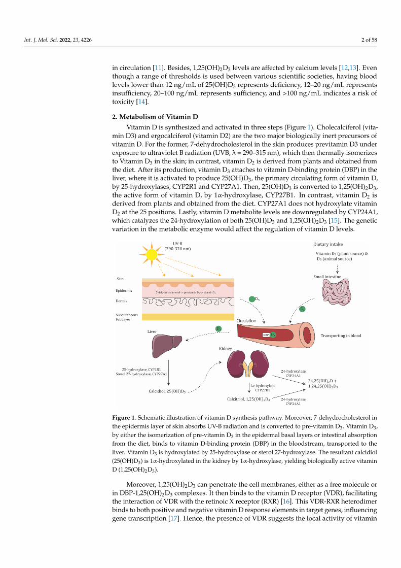
Vitamin D is synthesized and activated in three steps (Figure 1). Cholecalciferol (vita-min D3) and ergocalciferol (vitamin D2) are the two major biologically inert precursors ofvitamin D. For the former, 7-dehydrocholesterol in the skin produces previtamin D3 underexposure to ultraviolet B radiation (UVB, λ = 290–315 nm), which then thermally isomerizesto Vitamin D3 in the skin; in contrast, vitamin D2 is derived from plants and obtained fromthe diet. After its production, vitamin D3 attaches to vitamin D-binding protein (DBP) in theliver, where it is activated to produce 25(OH)D3, the primary circulating form of vitamin D,by 25-hydroxylases, CYP2R1 and CYP27A1. Then, 25(OH)D3 is converted to 1,25(OH)2D3,the active form of vitamin D, by 1α-hydroxylase, CYP27B1. In contrast, vitamin D2 isderived from plants and obtained from the diet. CYP27A1 does not hydroxylate vitaminD2 at the 25 positions. Lastly, vitamin D metabolite levels are downregulated by CYP24A1,which catalyzes the 24-hydroxylation of both 25(OH)D3 and 1,25(OH)2D3 [15]. The geneticvariation in the metabolic enzyme would affect the regulation of vitamin D levels.
Figure 1. Schematic illustration of vitamin D synthesis pathway. Moreover, 7-dehydrocholesterol inthe epidermis layer of skin absorbs UV-B radiation and is converted to pre-vitamin D3. Vitamin D3,by either the isomerization of pre-vitamin D3 in the epidermal basal layers or intestinal absorptionfrom the diet, binds to vitamin D-binding protein (DBP) in the bloodstream, transported to theliver. Vitamin D3 is hydroxylated by 25-hydroxylase or sterol 27-hydroxylase. The resultant calcidiol(25(OH)D3) is 1α-hydroxylated in the kidney by 1α-hydroxylase, yielding biologically active vitaminD (1,25(OH)2D3).
Moreover, 1,25(OH)2D3 can penetrate the cell membranes, either as a free molecule orin DBP-1,25(OH)2D3 complexes. It then binds to the vitamin D receptor (VDR), facilitatingthe interaction of VDR with the retinoic X receptor (RXR) [16]. This VDR-RXR heterodimerbinds to both positive and negative vitamin D response elements in target genes, influencinggene transcription [17]. Hence, the presence of VDR suggests the local activity of vitamin
Int. J. Mol. Sci. 2022, 23, 4226 3 of 58
D [18]. In particular, VDR has been detected in different parts of the eye, including theepithelium and endothelium of the cornea, lens, ciliary body, retinal ganglion cells (RGCs),inner nuclear layer, photoreceptors, and retinal pigment epithelium (RPE) [19,20]. Geneticalternations of the VDR gene could lead to defects in gene function, calcium metabolism,cell proliferation, and immune function. DBP is mainly responsible for the transportationof vitamin D and its metabolites.
Levels of active vitamin D in the body are regulated by the enzymes 25-hydroxylase,1α-hydroxylase, and 24-hydroxylase [15]. In a recent study, the 25(OH)D3 and 1,25(OH)2D3generating enzymes 25-hydroxylase (CYP2R1 and CYP27A1) and 1α-hydroxylase (CYP27B1),as well as the deactivating enzyme 24-hydroxylase (CYP24A1), were found to be stronglylocalized at the complementary regions of the ciliary body, RPE, neural retina, cornealepithelium and endothelium, and scleral fibroblast, suggesting that vitamin D in the eye islocally produced, activated, and regulated [21,22]. Moreover, vitamin D-dependent calciumbinding protein calbindin, a vitamin D metabolizing protein, was shown to be expressedthroughout the human retina [23]. Some of the cohort studies reported the correlation ofmetabolic enzymes in ocular diseases. In diabetic patients, retinal CYP27B1 was found tocorrelate strongly with VEGF-A in the eyes [24]. In a cohort of patients with Vogt-Koyanagi-Harada disease, a non-synonymous variant of CYP2R1 was found in 17 of 39 patients,suggesting that the variant in CYP2R1 may play a role in VKH pathogenesis [25].
Some of the vitamin D regulating proteins, such as ferredoxin reductase participatingin the activation of vitamin D in the kidney, are metalloproteins. Vitamin D is able tointeract with the matrix metalloproteinase. Metal deficiency may affect ocular condition.However, only one study found significantly lower serum calcium levels in blepharospasmpatients, but no significant difference in magnesium, phosphorus, or vitamin D [26].
Therefore, the potential of vitamin D to regulate various processes of potential rele-vance to ocular diseases has been acknowledged. Studies investigating the roles of vitaminD in ocular tissues and ocular disease pathogenic pathways have been carried out and willcontinue to contribute towards our understanding of ocular disease mechanisms and helpestablish effective intervention.
3. Vitamin D and Ocular Diseases
The potential effect of vitamin D deficiency on human health is a big concern. Recently,especially over the past few years, since the last published review articles related to vitaminD and ocular diseases, more and more studies investigating the relationship betweenserum vitamin D level and ocular diseases were published, including some prospectivestudies examining this relationship and therapeutic effects of vitamin D. Currently, reviewarticles related to vitamin D and ocular disease are available [18,20]. To update this andreach a comprehensive understanding, hence, we performed a systematic review here tosummarize the evidence revealing the association between vitamin D and ocular diseases.
3.1. Method of Literature Search
This systematic review was conducted in accordance with the Preferred ReportingItems for Systematic Reviews and Meta-Analyses guidelines [27]. The protocol is describedas follows.
3.1.1. Search Strategy
A systematic search on PubMed (Available online: http://www.ncbi.nlm.nih.gov/entrez/query.fcgi?DB5pubmed, accessed on 18 March 2022) and Web of Science (Availableonline: https://www.webofscience.com/wos/woscc/basic-search, accessed on 18 March2022) with coverage up to 18 March 2022 was conducted initially using the followingkeywords: vitamin D in combinations with eye (PubMED: 544; WOS: 1909), eye disease(PubMed: 824; WOS: 854), ocular (PubMed: 183; WOS: 534), cataract (PubMed: 158; WOS:355), lens opacity (PubMed: 118; WOS: 51), glaucoma (PubMed: 51; WOS: 151), intraocularpressure (PubMed: 24; WOS: 68), maculopathy (PubMed: 85; WOS: 97), diabetic retinopathy
Int. J. Mol. Sci. 2022, 23, 4226 4 of 58
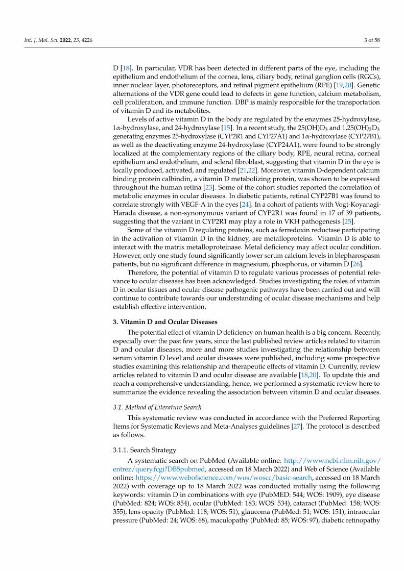
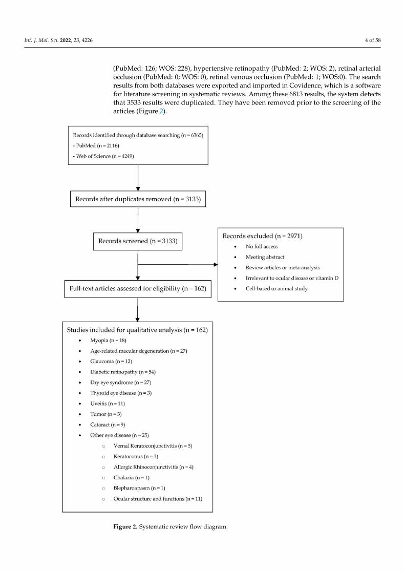
(PubMed: 126; WOS: 228), hypertensive retinopathy (PubMed: 2; WOS: 2), retinal arterialocclusion (PubMed: 0; WOS: 0), retinal venous occlusion (PubMed: 1; WOS:0). The searchresults from both databases were exported and imported in Covidence, which is a softwarefor literature screening in systematic reviews. Among these 6813 results, the system detectsthat 3533 results were duplicated. They have been removed prior to the screening of thearticles (Figure 2).
Figure 2. Systematic review flow diagram.
Int. J. Mol. Sci. 2022, 23, 4226 5 of 58
3.1.2. Inclusion and Exclusion Criteria
The inclusion criteria for studies were: (1) written in English; (2) evaluating the associa-tion between blood vitamin D and different ocular diseases in a randomized controlled trial,prospective study, cross-sectional study, or case-control study. After a review of abstracts,relevant articles were retrieved and reviewed. Bibliographies of these articles providedfurther references. All retrieved records were reviewed by two independent reviewers(HNC and XL). Uncertainties were resolved via discussion with another reviewer (XJZ).
3.1.3. Risk of Bias Assessment
Included interventional studies (both randomized controlled trials and clinical con-trolled trials) were assessed for quality according to the RoB tool for randomized trials fromthe Effective Practice and Organisation of Care (EPOC) Group. The assessment for the clini-cal controlled trial was assessed according to the suggestion from previous literature thatboth “random sequence generation” and “allocation concealment” were scored as “highrisk”, while grading the remaining items as RCT [28]. We further modified the RoB tool byallocating 1 point to “low risk”, 0.5 point to “unclear risk” and 0 points to “high risk”. Thereare a total of 9 items to be assessed using the RoB tool, and hence, the total number of pointsfor the RoB tool is 9 points, while those cohort, case-control, cross-sectional studies wereassessed for quality according to the LEGEND (Let Evidence Guide Every New Decision)System designed for Cincinnati Children’s Hospital [29].
3.2. Myopia
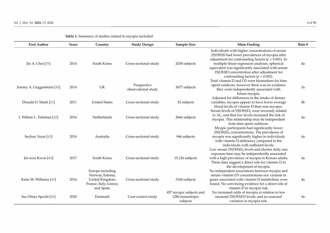
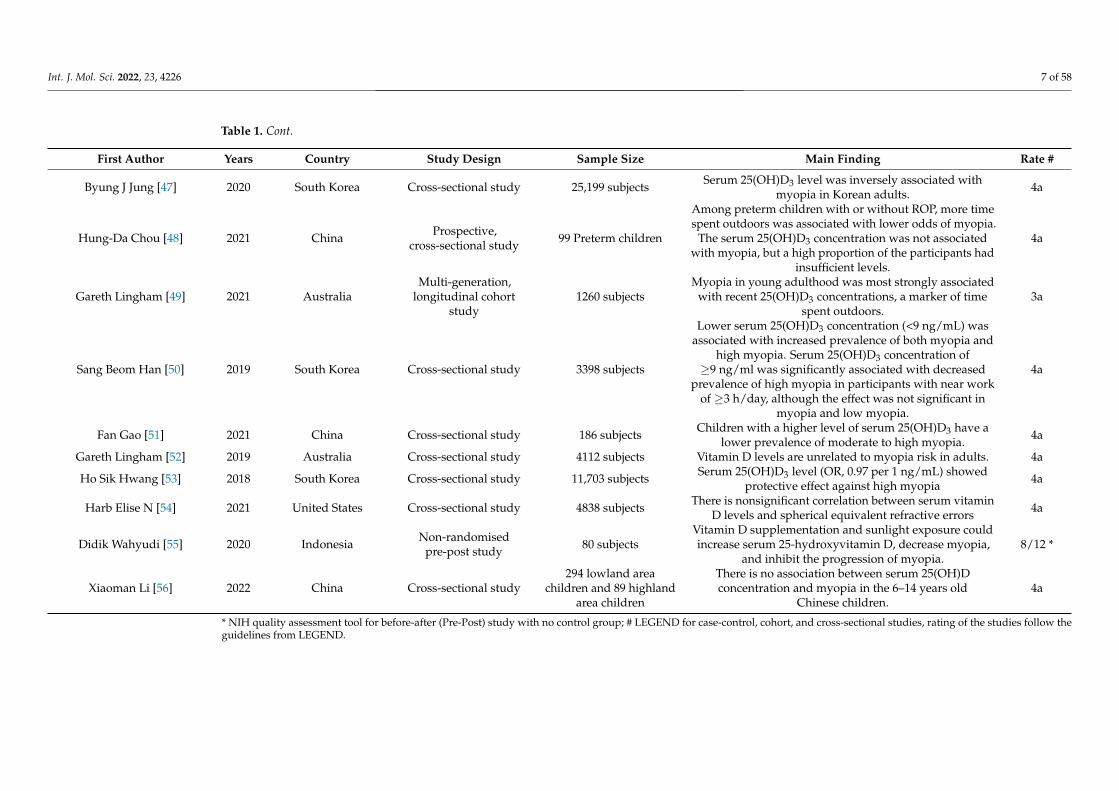
Myopia is an important public health problem worldwide [30]. The etiology of my-opia is complex, with both genetic and environmental risk factors [31–34]. Epidemiologicevidence indicates that time spent outdoors is a protective factor against myopia develop-ment [34–38], yet the underlying mechanism is unclear. Since the main source of vitaminD is sunlight exposure, vitamin D is linked to myopia, hypothesizing that a vitamin Dpathway may mediate the protective effect of time spent outdoors on myopia. Evidencefrom studies on the relationship of vitamin D and myopia is summarized in Table 1.
Int. J. Mol. Sci. 2022, 23, 4226 6 of 58
Table 1. Summary of studies related to myopia included.
First Author Years Country Study Design Sample Size Main Finding Rate #
Jin A. Choi [39] 2014 South Korea Cross-sectional study 2038 subjects
Individuals with higher concentrations of serum25(OH)D had lower prevalences of myopia after
adjustment for confounding factors (p < 0.001). Inmultiple linear regression analyses, spherical
equivalent was significantly associated with serum25(OH)D concentration after adjustment for
confounding factors (p = 0.002).
4a
Jeremy A. Guggenheim [40] 2014 UK Prospectiveobservational study 3677 subjects
Total vitamin D and D3 were biomarkers for timespent outdoors, however there was no evidence
they were independently associated withfuture myopia.
3a
Donald O. Mutti [41] 2011 United States Cross-sectional study 32 subjectsAdjusted for differences in the intake of dietaryvariables, myopes appear to have lower average
blood levels of vitamin D than non-myopes.4b
J. Willem L. Tideman [42] 2016 Netherlands Cross-sectional study 2666 subjects
Serum levels of 25(OH)D3 were inversely relatedto AL, and that low levels increased the risk ofmyopia. This relationship may be independent
from time spent outdoors.
4a
Seyhan Yazar [43] 2014 Australia Cross-sectional study 946 subjects
Myopic participants had significantly lower25(OH)D3 concentrations. The prevalence of
myopia was significantly higher in individualswith vitamin D deficiency compared to the
individuals with sufficient levels.
4a
Jin-woo Kwon [44] 2017 South Korea Cross-sectional study 15,126 subjects
Low serum 25(OH)D3 levels and shorter daily sunexposure time may be independently associated
with a high prevalence of myopia in Korean adults.These data suggest a direct role for vitamin D in
the development of myopia.
4a
Katie M. Williams [45] 2016
Europe includingNorway, Estonia,United Kingdom,
France, Italy, Greece,and Spain
Cross-sectional study 3168 subjects
No independent associations between myopia andserum vitamin D3 concentrations nor variants in
genes associated with vitamin D metabolism werefound. No convincing evidence for a direct role of
vitamin D in myopia risk.
4a
Ina Olmer Specht [46] 2020 Denmark Case-control study457 myopic subjects and
1280 emmetropicsubjects
No increased odds of myopia in relation to lowneonatal 25(OH)D3 levels, and no seasonal
variation in myopia risk.4a
Int. J. Mol. Sci. 2022, 23, 4226 7 of 58
Table 1. Cont.
First Author Years Country Study Design Sample Size Main Finding Rate #
Byung J Jung [47] 2020 South Korea Cross-sectional study 25,199 subjects Serum 25(OH)D3 level was inversely associated withmyopia in Korean adults. 4a
Hung-Da Chou [48] 2021 China Prospective,cross-sectional study 99 Preterm children
Among preterm children with or without ROP, more timespent outdoors was associated with lower odds of myopia.
The serum 25(OH)D3 concentration was not associatedwith myopia, but a high proportion of the participants had
insufficient levels.
4a
Gareth Lingham [49] 2021 AustraliaMulti-generation,
longitudinal cohortstudy
1260 subjectsMyopia in young adulthood was most strongly associated
with recent 25(OH)D3 concentrations, a marker of timespent outdoors.
3a
Sang Beom Han [50] 2019 South Korea Cross-sectional study 3398 subjects
Lower serum 25(OH)D3 concentration (<9 ng/mL) wasassociated with increased prevalence of both myopia and
high myopia. Serum 25(OH)D3 concentration of≥9 ng/ml was significantly associated with decreased
prevalence of high myopia in participants with near workof ≥3 h/day, although the effect was not significant in
myopia and low myopia.
4a
Fan Gao [51] 2021 China Cross-sectional study 186 subjects Children with a higher level of serum 25(OH)D3 have alower prevalence of moderate to high myopia. 4a
Gareth Lingham [52] 2019 Australia Cross-sectional study 4112 subjects Vitamin D levels are unrelated to myopia risk in adults. 4a
Ho Sik Hwang [53] 2018 South Korea Cross-sectional study 11,703 subjects Serum 25(OH)D3 level (OR, 0.97 per 1 ng/mL) showedprotective effect against high myopia 4a
Harb Elise N [54] 2021 United States Cross-sectional study 4838 subjects There is nonsignificant correlation between serum vitaminD levels and spherical equivalent refractive errors 4a
Didik Wahyudi [55] 2020 Indonesia Non-randomisedpre-post study 80 subjects
Vitamin D supplementation and sunlight exposure couldincrease serum 25-hydroxyvitamin D, decrease myopia,
and inhibit the progression of myopia.8/12 *
Xiaoman Li [56] 2022 China Cross-sectional study294 lowland area
children and 89 highlandarea children
There is no association between serum 25(OH)Dconcentration and myopia in the 6–14 years old
Chinese children.4a
* NIH quality assessment tool for before-after (Pre-Post) study with no control group; # LEGEND for case-control, cohort, and cross-sectional studies, rating of the studies follow theguidelines from LEGEND.
Int. J. Mol. Sci. 2022, 23, 4226 8 of 58
As demonstrated in Table 1, the association between vitamin D and myopia is con-troversial in cross-sectional studies. Many studies suggest that the serum 25(OH)D3level shows an inverse association with myopia and may have a protective effect on my-opia [39,41–44,47,50,51,53,55]. However, several case-control studies from Australia [52],Denmark [46], and the US [54] found that the risks of myopia are not related to theirneonatal vitamin D levels.
Nevertheless, it is important to distinguish the causation between vitamin D andmyopia. A large longitudinal cohort study found that 25(OH)D3 levels correlated withself-reported time spent outdoors; however, no evidence suggested that the participants’serum vitamin D levels were independently associated with myopia [40]. Another studyof preterm children also suggested that more time spent outdoors was associated with alower risk of myopia, despite serum 25(OH)D3 concentrations not being shown to relateto myopia [48]. However, an Australian perspective study showed that, in young adults,myopia was most strongly associated with recent 25(OH)D3 concentrations, which is amarker of time spent outdoors [49].
Our meta-analysis found that the risk of myopia is inversely associated with blood25(OH)D3 concentration after adjusting for sunlight exposure or time spent outdoors. How-ever, this relationship was not significant among individuals under 18 years of age [57].Polymorphisms in the vitamin D pathway genes may affect the development of myopia.One study reported the association of VDR polymorphisms, rs2853559, with myopia [58].However, the results of other studies suggested that the true contribution of the vitamin Dpathway to myopia could be negligible [42,45,59]. Our meta-analysis suggested that poly-morphisms in the VDR gene are not associated with myopia [57]. On the other hand, animalstudies proved that violet light (VL, λ = 360–400 nm) can suppress myopia progression,whereas no therapeutic effects were observed with UVB radiation (λ = 290–315 nm) [60],suggesting that UVB exposure and its dependent vitamin D synthetic pathway may nothave a protective effect on myopia progression.
In conclusion, from the literature evidence, we know that, although blood 25(OH)D3concentration is inversely associated with the risk of myopia, it seems unlikely that vitaminD has a direct protective effect on myopia progression. Instead, vitamin D levels may onlyserve as a biomarker for outdoor exposure.
3.3. Age-Related Macular Degeneration
As a chronic, progressive, degenerative disease, age-related macular degeneration(AMD) is a major cause of central blindness among people aged 60 years or over world-wide [61,62]. Oxidation, inflammation, and angiogenesis contribute to the pathogenesisof AMD, resulting in the dysfunction of RPE [63], Bruch’s membrane, and choriocapil-laries [64]. In an aging retina, the complement cascade [65,66] and the tissue residentmacrophage (retinal microglia) activation pathway [67] ultimately cause protein damageand aggregation, and degeneration of the RPE [68]. Angiogenesis, often caused by oxidativestress and inflammatory reactions, plays a major role in the development and progressionof exudative AMD, potentially leading to severe and permanent visual impairment.
The results of studies on cell lines and animal models have shown that vitamin D canprotect cells or reduce oxidative stress [69–71]. Vitamin D has an anti-inflammatory role inchronic inflammatory diseases by decreasing the proliferation of T-cells and the productionof pro-inflammatory agents [72,73]. On the other hand, vitamin D exerted an inhibitoryeffect on the angiogenesis signaling pathway [74,75], which may play a protective role inexudative AMD development and/or progression. Morrison et al. studied the variantsin the vitamin D catabolizing enzyme, CYP24A1, and reported that variants (rs1570669,rs1570670, rs2274130, rs2296239, and rs4809957) were associated with reduced risk forAMD [76].
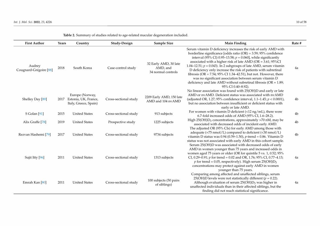
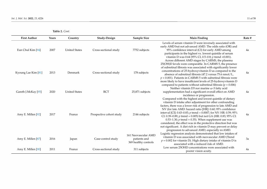
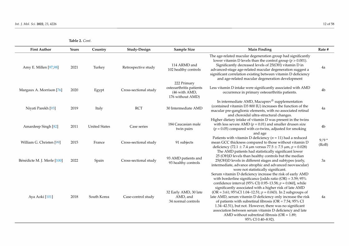
Table 2 summarized the studies on vitamin D and AMD. Case-control studies withsmall sample sizes suggest that AMD patients always have relatively low levels of serumvitamin D [77–87], except in a Iranian study, which did not find any significant correlation

Int. J. Mol. Sci. 2022, 23, 4226 9 of 58
between serum vitamin D level and AMD [79]. However, this association seems to changein cross-sectional studies with larger sample sizes. Population-based studies held inFrance [88], the United States [89,90], and Israel [91] did not support a specific role forvitamin D in AMD, but vitamin D may work in some specific populations. An analysis of asample of 1313 US participants indicated that high serum 25(OH)D3 concentrations mayprotect against early AMD in women less than 75 years old [92], while another US studysupported the fact that levels of serum vitamin D were inversely associated with early AMDbut not advanced AMD [93]. A Korean study had 17,045 participants and found that a highlevel of vitamin D was inversely associated with late AMD in men but not women [94].Vitamin D deficiency in the European population was found to be associated with nvAMD,but the adjusted OR was small, and cannot exclude residual confounding [95].
Int. J. Mol. Sci. 2022, 23, 4226 10 of 58
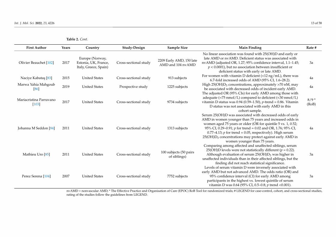
Table 2. Summary of studies related to age-related macular degeneration included.
First Author Years Country Study-Design Sample Size Main Finding Rate #
AudreyCougnard-Grégoire [88] 2018 South Korea Case-control study
32 Early AMD, 30 lateAMD, and
34 normal controls
Serum vitamin D deficiency increases the risk of early AMD withborderline significance [odds ratio (OR) = 3.59; 95% confidence
interval (95% CI) 0.95–13.58; p = 0.060], while significantlyassociated with a higher risk of late AMD (OR = 3.61; 95%CI
1.04–12.51; p = 0.043). In 2 subgroups of late AMD, serum vitaminD deficiency only increase the risk of patients with subretinal
fibrosis (OR = 7.54; 95% CI 1.34–42.51), but not. However, therewas no significant association between serum vitamin D
deficiency and late AMD without subretinal fibrosis (OR = 1.89;95% CI 0.40–8.92).
4a
Shelley Day [89] 2017Europe (Norway,
Estonia, UK, France,Italy, Greece, Spain)
Cross-sectional study 2209 Early AMD, 150 lateAMD and 104 nvAMD
No linear association was found with 25(OH)D and early or lateAMD or nvAMD. Deficient status was associated with nvAMD
(adjusted OR, 1.27; 95% confidence interval, 1.1–1.45; p < 0.0001),but no association between insufficient or deficient status with
early or late AMD.
4b
S Golan [91] 2015 United States Cross-sectional study 913 subjects For women with vitamin D deficient (<12 ng/mL), there were6.7-fold increased odds of AMD (95% CI, 1.6–28.2). 4b
Alix Graffe [78] 2019 United States Prospective study 1225 subjects High 25(OH)D3 concentrations, approximately >70 nM, may beassociated with decreased odds of incident early AMD. 4b
Rezvan Hashemi [79] 2017 United States Cross-sectional study 9734 subjects
The adjusted OR (95% CIs) for early AMD among those withadequate (=75 nmol/L) compared to deficient (<30 nmol/L)
vitamin D status was 0.94 (0.59–1.50), p-trend = 0.86. Vitamin Dstatus was not associated with early AMD in this cohort sample.
4b
Sujit Itty [96] 2011 United States Cross-sectional study 1313 subjects
Serum 25(OH)D was associated with decreased odds of earlyAMD in women younger than 75 years and increased odds in
women aged 75 years or older (OR for quintile 5 vs. 1, 0.52; 95%CI, 0.29–0.91; p for trend = 0.02 and OR, 1.76; 95% CI, 0.77–4.13;
p for trend = 0.05, respectively). High serum 25(OH)D3concentrations may protect against early AMD in women
younger than 75 years.
4a
Emrah Kan [80] 2011 United States Cross-sectional study 100 subjects (50 pairsof siblings)
Comparing among affected and unaffected siblings, serum25(OH)D levels were not statistically different (p = 0.22).Although evaluation of serum 25(OH)D3 was higher in
unaffected individuals than in their affected siblings, but thefinding did not reach statistical significance.
4a
Int. J. Mol. Sci. 2022, 23, 4226 11 of 58
Table 2. Cont.
First Author Years Country Study-Design Sample Size Main Finding Rate #
Eun Chul Kim [94] 2007 United States Cross-sectional study 7752 subjects
Levels of serum vitamin D were inversely associated withearly AMD but not advanced AMD. The odds ratio (OR) and
95% confidence interval (CI) for early AMD amongparticipants in the highest vs. lowest quintile of serum
vitamin D was 0.64 (95% CI, 0.5–0.8; p trend <0.001).
4a
Kyoung Lae Kim [81] 2013 Denmark Cross-sectional study 178 subjects
Across different AMD stages by CARMS, the plasema25(OH)D levels were comparable. In CARMS 5, the presenceof subretinal fibrosis was associated with significantly lowerconcentrations of 25-hydroxyvitamin D as compared to the
absence of subretinal fibrosis (47.2 versus 75.6 nmol/L,p < 0.001). Patients in CARMS 5 with subretinal fibrosis weremore likely to have insufficient levels of 25-hydroxyvitamin Dcompared to patients without subretinal fibrosis (p = 0.006)
4a
Gareth J.McKay [95] 2020 United States RCT 25,871 subjectsNeither vitamin D3 nor marineω-3 fatty acid
supplementation had a significant overall effect on AMDincidence or progression.
4a
Amy E. Millen [92] 2017 France Prospective cohort study 2146 subjects
Compared with the highest and lowest quintile of dietaryvitamin D intake after adjustment for other confounding
facters, there was a lower risk of progression to late AMD andNV (for late AMD: hazard ratio [HR]: 0.60; 95% confidence
interval [CI]: 0.43–0.83; p trend = 0.0007; for NV: HR: 0.59; 95%CI: 0.39–0.89; p trend = 0.005) but not GA (HR: 0.83; 95% CI:
0.53–1.30; p trend = 0.35). When supplement use wasconsidered, the effect was in the protective direction but wasnot significant. A diet rich in vitamin D may prevent or delay
progression to advanced AMD, especially nvAMD.
4a
Amy E. Millen [87] 2016 Japan Case-control study161 Neovascular AMD
patients and369 healthy controls
Logistic regression analysis demonstrated that low intakes ofvitamin D was associated with neovascular AMD (Trend
p = 0.002 for vitamin D). High dietary intake of vitamin D isassociated with a reduced risk of AMD.
3a
Amy E. Millen [90] 2011 France Cross-sectional study 311 subjects Low serum 25OHD concentrations were associated withpoorer vision acuity. 4a
Int. J. Mol. Sci. 2022, 23, 4226 12 of 58
Table 2. Cont.
First Author Years Country Study-Design Sample Size Main Finding Rate #
Amy E. Millen [97,98] 2021 Turkey Retrospective study 114 ARMD and102 healthy controls
The age-related macular degeneration group had significantlylower vitamin D levels than the control group (p > 0.001).
Significantly decreased levels of 25(OH) vitamin D inadvanced-stage age-related macular degeneration suggest asignificant correlation existing between vitamin D deficiency
and age-related macular degeneration development
4a
Margaux A. Morrison [76] 2020 Egypt Cross-sectional study
222 Primaryosteoarthritis patients
(46 with AMD,176 without AMD)
Less vitamin D intake were significantly associated with AMDoccurrence in primary osteoarthritis patients. 4b
Niyati Parekh [93] 2019 Italy RCT 30 Intermediate AMD
In intermediate AMD, Macuprev® supplementation(contained vitamin D3 800 IU) increases the function of themacular pre-ganglionic elements, with no associated retinal
and choroidal ultra-structural changes.
4a
Amardeep Singh [82] 2011 United States Case series 184 Caucasian maletwin pairs
Higher dietary intake of vitamin D was present in the twinswith less severe AMD (p = 0.01) and smaller drusen size(p = 0.05) compared with co-twins, adjusted for smoking
and age
4b
William G. Christen [99] 2015 France Cross-sectional study 91 subjectsPatients with vitamin D deficiency (n = 11) had a reduced
mean GCC thickness compared to those without vitamin Ddeficiency (72.1 ± 7.4 µm versus 77.5 ± 7.5 µm, p = 0.028)
9/9 *(RoB)
Bénédicte M. J. Merle [100] 2022 Spain Cross-sectional study 93 AMD patients and93 healthy controls
The AMD patients had statistically significant lower25 (OH)D levels than healthy controls but the median25(OH)D levels in different stages and subtypes (early,
intermediate, advance atrophic and advanced neovascular)were not statistically significant.
3a
Aya Aoki [101] 2018 South Korea Case-control study32 Early AMD, 30 late
AMD, and34 normal controls
Serum vitamin D deficiency increase the risk of early AMDwith borderline significance [odds ratio (OR) = 3.59; 95%confidence interval (95% CI) 0.95–13.58; p = 0.060], whilesignificantly associated with a higher risk of late AMD
(OR = 3.61; 95%CI 1.04–12.51; p = 0.043). In 2 subgroups oflate AMD, serum vitamin D deficiency only increase the risk
of patients with subretinal fibrosis (OR = 7.54; 95% CI1.34–42.51), but not. However, there was no significant
association between serum vitamin D deficiency and lateAMD without subretinal fibrosis (OR = 1.89;
95% CI 0.40–8.92).
4a
Int. J. Mol. Sci. 2022, 23, 4226 13 of 58
Table 2. Cont.
First Author Years Country Study-Design Sample Size Main Finding Rate #
Olivier Beauchet [102] 2017Europe (Norway,
Estonia, UK, France,Italy, Greece, Spain)
Cross-sectional study 2209 Early AMD, 150 lateAMD and 104 nvAMD
No linear association was found with 25(OH)D and early orlate AMD or nvAMD. Deficient status was associated with
nvAMD (adjusted OR, 1.27; 95% confidence interval, 1.1–1.45;p < 0.0001), but no association between insufficient or
deficient status with early or late AMD.
3a
Naciye Kabatas [83] 2015 United States Cross-sectional study 913 subjects For women with vitamin D deficient (<12 ng/mL), there was6.7-fold increased odds of AMD (95% CI, 1.6–28.2). 4a
Marwa Yahia Mahgoub[86] 2019 United States Prospective study 1225 subjects High 25(OH)D3 concentrations, approximately >70 nM, may
be associated with decreased odds of incident early AMD. 4a
Mariacristina Parravano[103] 2017 United States Cross-sectional study 9734 subjects
The adjusted OR (95% CIs) for early AMD among those withadequate (=75 nmol/L) compared to deficient (<30 nmol/L)
vitamin D status was 0.94 (0.59–1.50), p-trend = 0.86. VitaminD status was not associated with early AMD in this
cohort sample.
8/9 *(RoB)
Johanna M Seddon [84] 2011 United States Cross-sectional study 1313 subjects
Serum 25(OH)D was associated with decreased odds of earlyAMD in women younger than 75 years and increased odds in
women aged 75 years or older (OR for quintile 5 vs. 1, 0.52;95% CI, 0.29–0.91; p for trend = 0.02 and OR, 1.76; 95% CI,
0.77–4.13; p for trend = 0.05, respectively). High serum25(OH)D3 concentrations may protect against early AMD in
women younger than 75 years.
4a
Mathieu Uro [85] 2011 United States Cross-sectional study 100 subjects (50 pairsof siblings)
Comparing among affected and unaffected siblings, serum25(OH)D levels were not statistically different (p = 0.22).Although evaluation of serum 25(OH)D3 was higher in
unaffected individuals than in their affected siblings, but thefinding did not reach statistical significance.
3a
Perez Serena [104] 2007 United States Cross-sectional study 7752 subjects
Levels of serum vitamin D were inversely associated withearly AMD but not advanced AMD. The odds ratio (OR) and
95% confidence interval (CI) for early AMD amongparticipants in the highest vs. lowest quintile of serum
vitamin D was 0.64 (95% CI, 0.5–0.8; p trend <0.001).
3a
nvAMD = nonvascular AMD; * The Effective Practice and Organisation of Care (EPOC) RoB Tool for randomized trials; # LEGEND for case-control, cohort, and cross-sectional studies,rating of the studies follow the guidelines from LEGEND.

Int. J. Mol. Sci. 2022, 23, 4226 14 of 58
Prospective studies, however, have not found a consistent association between vitaminD and the risk of developing AMD. In a large prospective cohort study of 2146 participantswith a mean follow-up time of over 9 years, high dietary intake of vitamin D was signifi-cantly associated with a 40% lower risk of progression to advanced AMD [99]. However,recently, a nationwide, placebo-controlled, randomized clinical trial found that supple-menting vitamin D had no significant overall effect on AMD incidence or progression inhealthy people [98]. For this trial, 25,871 participants with a median age of 67.1 years weredivided into four groups, receiving vitamin D supplements (2000 IU/day), ω-3 fatty acids(1 g/day), a combination of both, and placebo, respectively. After a median follow-upperiod of 5.3 years, no significant differences were found in the incidence or progression ofAMD when compared with baseline [98]. This study suffered from a lack of stratification byclinical manifestations of AMD, a relatively short follow-up period for chronic disease, anda reliance on self-reported AMD diagnosis, leading to inconsistencies with the previoustwo studies [98,104].
In summary, cross-sectional studies suggest that vitamin D may have a protectiveeffect on AMD formation, but this effect is small or may only work in a specific popula-tion. Furthermore, evidence from prospective cohort studies showed that continuouslysupplementing vitamin D may not reduce the risks of AMD over a period of several years.
3.4. Glaucoma
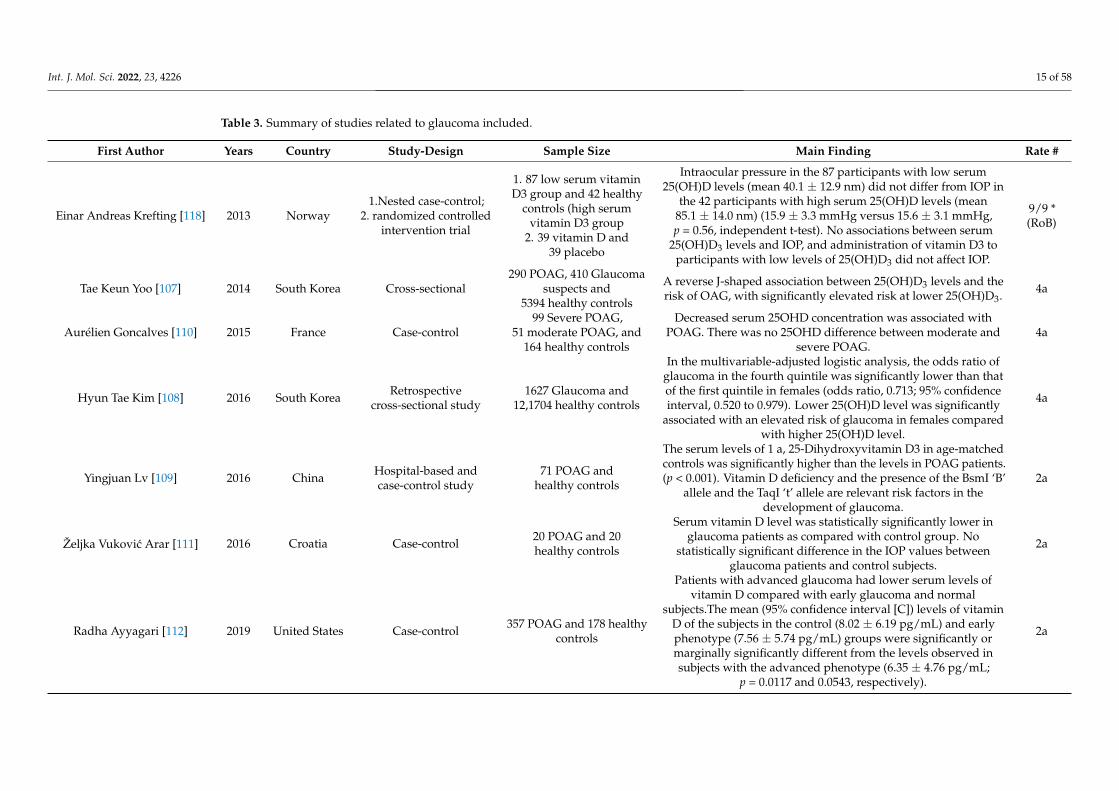
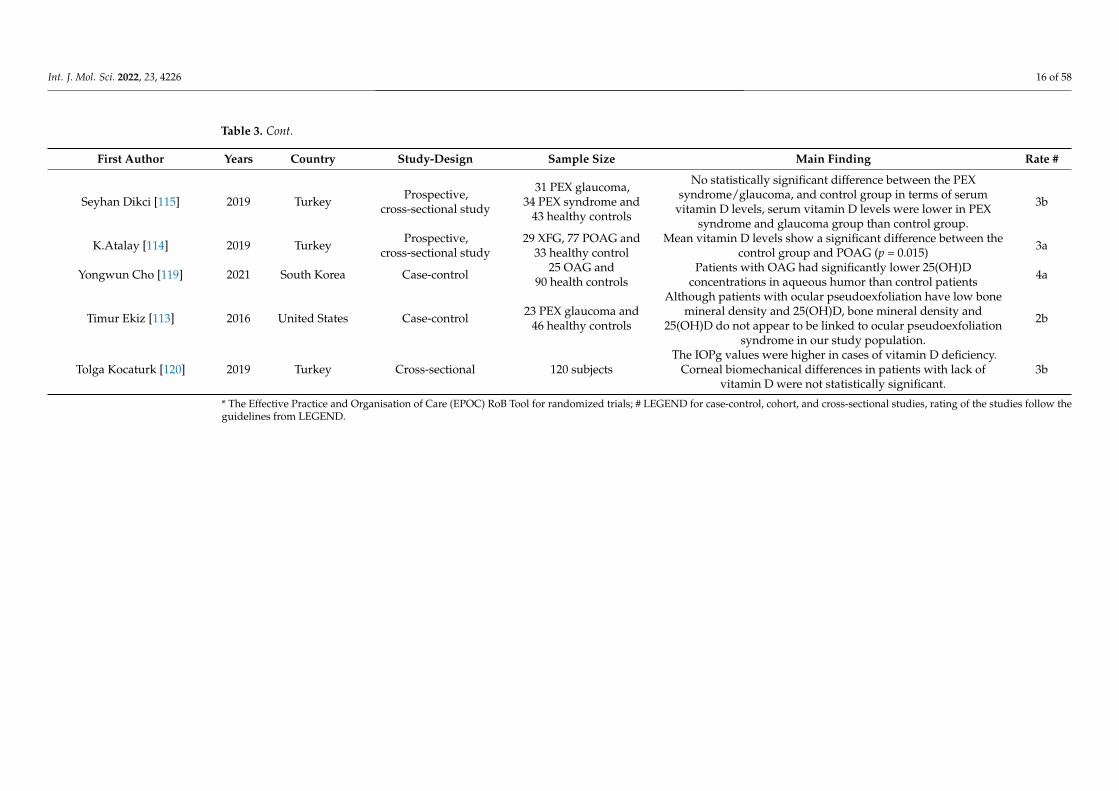
A leading cause of irreversible blindness, glaucoma is a group of optic neuropathiesinvolving the death of retinal ganglion cells (RGCs) and the loss of their axons [105,106].Two cross-sectional studies in South Korea reported that vitamin D deficiency is associatedwith glaucoma [107,108]. Similarly, a Chinese study found that the vitamin D deficiency,along with the presence of the BsmI ‘B’ allele and TaqI ‘t’ allele of the VDR gene, are relevantrisk factors for glaucoma development [109]. Other studies in France [110], Croatia [111],the United States [112,113], and Turkey [114] have demonstrated that glaucoma patientshave lower serum vitamin D levels compared to normal controls. However, another Turkishcase-control study found no statistically significant difference in serum vitamin D levelsbetween glaucoma patients and control subjects [115]. Similarly, a recent large-samplestudy in the United States showed that dietary intake, supplements, and serum levels ofvitamin D are not significantly related to the risk of glaucoma [116]. Notably, ethnicity maycontribute to the pathogenesis of glaucoma, giving rise to different conclusions amongthese studies [117]. Most of the literature reported the association between vitamin D andglaucoma and that a lower vitamin D concentration was found in glaucoma patients whencompared with the control group [108–111,114], however, there were no findings on theassociation between vitamin D and the severity. Increases in vitamin D were associatedwith lower risks of having glaucoma (fourth quintile versus first quintile, OR 0.713, 95%confidence interval, 0.520 to 0.979) [108]. Only a limited study reported no statisticallysignificant difference between the glaucoma group and the control group [108]; significantlylower vitamin D can only be found in advanced glaucoma patients [112] (Table 3).
Int. J. Mol. Sci. 2022, 23, 4226 15 of 58
Table 3. Summary of studies related to glaucoma included.
First Author Years Country Study-Design Sample Size Main Finding Rate #
Einar Andreas Krefting [118] 2013 Norway1.Nested case-control;
2. randomized controlledintervention trial
1. 87 low serum vitaminD3 group and 42 healthy
controls (high serumvitamin D3 group
2. 39 vitamin D and39 placebo
Intraocular pressure in the 87 participants with low serum25(OH)D levels (mean 40.1 ± 12.9 nm) did not differ from IOP in
the 42 participants with high serum 25(OH)D levels (mean85.1 ± 14.0 nm) (15.9 ± 3.3 mmHg versus 15.6 ± 3.1 mmHg,p = 0.56, independent t-test). No associations between serum
25(OH)D3 levels and IOP, and administration of vitamin D3 toparticipants with low levels of 25(OH)D3 did not affect IOP.
9/9 *(RoB)
Tae Keun Yoo [107] 2014 South Korea Cross-sectional290 POAG, 410 Glaucoma
suspects and5394 healthy controls
A reverse J-shaped association between 25(OH)D3 levels and therisk of OAG, with significantly elevated risk at lower 25(OH)D3. 4a
Aurélien Goncalves [110] 2015 France Case-control99 Severe POAG,
51 moderate POAG, and164 healthy controls
Decreased serum 25OHD concentration was associated withPOAG. There was no 25OHD difference between moderate and
severe POAG.4a
Hyun Tae Kim [108] 2016 South Korea Retrospectivecross-sectional study
1627 Glaucoma and12,1704 healthy controls
In the multivariable-adjusted logistic analysis, the odds ratio ofglaucoma in the fourth quintile was significantly lower than thatof the first quintile in females (odds ratio, 0.713; 95% confidenceinterval, 0.520 to 0.979). Lower 25(OH)D level was significantly
associated with an elevated risk of glaucoma in females comparedwith higher 25(OH)D level.
4a
Yingjuan Lv [109] 2016 China Hospital-based andcase-control study
71 POAG andhealthy controls
The serum levels of 1 a, 25-Dihydroxyvitamin D3 in age-matchedcontrols was significantly higher than the levels in POAG patients.(p < 0.001). Vitamin D deficiency and the presence of the BsmI ‘B’
allele and the TaqI ‘t’ allele are relevant risk factors in thedevelopment of glaucoma.
2a
Željka Vukovic Arar [111] 2016 Croatia Case-control 20 POAG and 20healthy controls
Serum vitamin D level was statistically significantly lower inglaucoma patients as compared with control group. No
statistically significant difference in the IOP values betweenglaucoma patients and control subjects.
2a
Radha Ayyagari [112] 2019 United States Case-control 357 POAG and 178 healthycontrols
Patients with advanced glaucoma had lower serum levels ofvitamin D compared with early glaucoma and normal
subjects.The mean (95% confidence interval [C]) levels of vitaminD of the subjects in the control (8.02 ± 6.19 pg/mL) and earlyphenotype (7.56 ± 5.74 pg/mL) groups were significantly ormarginally significantly different from the levels observed insubjects with the advanced phenotype (6.35 ± 4.76 pg/mL;
p = 0.0117 and 0.0543, respectively).
2a
Int. J. Mol. Sci. 2022, 23, 4226 16 of 58
Table 3. Cont.
First Author Years Country Study-Design Sample Size Main Finding Rate #
Seyhan Dikci [115] 2019 Turkey Prospective,cross-sectional study
31 PEX glaucoma,34 PEX syndrome and
43 healthy controls
No statistically significant difference between the PEXsyndrome/glaucoma, and control group in terms of serum
vitamin D levels, serum vitamin D levels were lower in PEXsyndrome and glaucoma group than control group.
3b
K.Atalay [114] 2019 Turkey Prospective,cross-sectional study
29 XFG, 77 POAG and33 healthy control
Mean vitamin D levels show a significant difference between thecontrol group and POAG (p = 0.015) 3a
Yongwun Cho [119] 2021 South Korea Case-control 25 OAG and90 health controls
Patients with OAG had significantly lower 25(OH)Dconcentrations in aqueous humor than control patients 4a
Timur Ekiz [113] 2016 United States Case-control 23 PEX glaucoma and46 healthy controls
Although patients with ocular pseudoexfoliation have low bonemineral density and 25(OH)D, bone mineral density and
25(OH)D do not appear to be linked to ocular pseudoexfoliationsyndrome in our study population.
2b
Tolga Kocaturk [120] 2019 Turkey Cross-sectional 120 subjectsThe IOPg values were higher in cases of vitamin D deficiency.
Corneal biomechanical differences in patients with lack ofvitamin D were not statistically significant.
3b
* The Effective Practice and Organisation of Care (EPOC) RoB Tool for randomized trials; # LEGEND for case-control, cohort, and cross-sectional studies, rating of the studies follow theguidelines from LEGEND.
Int. J. Mol. Sci. 2022, 23, 4226 17 of 58
High intraocular pressure (IOP) is an important risk factor for glaucoma. In ananimal study on non-human primates, vitamin D treatment modulated the expression ofIOP–regulating genes, with IOP falling in a dose-dependent manner [121]. However, ahuman study found no association between serum 25(OH)D3 levels and IOP, nor significantchanges in participants’ IOP levels after receiving 6 months of oral vitamin D supplements(20,000 IU twice weekly) compared to the placebo group [118]. This contradiction may bedue to the oral intake of vitamin D, which may lower the availability of vitamin D in theeye. Patients with glaucoma were found to have lower 25(OH)D concentrations in aqueoushumor [119], and the IOP values were higher in cases of vitamin D deficiency [120]. Furtherstudies are required to determine if vitamin D can be a potential intervention for glaucoma,especially through testing different supplement approaches.
Some studies have identified vitamin D as an independent risk factor for glaucoma;however, the role that vitamin D plays in relation to glaucoma remains uncertain. Apartfrom the elevated IOP pathway, vitamin D may participate in the oxidative stress pathwaydue to its anti-oxidation and anti-inflammatory abilities. In an in vivo study, 1,25(OH)2D3ameliorated the effects of oxidative stress from hydrogen peroxide-induced toxicity inhuman RPE cells through antioxidant signaling pathways, leading to lower levels of reactiveoxygen species (ROS), cytokines, and vascular endothelial growth factor (VEGF) [122].Another study demonstrated that vitamin D significantly altered the inflammatory-relatedgenes in glaucoma, suppressing the expression of the angiotensin I–converting enzyme(ACE), carbonic anhydrase (CA), and Ras homologue gene family member A (RhoA), whilesignificantly increasing the expression of the cytokine A20 precursor (CCL20) in the smallintestines of rats [123]. ACE inhibitors are neuroprotective for cultured retinal neurons andcan lower IOP in humans [124,125], while CA inhibitors can lower IOP and increase bloodflow in the retinal vasculature and optic nerve [126]. The suppression of RhoA throughsubsequent vitamin D treatment can reduce aqueous outflow resistance and enhance fluidoutflow [127,128]. Lastly, CCL2, an intraocular pressure responsive cytokine, possesses apotential role in intraocular pressure regulation [129].
In summary, all reported studies are cross-sectional studies (case-control studies andpopulation surveys) and suggested the protective associations of vitamin D on glaucoma.Future studies should employ randomized clinical trial designs to investigate the causalrelationship between glaucoma and low vitamin D levels or calcitriol deficiency.
3.5. Diabetic Retinopathy
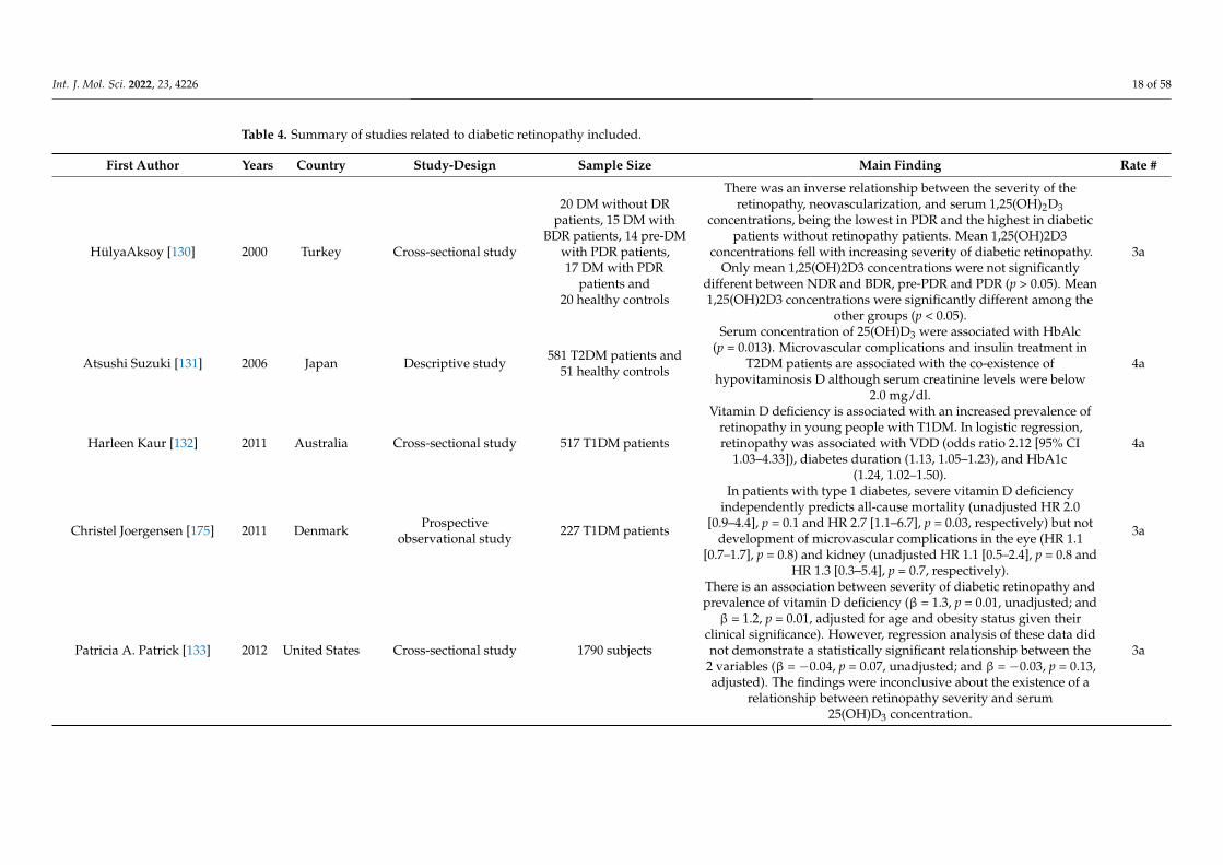
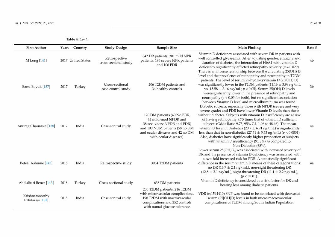
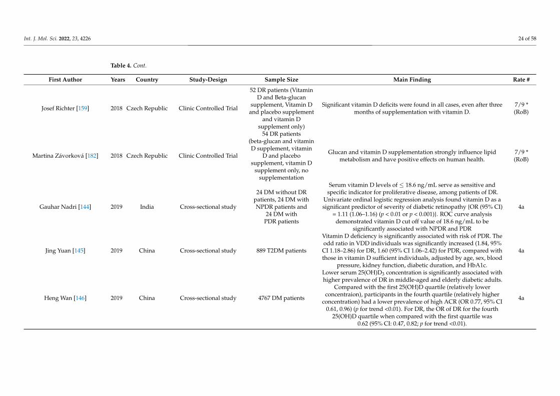
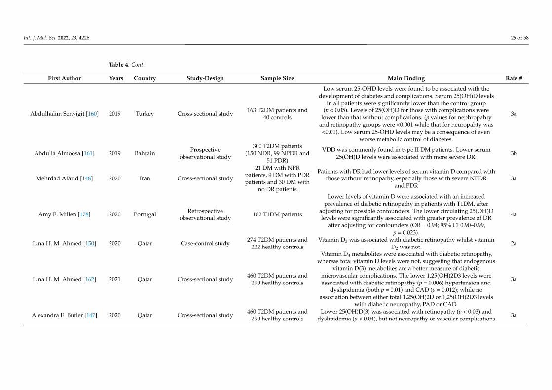
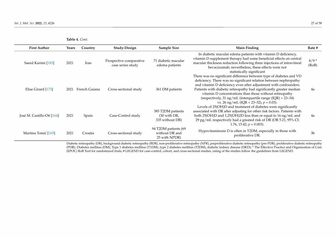
Because of its ability to inhibit neovascularization, vitamin D has been studied in thedevelopment of diabetic retinopathy (DR). Many observational studies have examined therelationship between vitamin D levels and the prevalence or severity of DR, with mostidentifying an inverse association with both type 1 and 2 diabetes [130–165]. However, aChinese study has reported a lack of association between vitamin D deficiency and DRafter adjusting for all potential covariates, such as demographics, physical measurements,laboratory measurements, related complications, comorbidities, and medications [166].Another Indian study suggested a possible association of vitamin D deficiency with type2 diabetes, but not specifically with DR [167]. As demonstrated in Table 4, in general, someof the studies reported an inverse correlation between the serum vitamin D and severity ofretinopathy [130,132–139,152,157,168,169]; similar findings were also reported, for example,the co-existence of low vitamin D and microvascular complications [131] or the associationbetween the severity of DR and the prevalence of vitamin D deficiency [133,161,165](Table 4); while some studies reported either no association or no significant differencebetween DR patients and healthy controls [170–173]. The agreement of the associationbetween vitamin D deficiency and neuropathy is lower when compared with retinopathy.While some studies report that the risk for having diabetic neuropathy is higher in thosewith vitamin D deficiency [134,137], there is limited research on contrasting findings [174].Further investigations are warranted.
Int. J. Mol. Sci. 2022, 23, 4226 18 of 58
Table 4. Summary of studies related to diabetic retinopathy included.
First Author Years Country Study-Design Sample Size Main Finding Rate #
HülyaAksoy [130] 2000 Turkey Cross-sectional study
20 DM without DRpatients, 15 DM with
BDR patients, 14 pre-DMwith PDR patients,17 DM with PDR
patients and20 healthy controls
There was an inverse relationship between the severity of theretinopathy, neovascularization, and serum 1,25(OH)2D3
concentrations, being the lowest in PDR and the highest in diabeticpatients without retinopathy patients. Mean 1,25(OH)2D3
concentrations fell with increasing severity of diabetic retinopathy.Only mean 1,25(OH)2D3 concentrations were not significantly
different between NDR and BDR, pre-PDR and PDR (p > 0.05). Mean1,25(OH)2D3 concentrations were significantly different among the
other groups (p < 0.05).
3a
Atsushi Suzuki [131] 2006 Japan Descriptive study 581 T2DM patients and51 healthy controls
Serum concentration of 25(OH)D3 were associated with HbAlc(p = 0.013). Microvascular complications and insulin treatment in
T2DM patients are associated with the co-existence ofhypovitaminosis D although serum creatinine levels were below
2.0 mg/dl.
4a
Harleen Kaur [132] 2011 Australia Cross-sectional study 517 T1DM patients
Vitamin D deficiency is associated with an increased prevalence ofretinopathy in young people with T1DM. In logistic regression,retinopathy was associated with VDD (odds ratio 2.12 [95% CI
1.03–4.33]), diabetes duration (1.13, 1.05–1.23), and HbA1c(1.24, 1.02–1.50).
4a
Christel Joergensen [175] 2011 Denmark Prospectiveobservational study 227 T1DM patients
In patients with type 1 diabetes, severe vitamin D deficiencyindependently predicts all-cause mortality (unadjusted HR 2.0
[0.9–4.4], p = 0.1 and HR 2.7 [1.1–6.7], p = 0.03, respectively) but notdevelopment of microvascular complications in the eye (HR 1.1
[0.7–1.7], p = 0.8) and kidney (unadjusted HR 1.1 [0.5–2.4], p = 0.8 andHR 1.3 [0.3–5.4], p = 0.7, respectively).
3a
Patricia A. Patrick [133] 2012 United States Cross-sectional study 1790 subjects
There is an association between severity of diabetic retinopathy andprevalence of vitamin D deficiency (β = 1.3, p = 0.01, unadjusted; andβ = 1.2, p = 0.01, adjusted for age and obesity status given their
clinical significance). However, regression analysis of these data didnot demonstrate a statistically significant relationship between the
2 variables (β = −0.04, p = 0.07, unadjusted; and β = −0.03, p = 0.13,adjusted). The findings were inconclusive about the existence of a
relationship between retinopathy severity and serum25(OH)D3 concentration.
3a
Int. J. Mol. Sci. 2022, 23, 4226 19 of 58
Table 4. Cont.
First Author Years Country Study-Design Sample Size Main Finding Rate #
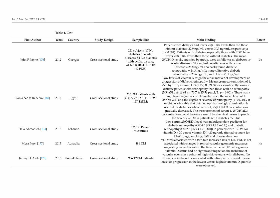
John F Payne [176] 2012 Georgia Cross-sectional study
221 subjects (17 Nodiabetes or ocular
diseases; 51 No diabeteswith ocular disease;
41 No BDR; 40 NPDR;42 PDR)
Patients with diabetes had lower 25(OH)D levels than did thosewithout diabetes (22.9 ng/mL versus 30.3 ng/mL, respectively;
p < 0.001). Patients with diabetes, especially those with PDR, havelower 25(OH)D levels than those without diabetes. The mean
25(OH)D levels, stratified by group, were as follows: no diabetes orocular disease = 31.9 ng/mL; no diabetes with ocular
disease = 28.8 ng/mL; no background diabeticretinopathy = 24.3 ng/mL; nonproliferative diabeticretinopathy = 23.6 ng/mL; and PDR = 21.1 ng/mL
3a
Rania NAM Reheem [168] 2013 Egypt Cross-sectional study200 DM patients with
suspected DR (43 T1DM;157 T2DM)
Low levels of vitamin D might be a risk marker of development orprogression of diabetic retinopathy. Mean serum concentration of 1,25 dihydroxy vitamin D 3 (1,25(OH)2D3) was significantly lower indiabetic patients with retinopathy than those with no retinopathy
(NR) (51.4 ± 16.64 vs. 70.7 ± 15.56 pmol/L, p < 0.001). There was asignificant negative correlation between the mean level of 1,
25(OH)2D3 and the degree of severity of retinopathy (p < 0.001). Itmight be advisable that detailed ophthalmologic examination isneeded for diabetics whose serum 1, 25(OH)2D3 concentrationsgradually decreased. The measurement of serum 1, 25(OH)2D3
concentrations could become a useful biochemical means to predictthe severity of DR in patients with diabetes mellitus
3a
Hala Ahmadieh [134] 2013 Lebanon Cross-sectional study 136 T2DM and74 controls
Low serum 25(OH)D3 level was an independent predictor fordiabetic neuropathy (OR 4.5 [95% CI 1.6–12]) and diabetic
retinopathy (OR 2.8 [95% CI 2.1–8.0]) in patients with T2DM forvitamin D < 20 versus vitamin D ≥ 20 ng/mL after adjustment for
HbA1c, age, smoking, BMI and disease duration.
4a
Myra Poon [177] 2013 Australia Cross-sectional study 481 DMVDD was associated with a two-fold increased risk of DR. VDD is not
associated with changes in retinal vascular geometric measures,suggesting an earlier role in the time course of DR pathogenesis.
4a
Jimmy D. Alele [170] 2013 United States Cross-sectional study 936 T2DM patients
Vitamin D status had no significant impact on the incidence ofvascular events in a cohort of high-risk veterans with diabetes. Nodifferences in the odds associated with retinopathy or renal diseaseonset or progression in the lowest versus highest vitamin D quartile
were observed.
4b
Int. J. Mol. Sci. 2022, 23, 4226 20 of 58
Table 4. Cont.
First Author Years Country Study-Design Sample Size Main Finding Rate #
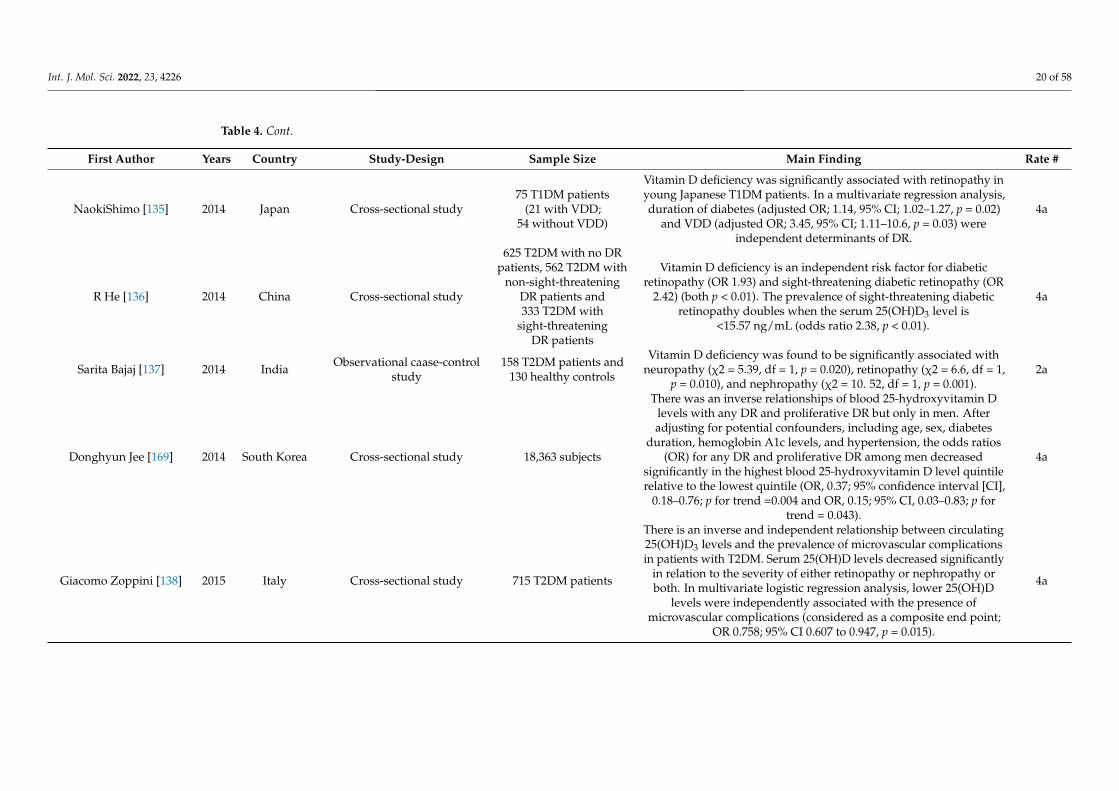
NaokiShimo [135] 2014 Japan Cross-sectional study75 T1DM patients
(21 with VDD;54 without VDD)
Vitamin D deficiency was significantly associated with retinopathy inyoung Japanese T1DM patients. In a multivariate regression analysis,duration of diabetes (adjusted OR; 1.14, 95% CI; 1.02–1.27, p = 0.02)
and VDD (adjusted OR; 3.45, 95% CI; 1.11–10.6, p = 0.03) wereindependent determinants of DR.
4a
R He [136] 2014 China Cross-sectional study
625 T2DM with no DRpatients, 562 T2DM with
non-sight-threateningDR patients and333 T2DM with
sight-threateningDR patients
Vitamin D deficiency is an independent risk factor for diabeticretinopathy (OR 1.93) and sight-threatening diabetic retinopathy (OR
2.42) (both p < 0.01). The prevalence of sight-threatening diabeticretinopathy doubles when the serum 25(OH)D3 level is
<15.57 ng/mL (odds ratio 2.38, p < 0.01).
4a
Sarita Bajaj [137] 2014 India Observational caase-controlstudy
158 T2DM patients and130 healthy controls
Vitamin D deficiency was found to be significantly associated withneuropathy (χ2 = 5.39, df = 1, p = 0.020), retinopathy (χ2 = 6.6, df = 1,
p = 0.010), and nephropathy (χ2 = 10. 52, df = 1, p = 0.001).2a
Donghyun Jee [169] 2014 South Korea Cross-sectional study 18,363 subjects
There was an inverse relationships of blood 25-hydroxyvitamin Dlevels with any DR and proliferative DR but only in men. Afteradjusting for potential confounders, including age, sex, diabetes
duration, hemoglobin A1c levels, and hypertension, the odds ratios(OR) for any DR and proliferative DR among men decreased
significantly in the highest blood 25-hydroxyvitamin D level quintilerelative to the lowest quintile (OR, 0.37; 95% confidence interval [CI],
0.18–0.76; p for trend =0.004 and OR, 0.15; 95% CI, 0.03–0.83; p fortrend = 0.043).
4a
Giacomo Zoppini [138] 2015 Italy Cross-sectional study 715 T2DM patients
There is an inverse and independent relationship between circulating25(OH)D3 levels and the prevalence of microvascular complicationsin patients with T2DM. Serum 25(OH)D levels decreased significantly
in relation to the severity of either retinopathy or nephropathy orboth. In multivariate logistic regression analysis, lower 25(OH)D
levels were independently associated with the presence ofmicrovascular complications (considered as a composite end point;
OR 0.758; 95% CI 0.607 to 0.947, p = 0.015).
4a
Int. J. Mol. Sci. 2022, 23, 4226 21 of 58
Table 4. Cont.
First Author Years Country Study-Design Sample Size Main Finding Rate #
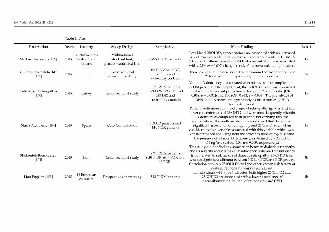
Markus Herrmann [139] 2015Australia, NewZealand, and
Finland
Multinational,double-blind,
placebo-controlled trial9795 T2DM patients
Low blood 25(OH)D3 concentrations are associated with an increasedrisk of macrovascular and microvascular disease events in T2DM. A50 nmol/L difference in blood 25OH-D concentration was associatedwith a 23% (p = 0.007) change in risk of macrovascular complications.
4a
G Bhanuprakash Reddy[167] 2015 India Cross-sectional
case-control study
82 T2DM with DRpatients and
99 healthy controls
There is a possible association between vitamin D deficiency and type2 diabetes, but not specifically with retinopathy. 3a
Celil Alper Usluogullari[140] 2015 Turkey Cross-sectional study
557 T2DM patients(299 DPN, 223 DN and
235 DR) and112 healthy controls
Vitamin D deficiency is associated with microvascular complicationsin DM patients. After adjustment, the 25 (OH) D level was confirmed
to be an independent protective factor for DPN (odds ratio [OR]:0.968, p = 0.004]) and DN (OR: 0.962, p = 0.006). The prevalence of
DPN and DN increased significantly as the serum 25 (OH) Dlevels decreased.
4a
Nuria Alcubierre [154] 2015 Spain Case-Control study 139 DR patients and144 NDR patients
Patients with more advanced stages of retinopathy (grades 2–4) hadlower concentrations of 25(OH)D and were more frequently vitamin
D deficient as compared with patients not carrying this eyecomplication. The multivariate analyses showed that there was asignificant association of retinopathy and 25(OH)D, even when
considering other variables associated with this variable which wereconsistent when analyzing both the concentrations of 25(OH)D and
the presence of vitamin D deficiency, as defined by a 25(OH)D<15 ng/mL (values 0.04 and 0.009, respectively.)
2a
Shokoufeh Bonakdaran[174] 2015 Iran Cross-sectional study
235 T2DM patients(153 NDR, 64 NPDR and
18 PDR)
This study did not find any association between diabetic retinopathyand its severity and vitamin D insufficiency. Vitamin D insufficiencyis not related to risk factors of diabetic retinopathy. 25(OH)D level
was not significant different between NDR, NPDR and PDR groups.Correlation between 25 (OH) D level and other known risk factors of
diabetic retinopathy was not significant.
3b
Lian Engelen [155] 2015 16 Europeancountries Prospective cohort study 532 T1DM patients
In individuals with type 1 diabetes, both higher 25(OH)D2 and25(OH)D3 are associated with a lower prevalence ofmacroalbuminuria, but not of retinopathy and CVD.
3b
Int. J. Mol. Sci. 2022, 23, 4226 22 of 58
Table 4. Cont.
First Author Years Country Study-Design Sample Size Main Finding Rate #
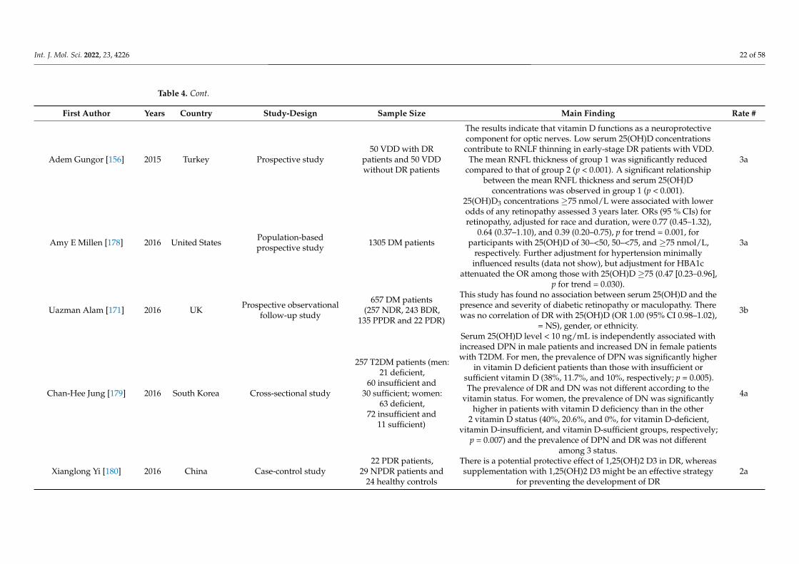
Adem Gungor [156] 2015 Turkey Prospective study50 VDD with DR
patients and 50 VDDwithout DR patients
The results indicate that vitamin D functions as a neuroprotectivecomponent for optic nerves. Low serum 25(OH)D concentrationscontribute to RNLF thinning in early-stage DR patients with VDD.
The mean RNFL thickness of group 1 was significantly reducedcompared to that of group 2 (p < 0.001). A significant relationship
between the mean RNFL thickness and serum 25(OH)Dconcentrations was observed in group 1 (p < 0.001).
3a
Amy E Millen [178] 2016 United States Population-basedprospective study 1305 DM patients
25(OH)D3 concentrations ≥75 nmol/L were associated with lowerodds of any retinopathy assessed 3 years later. ORs (95 % CIs) forretinopathy, adjusted for race and duration, were 0.77 (0.45–1.32),
0.64 (0.37–1.10), and 0.39 (0.20–0.75), p for trend = 0.001, forparticipants with 25(OH)D of 30–<50, 50–<75, and ≥75 nmol/L,
respectively. Further adjustment for hypertension minimallyinfluenced results (data not show), but adjustment for HBA1c
attenuated the OR among those with 25(OH)D ≥75 (0.47 [0.23–0.96],p for trend = 0.030).
3a
Uazman Alam [171] 2016 UK Prospective observationalfollow-up study
657 DM patients(257 NDR, 243 BDR,
135 PPDR and 22 PDR)
This study has found no association between serum 25(OH)D and thepresence and severity of diabetic retinopathy or maculopathy. Therewas no correlation of DR with 25(OH)D (OR 1.00 (95% CI 0.98–1.02),
= NS), gender, or ethnicity.
3b
Chan-Hee Jung [179] 2016 South Korea Cross-sectional study
257 T2DM patients (men:21 deficient,
60 insufficient and30 sufficient; women:
63 deficient,72 insufficient and
11 sufficient)
Serum 25(OH)D level < 10 ng/mL is independently associated withincreased DPN in male patients and increased DN in female patientswith T2DM. For men, the prevalence of DPN was significantly higher
in vitamin D deficient patients than those with insufficient orsufficient vitamin D (38%, 11.7%, and 10%, respectively; p = 0.005).The prevalence of DR and DN was not different according to the
vitamin status. For women, the prevalence of DN was significantlyhigher in patients with vitamin D deficiency than in the other
2 vitamin D status (40%, 20.6%, and 0%, for vitamin D-deficient,vitamin D-insufficient, and vitamin D-sufficient groups, respectively;
p = 0.007) and the prevalence of DPN and DR was not differentamong 3 status.
4a
Xianglong Yi [180] 2016 China Case-control study22 PDR patients,
29 NPDR patients and24 healthy controls
There is a potential protective effect of 1,25(OH)2 D3 in DR, whereassupplementation with 1,25(OH)2 D3 might be an effective strategy
for preventing the development of DR2a
Int. J. Mol. Sci. 2022, 23, 4226 23 of 58
Table 4. Cont.
First Author Years Country Study-Design Sample Size Main Finding Rate #
M Long [141] 2017 United States Retrospectivecross-sectional study
842 DR patients, 301 mild NPRpatients, 195 severe NPR patients
and 106 PDR
Vitamin D deficiency associated with severe DR in patients withwell controlled glycasemia. After adjusting gender, ethnicity and
duration of diabetes, the interaction of HbA1 with vitamin Ddeficiency significantly affected retinopathy severity (p = 0.029).
4b
Banu Boyuk [157] 2017 Turkey Cross-sectionalcase-control study
206 T2DM patients and34 healthy controls
There is an inverse relationship between the circulating 25(OH) Dlevel and the prevalence of retinopathy and neuropathy in T2DM
patients. The level of serum 25-hydroxyvitamin D (25(OH) D)was significantly lower in the T2DM patients (11.16 ± 3.99 ng/mL
vs. 15.58 ± 3.16 ng/mL; p < 0.05). Serum 25(OH) D levelsweresignificantly lower in the presence of retinopathy and
neuropathy (p < 0.05 for both), but no significant associationbetween Vitamin D level and microalbuminuria was found.
3b
Anurag Chaurasia [158] 2017 India Case-control study
120 DM patients (40 No BDR,42 mild-mod NPDR and
38 sev–v.serv NPDR and PDR)and 100 NDM patients (58 no DMand ocular diseases and 42 no DM
with ocular diseases)
Diabetic subjects, especially those with NPDR (severe and verysevere grade) and PDR have lower Vitamin D levels than those
without diabetes. Subjects with vitamin D insufficiency are at riskof having retinopathy 9.75 times that of vitamin D sufficientsubjects (Odds Ratio 9.75; 95% C.I. 1.96 to 48.46). The mean
vitamin D level in Diabetics (20.7 ± 6.91 ng/mL) is significantlyless than that in non-diabetics (27.51 ± 5.53 ng/mL) (p < 0.0001).Also, diabetics have significantly higher proportion of subjects
with vitamin D insufficiency (91.3%) as compared toNon-Diabetics (68%).
4a
Beteal Ashinne [142] 2018 India Retrospective study 3054 T2DM patients
Lower serum 25(OH)D3 was associated with increased severity ofDR and the presence of vitamin D deficiency was associated with
a two-fold increased risk for PDR. A statistically significantdifference in the serum vitamin D means of these categorizations:
no DR (13.7 ± 2.1 ng/mL), non-sight threatening DR(12.8 ± 2.1 ng/mL), sight threatening DR (11.1 ± 2.2 ng/mL),
(p < 0.001).
4a
Abdulbari Bener [143] 2018 Turkey Cross-sectional study 638 DM patients Vitamin D deficiency is considered as a risk factor for DR andhearing loss among diabetic patients.
KrishnamoorthyEzhilarasi [181] 2018 India Case-control study
200 T2DM patients, 216 T2DMwith microvascular complications,
198 T2DM with macrovascularcomplications and 252 controlswith nomal glucose tolerance
VDR (rs1544410) SNP was found to be associated with decreasedserum (25[OH]D) levels in both micro-macrovascular
complications of T2DM among South Indian Population.4a
Int. J. Mol. Sci. 2022, 23, 4226 24 of 58
Table 4. Cont.
First Author Years Country Study-Design Sample Size Main Finding Rate #
Josef Richter [159] 2018 Czech Republic Clinic Controlled Trial
52 DR patients (VitaminD and Beta-glucan
supplement, Vitamin Dand placebo supplement
and vitamin Dsupplement only)
Significant vitamin D deficits were found in all cases, even after threemonths of supplementation with vitamin D.
7/9 *(RoB)
Martina Závorková [182] 2018 Czech Republic Clinic Controlled Trial
54 DR patients(beta-glucan and vitaminD supplement, vitamin
D and placebosupplement, vitamin D
supplement only, nosupplementation
Glucan and vitamin D supplementation strongly influence lipidmetabolism and have positive effects on human health.
7/9 *(RoB)
Gauhar Nadri [144] 2019 India Cross-sectional study
24 DM without DRpatients, 24 DM withNPDR patients and
24 DM withPDR patients
Serum vitamin D levels of ≤ 18.6 ng/mL serve as sensitive andspecific indicator for proliferative disease, among patients of DR.
Univariate ordinal logistic regression analysis found vitamin D as asignificant predictor of severity of diabetic retinopathy {OR (95% CI)
= 1.11 (1.06–1.16) (p < 0.01 or p < 0.001)}. ROC curve analysisdemonstrated vitamin D cut off value of 18.6 ng/mL to be
significantly associated with NPDR and PDR
4a
Jing Yuan [145] 2019 China Cross-sectional study 889 T2DM patients
Vitamin D deficiency is significantly associated with risk of PDR. Theodd ratio in VDD individuals was significantly increased (1.84, 95%CI 1.18–2.86) for DR, 1.60 (95% CI 1.06–2.42) for PDR, compared withthose in vitamin D sufficient individuals, adjusted by age, sex, blood
pressure, kidney function, diabetic duration, and HbA1c.
4a
Heng Wan [146] 2019 China Cross-sectional study 4767 DM patients
Lower serum 25(OH)D3 concentration is significantly associated withhigher prevalence of DR in middle-aged and elderly diabetic adults.
Compared with the first 25(OH)D quartile (relatively lowerconcentraion), participants in the fourth quartile (relatively higher
concentration) had a lower prevalence of high ACR (OR 0.77, 95% CI0.61, 0.96) (p for trend <0.01). For DR, the OR of DR for the fourth
25(OH)D quartile when compared with the first quartile was0.62 (95% CI: 0.47, 0.82; p for trend <0.01).
4a
Int. J. Mol. Sci. 2022, 23, 4226 25 of 58
Table 4. Cont.
First Author Years Country Study-Design Sample Size Main Finding Rate #
Abdulhalim Senyigit [160] 2019 Turkey Cross-sectional study 163 T2DM patients and40 controls
Low serum 25-OHD levels were found to be associated with thedevelopment of diabetes and complications. Serum 25(OH)D levels
in all patients were significantly lower than the control group(p < 0.05). Levels of 25(OH)D for those with complications were
lower than that without complications. (p values for nephropahtyand retinopathy groups were <0.001 while that for neuropahty was<0.01). Low serum 25-OHD levels may be a consequence of even
worse metabolic control of diabetes.
3a
Abdulla Almoosa [161] 2019 Bahrain Prospectiveobservational study
300 T2DM patients(150 NDR, 99 NPDR and
51 PDR)
VDD was commonly found in type II DM patients. Lower serum25(OH)D levels were associated with more severe DR. 3b
Mehrdad Afarid [148] 2020 Iran Cross-sectional study
21 DM with NPRpatients, 9 DM with PDRpatients and 30 DM with
no DR patients
Patients with DR had lower levels of serum vitamin D compared withthose without retinopathy, especially those with severe NPDR
and PDR3a
Amy E. Millen [178] 2020 Portugal Retrospectiveobservational study 182 T1DM patients
Lower levels of vitamin D were associated with an increasedprevalence of diabetic retinopathy in patients with T1DM, after
adjusting for possible confounders. The lower circulating 25(OH)Dlevels were significantly associated with greater prevalence of DR
after adjusting for confounders (OR = 0.94; 95% CI 0.90–0.99,p = 0.023).
4a
Lina H. M. Ahmed [150] 2020 Qatar Case-control study 274 T2DM patients and222 healthy controls
Vitamin D3 was associated with diabetic retinopathy whilst vitaminD2 was not. 2a
Lina H. M. Ahmed [162] 2021 Qatar Cross-sectional study 460 T2DM patients and290 healthy controls
Vitamin D3 metabolites were associated with diabetic retinopathy,whereas total vitamin D levels were not, suggesting that endogenous
vitamin D(3) metabolites are a better measure of diabeticmicrovascular complications. The lower 1,25(OH)2D3 levels wereassociated with diabetic retinopathy (p = 0.006) hypertension and
dyslipidemia (both p = 0.01) and CAD (p = 0.012); while noassociation between either total 1,25(OH)2D or 1,25(OH)2D3 levels
with diabetic neuropathy, PAD or CAD.
3a
Alexandra E. Butler [147] 2020 Qatar Cross-sectional study 460 T2DM patients and290 healthy controls
Lower 25(OH)D(3) was associated with retinopathy (p < 0.03) anddyslipidemia (p < 0.04), but not neuropathy or vascular complications 3a

Int. J. Mol. Sci. 2022, 23, 4226 26 of 58
Table 4. Cont.
First Author Years Country Study-Design Sample Size Main Finding Rate #
Ying Xiao [166] 2020 China Cross-sectional study 4284 T2DM patients
In unadjusted analyses, DR was associated with VDD status (PR:1.147; 95% CI: 1.025–1.283), the associate retained after adjusted with
age and sex and other demographic and physical measurements.However, the significance diminished after adjusting all confounders
(PR: 1.093; 95% CI: 0.983–1.215).
4a
Gauhar Nadri [163] 2021 India Cross-sectional study66 T2DM patients (22 No
DR, 22 NPDR and22 PDR) and 22 controls
Low serum vitamin D levels correlate with increased severity of DR. 4a
Li Lu [151] 2021 China Retrospective study
55 PDR patients,25 non-diabetic patientswith idiopathic macular
hole patients and10 NDR patients
In ROC-curve analyses, both serum and vitreous 25(OH)D showeddiscriminatory ability in predicting DR (NPDR and PDR) and PDR.
In DR prediction, they obtained the same area under curve (AUC) of0.77. Serum 25 (OH) D has a better predictive value (AUC: 0.77) than
serum 25 (OH) D (AUC: 0.66) in PDR prediction.
4a
Wei-Jing Zhao [152] 2021 China Cross-sectional study 815 T2DM patients
Vitamin D deficiency is independently associated with higher risk ofdiabetic peripheral neuropathy and DR, but not diabetic retinopathy,
in T2DM patients. Univariate analysis showed that the 25 (OH) Dwas significantly correlated with DPN (odds ratio [OR]: 0.969, 95%confidence interval [CI]: 0.950–0.989, p = 0.003) and DN (OR: 0.950,95% CI: 0.928–0.973, p < 0.001), but not with DR (OR: 1.014, 95% CI:
0.994–1.034, p = 0.165). Multiple logistic regression analysis afteradjustment showed that the 25 (OH) D level was an independent
protective factor for DPN and DN.
4a
Xin Zhao [153] 2021 China Retrospective study636 T2DM patients
(466 NDR, 120 BDR and50 PDR)
A close association was observed between 25(OH)D3 level and DR inthe elderly male patients and postmenopausal women with T2DM.There was a significant difference was observed among the three
groups in men and women (men: χ2 = 7:75, p < 0.05; women:χ2 = 7:75, p < 0.05)
4a
Mehmet Balbaba [172] 2021 Turkey Prospective study20 T2DM-DR patients,
20 T2DM-NDR patientsand 20 healthy controls
Vitamin D levels were similar between diabetic patients with andwithout DR and healthy control subjects 3b
Int. J. Mol. Sci. 2022, 23, 4226 27 of 58
Table 4. Cont.
First Author Years Country Study-Design Sample Size Main Finding Rate #
Saeed Karimi [183] 2021 Iran Prospective comparativecase series study
71 diabetic macularedema patients
In diabetic macular edema patients with vitamin D deficiency,vitamin D supplement therapy had some beneficial effects on centralmacular thickness reduction following three injections of intravitreal
bevacizumab; nevertheless, these effects were notstatistically significant
8/9 *(RoB)
Elise Girard [173] 2021 French Guiana Cross-sectional study 361 DM patients
There was no significant difference between type of diabetes and VDdeficiency. There was no significant relation between nephropathyand vitamin D deficiency even after adjustment with confounders.Patients with diabetic retinopathy had significantly greater median
vitamin D concentrations than those without retinopathy(respectively, 31 ng/mL (interquartile range (IQR) = 23–34)
vs. 26 ng/mL (IQR = 23–32), p = 0.03).
4a
José M. Castillo-Otí [164] 2021 Spain Case-Control study385 T2DM patients
(30 with DR,335 without DR)
Levels of 25(OH)D and treatment of diabetes were significantlyassociated with DR after adjusting for other risk factors. Patients withboth 25(OH)D and 1,25(OH)2D less than or equal to 16 ng/mL and29 pg/mL respectively had a greated risk of DR (OR 5.21, 95% CI:
1.76, 15.42; p = 0.003).
4a
Martina Tomic [165] 2021 Croatia Cross-sectional study94 T2DM patients (69
without DR and25 with NPDR)
Hypovitaminosis D is often in T2DM, especially in those withproliferative DR. 3b
Diabetic retinopathy (DR), background diabetic retinopathy (BDR), non-proliferative retinopathy (NPR), preproliferative diabetic retinopathy (pre-PDR), proliferative diabetic retinopathy(PDR), Diabetes mellitus (DM), Type 1 diabetes mellitus (T1DM), type 2 diabetes mellitus (T2DM), diabetic kidney disease (DKD); * The Effective Practice and Organisation of Care(EPOC) RoB Tool for randomized trials; # LEGEND for case-control, cohort, and cross-sectional studies, rating of the studies follow the guidelines from LEGEND.
Int. J. Mol. Sci. 2022, 23, 4226 28 of 58
Besides cross-sectional studies, a population-based prospective study also showed thata high level of vitamin D was associated with a lower risk of DR after 3 years [178]. A double-blind, placebo-controlled trial found that low blood 25(OH)D3 levels were associatedwith an increased risk of macrovascular and microvascular disease events among type2 diabetics [139].
DR is a serious microvascular complication of diabetes. The characteristics of early DRinclude the loss of pericytes from retinal capillaries, the appearance of acellular capillariesand microaneurysms, and the breakdown of the blood-retinal barrier. In the proliferativephase of DR, neovascularization in the retina may occur, which significantly increasesthe probability of vision loss [184,185]. Potential mechanisms that explain how vitaminD can prevent DR include insulin resistance, immune regulation, anti-inflammation, andanti-angiogenesis. Animal studies have shown that vitamin D is important for insulinsynthesis and can improve the body’s sensitivity to insulin, reducing the risk of insulin re-sistance [186,187]. Other studies have found that vitamin D treatment decreased the retinalexpression of VEGF and the transforming growth factor TGF-β1 in rats [188], which mayhave protective effects on the retina. VDR has also been implicated in the pathogenesis ofDR [189]. A meta-analysis of seven studies evaluating the association of the VDR gene poly-morphisms with DR found that the FokI polymorphism of the VDR gene has a significantassociation with DR susceptibility [190]. Apart from the VDR polymorphism, other studieshave proposed different protective mechanisms of vitamin D on DR, including protectingthe vasculature [191–193], reducing oxidative stress [194,195], modulating inflammationand immune responses [180,196–198], inhibiting the renin-angiotensin aldosterone sys-tem [199,200], reducing the effects of advanced glycation end products [201,202], reducingendoplasmic reticulum stress [203,204], regulating endothelial cells apoptosis [205], andregulating diabetic leukostasis [206]. Further studies are needed to determine the exactmechanisms of vitamin D on DR.
In summary, even though there are no consistent associations between vitamin D leveland DR in observational studies, more than 30 reports suggested an inverse relationship.The same conclusion is made in perspective studies, although the causal relationship hasnot been identified. Further studies should investigate whether vitamin D supplementationcan reduce the risk of DR.
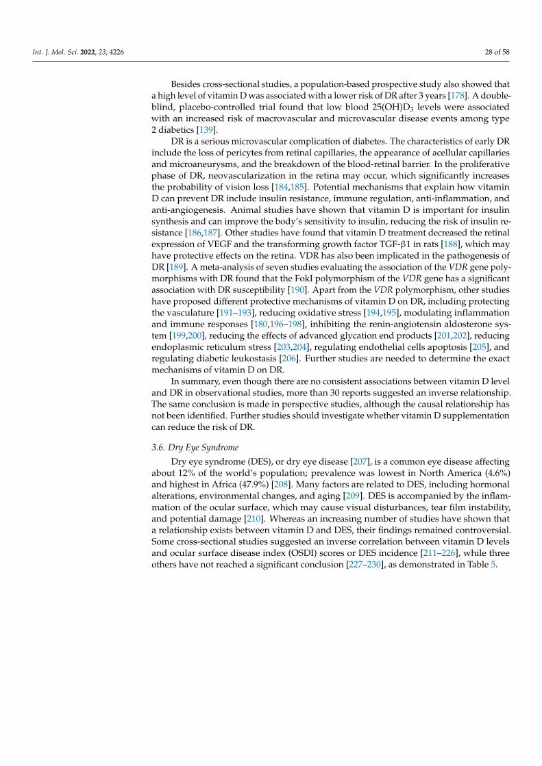
3.6. Dry Eye Syndrome
Dry eye syndrome (DES), or dry eye disease [207], is a common eye disease affectingabout 12% of the world’s population; prevalence was lowest in North America (4.6%)and highest in Africa (47.9%) [208]. Many factors are related to DES, including hormonalalterations, environmental changes, and aging [209]. DES is accompanied by the inflam-mation of the ocular surface, which may cause visual disturbances, tear film instability,and potential damage [210]. Whereas an increasing number of studies have shown thata relationship exists between vitamin D and DES, their findings remained controversial.Some cross-sectional studies suggested an inverse correlation between vitamin D levelsand ocular surface disease index (OSDI) scores or DES incidence [211–226], while threeothers have not reached a significant conclusion [227–230], as demonstrated in Table 5.
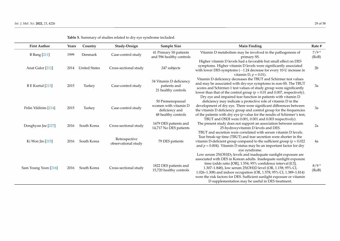
Int. J. Mol. Sci. 2022, 23, 4226 29 of 58
Table 5. Summary of studies related to dry eye syndrome included.
First Author Years Country Study-Design Sample Size Main Finding Rate #
B Bang [211] 1999 Denmark Case-control study 41 Primary SS patientsand 596 healthy controls
Vitamin D metabolism may be involved in the pathogenesis ofprimary SS.
7/9 *(RoB)
Anat Galor [212] 2014 United States Cross-sectional study 247 subjects
Higher vitamin D levels had a favorable but small effect on DESsymptoms. Higher vitamin D levels were significantly associated
with lower DES symptoms (−1.24 decrease for every 10-U increase invitamin D, p = 0.01).
2b
B E Kurtul [213] 2015 Turkey Case-control study34 Vitamin D deficiency
patients and21 healthy controls
Vitamin D deficiency decreases the TBUT and Schirmer test valuesand may be associated with dry-eye symptoms in non-SS. The TBUTscores and Schirmer-1 test values of study group were significantly
lower than that of the control group (p = 0.01 and 0.007, respectively).
3a
Pelin Yildirim [214] 2015 Turkey Case-control study
50 Premenopausalwomen with vitamin D
deficiency and48 healthy controls
Dry eye and impaired tear function in patients with vitamin Ddeficiency may indicate a protective role of vitamin D in the
development of dry eye. There were significant differences betweenthe vitamin D deficiency group and control group for the frequenciesof the patients with dry eye (p-value for the results of Schirmer’s test,
TBUT and OSDI were 0.001, 0.001 and 0.003 respectively).
3a
Donghyun Jee [227] 2016 South Korea Cross-sectional study 1679 DES patients and14,717 No DES patients
The present study does not support an association between serum25-hydroxyvitamin D levels and DES. 2a
Ki Won Jin [215] 2016 South Korea Retrospectiveobservational study 79 DES patients
TBUT and secretion were correlated with serum vitamin D levels.Tear break-up time (TBUT) and tear secretion were shorter in the
vitamin D-deficient group compared to the sufficient group (p = 0.022and p = 0.004). Vitamin D status may be an important factor for dry
eye syndrome.
4a
Sam Young Yoon [216] 2016 South Korea Cross-sectional study 1822 DES patients and15,720 healthy controls
Low serum 25(OH)D3 levels and inadequate sunlight exposure areassociated with DES in Korean adults. Inadequate sunlight exposure
time (odds ratio [OR], 1.554; 95% confidence interval [CI],1.307–1.848), low serum 25(OH)D level (OR, 1.158; 95% CI,
1.026–1.308) and indoor occupation (OR, 1.578; 95% CI, 1.389–1.814)were the risk factors for DES. Sufficient sunlight exposure or vitamin
D supplementation may be useful in DES treatment.
8/9 *(RoB)
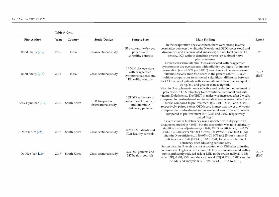
Int. J. Mol. Sci. 2022, 23, 4226 30 of 58
Table 5. Cont.
First Author Years Country Study-Design Sample Size Main Finding Rate #
Rohit Shetty [217] 2016 India Cross-sectional study52 evaporative dry eye
patients and43 healthy controls
In the evaporative dry eye cohort, there were strong inversecorrelation between the vitamin D levels and OSDI scores (total anddiscomfort- and vision-related subscales) but not total corneal DC
density, DCs without dendritic process, or subbasal nerveplexus features.
2b
Rohit Shetty [218] 2016 India Cross-sectional study
19 Mild dry eye signswith exaggerated
symptoms patients and19 healthy controls
Decreased serum vitamin D was associated with exaggeratedsymptoms in dry eye patients with mild dry eye signs. An inversecorrelation (r = −0.569; p = 0.0110) was observed between serumvitamin D levels and OSDI score in the patient cohort. Tukey’s
multiple comparisons test showed a significant difference betweenthe OSDI score of patients with serum vitamin D less than or equal to
10 ng/mL and greater than 20 ng/mL.
7/9 *(RoB)
Seok Hyun Bae [219] 2016 South Korea Retrospectiveobservational study
105 DES refractory toconventional treatment
and vitamin Ddeficiency patients
Vitamin D supplementation is effective and useful in the treatment ofpatients with DES refractory to conventional treatment and with
vitamin D deficiency. The TBUT in males was increased after 2 weekscompared to pre–treatment and in female it was increased after 2 and
6 weeks compared to pre-treatment (p = 0.041, <0.001 and <0.001,respectively, paired t-test). OSDI score in men was lower at 6 weekscompared to pre-treatment and in women it was lower at 10 weeks
compared to pre-treatment (p = 0.033 and 0.012, respectively,paired t-test).
3a
Min Ji Kim [228] 2017 South Korea Cross-sectional study 1428 DES patients and7921 healthy controls
Severe vitamin D deficiency was associated with dry eye in anunadjusted model (p = 0.01), but the association was not statistically
significant after adjustment (p = 0.49, Vit D insufficiency; p = 0.33,VDD; p = 0.18, sever VDD). OR was 1.24 (95% CI, 0.66 to 2.41) forvitamin D insufficiency, 1.30 (95% CI, 0.75 to 2.25) for vitamin Ddeficiency, and 1.42 (95% CI, 0.83 to 2.41) for severe vitamin D
deficiency after adjusting confounders.
4a
Da-Hye Jeon [229] 2017 South Korea Cross-sectional study 393 DES patients and347 healthy controls
Serum vitamin D levels are not associated with DES after adjustingconfounders. Higher serum vitamin D levels were associated with a
non-significantly reduced risk of DED in the crude analysis (oddsratio [OR], 0.991; 95% confidence interval [CI], 0.971 to 1.011) and in
the adjusted analysis (OR, 0.988; 95% CI, 0.966 to 1.010).
8/9 *(RoB)
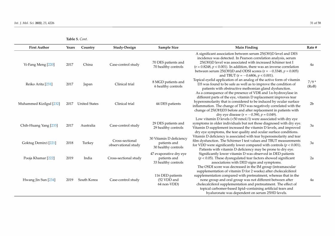
Int. J. Mol. Sci. 2022, 23, 4226 31 of 58
Table 5. Cont.
First Author Years Country Study-Design Sample Size Main Finding Rate #
Yi-Fang Meng [220] 2017 China Case-control study 70 DES patients and70 healthy controls
A significant association between serum 25(OH)D level and DESincidence was detected. In Pearson correlation analysis, serum
25(OH)D level was associated with increased Schimer test I(r = 0.8248, p < 0.001). In addition, there was an inverse correlationbetween serum 25(OH)D and ODSI scores (r = −0.3348, p = 0.005)
and TBUT (r = −0.6806, p < 0.001).
4a
Reiko Arita [231] 2017 Japan Clinical trial 8 MGD patients and6 healthy controls
Topical eyelid application of an analog of the active form of vitaminD3 was found to be safe as well as to improve the condition of
patients with obstructive meibomian gland dysfunction.
7/9 *(RoB)
Muhammed Kizilgul [232] 2017 United States Clinical trial 44 DES patients
As a consequence of the presence of VDR and 1α-hydroxylase indifferent parts of the eye, vitamin D replacement improves tear
hyperosmolarity that is considered to be induced by ocular surfaceinflammation. The change of TFO was negatively correlated with the
change of 25(OH)D3 before and after replacement in patients withdry eye disease (r = −0.390, p = 0.049).
2b
Chih-Huang Yang [233] 2017 Australia Case-control study 29 DES patients and29 healthy controls
Low vitamin D levels (<50 nmol/l) were associated with dry eyesymptoms in older individuals but not those diagnosed with dry eye.Vitamin D supplement increased the vitamin D levels, and improved
dry eye symptoms, the tear quality and ocular surface conditions.
3a
Goktug Demirci [221] 2018 Turkey Cross-sectionalobservational study
30 Vitamin D deficiencypatients and
30 healthy controls
Vitamin D deficiency is associated with tear hyperosmolarity and tearfilm dysfunction. The Schirmer I test values and TBUT measurementsfor VDD were significantly lower compared with controls (p < 0.001).
Patients with vitamin D deficiency may be prone to dry eye.
3a
Pooja Khamar [222] 2019 India Cross-sectional study47 evaporative dry eye
patients and33 healthy controls
Significantly lower vitamin D was observed in DED patients(p < 0.05). These dysregulated tear factors showed significant
associations with DED signs and symptoms.2a
Hwang Jin Sun [234] 2019 South Korea Case-control study116 DED patients
(52 VDD and64 non-VDD)
The OSDI score was decreased in the IM group (intramascularsupplementation of vitamin D for 2 weeks) after cholecalciferol
supplementation compared with pretreatment, whereas that in thenone group and oral group was not different between after
cholecalciferol supplementation and pretreatment. The effect oftopical carbomer-based lipid–containing artificial tears and
hyaluronate was dependent on serum 25HD levels.
4a
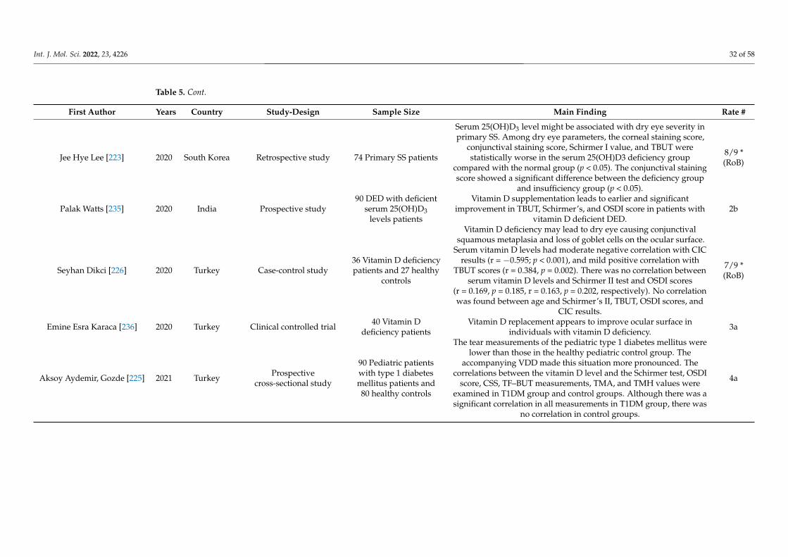
Int. J. Mol. Sci. 2022, 23, 4226 32 of 58
Table 5. Cont.
First Author Years Country Study-Design Sample Size Main Finding Rate #
Jee Hye Lee [223] 2020 South Korea Retrospective study 74 Primary SS patients
Serum 25(OH)D3 level might be associated with dry eye severity inprimary SS. Among dry eye parameters, the corneal staining score,
conjunctival staining score, Schirmer I value, and TBUT werestatistically worse in the serum 25(OH)D3 deficiency group
compared with the normal group (p < 0.05). The conjunctival stainingscore showed a significant difference between the deficiency group
and insufficiency group (p < 0.05).
8/9 *(RoB)
Palak Watts [235] 2020 India Prospective study90 DED with deficient
serum 25(OH)D3levels patients
Vitamin D supplementation leads to earlier and significantimprovement in TBUT, Schirmer’s, and OSDI score in patients with
vitamin D deficient DED.2b
Seyhan Dikci [226] 2020 Turkey Case-control study36 Vitamin D deficiencypatients and 27 healthy
controls
Vitamin D deficiency may lead to dry eye causing conjunctivalsquamous metaplasia and loss of goblet cells on the ocular surface.
Serum vitamin D levels had moderate negative correlation with CICresults (r = −0.595; p < 0.001), and mild positive correlation with
TBUT scores (r = 0.384, p = 0.002). There was no correlation betweenserum vitamin D levels and Schirmer II test and OSDI scores
(r = 0.169, p = 0.185, r = 0.163, p = 0.202, respectively). No correlationwas found between age and Schirmer’s II, TBUT, OSDI scores, and
CIC results.
7/9 *(RoB)
Emine Esra Karaca [236] 2020 Turkey Clinical controlled trial 40 Vitamin Ddeficiency patients
Vitamin D replacement appears to improve ocular surface inindividuals with vitamin D deficiency. 3a
Aksoy Aydemir, Gozde [225] 2021 Turkey Prospectivecross-sectional study
90 Pediatric patientswith type 1 diabetesmellitus patients and80 healthy controls
The tear measurements of the pediatric type 1 diabetes mellitus werelower than those in the healthy pediatric control group. The
accompanying VDD made this situation more pronounced. Thecorrelations between the vitamin D level and the Schirmer test, OSDI
score, CSS, TF–BUT measurements, TMA, and TMH values wereexamined in T1DM group and control groups. Although there was asignificant correlation in all measurements in T1DM group, there was
no correlation in control groups.
4a
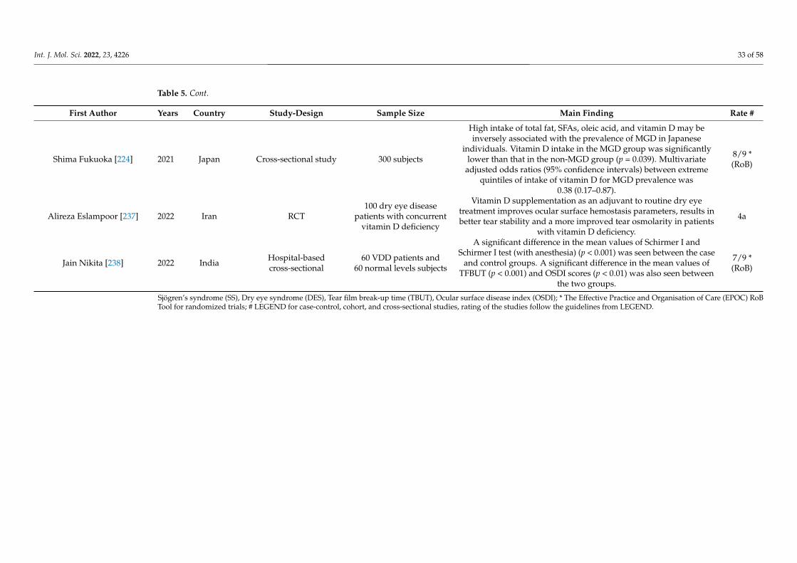
Int. J. Mol. Sci. 2022, 23, 4226 33 of 58
Table 5. Cont.
First Author Years Country Study-Design Sample Size Main Finding Rate #
Shima Fukuoka [224] 2021 Japan Cross-sectional study 300 subjects
High intake of total fat, SFAs, oleic acid, and vitamin D may beinversely associated with the prevalence of MGD in Japanese
individuals. Vitamin D intake in the MGD group was significantlylower than that in the non-MGD group (p = 0.039). Multivariate
adjusted odds ratios (95% confidence intervals) between extremequintiles of intake of vitamin D for MGD prevalence was
0.38 (0.17–0.87).
8/9 *(RoB)
Alireza Eslampoor [237] 2022 Iran RCT100 dry eye disease
patients with concurrentvitamin D deficiency
Vitamin D supplementation as an adjuvant to routine dry eyetreatment improves ocular surface hemostasis parameters, results inbetter tear stability and a more improved tear osmolarity in patients
with vitamin D deficiency.
4a
Jain Nikita [238] 2022 India Hospital-basedcross-sectional
60 VDD patients and60 normal levels subjects
A significant difference in the mean values of Schirmer I andSchirmer I test (with anesthesia) (p < 0.001) was seen between the case
and control groups. A significant difference in the mean values ofTFBUT (p < 0.001) and OSDI scores (p < 0.01) was also seen between
the two groups.
7/9 *(RoB)
Sjögren’s syndrome (SS), Dry eye syndrome (DES), Tear film break-up time (TBUT), Ocular surface disease index (OSDI); * The Effective Practice and Organisation of Care (EPOC) RoBTool for randomized trials; # LEGEND for case-control, cohort, and cross-sectional studies, rating of the studies follow the guidelines from LEGEND.
Int. J. Mol. Sci. 2022, 23, 4226 34 of 58
Several clinical trials investigated the treatment effects of vitamin D supplementationon DES symptoms, as demonstrated. Some of them only involved DES patients [232,236,237],while four other studies set healthy controls [219,231,233,235]. All these studies concludedthat vitamin D can improve the DES symptoms of tear quality and ocular surface conditions.However, these studies may suffer from a small sample size or lack of a placebo-controlgroup. Further well-design clinical trials with more samples are required to better under-stand the relationship between vitamin D and DES.
The key mechanism of vitamin D on DES may involve its antioxidation, anti-inflammatory,and immune-regulatory effects [239–241]. Vitamin D deficiency may cause the inflamma-tion of the ocular surface and ultimately DES [233]. Conversely, vitamin D may relieve DESthrough inhibiting the interleukin-6 (IL-6) inhibitor [233], the key mediator of localized in-flammation [242]. Moreover, vitamin D can suppress the release of inflammatory cytokinesand stimulate the release of antioxidant cytokines in tears. Lastly, vitamin D can improvecorneal epithelial barrier functions [233], which may improve ocular conditions. Apartfrom the role of vitamin D in the pathogenesis of DES, a study reported that the expressionof VDR and CYP27B1 (vitamin D metabolism enzyme) was significantly decreased in DEDpatients, suggesting the possible involvement of the vitamin D regulatory enzyme in pro-tecting the human eye from dry eye [243]. A study on SNPs of the VDR gene Apa-1, Bsm-1,Fok-1 and Taq-1, reported the association of Apa-1 and Taq-1 with the risk of DES [244].
In conclusion, the exact mechanisms of vitamin D on DES are unclear, but evidencefrom cross-sectional studies seems to suggest that vitamin D has a protective effect againstDES. Limit evidence from clinical trials also suggests that vitamin D supplementationcould help to improve DES symptoms, but a further placebo RCT is needed to verify thistreatment effect.
3.7. Thyroid Eye Diseases
Thyroid eye disease (TED), also known as Graves’ ophthalmopathy (GO), is an autoim-mune inflammatory disorder. Few studies have examined the relationship between vitaminD and TED (Table 6). A pilot study in Texas, United States found prevalence rates of 20%and 31% for vitamin D deficiency and insufficiency among TED patients, respectively [245].Another retrospective case-control study comparing vitamin D levels between Graves’disease patients and TED patients found that low serum vitamin D was associated withTED [246]. Assessing and supplementing vitamin D levels may be an important additionto the early management strategies of GD patients. Since vitamin D can regulate immuneresponses and reduce inflammation, there is a definite need to further evaluate the role ofvitamin D deficiency in TED patients. The current evidence for TED suggests that vitaminD would be associated with TED, however, the evidence is limited. It is worthwhile tofurther investigate the effect of vitamin D on TED.
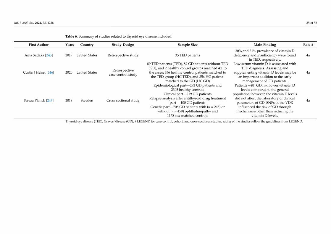
Int. J. Mol. Sci. 2022, 23, 4226 35 of 58
Table 6. Summary of studies related to thyroid eye disease included.
First Author Years Country Study-Design Sample Size Main Finding Rate #
Ama Sadaka [245] 2019 United States Retrospective study 35 TED patients20% and 31% prevalence of vitamin D
deficiency and insufficiency were foundin TED, respectively.
4a
Curtis J Heisel [246] 2020 United States Retrospectivecase-control study
89 TED patients (TED), 89 GD patients without TED(GD), and 2 healthy control groups matched 4:1 tothe cases; 356 healthy control patients matched to
the TED group (HC TED), and 356 HC patientsmatched to the GD (HC GD)
Low serum vitamin D is associated withTED diagnosis. Assessing and
supplementing vitamin D levels may bean important addition to the early
management of GD patients.
4a
Tereza Planck [247] 2018 Sweden Cross sectional study
Epidemiological part—292 GD patients and2305 healthy controls
Clinical part—219 GD patientsRelapse analysis after antithyroid drug treatment
part —100 GD patientsGenetic part—708 GD patients with (n = 245) or
without (n = 459) ophthalmopathy and1178 sex-matched controls
Patients with GD had lower vitamin Dlevels compared to the general
population; however, the vitamin D levelsdid not affect the laboratory or clinicalparameters of GD. SNPs in the VDR
influenced the risk of GD throughmechanisms other than reducing the
vitamin D levels.
4a
Thyroid eye disease (TED), Graves’ disease (GD); # LEGEND for case-control, cohort, and cross-sectional studies, rating of the studies follow the guidelines from LEGEND.

Int. J. Mol. Sci. 2022, 23, 4226 36 of 58
3.8. Uveitis
Uveitis is the inflammation of the uvea driven by the T-cells [248]. It can be describedas a failure of the ocular immune system, with the disease resulting from inflammation andtissue destruction. Since vitamin D can inhibit inflammation, influence T-cell responses, andregulate the immune system, it is necessary to examine its involvement in the developmentof uveitis. In an experimental autoimmune uveitis model, the oral administration ofcalcitriol was found to prevent and reverse the progression of uveitis by reducing theimmunological response [249].
Although in a population-based study, none of the 25 uveitis patients were found tohave vitamin D deficiency [250], two large retrospective case-control studies in the UnitedStates found an association of lower vitamin D levels with uveitis and scleritis, respec-tively [251,252]. Other case-control studies have been conducted on patients with specifictypes of uveitis, such as anterior uveitis (AAU) or non-infectious anterior uveitis [253–255],Vogt-Koyanagi-Harada (VKH) disease [256], sarcoidosis-associated uveitis [257], and juve-nile idiopathic arthritis (JIA)-associated uveitis [258] (Table 7). All these studies suggestedthat the vitamin D deficiency is associated with uveitis development. A recent prospectivecase-control study consistently reported the association of vitamin D levels with active andinactive non-infectious uveitis patients. Vitamin D levels are related to uveitis severity [259].
There are reported associations between VDR polymorphisms and uveitis. Single-nucleotide polymorphisms (SNPs) of the CYP2R1, CYP27B1, CYP24A1 and DHCR7 genesare linked with lower circulating vitamin D levels [260–263]. A study in a Chinese cohortfound that the frequencies of the genotype TT and T allele of DHCR7 rs12785878 were bothsignificantly higher among ocular Behçet disease (BD) patients compared with healthycontrols; however, similar associations were not found for the VKH, AAU with ankylosingspondylitis (AS), or pediatric uveitis [263]. Another cross-sectional study found that genevariants involved in vitamin D anabolism and catabolism may be important for VKHpathology [25].
In summary, much evidence has shown that the onset and activity of uveitis areassociated with vitamin D level, but most of the above studies were observational anddid not provide evidence for causality. In the future, randomized-controlled studies arerequired to evaluate whether vitamin D can be an option for treating uveitis.
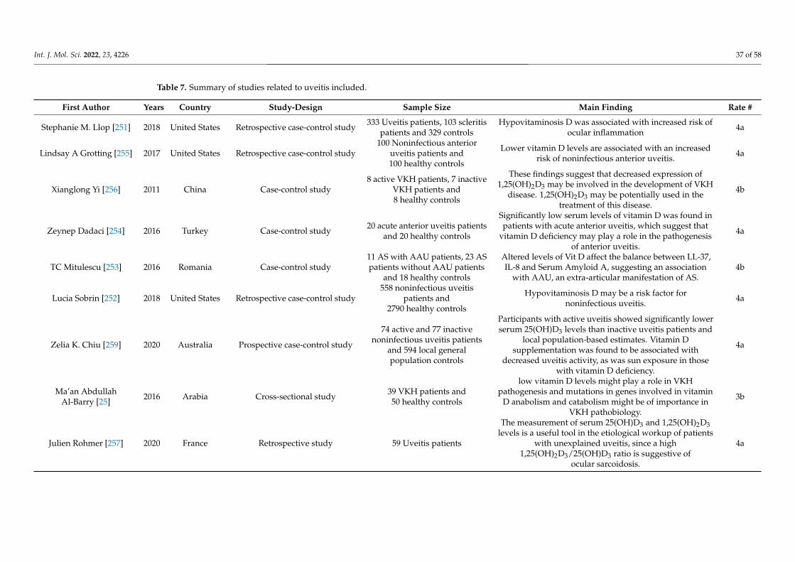
Int. J. Mol. Sci. 2022, 23, 4226 37 of 58
Table 7. Summary of studies related to uveitis included.
First Author Years Country Study-Design Sample Size Main Finding Rate #
Stephanie M. Llop [251] 2018 United States Retrospective case-control study 333 Uveitis patients, 103 scleritispatients and 329 controls
Hypovitaminosis D was associated with increased risk ofocular inflammation 4a
Lindsay A Grotting [255] 2017 United States Retrospective case-control study100 Noninfectious anterior
uveitis patients and100 healthy controls
Lower vitamin D levels are associated with an increasedrisk of noninfectious anterior uveitis. 4a
Xianglong Yi [256] 2011 China Case-control study8 active VKH patients, 7 inactive
VKH patients and8 healthy controls
These findings suggest that decreased expression of1,25(OH)2D3 may be involved in the development of VKH
disease. 1,25(OH)2D3 may be potentially used in thetreatment of this disease.
4b
Zeynep Dadaci [254] 2016 Turkey Case-control study 20 acute anterior uveitis patientsand 20 healthy controls
Significantly low serum levels of vitamin D was found inpatients with acute anterior uveitis, which suggest that
vitamin D deficiency may play a role in the pathogenesisof anterior uveitis.
4a
TC Mitulescu [253] 2016 Romania Case-control study11 AS with AAU patients, 23 ASpatients without AAU patients
and 18 healthy controls
Altered levels of Vit D affect the balance between LL-37,IL-8 and Serum Amyloid A, suggesting an association
with AAU, an extra-articular manifestation of AS.4b
Lucia Sobrin [252] 2018 United States Retrospective case-control study558 noninfectious uveitis
patients and2790 healthy controls
Hypovitaminosis D may be a risk factor fornoninfectious uveitis. 4a
Zelia K. Chiu [259] 2020 Australia Prospective case-control study
74 active and 77 inactivenoninfectious uveitis patients
and 594 local generalpopulation controls
Participants with active uveitis showed significantly lowerserum 25(OH)D3 levels than inactive uveitis patients and
local population-based estimates. Vitamin Dsupplementation was found to be associated with
decreased uveitis activity, as was sun exposure in thosewith vitamin D deficiency.
4a
Ma’an AbdullahAl-Barry [25] 2016 Arabia Cross-sectional study 39 VKH patients and
50 healthy controls
low vitamin D levels might play a role in VKHpathogenesis and mutations in genes involved in vitamin
D anabolism and catabolism might be of importance inVKH pathobiology.
3b
Julien Rohmer [257] 2020 France Retrospective study 59 Uveitis patients
The measurement of serum 25(OH)D3 and 1,25(OH)2D3levels is a useful tool in the etiological workup of patients
with unexplained uveitis, since a high1,25(OH)2D3/25(OH)D3 ratio is suggestive of
ocular sarcoidosis.
4a
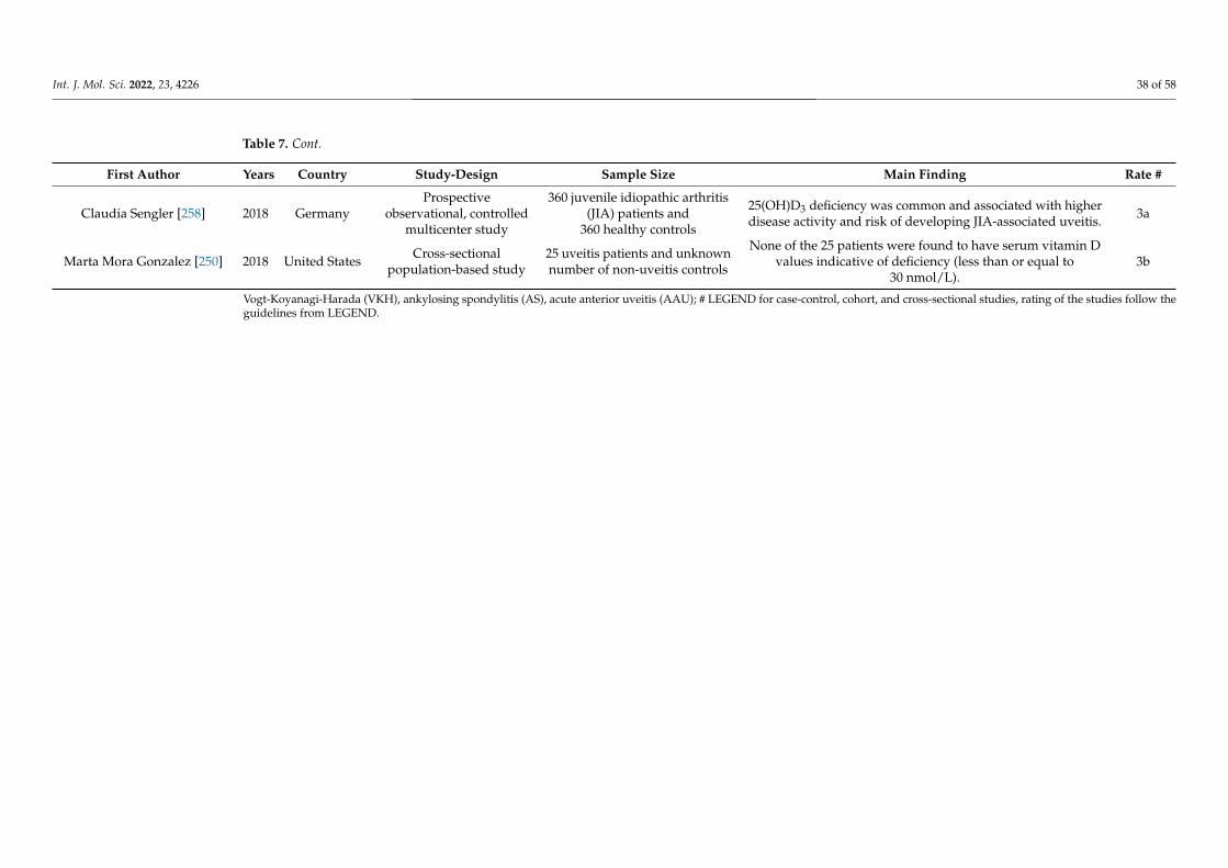
Int. J. Mol. Sci. 2022, 23, 4226 38 of 58
Table 7. Cont.
First Author Years Country Study-Design Sample Size Main Finding Rate #
Claudia Sengler [258] 2018 GermanyProspective
observational, controlledmulticenter study
360 juvenile idiopathic arthritis(JIA) patients and
360 healthy controls
25(OH)D3 deficiency was common and associated with higherdisease activity and risk of developing JIA-associated uveitis. 3a
Marta Mora Gonzalez [250] 2018 United States Cross-sectionalpopulation-based study
25 uveitis patients and unknownnumber of non-uveitis controls
None of the 25 patients were found to have serum vitamin Dvalues indicative of deficiency (less than or equal to
30 nmol/L).3b
Vogt-Koyanagi-Harada (VKH), ankylosing spondylitis (AS), acute anterior uveitis (AAU); # LEGEND for case-control, cohort, and cross-sectional studies, rating of the studies follow theguidelines from LEGEND.
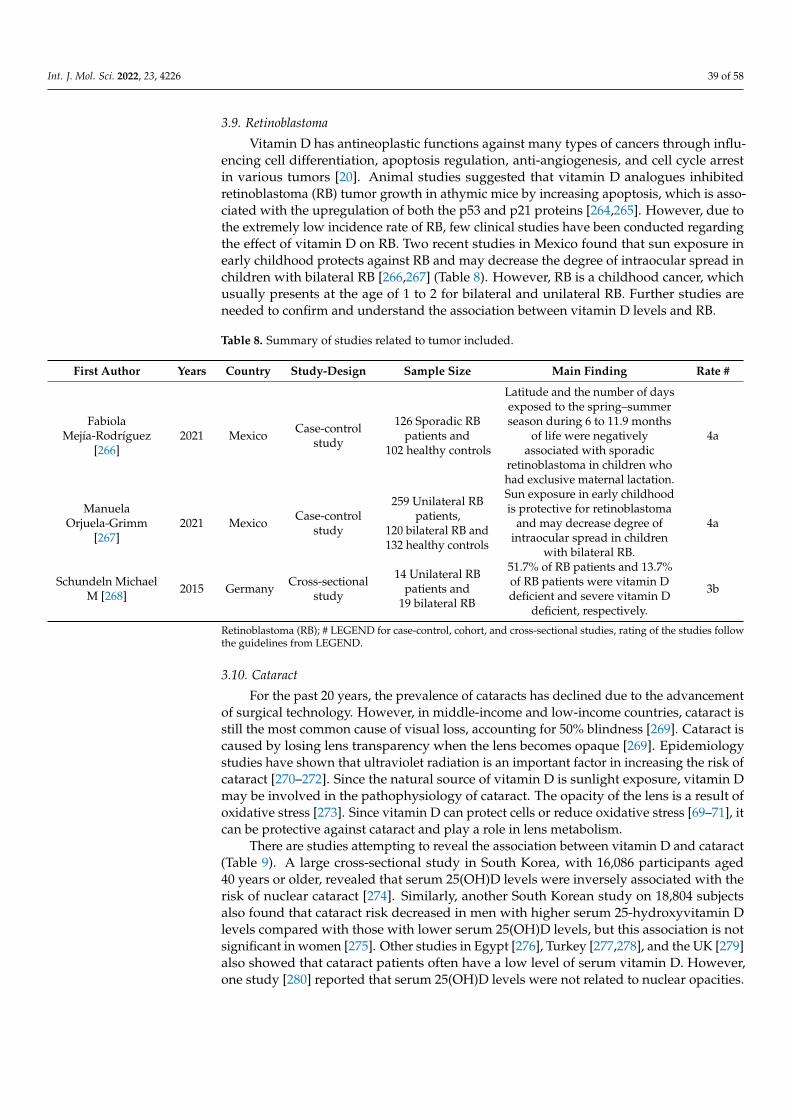
Int. J. Mol. Sci. 2022, 23, 4226 39 of 58
3.9. Retinoblastoma
Vitamin D has antineoplastic functions against many types of cancers through influ-encing cell differentiation, apoptosis regulation, anti-angiogenesis, and cell cycle arrestin various tumors [20]. Animal studies suggested that vitamin D analogues inhibitedretinoblastoma (RB) tumor growth in athymic mice by increasing apoptosis, which is asso-ciated with the upregulation of both the p53 and p21 proteins [264,265]. However, due tothe extremely low incidence rate of RB, few clinical studies have been conducted regardingthe effect of vitamin D on RB. Two recent studies in Mexico found that sun exposure inearly childhood protects against RB and may decrease the degree of intraocular spread inchildren with bilateral RB [266,267] (Table 8). However, RB is a childhood cancer, whichusually presents at the age of 1 to 2 for bilateral and unilateral RB. Further studies areneeded to confirm and understand the association between vitamin D levels and RB.
Table 8. Summary of studies related to tumor included.
First Author Years Country Study-Design Sample Size Main Finding Rate #
FabiolaMejía-Rodríguez
[266]2021 Mexico Case-control
study
126 Sporadic RBpatients and
102 healthy controls
Latitude and the number of daysexposed to the spring–summerseason during 6 to 11.9 months
of life were negativelyassociated with sporadic
retinoblastoma in children whohad exclusive maternal lactation.
4a
ManuelaOrjuela-Grimm
[267]2021 Mexico Case-control
study
259 Unilateral RBpatients,
120 bilateral RB and132 healthy controls
Sun exposure in early childhoodis protective for retinoblastoma
and may decrease degree ofintraocular spread in children
with bilateral RB.
4a
Schundeln MichaelM [268] 2015 Germany Cross-sectional
study
14 Unilateral RBpatients and
19 bilateral RB
51.7% of RB patients and 13.7%of RB patients were vitamin Ddeficient and severe vitamin D
deficient, respectively.
3b
Retinoblastoma (RB); # LEGEND for case-control, cohort, and cross-sectional studies, rating of the studies followthe guidelines from LEGEND.
3.10. Cataract
For the past 20 years, the prevalence of cataracts has declined due to the advancementof surgical technology. However, in middle-income and low-income countries, cataract isstill the most common cause of visual loss, accounting for 50% blindness [269]. Cataract iscaused by losing lens transparency when the lens becomes opaque [269]. Epidemiologystudies have shown that ultraviolet radiation is an important factor in increasing the risk ofcataract [270–272]. Since the natural source of vitamin D is sunlight exposure, vitamin Dmay be involved in the pathophysiology of cataract. The opacity of the lens is a result ofoxidative stress [273]. Since vitamin D can protect cells or reduce oxidative stress [69–71], itcan be protective against cataract and play a role in lens metabolism.
There are studies attempting to reveal the association between vitamin D and cataract(Table 9). A large cross-sectional study in South Korea, with 16,086 participants aged40 years or older, revealed that serum 25(OH)D levels were inversely associated with therisk of nuclear cataract [274]. Similarly, another South Korean study on 18,804 subjectsalso found that cataract risk decreased in men with higher serum 25-hydroxyvitamin Dlevels compared with those with lower serum 25(OH)D levels, but this association is notsignificant in women [275]. Other studies in Egypt [276], Turkey [277,278], and the UK [279]also showed that cataract patients often have a low level of serum vitamin D. However,one study [280] reported that serum 25(OH)D levels were not related to nuclear opacities.
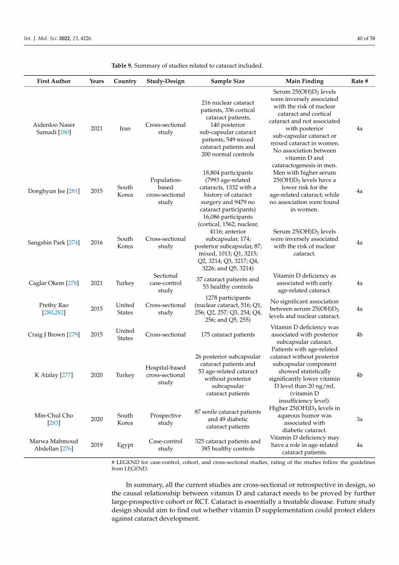
Int. J. Mol. Sci. 2022, 23, 4226 40 of 58
Table 9. Summary of studies related to cataract included.
First Author Years Country Study-Design Sample Size Main Finding Rate #
Aidenloo NaserSamadi [280] 2021 Iran Cross-sectional
study
216 nuclear cataractpatients, 336 cortical
cataract patients,140 posterior
sub-capsular cataractpatients, 549 mixed
cataract patients and200 normal controls
Serum 25(OH)D3 levelswere inversely associated
with the risk of nuclearcataract and cortical
cataract and not associatedwith posterior
sub-capsular cataract ormixed cataract in women.No association between
vitamin D andcataractogenesis in men.
4a
Donghyun Jee [281] 2015 SouthKorea
Population-based
cross-sectionalstudy
18,804 participants(7993 age-related
cataracts, 1332 with ahistory of cataract
surgery and 9479 nocataract participants)
Men with higher serum25(OH)D3 levels have a
lower risk for theage-related cataract; whileno association were found
in women.
4a
Sangshin Park [274] 2016 SouthKorea
Cross-sectionalstudy
16,086 participants(cortical, 1562; nuclear,
4116; anteriorsubcapsular, 174;
posterior subcapsular, 87;mixed, 1013; Q1, 3215;Q2, 3214; Q3, 3217; Q4,
3226; and Q5, 3214)
Serum 25(OH)D3 levelswere inversely associated
with the risk of nuclearcataract.
4a
Caglar Okem [278] 2021 TurkeySectional
case-controlstudy
37 cataract patients and53 healthy controls
Vitamin D deficiency asassociated with earlyage-related cataract.
4a
Prethy Rao[280,282] 2015 United
StatesCross-sectional
study
1278 participants(nuclear cataract, 516; Q1,256; Q2, 257; Q3, 254; Q4,
256; and Q5, 255)
No significant associationbetween serum 25(OH)D3levels and nuclear cataract.
4a
Craig J Brown [279] 2015 UnitedStates Cross-sectional 175 cataract patients
Vitamin D deficiency wasassociated with posterior
subcapsular cataract.4b
K Atalay [277] 2020 TurkeyHospital-basedcross-sectional
study
26 posterior subcapsularcataract patients and
53 age-related cataractwithout posterior
subcapsularcataract patients
Patients with age-relatedcataract without posteriorsubcapsular component
showed statisticallysignificantly lower vitamin
D level than 20 ng/mL(vitamin D
insufficiency level).
4b
Min-Chul Cho[283] 2020 South
KoreaProspective
study
87 senile cataract patientsand 49 diabetic
cataract patients
Higher 25(OH)D3 levels inaqueous humor was
associated withdiabetic cataract.
3a
Marwa MahmoudAbdellan [276] 2019 Egypt Case-control
study325 cataract patients and
385 healthy controls
Vitamin D deficiency mayhave a role in age-related
cataract patients.4a
# LEGEND for case-control, cohort, and cross-sectional studies, rating of the studies follow the guidelinesfrom LEGEND.
In summary, all the current studies are cross-sectional or retrospective in design, sothe causal relationship between vitamin D and cataract needs to be proved by furtherlarge-prospective cohort or RCT. Cataract is essentially a treatable disease. Future studydesign should aim to find out whether vitamin D supplementation could protect eldersagainst cataract development.
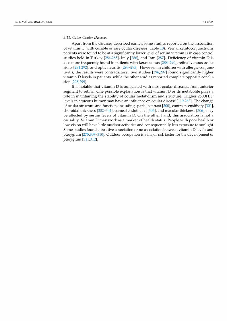
Int. J. Mol. Sci. 2022, 23, 4226 41 of 58
3.11. Other Ocular Diseases
Apart from the diseases described earlier, some studies reported on the associationof vitamin D with curable or rare ocular diseases (Table 10). Vernal keratoconjunctivitispatients were found to be at a significantly lower level of serum vitamin D in case-controlstudies held in Turkey [284,285], Italy [286], and Iran [287]. Deficiency of vitamin D isalso more frequently found in patients with keratoconus [288–290], retinal venous occlu-sions [291,292], and optic neuritis [293–295]. However, in children with allergic conjunc-tivitis, the results were contradictory: two studies [296,297] found significantly highervitamin D levels in patients, while the other studies reported complete opposite conclu-sion [298,299].
It is notable that vitamin D is associated with most ocular diseases, from anteriorsegment to retina. One possible explanation is that vitamin D or its metabolite plays arole in maintaining the stability of ocular metabolism and structure. Higher 25(OH)Dlevels in aqueous humor may have an influence on ocular disease [119,283]. The changeof ocular structure and function, including spatial contrast [300], contrast sensitivity [301],choroidal thickness [302–304], corneal endothelial [305], and macular thickness [306], maybe affected by serum levels of vitamin D. On the other hand, this association is not acausality. Vitamin D may work as a marker of health status. People with poor health orlow vision will have little outdoor activities and consequentially less exposure to sunlight.Some studies found a positive association or no association between vitamin D levels andpterygium [275,307–310]. Outdoor occupation is a major risk factor for the development ofpterygium [311,312].
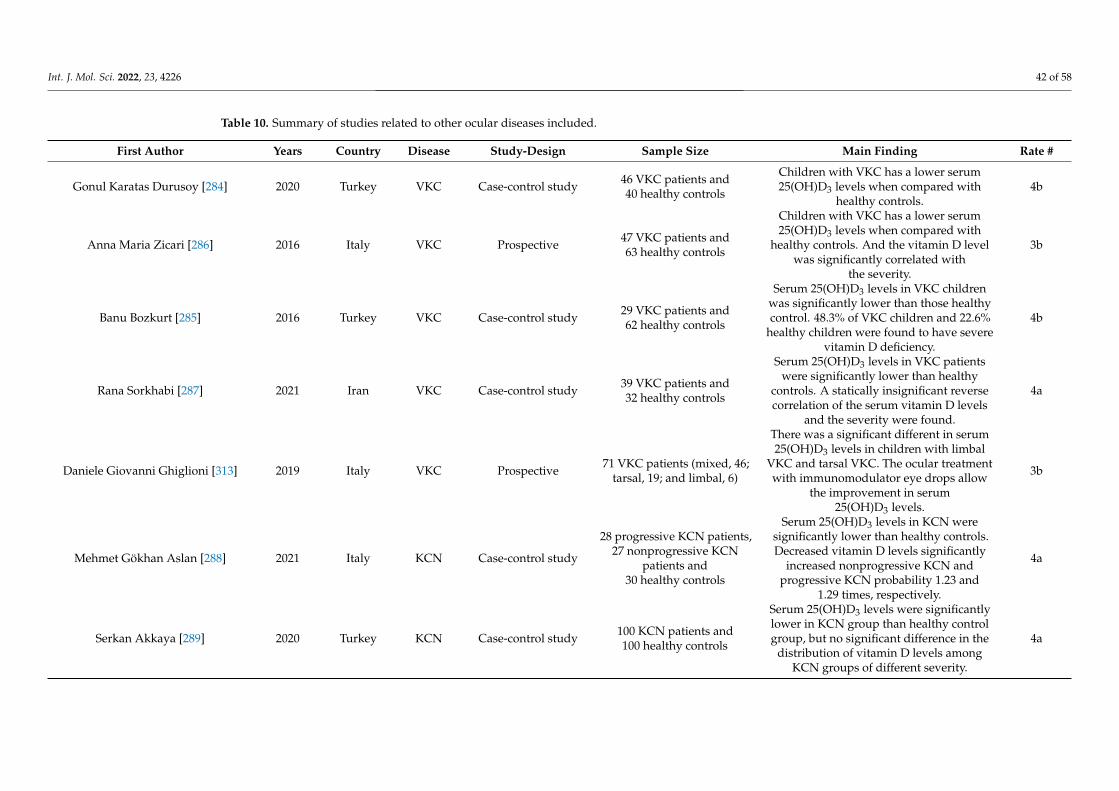
Int. J. Mol. Sci. 2022, 23, 4226 42 of 58
Table 10. Summary of studies related to other ocular diseases included.
First Author Years Country Disease Study-Design Sample Size Main Finding Rate #
Gonul Karatas Durusoy [284] 2020 Turkey VKC Case-control study 46 VKC patients and40 healthy controls
Children with VKC has a lower serum25(OH)D3 levels when compared with
healthy controls.4b
Anna Maria Zicari [286] 2016 Italy VKC Prospective 47 VKC patients and63 healthy controls
Children with VKC has a lower serum25(OH)D3 levels when compared with
healthy controls. And the vitamin D levelwas significantly correlated with
the severity.
3b
Banu Bozkurt [285] 2016 Turkey VKC Case-control study 29 VKC patients and62 healthy controls
Serum 25(OH)D3 levels in VKC childrenwas significantly lower than those healthycontrol. 48.3% of VKC children and 22.6%
healthy children were found to have severevitamin D deficiency.
4b
Rana Sorkhabi [287] 2021 Iran VKC Case-control study 39 VKC patients and32 healthy controls
Serum 25(OH)D3 levels in VKC patientswere significantly lower than healthy
controls. A statically insignificant reversecorrelation of the serum vitamin D levels
and the severity were found.
4a
Daniele Giovanni Ghiglioni [313] 2019 Italy VKC Prospective 71 VKC patients (mixed, 46;tarsal, 19; and limbal, 6)
There was a significant different in serum25(OH)D3 levels in children with limbal
VKC and tarsal VKC. The ocular treatmentwith immunomodulator eye drops allow
the improvement in serum25(OH)D3 levels.
3b
Mehmet Gökhan Aslan [288] 2021 Italy KCN Case-control study
28 progressive KCN patients,27 nonprogressive KCN
patients and30 healthy controls
Serum 25(OH)D3 levels in KCN weresignificantly lower than healthy controls.Decreased vitamin D levels significantly
increased nonprogressive KCN andprogressive KCN probability 1.23 and
1.29 times, respectively.
4a
Serkan Akkaya [289] 2020 Turkey KCN Case-control study 100 KCN patients and100 healthy controls
Serum 25(OH)D3 levels were significantlylower in KCN group than healthy controlgroup, but no significant difference in the
distribution of vitamin D levels amongKCN groups of different severity.
4a
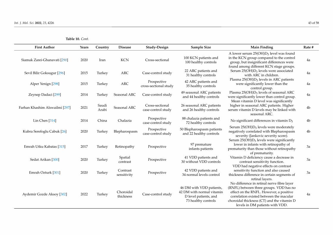
Int. J. Mol. Sci. 2022, 23, 4226 43 of 58
Table 10. Cont.
First Author Years Country Disease Study-Design Sample Size Main Finding Rate #
Siamak Zarei-Ghanavati [290] 2020 Iran KCN Cross-sectional 100 KCN patients and100 healthy controls
A lower serum 25(OH)D3 level was foundin the KCN group compared to the controlgroup, but insignificant differences were
found among different KCN stage groups.
4a
Sevil Bilir Goksugur [296] 2015 Turkey ARC Case-control study 22 ARC patients and31 healthy controls
Serum 25(OH)D3 levels were associatedwith ARC in children. 4a
Alper Yenign [298] 2015 Turkey ARC Prospectivecross-sectional study
42 ARC patients and35 healthy controls
Plasma 25(OH)D3 levels in ARC patientswere significantly lower than the
control group.4a
Zeynep Dadaci [299] 2014 Turkey Seasonal ARC Case-control study 49 seasonal ARC patientsand 44 healthy controls
Plasma 25(OH)D3 levels of seasonal ARCwere significantly lower than control group. 4a
Farhan Khashim Alswailmi [297] 2021 SaudiArabi Seasonal ARC Cross-sectional
case-control study26 seasonal ARC patientsand 26 healthy controls
Mean vitamin D level was significantlyhigher in seasonal ARC patients. Higher
serum vitamin D levels may be linked withseasonal ARC.
4b
Lin Chen [314] 2014 China Chalazia Prospectivecase-control study
88 chalazia patients and72 healthy controls No significant differences in vitamin D3 4b
Kubra Serefoglu Cabuk [26] 2020 Turkey Blepharospasm Prospectivecase-control study
50 Blepharospasm patientsand 22 healthy controls
Serum 25(OH)D3 levels were moderatelynegatively correlated with Blepharospasm
severity (Jankovic severity score).4b
Emrah Utku Kabatas [315] 2017 Turkey Retinopathy Prospective 97 prematureinfants patients
Serum 25(OH)D3 levels were significantlylower in infants with retinopathy of
prematurity than those without retinopathyof prematurity.
3a
Sedat Arikan [300] 2020 Turkey Spatialcontrast Prospective 41 VDD patients and
30 without VDD controlsVitamin D deficiency cause a decrease in
contrast sensitivity function. 3a
Emrah Ozturk [301] 2020 Turkey Contrastsensitivity Prospective 42 VDD patients and
34 normal levels control
VDD had negative effects on contrastsensitivity function and also caused
thickness difference in certain segments ofretinal layers.
3a
Aydemir Gozde Aksoy [302] 2022 Turkey Choroidalthickness Case-control study
46 DM with VDD patients,42 DM with normal vitamin
D level patients, and73 healthy controls
No difference in retinal nerve fibre layer(RNFL) between three groups. VDD has no
effect on the RNFL. However, a positivecorrelation existed between the macular
choroidal thickness (CT) and the vitamin Dlevels in DM patients with VDD.
4a
Int. J. Mol. Sci. 2022, 23, 4226 44 of 58
Table 10. Cont.
First Author Years Country Disease Study-Design Sample Size Main Finding Rate #
Esra Vural [303] 2020 Turkey Choroidalthickness
Prospectivecase-control study
30 VDD patients and30 normal level controls
A positive correlation was found between vitaminD levels and subfoveal choroidal thickness and
inferior and nasal peripapillary choroidal thicknessin all participants.
4a
Hasan Oncul [304] 2020 Turkey Choroidalthickness
Prospectivecase-control study
65 VDD patients and60 normal level controls
VDD patients have a thinner choroid and thechoroidal thickness increased after vitamin D
replacement therapy.4a
Cem Cankaya [305] 2018 UnitedStates
Cornealendothelial Case-control study 58 VDD patients and
40 normal level controls
The mean corneal endothelial cell density andmean hexagonal cell ratio in VDD patients were
lower than healthy controls. The mean coefficientof variation in VDD patients were higher thanhealthy controls. VDD may affect the corneal
endothelial layer.
4a
Alix Graffe [306] 2014 France Macularthickness Cross-sectional
62 patients (17 and 45 withvitamin D insufficiency and
sufficiency, respectively)
Vitamin D insufficiency was associated withreduced macular thickness with no patent
macular dysfunction.4a
Unal Mutlu [316] 2016 Netherlands Retinal mi-crovascular
Prospectivepopulation-based
study
5675 subjects (sample) and2973 subjects (subsample)
Individuals with lower vitamin D levels were morelikely to have retinopathy. Lower vitamin D levels
were associated with wider venular calibers.3a
Hatice Daldal [317] 2021 Turkey Ocularfindings Prospective
98 patients (41, 45 and12 were vitamin D severe
deficient, deficient andinsufficient, respectively)
Vitamin D may be related to thinning in macularand nasal of RNFL. 3a
Karabulut Mujdat [318] 2022 TurkeyRetinal
microvascu-larity
Case-control study 98 VDD patients and96 healthy controls
There was a strong negative correlation betweenthe serum vitamin D level and vessel density in thewhole image, parafoveal, and perifoveal regions of
the deep capillary plexus in the study group(Spearman’s rho = −0.71, p = 0.043; Spearman’s
rho = −0.79, p = 0.011; and Spearman’s rho =−0.74, p = 0.032; respectively).
4a
VKC: vernal keratoconjunctivitis; KCN: keratoconus; ARC: allergic rhinoconjunctivitis; VDD: vitamin D deficiency; # LEGEND for case-control, cohort, and cross-sectional studies, ratingof the studies follow the guidelines from LEGEND.
Int. J. Mol. Sci. 2022, 23, 4226 45 of 58
4. Perspective
In this systematic review, we summarize the evidence of vitamin D’s effect on differ-ent ocular diseases. However, there are significant limitations in many studies, and theinterpretation of results should be within these limitations.
First, the definition of vitamin D deficiency is not consistent in different studies, whichaffects the objective comparison of results from different studies. Until 1998, vitamin D de-ficiency was defined as a blood level of 25(OH)D, which represents a total concentration ofboth 25-hydroxyvitamin D2 and 25-hydroxyvitamin D3 of less than 10 ng/mL (25 nmol/L).However, this definition was redefined in 1998 as a blood level of 25(OH)D < 20 ng/mL(50 nmol/L). One reason for the change in the involvement of PTH: adults may requirea serum 25(OH)D of at least 50 nmol/L to achieve optimum PTH levels [319]. Moreover,some studies use quintiles or quartiles to perform data analysis. We suggest that futurestudies should use the common classification to analyze serum vitamin D levels.
Second, vitamin D levels are related to different socioeconomic, lifestyle, and dietaryfactors. These factors are very important when considering the onset or developmentof ocular diseases. Future studies should determine these factors and consider them asconfounders in statistical analysis.
Finally, few studies provided causal evidence on whether supplementing vitamin Dcan reduce the prevalence of ocular diseases. Currently, available evidence is insufficientto confirm how vitamin D works in ocular diseases. Moreover, investigations of thosemetabolic enzymes are also important to understand why significantly low vitamin Dis found in patients with ocular diseases. In summary, among all ocular diseases, theassociation between vitamin D and AMD was unclear, while clinical trials in DES or DRrequired bigger sample sizes in different ethnic populations. Other ocular diseases such ascataract, myopia, and uveitis, no RCTs or perspective cohorts have been reported, so theirassociations with vitamin D are still unclear.
5. Vitamin D and Eye Care
Vitamin D can be a potential intervention for different ocular diseases. Althoughfindings for certain ocular diseases are inconsistent, they can still serve as referencesfor further studies that examine the therapeutic effects of vitamin D on ocular diseases,considering that vitamin D deficiency is a common health issue worldwide, and vitaminD has wide safety doses and rare side effects. Even though more evidence from therandomized controlled trial is needed to confirm the effect of vitamin D on various oculardiseases, it is recommended to maintain blood 25(OH)D3 at a desirable level (25–50 nmol/L)by spending a short period of time outdoors, baring skin to the sun, and boosting vitaminD intake by a daily supplement of 400–800 international units (10 to 20 µg).
The human body can also obtain vitamin D naturally from sunlight exposure, however,both extended exposure to unprotected sunshine, which also increases the risk for cataracts,and AMD and completely avoiding sunlight by applying UV B sunscreen should beavoided. Several factors affect vitamin D production, and a more efficient productionof vitamin D can be achieved when someone is closer to the equator, has a lighter skincolor, and/or exposes larger surfaces of skin during summer midday without sunscreen.Generally, 5–30 min of sun exposure on the unprotected face, arms, legs, or back between10 a.m. and 3 p.m. twice to three times a week is enough for sufficient vitamin D. Eventhough vitamin D can be beneficial to our ocular health, long term prolonged sun exposureis also associated with corneal sunburn, tissue growths on sclera, cataracts, and maculardegeneration, and wearing hats, and sunglasses are recommended to protect the eyes fromUV damage.
6. Conclusions
Various epidemiological and clinical studies have demonstrated a connection betweenvitamin D deficiency and ocular diseases, such as myopia, age-related macular degen-eration, glaucoma, diabetic retinopathy, dry eye syndrome, thyroid eye disease, uveitis,
Int. J. Mol. Sci. 2022, 23, 4226 46 of 58
retinoblastoma, and cataract, among others. While vitamin D is associated with potentialpathways related to these respective diseases, their pathogeneses are complicated, andcurrent understandings of the underlying mechanisms remain limited. Vitamin D notonly affects mineral metabolism homeostasis, but also possesses antioxidation and anti-inflammatory properties. It also plays a role in anti-angiogenesis, modulating cell cycleincluding cell proliferation, differentiation, and apoptosis. VDR and vitamin D regulatoryenzymes are present in ocular tissues, and studies have demonstrated that ocular tissuescan activate and regulate vitamin D, suggesting the importance of vitamin D in maintainingocular health.
Author Contributions: Conceptualization, H.-N.C., X.-J.Z., J.C.Y., C.-P.P.; methodology, H.-N.C. andX.-J.Z.; validation, H.-N.C., X.-J.Z. and X.-T.L.; formal analysis, H.-N.C., X.-J.Z. and X.-T.L.; investiga-tion, H.-N.C. and X.-J.Z.; writing—original draft preparation, H.-N.C., X.-J.Z., X.-T.L. and C.H.-T.B.;writing—review and editing, Y.-M.W., P.I., W.-K.C., L.-J.C., C.C.T., J.C.Y. and C.-P.P.; supervision,J.C.Y. and C.-P.P. All authors have read and agreed to the published version of the manuscript.
Funding: This study was supported in part by the General Research Fund (GRF), Research GrantsCouncil, Hong Kong (14111515 and 14103419 [JCY]); Collaborative Research Fund (C7149-20G [JCY]);Health and Medical Research Fund (HMRF), Hong Kong (5160836, [LJC] and 07180826 [XJZ]), andthe Direct Grants of the Chinese University of Hong Kong, (4054193 [LJC] and 4054121 & 4054199[JCY] and 4054634 [XJZ]), the Innovation and Technology Fund (7010590 [JCY]), the UBS OptimusFoundation Grant 8984 (JCY); the Centaline Myopia Fund [JCY]; the CUHK Jockey Club Children’sEye Care Programme; and the CUHK Jockey Club Myopia Prevention Programme.
Institutional Review Board Statement: Not applicable.
Informed Consent Statement: Not applicable.
Data Availability Statement: Not applicable.
Conflicts of Interest: The authors declare no conflict of interest.
References1. Plum, L.A.; DeLuca, H.F. Vitamin D, disease and therapeutic opportunities. Nat. Rev. Drug Discov. 2010, 9, 941–955. [CrossRef]2. Zmijewski, M.A. Vitamin D and Human Health. Int. J. Mol. Sci. 2019, 20, 10145. [CrossRef]3. Zhang, R.; Naughton, D.P. Vitamin D in health and disease: Current perspectives. Nutr. J. 2010, 9, 65. [CrossRef] [PubMed]4. Al-Ishaq, R.K.; Kubatka, P.; Brozmanova, M.; Gazdikova, K.; Caprnda, M.; Büsselberg, D. Health implication of vitamin D on the
cardiovascular and the renal system. Arch. Physiol. Biochem. 2021, 127, 195–209. [CrossRef] [PubMed]5. Rai, V.; Agrawal, D.K. Role of Vitamin D in Cardiovascular Diseases. Endocrinol. Metab. Clin. N. Am. 2017, 46, 1039–1059.
[CrossRef] [PubMed]6. Wang, T.J.; Pencina, M.J.; Booth, S.L.; Jacques, P.F.; Ingelsson, E.; Lanier, K.; Benjamin, E.J.; D’Agostino, R.B.; Wolf, M.; Vasan, R.S.
Vitamin D deficiency and risk of cardiovascular disease. Circulation 2008, 117, 503–511. [CrossRef]7. Adamczak, M.; Surma, S.; Wiecek, A. Vitamin D and Arterial Hypertension: Facts and Myths. Curr. Hypertens. Rep. 2020, 22, 57.
[CrossRef] [PubMed]8. Seshadri, K.G.; Tamilselvan, B.; Rajendran, A. Role of Vitamin D in Diabetes. J. Endocrinol. Metab. 2011, 1, 47–56. [CrossRef]9. Jani, R.; Mhaskar, K.; Tsiampalis, T.; Kassaw, N.A.; González, M.Á.M.; Panagiotakos, D.B. Circulating 25-hydroxy-vitamin D and
the risk of cardiovascular diseases. Systematic review and meta-analysis of prospective cohort studies. Nutr. Metab. Cardiovasc.Dis. 2021, 31, 3282–3304. [CrossRef]
10. Jeon, S.M.; Shin, E.A. Exploring vitamin D metabolism and function in cancer. Exp. Mol. Med. 2018, 50, 1–14. [CrossRef]11. Holick, M.F.; Binkley, N.C.; Bischoff-Ferrari, H.A.; Gordon, C.M.; Hanley, D.A.; Heaney, R.P.; Murad, M.H.; Weaver, C.M.
Evaluation, treatment, and prevention of vitamin D deficiency: An Endocrine Society clinical practice guideline. J. Clin. Endocrinol.Metab. 2011, 96, 1911–1930. [CrossRef] [PubMed]
12. Brown, E.M.; Gamba, G.; Riccardi, D.; Lombardi, M.; Butters, R.; Kifor, O.; Sun, A.; Hediger, M.A.; Lytton, J.; Hebert, S.C.Cloning and characterization of an extracellular Ca(2+)–sensing receptor from bovine parathyroid. Nature 1993, 366, 575–580.[CrossRef] [PubMed]
13. Holick, M.F. Vitamin D status: Measurement, interpretation, and clinical application. Ann. Epidemiol. 2009, 19, 73–78. [CrossRef]14. Dominguez, L.J.; Farruggia, M.; Veronese, N.; Barbagallo, M. Vitamin D Sources, Metabolism, and Deficiency: Available
Compounds and Guidelines for Its Treatment. Metabolites 2021, 11, 255. [CrossRef] [PubMed]15. Bikle, D.D. Vitamin D Metabolism, Mechanism of Action, and Clinical Applications. Chem. Biol. 2014, 21, 319–329.
[CrossRef] [PubMed]
Int. J. Mol. Sci. 2022, 23, 4226 47 of 58
16. Zmijewski, M.A.; Carlberg, C. Vitamin D receptor(s): In the nucleus but also at membranes? Exp. Dermatol. 2020, 29, 876–884. [CrossRef]17. Kim, S.; Shevde, N.K.; Pike, J.W. 1,25-Dihydroxyvitamin D3 stimulates cyclic vitamin D receptor/retinoid X receptor DNA–
binding, co-activator recruitment, and histone acetylation in intact osteoblasts. J. Bone Miner. Res. 2005, 20, 305–317. [CrossRef]18. Skowron, K.; Pawlicka, I.; Gil, K. The role of vitamin D in the pathogenesis of ocular diseases. Folia Med. Cracov. 2018, 58, 103–118.19. Jamali, N.; Wang, S.; Darjatmoko, S.R.; Sorenson, C.M.; Sheibani, N. Vitamin D receptor expression is essential during retinal
vascular development and attenuation of neovascularization by 1, 25(OH)2D3. PLoS ONE 2017, 12, e0190131. [CrossRef]20. Reins, R.Y.; McDermott, A.M. Vitamin D: Implications for ocular disease and therapeutic potential. Exp. Eye Res. 2015, 134,
101–110. [CrossRef]21. Alsalem, J.A.; Patel, D.; Susarla, R.; Coca-Prados, M.; Bland, R.; Walker, E.A.; Rauz, S.; Wallace, G.R. Characterization of vitamin
D production by human ocular barrier cells. Investig. Ophthalmol. Vis. Sci. 2014, 55, 2140–2147. [CrossRef] [PubMed]22. Yin, Z.; Pintea, V.; Lin, Y.; Hammock, B.D.; Watsky, M.A. Vitamin D enhances corneal epithelial barrier function. Investig.
Ophthalmol. Vis. Sci. 2011, 52, 7359–7364. [CrossRef] [PubMed]23. Verstappen, A.; Parmentier, M.; Chirnoaga, M.; Lawson, D.E.; Pasteels, J.L.; Pochet, R. Vitamin D–dependent calcium binding
protein immunoreactivity in human retina. Ophthalmic Res. 1986, 18, 209–214. [CrossRef]24. Rullo, J.; Pennimpede, T.; Mehraban Far, P.; Strube, Y.N.; Irrcher, I.; Urton, T.; Bona, M.; Gonder, T.; Campbell, R.J.; Ten Hove, M.;
et al. Intraocular calcidiol: Uncovering a role for vitamin D in the eye. J. Steroid Biochem. Mol. Biol. 2020, 197, 105536.[CrossRef] [PubMed]
25. Al-Barry, M.A.; Albalawi, A.M.; Sayf, M.A.; Badawi, A.; Afzal, S.; Latif, M.; Samman, M.I.; Basit, S. Sequence analysis of fourvitamin D family genes (VDR, CYP24A1, CYP27B1 and CYP2R1) in Vogt-Koyanagi-Harada (VKH) patients: Identification of apotentially pathogenic variant in CYP2R1. BMC Ophthalmol. 2016, 16, 172. [CrossRef]
26. Serefoglu Cabuk, K.; Tunc, U.; Ozturk Karabulut, G.; Fazil, K.; Karaagac Gunaydin, Z.; Asik Nacaroglu, S.; Taskapili, M. Serumcalcium, magnesium, phosphorus, and vitamin D in benign essential blepharospasm. Graefe’s Arch. Clin. Exp. Ophthalmol. 2020,258, 1293–1297. [CrossRef]
27. Moher, D.; Liberati, A.; Tetzlaff, J.; Altman, D.G.; The PRISMA Group. Preferred Reporting Items for Systematic Reviews andMeta–Analyses: The PRISMA Statement. PLoS Med. 2009, 6, e1000097. [CrossRef]
28. Ma, L.-L.; Wang, Y.-Y.; Yang, Z.-H.; Huang, D.; Weng, H.; Zeng, X.-T. Methodological quality (risk of bias) assessment tools forprimary and secondary medical studies: What are they and which is better? Mil. Med. Res. 2020, 7, 7.
29. Clark, E.; Burkett, K.; Stanko-Lopp, D. Let Evidence Guide Every New Decision (LEGEND): An evidence evaluation system forpoint-of-care clinicians and guideline development teams. J. Eval. Clin. Pract. 2009, 15, 1054–1060. [CrossRef]
30. Dolgin, E. The myopia boom. Nature 2015, 519, 276–278. [CrossRef]31. Zadnik, K.; Satariano, W.A.; Mutti, D.O.; Sholtz, R.I.; Adams, A.J. The effect of parental history of myopia on children’s eye size.
JAMA 1994, 271, 1323–1327. [CrossRef] [PubMed]32. Lam, D.S.; Fan, D.S.; Lam, R.F.; Rao, S.K.; Chong, K.S.; Lau, J.T.; Lai, R.Y.; Cheung, E.Y. The effect of parental history of
myopia on children’s eye size and growth: Results of a longitudinal study. Investig. Ophthalmol. Vis. Sci. 2008, 49, 873–876.[CrossRef] [PubMed]
33. Fan, Q.; Verhoeven, V.J.; Wojciechowski, R.; Barathi, V.A.; Hysi, P.G.; Guggenheim, J.A.; Hohn, R.; Vitart, V.; Khawaja, A.P.;Yamashiro, K.; et al. Meta–analysis of gene–environment–wide association scans accounting for education level identifiesadditional loci for refractive error. Nat. Commun. 2016, 7, 11008. [CrossRef] [PubMed]
34. Rose, K.A.; Morgan, I.G.; Ip, J.; Kifley, A.; Huynh, S.; Smith, W.; Mitchell, P. Outdoor activity reduces the prevalence of myopia inchildren. Ophthalmology 2008, 115, 1279–1285. [CrossRef] [PubMed]
35. Dirani, M.; Tong, L.; Gazzard, G.; Zhang, X.; Chia, A.; Young, T.L.; Rose, K.A.; Mitchell, P.; Saw, S.M. Outdoor activity and myopiain Singapore teenage children. Br. J. Ophthalmol. 2009, 93, 997–1000. [CrossRef] [PubMed]
36. Jones, L.A.; Sinnott, L.T.; Mutti, D.O.; Mitchell, G.L.; Moeschberger, M.L.; Zadnik, K. Parental history of myopia, sports andoutdoor activities, and future myopia. Investig. Ophthalmol. Vis. Sci. 2007, 48, 3524–3532. [CrossRef] [PubMed]
37. Low, W.; Dirani, M.; Gazzard, G.; Chan, Y.H.; Zhou, H.J.; Selvaraj, P.; Au Eong, K.G.; Young, T.L.; Mitchell, P.; Wong, T.Y.; et al.Family history, near work, outdoor activity, and myopia in Singapore Chinese preschool children. Br. J. Ophthalmol. 2010, 94,1012–1016. [CrossRef] [PubMed]
38. He, M.; Xiang, F.; Zeng, Y.; Mai, J.; Chen, Q.; Zhang, J.; Smith, W.; Rose, K.; Morgan, I.G. Effect of Time Spent Outdoors at Schoolon the Development of Myopia Among Children in China: A Randomized Clinical Trial. JAMA 2015, 314, 1142–1148. [CrossRef]
39. Choi, J.A.; Han, K.; Park, Y.M.; La, T.Y. Low serum 25-hydroxyvitamin D is associated with myopia in Korean adolescents.Investig. Ophthalmol. Vis. Sci. 2014, 55, 2041–2047. [CrossRef]
40. Guggenheim, J.A.; Williams, C.; Northstone, K.; Howe, L.D.; Tilling, K.; St. Pourcain, B.; McMahon, G.; Lawlor, D.A. Does vitaminD mediate the protective effects of time outdoors on myopia? Findings from a prospective birth cohort. Investig. Ophthalmol. Vis.Sci. 2014, 55, 8550–8558. [CrossRef]
41. Mutti, D.O.; Marks, A.R. Blood levels of vitamin D in teens and young adults with myopia. Optom. Vis. Sci. 2011, 88, 377–382.[CrossRef] [PubMed]
42. Tideman, J.W.; Polling, J.R.; Voortman, T.; Jaddoe, V.W.; Uitterlinden, A.G.; Hofman, A.; Vingerling, J.R.; Franco, O.H.; Klaver, C.C.Low serum vitamin D is associated with axial length and risk of myopia in young children. Eur. J. Epidemiol. 2016, 31, 491–499.[CrossRef] [PubMed]
Int. J. Mol. Sci. 2022, 23, 4226 48 of 58
43. Yazar, S.; Hewitt, A.W.; Black, L.J.; McKnight, C.M.; Mountain, J.A.; Sherwin, J.C.; Oddy, W.H.; Coroneo, M.T.; Lucas, R.M.;Mackey, D.A. Myopia is associated with lower vitamin D status in young adults. Investig. Ophthalmol. Vis. Sci. 2014, 55, 4552–4559.[CrossRef] [PubMed]
44. Kwon, J.W.; Choi, J.A.; La, T.Y.; Epidemiologic Survey Committee of the Korean Ophthalmological Society. Serum 25-hydroxyvitamin D level is associated with myopia in the Korea national health and nutrition examination survey. Medicine 2016,95, 5012. [CrossRef] [PubMed]
45. Williams, K.M.; Bentham, G.C.; Young, I.S.; McGinty, A.; McKay, G.J.; Hogg, R.; Hammond, C.J.; Chakravarthy, U.; Rahu,M.; Seland, J.; et al. Association Between Myopia, Ultraviolet B Radiation Exposure, Serum Vitamin D Concentrations, andGenetic Polymorphisms in Vitamin D Metabolic Pathways in a Multicountry European Study. JAMA Ophthalmol. 2017, 135,47–53. [CrossRef]
46. Specht, I.O.; Jacobsen, N.; Frederiksen, P.; Heitmann, B.L. Neonatal vitamin D status and myopia in young adult men. ActaOphthalmol. 2020, 98, 500–505. [CrossRef]
47. Jung, B.J.; Jee, D. Association between serum 25-hydroxyvitamin D levels and myopia in general Korean adults. Indian J.Ophthalmol. 2020, 68, 15–22.
48. Chou, H.D.; Yao, T.C.; Huang, Y.S.; Huang, C.Y.; Yang, M.L.; Sun, M.H.; Chen, H.C.; Liu, C.H.; Chu, S.M.; Hsu, J.F.; et al. Myopiain school-aged children with preterm birth: The roles of time spent outdoors and serum vitamin D. Br. J. Ophthalmol. 2021, 105,468–472. [CrossRef]
49. Lingham, G.; Mackey, D.A.; Zhu, K.; Lucas, R.M.; Black, L.J.; Oddy, W.H.; Holt, P.; Walsh, J.P.; Sanfilippo, P.G.; Chan She Ping-Delfos, W.; et al. Time spent outdoors through childhood and adolescence–assessed by 25-hydroxyvitamin D concentration–andrisk of myopia at 20 years. Acta Ophthalmol. 2021, 99, 679–687. [CrossRef]
50. Han, S.B.; Jang, J.; Yang, H.K.; Hwang, J.M.; Park, S.K. Prevalence and risk factors of myopia in adult Korean population: Koreanational health and nutrition examination survey 2013–2014 (KNHANES VI). PLoS ONE 2019, 14, e0211204. [CrossRef]
51. Gao, F.; Li, P.; Liu, Y.Q.; Chen, Y. Association study of the serum 25(OH)D concentration and myopia in Chinese children. Medicine2021, 100, e26570. [CrossRef] [PubMed]
52. Lingham, G.; Yazar, S.; Lucas, R.M.; Walsh, J.P.; Zhu, K.; Hunter, M.; Lim, E.M.; Cooke, B.R.; Mackey, D.A. Low 25-HydroxyvitaminD Concentration Is Not Associated With Refractive Error in Middle-Aged and Older Western Australian Adults. Transl. Vis. Sci.Technol. 2019, 8, 13. [CrossRef] [PubMed]
53. Hwang, H.S.; Chun, M.Y.; Kim, J.S.; Oh, B.; Yoo, S.H.; Cho, B.J. Risk Factors for High Myopia in Koreans: The Korea NationalHealth and Nutrition Examination Survey. Curr. Eye Res. 2018, 43, 1052–1060. [CrossRef] [PubMed]
54. Harb, E.N.; Wildsoet, C.F. Nutritional Factors and Myopia: An Analysis of National Health and Nutrition Examination SurveyData. Optom. Vis. Sci. 2021, 98, 458–468. [CrossRef]
55. Wahyudi, D.; Reiki, W.; Hardhono; Suhartono. The Effect of Vitamin-D and Sunlight to Progressive Myopia in Students withGlasses Correction. Pak. J. Med. Health Sci. 2020, 14, 1588–1591.
56. Li, X.; Lin, H.; Jiang, L.; Chen, X.; Chen, J.; Lu, F. Low Serum Vitamin D Is Not Correlated with Myopia in Chinese Children andAdolescents. Front. Med. 2022, 9, 809787. [CrossRef]
57. Tang, S.M.; Lau, T.; Rong, S.S.; Yazar, S.; Chen, L.J.; Mackey, D.A.; Lucas, R.M.; Pang, C.P.; Yam, J.C. Vitamin D and its pathwaygenes in myopia: Systematic review and meta-analysis. Br. J. Ophthalmol. 2019, 103, 8–17. [CrossRef]
58. Mutti, D.O.; Cooper, M.E.; Dragan, E.; Jones-Jordan, L.A.; Bailey, M.D.; Marazita, M.L.; Murray, J.C.; Zadnik, K.; Group, C.S.Vitamin D receptor (VDR) and group–specific component (GC, vitamin D–binding protein) polymorphisms in myopia. Investig.Ophthalmol. Vis. Sci. 2011, 52, 3818–3824. [CrossRef]
59. Cuellar-Partida, G.; Williams, K.M.; Yazar, S.; Guggenheim, J.A.; Hewitt, A.W.; Williams, C.; Wang, J.J.; Kho, P.F.; Saw, S.M.;Cheng, C.Y.; et al. Genetically low vitamin D concentrations and myopic refractive error: A Mendelian randomization study. Int.J. Epidemiol. 2017, 46, 1882–1890. [CrossRef]
60. Torii, H.; Kurihara, T.; Seko, Y.; Negishi, K.; Ohnuma, K.; Inaba, T.; Kawashima, M.; Jiang, X.; Kondo, S.; Miyauchi, M.; et al. VioletLight Exposure Can Be a Preventive Strategy Against Myopia Progression. eBioMedicine 2017, 15, 210–219. [CrossRef]
61. Ramin, S.; Soheilian, M.; Habibi, G.; Ghazavi, R.; Gharebaghi, R.; Heidary, F. Age-Related Macular Degeneration: A ScientometricAnalysis. Med. Hypothesis Discov. Innov. Ophthalmol. 2015, 4, 39–49. [PubMed]
62. Apte, R.S. Age-Related Macular Degeneration. N. Engl. J. Med. 2021, 385, 539–547. [CrossRef] [PubMed]63. Coppé, A.M.; Ripandelli, G.; Parisi, V.; Varano, M.; Stirpe, M. Prevalence of asymptomatic macular holes in highly myopic eyes.
Ophthalmology 2005, 112, 2103–2109. [CrossRef] [PubMed]64. Yonekawa, Y.; Miller, J.W.; Kim, I.K. Age-Related Macular Degeneration: Advances in Management and Diagnosis. J. Clin. Med.
2015, 4, 343–359. [CrossRef]65. Khandhadia, S.; Cipriani, V.; Yates, J.R.; Lotery, A.J. Age-related macular degeneration and the complement system. Immunobiology
2012, 217, 127–146. [CrossRef]66. Park, Y.G.; Park, Y.S.; Kim, I.B. Complement System and Potential Therapeutics in Age-Related Macular Degeneration. Int. J. Mol.
Sci. 2021, 22, 36851. [CrossRef]67. Chen, M.; Muckersie, E.; Forrester, J.V.; Xu, H. Immune activation in retinal aging: A gene expression study. Investig. Ophthalmol.
Vis. Sci. 2010, 51, 5888–5896. [CrossRef]
Int. J. Mol. Sci. 2022, 23, 4226 49 of 58
68. Layana, A.G.; Minnella, A.M.; Garhofer, G.; Aslam, T.; Holz, F.G.; Leys, A.; Silva, R.; Delcourt, C.; Souied, E.; Seddon, J.M. VitaminD and Age-Related Macular Degeneration. Nutrients 2017, 9, 1120. [CrossRef]
69. Peng, X.; Vaishnav, A.; Murillo, G.; Alimirah, F.; Torres, K.E.; Mehta, R.G. Protection against cellular stress by 25-hydroxyvitaminD3 in breast epithelial cells. J. Cell. Biochem. 2010, 110, 1324–1333. [CrossRef]
70. Diker-Cohen, T.; Koren, R.; Ravid, A. Programmed cell death of stressed keratinocytes and its inhibition by vitamin D: The role ofdeath and survival signaling pathways. Apoptosis 2006, 11, 519–534. [CrossRef]
71. Tohari, A.M.; Zhou, X.; Shu, X. Protection against oxidative stress by vitamin D in cone cells. Cell Biochem. Funct. 2016, 34, 82–94.[CrossRef] [PubMed]
72. Schleithoff, S.S.; Zittermann, A.; Tenderich, G.; Berthold, H.K.; Stehle, P.; Koerfer, R. Vitamin D supplementation improvescytokine profiles in patients with congestive heart failure: A double–blind, randomized, placebo-controlled trial. Am. J. Clin.Nutr. 2006, 83, 754–759. [CrossRef] [PubMed]
73. Hewison, M. Vitamin D and the immune system: New perspectives on an old theme. Rheum. Dis. Clin. N. Am. 2012, 38, 125–139.[CrossRef] [PubMed]
74. Mantell, D.J.; Owens, P.E.; Bundred, N.J.; Mawer, E.B.; Canfield, A.E. 1 alpha,25-dihydroxyvitamin D(3) inhibits angiogenesisin vitro and in vivo. Circ. Res. 2000, 87, 214–220. [CrossRef] [PubMed]
75. Albert, D.M.; Scheef, E.A.; Wang, S.; Mehraein, F.; Darjatmoko, S.R.; Sorenson, C.M.; Sheibani, N. Calcitriol is a potent inhibitor ofretinal neovascularization. Investig. Ophthalmol. Vis. Sci. 2007, 48, 2327–2334. [CrossRef]
76. Morrison, M.A.; Silveira, A.C.; Huynh, N.; Jun, G.; Smith, S.E.; Zacharaki, F.; Sato, H.; Loomis, S.; Andreoli, M.T.; Adams, S.M.;et al. Systems biology–based analysis implicates a novel role for vitamin D metabolism in the pathogenesis of age-related maculardegeneration. Hum. Genom. 2011, 5, 538–568. [CrossRef]
77. Song, W.S.; Yoon, W.T.; Kim, Y.K.; Park, S.P. Analysis of 25-Hydroxy Vitamin D in the Aqueous Humor of Age-related MacularDegeneration Patients. J. Korean Ophthalmol. Soc. 2018, 59, 1024–1029. [CrossRef]
78. Graffe, A.; Milea, D.; Annweiler, C.; Beauchet, O.; Mauget-Faysse, M.; Beauchet, O.; Kodjikian, L.; Milea, D. Association betweenhypovitaminosis D and late stages of age-related macular degeneration: A case-control study. J. Am. Geriatr. Soc. 2012, 60,1367–1369. [CrossRef]
79. Hashemi, R.; Bandarian, M.; Abedi-Taleb, E.; Khojasteh, H.; Khedmat, L.; Asadollahi, E.; Beytollahi, M.; Jelodar, A.M. Theassociation between blood vitamins D and E with age-related macular degeneration: A pilot study. Interv. Med. Appl. Sci. 2018,10, 127–132.
80. Kan, E.; Kan, E.K.; Yucel, O.E. The Possible Link Between Vitamin D Levels and Exudative Age-related Macular Degeneration.Oman. Med. J. 2020, 35, e83. [CrossRef]
81. Kim, K.L.; Park, S.P. Association between serum vitamin D deficiency and age-related macular degeneration in Koreans: Clinicalcase-control pilot study. Medicine 2018, 97, e11908. [CrossRef] [PubMed]
82. Singh, A.; Falk, M.K.; Subhi, Y.; Sorensen, T.L. The association between plasma 25-hydroxyvitamin D and subgroups in age-relatedmacular degeneration: A cross-sectional study. PLoS ONE 2013, 8, e70948. [CrossRef] [PubMed]
83. Kabatas, N.; Dogan, A.S.; Yilmaz, M.; Kabatas, E.U.; Bicer, T.; Caliskan, S.; Celikay, O.; Ucar, F.; Gurdal, C. Association betweenage-related macular degeneration and 25(OH) vitamin D levels in the Turkish population. Arq. Bras. Oftalmol. 2022, 85, 7–12.[CrossRef] [PubMed]
84. Seddon, J.M.; Reynolds, R.; Shah, H.R.; Rosner, B. Smoking, dietary betaine, methionine, and vitamin D in monozygotic twinswith discordant macular degeneration: Epigenetic implications. Ophthalmology 2011, 118, 1386–1394. [CrossRef]
85. Uro, M.; Beauchet, O.; Cherif, M.; Graffe, A.; Milea, D.; Annweiler, C. Age-Related Vitamin D Deficiency Is Associated withReduced Macular Ganglion Cell Complex: A Cross-Sectional High–Definition Optical Coherence Tomography Study. PLoS ONE2015, 10, e0130879. [CrossRef]
86. Mahgoub, M.Y.; Abou Ghanima, A.T.; Elmohamady, M.N.; Abdul Basset, S. Age-Related Macular Degeneration in PrimaryOsteoarthritis Egyptian Patients. Open Access Rheumatol. 2020, 12, 35–40. [CrossRef]
87. Millen, A.E.; Nie, J.; Mares, J.A.; Lutsey, P.L.; LaMonte, M.J.; Meuer, S.M.; Sahli, M.W.; Andrews, C.A.; Klein, B.E.K.; Klein, R.Serum 25-Hydroxyvitamin D Concentrations and Incidence of Age-Related Macular Degeneration: The Atherosclerosis Risk inCommunities Study. Investig. Ophthalmol. Vis. Sci. 2019, 60, 1362–1371. [CrossRef]
88. Cougnard-Gregoire, A.; Merle, B.M.; Korobelnik, J.F.; Rougier, M.B.; Delyfer, M.N.; Feart, C.; Le Goff, M.; Dartigues, J.F.;Barberger-Gateau, P.; Delcourt, C. Vitamin D Deficiency in Community–Dwelling Elderly Is Not Associated with Age-RelatedMacular Degeneration. J. Nutr. 2015, 145, 1865–1872. [CrossRef]
89. Day, S.; Acquah, K.; Platt, A.; Lee, P.P.; Mruthyunjaya, P.; Sloan, F.A. Association of vitamin D deficiency and age-related maculardegeneration in medicare beneficiaries. Arch. Ophthalmol. 2012, 130, 1070–1071. [CrossRef]
90. Millen, A.E.; Nie, J.; Sahli, M.W.; Mares, J.A.; Meyers, K.J.; Klein, B.E.K.; LaMonte, M.J.; Lutsey, P.L.; Andrews, C.A.; Klein,R. Vitamin D Status and Prevalent Early Age-Related Macular Degeneration in African Americans and Caucasians: TheAtherosclerosis Risk in Communities (ARIC) Study. J. Nutr. Health Aging 2017, 21, 772–780. [CrossRef]
91. Golan, S.; Shalev, V.; Treister, G.; Chodick, G.; Loewenstein, A. Reconsidering the connection between vitamin D levels andage-related macular degeneration. Eye 2011, 25, 1122–1129. [CrossRef] [PubMed]
Int. J. Mol. Sci. 2022, 23, 4226 50 of 58
92. Millen, A.E.; Meyers, K.J.; Liu, Z.; Engelman, C.D.; Wallace, R.B.; LeBlanc, E.S.; Tinker, L.F.; Iyengar, S.K.; Robinson, J.G.; Sarto,G.E.; et al. Association between vitamin D status and age-related macular degeneration by genetic risk. JAMA Ophthalmol. 2015,133, 1171–1179. [CrossRef] [PubMed]
93. Parekh, N.; Chappell, R.J.; Millen, A.E.; Albert, D.M.; Mares, J.A. Association between vitamin D and age-related maculardegeneration in the Third National Health and Nutrition Examination Survey, 1988 through 1994. Arch. Ophthalmol. 2007, 125,661–669. [CrossRef] [PubMed]
94. Kim, E.C.; Han, K.; Jee, D. Inverse relationship between high blood 25-hydroxyvitamin D and late stage of age-related maculardegeneration in a representative Korean population. Investig. Ophthalmol. Vis. Sci. 2014, 55, 4823–4831. [CrossRef]
95. McKay, G.J.; Young, I.S.; McGinty, A.; Bentham, G.C.; Chakravarthy, U.; Rahu, M.; Seland, J.; Soubrane, G.; Tomazzoli, L.;Topouzis, F.; et al. Associations between Serum Vitamin D and Genetic Variants in Vitamin D Pathways and Age-Related MacularDegeneration in the European Eye Study. Ophthalmology 2017, 124, 90–96. [CrossRef]
96. Itty, S.; Day, S.; Lyles, K.W.; Stinnett, S.S.; Vajzovic, L.M.; Mruthyunjaya, P. Vitamin D deficiency in neovascular versusnonneovascular age-related macular degeneration. Retina 2014, 34, 1779–1786. [CrossRef] [PubMed]
97. Millen, A.E.; Voland, R.; Sondel, S.A.; Parekh, N.; Horst, R.L.; Wallace, R.B.; Hageman, G.S.; Chappell, R.; Blodi, B.A.; Klein, M.L.;et al. Vitamin D status and early age-related macular degeneration in postmenopausal women. Arch. Ophthalmol. 2011, 129,481–489. [CrossRef] [PubMed]
98. Christen, W.G.; Cook, N.R.; Manson, J.E.; Buring, J.E.; Chasman, D.I.; Lee, I.M.; Bubes, V.; Li, C.; Haubourg, M.; Schaumberg,D.A.; et al. Effect of Vitamin D and omega–3 Fatty Acid Supplementation on Risk of Age-Related Macular Degeneration: AnAncillary Study of the VITAL Randomized Clinical Trial. JAMA Ophthalmol. 2020, 138, 1280–1289. [CrossRef] [PubMed]
99. Merle, B.M.J.; Silver, R.E.; Rosner, B.; Seddon, J.M. Associations Between Vitamin D Intake and Progression to Incident AdvancedAge-Related Macular Degeneration. Investig. Ophthalmol. Vis. Sci. 2017, 58, 4569–4578. [CrossRef]
100. Aoki, A.; Inoue, M.; Nguyen, E.; Obata, R.; Kadonosono, K.; Shinkai, S.; Hashimoto, H.; Sasaki, S.; Yanagi, Y. Dietary n-3 FattyAcid, alpha-Tocopherol, Zinc, vitamin D, vitamin C, and beta-carotene are Associated with Age-Related Macular Degeneration inJapan. Sci. Rep. 2016, 6, 20723. [CrossRef]
101. Beauchet, O.; Milea, D.; Graffe, A.; Fantino, B.; Annweiler, C. Association between serum 25-hydroxyvitamin D concentrationsand vision: A cross-sectional population-based study of older adults. J. Am. Geriatr. Soc. 2011, 59, 568–570. [CrossRef] [PubMed]
102. Parravano, M.; Tedeschi, M.; Manca, D.; Costanzo, E.; Di Renzo, A.; Giorno, P.; Barbano, L.; Ziccardi, L.; Varano, M.; Parisi,V. Effects of Macuprev((R)) Supplementation in Age-Related Macular Degeneration: A Double-Blind Randomized Morpho–Functional Study Along 6 Months of Follow-Up. Adv. Ther. 2019, 36, 2493–2505. [CrossRef] [PubMed]
103. Pérez Serena, A.; Martínez Betancourt, D.P.; González del Valle, F.; Ruiz-Moreno, J.M. Serum 25-hydroxy vitamin D levels inage-related macular degeneration. Int. J. Retin. Vitr. 2022, 8, 17. [CrossRef]
104. Jacob, J.; Mangelschots, E.; Michez, M.; Sanak, S.N.; Leys, A. Cross-Sectional Study on Vitamin D, Zinc Oxide and Fatty AcidStatus in a Population with a Moderate to High Risk of AMD Identified by the STARS((R)) Questionnaire. Ophthalmol. Ther. 2021,10, 299–311. [CrossRef] [PubMed]
105. Resnikoff, S.; Pascolini, D.; Etya’ale, D.; Kocur, I.; Pararajasegaram, R.; Pokharel, G.P.; Mariotti, S.P. Global data on visualimpairment in the year 2002. Bull. World Health Organ. 2004, 82, 844–851.
106. Mantravadi, A.V.; Vadhar, N. Glaucoma. Prim. Care 2015, 42, 437–449. [CrossRef]107. Yoo, T.K.; Oh, E.; Hong, S. Is vitamin D status associated with open–angle glaucoma? A cross-sectional study from South Korea.
Public Health Nutr. 2014, 17, 833–843. [CrossRef]108. Kim, H.T.; Kim, J.M.; Kim, J.H.; Lee, M.Y.; Won, Y.S.; Lee, J.Y.; Park, K.H. The Relationship between Vitamin D and Glaucoma: A
Kangbuk Samsung Health Study. Korean J. Ophthalmol. 2016, 30, 426–433. [CrossRef]109. Lv, Y.; Yao, Q.; Ma, W.; Liu, H.; Ji, J.; Li, X. Associations of vitamin D deficiency and vitamin D receptor (Cdx-2, Fok I, Bsm I and
Taq I) polymorphisms with the risk of primary open–angle glaucoma. BMC Ophthalmol. 2016, 16, 116. [CrossRef]110. Goncalves, A.; Milea, D.; Gohier, P.; Jallet, G.; Leruez, S.; Baskaran, M.; Aung, T.; Annweiler, C. Serum vitamin D status is
associated with the presence but not the severity of primary open angle glaucoma. Maturitas 2015, 81, 470–474. [CrossRef]111. Vukovic Arar, Z.; Knezevic Pravecek, M.; Miskic, B.; Vatavuk, Z.; Vukovic Rodriguez, J.; Sekelj, S. Association Between Serum
Vitamin D Level and Glaucoma in Women. Acta Clin. Croat. 2016, 55, 203–208. [CrossRef] [PubMed]112. Ayyagari, R.; Chen, Y.I.; Zangwill, L.M.; Holman, M.; Dirkes, K.; Hai, Y.; Arzumanyan, Z.; Slight, R.; Hammel, N.; Girkin, C.A.;
et al. Association of severity of primary open–angle glaucoma with serum vitamin D levels in patients of African descent. Mol.Vis. 2019, 25, 438–445. [PubMed]
113. Ekiz, T.; Helvaci, S. Bone Mineral Density and 25-Hydroxyvitamin D Levels in Patients with Ocular Pseudoexfoliation Syndrome:A Case-Control Study. J. Clin. Densitom. 2016, 19, 419–422. [CrossRef] [PubMed]
114. Atalay, K.; Savur, F.G.; Kirgiz, A.; Kaldirim, H.E.; Zengi, O. Serum levels of thyroid hormone, vitamin D, vitamin B12, folic acid,C–reactive protein, and hemoglobin in Pseudoexfoliation and primary open angle Glaucoma. J. Fr. Ophtalmol. 2019, 42, 730–738.[CrossRef] [PubMed]
115. Dikci, S.; Ozturk, E.; Firat, P.G.; Yilmaz, T.; Taskapan, M.C.; Yologlu, S. The Association of Serum Vitamin D Levels withPseudoexfoliation Glaucoma/Syndrome. Endocr. Metab. Immune Disord. Drug Targets 2019, 19, 166–170. [CrossRef] [PubMed]
116. Carbone, L.D.; Johnson, K.; Larson, J.C.; Thomas, F.; Wactawski-Wende, J.; Bollinger, K.; Chen, Z.; Watsky, M. Association ofvitamin D with incident glaucoma: Findings from the Women’s Health Initiative. J. Investig. Med. 2021, 69, 843–850. [CrossRef]
Int. J. Mol. Sci. 2022, 23, 4226 51 of 58
117. Rudnicka, A.R.; Mt-Isa, S.; Owen, C.G.; Cook, D.G.; Ashby, D. Variations in primary open–angle glaucoma prevalence by age,gender, and race: A Bayesian meta-analysis. Investig. Ophthalmol. Vis. Sci. 2006, 47, 4254–4261. [CrossRef]
118. Krefting, E.A.; Jorde, R.; Christoffersen, T.; Grimnes, G. Vitamin D and intraocular pressure—Results from a case-control and anintervention study. Acta Ophthalmol. 2014, 92, 345–349. [CrossRef]
119. Cho, Y.; Yun, S.P.; Yoo, W.S.; Kim, R.B.; Cho, M.C.; Kim, S.J. Reduced 25-hydroxyvitamin D concentration in the aqueous humorof cataract patients with open–angle glaucoma. Sci. Rep. 2021, 11, 18785. [CrossRef]
120. Kocaturk, T.; Bekmez, S.; Unubol, M. Effects of vitamin D deficiency on intraocular pressure values obtained by ocular responseanalyzer. Int. Ophthalmol. 2020, 40, 697–701. [CrossRef]
121. Kutuzova, G.D.; Gabelt, B.T.; Kiland, J.A.; Hennes-Beann, E.A.; Kaufman, P.L.; DeLuca, H.F. 1alpha,25-Dihydroxyvitamin D(3)and its analog, 2-methylene-19-nor-(20S)-1alpha,25-dihydroxyvitamin D(3) (2MD), suppress intraocular pressure in non-humanprimates. Arch. Biochem. Biophys. 2012, 518, 53–60. [CrossRef] [PubMed]
122. Tohari, A.M.; Alhasani, R.H.; Biswas, L.; Patnaik, S.R.; Reilly, J.; Zeng, Z.; Shu, X. Vitamin D Attenuates Oxidative Damage andInflammation in Retinal Pigment Epithelial Cells. Antioxidants 2019, 8, 341. [CrossRef] [PubMed]
123. Kutuzova, G.D.; Deluca, H.F. Gene expression profiles in rat intestine identify pathways for 1,25-dihydroxyvitamin D(3) stimulatedcalcium absorption and clarify its immunomodulatory properties. Arch. Biochem. Biophys. 2004, 432, 152–166. [CrossRef]
124. Hirooka, K.; Shiraga, F. Potential role for angiotensin–converting enzyme inhibitors in the treatment of glaucoma. Clin. Ophthalmol.2007, 1, 217–223.
125. Constad, W.H.; Fiore, P.; Samson, C.; Cinotti, A.A. Use of an angiotensin converting enzyme inhibitor in ocular hypertension andprimary open–angle glaucoma. Am. J. Ophthalmol. 1988, 105, 674–677. [CrossRef]
126. Harris, A.; Arend, O.; Kagemann, L.; Garrett, M.; Chung, H.S.; Martin, B. Dorzolamide, visual function and ocular hemodynamicsin normal–tension glaucoma. J. Ocul. Pharmacol. Ther. 1999, 15, 189–197. [CrossRef]
127. Faralli, J.A.; Schwinn, M.K.; Gonzalez, J.M., Jr.; Filla, M.S.; Peters, D.M. Functional properties of fibronectin in the trabecularmeshwork. Exp. Eye Res. 2009, 88, 689–693. [CrossRef]
128. Tian, B.; Gabelt, B.T.; Geiger, B.; Kaufman, P.L. The role of the actomyosin system in regulating trabecular fluid outflow. Exp. EyeRes. 2009, 88, 713–717. [CrossRef]
129. Comes, N.; Borrás, T. Individual molecular response to elevated intraocular pressure in perfused postmortem human eyes.Physiol. Genom. 2009, 38, 205–225. [CrossRef]
130. Aksoy, H.; Akcay, F.; Kurtul, N.; Baykal, O.; Avci, B. Serum 1,25 dihydroxy vitamin D (1,25(OH)2D3), 25 hydroxy vitamin D(25(OH)D) and parathormone levels in diabetic retinopathy. Clin. Biochem. 2000, 33, 47–51. [CrossRef]
131. Suzuki, A.; Kotake, M.; Ono, Y.; Kato, T.; Oda, N.; Hayakawa, N.; Hashimoto, S.; Itoh, M. Hypovitaminosis D in type 2 diabetesmellitus: Association with microvascular complications and type of treatment. Endocr. J. 2006, 53, 503–510. [CrossRef] [PubMed]
132. Kaur, H.; Donaghue, K.C.; Chan, A.K.; Benitez-Aguirre, P.; Hing, S.; Lloyd, M.; Cusumano, J.; Pryke, A.; Craig, M.E. VitaminD deficiency is associated with retinopathy in children and adolescents with type 1 diabetes. Diabetes Care 2011, 34, 1400–1402.[CrossRef] [PubMed]
133. Patrick, P.A.; Visintainer, P.F.; Shi, Q.; Weiss, I.A.; Brand, D.A. Vitamin D and retinopathy in adults with diabetes mellitus. Arch.Ophthalmol. 2012, 130, 756–760. [CrossRef] [PubMed]
134. Ahmadieh, H.; Azar, S.T.; Lakkis, N.; Arabi, A. Hypovitaminosis d in patients with type 2 diabetes mellitus: A relation to diseasecontrol and complications. ISRN Endocrinol. 2013, 2013, 641098. [CrossRef]
135. Shimo, N.; Yasuda, T.; Kaneto, H.; Katakami, N.; Kuroda, A.; Sakamoto, F.; Takahara, M.; Irie, Y.; Horikawa, K.; Miyashita, K.;et al. Vitamin D deficiency is significantly associated with retinopathy in young Japanese type 1 diabetic patients. Diabetes Res.Clin. Pract. 2014, 106, e41–e43. [CrossRef]
136. He, R.; Shen, J.; Liu, F.; Zeng, H.; Li, L.; Yu, H.; Lu, H.; Lu, F.; Wu, Q.; Jia, W. Vitamin D deficiency increases the risk of retinopathyin Chinese patients with type 2 diabetes. Diabet. Med. 2014, 31, 1657–1664. [CrossRef]
137. Bajaj, S.; Singh, R.P.; Dwivedi, N.C.; Singh, K.; Gupta, A.; Mathur, M. Vitamin D levels and microvascular complications in type 2diabetes. Indian J. Endocrinol. Metab. 2014, 18, 537–541. [CrossRef]
138. Zoppini, G.; Galletti, A.; Targher, G.; Brangani, C.; Pichiri, I.; Trombetta, M.; Negri, C.; De Santi, F.; Stoico, V.; Cacciatori, V.; et al.Lower levels of 25-hydroxyvitamin D3 are associated with a higher prevalence of microvascular complications in patients withtype 2 diabetes. BMJ Open Diabetes Res. Care 2015, 3, e000058. [CrossRef]
139. Herrmann, M.; Sullivan, D.R.; Veillard, A.S.; McCorquodale, T.; Straub, I.R.; Scott, R.; Laakso, M.; Topliss, D.; Jenkins, A.J.;Blankenberg, S.; et al. Serum 25-hydroxyvitamin D: A predictor of macrovascular and microvascular complications in patientswith type 2 diabetes. Diabetes Care 2015, 38, 521–528. [CrossRef]
140. Usluogullari, C.A.; Balkan, F.; Caner, S.; Ucler, R.; Kaya, C.; Ersoy, R.; Cakir, B. The relationship between microvascularcomplications and vitamin D deficiency in type 2 diabetes mellitus. BMC Endocr. Disord. 2015, 15, 33. [CrossRef]
141. Long, M.; Wang, C.; Liu, D. Glycated hemoglobin A1C and vitamin D and their association with diabetic retinopathy severity.Nutr. Diabetes 2017, 7, e281. [CrossRef] [PubMed]
142. Ashinne, B.; Rajalakshmi, R.; Anjana, R.M.; Narayan, K.M.V.; Jayashri, R.; Mohan, V.; Hendrick, A.M. Association of serumvitamin D levels and diabetic retinopathy in Asian Indians with type 2 diabetes. Diabetes Res. Clin. Pract. 2018, 139, 308–313.[CrossRef] [PubMed]
Int. J. Mol. Sci. 2022, 23, 4226 52 of 58
143. Bener, A.; Eliacik, M.; Cincik, H.; Ozturk, M.; DeFronzo, R.A.; Abdul-Ghani, M. The Impact of Vitamin D Deficiency onRetinopathy and Hearing Loss among Type 2 Diabetic Patients. Biomed. Res. Int. 2018, 2018, 2714590. [CrossRef] [PubMed]
144. Nadri, G.; Saxena, S.; Mahdi, A.A.; Kaur, A.; Ahmad, M.K.; Garg, P.; Meyer, C.H. Serum vitamin D is a biomolecular biomarkerfor proliferative diabetic retinopathy. Int. J. Retin. Vitr. 2019, 5, 31. [CrossRef] [PubMed]
145. Yuan, J.; Zhou, J.B.; Zhao, W.; Zhang, R.H.; Cai, Y.H.; Shu, L.P.; Qi, L.; Yang, J.K. Could Vitamin D be Associated with ProliferativeDiabetic Retinopathy? Evidence from Pooling Studies. Horm. Metab. Res. 2019, 51, 729–734. [CrossRef]
146. Wan, H.; Wang, Y.; Zhang, K.; Chen, Y.; Fang, S.; Zhang, W.; Wang, C.; Li, Q.; Xia, F.; Wang, N.; et al. Associations between VitaminD and Microvascular Complications in Middle-Aged and Elderly Diabetic Patients. Endocr. Pract. 2019, 25, 809–816. [CrossRef]
147. Butler, A.E.; Dargham, S.R.; Latif, A.; Mokhtar, H.R.; Robay, A.; Chidiac, O.M.; Jayyousi, A.; Al Suwaidi, J.; Crystal, R.G.; AbiKhalil, C.; et al. Association of vitamin D3 and its metabolites in patients with and without type 2 diabetes and their relationshipto diabetes complications. Ther. Adv. Chronic. Dis. 2020, 11, 1–10. [CrossRef]
148. Afarid, M.; Ghattavi, N.; Johari, M. Serum Levels of Vitamin D in Diabetic Patients with and Without Retinopathy. J. OphthalmicVis. Res. 2020, 15, 172–177. [CrossRef]
149. Lopes, M.; Laiginhas, R.; Madeira, C.; Neves, J.S.; Barbosa, M.; Rosas, V.; Carvalho, D.; Falcao-Reis, F.; Falcao, M. Associationbetween Serum Vitamin D and Diabetic Retinopathy in Portuguese Patients with Type 1 Diabetes. Acta Med. Port. 2020, 33,459–465. [CrossRef]
150. Ahmed, L.H.M.; Butler, A.E.; Dargham, S.R.; Latif, A.; Robay, A.; Chidiac, O.M.; Jayyousi, A.; Al Suwaidi, J.; Crystal, R.G.; Atkin,S.L.; et al. Association of vitamin D2 and D3 with type 2 diabetes complications. BMC Endocr. Disord. 2020, 20, 65. [CrossRef]
151. Lu, L.; Zou, G.; Chen, L.; Lu, Q.; Wu, M.; Li, C. Elevated NLRP3 Inflammasome Levels Correlate with Vitamin D in the Vitreousof Proliferative Diabetic Retinopathy. Front. Med. 2021, 8, 736316. [CrossRef] [PubMed]
152. Zhao, W.J.; Xia, X.Y.; Yin, J. Relationship of serum vitamin D levels with diabetic microvascular complications in patients withtype 2 diabetes mellitus. Chin. Med. J. 2021, 134, 814–820. [CrossRef] [PubMed]
153. Zhao, X.; Huo, L.; Yu, X.; Zhang, X. Association of Bone Metabolism Indices and Bone Mineral Density with Diabetic Retinopathyin Elderly Patients with Type 2 Diabetes Mellitus: A Cross-Sectional Inpatient Study in China. J. Diabetes Res. 2021, 2021, 8853622.[CrossRef] [PubMed]
154. Alcubierre, N.; Valls, J.; Rubinat, E.; Cao, G.; Esquerda, A.; Traveset, A.; Granado-Casas, M.; Jurjo, C.; Mauricio, D. Vitamin DDeficiency Is Associated with the Presence and Severity of Diabetic Retinopathy in Type 2 Diabetes Mellitus. J. Diabetes Res. 2015,2015, 374178. [CrossRef]
155. Engelen, L.; Schalkwijk, C.G.; Eussen, S.J.; Scheijen, J.L.; Soedamah-Muthu, S.S.; Chaturvedi, N.; Fuller, J.H.; Stehouwer, C.D. Low25-hydroxyvitamin D2 and 25-hydroxyvitamin D3 levels are independently associated with macroalbuminuria, but not withretinopathy and macrovascular disease in type 1 diabetes: The EURODIAB prospective complications study. Cardiovasc. Diabetol.2015, 14, 67. [CrossRef]
156. Gungor, A.; Ates, O.; Bilen, H.; Kocer, I. Retinal Nerve Fiber Layer Thickness in Early-Stage Diabetic Retinopathy with Vitamin DDeficiency. Investig. Ophthalmol. Vis. Sci. 2015, 56, 6433–6437. [CrossRef]
157. Boyuk, B.; Atalay, H.; Degirmencioglu, S.; Altay, M.; Guzel, S.; Celebi, A.; Ekizoglu, I.; Ayar, Y. Association between Vitamin DLevel and Microvascular Complications in Patients with Type 2 Diabetes. Eurasian. J. Med. Oncol. 2017, 1, 190–196.
158. Chaurasia, A.; Jain, S. Serum Vitamin D Levels in Diabetics and Its Correlation with Diabetic Retinopathy. J. Evol. Med. Dent. Sci.2017, 6, 2735–2740. [CrossRef]
159. Richter, J.; Zavorkova, M.; Vetvicka, V.; Liehneova, I.; Kral, V.; Rajnohova Dobiasova, L. Effects of beta-glucan and Vitamin DSupplementation on Inflammatory Parameters in Patients with Diabetic Retinopathy. J. Diet. Suppl. 2019, 16, 369–378. [CrossRef]
160. Senyigit, A. The association between 25-hydroxy vitamin D deficiency and diabetic complications in patients with type 2 diabetesmellitus. Diabetes Metab. Syndr. 2019, 13, 1381–1386. [CrossRef]
161. Almoosa, A.; Ayachit, S.; Aldoseri, A.; Wagih, W. Incidence of Vitamin D Deficiency in Patients with Type II Diabetes Mellitusand its Relation to the Severity of Retinopathy. Bahrain Med. Bull. 2019, 41, 238–240.
162. Ahmed, L.H.M.; Butler, A.E.; Dargham, S.R.; Latif, A.; Ahmed, E.A.; Hassan, A.; Atkin, S.L. Relationship between total vitamin Dmetabolites and complications in patients with type 2 diabetes. Biomed. Rep. 2021, 14, 18. [CrossRef] [PubMed]
163. Nadri, G.; Saxena, S.; Kaur, A.; Ahmad, K.; Garg, P.; Mahdi, A.A.; Akduman, L.; Gazdikova, K.; Caprnda, M.; Vesely, P.; et al.Correlation between vitamin D serum levels and severity of diabetic retinopathy in patients with type 2 diabetes mellitus. J.Endocrinol. Metab. Diabetes South Afr. 2021, 26, 82–88. [CrossRef]
164. Castillo-Oti, J.M.; Galvan-Manso, A.I.; Callejas-Herrero, M.R.; Vara-Gonzalez, L.A.; Salas-Herrera, F.; Munoz-Cacho, P. VitaminD Deficiency Is Significantly Associated with Retinopathy in Type 2 Diabetes Mellitus: A Case-Control Study. Nutrients 2021,14, 84. [CrossRef]
165. Tomic, M.; Vrabec, R.; Ljubic, S.; Bulum, T.; Rahelic, D. Plasma homocysteine is associated with nonproliferative retinopathy inpatients with type 2 diabetes without renal disease. Diabetes Metab. Syndr. 2022, 16, 102355. [CrossRef]
166. Xiao, Y.; Wei, L.; Xiong, X.; Yang, M.; Sun, L. Association Between Vitamin D Status and Diabetic Complications in Patients WithType 2 Diabetes Mellitus: A Cross-Sectional Study in Hunan China. Front. Endocrinol. 2020, 11, 564738. [CrossRef]
167. Reddy, G.B.; Sivaprasad, M.; Shalini, T.; Satyanarayana, A.; Seshacharyulu, M.; Balakrishna, N.; Viswanath, K.; Sahay, M. Plasmavitamin D status in patients with type 2 diabetes with and without retinopathy. Nutrition 2015, 31, 959–963. [CrossRef]
Int. J. Mol. Sci. 2022, 23, 4226 53 of 58
168. Reheem, R.; Fattah, M. Serum vitamin D and parathormone (PTH) concentrations as predictors of the development and severityof diabetic retinopathy. Alex. J. Med. 2013, 49, 119–123. [CrossRef]
169. Jee, D.; Han, K.; Kim, E.C. Inverse association between high blood 25-hydroxyvitamin D levels and diabetic retinopathy in arepresentative Korean population. PLoS ONE 2014, 9, e115199. [CrossRef]
170. Alele, J.D.; Luttrell, L.M.; Hollis, B.W.; Luttrell, D.K.; Hunt, K.J.; Group, V.S. Relationship between vitamin D status and incidenceof vascular events in the Veterans Affairs Diabetes Trial. Atherosclerosis 2013, 228, 502–507. [CrossRef]
171. Alam, U.; Amjad, Y.; Chan, A.W.; Asghar, O.; Petropoulos, I.N.; Malik, R.A. Vitamin D Deficiency Is Not Associated with DiabeticRetinopathy or Maculopathy. J. Diabetes Res. 2016, 2016, 6156217. [CrossRef] [PubMed]
172. Balbaba, M.; Ulas, F.; Erdag, M.; Yildirim, H.; Celiker, U.; Aydin, S. Evaluation of aqueous humor and serum cortistatin levels indiabetic patients with and without diabetic retinopathy. Eur. J. Ophthalmol. 2021, 31, 638–642. [CrossRef] [PubMed]
173. Girard, E.; Nacher, M.; Bukasa-Kakamba, J.; Fahrasmane, A.; Adenis, A.; Massicard, M.; Drak Alsibai, K.; De Toffol, B.; Bekima,R.; Thelusme, L.; et al. Vitamin D Deficiency in Patients with Diabetes in French Guiana: Epidemiology and Relation withMicrovascular and Macrovascular Complications. Nutrients 2021, 13, 4302. [CrossRef] [PubMed]
174. Bonakdaran, S.; Shoeibi, N. Is there any correlation between vitamin D insufficiency and diabetic retinopathy? Int. J. Ophthalmol.2015, 8, 326–331. [PubMed]
175. Joergensen, C.; Hovind, P.; Schmedes, A.; Parving, H.H.; Rossing, P. Vitamin D levels, microvascular complications, and mortalityin type 1 diabetes. Diabetes Care 2011, 34, 1081–1085. [CrossRef]
176. Payne, J.F.; Ray, R.; Watson, D.G.; Delille, C.; Rimler, E.; Cleveland, J.; Lynn, M.J.; Tangpricha, V.; Srivastava, S.K. Vitamin Dinsufficiency in diabetic retinopathy. Endocr. Pract. 2012, 18, 185–193. [CrossRef]
177. Poon, M.; Craig, M.E.; Kaur, H.; Cusumano, J.; Sasongko, M.B.; Wong, T.Y.; Donaghue, K.C. Vitamin D deficiency is not associatedwith changes in retinal geometric parameters in young people with type 1 diabetes. J. Diabetes Res. 2013, 2013, 280691. [CrossRef]
178. Millen, A.E.; Sahli, M.W.; Nie, J.; LaMonte, M.J.; Lutsey, P.L.; Klein, B.E.; Mares, J.A.; Meyers, K.J.; Andrews, C.A.; Klein, R.Adequate vitamin D status is associated with the reduced odds of prevalent diabetic retinopathy in African Americans andCaucasians. Cardiovasc. Diabetol. 2016, 15, 128. [CrossRef]
179. Jung, C.H.; Kim, K.J.; Kim, B.Y.; Kim, C.H.; Kang, S.K.; Mok, J.O. Relationship between vitamin D status and vascular complica-tions in patients with type 2 diabetes mellitus. Nutr. Res. 2016, 36, 117–124. [CrossRef]
180. Yi, X.; Sun, J.; Li, L.; Wei, Q.; Qian, Y.; Chen, X.; Ma, L. 1,25-Dihydroxyvitamin D3 Deficiency is Involved in the Pathogenesis ofDiabetic Retinopathy in the Uygur Population of China. IUBMB Life 2016, 68, 445–451. [CrossRef]
181. Ezhilarasi, K.; Dhamodharan, U.; Vijay, V. BSMI single nucleotide polymorphism in vitamin D receptor gene is associated withdecreased circulatory levels of serum 25-hydroxyvitamin D among micro and macrovascular complications of type 2 diabetesmellitus. Int. J. Biol. Macromol. 2018, 116, 346–353. [CrossRef] [PubMed]
182. Zavorkova, M.; Vetvicka, V.; Richter, J.; Kral, V.; Liehnova, I.; Rajnohova, D.L. Effects of Glucan and Vitamin D Supplementationon Obesity and Lipid Metabolism in Diabetic Retinopathy. Open Biochem. J. 2018, 12, 36–45. [CrossRef] [PubMed]
183. Karimi, S.; Movafaghi, V.; Arabi, A.; Shahraki, T.; Safi, S. Effects of Oral Vitamin D Supplement Therapy on Clinical Outcomes ofIntravitreal Bevacizumab in Diabetic Macular Edema. J. Ophthalmic. Vis. Res. 2021, 16, 34–41. [CrossRef] [PubMed]
184. Wan, T.T.; Li, X.F.; Sun, Y.M.; Li, Y.B.; Su, Y. Recent advances in understanding the biochemical and molecular mechanism ofdiabetic retinopathy. Biomed. Pharmacother. 2015, 74, 145–147. [CrossRef] [PubMed]
185. Milne, R.; Brownstein, S. Advanced glycation end products and diabetic retinopathy. Amino Acids 2013, 44, 1397–1407.[CrossRef] [PubMed]
186. Norman, A.W.; Frankel, J.B.; Heldt, A.M.; Grodsky, G.M. Vitamin D deficiency inhibits pancreatic secretion of insulin. Science1980, 209, 823–825. [CrossRef] [PubMed]
187. Cade, C.; Norman, A.W. Vitamin D3 improves impaired glucose tolerance and insulin secretion in the vitamin D-deficient ratin vivo. Endocrinology 1986, 119, 84–90. [CrossRef]
188. Ren, Z.; Li, W.; Zhao, Q.; Ma, L.; Zhu, J. The impact of 1,25-dihydroxy vitamin D3 on the expressions of vascular endothelialgrowth factor and transforming growth factor–beta(1) in the retinas of rats with diabetes. Diabetes Res. Clin. Pract. 2012, 98,474–480. [CrossRef]
189. Zhang, J.; Upala, S.; Sanguankeo, A. Relationship between vitamin D deficiency and diabetic retinopathy: A meta-analysis. Can. J.Ophthalmol. 2017, 52, 219–224. [CrossRef]
190. Zhang, Y.; Xia, W.; Lu, P.; Yuan, H. The Association between VDR Gene Polymorphisms and Diabetic Retinopathy Susceptibility:A Systematic Review and Meta–Analysis. Biomed. Res. Int. 2016, 2016, 5305282. [CrossRef]
191. Sugden, J.A.; Davies, J.I.; Witham, M.D.; Morris, A.D.; Struthers, A.D. Vitamin D improves endothelial function in patients withType 2 diabetes mellitus and low vitamin D levels. Diabet. Med. 2008, 25, 320–325. [CrossRef] [PubMed]
192. Jha, P.; Dolan, L.M.; Khoury, P.R.; Urbina, E.M.; Kimball, T.R.; Shah, A.S. Low Serum Vitamin D Levels Are Associated withIncreased Arterial Stiffness in Youth With Type 2 Diabetes. Diabetes Care 2015, 38, 1551–1557. [CrossRef] [PubMed]
193. Ertek, S.; Akgül, E.; Cicero, A.F.; Kütük, U.; Demirtas, S.; Cehreli, S.; Erdogan, G. 25-Hydroxy vitamin D levels and endothelialvasodilator function in normotensive women. Arch. Med. Sci. 2012, 8, 47–52. [CrossRef] [PubMed]
194. Tohari, A.M.; Almarhoun, M.; Alhasani, R.H.; Biswas, L.; Zhou, X.; Reilly, J.; Zeng, Z.; Shu, X. Protection by vitamin D againsthigh–glucose–induced damage in retinal pigment epithelial cells. Exp. Cell Res. 2020, 392, 112023. [CrossRef] [PubMed]
Int. J. Mol. Sci. 2022, 23, 4226 54 of 58
195. Sinha, A.; Hollingsworth, K.G.; Ball, S.; Cheetham, T. Improving the vitamin D status of vitamin D deficient adults is asso-ciated with improved mitochondrial oxidative function in skeletal muscle. J. Clin. Endocrinol. Metab. 2013, 98, E509–E513.[CrossRef] [PubMed]
196. Ye, X.; Zuo, D.; Yu, L.; Zhang, L.; Tang, J.; Cui, C.; Bao, L.; Zan, K.; Zhang, Z.; Yang, X.; et al. ROS/TXNIP pathway contributes tothrombin induced NLRP3 inflammasome activation and cell apoptosis in microglia. Biochem. Biophys. Res. Commun. 2017, 485,499–505. [CrossRef]
197. Lu, L.; Lu, Q.; Chen, W.; Li, J.; Li, C.; Zheng, Z. Vitamin D(3) Protects against Diabetic Retinopathy by Inhibiting High–Glucose–Induced Activation of the ROS/TXNIP/NLRP3 Inflammasome Pathway. J. Diabetes Res. 2018, 2018, 8193523. [CrossRef]
198. Lee, V.; Rekhi, E.; Hoh Kam, J.; Jeffery, G. Vitamin D rejuvenates aging eyes by reducing inflammation, clearing amyloid beta andimproving visual function. Neurobiol. Aging 2012, 33, 2382–2389. [CrossRef]
199. Li, Y.C.; Kong, J.; Wei, M.; Chen, Z.F.; Liu, S.Q.; Cao, L.P. 1,25-Dihydroxyvitamin D(3) is a negative endocrine regulator of therenin–angiotensin system. J. Clin. Investig. 2002, 110, 229–238. [CrossRef]
200. Burgess, E.D.; Hawkins, R.G.; Watanabe, M. Interaction of 1,25-dihydroxyvitamin D and plasma renin activity in high reninessential hypertension. Am. J. Hypertens. 1990, 3, 903–905. [CrossRef]
201. Talmor, Y.; Golan, E.; Benchetrit, S.; Bernheim, J.; Klein, O.; Green, J.; Rashid, G. Calcitriol blunts the deleterious impact ofadvanced glycation end products on endothelial cells. Am. J. Physiol. Renal. Physiol. 2008, 294, F1059–F1064. [CrossRef] [PubMed]
202. Omidian, M.; Djalali, M.; Javanbakht, M.H.; Eshraghian, M.R.; Abshirini, M.; Omidian, P.; Alvandi, E.; Mahmoudi, M. Effectsof vitamin D supplementation on advanced glycation end products signaling pathway in T2DM patients: A randomized,placebo-controlled, double blind clinical trial. Diabetol. Metab. Syndr. 2019, 11, 86. [CrossRef] [PubMed]
203. Yuan, Y.F.; Das, S.K.; Li, M.Q. Vitamin D Ameliorates Impaired Wound Healing in Streptozotocin–Induced Diabetic Mice bySuppressing Endoplasmic Reticulum Stress. J. Diabetes Res. 2018, 2018, 1757925. [CrossRef] [PubMed]
204. Riek, A.E.; Oh, J.; Sprague, J.E.; Timpson, A.; de las Fuentes, L.; Bernal-Mizrachi, L.; Schechtman, K.B.; Bernal-Mizrachi, C.Vitamin D suppression of endoplasmic reticulum stress promotes an antiatherogenic monocyte/macrophage phenotype in type 2diabetic patients. J. Biol. Chem. 2012, 287, 38482–38494. [CrossRef]
205. Jeremy, M.; Gurusubramanian, G.; Roy, V.K. Vitamin D3 treatment regulates apoptosis, antioxidant defense system, and DNAintegrity in the epididymal sperm of an aged rat model. Mol. Reprod. Dev. 2019, 86, 1951–1962. [CrossRef]
206. Van der Wijk, A.E.; Hughes, J.M.; Klaassen, I.; Van Noorden, C.J.F.; Schlingemann, R.O. Is leukostasis a crucial step or epiphe-nomenon in the pathogenesis of diabetic retinopathy? J. Leukoc. Biol. 2017, 102, 993–1001. [CrossRef]
207. Sabbaghi, H.; Daftarian, N.; Suri, F.; Mirrahimi, M.; Madani, S.; Sheikhtaheri, A.; Khorrami, F.; Saviz, P.; Nejad, M.Z.; Tivay, A.;et al. The First Inherited Retinal Disease Registry in Iran: Research Protocol and Results of a Pilot Study. Arch. Iran. Med. 2020, 23,445–454. [CrossRef]
208. Papas, E.B. The global prevalence of dry eye disease: A Bayesian view. Ophthalmic Physiol. Opt. 2021, 41, 1254–1266. [CrossRef]209. Kanellopoulos, A.J.; Asimellis, G. In pursuit of objective dry eye screening clinical techniques. Eye Vis. 2016, 3, 1. [CrossRef]210. Kastelan, S.; Tomic, M.; Salopek-Rabatic, J.; Novak, B. Diagnostic procedures and management of dry eye. Biomed. Res. Int. 2013,
2013, 309723. [CrossRef]211. Bang, B.; Asmussen, K.; Sorensen, O.H.; Oxholm, P. Reduced 25-hydroxyvitamin D levels in primary Sjogren’s syndrome.
Correlations to disease manifestations. Scand. J. Rheumatol. 1999, 28, 180–183. [PubMed]212. Galor, A.; Gardener, H.; Pouyeh, B.; Feuer, W.; Florez, H. Effect of a Mediterranean dietary pattern and vitamin D levels on Dry
Eye syndrome. Cornea 2014, 33, 437–441. [CrossRef] [PubMed]213. Kurtul, B.E.; Ozer, P.A.; Aydinli, M.S. The association of vitamin D deficiency with tear break-up time and Schirmer testing in
non-Sjogren dry eye. Eye 2015, 29, 1081–1084. [CrossRef] [PubMed]214. Yildirim, P.; Garip, Y.; Karci, A.A.; Guler, T. Dry eye in vitamin D deficiency: More than an incidental association. Int. J. Rheum.
Dis. 2016, 19, 49–54. [CrossRef]215. Jin, K.W.; Ro, J.W.; Shin, Y.J.; Hyon, J.Y.; Wee, W.R.; Park, S.G. Correlation of vitamin D levels with tear film stability and secretion
in patients with dry eye syndrome. Acta Ophthalmol. 2017, 95, e230–e235. [CrossRef] [PubMed]216. Yoon, S.Y.; Bae, S.H.; Shin, Y.J.; Park, S.G.; Hwang, S.H.; Hyon, J.Y.; Wee, W.R. Low Serum 25-Hydroxyvitamin D Levels Are
Associated with Dry Eye Syndrome. PLoS ONE 2016, 11, e0147847. [CrossRef] [PubMed]217. Shetty, R.; Sethu, S.; Deshmukh, R.; Deshpande, K.; Ghosh, A.; Agrawal, A.; Shroff, R. Corneal Dendritic Cell Density Is Associated
with Subbasal Nerve Plexus Features, Ocular Surface Disease Index, and Serum Vitamin D in Evaporative Dry Eye Disease.Biomed. Res. Int. 2016, 2016, 4369750. [CrossRef]
218. Shetty, R.; Sethu, S.; Chevour, P.; Deshpande, K.; Pahuja, N.; Nagaraja, H.; Pindipapanahalli, N.; Ghosh, A. Lower Vitamin DLevel and Distinct Tear Cytokine Profile Were Observed in Patients with Mild Dry Eye Signs but Exaggerated Symptoms. Transl.Vis. Sci. Technol. 2016, 5, 16. [CrossRef]
219. Bae, S.H.; Shin, Y.J.; Kim, H.K.; Hyon, J.Y.; Wee, W.R.; Park, S.G. Vitamin D Supplementation for Patients with Dry Eye SyndromeRefractory to Conventional Treatment. Sci. Rep. 2016, 6, 33083. [CrossRef]
220. Meng, Y.F.; Lu, J.; Xing, Q.; Tao, J.J.; Xiao, P. Lower Serum Vitamin D Level Was Associated with Risk of Dry Eye Syndrome. Med.Sci. Monit. 2017, 23, 2211–2216. [CrossRef]
221. Demirci, G.; Karaman Erdur, S.; Ozsutcu, M.; Eliacik, M.; Olmuscelik, O.; Aydin, R.; Kocabora, M.S. Dry Eye Assessment inPatients With Vitamin D Deficiency. Eye Contact Lens 2018, 44 (Suppl. S1), S62–S65. [CrossRef] [PubMed]
Int. J. Mol. Sci. 2022, 23, 4226 55 of 58
222. Khamar, P.; Nair, A.P.; Shetty, R.; Vaidya, T.; Subramani, M.; Ponnalagu, M.; Dhamodaran, K.; D’Souza, S.; Ghosh, A.; Pahuja,N.; et al. Dysregulated Tear Fluid Nociception–Associated Factors, Corneal Dendritic Cell Density, and Vitamin D Levels inEvaporative Dry Eye. Investig. Ophthalmol. Vis. Sci. 2019, 60, 2532–2542. [CrossRef] [PubMed]
223. Lee, J.H.; Kim, S.J.; Byun, Y.S.; Lee, J.; Park, S.H.; Chung, S.H. The Association of Serum Vitamin D Level With the Severity of DryEye Parameters in Primary Sjogren Syndrome. Cornea 2020, 39, 702–705. [CrossRef] [PubMed]
224. Fukuoka, S.; Arita, R.; Mizoguchi, T.; Kawashima, M.; Koh, S.; Shirakawa, R.; Suzuki, T.; Sasaki, S.; Morishige, N. Relation ofDietary Fatty Acids and Vitamin D to the Prevalence of Meibomian Gland Dysfunction in Japanese Adults: The Hirado–TakushimaStudy. J. Clin. Med. 2021, 10, 350. [CrossRef] [PubMed]
225. Aksoy Aydemir, G.; Aydemir, E.; Asik, A. Changes in Tear Meniscus Analysis of Children Who Have Type 1 Diabetes Mellitus,With and Without Vitamin D Deficiency. Cornea 2021. [CrossRef]
226. Dikci, S.; Akatli, A.N.; Yildirim, T. Conjunctival impression cytology and tear–film changes in cases with vitamin D deficiency.Int. Ophthalmol. 2020, 40, 1687–1694. [CrossRef]
227. Jee, D.; Kang, S.; Yuan, C.; Cho, E.; Arroyo, J.G.; The Epidemiologic Survey Committee of the Korean Ophthalmologic Society.Serum 25-Hydroxyvitamin D Levels and Dry Eye Syndrome: Differential Effects of Vitamin D on Ocular Diseases. PLoS ONE2016, 11, e0149294.
228. Kim, M.J.; Hwang, H.R.; Kim, Y.J.; Lee, S.Y.; Lee, J.G.; Jeong, D.W.; Kim, Y.H. Association Between Serum 25-Hydroxyvitamin DLevels and Dry Eye in Korean Adults: A Study Based on Korean National Health and Nutrition Examination Survey, 2010–2011.Korean J. Fam. Med. 2017, 38, 81–85. [CrossRef]
229. Jeon, D.H.; Yeom, H.; Yang, J.; Song, J.S.; Lee, H.K.; Kim, H.C. Are Serum Vitamin D Levels Associated With Dry Eye Disease?Results From the Study Group for Environmental Eye Disease. J. Prev. Med. Public Health 2017, 50, 369–376. [CrossRef]
230. Arman, A.; Petricli, I.S.; Kara, C.; Koksal, Z.; Gucel, F. The relationship between serum vitamin D levels and dry eye syndrome inpostmenopausal women. Ann. Clin. Anal. Med. 2020, 11, 91–94.
231. Arita, R.; Kawashima, M.; Ito, M.; Tsubota, K. Clinical safety and efficacy of vitamin D3 analog ointment for treatment ofobstructive meibomian gland dysfunction. BMC Ophthalmol. 2017, 17, 84. [CrossRef]
232. Kizilgul, M.; Kan, S.; Ozcelik, O.; Beysel, S.; Apaydin, M.; Ucan, B.; Cakal, E. Vitamin D Replacement Improves Tear Osmolarityin Patients with Vitamin D Deficiency. Semin. Ophthalmol. 2018, 33, 589–594. [CrossRef] [PubMed]
233. Yang, C.H.; Albietz, J.; Harkin, D.G.; Kimlin, M.G.; Schmid, K.L. Impact of oral vitamin D supplementation on the ocular surfacein people with dry eye and/or low serum vitamin D. Cont. Lens Anterior Eye 2018, 41, 69–76. [CrossRef] [PubMed]
234. Hwang, J.S.; Lee, Y.P.; Shin, Y.J. Vitamin D Enhances the Efficacy of Topical Artificial Tears in Patients With Dry Eye Disease.Cornea 2019, 38, 304–310. [CrossRef] [PubMed]
235. Watts, P.; Sahai, A.; Kumar, P.R.; Shamshad, M.A.; Trivedi, G.K.; Tyagi, L. A prospective study to assess the role of vitamin Dindividually and in combination with cyclosporine in the treatment of dry eye in patients with deficient serum 25(OH)D levels.Indian J. Ophthalmol. 2020, 68, 1020–1026. [CrossRef]
236. Karaca, E.E.; Kemer, O.E.; Ozek, D.; Berker, D.; Imga, N.N. Clinical outcomes of ocular surface in patients treated with vitamin Doral replacement. Arq. Bras. Oftalmol. 2020, 83, 312–317. [CrossRef] [PubMed]
237. Eslampoor, A.; Najjaran, M.; Arjmand Askari, E.; Zarei-Ghanavati, S.; Ziaei, M. Effect of oral vitamin D supplementation on dryeye disease patients with vitamin D deficiency. Clin. Exp. Optom. 2022, 1–6. [CrossRef]
238. Jain, N.; Sharma, P.; Chouhan, J.K. A study of the association between Vitamin D deficiency and Dry Eye Syndrome (DES) in theIndian population. Indian J. Ophthalmol. 2022, 70, 500–504.
239. Shab-Bidar, S.; Neyestani, T.R.; Djazayery, A.; Eshraghian, M.R.; Houshiarrad, A.; Gharavi, A.; Kalayi, A.; Shariatzadeh, N.;Zahedirad, M.; Khalaji, N.; et al. Regular consumption of vitamin D-fortified yogurt drink (Doogh) improved endothelialbiomarkers in subjects with type 2 diabetes: A randomized double-blind clinical trial. BMC Med. 2011, 9, 125. [CrossRef]
240. Nagpal, S.; Na, S.; Rathnachalam, R. Noncalcemic actions of vitamin D receptor ligands. Endocr. Rev. 2005, 26, 662–687. [CrossRef]241. Froicu, M.; Weaver, V.; Wynn, T.A.; McDowell, M.A.; Welsh, J.E.; Cantorna, M.T. A crucial role for the vitamin D receptor in
experimental inflammatory bowel diseases. Mol. Endocrinol. 2003, 17, 2386–2392. [CrossRef] [PubMed]242. Fattori, E.; Cappelletti, M.; Costa, P.; Sellitto, C.; Cantoni, L.; Carelli, M.; Faggioni, R.; Fantuzzi, G.; Ghezzi, P.; Poli, V. Defective
inflammatory response in interleukin 6-deficient mice. J. Exp. Med. 1994, 180, 1243–1250. [CrossRef] [PubMed]243. Bascom, A.; Redfern, R.L.; Reins, R.Y. Characterization of Vitamin D Levels in Ocular Surface Tissues and their Association with
Dry Eye Disease. Investig. Ophthalmol. Vis. Sci. 2019, 60, 1414.244. Meng, Y.F.; Xin, Q.; Lu, J.; Xiao, P.; Li, J. Association Between Single Nucleotide Polymorphisms in the Vitamin D Receptor and
Incidence of Dry Eye Disease in Chinese Han Population. Med. Sci. Monit. 2019, 25, 4759–4765. [CrossRef]245. Sadaka, A.; Nguyen, K.; Malik, A.; Brito, R.; Berry, S.; Lee, A.G. Vitamin D and Selenium in a Thyroid Eye Disease Population in
Texas. Neuroophthalmology 2019, 43, 291–294. [CrossRef]246. Heisel, C.J.; Riddering, A.L.; Andrews, C.A.; Kahana, A. Serum Vitamin D Deficiency Is an Independent Risk Factor for Thyroid
Eye Disease. Ophthalmic Plast Reconstr. Surg. 2020, 36, 17–20. [CrossRef]247. Planck, T.; Shahida, B.; Malm, J.; Manjer, J. Vitamin D in Graves Disease: Levels, Correlation with Laboratory and Clinical
Parameters, and Genetics. Eur. Thyroid J. 2018, 7, 27–33. [CrossRef]248. Nian, H.; Liang, D.; Zuo, A.; Wei, R.; Shao, H.; Born, W.K.; Kaplan, H.J.; Sun, D. Characterization of autoreactive and bystander
IL-17+ T cells induced in immunized C57BL/6 mice. Investig. Ophthalmol. Vis. Sci. 2012, 53, 897–905. [CrossRef]
Int. J. Mol. Sci. 2022, 23, 4226 56 of 58
249. Tang, J.; Zhou, R.; Luger, D.; Zhu, W.; Silver, P.B.; Grajewski, R.S.; Su, S.B.; Chan, C.-C.; Adorini, L.; Caspi, R.R. CalcitriolSuppresses Antiretinal Autoimmunity through Inhibitory Effects on the Th17 Effector Response. J. Immunol. 2009, 182,4624–4632. [CrossRef]
250. González, M.M.; Solano, M.M.; Porco, T.C.; Oldenburg, C.E.; Acharya, N.R.; Lin, S.C.; Chan, M.F. Epidemiology of uveitis in a USpopulation-based study. J. Ophthalmic Inflamm. Infect. 2018, 8, 6. [CrossRef]
251. Llop, S.M.; Davoudi, S.; Stanwyck, L.K.; Sathe, S.; Tom, L.; Ahmadi, T.; Grotting, L.; Papaliodis, G.N.; Sobrin, L. Association ofLow Vitamin D Levels with Noninfectious Uveitis and Scleritis. Ocul. Immunol. Inflamm. 2019, 27, 602–609. [CrossRef] [PubMed]
252. Sobrin, L.; Stanwyck, L.K.; Pan, W.; Hubbard, R.A.; Kempen, J.H.; VanderBeek, B.L. Association of Hypovitaminosis D WithIncreased Risk of Uveitis in a Large Health Care Claims Database. JAMA Ophthalmol. 2018, 136, 548–552. [CrossRef] [PubMed]
253. Mitulescu, T.C.; Stavaru, C.; Voinea, L.M.; Banica, L.M.; Matache, C.; Predeteanu, D. The role of Vitamin D in immuno–inflammatory responses in Ankylosing Spondylitis patients with and without Acute Anterior Uveitis. J. Med. Life 2016, 9, 26–33.
254. Dadaci, Z.; Cetinkaya, S.; Oncel Acir, N.; Oncel, M.; Borazan, M. Serum Vitamin D Levels in Patients with Acute Anterior Uveitis.Ocul. Immunol. Inflamm. 2017, 25, 492–496. [CrossRef] [PubMed]
255. Grotting, L.A.; Davoudi, S.; Palenzuela, D.; Papaliodis, G.N.; Sobrin, L. Association of Low Vitamin D Levels With NoninfectiousAnterior Uveitis. JAMA Ophthalmol. 2017, 135, 150–153. [CrossRef]
256. Yi, X.; Yang, P.; Sun, M.; Yang, Y.; Li, F. Decreased 1,25-Dihydroxyvitamin D3 level is involved in the pathogenesis of Vogt-Koyanagi-Harada (VKH) disease. Mol. Vis. 2011, 17, 673–679. [PubMed]
257. Rohmer, J.; Hadjadj, J.; Bouzerara, A.; Salah, S.; Paule, R.; Groh, M.; Blanche, P.; Mouthon, L.; Monnet, D.; Le Jeunne, C.; et al.Serum 1,25(OH)2 Vitamin D and 25(OH) Vitamin D Ratio for the Diagnosis of Sarcoidosis–Related Uveitis. Ocul. Immunol.Inflamm. 2020, 28, 341–347. [CrossRef]
258. Sengler, C.; Zink, J.; Klotsche, J.; Niewerth, M.; Liedmann, I.; Horneff, G.; Kessel, C.; Ganser, G.; Thon, A.; Haas, J.P.; et al. VitaminD deficiency is associated with higher disease activity and the risk for uveitis in juvenile idiopathic arthritis—Data from a Germaninception cohort. Arthritis Res. Ther. 2018, 20, 276. [CrossRef] [PubMed]
259. Chiu, Z.K.; Lim, L.L.; Rogers, S.L.; Hall, A.J. Patterns of Vitamin D Levels and Exposures in Active and Inactive NoninfectiousUveitis Patients. Ophthalmology 2020, 127, 230–237. [CrossRef]
260. Cooper, J.D.; Smyth, D.J.; Walker, N.M.; Stevens, H.; Burren, O.S.; Wallace, C.; Greissl, C.; Ramos-Lopez, E.; Hypponen, E.; Dunger,D.B.; et al. Inherited variation in vitamin D genes is associated with predisposition to autoimmune disease type 1 diabetes.Diabetes 2011, 60, 1624–1631. [CrossRef]
261. Sawcer, S.; Hellenthal, G.; Pirinen, M.; Spencer, C.C.; Patsopoulos, N.A.; Moutsianas, L.; Dilthey, A.; Su, Z.; Freeman, C.; Hunt,S.E.; et al. Genetic risk and a primary role for cell–mediated immune mechanisms in multiple sclerosis. Nature 2011, 476,214–219. [PubMed]
262. Pillar, S.; Amer, R. The association between vitamin D and uveitis: A comprehensive review. Surv. Ophthalmol. 2021, 67, 321–330.[CrossRef] [PubMed]
263. Fang, J.; Hou, S.; Xiang, Q.; Qi, J.; Yu, H.; Shi, Y.; Zhou, Y.; Kijlstra, A.; Yang, P. Polymorphisms in genetics of vitamin Dmetabolism confer susceptibility to ocular Behcet disease in a Chinese Han population. Am. J. Ophthalmol. 2014, 157, 488–494.e6.[CrossRef] [PubMed]
264. Audo, I.; Darjatmoko, S.R.; Schlamp, C.L.; Lokken, J.M.; Lindstrom, M.J.; Albert, D.M.; Nickells, R.W. Vitamin D analoguesincrease p53, p21, and apoptosis in a xenograft model of human retinoblastoma. Investig. Ophthalmol. Vis. Sci. 2003, 44,4192–4199. [CrossRef]
265. Albert, D.M.; Kumar, A.; Strugnell, S.A.; Darjatmoko, S.R.; Lokken, J.M.; Lindstrom, M.J.; Patel, S. Effectiveness of vitaminD analogues in treating large tumors and during prolonged use in murine retinoblastoma models. Arch. Ophthalmol. 2004,122, 1357–1362.
266. Mejia-Rodriguez, F.; Flores-Aldana, M.E.; Quezada-Sanchez, A.D.; Shamah-Levy, T.; Villalpando, S.; Contreras-Manzano, A.;Bhatt-Carreno, S.; Orjuela-Grimm, M. Association between Predictors of Vitamin D Serum Levels and Risk of Retinoblastoma inChildren: A Case-Control Study. Nutrients 2021, 13, 2510. [CrossRef]
267. Orjuela-Grimm, M.; Carreno, S.B.; Liu, X.; Ruiz, A.; Medina, P.; Ramirez Ortiz, M.A.; Rendon, J.R.; Molina, N.C.L.; Pinilla, H.;Hinojosa, D.; et al. Sunlight exposure in infancy decreases risk of sporadic retinoblastoma, extent of intraocular disease. CancerRep. 2021, 4, e1409. [CrossRef]
268. Schündeln, M.M.; Hauffa, P.K.; Bauer, J.J.; Temming, P.; Sauerwein, W.; Biewald, E.; Bornfeld, N.; Hauffa, B.P.; Grasemann, C.Pediatric Survivors of Retinoblastoma Are at Risk for Altered Bone Metabolism After Chemotherapy Treatment Early in Life.Pediatr. Hematol. Oncol. 2015, 32, 455–466. [CrossRef]
269. Liu, Y.C.; Wilkins, M.; Kim, T.; Malyugin, B.; Mehta, J.S. Cataracts. Lancet 2017, 390, 600–612. [CrossRef]270. Delcourt, C.; Cougnard-Gregoire, A.; Boniol, M.; Carriere, I.; Dore, J.F.; Delyfer, M.N.; Rougier, M.B.; Le Goff, M.; Dartigues, J.F.;
Barberger-Gateau, P.; et al. Lifetime exposure to ambient ultraviolet radiation and the risk for cataract extraction and age-relatedmacular degeneration: The Alienor Study. Investig. Ophthalmol. Vis. Sci. 2014, 55, 7619–7627. [CrossRef]
271. Zhu, M.; Yu, J.; Gao, Q.; Wang, Y.; Hu, L.; Zheng, Y.; Wang, F.; Liu, Y. The relationship between disability–adjusted life years ofcataracts and ambient erythemal ultraviolet radiation in China. J. Epidemiol. 2015, 25, 57–65. [CrossRef] [PubMed]
272. Yu, J.M.; Yang, D.Q.; Wang, H.; Xu, J.; Gao, Q.; Hu, L.W.; Wang, F.; Wang, Y.; Yan, Q.C.; Zhang, J.S.; et al. Prevalence and risk factorsof lens opacities in rural populations living at two different altitudes in China. Int. J. Ophthalmol. 2016, 9, 610–616. [PubMed]
Int. J. Mol. Sci. 2022, 23, 4226 57 of 58
273. Vinson, J.A. Oxidative stress in cataracts. Pathophysiology 2006, 13, 151–162. [CrossRef] [PubMed]274. Park, S.; Choi, N.K. Serum 25-hydroxyvitamin D and Age-Related Cataract. Ophthalmic Epidemiol. 2017, 24, 281–286.
[CrossRef] [PubMed]275. Jee, D.; Kim, E.C. Association between serum 25-hydroxyvitamin D levels and age-related cataracts. J. Cataract Refract. Surg. 2015,
41, 1705–1715. [CrossRef]276. Abdellah, M.M.; Mohamed Mostafa, E.; Salama, E.H.; Roshdy Mohamed, E. Association of Serum 25-Hydroxyl Vitamin D
Deficiency and Age-Related Cataract: A Case-Control Study. J. Ophthalmol. 2019, 2019, 9312929. [CrossRef]277. Atalay, K.; Gezer Savur, F.; Kirgiz, A.; Erdogan Kaldirim, H.; Zengi, O. Serum Vitamin D Levels in Different Morphologic Forms
of Age Related Cataract. Acta Endocrinol. 2020, 16, 178–182. [CrossRef]278. Oktem, C.; Aslan, F. Vitamin D Levels in Young Adult Cataract Patients: A Case-Control Study. Ophthalmic Res. 2021, 64, 116–120.279. Brown, C.J.; Akaichi, F. Vitamin D deficiency and posterior subcapsular cataract. Clin. Ophthalmol. 2015, 9, 1093–1098. [CrossRef]280. Rao, P.; Millen, A.E.; Meyers, K.J.; Liu, Z.; Voland, R.; Sondel, S.; Tinker, L.; Wallace, R.B.; Blodi, B.A.; Binkley, N.; et al. The
Relationship Between Serum 25-Hydroxyvitamin D Levels and Nuclear Cataract in the Carotenoid Age-Related Eye Study(CAREDS), an Ancillary Study of the Women’s Health Initiative. Investig. Ophthalmol. Vis. Sci. 2015, 56, 4221–4230. [CrossRef]
281. Jee, D.; Kim, E.C.; Cho, E.; Arroyo, J.G. Positive Association between Blood 25-Hydroxyvitamin D Levels and Pterygium afterControl for Sunlight Exposure. PLoS ONE 2016, 11, e0157501. [CrossRef] [PubMed]
282. Samadi Aidenloo, N.; Motarjemizadeh, Q.; Moradi Nahriq, S. Serum concentrations of 25-hydroxyvitamin D and differentsubtypes of age-related cataract; a case-control study. Immunopathol. Persa 2022, 8, e16. [CrossRef]
283. Cho, M.C.; Kim, R.B.; Ahn, J.Y.; Yoo, W.S.; Kim, S.J. Aqueous humor and serum 25-Hydroxyvitamin D levels in patients withcataracts. BMC Ophthalmol. 2020, 20, 6. [CrossRef] [PubMed]
284. Karatas Durusoy, G.; Karakucuk, Y.; Tulu Aygun, B.; Solmaz, B. Serum Vitamin D Level and Body Mass Index in Children withVernal Keratoconjunctivitis. Beyoglu Eye J. 2020, 5, 102–107. [CrossRef]
285. Bozkurt, B.; Artac, H.; Ozdemir, H.; Ünlü, A.; Bozkurt, M.K.; Irkec, M. Serum Vitamin D Levels in Children with VernalKeratoconjunctivitis. Ocul. Immunol. Inflamm. 2018, 26, 435–439. [CrossRef] [PubMed]
286. Zicari, A.M.; Cafarotti, A.; Occasi, F.; Lollobrigida, V.; Nebbioso, M.; Pecorella, I.; De Castro, G.; Spalice, A.; Loffredo, L.; Villa,M.P.; et al. Vitamin D levels in children affected by vernal keratoconjunctivitis. Curr. Med. Res. Opin. 2017, 33, 269–274.[CrossRef] [PubMed]
287. Sorkhabi, R.; Ahoor, M.H.; Ghorbanihaghjo, A.; Jafari, S. Serum vitamin D levels in patients with vernal keratoconjunctivitis andits relationship with disease severity. Eur. J. Ophthalmol. 2021, 31, 3259–3264. [CrossRef]
288. Aslan, M.G.; Findik, H.; Okutucu, M.; Aydin, E.; Oruç, Y.; Arpa, M.; Uzun, F. Serum 25-Hydroxy Vitamin D, Vitamin B12, andFolic Acid Levels in Progressive and Nonprogressive Keratoconus. Cornea 2021, 40, 334–341. [CrossRef]
289. Akkaya, S.; Ulusoy, D.M. Serum Vitamin D Levels in Patients with Keratoconus. Ocul. Immunol. Inflamm. 2020, 28,348–353. [CrossRef]
290. Zarei-Ghanavati, S.; Yahaghi, B.; Hassanzadeh, S.; Mobarhan, M.G.; Hakimi, H.R.; Eghbali, P. Serum 25-Hydroxyvitamin D,Selenium, Zinc and Copper in Patients with Keratoconus. J. Curr. Ophthalmol. 2020, 32, 26–31. [CrossRef]
291. Epstein, D.; Kvanta, A.; Lindqvist, P.G. Vitamin D Deficiency in Patients with Central Retinal Vein Occlusion: A Case ControlStudy. Curr. Eye Res. 2017, 42, 448–451. [CrossRef] [PubMed]
292. Oli, A.; Joshi, D. Serum vitamin D levels in Indian patients with retinal venous occlusions. Saudi J. Ophthalmol. 2017, 31, 76–79.[CrossRef] [PubMed]
293. Gao, M.; Yao, X.; Ding, J.; Hong, R.; Wu, Y.; Huang, H.; Zhuang, L.; Li, Z.; Wang, Y.; Zhang, Y.; et al. Low levels of vitamin D andthe relationship between vitamin D and Th2 axis–related cytokines in neuromyelitis optica spectrum disorders. J. Clin. Neurosci.2019, 61, 22–27. [CrossRef]
294. Jitprapaikulsan, J.; Siritho, S.; Prayoonwiwat, N. Vitamin D level status in Thai neuromyelitis optica patients. J. Neuroimmunol.2016, 295–296, 75–78. [CrossRef] [PubMed]
295. Kusumadewi, W.; Imran, D.; Witjaksono, F.; Pakasi, T.A.; Rusmana, A.I.; Pangeran, D.; Marwadhani, S.S.; Maharani, K.; Estiasari,R. Low vitamin D-25(OH) level in Indonesian multiple sclerosis and neuromyelitis optic patients. Mult. Scler. Relat. Disord. 2018,25, 329–333. [CrossRef] [PubMed]
296. Goksugur, S.B.; Erdurmus, M.; Bekdas, M.; Erkocoglu, M.; Agca, S.; Tosun, M.; Goksugur, N.; Demircioglu, F. Tear and serumvitamin D levels in children with allergic rhinoconjunctivitis. Allergol. Immunopathol. 2015, 43, 533–537. [CrossRef] [PubMed]
297. Al-wailmi, F.K.; Shah, S.I.A.; Al-Mazaideh, G.M.; Sikandar, M.Z. Serum Vitamin D and Immunoglobulin E Levels in PatientsWith Seasonal Allergic Conjunctivitis. IIUM Med. J. Malays. 2021, 20. [CrossRef]
298. Yenigun, A.; Dadaci, Z.; Oncel, M. Plasma vitamin D levels of patients with allergic rhino–conjunctivitis with positive skin pricktest. Am. J. Rhinol. Allergy 2015, 29, e46–e49. [CrossRef]
299. Dadaci, Z.; Borazan, M.; Kiyici, A.; Oncel Acir, N. Plasma vitamin D and serum total immunoglobulin E levels in patients withseasonal allergic conjunctivitis. Acta Ophthalmol. 2014, 92, e443–e446. [CrossRef]
300. Arikan, S.; Kamis, F. Effect of vitamin D deficiency on spatial contrast sensitivity function. Clin. Exp. Optom. 2021, 1–7. [CrossRef]301. Ozturk, E.; Cankaya, C. Effect of Vitamin D Deficiency on Contrast Sensitivity Function. Curr. Eye Res. 2020, 45, 1619–1624.
[CrossRef] [PubMed]
Int. J. Mol. Sci. 2022, 23, 4226 58 of 58
302. Aksoy Aydemir, G.; Yetkin, E.; Aydemir, E.; Bolu, S.; Asik, A. Changes in the macular choroidal thickness of children who havetype–1 diabetes mellitus, with and without vitamin D deficiency. Int. Ophthalmol. 2022, in press. [CrossRef] [PubMed]
303. Vural, E.; Hazar, L.; Çaglayan, M.; Seker, Ö.; Çelebi, A.R.C. Peripapillary choroidal thickness in patients with vitamin D deficiency.Eur. J. Ophthalmol. 2021, 31, 578–583. [CrossRef] [PubMed]
304. Öncül, H.; Alakus, M.F.; Çaglayan, M.; Öncül, F.Y.; Dag, U.; Arac, E. Changes in choroidal thickness after vitamin D supplementa-tion in patients with vitamin D deficiency. Can. J. Ophthalmol. 2020, 55, 486–491. [CrossRef]
305. Cankaya, C.; Cumurcu, T.; Gunduz, A. Corneal endothelial changes in patients with vitamin D deficiency. Indian J. Ophthalmol.2018, 66, 1256. [CrossRef]
306. Graffe, A.; Beauchet, O.; Fantino, B.; Milea, D.; Annweiler, C. Vitamin D and Macular Thickness in the Elderly: An OpticalCoherence Tomography Study. Investig. Ophthalmol. Vis. Sci. 2014, 55, 5298–5303. [CrossRef]
307. Kara, N.; Ceri, S. Vitamin D level in patients with pterygium. Arq. Bras. Oftalmol. 2017, 80, 229–233. [CrossRef]308. Lee, Y.B.; Kim, S.Y.; Park, Y.G.; Han, K.D.; Kim, J.W.; Chae, H.S.; Lee, Y.C. Evaluation of socioeconomic status as a risk factor
of pterygium using the Korean National Health and Nutrition Examination Survey 2010 to 2011: A STROBE–compliant article.Medicine 2017, 96, e6343. [CrossRef]
309. Maxia, C.; Murtas, D.; Corrias, M.; Zucca, I.; Minerba, L.; Piras, F.; Marinelli, C.; Perra, M.T. Vitamin D and vitamin D receptor inpatients with ophthalmic pterygium. Eur. J. Histochem. 2017, 61, 2837. [CrossRef]
310. Ornek, N.; Ogurel, T.; Kisa, U. Tear Fluid and Serum Vitamin D Concentrations in Unilateral Pterygium. Optom. Vis. Sci. 2021, 98,170–174. [CrossRef]
311. Droutsas, K.; Sekundo, W. Epidemiology of pterygium. A review. Ophthalmologe 2010, 107, 511–512, 514–516. [CrossRef] [PubMed]312. Singh, S.K. Pterygium: Epidemiology prevention and treatment. Community Eye Health 2017, 30, S5–S6. [PubMed]313. Ghiglioni, D.G.; Bruschi, G.; Gandini, S.; Osnaghi, S.; Peroni, D.; Marchisio, P. Vitamin D serum levels in children with vernal
keratoconjunctivitis and disease control. Int. J. Immunopathol. Pharmacol. 2019, 33, 1–5. [CrossRef] [PubMed]314. Chen, L.; Chen, X.; Xiang, Q.; Zheng, Y.; Pi, L.; Liu, Q.; Xiao, J.; Ke, N.; Fang, J. Prevalence of Low Serum Vitamin A Levels in
Young Children with Chalazia in Southwest China. Am. J. Ophthalmol. 2014, 157, 1103–1108.e2. [CrossRef] [PubMed]315. Kabatas, E.U.; Dinlen, N.F.; Zenciroglu, A.; Dilli, D.; Beken, S.; Okumus, N. Relationship between serum 25-hydroxy vitamin D
levels and retinopathy of prematurity. Scott. Med. J. 2017, 62, 129–135. [CrossRef] [PubMed]316. Mutlu, U.; Ikram, M.A.; Hofman, A.; de Jong, P.; Uitterlinden, A.G.; Klaver, C.C.W.; Ikram, M.K. Vitamin D and retinal
microvascular damage: The Rotterdam Study. Medicine 2016, 95, e5477. [CrossRef] [PubMed]317. Daldal, H.; Gokmen Salici, A. Ocular Findings Among Patients with Vitamin D Deficiency. Cureus 2021, 13, e15159. [CrossRef]318. Karabulut, M.; Karabulut, S.; Canbek, T.D.; Sül, S.; Karalezli, A. The effect of vitamin D deficiency on the retinal microvasculature:
An observational case-control study. Arq. Bras. Oftalmol. 2022. [CrossRef]319. Malabanan, A.; Veronikis, I.E.; Holick, M.F. Redefining vitamin D insufficiency. Lancet 1998, 351, 805–806. [CrossRef]


