Embed Size (px)
Citation preview
RESEARCH ARTICLE
Wild Type Beta-2 Microglobulin and DE LoopMutants Display a Common FibrillarArchitectureAntonino Natalello1☯, Annalisa Relini2☯, Amanda Penco2, Levon Halabelian3,Martino Bolognesi3,4, Silvia Maria Doglia1, Stefano Ricagno3*
1 Dipartimento di Fisica G. Occhialini and Dipartimento di Biotecnologie e Bioscienze, Università di Milano-Bicocca, P.zza della Scienza 2, Milano, Italy, 2 Dipartimento di Fisica, Universita di Genova, via Dodecaneso33, Genova, Italy, 3 Dipartimento di Bioscienze, Universita di Milano, Via Celoria 26, Milano, Italy,4 CIMAINA e Istituto CNR di Biofisica, Milano, Italy
☯ These authors contributed equally to this work.* [email protected]
AbstractBeta-2 microglobulin (β2m) is the protein responsible for a pathologic condition known as di-
alysis related amyloidosis. In recent years an important role has been assigned to the pep-
tide loop linking strands D and E (DE loop) in determining β2m stability and amyloid
propensity. Several mutants of the DE loop have been studied, showing a good correlation
between DE loop geometrical strain, protein stability and aggregation propensity. However,
it remains unclear whether the aggregates formed by wild type (wt) β2m and by the DE loop
variants are of the same kind, or whether the mutations open new aggregation pathways. In
order to address this question, fibrillar samples of wt and mutated β2m variants have been
analysed by means of atomic force microscopy and infrared spectroscopy. The data here
reported indicate that the DE loop mutants form aggregates with morphology and structural
organisation very similar to the wt protein. Therefore, the main effect of β2m DE loop muta-
tions is proposed to stem from the different stabilities of the native fold. Considerations on
the structural role of the DE loop in the free monomeric β2m and as part of the Major Histo-
compatibility Complex are also presented.
IntroductionAmyloidosis is characterized by the conversion of a protein from its native state into insolublehighly organized fibrillar aggregates, being at the roots of several protein misfolding diseases inman, such as Alzheimer, Parkinson and Huntington diseases [1]. β2-microglobulin (β2m) isthe light chain of class I major histocompatibility complex (MHC-I) [2]. It is a 99-residue pro-tein displaying a classic immunoglobulin fold, based on two facing β-sheets that are linked by adisulphide bond. Under physiological conditions, β2m turnover takes place in kidneys, whereit is degraded. In case of renal failure, the degradation of β2m does not occur, and the proteinaccumulates in the blood increasing its concentration up to 50-fold in hemodialysed patients
PLOSONE | DOI:10.1371/journal.pone.0122449 March 24, 2015 1 / 14
OPEN ACCESS
Citation: Natalello A, Relini A, Penco A, HalabelianL, Bolognesi M, Doglia SM, et al. (2015) Wild TypeBeta-2 Microglobulin and DE Loop Mutants Display aCommon Fibrillar Architecture. PLoS ONE 10(3):e0122449. doi:10.1371/journal.pone.0122449
Academic Editor: Jie Zheng, University of Akron,UNITED STATES
Received: October 17, 2014
Accepted: February 15, 2015
Published: March 24, 2015
Copyright: © 2015 Natalello et al. This is an openaccess article distributed under the terms of theCreative Commons Attribution License, which permitsunrestricted use, distribution, and reproduction in anymedium, provided the original author and source arecredited.
Data Availability Statement: All relevant data arewithin the paper.
Funding: This work was supported by the ItalianMinistry of University and Research Project FIRBRBFR109EOS, Cariplo Foundation Milano (ItalyProject n 2013-0964) and the University of Genoa"Fondi di Ateneo". The funders had no role in studydesign, data collection and analysis, decision topublish, or preparation of the manuscript.
Competing Interests: The authors have declaredthat no competing interests exist.
[3]. When such high β2m blood level is retained over the years, the protein self-associates intoamyloid fibrils [4], which accumulate around the skeletal joints, bones and muscles. Such con-dition, known as dialysis related amyloidosis, is characterized by typical symptoms such asmovement impairment, bone fragility, and carpal syndrome [4].
Somehow contradictory is the observation that in vitro β2m is a very stable protein, reachingmM concentrations without precipitating or aggregating, and yet in vivo at much lower con-centration β2m turns into amyloid deposits [5]. Although several factors, such as type I colla-gen and glycosaminoglycans (GAGs) [6–8] have been reported to facilitate β2m aggregation,very little light has been shed on the β2m regions that trigger or mediate the fibril formation.
Many studies have shown the relevance of different β2m stretches in determining the pro-tein aggregation propensity. Among others, the cis to trans isomerisation of Pro32, in the BC-loop, has been shown to be one of the fundamental steps of the conversion from the native foldto the amyloidogenic intermediate [9]; in keeping with this observation, no cis-Pro residueshave been observed in the mature β2m fibrils [10]. Moreover, the N-terminal six residues areknown to stabilise the β2m fold; accordingly, the natural variant ΔN6 (i.e. β2m devoid of thefirst six residues) is highly amyloidogenic [11]. Notably, the natural β2m mutant (D76N), re-cently discovered, highlights the role of the EF loop in determining β2m stability and aggrega-tion propensity [12].
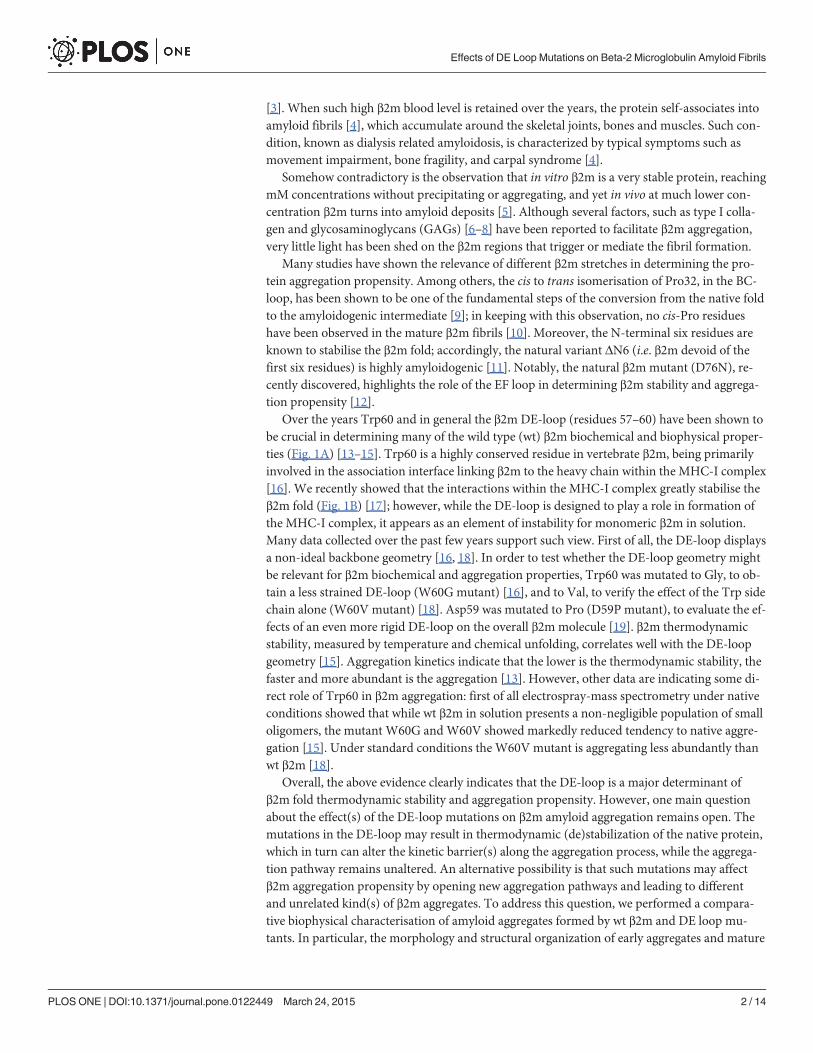
Over the years Trp60 and in general the β2m DE-loop (residues 57–60) have been shown tobe crucial in determining many of the wild type (wt) β2m biochemical and biophysical proper-ties (Fig. 1A) [13–15]. Trp60 is a highly conserved residue in vertebrate β2m, being primarilyinvolved in the association interface linking β2m to the heavy chain within the MHC-I complex[16]. We recently showed that the interactions within the MHC-I complex greatly stabilise theβ2m fold (Fig. 1B) [17]; however, while the DE-loop is designed to play a role in formation ofthe MHC-I complex, it appears as an element of instability for monomeric β2m in solution.Many data collected over the past few years support such view. First of all, the DE-loop displaysa non-ideal backbone geometry [16, 18]. In order to test whether the DE-loop geometry mightbe relevant for β2m biochemical and aggregation properties, Trp60 was mutated to Gly, to ob-tain a less strained DE-loop (W60G mutant) [16], and to Val, to verify the effect of the Trp sidechain alone (W60V mutant) [18]. Asp59 was mutated to Pro (D59P mutant), to evaluate the ef-fects of an even more rigid DE-loop on the overall β2m molecule [19]. β2m thermodynamicstability, measured by temperature and chemical unfolding, correlates well with the DE-loopgeometry [15]. Aggregation kinetics indicate that the lower is the thermodynamic stability, thefaster and more abundant is the aggregation [13]. However, other data are indicating some di-rect role of Trp60 in β2m aggregation: first of all electrospray-mass spectrometry under nativeconditions showed that while wt β2m in solution presents a non-negligible population of smalloligomers, the mutant W60G and W60V showed markedly reduced tendency to native aggre-gation [15]. Under standard conditions the W60V mutant is aggregating less abundantly thanwt β2m [18].
Overall, the above evidence clearly indicates that the DE-loop is a major determinant ofβ2m fold thermodynamic stability and aggregation propensity. However, one main questionabout the effect(s) of the DE-loop mutations on β2m amyloid aggregation remains open. Themutations in the DE-loop may result in thermodynamic (de)stabilization of the native protein,which in turn can alter the kinetic barrier(s) along the aggregation process, while the aggrega-tion pathway remains unaltered. An alternative possibility is that such mutations may affectβ2m aggregation propensity by opening new aggregation pathways and leading to differentand unrelated kind(s) of β2m aggregates. To address this question, we performed a compara-tive biophysical characterisation of amyloid aggregates formed by wt β2m and DE loop mu-tants. In particular, the morphology and structural organization of early aggregates and mature
Effects of DE Loop Mutations on Beta-2 Microglobulin Amyloid Fibrils
PLOS ONE | DOI:10.1371/journal.pone.0122449 March 24, 2015 2 / 14
fibrils have been analysed by Atomic Force Microscopy (AFM); then, the hydrogen/deuterium(H/D) exchange kinetics and the overall secondary structure content for the fibrils of the fourβ2m variants have been monitored by Fourier transform infrared (FTIR) spectroscopy. Previ-ous data showed that the DE loop mutations exert a major effect on the stability of the β2m na-tive fold and affect the aggregation kinetics [13, 15] while the experiments here presentedindicate that the fibrils of the DE loop variants and of wt β2m have comparable architectureand dynamics, suggesting that mutations at the DE loop effects are limited to the β2m aggrega-tion process, and do not alter the end stage of the aggregation pathway.
Fig 1. DE loop in monomeric β2m and in interaction within the MHC-I. (A) Ribbon representation ofmonomeric β2m (PDB code 2YXF). The DE loop residues are shown in yellow sticks. (B) Stereo view of theDE loop and Phe56 (yellow sticks) when interacting with the heavy chain in the MHC-I (electrostatic surfaceand green sticks). Trp60 is establishing a H-bond with Asp122 from the heavy chain (PDB code 4L29).
doi:10.1371/journal.pone.0122449.g001
Effects of DE Loop Mutations on Beta-2 Microglobulin Amyloid Fibrils
PLOS ONE | DOI:10.1371/journal.pone.0122449 March 24, 2015 3 / 14
Results
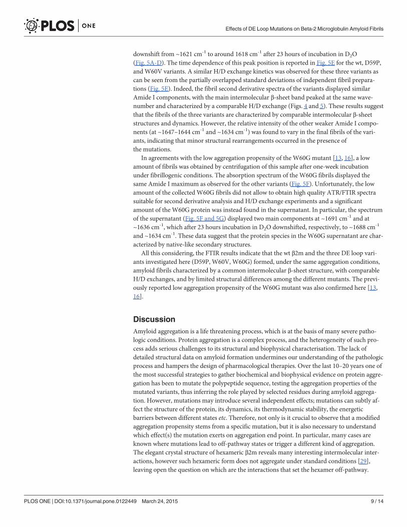
Amyloid fibril preparationPurified samples of wt β2m and of the three DE loop variants have been placed under standardaggregation conditions (see methods). Aggregated samples after 24 hours and after one-weekincubation have been tested for thioflavin fluorescence (Table 1) and have been analysed bymeans of AFM and FTIR.
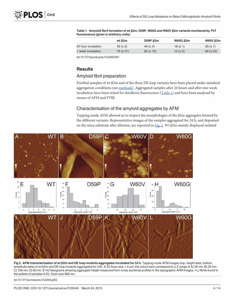
Characterisation of the amyloid aggregates by AFMTapping mode AFM allowed us to inspect the morphologies of the β2m aggregates formed bythe different variants. Representative images of the samples aggregated for 24 h, and depositedon the mica substrate after dilution, are reported in Fig. 2. Wt β2m mainly displayed isolated
Table 1. Amyloid fibril formation of wt β2m, D59P, W60G andW60V β2m variants monitored by ThTfluorescence (given in arbitrary units).
wt β2m D59P β2m W60G β2m W60V β2m
24 hour incubation 40 (± 2) 46 (± 4) 18 (± 1) 29 (± 1)
1 week incubation 79 (± 21) 62 (± 15) 12 (± 2) 69 (± 22)
doi:10.1371/journal.pone.0122449.t001
Fig 2. AFM characterisation of wt β2m and DE loopmutants aggregates incubated for 24 h. Tapping mode AFM images (top, height data; bottom,amplitude data) of wt β2m and DE loop mutants aggregated for 24h. A-D) Scan size 1.4 μm; the colour bars correspond to a Z range of A) 30 nm; B) 35 nm;C) 100 nm; D) 60 nm. E-H) histograms showing aggregate height measured from cross-sectional profiles in the topographic AFM images. I-L) fibrils found inthe pellets of samples A-D). Scan size 860 nm.
doi:10.1371/journal.pone.0122449.g002
Effects of DE Loop Mutations on Beta-2 Microglobulin Amyloid Fibrils
PLOS ONE | DOI:10.1371/journal.pone.0122449 March 24, 2015 4 / 14
oligomers with a mean height of 6.2±0.3 nm, or oligomer chains (Fig. 2A and 2E), while theD59P variant extensively formed overlying planar sheets of filaments and thin fibrils, respec-tively about 1 and 4 nm high (Fig. 2B and 2F). The height of these fibrils is significantly lowerthan that measured for mature fibrils, suggesting that the thin fibrils are either intermediatestructures or structures resulting from an epitaxial growth induced by the mica substrate. Inany case, they reflect a strong tendency to fast aggregation, which is distinctive for this variant.Large spheroidal aggregates (mean height 25 ± 1 nm), probably resulting from oligomer clus-tering, were observed for the W60V variant (Fig. 2C and 2G). For the W60G variant, smalloligomers (mean height 3.1 ± 0.2 nm) were found to coexist with larger oligomers (meanheight 12 ± 1 nm), and very short protofibrils formed by few units of the latter (Fig. 2D and2H). Mature fibrils were not observed for any of the variants, probably due to the relatively lowfraction of fibrillar material. Therefore, samples were centrifuged and the pellet was analysed tocheck for the presence of mature fibrils. In all cases except for W60G β2m, mature fibrils wereabundant in the pellet and were intertwined in clusters (Fig. 2I-K). A different behaviour wasfound for the W60G variant, which exhibited only very short, almost isolated, fibrillar struc-tures (Fig. 2L).
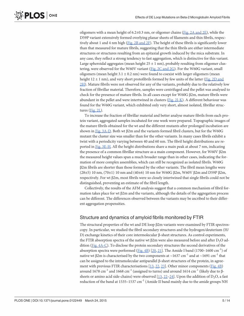
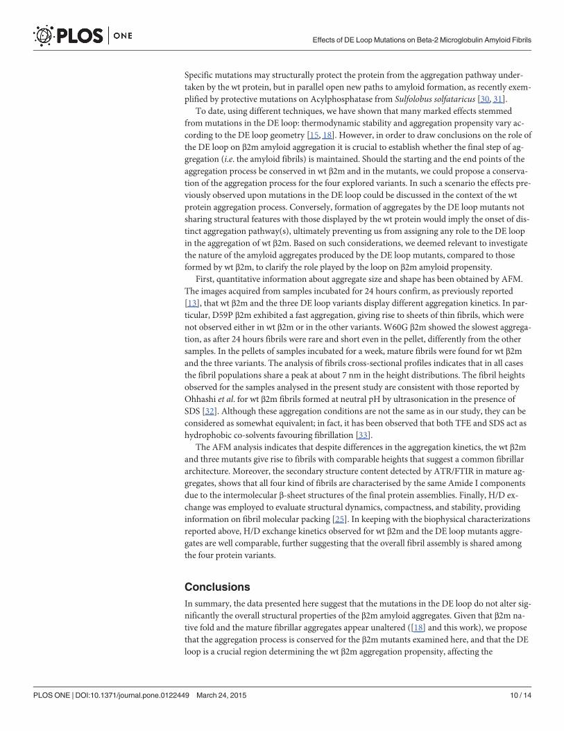
To increase the fraction of fibrillar material and better analyse mature fibrils from each pro-tein variant, aggregated samples incubated for one week were prepared. Topographic images ofthe mature fibrils obtained for the wt and the different mutants after prolonged incubation areshown in Fig. 3A-D. Both wt β2m and the variants formed fibril clusters, but for the W60Gmutant the cluster size was smaller than for the other variants. In many cases fibrils exhibit atwist with a periodicity varying between 40 and 60 nm. The fibril height distributions are re-ported in Fig. 3E-H. All the height distributions share a main peak at about 7 nm, indicatingthe presence of a common fibrillar structure as a main component. However, for W60V β2mthe measured height values span a much broader range than in other cases, indicating the for-mation of more complex assemblies, which can still be recognized as isolated fibrils. W60Gβ2m fibrils are shorter than those formed by the other variants. The fibril mean lengths were(28±5)�10 nm, (70±1)�10 nm and (40±6)�10 nm for W60G β2m, W60V β2m and D59P β2m,respectively. For wt β2m, most fibrils were so closely intertwined that single fibrils could not bedistinguished, preventing an estimate of the fibril length.
Collectively, the results of the AFM analysis suggest that a common mechanism of fibril for-mation takes place for wt β2m and the variants, although the details of the aggregation processcan be different. The differences observed between the variants may be ascribed to their differ-ent aggregation propensities.
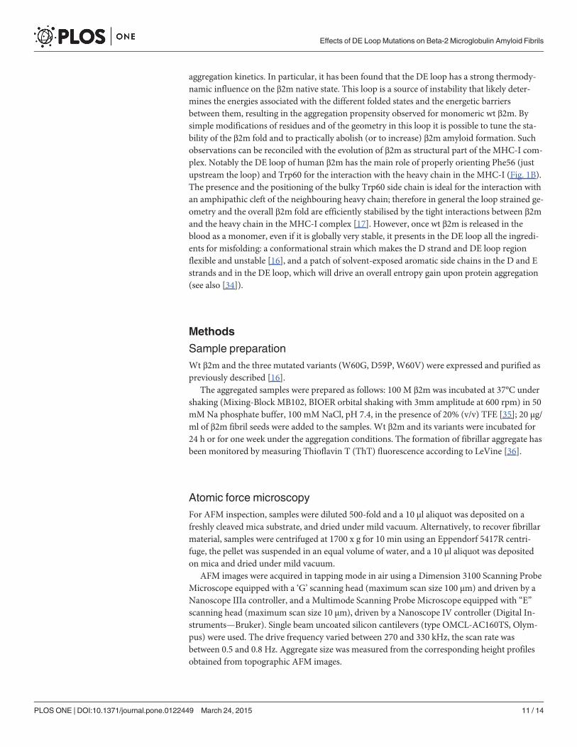
Structure and dynamics of amyloid fibrils monitored by FTIRThe structural properties of the wt and DE loop β2m variants were examined by FTIR spectros-copy. In particular, we studied the fibril secondary structures and the hydrogen/deuterium (H/D) exchange kinetics of their core intermolecular β-sheet structures. As control experiments,the FTIR absorption spectra of the native wt β2m were also measured before and after D2O ad-dition (Fig. 4A-C). To disclose the protein secondary structures the second derivatives of theabsorption spectra were performed (Fig. 4B) [20, 21]. The Amide I band (1700–1600 cm-1) ofnative wt β2m is characterised by the two components at ~1637 cm-1 and at ~1691 cm-1 thatcan be assigned to the intramolecular antiparallel β-sheet structures of the protein, in agree-ment with previous FTIR characterisations [13, 22, 23]. Other minor components (Fig. 4B)around 1678 cm-1 and 1668 cm-1 (assigned to turns) and around 1614 cm-1 (likely due to β-sheets or amino acid side-chains) were observed [13, 22–24]. Upon the addition of D2O, a fastreduction of the band at 1535–1537 cm-1 (Amide II band mainly due to the amide groups NH
Effects of DE Loop Mutations on Beta-2 Microglobulin Amyloid Fibrils
PLOS ONE | DOI:10.1371/journal.pone.0122449 March 24, 2015 5 / 14
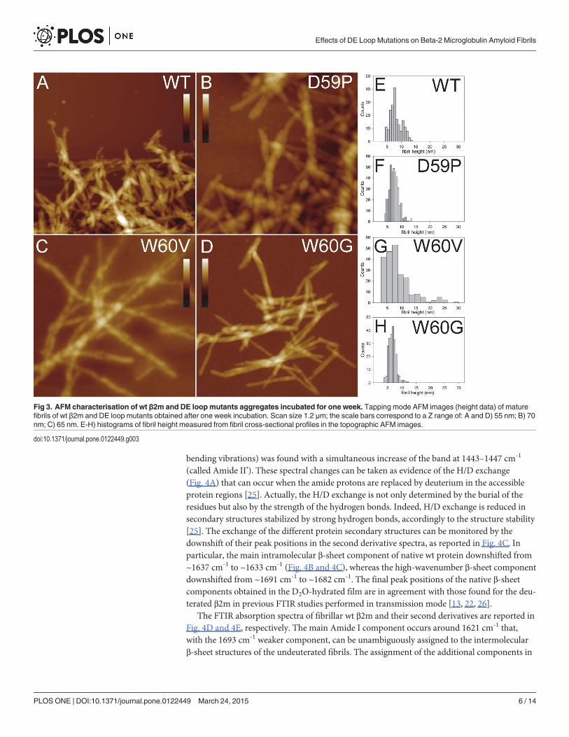
bending vibrations) was found with a simultaneous increase of the band at 1443–1447 cm-1
(called Amide II’). These spectral changes can be taken as evidence of the H/D exchange(Fig. 4A) that can occur when the amide protons are replaced by deuterium in the accessibleprotein regions [25]. Actually, the H/D exchange is not only determined by the burial of theresidues but also by the strength of the hydrogen bonds. Indeed, H/D exchange is reduced insecondary structures stabilized by strong hydrogen bonds, accordingly to the structure stability[25]. The exchange of the different protein secondary structures can be monitored by thedownshift of their peak positions in the second derivative spectra, as reported in Fig. 4C. Inparticular, the main intramolecular β-sheet component of native wt protein downshifted from~1637 cm-1 to ~1633 cm-1 (Fig. 4B and 4C), whereas the high-wavenumber β-sheet componentdownshifted from ~1691 cm-1 to ~1682 cm-1. The final peak positions of the native β-sheetcomponents obtained in the D2O-hydrated film are in agreement with those found for the deu-terated β2m in previous FTIR studies performed in transmission mode [13, 22, 26].
The FTIR absorption spectra of fibrillar wt β2m and their second derivatives are reported inFig. 4D and 4E, respectively. The main Amide I component occurs around 1621 cm-1 that,with the 1693 cm-1 weaker component, can be unambiguously assigned to the intermolecularβ-sheet structures of the undeuterated fibrils. The assignment of the additional components in
Fig 3. AFM characterisation of wt β2m and DE loopmutants aggregates incubated for one week. Tapping mode AFM images (height data) of maturefibrils of wt β2m and DE loop mutants obtained after one week incubation. Scan size 1.2 μm; the scale bars correspond to a Z range of: A and D) 55 nm; B) 70nm; C) 65 nm. E-H) histograms of fibril height measured from fibril cross-sectional profiles in the topographic AFM images.
doi:10.1371/journal.pone.0122449.g003
Effects of DE Loop Mutations on Beta-2 Microglobulin Amyloid Fibrils
PLOS ONE | DOI:10.1371/journal.pone.0122449 March 24, 2015 6 / 14
the 1675–1634 cm-1 spectral region is not unequivocal. Indeed, as discussed in the literature[20, 22, 26–28], they can be assigned to turns (typically in the 1686–1660 cm-1 range), to loops(typically in the 1650–1640 cm-1 range), to native-like structures (around 1634 cm-1) or to a pe-culiar arrangement of the β-strands in the protein supramolecular assemblies [26]. We shouldnote that the relative intensities of these components displayed a certain heterogeneity as
Fig 4. ATR/FTIR characterisation of wt β2m in the native and the fibrillar state. A) The absorption spectra of the native β2m in form of a protein film werecollected before and after incubation in D2O for different times. Spectra are reported in the regions of Amide I (AI), Amide II (AII), and Amide II’ (AII’). Arrowspoint at increasing incubation time in D2O. Absorption spectra are normalized at the Amide I maximum. B) Second derivatives of the absorption spectra of (A)in the Amide I region. The spectra collected after D2O addition were normalized at the tyrosine band [27]. The marked peak positions of the two componentsdue to the native antiparallel β-sheet structures refer to the spectrum of the undeuterated sample. C) Time course of the peak position of the main native β-sheet component reported after D2O addition to the protein film. Error bars represent the standard deviation of three independent samples. The peakpositions were taken from the second derivative spectra. D) Absorption spectra of the wt β2m fibrils collected before and after incubation in D2O, reported asin (A). E) Second derivatives of the absorption spectra of (D) in the Amide I region. Spectra of two undeuterated fibrils obtained from independentpreparations are compared to show fibril heterogeneity. The spectra collected after D2O addition were normalized at the tyrosine band [27]. The peakpositions of the main components are indicated. F) Time course of the peak position of the main intermolecular β-sheet component is reported after D2Oaddition. Error bars represent the standard deviation of three independent fibril preparations. The peak positions were taken from the secondderivative spectra.
doi:10.1371/journal.pone.0122449.g004
Effects of DE Loop Mutations on Beta-2 Microglobulin Amyloid Fibrils
PLOS ONE | DOI:10.1371/journal.pone.0122449 March 24, 2015 7 / 14
observed for independent fibril preparations and illustrated in Fig. 4E, where two representa-tive second derivative spectra of wt (undeuterated) fibril films are reported.
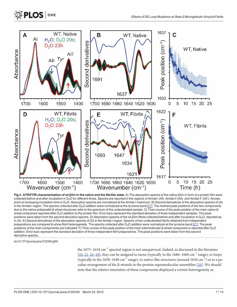
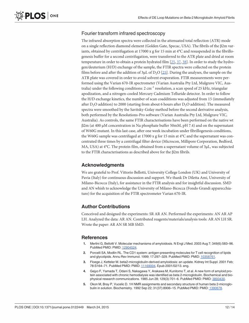
To study the solvent accessibility and dynamics [25] of the fibril core β-sheet structure, H/Dexchange experiments were performed, as reported in Fig. 4D-F. In particular, during incuba-tion in D2O, the main intermolecular β-sheet peak downshifted from ~1621 cm-1 in theundeuterated fibrils to ~1618 cm-1 after 23 hours of incubation in D2O (Fig. 4E and 4F). Thesame characterisations were performed on the fibrils of the DE-loop mutants (Fig. 5).
The undeuterated fibrils of D59P and of W60V variants share with aggregates of wt β2m thesame two main Amide I components around 1693 cm-1 and 1621cm-1 that can be assigned tothe intermolecular β-sheet structures (Fig. 5B and 5D). This result is in agreement with previ-ous FTIR characterizations [13] on the same β2m variants studied here. Indeed, in these earlierexperiments, the aggregation of unseeded wt and DE loop mutants took place inside the infra-red cell, leading to final aggregates characterized by the same intermolecular β-sheet AmideI’ components.
Concerning the H/D exchange experiments presented in this work, as observed for wt fi-brils, the main intermolecular β-sheet peak of D59P and of W60V fibrils was found to
Fig 5. ATR/FTIR characterisation of DE loopmutants in the fibrillar state. A) The absorption spectra of the D59P fibrils were collected before and afterincubation in D2O for different times. Spectra are reported in the regions of Amide I, Amide II, and Amide II’ bands. Arrows point to the spectral changes atincreased incubation time in D2O. Absorption spectra are normalized at the Amide I maximum. B) Second derivatives of the absorption spectra of (A) in theAmide I region. The spectra collected after D2O additions were normalized at the tyrosine band [27]. The peak positions of the main components areindicated. C) The absorption spectra of theW60V fibrils were collected before and after incubation in D2O for different times and reported as in (A). D)Second derivatives of the absorption spectra of (C) in the Amide I region. E) Time course of the peak positions of the main intermolecular β-sheet componentof wt, D59P, andW60V amyloid fibrils are reported after D2O addition to the protein films. Error bars represent the standard deviation of at least threeindependent fibril preparations. The peak positions were taken from the second derivative spectra. F) The absorption spectra of W60G, wt, D59P, andW60Vfibrils and that of W60G supernatant are reported in the Amide I region. The intermolecular β-sheet structure absorption band is marked. G) Secondderivative spectra of the W60G supernatant collected before and after 23 hours from D2O addition. The peak positions of the main components are indicated.
doi:10.1371/journal.pone.0122449.g005
Effects of DE Loop Mutations on Beta-2 Microglobulin Amyloid Fibrils
PLOS ONE | DOI:10.1371/journal.pone.0122449 March 24, 2015 8 / 14
downshift from ~1621 cm-1 to around 1618 cm-1 after 23 hours of incubation in D2O(Fig. 5A-D). The time dependence of this peak position is reported in Fig. 5E for the wt, D59P,and W60V variants. A similar H/D exchange kinetics was observed for these three variants ascan be seen from the partially overlapped standard deviations of independent fibril prepara-tions (Fig. 5E). Indeed, the fibril second derivative spectra of the variants displayed similarAmide I components, with the main intermolecular β-sheet band peaked at the same wave-number and characterized by a comparable H/D exchange (Figs. 4 and 5). These results suggestthat the fibrils of the three variants are characterized by comparable intermolecular β-sheetstructures and dynamics. However, the relative intensity of the other weaker Amide I compo-nents (at ~1647–1644 cm-1 and ~1634 cm-1) was found to vary in the final fibrils of the vari-ants, indicating that minor structural rearrangements occurred in the presence ofthe mutations.
In agreements with the low aggregation propensity of the W60G mutant [13, 16], a lowamount of fibrils was obtained by centrifugation of this sample after one-week incubationunder fibrillogenic conditions. The absorption spectrum of the W60G fibrils displayed thesame Amide I maximum as observed for the other variants (Fig. 5F). Unfortunately, the lowamount of the collected W60G fibrils did not allow to obtain high quality ATR/FTIR spectrasuitable for second derivative analysis and H/D exchange experiments and a significantamount of the W60G protein was instead found in the supernatant. In particular, the spectrumof the supernatant (Fig. 5F and 5G) displayed two main components at ~1691 cm-1 and at~1636 cm-1, which after 23 hours incubation in D2O downshifted, respectively, to ~1688 cm-1
and ~1634 cm-1. These data suggest that the protein species in the W60G supernatant are char-acterized by native-like secondary structures.
All this considering, the FTIR results indicate that the wt β2m and the three DE loop vari-ants investigated here (D59P, W60V, W60G) formed, under the same aggregation conditions,amyloid fibrils characterized by a common intermolecular β-sheet structure, with comparableH/D exchanges, and by limited structural differences among the different mutants. The previ-ously reported low aggregation propensity of the W60G mutant was also confirmed here [13,16].
DiscussionAmyloid aggregation is a life threatening process, which is at the basis of many severe patho-logic conditions. Protein aggregation is a complex process, and the heterogeneity of such pro-cess adds serious challenges to its structural and biophysical characterisation. The lack ofdetailed structural data on amyloid formation undermines our understanding of the pathologicprocess and hampers the design of pharmacological therapies. Over the last 10–20 years one ofthe most successful strategies to gather biochemical and biophysical evidence on protein aggre-gation has been to mutate the polypeptide sequence, testing the aggregation properties of themutated variants, thus inferring the role played by selected residues during amyloid aggrega-tion. However, mutations may introduce several independent effects; mutations can subtly af-fect the structure of the protein, its dynamics, its thermodynamic stability, the energeticbarriers between different states etc. Therefore, not only is it crucial to observe that a modifiedaggregation propensity stems from a specific mutation, but it is also necessary to understandwhich effect(s) the mutation exerts on aggregation end point. In particular, many cases areknown where mutations lead to off-pathway states or trigger a different kind of aggregation.The elegant crystal structure of hexameric β2m reveals many interesting intermolecular inter-actions, however such hexameric form does not aggregate under standard conditions [29],leaving open the question on which are the interactions that set the hexamer off-pathway.
Effects of DE Loop Mutations on Beta-2 Microglobulin Amyloid Fibrils
PLOS ONE | DOI:10.1371/journal.pone.0122449 March 24, 2015 9 / 14
Specific mutations may structurally protect the protein from the aggregation pathway under-taken by the wt protein, but in parallel open new paths to amyloid formation, as recently exem-plified by protective mutations on Acylphosphatase from Sulfolobus solfataricus [30, 31].
To date, using different techniques, we have shown that many marked effects stemmedfrom mutations in the DE loop: thermodynamic stability and aggregation propensity vary ac-cording to the DE loop geometry [15, 18]. However, in order to draw conclusions on the role ofthe DE loop on β2m amyloid aggregation it is crucial to establish whether the final step of ag-gregation (i.e. the amyloid fibrils) is maintained. Should the starting and the end points of theaggregation process be conserved in wt β2m and in the mutants, we could propose a conserva-tion of the aggregation process for the four explored variants. In such a scenario the effects pre-viously observed upon mutations in the DE loop could be discussed in the context of the wtprotein aggregation process. Conversely, formation of aggregates by the DE loop mutants notsharing structural features with those displayed by the wt protein would imply the onset of dis-tinct aggregation pathway(s), ultimately preventing us from assigning any role to the DE loopin the aggregation of wt β2m. Based on such considerations, we deemed relevant to investigatethe nature of the amyloid aggregates produced by the DE loop mutants, compared to thoseformed by wt β2m, to clarify the role played by the loop on β2m amyloid propensity.
First, quantitative information about aggregate size and shape has been obtained by AFM.The images acquired from samples incubated for 24 hours confirm, as previously reported[13], that wt β2m and the three DE loop variants display different aggregation kinetics. In par-ticular, D59P β2m exhibited a fast aggregation, giving rise to sheets of thin fibrils, which werenot observed either in wt β2m or in the other variants. W60G β2m showed the slowest aggrega-tion, as after 24 hours fibrils were rare and short even in the pellet, differently from the othersamples. In the pellets of samples incubated for a week, mature fibrils were found for wt β2mand the three variants. The analysis of fibrils cross-sectional profiles indicates that in all casesthe fibril populations share a peak at about 7 nm in the height distributions. The fibril heightsobserved for the samples analysed in the present study are consistent with those reported byOhhashi et al. for wt β2m fibrils formed at neutral pH by ultrasonication in the presence ofSDS [32]. Although these aggregation conditions are not the same as in our study, they can beconsidered as somewhat equivalent; in fact, it has been observed that both TFE and SDS act ashydrophobic co-solvents favouring fibrillation [33].
The AFM analysis indicates that despite differences in the aggregation kinetics, the wt β2mand three mutants give rise to fibrils with comparable heights that suggest a common fibrillararchitecture. Moreover, the secondary structure content detected by ATR/FTIR in mature ag-gregates, shows that all four kind of fibrils are characterised by the same Amide I componentsdue to the intermolecular β-sheet structures of the final protein assemblies. Finally, H/D ex-change was employed to evaluate structural dynamics, compactness, and stability, providinginformation on fibril molecular packing [25]. In keeping with the biophysical characterizationsreported above, H/D exchange kinetics observed for wt β2m and the DE loop mutants aggre-gates are well comparable, further suggesting that the overall fibril assembly is shared amongthe four protein variants.
ConclusionsIn summary, the data presented here suggest that the mutations in the DE loop do not alter sig-nificantly the overall structural properties of the β2m amyloid aggregates. Given that β2m na-tive fold and the mature fibrillar aggregates appear unaltered ([18] and this work), we proposethat the aggregation process is conserved for the β2m mutants examined here, and that the DEloop is a crucial region determining the wt β2m aggregation propensity, affecting the
Effects of DE Loop Mutations on Beta-2 Microglobulin Amyloid Fibrils
PLOS ONE | DOI:10.1371/journal.pone.0122449 March 24, 2015 10 / 14
aggregation kinetics. In particular, it has been found that the DE loop has a strong thermody-namic influence on the β2m native state. This loop is a source of instability that likely deter-mines the energies associated with the different folded states and the energetic barriersbetween them, resulting in the aggregation propensity observed for monomeric wt β2m. Bysimple modifications of residues and of the geometry in this loop it is possible to tune the sta-bility of the β2m fold and to practically abolish (or to increase) β2m amyloid formation. Suchobservations can be reconciled with the evolution of β2m as structural part of the MHC-I com-plex. Notably the DE loop of human β2m has the main role of properly orienting Phe56 (justupstream the loop) and Trp60 for the interaction with the heavy chain in the MHC-I (Fig. 1B).The presence and the positioning of the bulky Trp60 side chain is ideal for the interaction withan amphipathic cleft of the neighbouring heavy chain; therefore in general the loop strained ge-ometry and the overall β2m fold are efficiently stabilised by the tight interactions between β2mand the heavy chain in the MHC-I complex [17]. However, once wt β2m is released in theblood as a monomer, even if it is globally very stable, it presents in the DE loop all the ingredi-ents for misfolding: a conformational strain which makes the D strand and DE loop regionflexible and unstable [16], and a patch of solvent-exposed aromatic side chains in the D and Estrands and in the DE loop, which will drive an overall entropy gain upon protein aggregation(see also [34]).
Methods
Sample preparationWt β2m and the three mutated variants (W60G, D59P, W60V) were expressed and purified aspreviously described [16].
The aggregated samples were prepared as follows: 100 M β2m was incubated at 37°C undershaking (Mixing-Block MB102, BIOER orbital shaking with 3mm amplitude at 600 rpm) in 50mMNa phosphate buffer, 100 mM NaCl, pH 7.4, in the presence of 20% (v/v) TFE [35]; 20 μg/ml of β2m fibril seeds were added to the samples. Wt β2m and its variants were incubated for24 h or for one week under the aggregation conditions. The formation of fibrillar aggregate hasbeen monitored by measuring Thioflavin T (ThT) fluorescence according to LeVine [36].
Atomic force microscopyFor AFM inspection, samples were diluted 500-fold and a 10 μl aliquot was deposited on afreshly cleaved mica substrate, and dried under mild vacuum. Alternatively, to recover fibrillarmaterial, samples were centrifuged at 1700 x g for 10 min using an Eppendorf 5417R centri-fuge, the pellet was suspended in an equal volume of water, and a 10 μl aliquot was depositedon mica and dried under mild vacuum.
AFM images were acquired in tapping mode in air using a Dimension 3100 Scanning ProbeMicroscope equipped with a ‘G’ scanning head (maximum scan size 100 μm) and driven by aNanoscope IIIa controller, and a Multimode Scanning Probe Microscope equipped with “E”scanning head (maximum scan size 10 μm), driven by a Nanoscope IV controller (Digital In-struments—Bruker). Single beam uncoated silicon cantilevers (type OMCL-AC160TS, Olym-pus) were used. The drive frequency varied between 270 and 330 kHz, the scan rate wasbetween 0.5 and 0.8 Hz. Aggregate size was measured from the corresponding height profilesobtained from topographic AFM images.
Effects of DE Loop Mutations on Beta-2 Microglobulin Amyloid Fibrils
PLOS ONE | DOI:10.1371/journal.pone.0122449 March 24, 2015 11 / 14
Fourier transform infrared spectroscopyThe infrared absorption spectra were collected in the attenuated total reflection (ATR) modeon a single reflection diamond element (Golden Gate, Specac, USA). The fibrils of the β2m var-iants, obtained by centrifugation at 17000 x g for 15 min at 4°C and resuspended in the fibrillo-genesis buffer for a second centrifugation, were transferred to the ATR plate and dried at roomtemperature in order to obtain a protein hydrated film [25, 37, 38]. In order to study the hydro-gen/deuterium (H/D) exchange of the sample, the FTIR spectra were collected on the proteinfilms before and after the addition of 3μL of D2O [25]. During the analyses, the sample on theATR plate was covered in order to avoid solvent evaporation. FTIR measurements were per-formed using the Varian 670-IR spectrometer (Varian Australia Pty Ltd, Mulgrave VIC, Aus-tralia) under the following conditions: 2 cm-1 resolution, a scan speed of 25 kHz, triangularapodization, and a nitrogen-cooled Mercury Cadmium Telluride detector. In order to followthe H/D exchange kinetics, the number of scan coadditions was adjusted from 15 (immediatelyafter D2O addition) to 2000 (starting from about 6 hours after D2O addition). The measuredspectra were smoothed by the Savitsky-Golay method before the second derivative analysis,both performed by the Resolutions-Pro software (Varian Australia Pty Ltd, Mulgrave VIC,Australia). As controls, the same FTIR characterisations have been performed on the native wtβ2m (at 400 μM concentration in Na phosphate buffer 50mM, pH 7.4) and on the supernatantof W60G mutant. In this last case, after one week incubation under fibrillogenesis conditions,the W60G sample was centrifuged at 17000 x g for 15 min at 4°C and the supernatant was con-centrated three times by a centrifugal filter device (Microcon, Millipore Corporation, Bedford,MA, USA) at 4°C. The protein film, obtained from a supernatant volume of 3μL, was subjectedto the FTIR characterisations as described above for the β2m fibrils.
AcknowledgmentsWe are grateful to Prof. Vittorio Bellotti, University College London (UK) and University ofPavia (Italy) for continuous discussion and support. We thank Dr Diletta Ami, University ofMilano-Bicocca (Italy), for assistance in the FTIR analysis and for insightful discussion. SMDand AN whish to acknowledge the University of Milano-Bicocca (Fondo Grandi apparecchia-ture) for the acquisition of the FTIR spectrometer Varian 670-IR.
Author ContributionsConceived and designed the experiments: SR AR AN. Performed the experiments: AN AR APLH. Analyzed the data: AR AN. Contributed reagents/materials/analysis tools: AR AN LH SR.Wrote the paper: AR AN SR MB SMD.
References1. Merlini G, Bellotti V. Molecular mechanisms of amyloidosis. N Engl J Med. 2003 Aug 7; 349(6):583–96.
PubMed PMID: PMID: 12904524.
2. Porcelli SA, Modlin RL. The CD1 system: antigen-presenting molecules for T cell recognition of lipidsand glycolipids. Annu Rev Immunol. 1999; 17:297–329. PubMed PMID: PMID: 10358761.
3. Floege J, Ketteler M. beta2-microglobulin-derived amyloidosis: an update. Kidney Int Suppl. 2001 Feb;78:S164–71. PubMed PMID: PMID: 11169004. Epub 2001/02/13. eng.
4. Gejyo F, Yamada T, Odani S, Nakagawa Y, Arakawa M, Kunitomo T, et al. A new form of amyloid pro-tein associated with chronic hemodialysis was identified as beta 2-microglobulin. Biochemical and bio-physical research communications. 1985 Jun 28; 129(3):701–6. PubMed PMID: PMID: 3893430.
5. Okon M, Bray P, Vucelic D. 1H NMR assignments and secondary structure of human beta 2-microglo-bulin in solution. Biochemistry. 1992 Sep 22; 31(37):8906–15. PubMed PMID: PMID: 1390678.
Effects of DE Loop Mutations on Beta-2 Microglobulin Amyloid Fibrils
PLOS ONE | DOI:10.1371/journal.pone.0122449 March 24, 2015 12 / 14
6. Myers SL, Jones S, Jahn TR, Morten IJ, Tennent GA, Hewitt EW, et al. A systematic study of the effectof physiological factors on beta2-microglobulin amyloid formation at neutral pH. Biochemistry. 2006Feb 21; 45(7):2311–21. PubMed PMID: PMID: 16475820. eng.
7. Relini A, Canale C, De Stefano S, Rolandi R, Giorgetti S, Stoppini M, et al. Collagen plays an activerole in the aggregation of beta2-microglobulin under physiopathological conditions of dialysis-relatedamyloidosis. The Journal of biological chemistry. 2006 Jun 16; 281(24):16521–9. PubMed PMID:PMID: 16601119.
8. Relini A, De Stefano S, Torrassa S, Cavalleri O, Rolandi R, Gliozzi A, et al. Heparin strongly enhancesthe formation of beta2-microglobulin amyloid fibrils in the presence of type I collagen. The Journal of bi-ological chemistry. 2008 Feb 22; 283(8):4912–20. PubMed PMID: PMID: 18056266. eng.
9. Eakin CM, Berman AJ, Miranker AD. A native to amyloidogenic transition regulated by a backbone trig-ger. Nature structural & molecular biology. 2006 Mar; 13(3):202–8. PubMed PMID: PMID: 16491088.eng.
10. Barbet-Massin E, Ricagno S, Lewandowski JR, Giorgetti S, Bellotti V, Bolognesi M, et al. Fibrillar vscrystalline full-length beta-2-microglobulin studied by high-resolution solid-state NMR spectroscopy.Journal of the American Chemical Society. 2010 Apr 28; 132(16):5556–7. PubMed PMID: PMID:20356307. eng. doi: 10.1021/ja1002839
11. Esposito G, Michelutti R, Verdone G, Viglino P, Hernandez H, Robinson CV, et al. Removal of the N-ter-minal hexapeptide from human beta2-microglobulin facilitates protein aggregation and fibril formation.Protein Sci. 2000 May; 9(5):831–45. PubMed PMID: PMID: 10850793.
12. Valleix S, Gillmore JD, Bridoux F, Mangione PP, Dogan A, Nedelec B, et al. Hereditary systemic amy-loidosis due to Asp76Asn variant beta2-microglobulin. N Engl J Med. 2012 Jun 14; 366(24):2276–83.PubMed PMID: PMID: 22693999. Epub 2012/06/15. eng. doi: 10.1056/NEJMoa1201356
13. Ami D, Ricagno S, Bolognesi M, Bellotti V, Doglia SM, Natalello A. Structure, stability, and aggregationof beta-2 microglobulin mutants: insights from a Fourier transform infrared study in solution and in thecrystalline state. Biophys J. 2012 Apr 4; 102(7):1676–84. PubMed PMID: PMID: 22500768. PubmedCentral PMCID: 3318121. doi: 10.1016/j.bpj.2012.02.045
14. Rennella E, Corazza A, Giorgetti S, Fogolari F, Viglino P, Porcari R, et al. Folding and fibrillogenesis:clues from beta2-microglobulin. Journal of molecular biology. 2010 Aug 13; 401(2):286–97. PubMedPMID: PMID: 20558175. eng. doi: 10.1016/j.jmb.2010.06.016
15. Santambrogio C, Ricagno S, Colombo M, Barbiroli A, Bonomi F, Bellotti V, et al. DE-loop mutations af-fect beta2 microglobulin stability, oligomerization, and the low-pH unfolded form. Protein Sci. 2010 Jul;19(7):1386–94. PubMed PMID: PMID: 20506535. eng. doi: 10.1002/pro.419
16. Esposito G, Ricagno S, Corazza A, Rennella E, Gumral D, Mimmi MC, et al. The controlling roles ofTrp60 and Trp95 in beta2-microglobulin function, folding and amyloid aggregation properties. Journalof molecular biology. 2008 May 9; 378(4):885–95. PubMed PMID: PMID: 18395224.
17. Halabelian L, Ricagno S, Giorgetti S, Santambrogio C, Barbiroli A, Pellegrino S, et al. Class I MajorHistocompatibility Complex, the Trojan Horse for Secretion of Amyloidogenic beta2-Microglobulin. TheJournal of biological chemistry. 2014 Feb 7; 289(6):3318–27. PubMed PMID: PMID: 24338476.Pubmed Central PMCID: 3916536. Epub 2013/12/18. eng. doi: 10.1074/jbc.M113.524157
18. Ricagno S, Raimondi S, Giorgetti S, Bellotti V, Bolognesi M. Human beta-2 microglobulin W60Vmutantstructure: Implications for stability and amyloid aggregation. Biochemical and biophysical researchcommunications. 2009 Mar 13; 380(3):543–7. PubMed PMID: PMID: 19284997. eng. doi: 10.1016/j.bbrc.2009.01.116
19. Ricagno S, ColomboM, de Rosa M, Sangiovanni E, Giorgetti S, Raimondi S, et al. DE loop mutationsaffect beta-2 microglobulin stability and amyloid aggregation. Biochemical and biophysical researchcommunications. 2008 Dec 5; 377(1):146–50. doi: 10.1016/j.bbrc.2008.09.108 PMID: 18835253
20. Barth A. Infrared spectroscopy of proteins. Bba-Bioenergetics. 2007 SEP; 1767(9):1073–101. PubMedPMID: ISI:000249671400001. PMID: 17692815
21. Susi H, Byler DM. Resolution-Enhanced Fourier-Transform Infrared-Spectroscopy of Enzymes. Meth-od Enzymol. 1986; 130:290–311. PubMed PMID: ISI:A1986E409500013. PMID: 3773736
22. Fabian H, Naumann D. Millisecond-to-Minute Protein Folding/Misfolding Events Monitored by FTIRSpectroscopy. In: Fabian H, Naumann D, editors. Protein Folding and Misfolding. Biological and Medi-cal Physics, Biomedical Engineering: Springer Berlin Heidelberg; 2012. p. 53–89.
23. Kardos J, Okuno D, Kawai T, Hagihara Y, Yumoto N, Kitagawa T, et al. Structural studies reveal thatthe diverse morphology of beta(2)-microglobulin aggregates is a reflection of different molecular archi-tectures. Bba-Proteins Proteom. 2005 NOV 10; 1753(1):108–20. PubMed PMID:ISI:000233450300013. PMID: 16185940
Effects of DE Loop Mutations on Beta-2 Microglobulin Amyloid Fibrils
PLOS ONE | DOI:10.1371/journal.pone.0122449 March 24, 2015 13 / 14
24. Valdivia AA, Barth A, Batista YR, Kumar S. Characterization of recombinant antibodies for cancer ther-apy by infrared spectroscopy. Biologicals. 2013; 41(2):104–10. doi: 10.1016/j.biologicals.2012.11.004PMID: 23290364
25. Goormaghtigh E, Raussens V, Ruysschaert J-M. Attenuated total reflection infrared spectroscopy ofproteins and lipids in biological membranes. BBA—Reviews on Biomembranes. 1999 1999///; 1422(2):105–85.
26. Fabian H, Gast K, Laue M, Jetzschmann KJ, Naumann D, Ziegler A, et al. IR spectroscopic analyses ofamyloid fibril formation of beta(2)-microglobulin using a simplified procedure for its in vitro generation atneutral pH. Biophysical Chemistry. 2013 Sep; 179:35–46. PubMed PMID: WOS:000322058700004.doi: 10.1016/j.bpc.2013.05.001 PMID: 23727989
27. Fabian H, Gast K, Laue M, Misselwitz R, Uchanska-Ziegler B, Ziegler A, et al. Early stages of misfoldingand association of beta(2)-microglobulin: Insights from infrared spectroscopy and dynamic light scatter-ing. Biochemistry. 2008 JUL 1; 47(26):6895–906. PubMed PMID: ISI:000257095400018. doi: 10.1021/bi800279y PMID: 18540682
28. Gelain F, Silva D, Caprini A, Taraballi F, Natalello A, Villa O, et al. BMHP1-Derived Self-AssemblingPeptides: Hierarchically Assembled Structures with Self-Healing Propensity and Potential for TissueEngineering Applications. Acs Nano. 2011 Mar; 5(3):1845–59. PubMed PMID: ISI:000288570600036.English. doi: 10.1021/nn102663a PMID: 21314189
29. Calabrese MF, Eakin CM, Wang JM, Miranker AD. A regulatable switch mediates self-association in animmunoglobulin fold. Nature structural & molecular biology. 2008 Aug 31. PubMed PMID: PMID:18758467. Eng.
30. de Rosa M, Bemporad F, Pellegrino S, Chiti F, Bolognesi M, Ricagno S. Edge strand engineering pre-vents native-like aggregation in Sulfolobus solfataricus acylphosphatase. FEBS J. 2014 Jun 3. PubMedPMID: PMID: 24893801.
31. Soldi G, Bemporad F, Chiti F. The degree of structural protection at the edge beta-strands determinesthe pathway of amyloid formation in globular proteins. Journal of the American Chemical Society. 2008Apr 2; 130(13):4295–302. PubMed PMID: PMID: 18335927. Epub 2008/03/14. eng. doi: 10.1021/ja076628s
32. Ohhashi Y, Kihara M, Naiki H, Goto Y. Ultrasonication-induced amyloid fibril formation of beta2-micro-globulin. The Journal of biological chemistry. 2005 Sep 23; 280(38):32843–8. PubMed PMID: PMID:16046408.
33. Yamaguchi K, Naiki H, Goto Y. Mechanism by which the amyloid-like fibrils of a beta 2-microglobulinfragment are induced by fluorine-substituted alcohols. Journal of molecular biology. 2006 Oct 13; 363(1):279–88. PubMed PMID: PMID: 16959264.
34. Platt GW, Routledge KE, Homans SW, Radford SE. Fibril growth kinetics reveal a region of beta2-microglobulin important for nucleation and elongation of aggregation. Journal of molecular biology.2008 Apr 18; 378(1):251–63. PubMed PMID: PMID: 18342332. eng. doi: 10.1016/j.jmb.2008.01.092
35. Yamamoto S, Yamaguchi I, Hasegawa K, Tsutsumi S, Goto Y, Gejyo F, et al. Glycosaminoglycans en-hance the trifluoroethanol-induced extension of beta 2-microglobulin-related amyloid fibrils at a neutralpH. J Am Soc Nephrol. 2004 Jan; 15(1):126–33. PubMed PMID: PMID: 14694164.
36. LeVine H 3rd. Thioflavine T interaction with synthetic Alzheimer's disease beta-amyloid peptides: de-tection of amyloid aggregation in solution. Protein Sci. 1993 Mar; 2(3):404–10. PubMed PMID: PMID:8453378.
37. Natalello A, Frana AM, Relini A, Apicella A, Invernizzi G, Casari C, et al. A Major Role for Side-ChainPolyglutamine Hydrogen Bonding in Irreversible Ataxin-3 Aggregation. Plos One. 2011 Apr 13; 6(4):e18789. PubMed PMID: ISI:000289458800042. English. doi: 10.1371/journal.pone.0018789 PMID:21533208
38. Natalello A, Mattoo RUH, Priya S, Sharma SK, Goloubinoff P, Doglia SM. Biophysical Characterizationof Two Different Stable Misfolded Monomeric Polypeptides That Are Chaperone-Amenable Substrates.Journal of molecular biology. 2013 2013; 425(7):1158–71. doi: 10.1016/j.jmb.2012.12.025 PMID:23306033
Effects of DE Loop Mutations on Beta-2 Microglobulin Amyloid Fibrils
PLOS ONE | DOI:10.1371/journal.pone.0122449 March 24, 2015 14 / 14


