Embed Size (px)
Citation preview

© 2010 McGraw-Hill Higher Education. All rights reserved.
Chapter 10: Lower Extremity Injury Recognition- Knee
Evaluation & InjuriesCore Concepts in Athletic Training and
TherapySusan Kay Hillman
© 2010 McGraw-Hill Higher Education. All rights reserved.
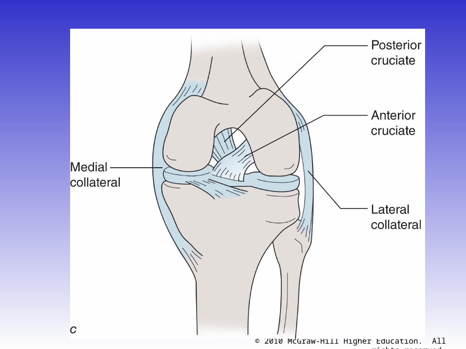
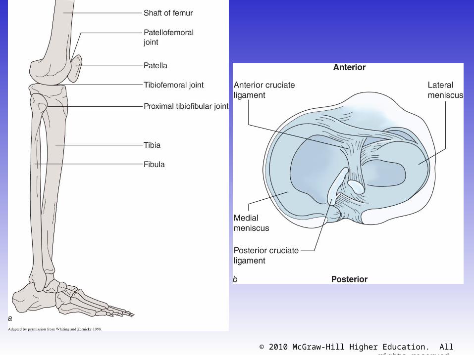
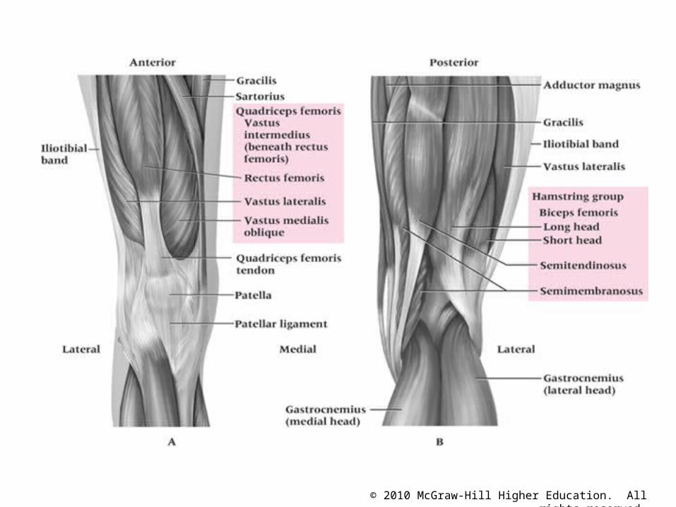
• Complex joint that endures great amounts of trauma due to extreme amounts of stress that are regularly applied
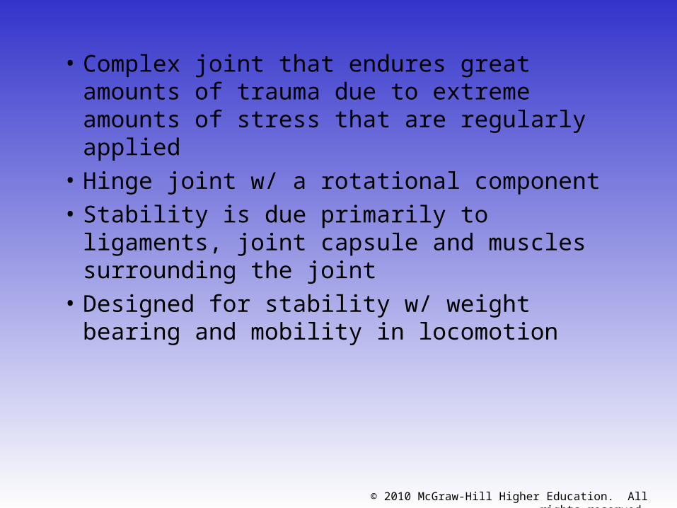
• Hinge joint w/ a rotational component• Stability is due primarily to ligaments, joint
capsule and muscles surrounding the joint• Designed for stability w/ weight bearing
and mobility in locomotion
© 2010 McGraw-Hill Higher Education. All rights reserved.
© 2010 McGraw-Hill Higher Education. All rights reserved.
© 2010 McGraw-Hill Higher Education. All rights reserved.
© 2010 McGraw-Hill Higher Education. All rights reserved.
© 2010 McGraw-Hill Higher Education. All rights reserved.
© 2010 McGraw-Hill Higher Education. All rights reserved.
© 2010 McGraw-Hill Higher Education. All rights reserved.
Prevention of Knee Injuries• Physical Conditioning and Rehabilitation
– Total body conditioning is required• Strength, flexibility, cardiovascular and muscular
endurance, agility, speed and balance
– Muscles around joint must be conditioned (flexibility and strength) to maximize stability
– Must avoid abnormal muscle action through flexibility– In an effort to prevent injury, extensibility of
hamstrings, erector spinae, groin, quadriceps and gastrocnemius is important
© 2010 McGraw-Hill Higher Education. All rights reserved.
• ACL Prevention Programs– Focus on strength, neuromuscular control,
balance– Series of different programs which address
balance board training, landing strategies, plyometric training, and single leg performance
– Can be implemented in rehabilitation and preventative training programs
• Shoe Type– Change in football footwear has drastically
reduced the incidence of knee injuries– Shoes w/ more shorter cleats does not allow foot
to become fixed while still allowing for control w/ running and cutting
© 2010 McGraw-Hill Higher Education. All rights reserved.
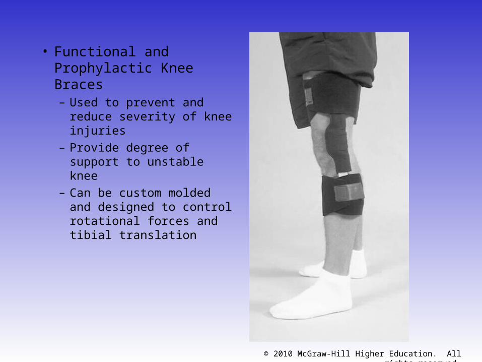
• Functional and Prophylactic Knee Braces– Used to prevent and
reduce severity of knee injuries
– Provide degree of support to unstable knee
– Can be custom molded and designed to control rotational forces and tibial translation
© 2010 McGraw-Hill Higher Education. All rights reserved.
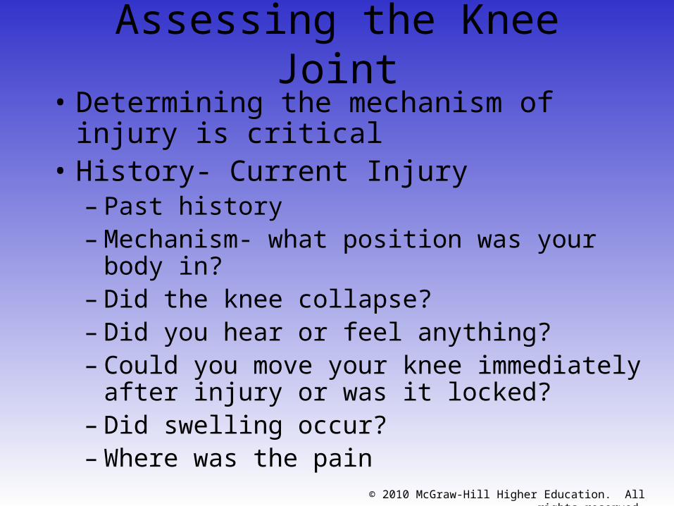
Assessing the Knee Joint• Determining the mechanism of injury is
critical• History- Current Injury
– Past history– Mechanism- what position was your body in?– Did the knee collapse?– Did you hear or feel anything?– Could you move your knee immediately after
injury or was it locked?– Did swelling occur?– Where was the pain
© 2010 McGraw-Hill Higher Education. All rights reserved.
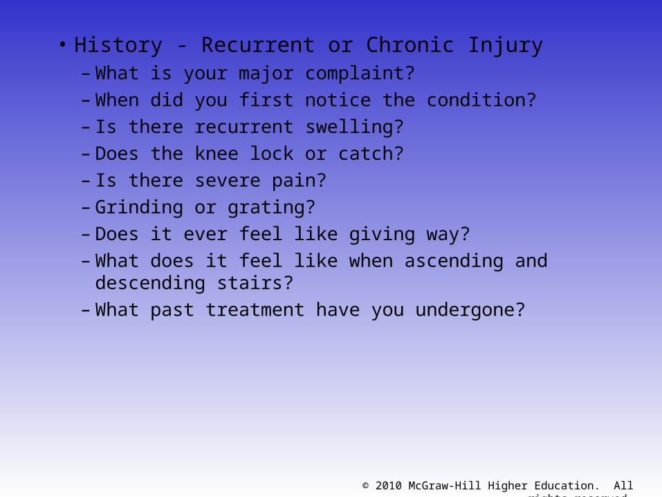
• History - Recurrent or Chronic Injury– What is your major complaint?
– When did you first notice the condition?
– Is there recurrent swelling?
– Does the knee lock or catch?
– Is there severe pain?
– Grinding or grating?
– Does it ever feel like giving way?
– What does it feel like when ascending and descending stairs?
– What past treatment have you undergone?
© 2010 McGraw-Hill Higher Education. All rights reserved.
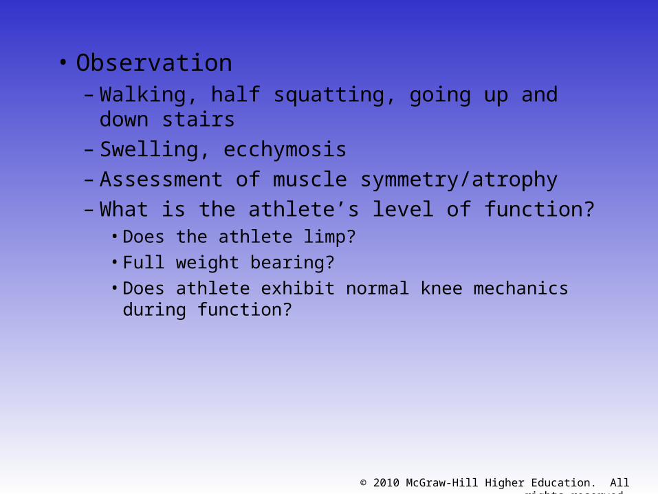
• Observation– Walking, half squatting, going up and down stairs– Swelling, ecchymosis– Assessment of muscle symmetry/atrophy– What is the athlete’s level of function?
• Does the athlete limp?• Full weight bearing?• Does athlete exhibit normal knee mechanics during
function?
© 2010 McGraw-Hill Higher Education. All rights reserved.
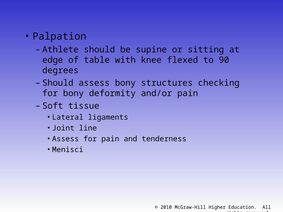
• Palpation– Athlete should be supine or sitting at edge of
table with knee flexed to 90 degrees– Should assess bony structures checking for
bony deformity and/or pain– Soft tissue
• Lateral ligaments
• Joint line
• Assess for pain and tenderness
• Menisci
© 2010 McGraw-Hill Higher Education. All rights reserved.
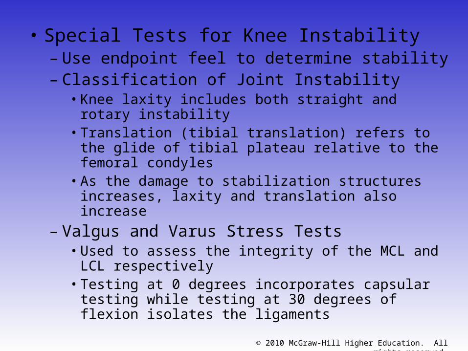
• Special Tests for Knee Instability– Use endpoint feel to determine stability– Classification of Joint Instability
• Knee laxity includes both straight and rotary instability
• Translation (tibial translation) refers to the glide of tibial plateau relative to the femoral condyles
• As the damage to stabilization structures increases, laxity and translation also increase
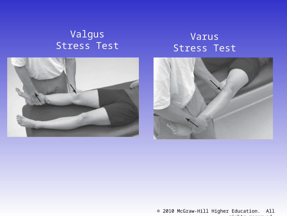
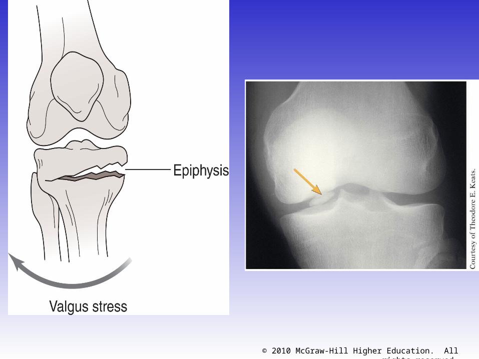
– Valgus and Varus Stress Tests• Used to assess the integrity of the MCL and LCL
respectively• Testing at 0 degrees incorporates capsular testing
while testing at 30 degrees of flexion isolates the ligaments
© 2010 McGraw-Hill Higher Education. All rights reserved.
Valgus Stress Test
Varus Stress Test
© 2010 McGraw-Hill Higher Education. All rights reserved.
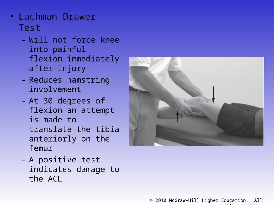
• Lachman Drawer Test– Will not force knee into
painful flexion immediately after injury
– Reduces hamstring involvement
– At 30 degrees of flexion an attempt is made to translate the tibia anteriorly on the femur
– A positive test indicates damage to the ACL
© 2010 McGraw-Hill Higher Education. All rights reserved.
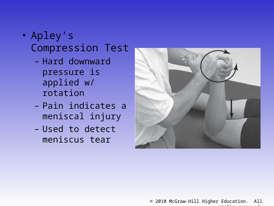
• Apley’s Compression Test– Hard downward
pressure is applied w/ rotation
– Pain indicates a meniscal injury
– Used to detect meniscus tear
© 2010 McGraw-Hill Higher Education. All rights reserved.
Recognition and Management of Specific Injuries
• Medial Collateral Ligament Sprain– Cause of Injury
• Result of severe blow or outward twist – valgus force
– Signs of Injury - Grade I• Little fiber tearing or stretching
• Stable valgus test
• Little or no joint effusion
• Some joint stiffness and point tenderness on lateral aspect
• Relatively normal ROM
© 2010 McGraw-Hill Higher Education. All rights reserved.
– Signs of Injury (Grade II)• Complete tear of deep capsular ligament and partial
tear of superficial layer of MCL• No gross instability; slight laxity• Slight swelling• Moderate to severe joint tightness w/ decreased ROM• Pain along medial aspect of knee
– Signs of Injury (Grade III)• Complete tear of supporting ligaments• Complete loss of medial stability, meniscus disruption• Minimum to moderate swelling• Immediate pain followed by ache• Loss of motion due to effusion and hamstring
guarding• Positive valgus stress test

© 2010 McGraw-Hill Higher Education. All rights reserved.
• Care– RICE for at least 24 hours– Crutches if necessary– Knee immobilizer may be
applied– Move from isometrics and
STLR exercises to bicycle riding and isokinetics
– Return to play when all areas have returned to normal
• Continued bracing may be required
© 2010 McGraw-Hill Higher Education. All rights reserved.
– Care• Conservative non-operative approach for isolated grade 2
and 3 injuries• Limited immobilization (w/ a brace); progressive weight
bearing for 2 weeks• Follow with 2-3 week period of protection with functional
hinge brace• When normal range, strength, power, flexibility, endurance
and coordination are regained athlete can return– Some additional bracing and taping may be required
© 2010 McGraw-Hill Higher Education. All rights reserved.
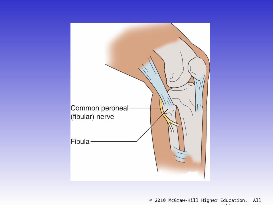
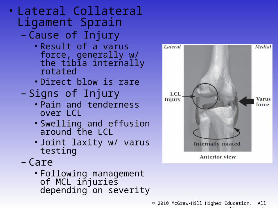
• Lateral Collateral Ligament Sprain– Cause of Injury
• Result of a varus force, generally w/ the tibia internally rotated
• Direct blow is rare– Signs of Injury
• Pain and tenderness over LCL
• Swelling and effusion around the LCL
• Joint laxity w/ varus testing
– Care• Following management of
MCL injuries depending on severity
© 2010 McGraw-Hill Higher Education. All rights reserved.
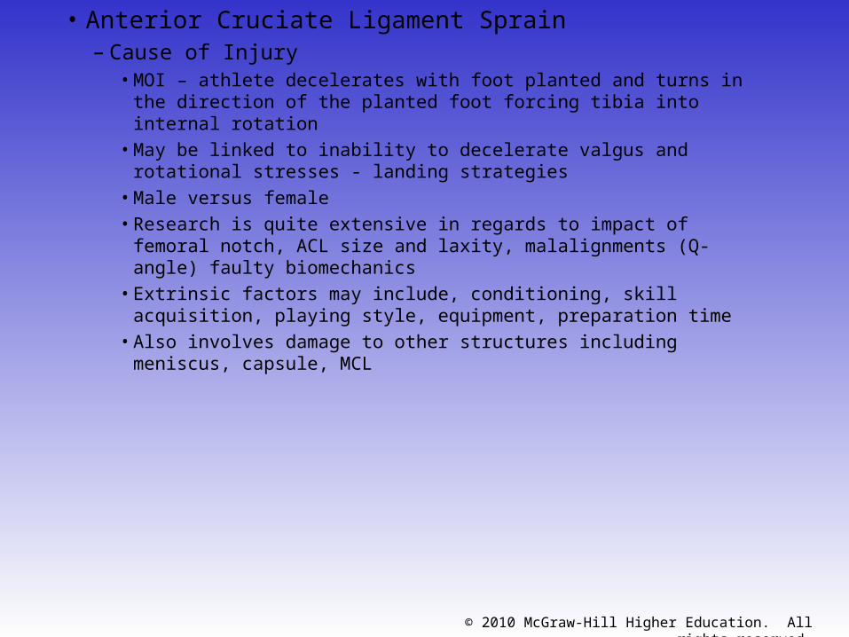
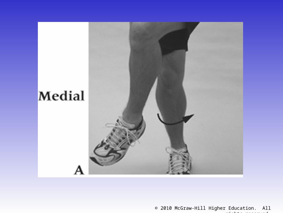
• Anterior Cruciate Ligament Sprain– Cause of Injury
• MOI – athlete decelerates with foot planted and turns in the direction of the planted foot forcing tibia into internal rotation
• May be linked to inability to decelerate valgus and rotational stresses - landing strategies
• Male versus female• Research is quite extensive in regards to impact of femoral notch,
ACL size and laxity, malalignments (Q-angle) faulty biomechanics• Extrinsic factors may include, conditioning, skill acquisition, playing
style, equipment, preparation time• Also involves damage to other structures including meniscus,
capsule, MCL
© 2010 McGraw-Hill Higher Education. All rights reserved.
© 2010 McGraw-Hill Higher Education. All rights reserved.
– Signs of Injury • Experience pop w/ severe pain and disability• Rapid swelling at the joint line• Positive anterior drawer and Lachman’s • Other ACL tests may also be positive
– Care• RICE; use of crutches• Arthroscopy may be necessary to determine extent
of injury• Could lead to major instability in incidence of high
performance• W/out surgery joint degeneration may result• Age and activity may factor into surgical option• Surgery may involve joint reconstruction w/ grafts
(tendon), transplantation of external structures– Will require brief hospital stay and 3-5 weeks of a brace– Also requires 4-6 months of rehab
© 2010 McGraw-Hill Higher Education. All rights reserved.
• Posterior Cruciate Ligament Sprain– Cause of Injury
• Most at risk during 90 degrees of flexion• Fall on bent knee is most common mechanism• Can also be damaged as a result of a rotational force
– Signs of Injury• Feel a pop in the back of the knee• Tenderness and relatively little swelling in the popliteal
fossa• Laxity w/ posterior sag test
– Care• RICE• Non-operative rehab of grade I and II injuries should focus
on quad strength• Surgical versus non-operative
– Surgery will require 6 weeks of immobilization in extension w/ full weight bearing on crutches
– ROM after 6 weeks and PRE at 4 months
© 2010 McGraw-Hill Higher Education. All rights reserved.
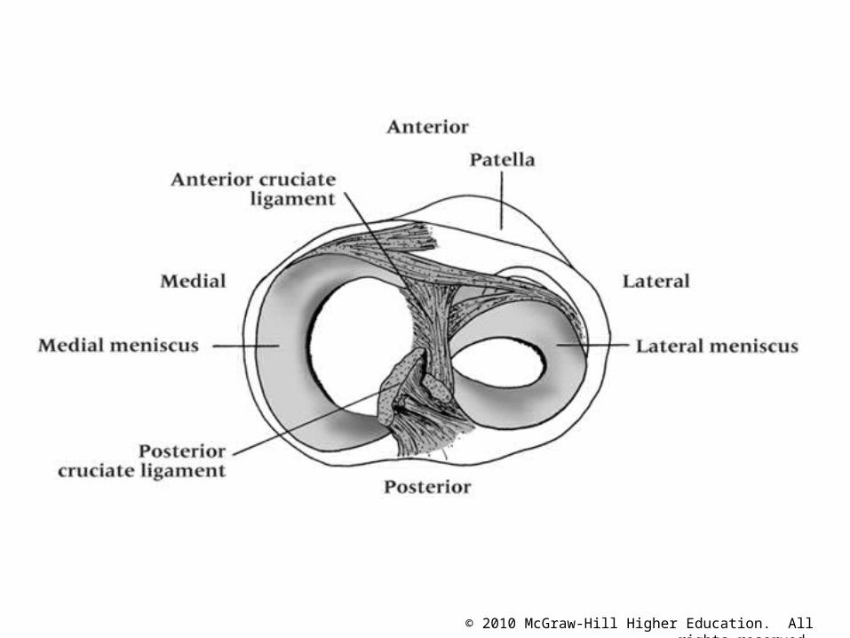
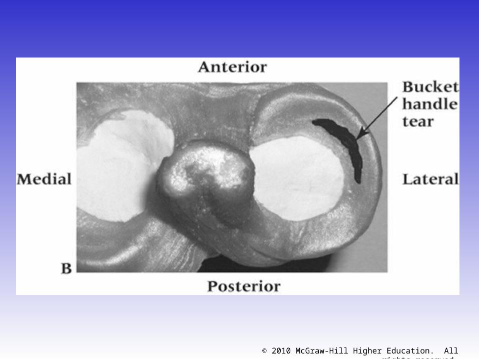
• Meniscus Injuries– Cause of Injury
• Medial meniscus is more commonly injured due to ligamentous attachments and decreased mobility
– Also more prone to disruption through torsional and valgus forces
• Most common MOI is rotary force w/ knee flexed or extended while weight bearing
– Signs of Injury • Diagnosis is difficult• Effusion developing over 48-72 hour period• Joint line pain and loss of motion• Intermittent locking and giving way• Pain w/ squatting
© 2010 McGraw-Hill Higher Education. All rights reserved.
© 2010 McGraw-Hill Higher Education. All rights reserved.
– Care• Immediate care = PRICE• If the knee is not locked, but indications of a
tear are present further diagnostic testing may be required
– Treatment should follow that of MCL injury
• If locking occurs, anesthesia may be necessary to unlock the joint w/ possible arthroscopic surgery follow-up
• W/ surgery all efforts are made to preserve the meniscus -- with full healing being dependent on location
• Torn meniscus may be repaired using sutures
© 2010 McGraw-Hill Higher Education. All rights reserved.
• Joint Contusions– Cause of Injury
• Blow to the muscles crossing the joint (vastus medialis)
– Signs of Injury• Present as knee sprain, severe pain, loss of
movement and signs of acute inflammation• Swelling, discoloration
– Care• RICE initially and continue if swelling persists• Gradual progression to normal activity following
return of ROM and padding for protection• If swelling does not resolve w/in a week a
chronic condition (synovitis or bursitis) may exist requiring more rest
© 2010 McGraw-Hill Higher Education. All rights reserved.
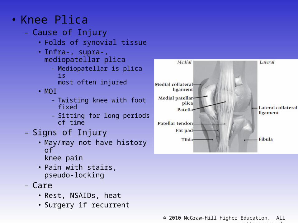
• Knee Plica– Cause of Injury
• Folds of synovial tissue• Infra-, supra-, mediopatellar
plica– Mediopatellar is plica is
most often injured
• MOI– Twisting knee with foot fixed– Sitting for long periods of
time
– Signs of Injury• May/may not have history of
knee pain• Pain with stairs, pseudo-
locking
– Care• Rest, NSAIDs, heat• Surgery if recurrent
© 2010 McGraw-Hill Higher Education. All rights reserved.
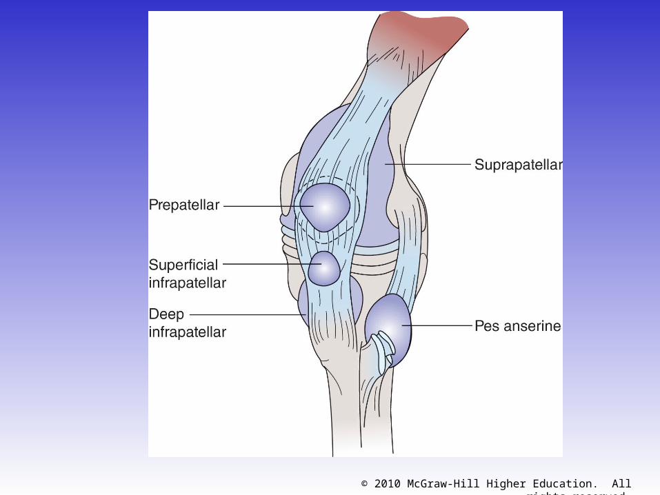
• Bursitis– Cause of Injury
• Acute, chronic or recurrent swelling• Prepatellar = continued kneeling• Infrapatellar = overuse of patellar tendon
– Signs of Injury• Prepatellar bursitis may be localized swelling
above knee that is ballotable• Presents with cardinal signs of inflammation• Swelling in popliteal fossa may indicate a
Baker’s cyst
– Care • Eliminate cause, RICE and NSAID’s• Aspiration and steroid injection if chronic
© 2010 McGraw-Hill Higher Education. All rights reserved.
© 2010 McGraw-Hill Higher Education. All rights reserved.
• Loose Bodies w/in the Knee– Cause
• Result of repeated trauma• Possibly stem from osteochondritis dissecans,
meniscal fragments, synovial tissue or cruciate ligaments
– Signs of Injury • May become lodged, causing locking or
popping• Pain and sensation of instability
– Care• If not surgically removed it can lead to
conditions causing joint degeneration
© 2010 McGraw-Hill Higher Education. All rights reserved.
© 2010 McGraw-Hill Higher Education. All rights reserved.
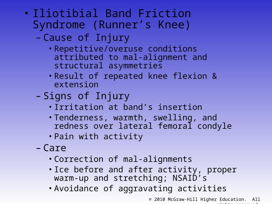
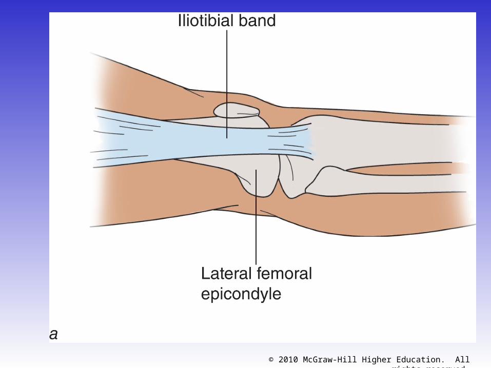
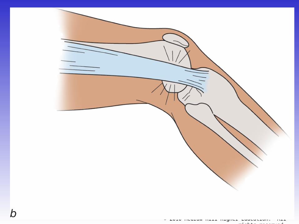
• Iliotibial Band Friction Syndrome (Runner’s Knee)– Cause of Injury
• Repetitive/overuse conditions attributed to mal-alignment and structural asymmetries
• Result of repeated knee flexion & extension– Signs of Injury
• Irritation at band’s insertion • Tenderness, warmth, swelling, and redness
over lateral femoral condyle• Pain with activity
– Care• Correction of mal-alignments• Ice before and after activity, proper warm-up
and stretching; NSAID’s• Avoidance of aggravating activities
© 2010 McGraw-Hill Higher Education. All rights reserved.
© 2010 McGraw-Hill Higher Education. All rights reserved.
© 2010 McGraw-Hill Higher Education. All rights reserved.
• Patellar Fracture– Cause of Injury
• Direct or indirect trauma (severe pull of tendon)
• Forcible contraction, falling, jumping or running
– Signs of Injury• Hemorrhaging and joint effusion w/ generalized
swelling
• Indirect fractures may cause capsular tearing, separation of bone fragments and possible quadriceps tendon tearing
• Little bone separation w/ direct injury
– Management• X-ray necessary for confirmation of findings
• RICE and splinting if fracture suspected
• Refer and immobilize for 2-3 months
© 2010 McGraw-Hill Higher Education. All rights reserved.
• Acute Patella Subluxation or Dislocation– Cause of Injury
• Deceleration w/ simultaneous cutting in opposite direction (valgus force at knee)
• Quad pulls the patella out of alignment• Some athletes may be predisposed to injury• Repetitive subluxation will impose stress to medial
restraints• More commonly seen in female athletes
– Signs of Injury• W/ subluxation, pain and swelling, restricted ROM,
palpable tenderness over adductor tubercle• Dislocations result in total loss of function• First time dislocation = assume fracture
© 2010 McGraw-Hill Higher Education. All rights reserved.
– Care• Immobilize and refer to physician for reduction• Ice around the joint• Following reduction, immobilization for at least
4 weeks w/ use of crutches • After immobilization period, horseshoe pad w/
elastic wrap should be used to support patella• Muscle rehab focusing on muscle around the
knee, thigh and hip are key (STLR’s are optimal for the knee)
© 2010 McGraw-Hill Higher Education. All rights reserved.
• Chondromalacia patella– Cause
• Softening and deterioration of the articular cartilage
• Possible abnormal patellar tracking due to genu valgum, external tibial torsion, foot pronation, femoral anteversion, patella alta, shallow femoral groove, increased Q angle, laxity of quad tendon
– Signs of Injury• Pain w/ walking, running, stairs and squatting• Possible recurrent swelling, grating sensation w/
flexion and extension– Care
• Conservative measures– RICE, NSAID’s, isometrics for strengthening– Avoid aggravating activities
• Surgical possibilities
© 2010 McGraw-Hill Higher Education. All rights reserved.
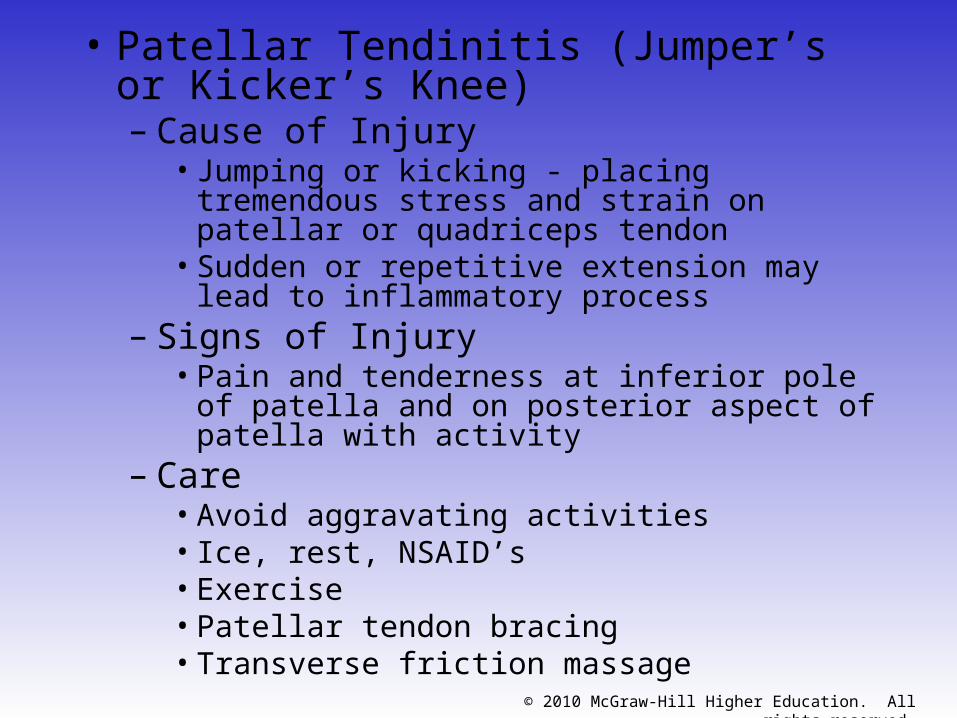
• Patellar Tendinitis (Jumper’s or Kicker’s Knee)– Cause of Injury
• Jumping or kicking - placing tremendous stress and strain on patellar or quadriceps tendon
• Sudden or repetitive extension may lead to inflammatory process
– Signs of Injury• Pain and tenderness at inferior pole of patella
and on posterior aspect of patella with activity– Care
• Avoid aggravating activities• Ice, rest, NSAID’s• Exercise• Patellar tendon bracing• Transverse friction massage
© 2010 McGraw-Hill Higher Education. All rights reserved.
© 2010 McGraw-Hill Higher Education. All rights reserved.
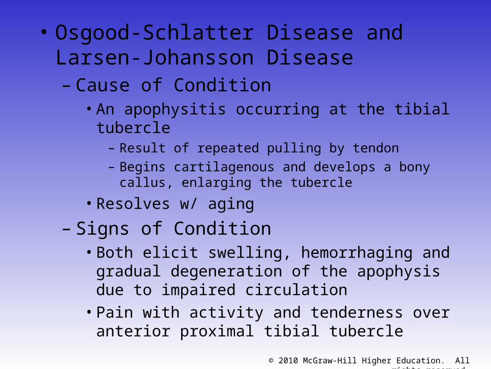
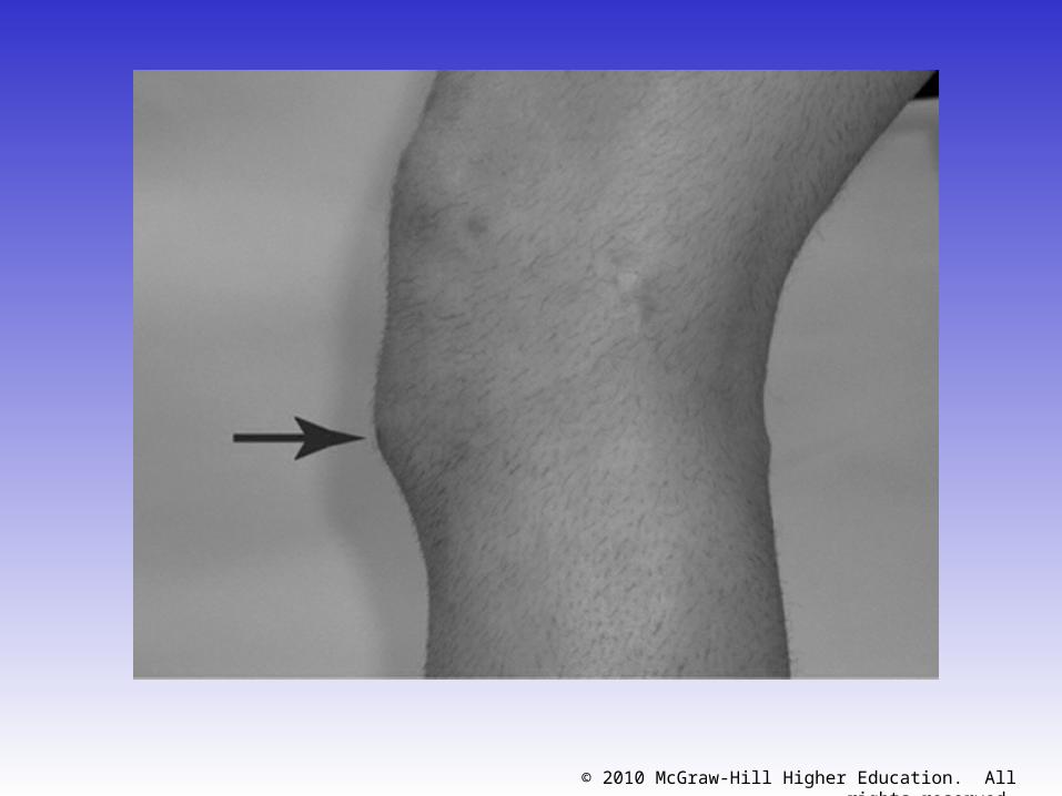
• Osgood-Schlatter Disease and Larsen-Johansson Disease– Cause of Condition
• An apophysitis occurring at the tibial tubercle – Result of repeated pulling by tendon– Begins cartilagenous and develops a bony callus,
enlarging the tubercle
• Resolves w/ aging
– Signs of Condition• Both elicit swelling, hemorrhaging and gradual
degeneration of the apophysis due to impaired circulation
• Pain with activity and tenderness over anterior proximal tibial tubercle
© 2010 McGraw-Hill Higher Education. All rights reserved.
© 2010 McGraw-Hill Higher Education. All rights reserved.
– Care• Conservative
– Reduce stressful activity until union occurs (6-12 months)
– Padding may be necessary for protection– Possible casting, ice before and after activity– Isometrics

![A Rolling Contact Joint Lower Extremity Exoskeleton Knee · migrates on an evolute while the knee is exed, rolling and sliding simultaneously [13]. Since the exoskeleton is placed](https://img.pdfslide.net/doc/110x75/5f0d42ae7e708231d43976ca/a-rolling-contact-joint-lower-extremity-exoskeleton-knee-migrates-on-an-evolute.jpg)