Embed Size (px)
Citation preview
1 | P a g e
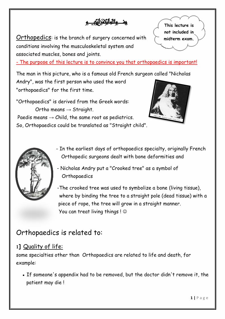
ميحرلا نمحرلا هللا مسب
Orthopedics: is the branch of surgery concerned with
conditions involving the musculoskeletal system and
associated muscles, bones and joints.
- The purpose of this lecture is to convince you that orthopaedics is important!
The man in this picture, who is a famous old French surgeon called "Nicholas
Andry", was the first person who used the word
"orthopaedics" for the first time.
"Orthopaedics" is derived from the Greek words:
Ortho means → Straight.
Paedis means → Child, the same root as pediatrics.
So, Orthopaedics could be translated as "Straight child".
- In the earliest days of orthopaedics specialty, originally French
Orthopedic surgeons dealt with bone deformities and
- Nicholas Andry put a "Crooked tree" as a symbol of
Orthopaedics
-The crooked tree was used to symbolize a bone (living tissue),
where by binding the tree to a straight pole (dead tissue) with a
piece of rope, the tree will grow in a straight manner.
You can treat living things !
Orthopaedics is related to:
1] Quality of life:
some specialties other than Orthopaedics are related to life and death, for
example:
▪ If someone's appendix had to be removed, but the doctor didn't remove it, the
patient may die !
This lecture is
not included in
midterm exam.

2 | P a g e
A patient with a stroke, if the doctor didn't do anything for him, definitely
he will die !
Orthopaedics, on the other hand, is related to the Quality of life, that is the
general well-being of individuals and societies. Like cars, mobile phones, clothes,
etc … !
2] Pain:
▪ pain is something pathological, normally you shouldn't feel pain, so you should
think of this pain as an underlying pathologic like Inflammation (inflammation signs
are redness, heat, swelling, pain and loss of function)
3] Deformity:
▪ Femur bone is a curved bone, if it becomes straight bone, it will be deformed.
▪ Radius has a radial bow, if this bone becomes straight, it will be deformed, so no
more pronation and supination.
▪ Deformity means deviation from normal anatomy, So you should know the normal
anatomy in the beginning to recognize the deformity.
4] Loss of function
** This is a longitudinal section of a child's femur: trabeculum
Articular surface
Growth plate
cortex
medulla
Pain, deformity and loss of function
are the major complaints of patients.
Epiphysis
Diaphysis
3 | P a g e
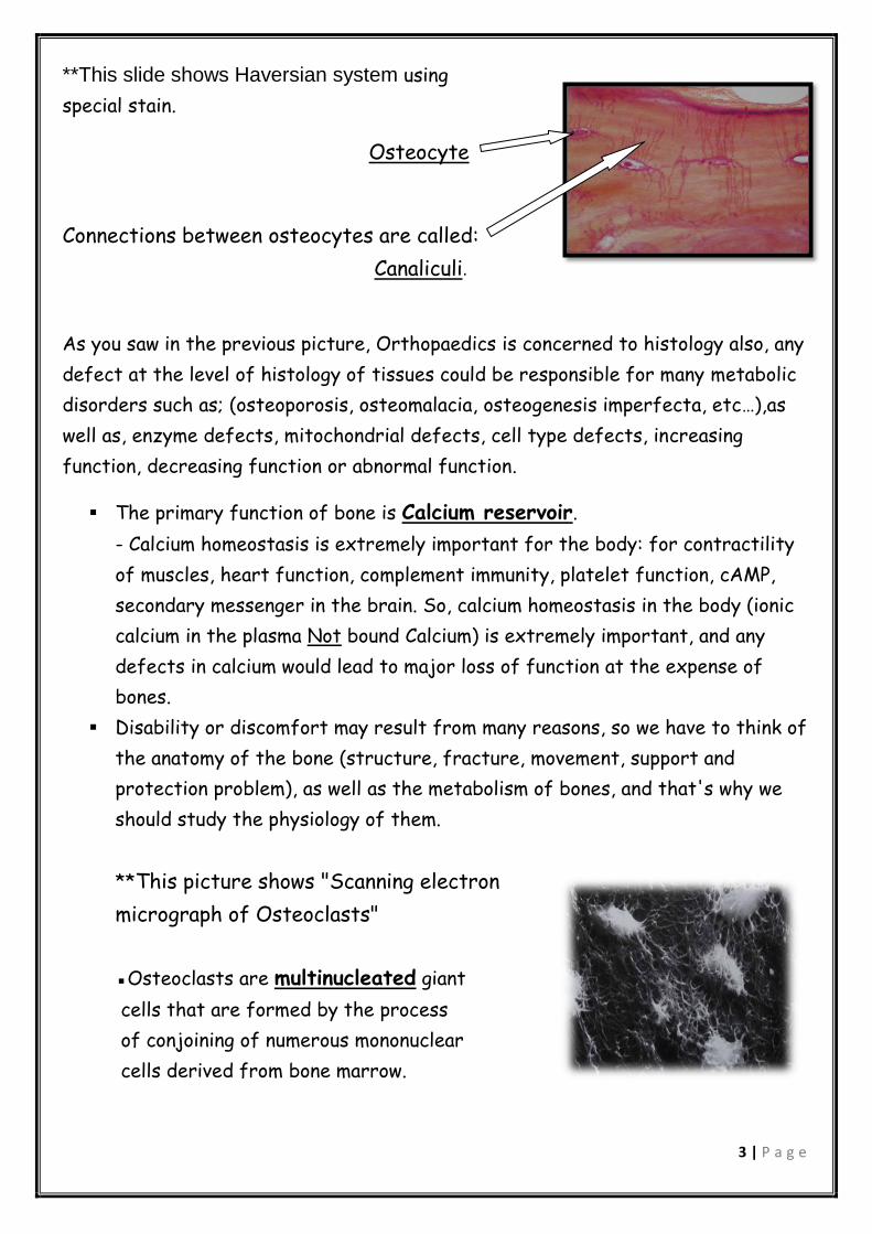
**This slide shows Haversian system using
special stain.
Osteocyte
Connections between osteocytes are called:
Canaliculi.
As you saw in the previous picture, Orthopaedics is concerned to histology also, any
defect at the level of histology of tissues could be responsible for many metabolic
disorders such as; (osteoporosis, osteomalacia, osteogenesis imperfecta, etc…),as
well as, enzyme defects, mitochondrial defects, cell type defects, increasing
function, decreasing function or abnormal function.
The primary function of bone is Calcium reservoir.
- Calcium homeostasis is extremely important for the body: for contractility
of muscles, heart function, complement immunity, platelet function, cAMP,
secondary messenger in the brain. So, calcium homeostasis in the body (ionic
calcium in the plasma Not bound Calcium) is extremely important, and any
defects in calcium would lead to major loss of function at the expense of
bones.
Disability or discomfort may result from many reasons, so we have to think of
the anatomy of the bone (structure, fracture, movement, support and
protection problem), as well as the metabolism of bones, and that's why we
should study the physiology of them.
**This picture shows "Scanning electron
micrograph of Osteoclasts"
▪Osteoclasts are multinucleated giant
cells that are formed by the process
of conjoining of numerous mononuclear
cells derived from bone marrow.

4 | P a g e
▪ Osteoclasts reabsorb calcium of the bones, on the other hand,
osteoblasts build bones, so bones are dynamic tissues.
▪ Enzymes are secreted from Ruffled border to destroy organic and
inorganic contents of bone matrix
Osteoclasts are found in pits in the bone
surface which are called Howship lacunae
Ruffled border
Nucleolus of Osteoclast
▪The strongest bone in the body is the proximal femur, When you are lying down
and raising your leg up, it will bear weight equal to your body weight multiplied by 4 !
Slide (12)
"layers of growth plate"
▪ each layer could have a disease such
as excessive or less function.
Examples of such diseases:
1) Chondroplasia: people with chondroplasia have
short stature.
2) Marfan syndrome: people with Marfan syndrome
are usually tall with long, thin arms and legs.
- These are two defects at the same level.
Slides (13-16)
Examination of (Joints, bones, muscles):
1] Look (Inspection):
▪ Deformity. ▪ Muscle wasting. ▪ Scars.
Familial diseases: raised in families, usually because of environmental connected. reasons.

5 | P a g e
2] Feel (Palpation):
▪ Temperature ▪ Tenderness ▪ Masses ▪ Anatomical landmarks
3] Move:
▪ Active ▪ Passive
Slide (17) Very important slide
" Causes of orthopedic diseases"
▪ Orthopaedic diseases are divided into 3 categories, and here are some examples:
1] Congenital means acquired before birth, and caused by:
Genetic disorders: the underlying cause is a genetic defect such as
Down syndrome. Genetic disorders may be recognized at birth or after
birth but it is acquired before birth.
Drugs.
Radiation.
2] Developmental means developed over a period of time, usually with slight
destruction ↓
- a man started to lose his hair, then after a period of time he became bald !
3] Acquired diseases: caused by:
Trauma infection: Here, it is extremely important to know the anatomy of
the body, because your goals are to restore function, correct deformity and
restore normal anatomy.
Congenital diseases
• Intrauterine infection.
• dislocation.
Developmental diseases Acquired diseases
Tendon injuries.
DDH "Developmental
dysplasia of the hip":
dislocation of the hip joint.
CTS, carpal tunnel syndrome.
Acquired
Immunodeficiency
Syndrome (AIDS).

6 | P a g e
Paralysis.
Infection.
Arthritis.
Neoplasm: and it is divided into:
1) Primary neoplasia: originates in the bone, and can be divided into
benign tumors and cancers.
2) Secondary neoplasia: originates in other sites and spread (metastasize)
to the bones to become easily fractured.
Slide (18)
This slide shows examples of congenital
diseases.
▪ The photo on the upper left side shows a
hand with an extra digit beside the
pathological (abnormal) thumb, this disorder
is known as "polydactyly".
Slide (20)
▪ An abnormal hand fitted with a prosthesis.
- This limb reduction, which is caused by a faulty gene, may be as a result of:
1] Thalidomide:
Thalidomide was a widely used drug in 1960s as an oral contraceptive. Later on, it
became apparent that thalidomide resulted in severe birth defects in thousands of
children, though it was banned in most countries at that time.
2] Chernobyl disaster:
Chernobyl disaster is an explosion released
large quantities of radioactive particles into the
atmosphere reached Russia ! Long-term cancers
and deformities are still being accounted for.
7 | P a g e
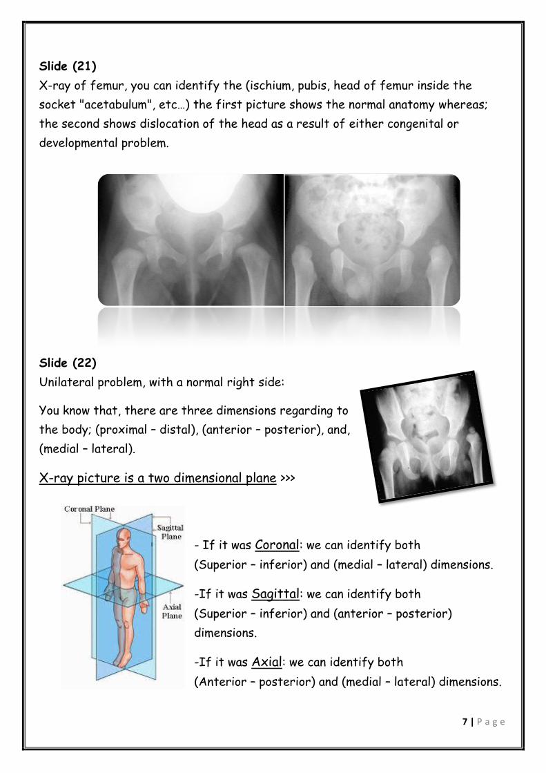
Slide (21)
X-ray of femur, you can identify the (ischium, pubis, head of femur inside the
socket "acetabulum", etc…) the first picture shows the normal anatomy whereas;
the second shows dislocation of the head as a result of either congenital or
developmental problem.
Slide (22)
Unilateral problem, with a normal right side:
You know that, there are three dimensions regarding to
the body; (proximal – distal), (anterior – posterior), and,
(medial – lateral).
X-ray picture is a two dimensional plane >>>
- If it was Coronal: we can identify both
(Superior – inferior) and (medial – lateral) dimensions.
-If it was Sagittal: we can identify both
(Superior – inferior) and (anterior – posterior)
dimensions.
-If it was Axial: we can identify both
(Anterior – posterior) and (medial – lateral) dimensions.

8 | P a g e
Because of this, we have to mention the type of section in the x- ray picture.
another important point is, to take the permission from the patient if you want
to use his/her photo, or you have to delete all his/her information.
Slide (23)
The clinical examination reviles an abnormal
anatomy for a baby female, having a limited
abduction + short leg syndrome
Your job is trying to restore the normal
anatomy. You have to know the normal size of
each organ, in order to distinguish normal from
abnormal.
Slide (24)
Femur neck fracture, very common case.
Doctors usually try to do reduction, fixation and
electrolyte balance in an attempt to restore the
normal anatomy.
- You should know the history of your patient i.e. if
she has diabetes, CDV disease, and so on.
Slide (25)
Painful deformity, an acquired (neither developmental
nor congenital) case, referred to as scoliosis, a
painful benign tumor.
This case is characterized by bow legs, and usually,
we need surgery in order to treat such cases.
9 | P a g e
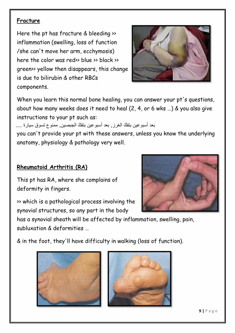
Fracture
Here the pt has fracture & bleeding >>
inflammation (swelling, loss of function
/she can't move her arm, ecchymosis)
here the color was red>> blue >> black >>
green>> yellow then disappears, this change
is due to bilirubin & other RBCs
components.
When you learn this normal bone healing, you can answer your pt's questions,
about how many weeks does it need to heal (2, 4, or 6 wks …) & you also give
instructions to your pt such as:
....ممنوع تسوق سيارة , بعد أسبوعين بتفّك الجبصين, بعد أسبوعين بتفّك الغرز
you can't provide your pt with these answers, unless you know the underlying
anatomy, physiology & pathology very well.
Rheumatoid Arthritis (RA)
This pt has RA, where she complains of
deformity in fingers.
>> which is a pathological process involving the
synovial structures, so any part in the body
has a synovial sheath will be affected by inflammation, swelling, pain,
subluxation & deformities …
& in the foot, they'll have difficulty in walking (loss of function).

10 | P a g e
Ankylosing Spondylitis "تشّمـع المفاصل"
this is called "bamboo sign"(مثل القصيب), where
there is a fusion in the pt's spin (pt moves as
one peice).
Knowing that the underlying pathology doesn't
involve the nerves, we can reassure the pt &
advise him/her to do physiotherapy & standing
posture (because otherwise the pt may
develop bended posture).
Then when the disease reaches & destroys
the hips, we'll replace both of them.
You should know anatomy very well to avoid
causing injury to the surrounding structures
(sciatic nerve, or femoral artery ...).
This is a lateral X-ray for the same pt.

11 | P a g e
This is a Cadaver, where we may have multiple
fractures and diseased factors (spongy bone, cortical
bone, inter-vertebral disc, lamina, pedicle, spinous
process, dimifacet joint, nerve root canal, neural
foramina, anterior & posterior longitudinal ligament …)
If you know the anatomy very well, you can interpret
problems & pathologies.
Osteogenesis Imperfecta
Systemic disease with multiple deformities (legs, hips, chest …).
Trauma
12 | P a g e
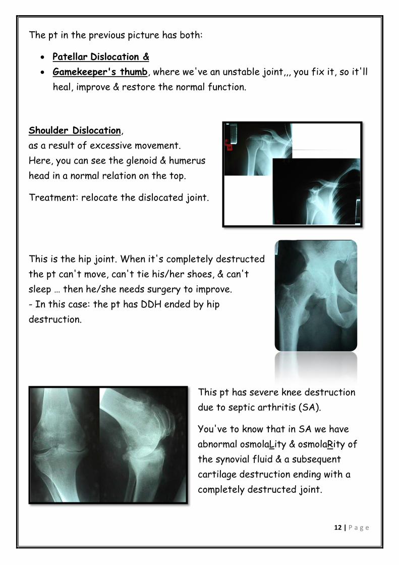
The pt in the previous picture has both:
Patellar Dislocation &
Gamekeeper's thumb, where we've an unstable joint,,, you fix it, so it'll
heal, improve & restore the normal function.
Shoulder Dislocation,
as a result of excessive movement.
Here, you can see the glenoid & humerus
head in a normal relation on the top.
Treatment: relocate the dislocated joint.
This is the hip joint. When it's completely destructed
the pt can't move, can't tie his/her shoes, & can't
sleep … then he/she needs surgery to improve.
- In this case: the pt has DDH ended by hip
destruction.
This pt has severe knee destruction
due to septic arthritis (SA).
You've to know that in SA we have
abnormal osmolaLity & osmolaRity of
the synovial fluid & a subsequent
cartilage destruction ending with a
completely destructed joint.

13 | P a g e
here, in total knee replacement surgery, one
of our important objectives is not to have
infections, so we provide a sterile condition
during this surgery, we give prophylactic, IV
antibiotics, & so many things to prevernt
infection.
Infected joint equals a disaster!
This is post surgery (post total knee
replacement).
Here the pt had bilateral hip joint
replacement. But as you can see it's
dislocated on the left side, because this pt
had a stroke & a subsequent abductor
muscles weakness (gluteus maximus, medius
and minimus). So pt will have difficulty in
walking.
This young pt had arthrodesis >> we removed
the cartilage & there'll be a fusion between
the two … complete loss of movement &
complete loss of pain! (شيء على حسب شيء).

14 | P a g e
This is ankle replacement
you can see the tibia, calcaneus, prosthetic joint.
Hip joint replacement: Is defined by WHO as the
best operation ever invented by Man! :) With a 90%
satisfaction rate, while ankle replacement for instance has a high failure rate
(60% of the best of these ankles survive 5 years, after that they're
damaged) & 8% post-operative infection rate (we still don't know exact
biomechanics of the ankle joint so we still unable to do ankle replacement
with a high success rate).
This is shoulder replacement; it's a successful surgery, we do it here in our
hospital, we should have intact muscles, otherwise it'll dislocate again
(empagement).
This is a pathological fracture, this is a
primary benign bone tumor
(osteochondroma), from its appearance you
can reassure the pt & his/her family that
it's simple; we hang it 6 weeks & it'll resolve

15 | P a g e
While
This pt is totally different! This is
osteosarcoma ("sun burst
appearance">> malignant tumor >>
amputations >> life & death)
Here, the humerus, scapula, thoracic muscles & … all were
removed to save the life of the pt.
You'll learn all these things & then you'll be able to
council pts & their families :)
A major surgery, removing all
distal femur with the knee,
connecting tibiae with proximal
femur.
This is a diabetic pt, she had a "below-knee"
amputation (due to diabetes >> decreased blood
supply >> ischemia >> amputation). & you should know
that pt may die because of ischemia & subsequent
infection.
Best wishes
Asmaa Al-Bitar & Abeer Odeh.