Embed Size (px)
Citation preview
1
Islamic University of GazaIslamic University of GazaFaculty of NursingFaculty of Nursing
Chapter 7Chapter 7
Assessment of respiratory systemAssessment of respiratory system
2
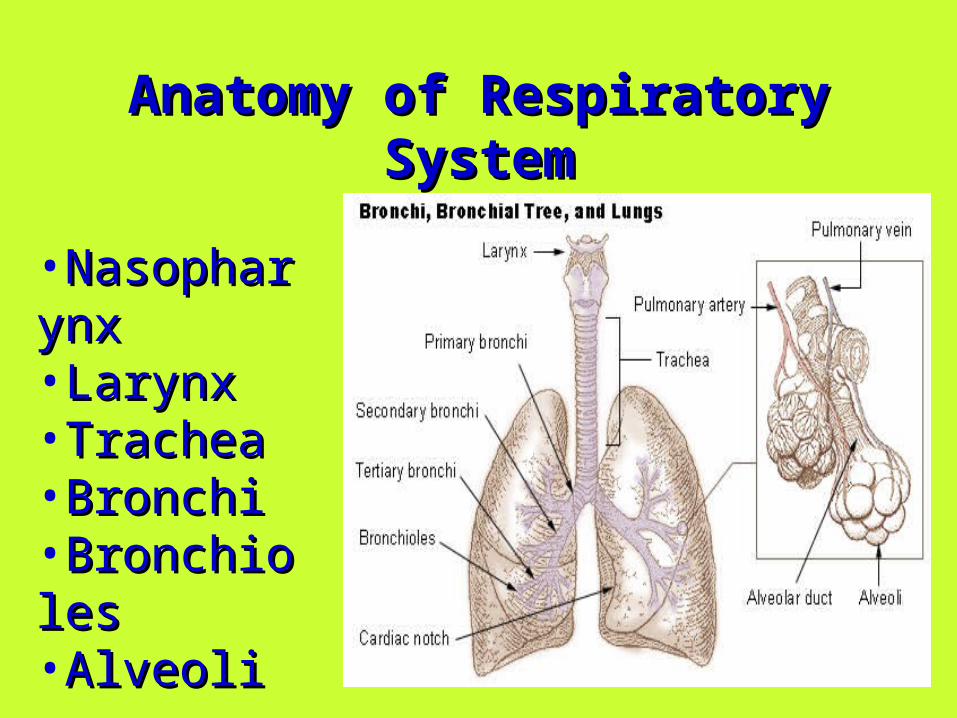
Anatomy of Respiratory SystemAnatomy of Respiratory System
•NasopharynxNasopharynx•LarynxLarynx•TracheaTrachea•BronchiBronchi•BronchiolesBronchioles•AlveoliAlveoli
3
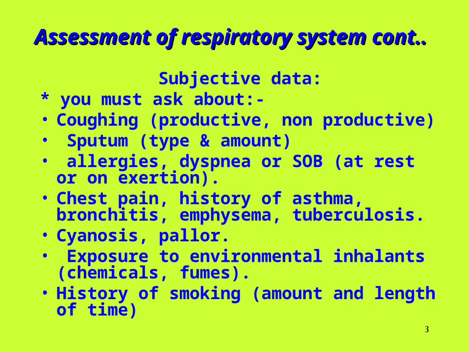
Assessment of respiratory system cont..Assessment of respiratory system cont..
Subjective data:* you must ask about:- • Coughing (productive, non productive) • Sputum (type & amount)• allergies, dyspnea or SOB (at rest or on
exertion).• Chest pain, history of asthma, bronchitis,
emphysema, tuberculosis.• Cyanosis, pallor.• Exposure to environmental inhalants
(chemicals, fumes).• History of smoking (amount and length of time)
4
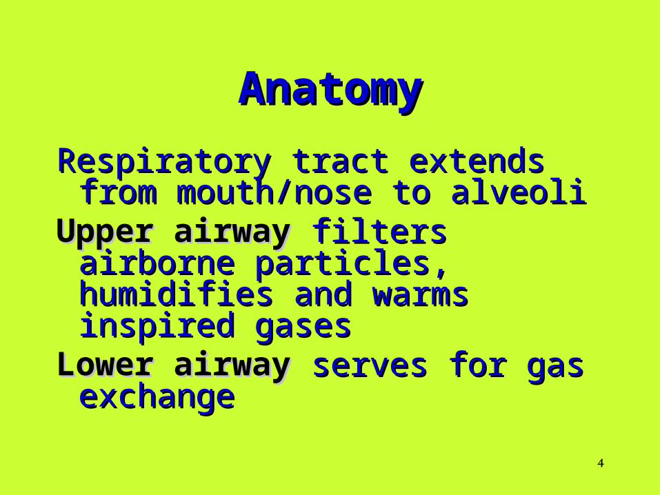
AnatomyAnatomy
Respiratory tract extends from Respiratory tract extends from mouth/nose to alveolimouth/nose to alveoli
Upper airwayUpper airway filters airborne filters airborne particles, humidifies and warms particles, humidifies and warms inspired gasesinspired gases
Lower airwayLower airway serves for gas serves for gas exchangeexchange
5
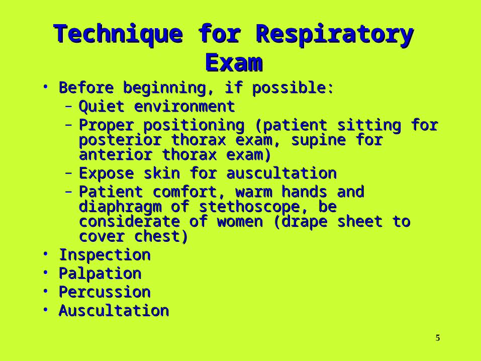
Technique for Respiratory ExamTechnique for Respiratory Exam
• Before beginning, if possible:Before beginning, if possible:– Quiet environmentQuiet environment– Proper positioning (patient sitting for posterior thorax Proper positioning (patient sitting for posterior thorax
exam, supine for anterior thorax exam) exam, supine for anterior thorax exam) – Expose skin for auscultationExpose skin for auscultation– Patient comfort, warm hands and diaphragm of Patient comfort, warm hands and diaphragm of
stethoscope, be considerate of women (drape sheet to stethoscope, be considerate of women (drape sheet to cover chest) cover chest)
• InspectionInspection• PalpationPalpation• PercussionPercussion• AuscultationAuscultation
6
Initial Respiratory SurveyInitial Respiratory Survey
• Observe the patient’s breathing patternObserve the patient’s breathing pattern
– Rate (normal vs. increased/decreased) Rate (normal vs. increased/decreased)
– Depth (shallow vs. deep)Depth (shallow vs. deep)
– Effort (any sign of accessory muscle use, Effort (any sign of accessory muscle use, inspect neck)inspect neck)
• Assess the patient’s colorAssess the patient’s color
– cyanosiscyanosis
7
Normal Respiratory RatesNormal Respiratory Rates
– Infant 30-60Infant 30-60
– Toddler 24-40Toddler 24-40
– Preschooler 22-34Preschooler 22-34
– School-age child 18-30School-age child 18-30
– Adolescent 12-16Adolescent 12-16
– Adult 16-20Adult 16-20
8
Assessment of respiratory system cont..
• Inspection for Measurement and assessment of respiration patterns.
• Assess the skin and overall symmetry and integrity of the thorax.
• Assess thoracic configuration. • ** Client must be uncovered to the waist, and in sitting
position without support. • * Observation of skin may give you knowledge about,
nutritional status of the client. • * Anterior- posterior diameter of thorax in normal
person less than the transverse diameter = (1 – 2).• * Assess for abnormality of configuration, e.g. pigeon
chest, funnel chest, spinal deformities.
9
• Assess ribs and inter spaces on respiration – may give you in formation about obstruction in air flow e.g. bulging of inter spaces on expiration may be from obstruction to air out flow “tumor, aneurysm, cardiac enlargement”
*Assess pattern of respiration: • Normally: men and children – breathe diaphragmatically
and Women breathe thoracically or costally. • Tachypnea: respiratory rate over than 20/m. • Bradypnea: respiratory rate less than 10/m.• * Palpation: palpate areas of chest especially areas of
abnormalities.• If clients complains: all chest areas must palpated
carefully for tenderness, bulges, or al movements
10
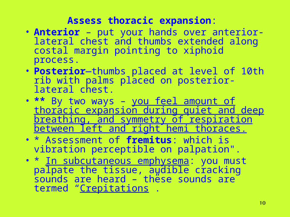
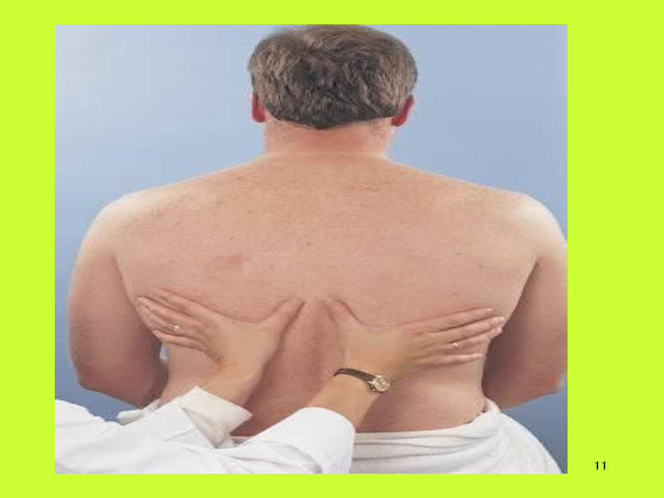
Assess thoracic expansion: • Anterior – put your hands over anterior-lateral
chest and thumbs extended along costal margin pointing to xiphoid process.
• Posterior—thumbs placed at level of 10th rib with palms placed on posterior-lateral chest.
• ** By two ways – you feel amount of thoracic expansion during quiet and deep breathing, and symmetry of respiration between left and right hemi thoraces.
• * Assessment of fremitus: which is vibration perceptible on palpation".
• * In subcutaneous emphysema: you must palpate the tissue, audible cracking sounds are heard – these sounds are termed “Crepitations”.
11
12
• Percussion of chest: to determine relative amounts of air, liquid, or solid material in the underlying lung, and to determine positions and boundaries of organs.
• * Percussion done for posterior and anterior and lateral aspects of chest with all directions, and with about “5”cms intervals.
• * Auscultation: To obtains information about the function of respiratory system & to detect any obstruction in the passages.
• * Instruct the client to breathe through the mouth more deeply and slowly than in usual respiration before beginning
• Auscultate all areas of chest for at least one complete respiration
13
Auscultation cont..
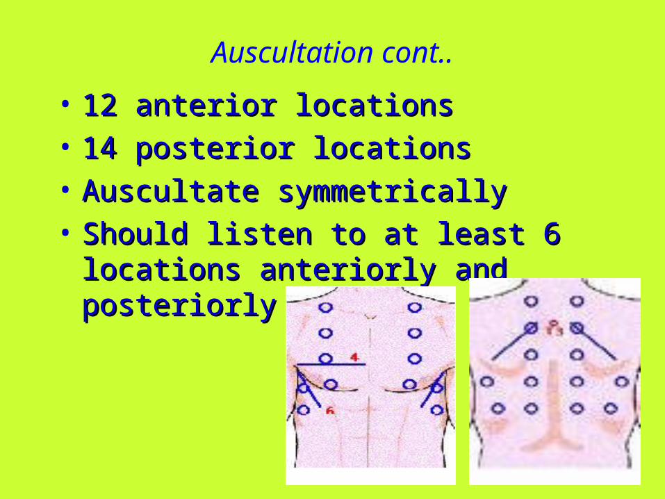
• 12 anterior locations12 anterior locations
• 14 posterior locations14 posterior locations
• Auscultate symmetricallyAuscultate symmetrically
• Should listen to at least 6 locations anteriorly Should listen to at least 6 locations anteriorly and posteriorly and posteriorly
14
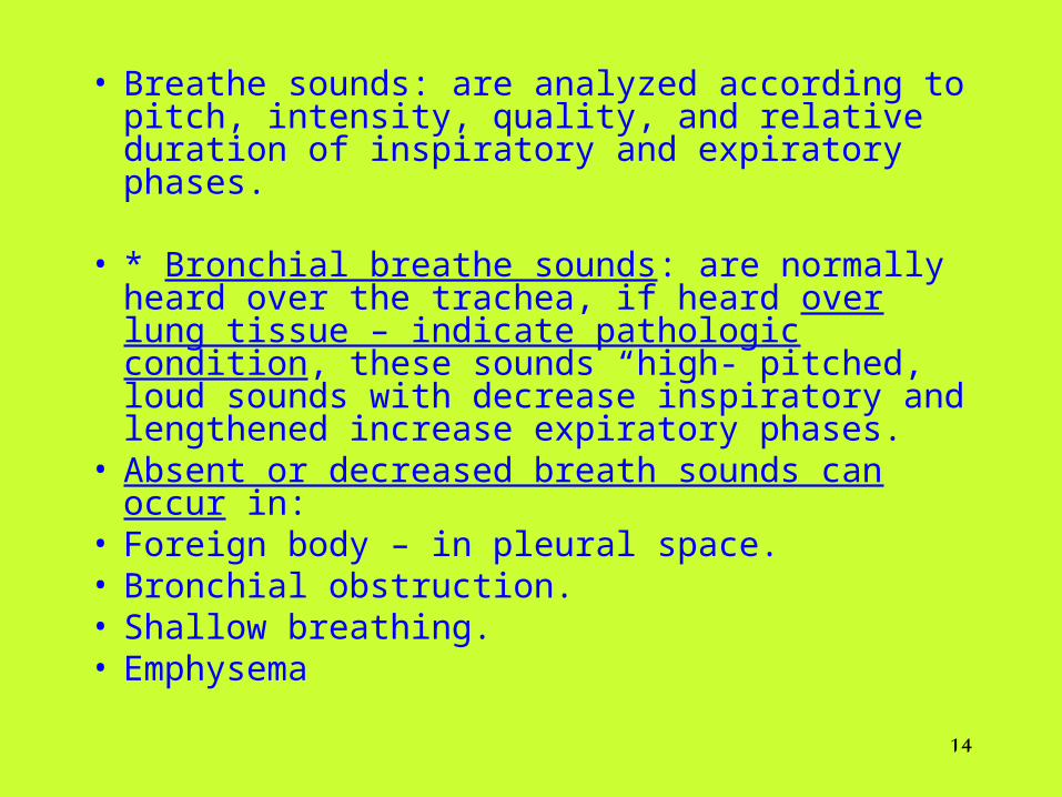
• Breathe sounds: are analyzed according to pitch, intensity, quality, and relative duration of inspiratory and expiratory phases.
• * Bronchial breathe sounds: are normally heard
over the trachea, if heard over lung tissue – indicate pathologic condition, these sounds “high- pitched, loud sounds with decrease inspiratory and lengthened increase expiratory phases.
• Absent or decreased breath sounds can occur in: • Foreign body – in pleural space. • Bronchial obstruction. • Shallow breathing. • Emphysema
15
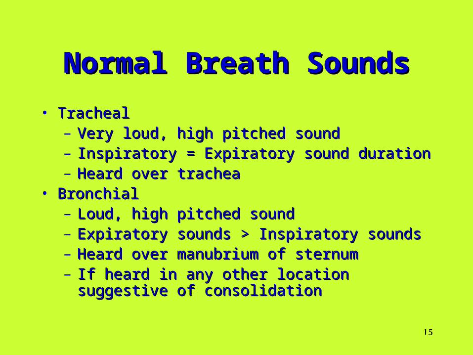
Normal Breath SoundsNormal Breath Sounds
• TrachealTracheal– Very loud, high pitched soundVery loud, high pitched sound– Inspiratory = Expiratory sound durationInspiratory = Expiratory sound duration– Heard over tracheaHeard over trachea
• BronchialBronchial– Loud, high pitched soundLoud, high pitched sound– Expiratory sounds > Inspiratory soundsExpiratory sounds > Inspiratory sounds– Heard over manubrium of sternumHeard over manubrium of sternum– If heard in any other location suggestive of If heard in any other location suggestive of
consolidationconsolidation
16
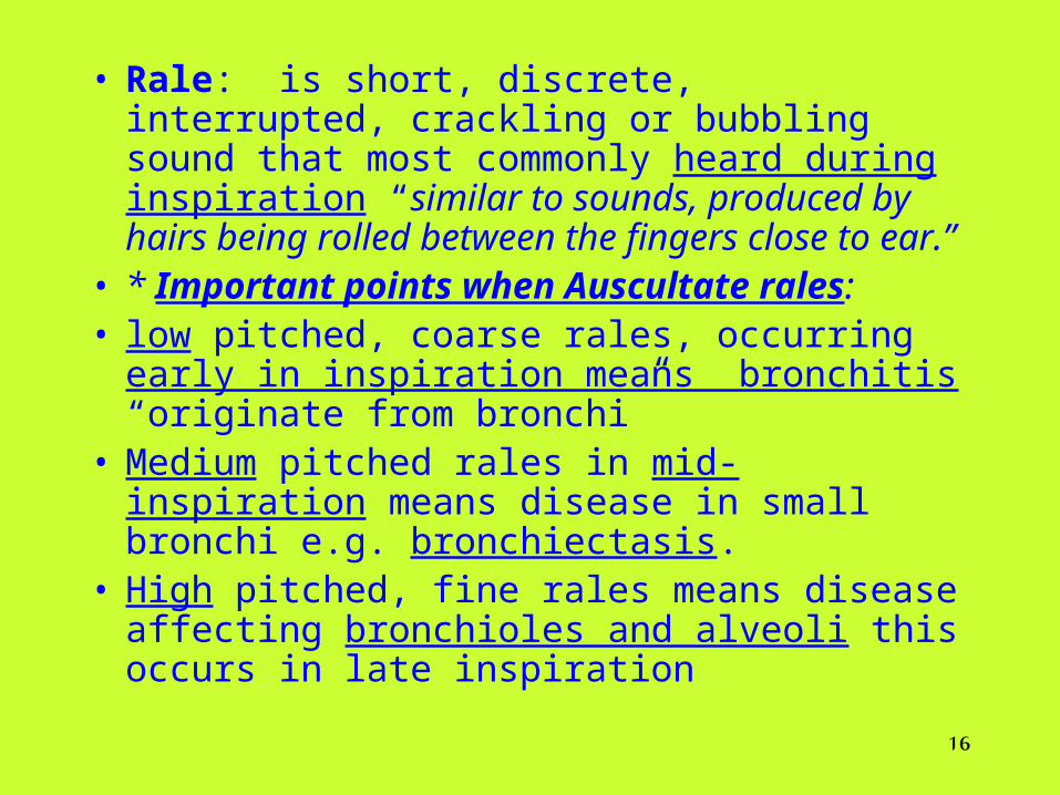
• Rale: is short, discrete, interrupted, crackling or bubbling sound that most commonly heard during inspiration “similar to sounds, produced by hairs being rolled between the fingers close to ear.”
• * Important points when Auscultate rales: • low pitched, coarse rales, occurring early in
inspiration means bronchitis “originate from bronchi”
• Medium pitched rales in mid-inspiration means disease in small bronchi e.g. bronchiectasis.
• High pitched, fine rales means disease affecting bronchioles and alveoli this occurs in late inspiration
17
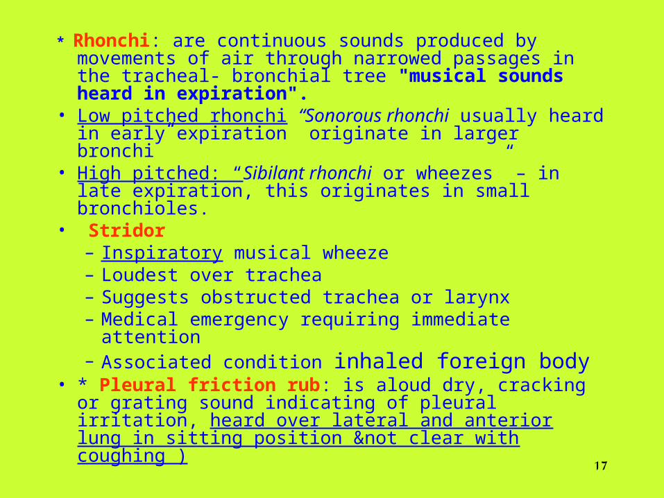
* Rhonchi: are continuous sounds produced by movements of air through narrowed passages in the tracheal- bronchial tree "musical sounds heard in expiration".
• Low pitched rhonchi “Sonorous rhonchi usually heard in early expiration originate in larger bronchi”
• High pitched: “Sibilant rhonchi or wheezes” – in late expiration, this originates in small bronchioles.
• Stridor– Inspiratory musical wheeze– Loudest over trachea– Suggests obstructed trachea or larynx– Medical emergency requiring immediate attention– Associated condition inhaled foreign body
• * Pleural friction rub: is aloud dry, cracking or grating sound indicating of pleural irritation, heard over lateral and anterior lung in sitting position ¬ clear with coughing )
18
Causes of Decreased or Absent Breath SoundsCauses of Decreased or Absent Breath Sounds
• AsthmaAsthma
• COPDCOPD
• Pleural EffusionPleural Effusion
• PneumothoraxPneumothorax
• AtelectasisAtelectasis
19
• Pneumonia: Pneumonia: Community-acquired pneumoniaCommunity-acquired pneumonia• Hospital-acquired pneumonia Hospital-acquired pneumonia • BacteriaBacteria• Viruses Viruses • Mycoplasma Mycoplasma • Fungi Fungi • Chemical Chemical
Common Respiratory Common Respiratory DisordersDisorders
20
• Pleural EffusionPleural EffusionAccumulation of pleural fluid Accumulation of pleural fluid secondary to increased fluid formation secondary to increased fluid formation – Increased capillary permeability Increased capillary permeability – Deceased colloid osmotic pressure of the blood Deceased colloid osmotic pressure of the blood – Increased intrapleural negative pressure Increased intrapleural negative pressure – Impaired lymphatic drainage Impaired lymphatic drainage – Increased pressure in the capillaries or lymphaticsIncreased pressure in the capillaries or lymphatics
Common Respiratory Disorders cont..Common Respiratory Disorders cont..
21
Common Respiratory Disorders cont..Common Respiratory Disorders cont..
• PneumothoraxPneumothoraxSudden onset of pleuritic chest Sudden onset of pleuritic chest painpain– Dyspnea, shortness of breath, increased work of Dyspnea, shortness of breath, increased work of
breathing breathing • Diagnostic testDiagnostic test
– CXR CXR • ManagementManagement
– Oxygen Oxygen – Possible placement of chest tubePossible placement of chest tube
22
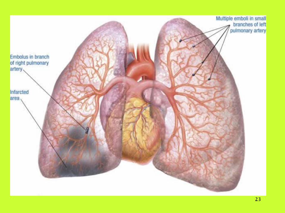
Common Respiratory Disorders cont..Common Respiratory Disorders cont..
• Pulmonary Embolism Pulmonary Embolism Part of a deep vein Part of a deep vein thrombosis that has traveled and lodged in thrombosis that has traveled and lodged in the pulmonary arteriesthe pulmonary arteries
• Severity depends on the extent of occlusionSeverity depends on the extent of occlusion
• Mismatch of ventilation and perfusion Mismatch of ventilation and perfusion
• Testing Testing ( pulmonary angiogram)( pulmonary angiogram)
23
24
• COPD COPD History History – Exposure to risk factors, co-morbidities, Exposure to risk factors, co-morbidities,
current medical treatment (beta blockers)current medical treatment (beta blockers)• Tests Tests
– Spirometry, ABGsSpirometry, ABGs• Management Management
– Oxygen, education, drug therapy, Oxygen, education, drug therapy, nutrition, exercise, surgical interventionnutrition, exercise, surgical intervention
Common Respiratory Disorders cont..Common Respiratory Disorders cont..
25
• AsthmaAsthma A chronic inflammatory disease of the airwaysA chronic inflammatory disease of the airways• Airway hyper responsivenessAirway hyper responsiveness• Variable airway obstruction Variable airway obstruction • Resolves spontaneously or after using a bronchodilatorResolves spontaneously or after using a bronchodilator• Testing :Testing :
– Spirometry Spirometry – Pulmonary function testingPulmonary function testing
• ManagementManagement– Education, prevent exacerbation, optimize Education, prevent exacerbation, optimize
pharmacotherapypharmacotherapy
Common Respiratory Disorders cont..Common Respiratory Disorders cont..
26
• Acute Respiratory FailureAcute Respiratory Failure A sudden and life–threatening A sudden and life–threatening deterioration in gas exchangedeterioration in gas exchange
• Type I – Acute hypoxemic respiratory failure Type I – Acute hypoxemic respiratory failure • Type II - Acute hypercapnic respiratory failureType II - Acute hypercapnic respiratory failure• Type III – Combined hypoxemic and hypercapnic failure Type III – Combined hypoxemic and hypercapnic failure
• TestsTests – ABGs, CXR, CT, thoracentesisABGs, CXR, CT, thoracentesis
• ManagementManagement– Correction of gases, oxygen therapy Correction of gases, oxygen therapy – Reversal of any narcoticsReversal of any narcotics– Possible mechanical ventilationPossible mechanical ventilation
Common Respiratory Disorders cont..Common Respiratory Disorders cont..

27
The end
Thank you